


Abstract
Xanthophylls are oxygenated carotenoids widely distributed in photosynthetic microorganisms, plants, algae, and certain invertebrates, where they function as key photoprotective and antioxidant pigments. Among them, xanthophylls containing vicinal 1,2-diol moieties exhibit unique chemical reactivity that enables reversible coordination with boron species naturally present in marine and terrestrial environments. The formation of cyclic borate esters between boron and diol-containing xanthophylls induces structural and electronic modifications that may enhance pigment stability and functional performance. Emerging evidence suggests that boron–xanthophyll complexes display improved resistance to photooxidative degradation, enhanced singlet oxygen quenching capacity, and increased radical-scavenging activity compared with their uncomplexed counterparts. In addition, boron coordination can influence molecular conformation, polarity, and supramolecular organization within lipid bilayers, thereby promoting membrane stabilization under conditions of high light exposure and oxidative stress. Together, these effects indicate a cooperative role of boron complexation in amplifying the intrinsic photoprotective and antioxidant properties of xanthophylls. A deeper understanding of the structural basis and biological implications of boron–xanthophyll interactions may provide new insights into adaptive stress tolerance in marine and photosynthetic organisms, as well as guide the development of advanced photoprotective systems for biomedical and technological applications.
1. Introduction
Ultraviolet (UV) radiation is a major environmental factor driving oxidative stress in biological systems [1,2,3]. Absorption of UV photons by endogenous chromophores in the skin generates excited states, reactive oxygen species (ROS), and secondary radical cascades that contribute to lipid peroxidation, protein oxidation, DNA damage, inflammation, photoaging, and carcinogenesis [4,5]. In particular, UVA radiation promotes sustained oxidative stress through photosensitized generation of singlet oxygen and superoxide, while UVB induces direct photochemical damage and amplifies inflammatory signaling [6,7]. Although conventional photoprotection strategies rely primarily on UV filters that absorb or reflect radiation, increasing attention has shifted toward molecular photoprotective systems capable of quenching excited states and neutralizing ROS at the cellular level [8,9,10,11].
Xanthophylls, a subclass of oxygenated carotenoids, are natural pigments widely distributed in cyanobacteria, microalgae, fungi, and higher plants. Structurally characterized by an extended conjugated polyene backbone and one or more oxygen-containing functional groups, xanthophylls are highly efficient quenchers of singlet oxygen and triplet excited states [12,13,14]. Through physical quenching and radical scavenging mechanisms, these compounds dissipate excess excitation energy as heat and protect lipid membranes from peroxidative damage. In biological membranes, their amphiphilic character allows strategic orientation across lipid bilayers, optimizing photoprotective efficiency. Several xanthophylls, including lutein, astaxanthin, fucoxanthin, and myxol, have demonstrated antioxidant and anti-inflammatory activities in UV-exposed cells and experimental models [15,16,17,18].
Among xanthophylls, a structurally significant subgroup contains vicinal diol (1,2-diol) moieties, typically located on terminal cyclic end groups. These diol functionalities introduce an additional dimension beyond classical carotenoid photophysics: the capacity to form reversible coordination complexes with boron species such as boric acid or borate [19,20,21]. Boron, a Lewis acid with a strong affinity for cis-vicinal diols, forms five-membered cyclic borate esters in aqueous or mildly alkaline environments, converting trigonal planar boron to tetrahedral borate complexes. This dynamic and pH-dependent coordination chemistry has been extensively studied in carbohydrate systems but remains largely unexplored in carotenoid photochemistry [19,20,21,22,23,24].
The formation of boron–vicinal diol xanthophyll complexes may influence multiple properties relevant to photoprotection. Boron coordination can modify electronic distribution within the diol-containing ring system, potentially affecting excited-state lifetimes and redox behavior [20,21]. Complexation may enhance structural rigidity, alter aggregation behavior, or stabilize specific conformations of the chromophore. In polyhydroxylated xanthophylls, boron may also facilitate supramolecular assembly through bridging interactions. Such effects could translate into improved photostability, enhanced ROS quenching efficiency, and controlled redox modulation under UV exposure [19,20,21,22,23,24].
Notably, many cyanobacteria that biosynthesize vicinal diol xanthophylls inhabit alkaline or mineral-rich environments where borate species are present (samples of boron-containing minerals are shown in Figure 1 and Figure 2). This ecological context raises the intriguing possibility that boron–xanthophyll interactions may occur naturally and contribute to adaptive photoprotection in these organisms. Extending this concept to engineered systems suggests a new strategy for molecular photoprotection: the deliberate design of boron-coordinated xanthophyll complexes as multifunctional antioxidants capable of mitigating UV-induced oxidative stress [18,25,26,27,28,29,30].
Figure 1.
Borax is one of the best-known naturally occurring boron-containing minerals. Chemically, it is sodium tetraborate decahydrate, a specific borate mineral within a broader group of naturally occurring boron-containing minerals. Borate minerals are commonly associated with evaporite deposits formed in ancient alkaline lakes located in arid and semi-arid regions. Borax is widely used in cleaning agents and laundry formulations because of its buffering, emulsifying, and dispersing properties. In addition, it has applications as an antiseptic and pesticide, and in metallurgy it is used as a flux in soldering and welding processes. The drawing was made using the programs PhotoSuite (version 4.0) and Adobe Elements (version 8.5.01). Reprinted from Ref. [31].
Figure 2.
Samples of boron-containing minerals: (a) boracite, (b) green datolite, (c) hydroboracite, and (d) sassolite. Boracite (Mg3B7O13Cl) is a magnesium borate chloride mineral that crystallizes mainly in the cubic system but may display lower-symmetry pseudomorphic forms. Datolite is a rare calcium borosilicate mineral commonly found in hydrothermal veins and cavities of mafic igneous rocks. Sassolite is a naturally occurring boric acid mineral (H3BO3), typically forming flaky, fibrous, or earthy aggregates and characterized by high solubility. Hydroboracite is a hydrated calcium magnesium borate mineral commonly associated with evaporitic borate deposits. The drawing was made using the programs PhotoSuite (version 4.0) and Adobe Elements (version 8.5.01). Reprinted from Ref. [31].
This review explores the structural chemistry, coordination mechanisms, photophysical implications, and biological relevance of boron–vicinal diol xanthophyll complexes. By integrating principles of coordination chemistry with carotenoid photochemistry and oxidative stress biology, we aim to evaluate whether these dynamic boron-mediated systems represent emerging platforms for advanced protection against UV-induced oxidative damage.
2. Boron Sequestration by Bacteria
Boron is widely distributed in nature, primarily occurring in the form of borate minerals that originate from volcanic activity, hydrothermal processes, and the evaporation of boron-rich waters [29,30,32,33,34]. The most important boron-containing minerals include borax (sodium tetraborate decahydrate, Figure 1), kernite (hydrated sodium borate), colemanite (calcium borate), ulexite (sodium calcium borate), and tourmaline (a complex borosilicate, or datolite, Figure 2) [35,36,37,38]. These minerals are commonly found in arid and semi-arid regions where evaporitic deposits concentrate borate salts, such as in salt flats and alkaline lakes. In aqueous environments, boron typically exists as boric acid (B(OH)3) or borate ions (B(OH)4−), depending on pH, and its mobility allows it to cycle between geological and biological systems. Certain microorganisms have evolved mechanisms to tolerate, transport, and sometimes accumulate boron from such environments. While boron is not universally required for all bacteria, it plays recognized roles in specific taxa, particularly in the form of boron-containing quorum-sensing molecules such as autoinducer-2 (AI-2) in some Gram-negative species [39,40,41]. In boron-rich habitats, including marine ecosystems and alkaline soils, bacteria may absorb boron passively as boric acid or actively regulate intracellular levels through specialized transport systems to prevent toxicity. Some species associated with saline or alkaline environments exhibit enhanced boron tolerance and may bind boron to cell wall components, extracellular polysaccharides, or diol-containing metabolites, thereby contributing to boron sequestration and biogeochemical cycling [41,42,43,44,45].
Additionally, recent studies demonstrate that certain bacteria form stable complexes between boron and biomolecules containing vicinal diols, with measurable consequences for membrane stability and stress adaptation. In environmental systems, boron-containing minerals constitute the principal reservoirs of this element, while boron-assimilating microorganisms actively mediate its mobilization, chemical transformation, and redistribution across terrestrial and marine ecosystems [20,21,41,42,43,44,45].
Boron accumulation is well documented in cyanobacteria, where it represents a physiologically relevant and experimentally supported process. In contrast, it is neither a defining nor a quantitatively established trait in most marine flavobacteria. Available evidence consistently indicates that cyanobacteria possess the most robust and mechanistically substantiated capacity for boron uptake and intracellular regulation, whereas reports for Robiginitalea, Nonlabens, and Aurantiacicella marina remain sparse and largely indirect [45,46,47,48,49,50].
In Synechocystis (e.g., PCC 6803), a widely studied unicellular model cyanobacterium, boron transport and homeostasis systems—including characterized efflux mechanisms—have been clearly established, confirming active and regulated control of intracellular boron levels. This organism is capable of accumulating boron from the external environment and tolerating elevated concentrations relative to many non-photosynthetic bacteria. Importantly, this accumulation is controlled and physiologically regulated rather than excessive, with boron distributed across the cytosol, membranes, and cell envelope components [51,52,53].
Anabaena variabilis and Anabaena spp. are filamentous, heterocyst-forming cyanobacteria that inhabit boron-containing aquatic environments and demonstrably take up measurable amounts of boron. Their thick extracellular polysaccharide (EPS) sheaths provide substantial boron-binding capacity, facilitating surface-associated accumulation through interactions with cis-diol-containing polymers. While not hyperaccumulators, these organisms consistently tolerate and retain boron at environmentally relevant concentrations [54,55,56,57,58]. This behavior reflects a regulated and physiologically integrated response rather than passive adsorption alone. In addition, heterocyst differentiation and nitrogen fixation impose distinct cellular states that may further influence boron uptake and compartmentalization. Collectively, these features support a functional role for boron in cell envelope stability and stress resilience under fluctuating environmental conditions.
Nostoc punctiforme exhibits closely related characteristics, producing extensive mucilaginous sheaths rich in polysaccharides capable of binding borate. Boron accumulation occurs both externally within EPS matrices and internally under regulated conditions, indicating coordinated control over its distribution. Its widespread presence in soils and freshwater systems exposes it to variable boron availability, reinforcing the importance of adaptive regulation rather than excessive storage [59,60,61,62,63,64,65]. EPS-mediated sequestration likely contributes to protection against desiccation and oxidative stress, while controlled intracellular levels suggest the involvement of specific transport and homeostasis mechanisms. This dual strategy—external binding combined with regulated internal handling—demonstrates a balanced and functionally relevant mode of boron utilization.
Oscillatoria agardhii, O. limosa, and related Oscillatoria species similarly produce substantial extracellular matrices and occupy freshwater or brackish habitats with variable boron concentrations. These organisms are capable of boron uptake; however, available evidence indicates that accumulation is predominantly associated with cell surface polysaccharides rather than large intracellular pools [66,67,68,69,70]. This distribution is consistent with a primarily structural or protective role of boron at the cell interface. Environmental fluctuations in boron availability may modulate EPS composition and binding capacity, further influencing surface-associated sequestration. In contrast to cyanobacteria with well-characterized intracellular regulation, Oscillatoria species appear to rely more heavily on extracellular interactions, reflecting a less specialized but still functionally significant mode of boron association.
Thermosynechococcus elongates, a thermophilic cyanobacterium, tolerates a range of dissolved minerals in hot spring environments. While specific high-level boron accumulation data are limited, its membrane systems and stress tolerance machinery suggest regulated boron uptake rather than strong hyperaccumulation [71,72,73,74,75,76].
All listed cyanobacteria can take up boron and regulate it; several (especially filamentous, EPS-rich genera like Nostoc and Anabaena) likely accumulate boron in extracellular polysaccharide matrices. However, none are known as extreme boron hyperaccumulators comparable to certain plants [54,55,56,57,58,59,60,61,62,63,64,65].
These marine flavobacteria, Robiginitalea, Nonlabens, and Aurantiacicella marina are heterotrophic bacteria that produce carotenoids such as myxol derivatives, but there is little direct evidence that they accumulate boron to unusually high levels. Marine environments contain dissolved boron (primarily as boric acid), so passive uptake occurs, but current literature does not identify these genera as significant boron accumulators. Any retention would likely be low-level and regulated [77,78,79,80,81].
Boron uptake and regulation are well established in several cyanobacteria, including Synechocystis, Anabaena spp., Nostoc punctiforme, Oscillatoria spp., and Thermosynechococcus elongatus. Among these, Nostoc and Anabaena exhibit the strongest potential for measurable accumulation, particularly at the cell surface, due to their extensive extracellular polysaccharide (EPS) matrices. In contrast, there is limited or no direct evidence for significant boron accumulation in Robiginitalea, Nonlabens, and Aurantiacicella marina [61,62,63,64,65,66,67]. Importantly, these cyanobacteria should be characterized as boron-tolerant and boron-regulating organisms rather than classical hyperaccumulators. Boron levels are maintained within a controlled physiological range, reflecting tight homeostatic regulation. Accumulation is generally moderate and linked to environmental exposure, cell envelope chemistry, and regulated transport systems rather than high-capacity storage. This distinction is critical for accurately interpreting boron dynamics in microbial systems. It also reinforces that boron association is functionally integrated into cellular processes rather than being a passive consequence of environmental availability.
The mechanisms by which marine bacteria absorb boron are increasingly understood, although they remain incompletely characterized across diverse taxa. In seawater, boron occurs at relatively stable concentrations (approximately 4–5 mg/L), predominantly as boric acid (B(OH)3) at physiological pH. Because boric acid is small and uncharged, it can diffuse passively across bacterial membranes. However, passive diffusion alone does not provide adequate control over intracellular boron concentrations, particularly given the potential toxicity of excess boron [20,21,41,42,43,44,45]. Consequently, cells must employ regulatory mechanisms to balance influx and efflux. This requirement underscores the importance of transport specificity and intracellular buffering systems. It also highlights that boron handling is an actively managed physiological process rather than a purely physicochemical phenomenon.
Marine bacteria therefore rely on regulated transport systems to maintain boron homeostasis. The best-characterized mechanism involves boron incorporation into quorum-sensing molecules. In many Gram-negative bacteria, boron is integrated into the signaling molecule autoinducer-2 (AI-2), forming a furanosyl borate diester. This boron-containing signal is transported via specific periplasmic binding proteins and ATP-binding cassette (ABC) transporters, such as the LuxP/LuxQ system in Vibrio species. In this context, boron is not simply taken up as a nutrient but becomes structurally embedded in a signaling framework that regulates gene expression and population-level behavior [82,83,84]. This represents a well-defined example of boron functioning in molecular communication. It further demonstrates that boron can have direct biochemical roles beyond passive association. Such mechanisms provide a conceptual basis for understanding more complex boron–biomolecule interactions.
In addition, bacteria possess boron efflux systems that prevent intracellular toxicity. Transport proteins, including Bor1-like exporters and related membrane channels, actively remove excess boron from the cytoplasm. Some marine bacteria may also transiently bind boron to intracellular metabolites containing vicinal diol groups (e.g., sugars or polyols), forming reversible borate esters that modulate cellular chemistry. These interactions may contribute to short-term buffering of intracellular boron levels. In boron-rich or dynamic environments, extracellular polysaccharides and cell surface components further enhance boron binding and sequestration [85,86,87,88]. Together, these processes define a coordinated system of uptake, utilization, buffering, and export. This integrated framework supports controlled boron homeostasis while enabling functional interactions with cellular biomolecules.
Overall, marine bacterial boron absorption involves a combination of passive diffusion of boric acid, regulated transporter-mediated uptake associated with signaling pathways, and active efflux mechanisms to maintain homeostasis. While the ecological role of boron in marine bacteria is still being explored, its participation in quorum sensing and potential interactions with diol-containing biomolecules represent the most clearly established biological functions to date [89,90,91].
3. Detection of Boron as Complex with Carotenoids
Borate–carotenoid complexes (BCCs) can be isolated and characterized using various chromatographic techniques, particularly high-performance liquid chromatography (HPLC). These complexes arise from the interaction between boric acid or borate anions and the hydroxyl groups of carotenoids, especially xanthophylls containing vicinal diol moieties. The separation process relies on differences in polarity, hydrophilicity, and structural features between native carotenoids and their corresponding borate complexes. Formation of a borate ester typically increases polarity and may alter retention time, allowing chromatographic discrimination between free pigments and boron-bound species. In some cases, boron complexation also induces subtle changes in UV–visible absorption spectra, which can be monitored by diode-array detection [21,92,93,94,95,96,97,98,99,100].
The use of borate in electrophoretic techniques, particularly capillary electrophoresis (CE), has been systematically exploited for the separation and analysis of carotenoids and structurally related compounds. Boric acid and borate anions selectively interact with molecules bearing cis-1,2-diol groups (Figure 3), including certain carbohydrates and diol-containing carotenoids. Formation of these borate complexes introduces a net negative charge, thereby significantly altering electrophoretic mobility and migration behavior. This property enables the selective detection and resolution of diol-containing carotenoids from non-coordinating analogues. The strength and reversibility of borate–diol interactions also allow dynamic modulation of separation conditions through pH and buffer composition. Consequently, borate-based systems provide both analytical selectivity and mechanistic insight into diol functionality. Furthermore, coupling CE or HPLC with mass spectrometry (MS) enables direct confirmation of boron complexation through characteristic mass shifts and isotope signatures of boron-containing species. These analytical approaches collectively offer a robust platform for probing boron–xanthophyll interactions at high sensitivity and resolution [21,42,44,45,93,94,95,96,97].
Figure 3.
General scheme illustrating the formation of boron–carotenoid complexes based on extensive experimental data from boron coordination chemistry, particularly the well-established interactions of boric acid and borate anions with vicinal diol-containing compounds. Column (A) shows structural fragments of xanthophylls containing a 1,2-vicinal diol moiety. Column (B) depicts interactions of these diol-containing fragments with boric acid under acidic conditions, leading to formation of trigonal planar boron complexes. Column (C) illustrates the corresponding interactions in alkaline media, where borate anions form negatively charged tetrahedral borate complexes. The presented reaction schemes are mechanistic models derived from established borate–diol chemistry and stereochemical principles. Adapted from Ref. [21].
Certain cyanobacteria, microalgae, and possibly some invertebrates tolerate and accumulate elevated boron concentrations by converting it into less bioactive forms through complexation and sequestration rather than exclusion. Because boron readily forms reversible complexes with cis-diol–containing molecules, carbohydrate-rich structures—including extracellular polymeric substances (EPS), cell walls, mucopolysaccharides, and intracellular polyols—serve as effective boron-binding reservoirs [101,102,103,104,105,106]. In these systems, boron is incorporated into borate–diol complexes and retained either within extracellular matrices or compartmentalized in vacuoles or specialized cellular regions, thereby limiting the concentration of free, metabolically disruptive boron in the cytosol.
This sequestration reduces boron bioavailability and mitigates its interference with enzymes, membranes, and nucleic acid–associated processes. Importantly, this mechanism represents an active physiological strategy rather than passive chemical association. It also provides a unifying explanation for how structurally diverse organisms maintain boron tolerance under variable environmental exposure. Concurrent stress responses, including enhanced antioxidant defenses and photoprotective mechanisms such as xanthophyll cycling, may further reduce secondary oxidative damage associated with boron stress. Although these storage and detoxification processes are likely saturable and influenced by environmental parameters such as pH and competing ligands, they define a coherent framework for understanding controlled boron accumulation in biological systems [20,43,102,103,104,105,106].
Figure 3 illustrates representative structural fragments of xanthophylls containing vicinal 1,2-diol groups and their interaction with boron species under varying pH conditions. Under acidic to neutral conditions, boric acid (B(OH)3) interacts with cis-1,2-diol groups to form boron–diol adducts in which boron retains trigonal planar (sp2) geometry. These interactions are typically characterized by partial ester formation and hydrogen-bond-assisted coordination, resulting in a dynamic equilibrium between free xanthophyll and boron-associated species [20,21,41,42,43]. This equilibrium is sensitive to local chemical conditions, including pH and ligand availability. As pH increases, conversion to tetrahedral borate species enhances complex stability and shifts the equilibrium toward more fully coordinated structures. Such pH-dependent behavior is central to both biological function and analytical detection of boron–diol complexes.
As the pH increases to neutral–alkaline and alkaline conditions, boric acid is progressively converted into the borate anion (B(OH)4−), which exhibits stronger Lewis acidity toward cis-vicinal diols. Under these conditions, deprotonation of hydroxyl groups promotes tighter coordination, resulting in the formation of a five-membered cyclic borate ester in which boron adopts a tetrahedral (sp3) configuration and carries a net negative charge [20,21,43]. This tetrahedral borate complex is thermodynamically more stable than the trigonal planar form and can substantially modify the polarity, solubility, and electronic distribution of the xanthophyll molecule. The shift toward tetrahedral coordination also increases the persistence of boron–diol interactions under physiological conditions. As a result, borate binding becomes more functionally significant in environments where pH is moderately alkaline. This transition has important implications for both biological systems and analytical detection strategies (Table 1).
Table 1.
Mechanistic Implications of Boron–Xanthophyll Interactions.
The equilibrium between trigonal and tetrahedral boron species is fully reversible and strongly governed by pH, solvent environment, and the stereochemical arrangement of the diol group. In xanthophylls, the cis configuration of the 1,2-diol is particularly well suited for forming low-strain, five-membered chelate rings, thereby favoring stable complex formation [44,45]. Borate coordination can further restrict conformational flexibility of the carotenoid end group, influencing molecular packing and orientation within lipid membranes. These structural effects may, in turn, alter aggregation behavior and intermolecular interactions. Additionally, changes in electronic structure induced by boron binding can affect light absorption and energy dissipation pathways. Consequently, borate complex formation represents a dynamic and tunable chemical interaction. This pH-responsive system provides a mechanistic basis for modulating photostability, redox behavior, and protection against UV-induced oxidative stress.
4. Stereochemistry of 1,2-Diol Xanthophylls
Vicinal 1,2-diols in xanthophylls play a decisive role in governing their coordination chemistry with boron and, consequently, their structural and photoprotective behavior in marine and photosynthetic organisms. In carotenoid end groups, hydroxyl substituents at positions such as 2,3 or 3,4 may adopt either cis (erythro-like) or trans (threo-like) orientations, depending on enzymatic hydroxylation pathways and ring conformation. The cis-1,2-diol configuration is stereochemically optimal for boron chelation, as it enables formation of a low-strain, five-membered cyclic borate ester [107,108,109]. In this geometry, boron coordinates simultaneously to both oxygen atoms, forming either a trigonal planar boron–diol adduct under acidic conditions or a tetrahedral borate complex under mildly alkaline conditions. This stereochemical compatibility maximizes orbital overlap and stabilizes the resulting complex. As a result, cis-diol–containing xanthophylls exhibit a markedly higher propensity for boron binding. This distinction is fundamental to understanding structure–function relationships in these systems. In contrast, trans-1,2-diols are geometrically less favorable for stable chelation, as the increased spatial separation of hydroxyl groups introduces ring strain and reduces coordination efficiency; consequently, complex formation is weaker or requires conformational adjustment [107,108,109,110,111,112,113].
In marine algae, cyanobacteria, and marine invertebrates—organisms frequently exposed to intense solar radiation—the presence of cis-vicinal diols in xanthophylls such as myxol, nostoxanthin, oscillol, crustaxanthin, and related polyhydroxy derivatives provides a robust structural platform for dynamic boron coordination [107,109,110,111,112]. Marine environments contain consistent levels of borate species, particularly in slightly alkaline seawater, creating conditions favorable for reversible borate ester formation. The stereochemical arrangement of diol groups therefore dictates not only binding strength but also the multiplicity and topology of boron interactions. Xanthophylls bearing multiple cis-1,2-diol sites can form mono- or bis-boron complexes, increasing structural diversity. In some cases, borate ions may act as bridging units linking adjacent carotenoid molecules into supramolecular assemblies. Such cross-linking introduces an additional level of structural organization within pigment systems. This capacity for intermolecular association may be particularly relevant in densely packed membrane environments [20,21,44,45,107,108,109,110,111,112,113].
The functional implications of this stereochemistry are significant for UV-protective antioxidant activity. Xanthophylls mitigate photooxidative stress primarily by quenching singlet oxygen, dissipating excess excitation energy, and scavenging free radicals generated under UV exposure. Boron coordination at cis-diol sites can enhance conformational rigidity of the end rings, reducing nonproductive rotational relaxation and potentially influencing excited-state lifetimes of the conjugated polyene chain. Increased structural organization through boron-mediated interactions may stabilize carotenoid alignment within lipid bilayers, thereby improving membrane resistance to lipid peroxidation. Furthermore, boron–diol complexation can modulate local electron density and redox potential, subtly affecting the efficiency of reactive oxygen species (ROS) quenching [12,13,14,18,114,115,116,117,118,119]. These effects suggest that boron binding is not merely structural but functionally integrated into photoprotective mechanisms. The combined structural and electronic influences may enhance overall pigment performance under stress conditions. This provides a mechanistic link between molecular coordination chemistry and biological function.
In cyanobacteria and algae inhabiting alkaline or mineral-rich waters, such reversible boron–xanthophyll interactions likely represent an adaptive mechanism contributing to photostability and oxidative resilience. In marine invertebrates, where carotenoids are acquired through diet and incorporated into membrane systems, similar stereochemically governed boron coordination may reinforce pigment assemblies and enhance long-term UV protection. These interactions are expected to be dynamic and responsive to environmental conditions, particularly pH and boron availability. Such responsiveness allows organisms to modulate pigment behavior in situ. Overall, the stereochemical distinction between cis- and trans-1,2-diols is not merely structural but directly determines the capacity of xanthophylls to engage in boron coordination. This, in turn, defines their role as dynamic, pH-responsive photoprotective agents in diverse aquatic ecosystems. Accordingly, boron–xanthophyll complexation should be considered a functionally relevant component of stress adaptation rather than a secondary chemical phenomenon [12,13,14,15,16,17,18,114,115,116,117,118,119,120,121].
5. Functions of 1,2-Diol Xanthophylls
Carotenoids containing 1,2-diol (vicinal diol) motifs constitute a specialized subgroup of xanthophylls that exhibit functional properties extending beyond those of non-oxygenated carotenoids. In bacteria, these compounds retain the canonical roles of carotenoids—photoprotection, antioxidant defense, and membrane stabilization—while the presence of terminal 1,2-diol groups confers additional chemical reactivity and functional versatility [12,13,14,15,16,17,18,20,21,44,45,114,115,116,117]. This structural feature enables specific interactions with environmental ligands, including boron species, that are not accessible to non-hydroxylated carotenoids. Consequently, diol-containing xanthophylls occupy a distinct functional niche within microbial physiology. Their dual role as both photoprotective agents and chemically responsive molecules underscores their adaptive significance. This expanded functionality is particularly relevant in chemically dynamic environments such as marine systems.
Like other carotenoids, diol-containing xanthophylls efficiently quench singlet oxygen and dissipate excess excitation energy, thereby protecting photosynthetic machinery and cellular components from photooxidative damage. The presence of hydroxyl groups increases molecular polarity, promoting favorable orientation across lipid bilayers and enabling these pigments to span or anchor within membranes more effectively than purely hydrophobic carotenes. This amphipathic character enhances membrane rigidity, reduces lipid peroxidation, and supports structural integrity under environmental stress conditions such as high irradiance, salinity, and temperature fluctuations [21,44,45]. In addition, membrane-spanning orientation may facilitate more efficient energy dissipation pathways. The interaction between polar end groups and lipid headgroups further stabilizes membrane architecture. These properties collectively contribute to improved cellular resilience under fluctuating environmental conditions.
The presence of vicinal diol groups also enables reversible complexation with boron species (e.g., boric acid or borate), leading to the formation of cyclic borate esters. In marine environments, where boron is consistently available, such boron–diol interactions can influence pigment conformation, electronic distribution, and photophysical behavior. These complexes may alter light absorption characteristics, enhance oxidative stability, and reduce susceptibility to photobleaching. Furthermore, boron coordination can promote supramolecular organization within membranes or extracellular matrices, stabilizing pigment assemblies and potentially facilitating cooperative interactions. This introduces an additional level of structural hierarchy within pigment systems. The dynamic and reversible nature of these interactions allows responsiveness to environmental changes. As a result, boron binding represents a tunable chemical modification rather than a static structural feature [21,44,45].
From a physiological perspective, boron–carotenoid complexes likely function as adaptive photoprotective systems, particularly in marine or alkaline habitats. By modulating antioxidant efficiency and membrane interactions, these complexes can enhance bacterial resilience under intense solar irradiation. The reversible nature of borate ester formation also suggests a potential role in regulating intracellular boron availability and mitigating toxicity. Such mechanisms may contribute to maintaining cellular homeostasis under variable environmental conditions. Although not yet fully elucidated, this regulatory dimension warrants further investigation. It also highlights the broader biological relevance of boron–diol chemistry beyond simple binding interactions [21,43,45].
Overall, carotenoids containing 1,2-diol motifs function as multifunctional photoprotective and membrane-active molecules, while their capacity to form boron complexes provides additional layers of structural stabilization and functional modulation. These interactions can influence photophysical properties, membrane organization, and stress tolerance. Importantly, this behavior reflects an integrated chemical–biological adaptation rather than an incidental interaction. The combined effects enhance the ecological fitness of organisms inhabiting boron-rich environments. Thus, boron–xanthophyll complexation should be regarded as a significant contributing factor to environmental adaptability and photoprotection in diverse microbial systems [20,41,42,43,44,45,114,115,116,117].
6. Xanthophylls with cis-1,2-Diol Configuration
Myxol (1, Figure 4) is a distinctive glycosylated xanthophyll widely produced by marine and freshwater bacteria, particularly cyanobacteria such as Synechocystis, Anabaena, Nostoc punctiforme, Oscillatoria, and Thermosynechococcus elongatus, as well as marine flavobacteria including Robiginitalea, Nonlabens, and Aurantiacicella marina [21,122,123,124]. Myxol commonly occurs as myxol 2′-glycoside, in which a sugar moiety increases polarity and confers amphipathic character, allowing the hydrophobic polyene chain to span lipid bilayers while polar hydroxyl and sugar groups interact with membrane headgroups. These structural features contribute to membrane stabilization, modulation of membrane fluidity, and photoprotection through efficient quenching of singlet oxygen and dissipation of excess excitation energy [122,123,124].
Figure 4.
Proposed structures of xanthophyll–boron complexes identified in bacterial species. The illustrated coordination models are based on extensive experimental knowledge of boron chemistry, particularly the well-established interaction of boric acid and borate anions with vicinal diol groups, leading to formation of cyclic borate esters. Coordination between boron atoms and vicinal diol moieties within xanthophyll structures may alter the electronic distribution along the conjugated polyene chain and thereby influence the photophysical, antioxidant, and membrane-associated properties of the pigments. Structural variations among bacterial species may contribute to differences in complex stability, membrane affinity, and photoprotective efficiency. Adapted from Ref. [21].
Marine bacterium strain P99-3, formerly designated Flavobacterium sp., was isolated from the sponge Homaxinella sp. and produces monocyclic myxol, the aglycone form of myxoxanthophyll [124]. Myxol is thought to be biosynthesized through γ-carotene or β,ψ-carotene intermediates and occurs either in free form or conjugated with fucosides and nitrogen-containing groups [21,122,123]. It is often a major carotenoid component in cyanobacteria such as Synechocystis PCC 6803 and Nostoc punctiforme, where it contributes to UV resistance, oxidative stress tolerance, and ecological fitness in fluctuating aquatic environments [124,125,126,127].
The biological activity of myxol is closely linked to its amphipathic structure. The extended conjugated polyene chain provides antioxidant and light-harvesting properties, whereas hydroxyl and glycosidic groups stabilize membrane association. In cyanobacteria, glycosylated carotenoids regulate membrane fluidity and protect cellular membranes under high light and oxidative stress. Mutants deficient in glycosylated carotenoids frequently display impaired membrane stability and increased stress sensitivity.
Myxol also plays an important role in photoprotection by scavenging reactive oxygen species and stabilizing photosystem complexes. In photosynthetic cyanobacteria, including Synechocystis, Anabaena, and Nostoc, myxol complements β-carotene and zeaxanthin in preventing photodamage [122,123,124,125,126,127]. Its localization within membranes enables rapid quenching of excited states and reactive oxygen species. Because myxol contains vicinal diol groups, boron coordination through reversible borate ester formation is chemically feasible and may further influence pigment conformation, membrane organization, and resistance to photooxidative stress.
In marine flavobacteria, myxol contributes to salt tolerance, UV resistance, and biofilm stability [126,127]. The abundance of boron in marine systems favors reversible borate–diol interactions that may enhance pigment stability and membrane organization. Consequently, boron–myxol interactions may contribute to environmental adaptability in both cyanobacteria and marine microbial communities.
The broad distribution and abundance of myxol glycosides in cyanobacteria and Bacteroidetes suggest that these pigments serve as important interfaces between carotenoid photoprotection and boron coordination chemistry. At the ecosystem level, myxol-producing organisms influence microbial mat stability, marine biofilm formation, and pigment cycling, linking environmental chemistry with adaptive photobiological function.
4-Ketomyxol (2) is a naturally occurring keto-xanthophyll structurally related to myxol and found mainly in cyanobacteria. Its defining feature is a keto group at the C-4 position of the β-ionone ring, which increases polarity and influences membrane interactions, chemical reactivity, and photoprotective activity [122,123].
In cyanobacteria, 4-ketomyxol contributes to membrane stabilization, oxidative stress resistance, and photoprotection through dissipation of excess excitation energy and quenching of reactive oxygen species. It commonly occurs in glycosylated forms that enhance membrane integration and solubility. Biosynthetically, 4-ketomyxol is produced through enzymatic oxidation of myxol by ketolase-mediated introduction of a C-4 keto group. Both myxol and 4-ketomyxol have been identified in Anabaena sp. PCC 7120, Anabaena variabilis IAM M-3, Nostoc punctiforme PCC 73102, and Nostoc sp. HK-01 [21].
Deoxymyxol (3) is a monocyclic xanthophyll structurally related to myxoxanthophyll, differing by the absence of one hydroxyl group. It occurs in marine and freshwater bacteria, including Corynebacterineae strain AIST-1 and Gordonia terrae NBRC 10016ᵀ [122,123,128]. Its vicinal 1,2-diol moiety enables reversible coordination with boric acid or borate ions, forming pH-dependent cyclic borate esters. Boron coordination may enhance conformational rigidity, photostability, and resistance to oxidative degradation while modulating electronic distribution along the polyene chain. These interactions suggest a direct relationship between boron coordination chemistry and photoprotective performance. Because deoxymyxol occurs in sun-exposed aquatic microorganisms, boron-mediated interactions may contribute to adaptive photoprotection (Figure 5) under natural environmental conditions [122,123,128].
Figure 5.
Polycarotenoids produced by the marine filamentous cyanobacterium Oscillatoria contain terminal 1′,2′-vicinal diol groups within their xanthophyll units. The number of linked xanthophyll units may vary from several to potentially hundreds, forming extended polycarotenoid structures. These terminal diol groups may participate in intermolecular interactions, including esterification and borate complex formation, contributing to supramolecular organization. The extended conjugated system may enhance light-harvesting efficiency and antioxidant capacity. Such structural diversity suggests adaptive roles in membrane stabilization and photoprotection under high-light marine conditions. Adapted from Ref. [21].
4-Ketodeoxymyxol (4) is an oxidized derivative of deoxymyxol containing both hydroxyl and keto groups on the terminal ring system [122,123,128,129]. This multifunctional structure increases polarity and expands its redox and coordination properties. The retained vicinal diol group enables formation of reversible cyclic borate esters under acidic, neutral, or mildly alkaline conditions. Boron coordination may enhance structural rigidity, stabilize excited states, and improve resistance to photooxidative degradation. The adjacent keto group may further influence electron distribution and complex stability. These features support its proposed role in photoprotection and oxidative stress adaptation in marine microorganisms [122,128,130].
4-Hydroxymyxol (5) is a polyoxygenated monocyclic xanthophyll identified in cyanobacteria such as Anabaena variabilis ATCC 29413 and Nostoc commune NIES-24 [122,123,131,132]. It contains both a vicinal 1,2-diol system and an additional C-4 hydroxyl group, providing favorable sites for boron coordination. Under alkaline conditions, interaction with borate ions may generate tetrahedral cyclic borate esters, whereas acidic and neutral conditions favor trigonal planar boron–diol complexes. Boron coordination may enhance photostability, alter excited-state dynamics, and contribute to membrane stabilization under oxidative stress. The occurrence of 4-hydroxymyxol in high-irradiance cyanobacteria supports its role in adaptive photoprotection [123,132,133].
Plectaniaxanthin (6) is a monocyclic xanthophyll containing an extended conjugated chromophore and multiple hydroxyl groups. Originally isolated from the fungus Plectania coccinea, it also occurs in yeasts including Cryptococcus laurentii, Dioszegia spp., and Rhodotorula aurantiaca [134,135,136]. Vicinal hydroxyl groups enable formation of cyclic borate esters through interaction with boric acid or borate ions. Boron coordination may alter excited-state dynamics, improve photostability, and promote supramolecular organization through weak cross-linking interactions. These effects may influence membrane organization and oxidative stress resistance (Table 2). Accordingly, plectaniaxanthin represents a useful model for studying boron-mediated modulation of carotenoid systems [134,135,136].
Table 2.
Proposed Effects on Membrane Structure and Photophysics.
Oscillol (7) is a polyhydroxy monocyclic xanthophyll identified in filamentous cyanobacteria including Oscillatoria spp. and Arthrospira species [137,138,139,140,141,142,143]. Its cis-oriented vicinal diol group provides a favorable site for boron coordination and cyclic borate ester formation. Additional hydroxyl groups enhance hydrogen bonding and intermolecular association. In marine cyanobacteria, oscillol may participate in borate-mediated supramolecular assemblies that influence carotenoid aggregation, membrane organization, and photostability. These interactions support a potential role for boron coordination in cellular adaptation to UV-induced oxidative stress [138,139,140,141,142,143].
Beyond its ecological distribution, oscillol possesses structural features—particularly terminal 1,2-diol groups—that make it a valuable model for studying boron–xanthophyll interactions. Boron coordination at these sites may increase conformational rigidity, modulate electronic distribution, and improve resistance to photooxidative degradation. Because oscillol contains diol groups at both terminal ends, borate ions may also promote intermolecular bridging and formation of supramolecular assemblies. Such dynamic interactions could contribute to membrane organization and stabilization in cyanobacteria exposed to high irradiance [139,141,143,144].
Filamentous cyanobacteria such as Oscillatoria limosa accumulate storage polymers including polyhydroxyalkanoates and polysaccharides, reflecting their capacity to organize polymeric cellular structures. By analogy, oscillol-linked borate assemblies may form reversible network-like structures within membranes or extracellular matrices. These interactions may enhance photostability, regulate excited-state dynamics, and contribute to cellular adaptation under UV-induced oxidative stress [141,142,143,144].
2,3-Dihydroxy-γ-carotene (9, Figure 6) is a polyoxygenated carotenoid containing adjacent hydroxyl groups at the 2 and 3 positions of a terminal ring. It has been identified in the red yeast Xanthophyllomyces dendrorhous and related species as a minor intermediate in astaxanthin biosynthesis [145,146,147,148]. The cis-oriented vicinal diol enables reversible coordination with boric acid or borate ions through formation of cyclic borate esters. Boron binding may enhance conformational rigidity, alter electronic distribution along the chromophore, and influence excited-state dynamics and photostability. These properties make 2,3-dihydroxy-γ-carotene a useful model for investigating boron–xanthophyll photochemistry [145,146,147,148].
Figure 6.
Bioactive boron-containing xanthophylls possessing 1,2-diol fragments from aquatic species. These compounds are mainly isolated from marine algae and other aquatic organisms, where the vicinal diol moiety enables reversible borate ester formation that may influence molecular stability and biological activity. Such structural features are associated with antioxidant, anti-inflammatory, and potential antimicrobial properties observed in marine-derived natural products. Adapted from Ref. [21].
2,3-Dihydroxy-β-carotene (10) is a polyoxygenated carotenoid derived from β-carotene and characterized by a vicinal 2,3-diol group on one β-ionone ring [149,150,151,152,153]. It occurs in carotenoid-producing yeasts, aquatic microorganisms, and photosynthetic microalgae, often as a biosynthetic intermediate leading to more oxygenated carotenoids. The cis-oriented 1,2-diol arrangement provides an optimal site for reversible boron coordination and cyclic borate ester formation. Boron binding may increase conformational rigidity and subtly modulate electronic properties, potentially affecting antioxidant activity, excited-state behavior, and photostability under UV exposure [21,149,150,151,152,153].
Caloxanthin (11) is a monocyclic xanthophyll containing multiple hydroxyl groups, including a vicinal 1,2-diol moiety. It occurs in cyanobacteria, microalgae, and other photosynthetic organisms exposed to high solar radiation [154,155,156,157]. Its extended conjugated polyene system contributes to antioxidant and photoprotective activity, while the vicinal diol functionality provides a favorable site for reversible boron coordination. These structural properties support a potential role for boron–caloxanthin interactions in adaptive photoprotection and oxidative stress resistance.
The most chemically significant feature of caloxanthin is its 1,2-vicinal diol (2,3-diol) fragment, typically located on a cyclic end group. This cis-oriented diol configuration provides a favorable geometry for chelation of boron species. In the presence of boric acid (B(OH)3), the two adjacent hydroxyl groups can coordinate to form a cyclic borate ester in which boron remains trigonal planar under acidic or neutral conditions. As the pH increases and borate anions (B(OH)4−) predominate, stronger chelation occurs, yielding a tetrahedral borate complex bearing a negative charge. The formation of these boron complexes is reversible and dependent on environmental pH and solvent conditions.
The vicinal diol functionality of caloxanthin enhances its chemical versatility beyond intrinsic antioxidant activity. Hydroxyl substitution increases polarity and membrane affinity, while boron coordination at the diol site may increase conformational rigidity and modulate electron distribution along the conjugated polyene chain. These effects may influence excited-state relaxation and singlet oxygen quenching, supporting the relevance of caloxanthin as a model for boron–xanthophyll photoprotective systems [154,155,156,157].
2-Hydroxyadonixanthin (12) is a polyoxygenated xanthophyll structurally related to adonixanthin and astaxanthin and identified mainly in the red yeast Xanthophyllomyces dendrorhous as a minor intermediate in astaxanthin biosynthesis [158,159,160,161,162]. The molecule contains both keto and vicinal 1,2-diol functionalities, creating a favorable site for reversible boron coordination through cyclic borate ester formation. Boron binding may alter conformational rigidity, aggregation behavior, and excited-state dynamics while improving photostability and oxidative stress resistance. These properties make 2-hydroxyadonixanthin a useful model for studying boron-mediated modulation of carotenoid photochemistry [158,159,160,161,162].
2-Hydroxyadonirubin (13) is a highly oxygenated ketocarotenoid related to adonirubin and astaxanthin and occurs mainly in carotenoid-producing red yeasts [21,22]. Its vicinal 1,2-diol group enables reversible pH-dependent coordination with boric acid or borate ions. Combined keto and hydroxyl substituents enhance antioxidant activity, membrane interactions, and photoprotective potential. Boron coordination may increase conformational rigidity and influence excited-state relaxation and resistance to photooxidative degradation, supporting its relevance as a boron-binding photoprotective carotenoid [21,22].
2′-Hydroxyadonixanthin (14) is another oxygenated ketocarotenoid identified in Xanthophyllomyces dendrorhous and related systems [158,163,164]. It contains both keto substituents and a vicinal diol moiety capable of reversible boron coordination. Formation of cyclic borate esters may influence electronic distribution, photostability, and antioxidant behavior under UV exposure. These structural features support its potential role in oxidative stress adaptation and photoprotection [158,163,164].
Idoxanthin (15) is an oxygenated marine carotenoid found in crustaceans, mollusks, and other marine invertebrates [165,166,167]. The molecule contains hydroxyl and keto substituents together with a vicinal 1,2-diol fragment capable of forming reversible cyclic borate esters. Boron coordination may modify conformational rigidity, excited-state relaxation, and singlet oxygen quenching efficiency. Because marine environments naturally contain borate species, such interactions may contribute to pigment stabilization and membrane organization under ecological conditions [165,166,167].
Lilixanthin (16) is a polyoxygenated xanthophyll identified mainly in marine mollusks and gastropods [168,169,170,171]. Its vicinal cis-1,2-diol functionality provides an efficient site for boron coordination and cyclic borate ester formation. Boron binding may improve photostability, alter excited-state dynamics, and enhance antioxidant performance. In marine systems, boron–lilixanthin interactions may also contribute to membrane stabilization and supramolecular organization [168,169,170,171].
The cyanobacterium Anacystis nidulans produces nostoxanthin (17, Figure 7), whereas crustaxanthin (18) occurs in Arctic char (Salvelinus alpinus) [21]. Related xanthophylls (19–22) identified in Xanthophyllomyces dendrorhous contain paired terminal vicinal diol groups capable of coordinating boron at both ends of the molecule. Similar to oscillol, these polyhydroxylated carotenoids may form oligomeric or supramolecular assemblies through borate-mediated interactions, potentially enhancing membrane stabilization, antioxidant capacity, and UV resistance [172,173,174,175].
Figure 7.
Major boron-containing xanthophyll isomers possessing 1,2-diol fragments from algal and yeast species. These isomeric forms differ in the stereochemistry and position of the vicinal diol groups, which may influence cyclic borate ester formation and overall chemical reactivity. Variations in double-bond configuration and hydroxyl orientation may also affect photoprotective and antioxidant properties in algal- and yeast-derived systems. Adapted from Ref. [21].
Nostoxanthin (17) is a polyhydroxy xanthophyll widely distributed in cyanobacteria and other photosynthetic microorganisms [176,177,178]. Multiple hydroxyl groups and vicinal diol motifs enable formation of mono-, bis-, or bridging borate complexes under environmentally relevant conditions. Boron coordination may influence membrane organization, aggregation behavior, and photostability while enhancing resistance to oxidative stress. These properties support a role for reversible boron–nostoxanthin interactions in adaptive photoprotection [21,177].
Crustaxanthin (18) is a highly oxygenated marine xanthophyll possessing two terminal cis-1,2-diol groups, one on each end ring [165,179,180]. This unusual architecture provides exceptional potential for boron coordination and supramolecular organization. Borate ions may form bridging interactions between adjacent crustaxanthin molecules, generating extended assemblies that enhance membrane stabilization, reduce photooxidative degradation, and improve singlet oxygen quenching. Such reversible interactions may contribute to adaptive photoprotection in boron-containing marine environments [179,180].
2,3,2′,3′-Tetrahydroxy-4-keto-β-carotene (19) and the related 2,3,2′,3′-tetrahydroxy-4,4′-keto-β-carotene are rare highly oxygenated carotenoids identified in Xanthophyllomyces dendrorhous and related microorganisms [21,172,173,174,175]. Their defining feature is the presence of two terminal vicinal cis-1,2-diol systems that provide highly favorable sites for boron coordination and potential supramolecular assembly formation.
Each vicinal 1,2-diol unit can independently coordinate boric acid or borate ions, forming trigonal planar boron–diol adducts under acidic or neutral conditions and tetrahedral cyclic borate esters under mildly alkaline conditions. Because these molecules contain two diol pairs, they are capable of forming mono-, bis-, or bridging boron complexes.
The dual diol architecture also enables boron-mediated intermolecular cross-linking, in which borate ions bridge adjacent carotenoid molecules to generate supramolecular chains or network-like assemblies. Additional keto substituents may further influence electron distribution and stabilize specific conformations of the conjugated polyene system. Boron coordination at both terminal rings may increase molecular rigidity, reduce rotational flexibility, and modulate aggregation behavior within lipid bilayers.
These highly oxygenated carotenoids retain strong singlet oxygen–quenching and radical-scavenging activity due to their extended conjugated systems. Boron complex formation may further enhance photostability and resistance to oxidative degradation by stabilizing diol-bearing rings and influencing excited-state relaxation pathways. In membrane environments, boron-bridged assemblies may reinforce bilayer organization and improve resistance to UV-induced lipid peroxidation.
Accordingly, 2,3,2′,3′-tetrahydroxy-4-keto-β-carotene and its 4,4′-diketo analogue (20) represent structurally remarkable vicinal diol xanthophylls with exceptional boron coordination potential. Their dual cis-1,2-diol motifs make them particularly suitable for borate complex formation and supramolecular organization, supporting their relevance as emerging models for boron-enhanced photoprotective systems [21,172,173,174,175].
2,3,4,2′,3′-Pentahydroxy-4′-keto-β-carotene (21a,b) and 2,3,4,2′,3′-pentahydroxy-β-carotene (22a,b) are rare highly oxygenated β-carotene derivatives containing five hydroxyl groups distributed across two terminal rings. The 4′-keto derivative additionally contains a carbonyl group that further increases polarity and electronic conjugation. These pigments have been identified mainly in metabolically specialized yeasts and microalgae and likely arise through extensive hydroxylation of β-carotene intermediates [21,172,173,175].
Structurally, both molecules contain two vicinal 1,2-diol systems together with an additional hydroxyl substituent. The cis-oriented 2,3- and 2′,3′-diol groups provide highly favorable sites for boron coordination, enabling formation of mono- or bis-borate complexes. The additional hydroxyl group may further stabilize boron-bound species through hydrogen bonding or electronic effects, particularly in the 4′-keto derivative.
Under acidic or neutral conditions, boric acid typically forms trigonal planar boron–diol adducts with vicinal hydroxyl groups, whereas mildly alkaline conditions favor formation of negatively charged tetrahedral cyclic borate esters. Because these carotenoids contain two chelating diol sites, borate ions may also act as bridging units linking adjacent molecules into supramolecular assemblies. Such boron-mediated cross-linking may enhance conformational rigidity, influence membrane aggregation behavior, and promote formation of polyxanthophyll networks [20,21,43,44,45].
These polyhydroxylated carotenoids retain extended conjugated polyene systems responsible for efficient singlet oxygen quenching and radical scavenging. Boron coordination at one or both diol sites may alter local electron density, modulate excited-state lifetimes, and improve resistance to photooxidative degradation. The presence of multiple coordination sites introduces structural flexibility and dynamic pH-responsive boron–carotenoid interactions.
Accordingly, 2,3,4,2′,3′-pentahydroxy-4′-keto-β-carotene and 2,3,4,2′,3′-pentahydroxy-β-carotene represent rare multi-diol xanthophylls with substantial boron-binding potential. Their capacity to form mono-, bis-, or bridging borate complexes makes them promising models for studying boron-enhanced supramolecular organization and photoprotective systems against UV-induced oxidative stress.
The red yeast Xanthophyllomyces dendrorhous, particularly mutant strain PR1-104, produces structurally diverse polyoxygenated carotenoids (19–23) containing multiple hydroxyl substituents [18,181,182,183,184,185,186,187,188,189,190,191,192,193]. Increased hydroxylation enhances polarity, chemical reactivity, and boron coordination capacity. Treatment with boric acid or borate ions generates multiple cyclic borate ester isomers, including compounds 23a–g (Figure 8). The diversity of these isomers likely reflects differences in coordination sites, ring conformations, and intra- versus intermolecular ester formation [21,188,189,190,191,192,193].
Figure 8.
Rare boron-containing xanthophyll isomers with six hydroxyl groups from red yeast species. The high degree of hydroxylation enhances their ability to interact with boron through multiple vicinal diol sites, potentially forming cyclic or polyborate ester structures. Extensive hydroxylation may also increase polarity and antioxidant capacity, contributing to adaptive stress responses in red yeast systems. Adapted from Ref. [21].
Compound 23, identified as 2,3,4,2′,3′,4′-hexahydroxy-β-carotene, is particularly notable because its multiple hydroxyl groups provide numerous potential boron-chelation sites. Boron coordination may influence electronic distribution, conjugation length, molecular stability, and supramolecular organization within membranes. Such effects may enhance resistance to oxidative degradation and modify photophysical behavior.
Functionally, these boron–carotenoid complexes may represent adaptive photoprotective systems. The combination of extended conjugation and boron-mediated stabilization could improve singlet oxygen quenching, antioxidant activity, and membrane organization. Increased polarity and potential cross-linking interactions may further promote ordered supramolecular assemblies in biological membranes.
A distinctive class of xanthophylls containing 5,6-vicinal diol groups (24–33, Figure 9) has been identified in cyanobacteria, mollusks, microalgae, and terrestrial plants [181,182,183,184,185,186,187,191,193]. These pigments likely originate from cyanobacterial biosynthesis and may subsequently undergo trophic transfer and metabolic modification in aquatic organisms.
Figure 9.
Rare boron-containing xanthophylls possessing a 1,2-diol group at the C-5 and C-6 positions. This vicinal diol arrangement provides a favorable site for cyclic borate ester formation, potentially influencing conformational stability and electronic distribution within the molecule. Such structural features may affect photochemical behavior and contribute to antioxidant, radical-scavenging, and metal-chelating properties. Adapted from Ref. [21].
Related 5,6-diol carotenoids also occur in Capsicum species, including 5,6-diepikarpoxanthin, 5,6-diepicapsokarpoxanthin, 5,6-diepilatoxanthin, 6-epikarpoxanthin, and latoxanthin [21,181,182,183,184,185,186,187,192]. These compounds contribute to fruit coloration and may participate in antioxidant defense during ripening.
Latoxanthin (30) is especially notable because it occurs in both paprika and several brown algae, including Fucus, Ascophyllum, and Laminaria species [194,195]. The coexistence of epoxide and vicinal diol functionalities increases structural complexity and coordination potential.
The 5,6-diol configuration provides additional chemical versatility compared with conventional hydroxylated xanthophylls. These diol groups may participate in reversible boron coordination, hydrogen-bonding networks, and membrane stabilization. Their widespread occurrence across aquatic and terrestrial organisms suggests an adaptive role combining antioxidant activity, photoprotection, and structural flexibility.
Heteroxanthin (24) is a relatively uncommon xanthophyll reported mainly in green algae and related photosynthetic microorganisms [196,197]. Unlike typical xanthophylls such as lutein or zeaxanthin, heteroxanthin contains adjacent hydroxyl groups capable of forming a vicinal 1,2-diol configuration favorable for boron coordination. The stereochemical arrangement of these hydroxyl groups enables potential formation of cyclic borate esters under appropriate conditions, although complex stability depends strongly on pH, membrane environment, and competition from other cellular diols.
5,6-Dihydroxy-5,6-dihydrolutein (25) is another unusual xanthophyll containing a vicinal 5,6-diol group on a β-end ring. Such cis-oriented 1,2-diols provide chemically plausible sites for reversible boron complexation. Related carotenoids, including 5,6-diepicarpoxanthin (26) and other paprika minor carotenoids, reflect a broader pattern of carotenoid diversification through epoxidation, hydration, and hydroxylation reactions. Similar hydrated carotenoids, such as hydratoperidinin and hydratoperidinol, have been identified in marine organisms, indicating that animals can metabolically modify algal carotenoids into highly oxygenated derivatives.
From the perspective of boron chemistry, vicinal diol carotenoids may act as localized membrane-associated boron-binding sites. Although carbohydrates and polyols dominate overall boron sequestration within cells, carotenoid diols positioned at membrane interfaces may locally modulate boron activity where oxidative and structural damage is most critical. Boron–diol interactions are reversible and strongly pH-dependent, with borate ester formation favored under alkaline conditions.
These pigments are also effective antioxidants and photoprotective agents. Their extended conjugated systems efficiently quench singlet oxygen and inhibit lipid peroxidation, while additional hydroxylation enhances polarity, hydrogen bonding, and membrane interactions. Consequently, structural variation at the vicinal diol site may significantly influence antioxidant performance and membrane stabilization.
Collectively, these observations support a model in which 5,6-diol carotenoids combine membrane-associated antioxidant activity with reversible boron coordination. Such interactions may enhance structural organization, regulate local chemical environments, and contribute to protection against boron-associated oxidative stress.
This group includes 5,6-dihydroxy-5,6-dihydrolutein (25), 5,6-diepicarpoxanthin (26), 5,6-diepicapsocarpoxanthin (27), 5,6-diepilatoxanthin (28), latoxanthin (29), hydratoperidinin (31), hydratoperidinol (32), and hydratopyrrohoxanthinol (33) [198,199,200,201,202,203]. These metabolites are biosynthesized by photosynthetic organisms or generated through metabolic transformation of dietary carotenoids in marine invertebrates. Their combination of vicinal diol functionality, antioxidant capacity, and potential boron coordination highlights their relevance as structurally distinctive photoprotective systems.
Pectenol (34, Figure 10) was first isolated from the marine mussel Mytilus coruscus and later identified in several marine bivalves, including Anadara kagoshimensis, Abra segmentum, and Mytilus edulis [21,184,204,205,206]. Its distribution suggests dietary acquisition followed by metabolic transformation within molluscs. Oxidative derivatives of pectenol and related xanthophylls indicate extensive carotenoid modification pathways that may enhance membrane localization and antioxidant activity.
Figure 10.
Boron-containing xanthophylls with 1,2-diol groups isolated from marine snails. These metabolites may form cyclic borate esters through vicinal diol interactions, influencing molecular stability and chemical reactivity in marine environments. Their occurrence in marine snails suggests possible roles in antioxidant defense and photoprotection in intertidal and shallow-water habitats. Adapted from Ref. [21].
Corbiculaxanthin (35) and its acetate derivative (36) occur in freshwater and brackish-water bivalves of the genus Corbicula and appear to be species-specific carotenoids [207,208]. Additional highly oxygenated xanthophylls, including 3,4,3′,4′-tetrahydroxypiraxanthin (37), 3,4,4′-trihydroxypiraxanthin (38), and related carotenoids, have been identified in marine gastropods and bivalves [209,210]. These compounds further illustrate the remarkable ability of molluscs to accumulate and enzymatically modify dietary carotenoids into structurally diverse polyhydroxyxanthophylls.
Collectively, these findings highlight the remarkable diversity of polyoxygenated carotenoids in marine and freshwater molluscs. The repeated occurrence of hydroxylated and oxidized derivatives suggests adaptive biochemical processing rather than simple passive accumulation. Increased oxygenation enhances antioxidant potential, facilitates membrane association, and may improve resistance to photooxidative stress in shallow or intertidal habitats. Moreover, the structural diversity observed across species underscores the ecological interplay between dietary microalgae and molluscan metabolism, positioning bivalves and gastropods as important biological reservoirs and transformers of complex xanthophyll derivatives.
7. Boron Complexes of Xanthophylls as Structural Elements of Biological Membranes in Bacteria
Boron–xanthophyll interactions represent a chemically plausible and functionally relevant mechanism contributing to membrane organization in bacteria, particularly in photosynthetic and marine microorganisms exposed to fluctuating environmental conditions. Xanthophylls containing vicinal diol groups are preferentially localized within lipid bilayers, where their polar end groups interact with membrane interfaces while the hydrophobic polyene chain aligns with lipid acyl chains. This amphipathic architecture positions diol-containing xanthophylls as ideal candidates for coordination with boron species present in the surrounding environment [211,212,213,214,215,216]. The resulting borate–diol complexes may therefore be formed directly within membrane-associated regions. Such localization enables boron interactions to exert structural effects precisely where membrane integrity is most critical (Table 3).
Table 3.
Biological Context and Relevance.
The formation of cyclic borate esters at xanthophyll diol sites can influence membrane properties at multiple levels. Coordination of boron increases conformational rigidity of the carotenoid end groups, which may reduce rotational freedom and stabilize pigment orientation within the bilayer. This effect can enhance lipid packing and decrease membrane fluidity under stress conditions such as high irradiance, temperature fluctuations, or osmotic variation. In addition, boron-mediated interactions may promote lateral organization of xanthophyll molecules, contributing to microdomain formation or ordered assemblies within the membrane. These structural modifications are likely to improve resistance to lipid peroxidation and mechanical destabilization. Consequently, boron–xanthophyll complexes may act as stabilizing elements that reinforce membrane architecture [31,217,218,219].
An important feature of boron–diol chemistry is its reversibility and sensitivity to environmental parameters, particularly pH and boron availability. This dynamic behavior allows membrane-associated xanthophylls to function as responsive structural components (Figure 11). Under alkaline or boron-rich conditions, increased formation of tetrahedral borate complexes may enhance membrane stabilization through stronger coordination. Conversely, under acidic conditions, dissociation of borate esters restores the uncomplexed xanthophyll state, maintaining membrane flexibility. This reversible modulation provides a mechanism for adaptive tuning of membrane properties in response to environmental changes. It also distinguishes boron-mediated interactions from permanent structural modifications. As a result, boron–xanthophyll complexes can be viewed as dynamic regulators of membrane organization [220,221,222].
Figure 11.
Conceptual illustration of boron–xanthophyll complexes as photostabilizing structural elements in biological membranes. The upper panel depicts archaea inhabiting hypersaline environments of early Earth, where isoprenoid lipid membranes enriched with boron–xanthophyll complexes may provide protection against intense solar radiation. The lower panel illustrates bacteria with glycerolipid-based membranes, in which similar complexes may contribute to membrane stabilization and photoprotection in aquatic environments.
In bacterial systems, particularly cyanobacteria and marine flavobacteria, membranes are sites of intense oxidative and photochemical stress. The presence of boron-coordinated xanthophylls at these interfaces may provide dual functionality by combining structural stabilization with enhanced antioxidant performance. Increased rigidity and ordered packing can limit oxygen diffusion and reduce susceptibility to lipid peroxidation, while the intrinsic photoprotective properties of xanthophylls mitigate reactive oxygen species (ROS) formation. Boron coordination may further modulate electronic properties of the chromophore, potentially influencing energy dissipation and singlet oxygen quenching efficiency. This coupling of structural and photochemical effects represents a synergistic mechanism of protection. It highlights the integration of coordination chemistry into biological function [31,211,212,216,217,218,219].
Moreover, the potential for boron to act as a bridging element between adjacent diol-containing xanthophylls introduces an additional level of supramolecular organization. Such borate-mediated cross-linking could generate transient networks or clusters of carotenoids within membranes or at membrane–matrix interfaces. Although likely limited in extent due to competition with other cellular diols, these interactions may be locally significant. They could contribute to the formation of stabilized pigment assemblies or membrane-associated complexes. This behavior is particularly relevant in extracellular polymeric substances (EPS) or biofilm matrices, where boron and diol-rich biomolecules coexist. Thus, boron coordination may extend beyond individual molecules to influence higher-order organization [31,217,218].
Overall, boron complexes of xanthophylls should be considered dynamic structural elements of bacterial membranes rather than incidental chemical species. Their formation integrates environmental chemistry with membrane biophysics, enabling localized and reversible modulation of membrane properties. This interaction contributes to enhanced stability, optimized photoprotection, and improved resilience under environmental stress. Importantly, the effect is spatially targeted, occurring at membrane interfaces where protection is most needed. These insights support a broader conceptual framework in which boron–xanthophyll coordination plays an active role in microbial adaptation. Future studies should aim to quantify these interactions in vivo and elucidate their contribution to membrane function at the molecular level (Table 4).
Table 4.
Key Open Questions and Research Directions.
8. Challenges, Limitations, and Future Perspectives
Despite increasing interest in boron–vicinal diol xanthophyll interactions, direct experimental evidence for many proposed boron–xanthophyll complexes remains limited. Most current models are derived from established borate–diol coordination chemistry and structural analogies with carbohydrates, polyols, and catechol-containing systems. A major challenge in this field is the isolation and structural characterization of naturally occurring boron–xanthophyll complexes using advanced analytical approaches such as high-resolution NMR spectroscopy, mass spectrometry, synchrotron-based techniques, and cryogenic electron microscopy. The dynamic and reversible nature of borate ester formation, together with strong pH dependence and environmental variability, further complicates direct experimental observation under biological conditions.
Another important limitation concerns the incomplete understanding of how boron coordination influences excited-state dynamics, membrane organization, photostability, and antioxidant activity at the molecular level. Although boron complexation may alter conformational rigidity, electronic distribution, and supramolecular assembly behavior, the precise mechanisms linking these structural changes to enhanced photoprotective performance remain insufficiently explored. Future studies integrating photochemistry, membrane biophysics, computational chemistry, and coordination chemistry will therefore be essential.
In addition, the ecological and physiological relevance of boron–xanthophyll interactions remains largely unresolved. Marine environments naturally contain dissolved borate species, suggesting that reversible boron coordination may occur under environmentally relevant conditions; however, the extent to which these interactions contribute to stress adaptation, membrane stabilization, or oxidative protection in living organisms requires further biological validation. Experimental investigations using cyanobacteria, algae, marine microorganisms, and model membrane systems may provide important insight into these adaptive processes.
From an applied perspective, boron–xanthophyll complexes represent promising candidates for the development of advanced photoprotective materials, antioxidant systems, membrane-stabilizing agents, and bioinspired functional assemblies. The combination of reversible coordination chemistry, strong antioxidant activity, and self-organizing potential may also have relevance in nanotechnology, biomaterials research, and pharmaceutical formulations designed to mitigate UV-induced oxidative damage.
Overall, boron–vicinal diol xanthophyll systems represent an emerging interdisciplinary research area at the interface of coordination chemistry, photobiology, membrane science, and natural product chemistry. Continued investigation of these dynamic interactions may provide new insights into both adaptive biological photoprotection and the design of next-generation bioinspired photoprotective technologies.
9. Conclusions
Xanthophylls are oxygenated carotenoids characterized by their distinctive yellow to red coloration and are widely distributed in plants, algae, invertebrates, and certain bacteria. These pigments play essential roles in photosynthetic organisms, where they function as photoprotective agents by dissipating excess excitation energy and preventing photooxidative damage. In addition to their light-harvesting and protective roles, xanthophylls exhibit strong antioxidant properties that contribute to cellular stability and overall physiological resilience.
Like other carotenoids and lipids, xanthophylls are predominantly hydrophobic and therefore insoluble in aqueous environments. As a result, their absorption, transport, and distribution in animal systems resemble those of lipids. Following dietary intake, xanthophylls are incorporated into micelles, absorbed in the intestine, and transported in the bloodstream via lipoproteins without significant structural modification. Tissue uptake occurs through receptor-mediated processes involving lipid-associated membrane carriers. Consequently, circulating levels and tissue distribution patterns of xanthophylls are strongly influenced by dietary composition, bioavailability, and metabolic factors.
Although xanthophylls are not classified as essential nutrients, accumulating evidence suggests that their regular consumption may be associated with a reduced risk of cardiovascular disease, age-related macular degeneration, cognitive decline, and certain cancers. Their biological activity is largely attributed to their conjugated carbon–carbon double-bond system, which enables efficient quenching of reactive oxygen species, particularly singlet oxygen. Through this mechanism, xanthophylls inhibit lipid peroxidation, protect nucleic acids from oxidative damage, and help preserve the integrity of cellular signaling pathways. By mitigating oxidative stress, these pigments may reduce chronic inflammation and lower the risk of degenerative pathologies.
Xanthophylls, acting as potent antioxidants, therefore represent promising dietary components with potential protective effects against cancer and cardiovascular disorders. However, it remains to be fully elucidated whether these benefits arise solely from xanthophylls or from synergistic interactions with other bioactive compounds present in whole foods.
In summary, xanthophylls are multifunctional pigments with critical roles in photoprotection, antioxidant defense, and membrane stabilization across diverse biological systems. In humans, their dietary intake is associated with important health-promoting effects, particularly in conditions linked to oxidative stress. While substantial progress has been made in understanding their biochemical and physiological functions, further well-controlled clinical and mechanistic studies are necessary to clarify their specific contributions to disease prevention. Given the global increase in life expectancy and the rising burden of chronic diseases, continued research into xanthophylls and related carotenoids remains both scientifically and medically relevant.
Author Contributions
Conceptualization, V.M.D.; methodology, V.M.D.; software, A.O.T.; investigation, V.M.D.; resources, V.M.D.; writing—original draft preparation, A.O.T. and V.M.D.; writing—review and editing, A.O.T. and V.M.D. All authors have read and agreed to the published version of the manuscript.
Funding
This research received no external funding.
Institutional Review Board Statement
Not applicable.
Informed Consent Statement
Not applicable.
Data Availability Statement
No new data were created or analyzed in this study. Data sharing is not applicable to this article.
Acknowledgments
During the preparation of this manuscript/study, the authors used [GPT 4.5] to generate images. The authors have reviewed and edited the output and take full responsibility for the content of this publication.
Conflicts of Interest
Author Valery M. Dembitsky was affiliated with Bio-Pharm Laboratories. The remaining author declare that the study was conducted in the absence of any commercial or financial ties that could be construed as a potential conflict of interest.
References
- Brar, G.; Dhaliwal, A.; Brar, A.S.; Sreedevi, M.; Ahmadi, Y.; Irfan, M.; Abarca-Pineda, Y.A. A comprehensive review of the role of UV radiation in photoaging processes between different types of skin. Cureus 2025, 17, e81109. [Google Scholar] [CrossRef] [Scilit]
- Yu, S.L.; Lee, S.K. Ultraviolet radiation: DNA damage, repair, and human disorders. Mol. Cell. Toxicol. 2017, 13, 21–28. [Google Scholar] [CrossRef] [Scilit]
- Ichihashi, M.; Ueda, M.; Budiyanto, A.; Bito, T.; Oka, M.; Fukunaga, M.; Horikawa, T. UV-induced skin damage. Toxicology 2003, 189, 21–39. [Google Scholar] [CrossRef] [Scilit] [PubMed]
- Matsui, Y.; Shimizu, T. The role of macrophage migration inhibitory factor and its homolog D-dopachrome tautomerase in ultraviolet radiation-induced carcinogenesis: New insights into skin cancer mechanisms. Photodermatol. Photoimmunol. Photomed. 2025, 41, e70046. [Google Scholar] [CrossRef] [Scilit]
- Yu, Z.W.; Zheng, M.; Fan, H.Y.; Liang, X.H.; Tang, Y.L. Ultraviolet (UV) radiation: A double-edged sword in cancer development and therapy. Mol. Biomed. 2024, 5, 49. [Google Scholar] [CrossRef] [Scilit]
- Karimi, M.; Sadeghi, E.; Bigdeli, S.K.; Zahedifar, M. Optical properties, singlet oxygen, and free radical production ability with different UV irradiations and antimicrobial inhibitors against various bacterial species of ZnO:Eu nanoparticles. Radiat. Phys. Chem. 2023, 212, 111132. [Google Scholar] [CrossRef] [Scilit]
- Wang, P.; Wang, L.; Xiao, R.; Qiu, S.; Cao, J.; Fu, Y.; Wang, Z. New evidence for the involvement of superoxide and singlet oxygen in UV-activated peroxydisulfate system under acidic conditions. Chem. Eng. J. 2025, 505, 159531. [Google Scholar] [CrossRef] [Scilit]
- Miranda, J.A.; Cruz, Y.F.D.; Girão, Í.C.; Souza, F.J.J.D.; Oliveira, W.N.D.; Alencar, É.D.N.; Egito, E.S.T.D. Beyond traditional sunscreens: A review of liposomal-based systems for photoprotection. Pharmaceutics 2024, 16, 661. [Google Scholar] [CrossRef] [Scilit]
- Dharmalingam, V.; Patrick, A.P.R.; Venkatesan, V.; Subramani, N. Bioprospecting of lithophytic microalgae for photoprotective compounds against UV-B radiation: A review. Bioresour. Technol. Rep. 2026, 12, 102527. [Google Scholar] [CrossRef] [Scilit]
- Verma, A.; Zanoletti, A.; Kareem, K.Y.; Adelodun, B.; Kumar, P.; Ajibade, F.O.; Dwivedi, A. Skin protection from solar ultraviolet radiation using natural compounds: A review. Environ. Chem. Lett. 2024, 22, 273–295. [Google Scholar] [CrossRef] [Scilit]
- Řezanka, T.; Temina, M.; Tolstikov, A.G.; Dembitsky, V.M. Natural microbial UV radiation filters—Mycosporine-like amino acids. Folia Microbiol. 2004, 49, 339–352. [Google Scholar] [CrossRef] [Scilit]
- Thomas, S.E.; Johnson, E.J. Xanthophylls. Adv. Nutr. 2018, 9, 160–162. [Google Scholar] [CrossRef] [Scilit]
- Jackson, H.; Braun, C.L.; Ernst, H. The chemistry of novel xanthophyll carotenoids. Am. J. Cardiol. 2008, 101, S50–S57. [Google Scholar] [CrossRef] [Scilit]
- Zaripheh, S.; Erdman, J.W., Jr. Factors that influence the bioavailability of xanthophylls. J. Nutr. 2002, 132, 531S–534S. [Google Scholar] [CrossRef] [Scilit]
- Goss, R.; Jakob, T. Regulation and function of xanthophyll cycle-dependent photoprotection in algae. Photosynth. Res. 2010, 106, 103–122. [Google Scholar] [CrossRef] [Scilit] [PubMed]
- Giossi, C.; Cartaxana, P.; Cruz, S. Photoprotective role of neoxanthin in plants and algae. Molecules 2020, 25, 4617. [Google Scholar] [CrossRef] [Scilit]
- Rastogi, R.P.; Richa; Sinha, R.P.; Singh, S.P.; Häder, D.P. Photoprotective compounds from marine organisms. J. Ind. Microbiol. Biotechnol. 2010, 37, 537–558. [Google Scholar] [CrossRef] [Scilit] [PubMed]
- Bhosale, P.; Bernstein, P.S. Microbial xanthophylls. Appl. Microbiol. Biotechnol. 2005, 68, 445–455. [Google Scholar] [CrossRef] [Scilit]
- Tyman, J.H.P. The chemistry of some natural colourants. Stud. Nat. Prod. Chem. 1997, 20, 719–788. [Google Scholar]
- Dembitsky, V.M.; Terent’ev, A.O.; Baranin, S.V.; Gursky, M.E. Aromatic compounds and their fascinating boron complexes as potential quorum sensing molecules. Vietnam J. Chem. 2025, 63, 883–911. [Google Scholar] [CrossRef] [Scilit]
- Dembitsky, V.M.; Baranin, S.V.; Terent’ev, A.O. 1,2-Diol xanthophylls and their boron complexes. MarineMedicine 2025, 1, 11. [Google Scholar] [CrossRef] [Scilit]
- Iga, D.P. Carotenoid structures, an illustration of a new kind of symmetry in chemistry. Chem. Res. J. 2021, 6, 20–48. [Google Scholar]
- Jerković, I.; Kuś, P.M. Terpenes in honey: Occurrence, origin and their role as chemical biomarkers. RSC Adv. 2014, 4, 31710–31728. [Google Scholar] [CrossRef] [Scilit]
- Khurana, A.L. Food analysis on silica-bound HPLC phases. Crit. Rev. Food Sci. Nutr. 1990, 29, 197–235. [Google Scholar] [CrossRef] [Scilit] [PubMed]
- Smaoui, S.; Barkallah, M.; Ben Hlima, H.; Fendri, I.; Mousavi Khaneghah, A.; Michaud, P.; Abdelkafi, S. Microalgae xanthophylls: From biosynthesis pathway and production techniques to encapsulation development. Foods 2021, 10, 2835. [Google Scholar] [CrossRef] [Scilit]
- Saini, D.K.; Pabbi, S.; Shukla, P. Cyanobacterial pigments: Perspectives and biotechnological approaches. Food Chem. Toxicol. 2018, 120, 616–624. [Google Scholar] [CrossRef] [Scilit]
- Bailey, S.; Grossman, A. Photoprotection in cyanobacteria: Regulation of light harvesting. Photochem. Photobiol. 2008, 84, 1410–1420. [Google Scholar] [CrossRef] [Scilit]
- MacIntyre, H.L.; Kana, T.M.; Anning, T.; Geider, R.J. Photoacclimation of photosynthesis irradiance response curves and photosynthetic pigments in microalgae and cyanobacteria. J. Phycol. 2002, 38, 17–38. [Google Scholar] [CrossRef] [Scilit]
- Kim, K.C.; Kim, N.I.; Jiang, T.; Kim, J.C.; Kang, C.I. Boron recovery from salt lake brine, seawater, and wastewater—A review. Hydrometallurgy 2023, 218, 106062. [Google Scholar]
- Bulut, G.; Aydın, Ş.B.; Perek, K.T.; Arslan, F. Enrichment of boron using physical and chemical methods: A review. Miner. Process. Extr. Metall. Rev. 2025, 47, 426–443. [Google Scholar] [CrossRef] [Scilit]
- Dembitsky, V.M.; Terent’ev, A.O.; Baranin, S.V. Life with Boron: Microbial Boron-Binding Siderophores, Adaptation, and Function. Microbiol. Res. 2026, 17, 57. [Google Scholar] [CrossRef] [Scilit]
- Zhu, S.; Khan, M.S.; Xiao, K.; Chang, C.M.; Zhao, Q.; Chung, T.S.; Chen, S.B. Brick-and-cement structured polyamide membranes enabling to selectively separate boron from brackish water and real seawater permeate. J. Membr. Sci. 2026, 26, 125198. [Google Scholar] [CrossRef] [Scilit]
- Mutlu-Salmanli, O.; Koyuncu, I. Boron removal and recovery from water and wastewater. Rev. Environ. Sci. Biotechnol. 2022, 21, 635–664. [Google Scholar] [CrossRef] [Scilit]
- Sharkh, B.A.; Al-Amoudi, A.A.; Farooque, M.; Fellows, C.M.; Ihm, S.; Lee, S.; Voutchkov, N. Seawater desalination concentrate—A new frontier for sustainable mining of valuable minerals. npj Clean Water 2022, 5, 9. [Google Scholar] [CrossRef] [Scilit]
- Xuemin, L.; Xu, W.; Yuanliang, Y.; Dabin, Y.; Xingjian, L.; Pufeng, Z.; Bingli, L. Pegmatite evolution and mineralization: Insights from tourmaline geochemistry and boron isotopes. Ore Geol. Rev. 2026, 190, 107156. [Google Scholar] [CrossRef] [Scilit]
- Kang, N.; Zhou, W.; Qi, Z.; Li, Y.; Wang, Z.; Li, Q.; Lv, K. Recent progress of natural mineral materials in environmental remediation. Catalysts 2022, 12, 996. [Google Scholar] [CrossRef] [Scilit]
- Halvacı, E.; İkballı, D.; Özengül, A.; Sevimli, B.; Tumeh, A.A.; Güzel, Z.; Şen, F. The role of boron in new generation technologies and sustainable future. Int. J. Boron Sci. Nanotechnol. 2024, 1, 86–102. [Google Scholar]
- Micus, J.L.; Altman, I.; Pantoya, M.L. Fundamental calorimetric measurements of boron oxidation from dry powder suspensions. Chem. Eng. J. 2026, 18, 172628. [Google Scholar] [CrossRef] [Scilit]
- Guo, S.; Mao, B.; Tang, X.; Zhang, Q.; Zhao, J.; Chen, W.; Cui, S. Autoinducer-2 as a universal language in microbial consortia: Decoding molecular mechanisms, ecological impacts, and application. Gut Microbes 2026, 18, 2615494. [Google Scholar] [CrossRef] [Scilit]
- Konaklieva, M.I.; Plotkin, B.J. Activity of organoboron compounds against biofilm-forming pathogens. Antibiotics 2024, 13, 929. [Google Scholar] [CrossRef] [Scilit]
- Dembitsky, V.M.; Terent’ev, A.O.; Stolbov, L.A.; Pogodin, P.V.; Filimonov, D.A.; Poroikov, V.V. Salicylic acid and its boron complexes as quorum sensing molecules. Mol. Pharm. 2025, 22, 6499–6509. [Google Scholar] [CrossRef] [Scilit]
- Dembitsky, V.M.; Terent’ev, A.O.; Baranin, S.V. Boronosteroids as potential antitumor drugs: A review. Tumor Discov. 2025, 5, 14–33. [Google Scholar] [CrossRef] [Scilit]
- Dembitsky, V.M.; Terent’ev, A.O.; Gursky, M.E.; Baranin, S.V. Fascinating and intriguing biomolecules: The chemistry of boron complexes with carbohydrates, glycolipids, and steroids. Vietnam J. Chem. 2025, in press. [Google Scholar] [CrossRef] [Scilit]
- Dembitsky, V.M.; Al Quntar, A.A.A.; Srebnik, M. Natural and synthetic small boron-containing molecules as potential inhibitors of bacterial and fungal quorum sensing. Chem. Rev. 2011, 111, 209–237. [Google Scholar] [CrossRef] [Scilit]
- Dembitsky, V.M.; Smoum, R.; Al-Quntar, A.A.; Ali, H.A.; Pergament, I.; Srebnik, M. Natural occurrence of boron-containing compounds in plants, algae and microorganisms. Plant Sci. 2002, 163, 931–942. [Google Scholar] [CrossRef] [Scilit]
- Ahmed, I.; Zulfiqar, S.; Ali, M.; Abbas, S.; Fujiwara, T. Boron-tolerant bacteria in bioremediation: Mechanisms, applications, and prospects for environmental sustainability. Sustain. Microbiol. 2026, 3, qvaf034. [Google Scholar] [CrossRef] [Scilit]
- Shireen, F.; Nawaz, M.A.; Chen, C.; Zhang, Q.; Zheng, Z.; Sohail, H.; Bie, Z. Boron: Functions and approaches to enhance its availability in plants for sustainable agriculture. Int. J. Mol. Sci. 2018, 19, 1856. [Google Scholar] [CrossRef] [Scilit]
- Biţă, A.; Scorei, I.R.; Bălşeanu, T.A.; Ciocîlteu, M.V.; Bejenaru, C.; Radu, A.; Benner, S.A. New insights into boron essentiality in humans and animals. Int. J. Mol. Sci. 2022, 23, 9147. [Google Scholar] [CrossRef] [Scilit]
- Coulthurst, S.J.; Whitehead, N.A.; Welch, M.; Salmond, G.P.C. Can boron get bacteria talking? Trends Biochem. Sci. 2002, 27, 217–219. [Google Scholar] [CrossRef] [Scilit]
- Huang, Z.; Bai, L.; Liu, J.; Luo, Y. Boron-containing compounds as antimicrobial agents to tackle drug-resistant bacteria. Pharm. Front. 2024, 6, e336–e354. [Google Scholar] [CrossRef] [Scilit]
- Miwa, H.; Fujiwara, T. Isolation and identification of boron-accumulating bacteria from contaminated soils and active sludge. Soil. Sci. Plant Nutr. 2009, 55, 643–646. [Google Scholar] [CrossRef] [Scilit]
- Lima, S.; Matinha-Cardoso, J.; Giner-Lamia, J.; Couto, N.; Pacheco, C.C.; Florencio, F.J.; Oliveira, P. Extracellular vesicles as an alternative copper-secretion mechanism in bacteria. J. Hazard. Mater. 2022, 431, 128594. [Google Scholar] [CrossRef] [Scilit]
- Akai, M.; Onai, K.; Kusano, M.; Sato, M.; Redestig, H.; Toyooka, K.; Uozumi, N. Plasma membrane aquaporin AqpZ protein is essential for glucose metabolism during photomixotrophic growth of Synechocystis sp. PCC 6803. J. Biol. Chem. 2011, 286, 25224–25235. [Google Scholar] [CrossRef] [Scilit]
- Mateo, P.; Bonilla, I.; Fernandez-Valiente, E.; Sanchez-Maeso, E. Essentiality of boron for dinitrogen fixation in Anabaena sp. PCC 7119. Plant Physiol. 1986, 81, 430–433. [Google Scholar] [CrossRef] [Scilit]
- Abreu, I.; Orús, I.; Bolaños, L.; Bonilla, I. The interaction of boron with glycolipids is required to increase tolerance to stresses in Anabaena PCC 7120. Phytochemistry 2014, 106, 55–60. [Google Scholar] [CrossRef] [Scilit]
- Bonilla, I.; Bolaños, L.; Mateo, P. Interaction of boron and calcium in the cyanobacteria Anabaena and Synechococcus. Physiol. Plant. 1995, 94, 31–36. [Google Scholar] [CrossRef] [Scilit]
- Niemczyk, E.; Pogrzeba, J.; Adamczyk-Woźniak, A.; Lipok, J. Boronic acids of pharmaceutical importance affect the growth and photosynthetic apparatus of cyanobacteria in a dose-dependent manner. Toxins 2020, 12, 793. [Google Scholar] [CrossRef] [Scilit]
- Morsi, H.H.; Gharieb, M.M.; Abd El-Monem, A.M.; Doman, K.M. The influence of nutrient manipulation on growth and cultivation constituents of Anabaena variabilis. Egypt. J. Phycol. 2023, 24, 54–77. [Google Scholar] [CrossRef] [Scilit]
- Bonilla, I.; Garcia-Gonzalez, M.; Mateo, P. Boron requirement in cyanobacteria: Its possible role in the early evolution of photosynthetic organisms. Plant Physiol. 1990, 94, 1554–1560. [Google Scholar] [CrossRef] [Scilit]
- Dembitsky, V.M.; Rezanka, T. Metabolites produced by nitrogen-fixing Nostoc species. Folia Microbiol. 2005, 50, 363–391. [Google Scholar] [CrossRef] [Scilit]
- Řezanka, T.; Dor, I.; Prell, A.; Dembitsky, V.M. Fatty acid composition of six freshwater wild cyanobacterial species. Folia Microbiol. 2003, 48, 71–75. [Google Scholar] [CrossRef] [Scilit]
- Temina, M.; Rezankova, H.; Rezanka, T.; Dembitsky, V.M. Diversity of the fatty acids of the Nostoc species and their statistical analysis. Microbiol. Res. 2007, 162, 308–321. [Google Scholar] [CrossRef] [Scilit]
- Dembitsky, V.M.; Shkrob, I.; Dor, I. Separation and identification of hydrocarbons and other volatile compounds from cultured blue-green alga Nostoc sp. by gas chromatography–mass spectrometry using serially coupled capillary columns with consecutive nonpolar and semipolar stationary phases. J. Chromatogr. A 1999, 862, 221–229. [Google Scholar] [CrossRef] [Scilit]
- Dembitsky, V.M. Hydrobiological aspects of fatty acids: Unique, rare, and unusual fatty acids incorporated into linear and cyclic lipopeptides and their biological activity. Hydrobiology 2022, 1, 331–432. [Google Scholar] [CrossRef] [Scilit]
- Eyster, C. Necessity of boron for Nostoc muscorum. Nature 1952, 170, 755. [Google Scholar] [CrossRef] [Scilit]
- Taştan, B.E.; Bakir, B.; Dönmez, G. Boron bio-mining by high boron-tolerant native microalgae in Turkey and boron toxicity in the aquatic environment. Water Sci. Technol. 2023, 87, 2490–2503. [Google Scholar] [CrossRef] [Scilit]
- Deyhle, A.; Hodge, V.; Lewin, R.A. 33Boron in diatoms. J. Phycol. 2003, 39, 12–13. [Google Scholar] [CrossRef] [Scilit]
- Bala, S.; Garg, D.; Phutela, U.G.; Kaur, M.; Bhatia, S. Oscillatoria sancta cultivation using fruit and vegetable waste formulated media and its potential as a functional food: Assessment of cultivation optimization. Mol. Biotechnol. 2023, 1–19. [Google Scholar] [CrossRef] [Scilit]
- Singh, A.D.; Singh, G.P. Molybdenum stress and its effect on growth and biopigment profile of blue green alga Oscillatoria agardhii. Int. J. Sci. Technol. Res. 2019, 8, 789–792. [Google Scholar]
- Aoki, J.; Koshikawa, R.; Asayama, M. Recent progress in the cyanobacterial products and applications of phycocyanins. World J. Microbiol. Biotechnol. 2025, 41, 84. [Google Scholar] [CrossRef] [Scilit]
- Eberly, J.O.; Ely, R.L. Photosynthetic accumulation of carbon storage compounds under CO2 enrichment by the thermophilic cyanobacterium Thermosynechococcus elongatus. J. Ind. Microbiol. Biotechnol. 2012, 39, 843–850. [Google Scholar] [CrossRef] [Scilit]
- Eberly, J.O. Analysis of the Thermophilic cyanobacterium Thermosynechococcus elongatus as a Model Organism for Carbon Sequestration, Biofuel, and Biomaterial Production. Ph.D. Thesis, Oregon State University, Corvallis, OR, USA, 2010. [Google Scholar]
- Ohkubo, S.; Miyashita, H. A niche for cyanobacteria producing chlorophyll f within a microbial mat. ISME J. 2017, 11, 2368–2378. [Google Scholar] [CrossRef] [Scilit]
- Wang, B.; Zhang, Y.; Minteer, S.D. Renewable electron-driven bioinorganic nitrogen fixation: A superior route toward green ammonia? Energy Environ. Sci. 2023, 16, 404–420. [Google Scholar] [CrossRef] [Scilit]
- Mondal, N.; Dutta, S.; Chatterjee, S.; Sarkar, J.; Mondal, M.; Roy, C.; Ghosh, W. Aquificae overcomes competition by archaeal thermophiles, and crowding by bacterial mesophiles, to dominate the boiling vent-water of a Trans-Himalayan sulfur-borax spring. PLoS ONE 2024, 19, e0310595. [Google Scholar] [CrossRef] [Scilit]
- Hajiyeva, S.; Cankilic, M.Y.; Sariozlu, N.Y.; Student, P. Production, large-scale extraction, and purification of phycocyanin by different cyanobacteria isolated from various environments. Biointerface Res. Appl. Chem. 2024, 14, 91. [Google Scholar] [CrossRef] [Scilit]
- Falkiewicz-Dulik, M.; Janda, K.; Wypych, G. Handbook of Material Biodegradation, Biodeterioration, and Biostabilization; Elsevier: Amsterdam, The Netherlands, 2015. [Google Scholar]
- Hayes, P.R. Studies on marine flavobacteria. Microbiology 1963, 30, 1–19. [Google Scholar] [CrossRef] [Scilit]
- Weerasinghe, A.J.; Amin, S.A.; Barker, R.A.; Othman, T.; Romano, A.N.; Parker Siburt, C.J.; Crumbliss, A.L. Borate as a synergistic anion for Marinobacter algicola ferric binding protein, FbpA: A role for boron in iron transport in marine life. J. Am. Chem. Soc. 2013, 135, 14504–14507. [Google Scholar] [CrossRef] [Scilit]
- Ahmed, I.; Yokota, A.; Fujiwara, T. Chimaereicella boritolerans sp. nov., a boron-tolerant and alkaliphilic bacterium of the family Flavobacteriaceae isolated from soil. Int. J. Syst. Evol. Microbiol. 2007, 57, 986–992. [Google Scholar] [CrossRef] [Scilit]
- De Carvalho, C.C.; Fernandes, P. Production of metabolites as bacterial responses to the marine environment. Mar. Drugs 2010, 8, 705–727. [Google Scholar] [CrossRef] [Scilit]
- Strigul, N.; Vaccari, L.; Galdun, C.; Wazne, M.; Liu, X.; Christodoulatos, C.; Jasinkiewicz, K. Acute toxicity of boron, titanium dioxide, and aluminum nanoparticles to Daphnia magna and Vibrio fischeri. Desalination 2009, 248, 771–782. [Google Scholar] [CrossRef] [Scilit]
- Sayin, Z.; Ucan, U.S.; Sakmanoglu, A. Antibacterial and antibiofilm effects of boron on different bacteria. Biol. Trace Elem. Res. 2016, 173, 241–246. [Google Scholar] [CrossRef] [Scilit]
- Yaylacı, E.U. Antibacterial effects of boric acid against aquatic pathogens. J. Anatol. Environ. Anim. Sci. 2021, 6, 240–244. [Google Scholar] [CrossRef] [Scilit]
- Nakagawa, Y.; Hanaoka, H.; Kobayashi, M.; Miyoshi, K.; Miwa, K.; Fujiwara, T. Cell-type specificity of the expression of OsBOR1, a rice efflux boron transporter gene, is regulated in response to boron availability for efficient boron uptake and xylem loading. Plant Cell 2007, 19, 2624–2635. [Google Scholar] [CrossRef] [Scilit]
- Sharma, H.; Sharma, A.; Rajput, R.; Sidhu, S.; Dhillon, H.; Verma, P.C.; Upadhyay, S.K. Molecular characterization, evolutionary analysis, and expression profiling of BOR genes in important cereals. Plants 2022, 11, 911. [Google Scholar] [CrossRef] [Scilit]
- Zhang, Q.; Chen, H.; He, M.; Zhao, Z.; Cai, H.; Ding, G.; Xu, F. The boron transporter BnaC4.BOR1;1c is critical for inflorescence development and fertility under boron limitation in Brassica napus. Plant Cell Environ. 2017, 40, 1819–1833. [Google Scholar] [CrossRef] [Scilit]
- Takano, J.; Miwa, K.; Fujiwara, T. Boron transport mechanisms: Collaboration of channels and transporters. Trends Plant Sci. 2008, 13, 451–457. [Google Scholar] [CrossRef] [Scilit]
- Herrera-Rodríguez, M.B.; González-Fontes, A.; Rexach, J.; Camacho-Cristóbal, J.J.; Maldonado, J.M.; Navarro-Gochicoa, M.T. Role of boron in vascular plants and response mechanisms to boron stresses. Plant Stress 2010, 4, 115–122. [Google Scholar]
- Pandey, N. Update on boron—Physiological responses and homeostasis in plants. In Physiology of Nutrition and Environmental Stresses on Crop Productivity; Scientific Publishers: Jodhpur, India, 2014; p. 28. [Google Scholar]
- Wang, G. Molecular Mechanisms of Boron Toxicity Tolerance in Plants; Louisiana State University and Agricultural & Mechanical College: Baton Rouge, LA, USA, 2020. [Google Scholar]
- Lopalco, A.; Lopedota, A.A.; Laquintana, V.; Denora, N.; Stella, V.J. Boric acid, a Lewis acid with unique and unusual properties: Formulation implications. J. Pharm. Sci. 2020, 109, 2375–2386. [Google Scholar] [CrossRef] [Scilit]
- Weser, U. Chemistry and structure of some borate polyol compounds of biochemical interest. In Structure and Bonding; Jørgensen, C.K., Neilands, J.B., Nyholm, R.S., Reinen, D., Williams, R.J.P., Eds.; Springer: Berlin/Heidelberg, Germany, 1967; Volume 2. [Google Scholar]
- Sah, R.N.; Brown, P.H. Boron determination—A review of analytical methods. Microchem. J. 1997, 56, 285–304. [Google Scholar] [CrossRef] [Scilit]
- Türker, A.; Türker, A.R. A critical review on the determination of boron in various matrices. J. Boron 2019, 4, 31–38. [Google Scholar]
- Farhat, A.; Ahmad, F.; Arafat, H. Analytical techniques for boron quantification supporting desalination processes: A review. Desalination 2013, 310, 9–17. [Google Scholar] [CrossRef] [Scilit]
- Carrano, C.J.; Schellenberg, S.; Amin, S.A.; Green, D.H.; Küpper, F.C. Boron and marine life: A new look at an enigmatic bioelement. Mar. Biotechnol. 2009, 11, 431–440. [Google Scholar] [CrossRef] [Scilit]
- Hatcher, J.T.; Wilcox, L.V. Colorimetric determination of boron using carmine. Anal. Chem. 1950, 22, 567–569. [Google Scholar] [CrossRef] [Scilit]
- Smith, W.C., Jr.; Goudie, A.J.; Sivertson, J.N. Colorimetric determination of trace quantities of boric acid in biological materials. Anal. Chem. 1955, 27, 295–297. [Google Scholar] [CrossRef] [Scilit]
- Hill, W.H.; Merrill, J.M.; Palm, B.J. Direct determination of boranes by the carmine method. Am. Ind. Hyg. Assoc. J. 1958, 19, 461–463. [Google Scholar] [CrossRef] [Scilit] [PubMed]
- Pappin, B.; Kiefel, M.J.; Houston, T.A. Boron–carbohydrate interactions. In Carbohydrates—Comprehensive Studies on Glycobiology and Glycotechnology; IntechOpen: London, UK, 2012; pp. 37–54. [Google Scholar]
- Ferrier, R.J. Carbohydrate boronates. In Advances in Carbohydrate Chemistry and Biochemistry; Academic Press: New York, NY, USA, 1978; Volume 35, pp. 31–80. [Google Scholar]
- Lee, D. Boron–Diol Interactions as the Basis for Novel Catalytic Transformations. Doctoral Dissertation, University of Toronto, Toronto, ON, USA, 2014. [Google Scholar]
- Henderson, W.G.; How, M.J.; Kennedy, G.R.; Mooney, E.F. The interconversion of aqueous boron species and the interaction of borate with diols: A 11B NMR study. Carbohydr. Res. 1973, 28, 1–12. [Google Scholar]
- Gertsev, V.V.; Komissarov, S.A. Thermal degradation of carbohydrates, cellulose and their esters with boric acid. Polym. Sci. USSR 1972, 14, 511–516. [Google Scholar]
- Böeseken, J. The use of boric acid for the determination of the configuration of carbohydrates. In Advances in Carbohydrate Chemistry; Academic Press: New York, NY, USA, 1949; Volume 4, pp. 189–210. [Google Scholar]
- Weedon, B.C.L. Stereochemistry. In Carotenoids; Birkhäuser: Basel, Switzerland, 1971; pp. 267–323. [Google Scholar]
- Roy, S.; Dora, K.C.; Kumar, S.; Saklani, P.; Muthukumar, A.; Ozogul, F.; Harisankar, K.C.; Mutum, R.D.; Celine Hilda Mary, S.; Surasani, V.K.R.; et al. A critical review on technical advances and multifaceted role of carotenoids in human health with special emphasis on metabolic diseases. Phytochem. Rev. 2026, 25, 1067–1092. [Google Scholar] [CrossRef] [Scilit]
- Walton, T.J.; Britton, G.; Goodwin, T.W. Biosynthesis of xanthophylls in higher plants: Stereochemistry of hydroxylation at C-3. Biochem. J. 1969, 112, 383. [Google Scholar] [CrossRef] [Scilit]
- Eugster, C.H. New carotenoid structures and stereochemistry. In Carotenoid Chemistry and Biochemistry; Pergamon: Oxford, UK, 1982; pp. 1–26. [Google Scholar]
- Zechmeister, L.; Lemmon, R.M. Contribution to the stereochemistry of cryptoxanthin and zeaxanthin. J. Am. Chem. Soc. 1944, 66, 317–322. [Google Scholar] [CrossRef] [Scilit]
- Sujak, A.; Mazurek, P.; Gruszecki, W.I. Xanthophyll pigments lutein and zeaxanthin in lipid multibilayers formed with dimyristoylphosphatidylcholine. J. Photochem. Photobiol. B 2002, 68, 39–44. [Google Scholar] [CrossRef] [Scilit] [PubMed]
- Zechmeister, L. Cis–trans isomerization and stereochemistry of carotenoids and diphenyl-polyenes. Chem. Rev. 1944, 34, 267–344. [Google Scholar]
- Niyogi, K.K.; Björkman, O.; Grossman, A.R. The roles of specific xanthophylls in photoprotection. Proc. Natl. Acad. Sci. USA 1997, 94, 14162–14167. [Google Scholar]
- Cazzaniga, S.; Bressan, M.; Carbonera, D.; Agostini, A.; Dall’Osto, L. Differential roles of carotenes and xanthophylls in photosystem I photoprotection. Biochemistry 2016, 55, 3636–3649. [Google Scholar] [CrossRef] [Scilit]
- Kholili, U.; Wicaksono, A.B.; Hidayat, A.A.; Bintoro, U.Y.; Soetjipto, S.; Aryati, A.; Defianto, M.Z.F.; Miftahussurur, M. Xanthophyll-Rich Extracts from Garcinia dulcis Pulp as Potential Anti-Hepatocellular Carcinoma Functional Food. Nutrients 2026, 18, 670. [Google Scholar] [CrossRef] [Scilit]
- Baroli, I.; Niyogi, K.K. Molecular genetics of xanthophyll-dependent photoprotection in green algae and plants. Philos. Trans. R. Soc. Lond. B Biol. Sci. 2000, 355, 1385–1394. [Google Scholar] [CrossRef] [Scilit]
- Dall’Osto, L.; Fiore, A.; Cazzaniga, S.; Giuliano, G.; Bassi, R. Different roles of α- and β-branch xanthophylls in photosystem assembly and photoprotection. J. Biol. Chem. 2007, 282, 35056–35068. [Google Scholar] [CrossRef] [Scilit]
- Esteban, R.; Olano, J.M.; Castresana, J.; Fernández-Marín, B.; Hernández, A.; Becerril, J.M.; García-Plazaola, J.I. Distribution and evolutionary trends of photoprotective isoprenoids (xanthophylls and tocopherols) within the plant kingdom. Physiol. Plant. 2009, 135, 379–389. [Google Scholar] [CrossRef] [Scilit]
- Demmig-Adams, B.; Adams, W.W. Light stress and photoprotection related to the xanthophyll cycle. In Causes of Photooxidative Stress and Amelioration of Defense Systems in Plants; CRC Press: Boca Raton, FL, USA, 2019; pp. 105–126. [Google Scholar]
- Emiliani, J.; D’Andrea, L.; Falcone Ferreyra, M.L.; Maulión, E.; Rodriguez, E.; Rodriguez-Concepción, M.; Casati, P. A role for β,β-xanthophylls in Arabidopsis UV-B photoprotection. J. Exp. Bot. 2018, 69, 4921–4933. [Google Scholar] [CrossRef] [Scilit] [PubMed]
- Takaichi, S.; Maoka, T.; Masamoto, K. Myxoxanthophyll in Synechocystis sp. PCC 6803 is myxol 2′-dimethyl-fucoside, (3R,2′S)-myxol 2′-(2,4-di-O-methyl-α-L-fucoside), not rhamnoside. Plant Cell Physiol. 2001, 42, 756–762. [Google Scholar] [CrossRef] [Scilit] [PubMed]
- Takaichi, S.; Mochimaru, M.; Maoka, T. Presence of free myxol and 4-hydroxymyxol and absence of myxol glycosides in Anabaena variabilis ATCC 29413, and proposal of a biosynthetic pathway of carotenoids. Plant Cell Physiol. 2006, 47, 211–216. [Google Scholar] [CrossRef] [Scilit]
- Shindo, K.; Kikuta, K.; Suzuki, A.; Katsuta, A.; Kasai, H.; Yasumoto-Hirose, M.; Takaichi, S. Rare carotenoids, (3R)-saproxanthin and (3R,2′S)-myxol, isolated from novel marine bacteria (Flavobacteriaceae) and their antioxidative activities. Appl. Microbiol. Biotechnol. 2007, 74, 1350–1357. [Google Scholar] [CrossRef] [Scilit]
- Wan, L.L.; Zhuo, R.Y.; He, D.Y.; Li, J.X.; Sun, C.; Chen, C.; Xu, L. Salinimicrobium aquimaris sp. nov., a novel Flavobacteriaceae member isolated from seawater in the tidal region of Danzhou, Hainan Island. Intern. J. Syst. Evol. Microbiol. 2026, 76, 007064. [Google Scholar] [CrossRef] [Scilit]
- Teramoto, M.; Onodera, K.-I.; Moriyama, H.; Komatsu, A.; Akakabe, M.; Nishijima, M. Aurantiacicella marina gen. nov., sp. nov., a myxol-producing bacterium from surface seawater. Int. J. Syst. Evol. Microbiol. 2016, 66, 248–254. [Google Scholar] [CrossRef] [Scilit]
- Manh, H.D.; Matsuo, Y.; Katsuta, A.; Matsuda, S.; Shizuri, Y.; Kasai, H. Robiginitalea myxolifaciens sp. nov., a novel myxol-producing bacterium isolated from marine sediment, and emended description of the genus Robiginitalea. Int. J. Syst. Evol. Microbiol. 2008, 58, 1660–1664. [Google Scholar] [CrossRef] [Scilit] [PubMed]
- Misawa, N. Carotenoid β-ring hydroxylase and ketolase from marine bacteria—Promiscuous enzymes for synthesizing functional xanthophylls. Mar. Drugs 2011, 9, 757–771. [Google Scholar] [CrossRef] [Scilit]
- Mochimaru, M.; Masukawa, H.; Maoka, T.; Mohamed, H.E.; Vermaas, W.F.; Takaichi, S. Substrate specificities and availability of fucosyltransferase and β-carotene hydroxylase for myxol 2′-fucoside synthesis in Anabaena sp. strain PCC 7120 compared with Synechocystis sp. strain PCC 6803. J. Bacteriol. 2008, 190, 6726–6733. [Google Scholar] [CrossRef] [Scilit]
- Mai, T.T. A Study of Cyanobacteria Metabolites, Exopolysaccharides and Carotenoids, in Context of Applied Phycology. Doctoral Dissertation, New Mexico State University, Las Cruces, NM, USA, 2021. [Google Scholar]
- Takaichi, S.; Maoka, T.; Mochimaru, M. Unique carotenoids in the terrestrial cyanobacterium Nostoc commune NIES-24: 2-Hydroxymyxol 2′-fucoside, nostoxanthin and canthaxanthin. Curr. Microbiol. 2009, 59, 413–419. [Google Scholar] [CrossRef] [Scilit]
- Takaichi, S.; Mochimaru, M.; Maoka, T.; Katoh, H. Myxol and 4-ketomyxol 2′-fucosides, not rhamnosides, from Anabaena sp. PCC 7120 and Nostoc punctiforme PCC 73102, and proposal for the biosynthetic pathway of carotenoids. Plant Cell Physiol. 2005, 46, 497–504. [Google Scholar] [CrossRef] [Scilit]
- Madhour, A.; Anke, H.; Mucci, A.; Davoli, P.; Weber, R.W. Biosynthesis of the xanthophyll plectaniaxanthin as a stress response in the red yeast Dioszegia (Tremellales, Heterobasidiomycetes, Fungi). Phytochemistry 2005, 66, 2617–2626. [Google Scholar] [CrossRef] [Scilit]
- Bae, M.; Lee, T.H.; Yokoyama, H.; Boettger, H.G.; Chichester, C.O. The occurrence of plectaniaxanthin in Cryptococcus laurentii. Phytochemistry 1971, 10, 625–629. [Google Scholar] [CrossRef] [Scilit]
- Røonneberg, H.; Borch, G.; Buchecker, R.; Arpin, N.; Liaaen-Jensen, S. Chirality of plectaniaxanthin. Phytochemistry 1982, 21, 2087–2090. [Google Scholar] [CrossRef] [Scilit]
- Liu, I.; Yokoyama, H.; Simpson, K.L.; Chichester, C.O. Isolation and identification of 2-hydroxyplectaniaxanthin from Rhodotorula aurantiaca. Phytochemistry 1973, 12, 2953–2956. [Google Scholar] [CrossRef] [Scilit]
- Takaichi, S.; Maoka, T.; Takasaki, K.; Hanada, S. Carotenoids of Gemmatimonas aurantiaca (Gemmatimonadetes): Identification of a novel carotenoid, deoxyoscillol 2-rhamnoside, and proposed biosynthetic pathway of oscillol 2,2′-dirhamnoside. Microbiology 2010, 156, 757–763. [Google Scholar] [CrossRef] [Scilit] [PubMed]
- Foss, P.; Skulberg, O.M.; Kilaas, L.; Liaaen-Jensen, S. The carbohydrate moieties bound to the carotenoids myxol and oscillol and their chemosystematic applications. Phytochemistry 1986, 25, 1127–1132. [Google Scholar] [CrossRef] [Scilit]
- Tsuchiya, T.; Takaichi, S.; Misawa, N.; Maoka, T.; Miyashita, H.; Mimuro, M. The cyanobacterium Gloeobacter violaceus PCC 7421 uses bacterial-type phytoene desaturase in carotenoid biosynthesis. FEBS Lett. 2005, 579, 2125–2129. [Google Scholar] [CrossRef] [Scilit] [PubMed]
- Aakermann, T.; Skulberg, O.M. A comparison of the carotenoids of strains of Oscillatoria and Spirulina (Cyanobacteria). Biochem. Syst. Ecol. 1992, 20, 761–769. [Google Scholar] [CrossRef] [Scilit]
- Francis, G.W.; Hertzberg, S.; Andersen, K.; Liaaen-Jensen, S. New carotenoid glycosides from Oscillatoria limosa. Phytochemistry 1970, 9, 629–635. [Google Scholar] [CrossRef] [Scilit]
- Hertzberg, S.; Liaaen-Jensen, S.; Siegelman, H.W. The carotenoids of blue-green algae. Phytochemistry 1971, 10, 3121–3127. [Google Scholar] [CrossRef] [Scilit]
- Takaichi, S.; Mochimaru, M. Carotenoids, their diversity and carotenogenesis in cyanobacteria. In Handbook on Cyanobacteria: Biochemistry, Biotechnology and Applications; Nova Science Publishers: New York, NY, USA, 2009; pp. 399–428. [Google Scholar]
- Takaichi, S. Carotenoids in phototrophic microalgae: Distributions and biosynthesis. In Pigments from Microalgae Handbook; Springer International Publishing: Cham, Switzerland, 2020; pp. 19–41. [Google Scholar]
- Barahona, S.; Yuivar, Y.; Socias, G.; Alcaíno, J.; Cifuentes, V.; Baeza, M. Identification and characterization of yeasts isolated from sedimentary rocks of Union Glacier at the Antarctica. Extremophiles 2016, 20, 479–491. [Google Scholar] [CrossRef] [Scilit]
- López, G.D.; Álvarez-Rivera, G.; Carazzone, C.; Ibáñez, E.; Leidy, C.; Cifuentes, A. Bacterial carotenoids: Extraction, characterization, and applications. Crit. Rev. Anal. Chem. 2023, 53, 1239–1262. [Google Scholar] [CrossRef] [Scilit]
- Dufossé, L. Microbial pigments from bacteria, yeasts, fungi, and microalgae for the food and feed industries. In Natural and Artificial Flavoring Agents and Food Dyes; Academic Press: London, UK, 2018; pp. 113–132. [Google Scholar]
- Dufossé, L. Back to nature, microbial production of pigments and colorants for food use. In Advances in Food and Nutrition Research; Academic Press: London, UK, 2022; Volume 102, pp. 93–122. [Google Scholar]
- Martín, J.F.; Gudiña, E.; Barredo, J.L. Conversion of β-carotene into astaxanthin: Two separate enzymes or a bifunctional hydroxylase–ketolase protein? Microb. Cell Fact. 2008, 7, 3. [Google Scholar]
- Ojima, K.; Breitenbach, J.; Visser, H.; Setoguchi, Y.; Tabata, K.; Hoshino, T.; Sandmann, G. Cloning of the astaxanthin synthase gene from Xanthophyllomyces dendrorhous (Phaffia rhodozyma) and its assignment as a β-carotene 3-hydroxylase/4-ketolase. Mol. Genet. Genom. 2006, 275, 148–158. [Google Scholar] [CrossRef] [Scilit] [PubMed]
- Pollmann, H.; Breitenbach, J.; Wolff, H.; Bode, H.B.; Sandmann, G. Combinatorial biosynthesis of novel multi-hydroxy carotenoids in the red yeast Xanthophyllomyces dendrorhous. J. Fungi 2017, 3, 9. [Google Scholar] [CrossRef] [Scilit] [PubMed]
- Del Toro-Sánchez, L.; Sánchez, S.; Ortiz, M.A.; Villanueva, S.; Lugo-Cervantes, E. Generation of aroma compounds from Ditaxis heterantha by Saccharomyces cerevisiae. Appl. Microbiol. Biotechnol. 2006, 72, 155–162. [Google Scholar] [CrossRef] [Scilit]
- Linden, H. Carotenoid hydroxylase from Haematococcus pluvialis: cDNA sequence, regulation and functional complementation. Biochim. Biophys. Acta Gene Struct. Expr. 1999, 1446, 203–212. [Google Scholar]
- Osawa, A.; Harada, H.; Choi, S.K.; Misawa, N.; Shindo, K. Production of caloxanthin 3′-β-D-glucoside, zeaxanthin 3,3′-β-D-diglucoside, and nostoxanthin in a recombinant Escherichia coli expressing system harboring seven carotenoid biosynthesis genes, including crtX and crtG. Phytochemistry 2011, 72, 711–716. [Google Scholar]
- Buchecker, R.; Liaaen-Jensen, S.; Borch, G.; Siegelman, H.W. Carotenoids of Anacystis nidulans, structures of caloxanthin and nostoxanthin. Phytochemistry 1976, 15, 1015–1018. [Google Scholar] [CrossRef] [Scilit]
- Kosourov, S.; Murukesan, G.; Jokela, J.; Allahverdiyeva, Y. Carotenoid biosynthesis in Calothrix sp. 336/3: Composition of carotenoids on full medium, during diazotrophic growth and after long-term H2 photoproduction. Plant Cell Physiol. 2016, 57, 2269–2282. [Google Scholar] [CrossRef] [Scilit]
- Hager, A.T.; Stransky, H. Das Carotinoidmuster und die Verbreitung des lichtinduzierten Xanthophyllcyclus in verschiedenen Algenklassen. Arch. Mikrobiol. 1970, 71, 132–163. [Google Scholar] [CrossRef] [Scilit]
- Yokoyama, A.; Miki, W.; Izumida, H.; Shizuri, Y. New trihydroxy-keto-carotenoids isolated from an astaxanthin-producing marine bacterium. Biosci. Biotechnol. Biochem. 1996, 60, 200–203. [Google Scholar] [CrossRef] [Scilit]
- Asker, D. Isolation and characterization of a novel, highly selective astaxanthin-producing marine bacterium. J. Agric. Food Chem. 2017, 65, 9101–9109. [Google Scholar] [CrossRef] [Scilit]
- Asker, D.; Awad, T.S.; Beppu, T.; Ueda, K. Screening and profiling of natural ketocarotenoids from environmental aquatic bacterial isolates. Food Chem. 2018, 253, 247–254. [Google Scholar] [CrossRef] [Scilit]
- Asker, D. High throughput screening and profiling of high-value carotenoids from a wide diversity of bacteria in surface seawater. Food Chem. 2018, 261, 103–111. [Google Scholar] [CrossRef] [Scilit]
- De Ridder, E.; Vandamme, P.; Willems, A. Carotenoid biosynthesis in bacteria: The crt gene products and their functional roles in the carotenogenic pathways. Crit. Rev. Microbiol. 2025, 52, 64–83. [Google Scholar] [CrossRef] [Scilit]
- Kathiresan, A.; Kim, H.S.; Li, C.; Dong, K.; Srinivasan, S.; Lee, S.S. Litorerythrobacter xanthomarinus gen. nov., sp. nov., a novel marine bacterium with distinct phenotypic traits from tidal mudflat sediment. Antonie Leeuwenhoek 2026, 119, 34. [Google Scholar] [CrossRef] [Scilit]
- Liu, H.; Tan, K.S.; Zhang, X.; Zhang, H.; Cheng, D.; Ting, Y.; Zheng, H. Comparison of gut microbiota between golden and brown noble scallop Chlamys nobilis and its association with carotenoids. Front. Microbiol. 2020, 11, 36. [Google Scholar] [CrossRef] [Scilit]
- Bjerkeng, B.; Hatlen, B.; Jobling, M. Astaxanthin and its metabolites idoxanthin and crustaxanthin in flesh, skin, and gonads of sexually immature and maturing Arctic charr (Salvelinus alpinus L.). Comp. Biochem. Physiol. B 2000, 125, 395–404. [Google Scholar]
- Schiedt, K.; Mayer, H.; Vecchi, M.; Glinz, E.; Storebakken, T. Metabolism of carotenoids in salmonids. Part 2. Distribution and absolute configuration of idoxanthin in various organs and tissues of one Atlantic salmon (Salmo salar L.) fed with astaxanthin. Helv. Chim. Acta 1988, 71, 881–886. [Google Scholar] [CrossRef] [Scilit]
- Aas, G.H.; Bjerkeng, B.; Hatlen, B.; Storebakken, T. Idoxanthin, a major carotenoid in the flesh of Arctic charr (Salvelinus alpinus) fed diets containing astaxanthin. Aquaculture 1997, 150, 135–142. [Google Scholar] [CrossRef] [Scilit]
- Märki-Fischer, E.; Eugster, C.H. Das Carotinoidspektrum der Antheren und Petalen von Lilium tigrinum cv. ‘Red Night’. Helv. Chim. Acta 1985, 68, 1708–1715. [Google Scholar] [CrossRef] [Scilit]
- Goodwin, T.W. Distribution of carotenoids. In Methods in Enzymology; Academic Press: New York, NY, USA, 1992; Volume 213, pp. 167–172. [Google Scholar]
- Eugster, C.H. Recent progress in carotenoid structures. In Carotenoids: Chemistry and Biology; Springer: Berlin/Heidelberg, Germany, 1990; pp. 1–20. [Google Scholar]
- Jensen, S.L. The carotenoid group of natural products. In Second Supplements to the 2nd Edition of Rodd’s Chemistry of Carbon Compounds; Elsevier: Amsterdam, The Netherlands, 1991; pp. 57–101. [Google Scholar]
- Rodríguez-Sáiz, M.; de la Fuente, J.L.; Barredo, J.L. Xanthophyllomyces dendrorhous for the industrial production of astaxanthin. Appl. Microbiol. Biotechnol. 2010, 88, 645–658. [Google Scholar] [CrossRef] [Scilit]
- Barredo, J.L.; García-Estrada, C.; Kosalkova, K.; Barreiro, C. Biosynthesis of astaxanthin as a main carotenoid in the heterobasidiomycetous yeast Xanthophyllomyces dendrorhous. J. Fungi 2017, 3, 44. [Google Scholar] [CrossRef] [Scilit]
- Visser, H.; Sandmann, G.; Verdoes, J.C. Xanthophylls in fungi: Metabolic engineering of the astaxanthin biosynthetic pathway in Xanthophyllomyces dendrorhous. In Microbial Processes and Products; Humana Press: Totowa, NJ, USA, 2005; pp. 257–272. [Google Scholar]
- Huang, R.; Wang, J.; Ding, R.; Liu, Y.; Su, J.; Chen, Z.; Fan, Y.; Li, F.; Wang, L.; Liu, X.; et al. A conserved E3 ubiquitin lig-ase rewires carotenoid, sterol, and lipid metabolism in Xanthophyllomyces dendrorhous. Proc. Natl. Acad. Sci. USA 2026, 123, e2530496123. [Google Scholar]
- Kikukawa, H.; Okaya, T.; Maoka, T.; Miyazaki, M.; Murofushi, K.; Kato, T.; Hara, K.Y. Carotenoid nostoxanthin production by Sphingomonas sp. SG73 isolated from deep sea sediment. Mar. Drugs 2021, 19, 274. [Google Scholar] [CrossRef] [Scilit]
- Jiang, L.; Seo, J.; Peng, Y.; Jeon, D.; Lee, J.H.; Kim, C.Y.; Lee, J. Sphingomonas nostoxanthinifaciens sp. nov., a nostoxanthin-producing bacterium alleviating salt stress in Arabidopsis seedlings via reactive oxygen species scavenging. Front. Microbiol. 2023, 14, 1101150. [Google Scholar]
- Raman, J.; Kim, J.S.; Ko, Y.J.; Kim, S.J. Nostoxanthin biosynthesis by Sphingomonas species (COS14-R2): Isolation, identification, and optimization of culture conditions. Curr. Microbiol. 2024, 81, 453. [Google Scholar] [CrossRef] [Scilit]
- Czygan, F.C. Zum Vorkommen von Crustaxanthin (3,3′,4,4′-Tetraoxi-β-carotin) und Phoenicopteron (4-Oxo-α-carotin) in Aplanosporen von Haematococcus pluvialis. Flora 1968, 159, 339–345. [Google Scholar] [CrossRef] [Scilit]
- Czeczuga, B.; Czeczuga-Semeniuk, E.; Semeniuk, A. Carotenoids and carotenoproteins in Asellus aquaticus L. (Crustacea: Isopoda). Folia Biol. 2005, 53, 109–114. [Google Scholar] [CrossRef] [Scilit]
- Strain, H.H.; Benton, F.L.; Grandolfo, M.C.; Aitzetmüller, K.; Svec, W.; Katz, J. Heteroxanthin, diatoxanthin and diadinoxanthin from Tribonema aequale. Phytochemistry 1970, 9, 2561–2565. [Google Scholar] [CrossRef] [Scilit]
- Nitsche, H. Heteroxanthin in Euglena gracilis. Arch. Mikrobiol. 1973, 90, 151–155. [Google Scholar] [CrossRef] [Scilit]
- Strain, H.H.; Aitzetmüller, K.; Svec, W.A.; Katz, J.J. Structure of heteroxanthin, a unique xanthophyll from the Xanthophyceae (Heterokontae). J. Chem. Soc. D Chem. Commun. 1970, 14, 876–877. [Google Scholar] [CrossRef] [Scilit]
- Borodina, A.V. Features of carotenoid profile in Black Sea bivalve mollusks. J. Evol. Biochem. Physiol. 2022, 58, 943–954. [Google Scholar] [CrossRef] [Scilit]
- Tan, K.; Zhang, H.; Zheng, H. Carotenoid content and composition: A special focus on commercially important fish and shellfish. Crit. Rev. Food Sci. Nutr. 2024, 64, 544–561. [Google Scholar] [CrossRef] [Scilit]
- Mohd Hassan, N.; Yusof, N.A.; Yahaya, A.F.; Mohd Rozali, N.N.; Othman, R. Carotenoids of Capsicum fruits: Pigment profile and health-promoting functional attributes. Antioxidants 2019, 8, 469. [Google Scholar] [CrossRef] [Scilit]
- del Rocío Gómez-García, M.; Ochoa-Alejo, N. Biochemistry and molecular biology of carotenoid biosynthesis in chili peppers (Capsicum spp.). Int. J. Mol. Sci. 2013, 14, 19025–19053. [Google Scholar] [CrossRef] [Scilit]
- Sandmann, G. Generation of stable homozygous transformants of diploid yeasts such as Xanthophyllomyces dendrorhous. Appl. Microbiol. Biotechnol. 2022, 106, 4921–4927. [Google Scholar] [CrossRef] [Scilit]
- Sandmann, G. Carotenoids and their biosynthesis in fungi. Molecules 2022, 27, 1431. [Google Scholar] [CrossRef] [Scilit]
- Shahi, R.; Lohith Kumar, N.; Mishra, A. Microbial Carotenoids: Biosynthesis Pathways, Produc-tion Strategies, and Their Industrial Applications. In Biotechnological Solutions for a Sustainable Future: From Soil Health to Industrial Applications; Springer: Singapore, 2026; Volume 55, pp. 279–305. [Google Scholar]
- Johnson, E.A. Phaffia rhodozyma: Colorful odyssey. Int. Microbiol. 2003, 6, 169–174. [Google Scholar] [CrossRef] [Scilit] [PubMed]
- Tavares, D.Q.; Santos, G.C.; Mangussi, I.M.A.S.; Vital, L.R.; Nascimento, L.M.; Oliveira, C.R. Xanthophylls: Potential benefits in protecting against UV burns. Braz. J. Biol. 2025, 85, e288662. [Google Scholar] [CrossRef] [Scilit] [PubMed]
- Rezanka, T.; Palyzová, A. Yeast need not be used only in beer production. I. The carotenoid astaxanthin produced by yeast. Kvas. Prum. 2020, 66, 208–214. [Google Scholar]
- Haugan, J.A. Algal carotenoids 54. Carotenoids of brown algae (Phaeophyceae). Biochem. Syst. Ecol. 1994, 22, 31–41. [Google Scholar] [CrossRef] [Scilit]
- Nagy, V.; Agocs, A.; Turcsi, E.; Molnár, P.; Szabó, Z.; Deli, J. Latoxanthin, a minor carotenoid isolated from the fruits of yellow paprika (Capsicum annuum var. lycopersiciforme flavum). Tetrahedron Lett. 2007, 48, 9012–9014. [Google Scholar] [CrossRef] [Scilit]
- Takaichi, S. Distribution, biosynthesis, and function of carotenoids in oxygenic phototrophic algae. Mar. Drugs 2025, 23, 62. [Google Scholar] [CrossRef] [Scilit] [PubMed]
- Suzuki, T.; Ashihara, H.; Waller, G.R. Purine and purine alkaloid metabolism in Camellia and Coffea plants. Phytochemistry 1992, 31, 2575–2584. [Google Scholar] [CrossRef] [Scilit]
- Maoka, T.; Hashimoto, K.; Akimoto, N.; Fujiwara, Y. Structures of five new carotenoids from the oyster Crassostrea gigas. J. Nat. Prod. 2001, 64, 578–581. [Google Scholar] [CrossRef] [Scilit]
- Maoka, T.; Fujiwara, Y.; Hashimoto, K.; Akimoto, N. Structures of new carotenoids with a 3,4-dihydroxy-γ-end group from the oyster Crassostrea gigas. Chem. Pharm. Bull. 2005, 53, 1207–1209. [Google Scholar] [CrossRef] [Scilit]
- Dembitsky, V.M.; Maoka, T. Allenic and cumulenic lipids. Prog. Lipid Res. 2007, 46, 328–375. [Google Scholar] [CrossRef] [Scilit] [PubMed]
- Maoka, T.; Akimoto, N.; Yim, M.-J.; Hosokawa, M.; Miyashita, K. A new C37-skeletal carotenoid from the clam Paphia amabillis. J. Agric. Food Chem. 2008, 56, 12069–12072. [Google Scholar] [CrossRef] [Scilit]
- Maoka, T.; Fujiwara, Y.; Hashimoto, K. Carotenoids in three species of corbicula clams, Corbicula japonica, Corbicula sandai, and Corbicula sp. (Chinese freshwater corbicula clam). J. Agric. Food Chem. 2005, 53, 8357–8364. [Google Scholar] [CrossRef] [Scilit]
- Maoka, T.; Fujiwara, Y.; Hashimoto, K.; Akimoto, N. Structure of new carotenoids from the corbicula clam Corbicula japonica. J. Nat. Prod. 2005, 68, 1341–1344. [Google Scholar] [CrossRef] [Scilit]
- Siddiq, A.; Dembitsky, V.M. Acetylenic anticancer agents. Anti-Cancer Agents Med. Chem. 2008, 8, 132–170. [Google Scholar] [CrossRef] [Scilit]
- Kantha, S.S. Carotenoids of edible molluscs: A review. J. Food Biochem. 1989, 13, 429–442. [Google Scholar] [CrossRef] [Scilit]
- Dembitsky, V.M.; Levitsky, D.O.; Gloriozova, T.A.; Poroikov, V.V. Acetylenic aquatic anticancer agents and related compounds. Nat. Prod. Commun. 2006, 1, 773–811. [Google Scholar] [CrossRef] [Scilit]
- Maoka, T. Recent progress in structural studies of carotenoids in animals and plants. Arch. Biochem. Biophys. 2009, 483, 191–195. [Google Scholar] [CrossRef] [Scilit] [PubMed]
- Dembitsky, V.M. Anticancer activity of natural and synthetic acetylenic lipids. Lipids 2006, 41, 883–924. [Google Scholar] [CrossRef] [Scilit]
- Tsushima, M.; Maoka, T.; Matsuno, T. Structure of carotenoids with 5,6-dihydro-β-end group from the spindle shell Fusinus perplexus. J. Nat. Prod. 2001, 64, 1139–1142. [Google Scholar] [CrossRef] [Scilit]
- Maoka, T. Carotenoids in marine animals. Mar. Drugs 2011, 9, 278–293. [Google Scholar] [CrossRef] [Scilit]
- Dembitsky, V.M.; Rosenberg, G.S.; Zanfera, V.M. The Evolutionary Pathway to the Biomembrane: The Role of Low Molecular Weight Polyols in the Formation of the Protomembrane; IEVRB RAS Editorial Department: Moscow, Russia, 2023; 128p. [Google Scholar]
- Dembitsky, V.M. Ether lipids of the organic world: Formation and biotransformation. In Fats for the Future; Cambie, R.Q., Ed.; Ellis Harwood Series in Food Science and Technology; Van Nostrand Reinhold/Avi: London, UK, 1989; pp. 173–189. [Google Scholar]
- Deamer, D. Origins of life research: The conundrum between laboratory and field simulations of messy environments. Life 2022, 12, 1429. [Google Scholar] [CrossRef] [Scilit]
- Peretó, J. Prebiotic chemistry that led to life. In Handbook of Astrobiology; CRC Press: Boca Raton, FL, USA, 2019; pp. 219–233. [Google Scholar]
- Gómez-Márquez, J. The origin of life and cellular systems: A continuum from prebiotic chemistry to biodiversity. Life 2025, 15, 1745. [Google Scholar] [CrossRef] [Scilit]
- Anton, O.; Stirnemann, G. Computational studies of prebiotic chemistry at the age of machine learning: From recent breakthroughs to future revolutions. ChemSystemsChem 2026, 8, e00057. [Google Scholar] [CrossRef] [Scilit]
- Dembitsky, V.M.; Terent’ev, A.O.; Baranin, S.V.; Scorei, R.I. Life with Boron: Steroid Architecture and the Chemistry of Marine Boronosteroids. Mar. Drugs 2026, 24, 113. [Google Scholar] [CrossRef] [Scilit] [PubMed]
- Dembitsky, V.M.; Terent’ev, A.O.; Baranin, S.V.; Scorei, I.R. Boron’s Double Edge—Antibiotics, Toxins, and the Fine Line Between Them. Molecules 2026, 31, 1021. [Google Scholar] [CrossRef] [Scilit]
- Dembitsky, V.M.; Terent’ev, A.O.; Baranin, S.V. Functional Plasticity of Microbial Siderophores in Iron- and Boron-Rich Niches. Appl. Microbiol. 2026, 6, 50. [Google Scholar] [CrossRef] [Scilit]
- Le Blanc, O.H., Jr. Tetraphenylborate conductance through lipid bilayer membranes. Biochim. Biophys. Acta (BBA)-Biomembr. 1969, 193, 350–360. [Google Scholar] [CrossRef] [Scilit]
- Bakowsky, H.; Richter, T.; Kneuer, C.; Hoekstra, D.; Rothe, U.; Bendas, G.; Ehrhardt, C.; Bakowsky, U. Adhesion characteristics and stability assessment of lectin-modified liposomes for site-specific drug delivery. Biochim. Biophys. Acta (BBA)-Biomembr. 2008, 1778, 242–249. [Google Scholar] [CrossRef] [Scilit]
- Mautner-Culetto, A.; Huhn, M.; Schwarz, S.; Tian, L.; Hamballer, M.; Afonin, S.; Martinac, B.; Buth, G.; Watts, A.; Weinschenk, S.; et al. Role of the lipid matrix in the action of local anesthetics. Biochim. Biophys. Acta (BBA)-Biomembr. 2026, 1868, 184504. [Google Scholar] [CrossRef] [Scilit] [PubMed]
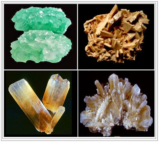
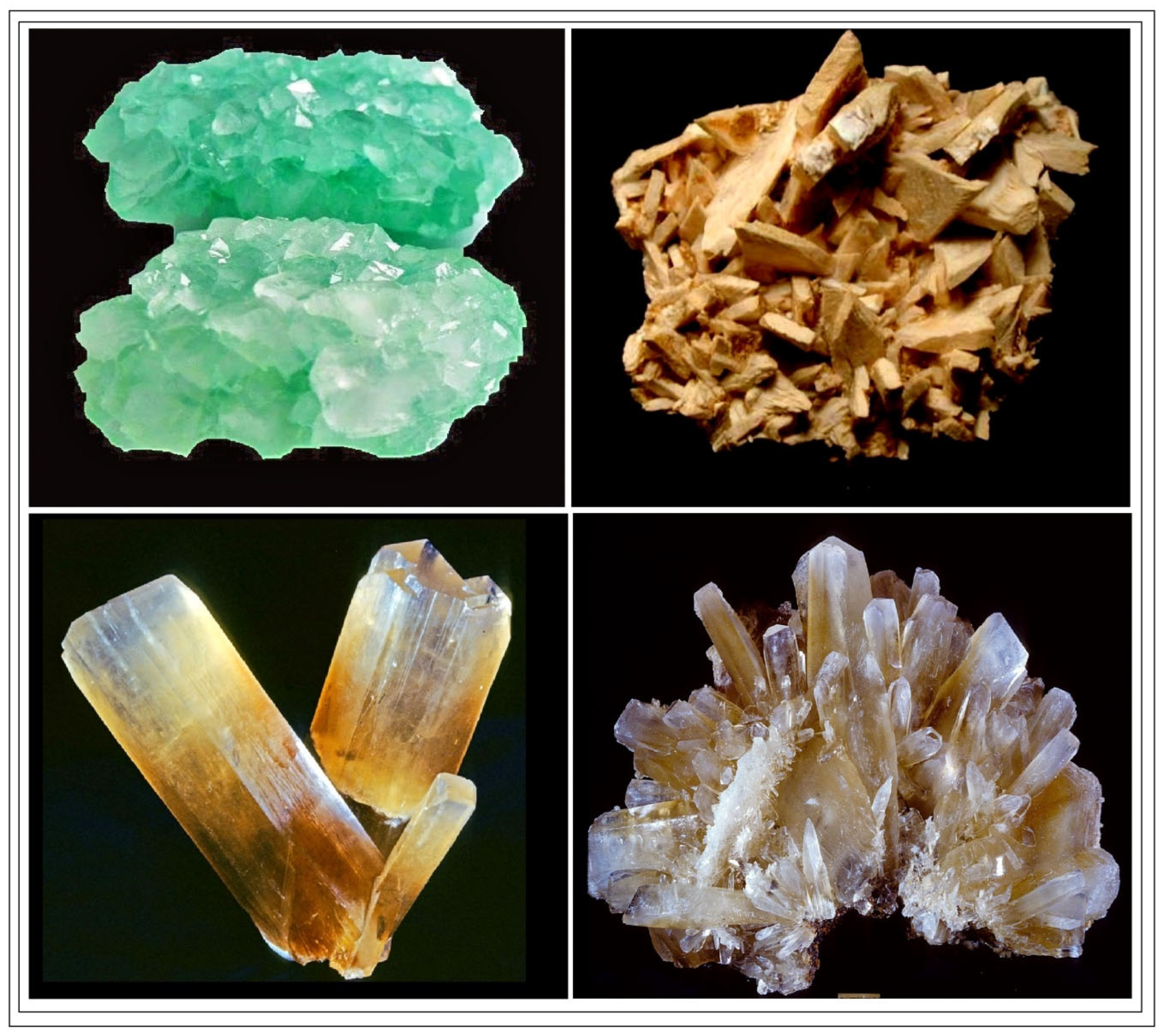
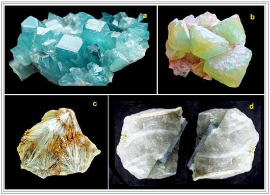
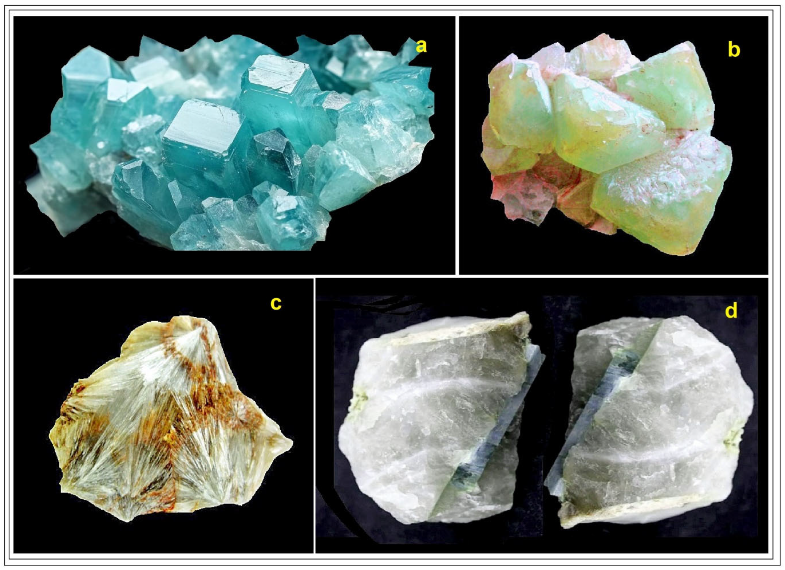
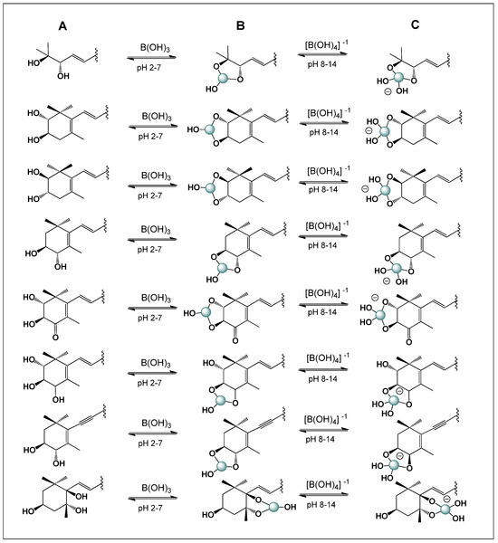
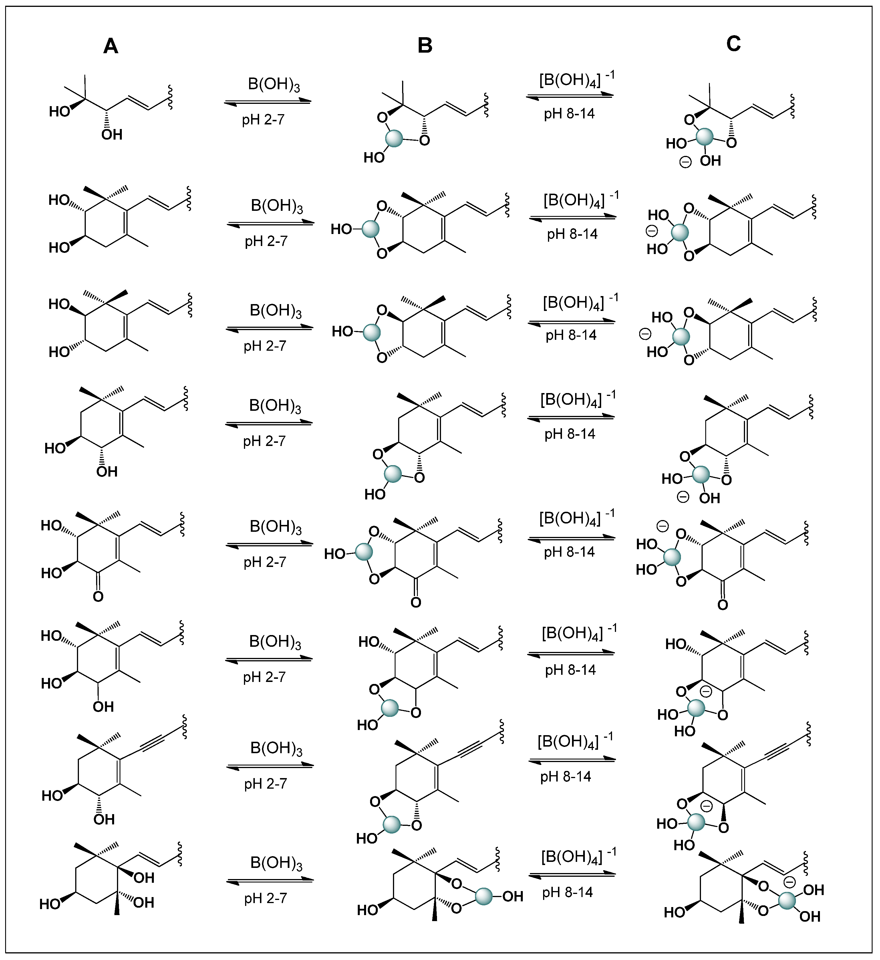
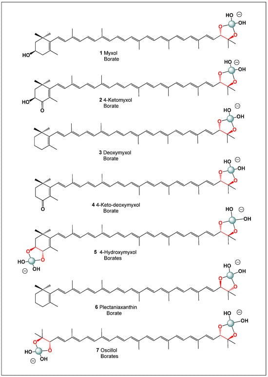
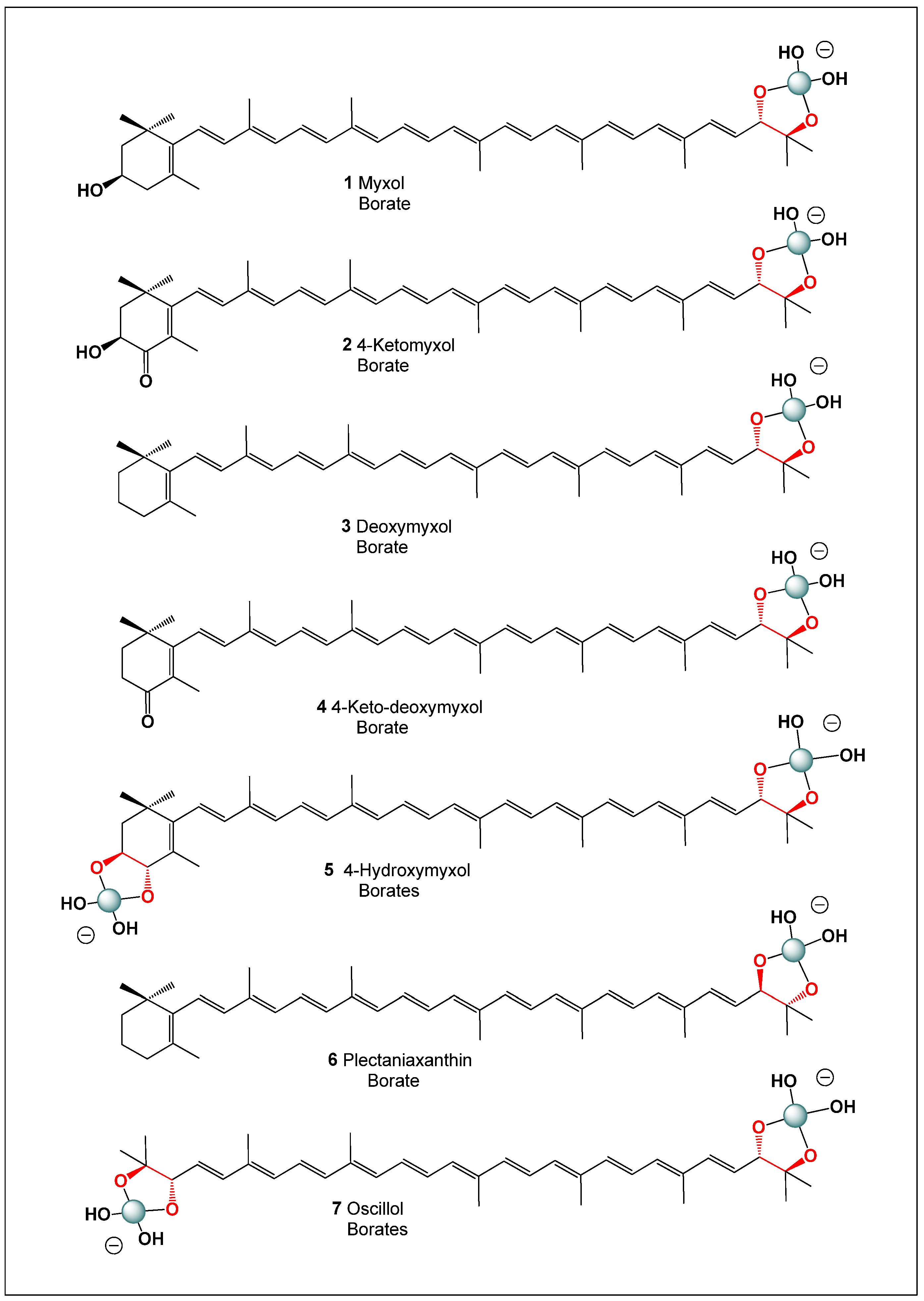
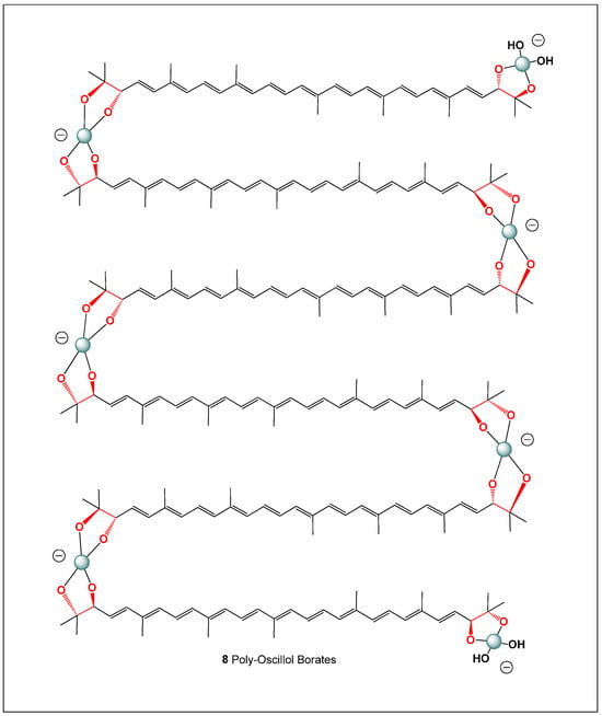
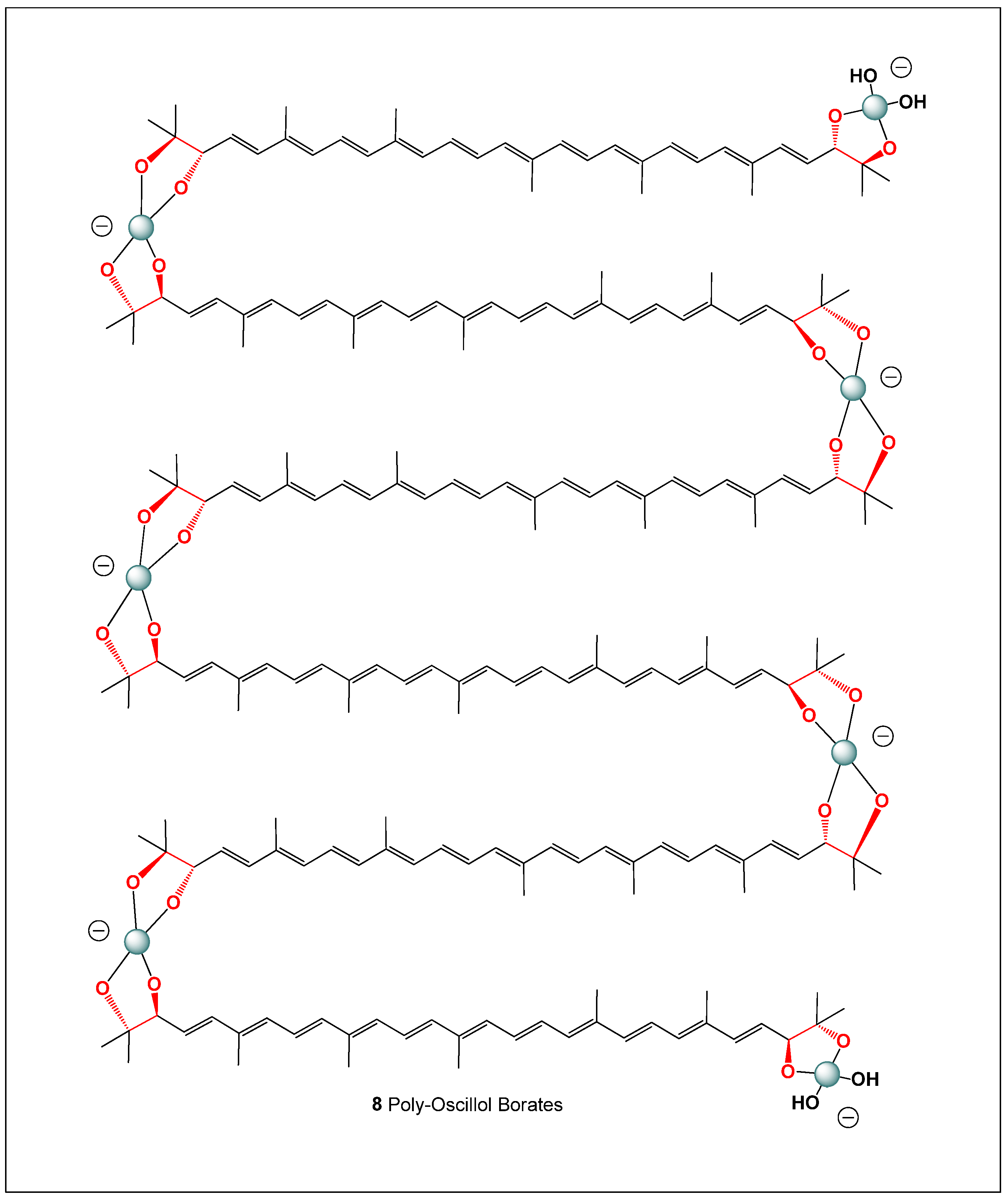
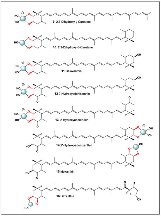
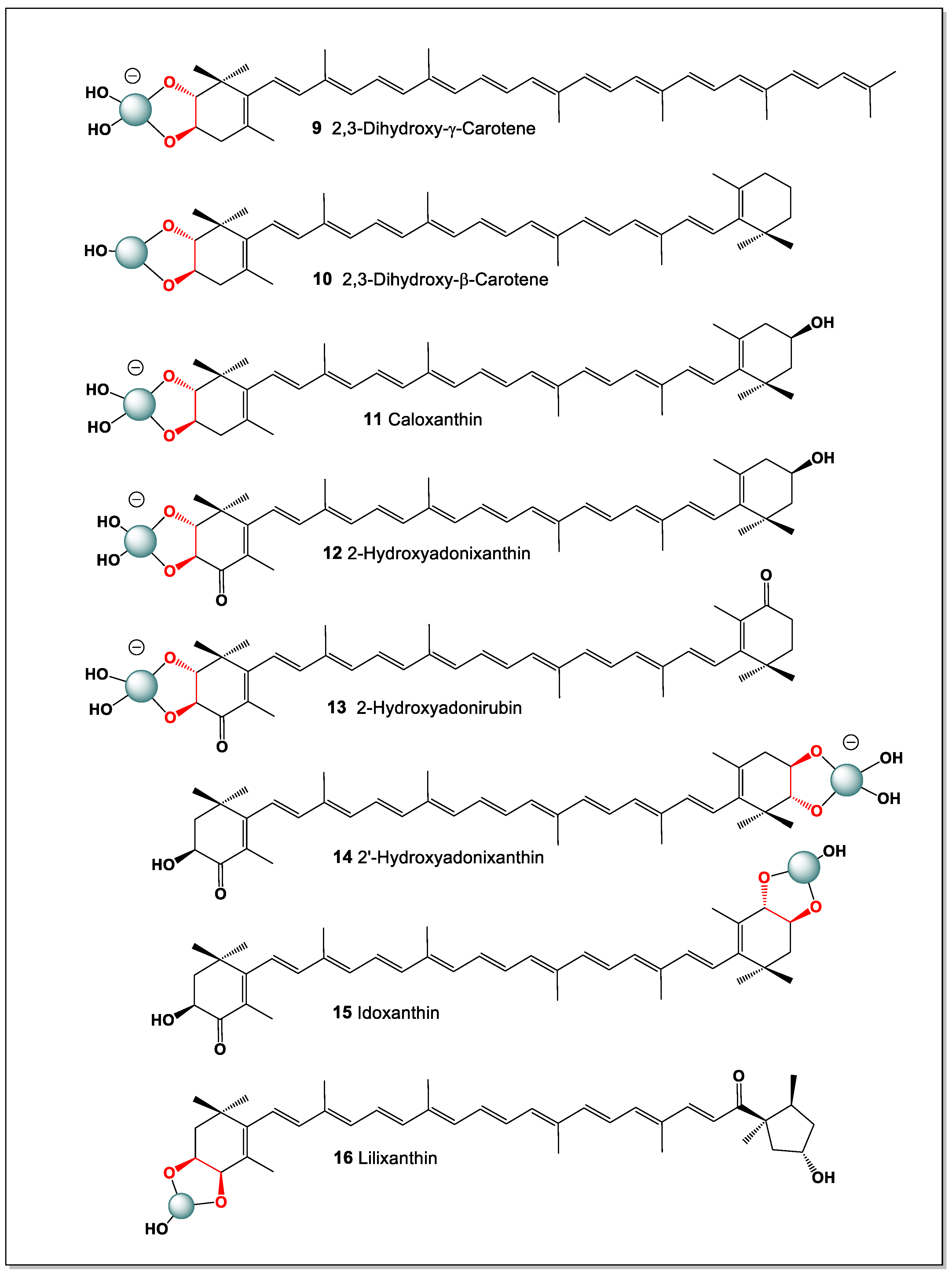
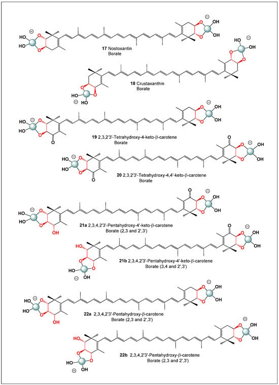
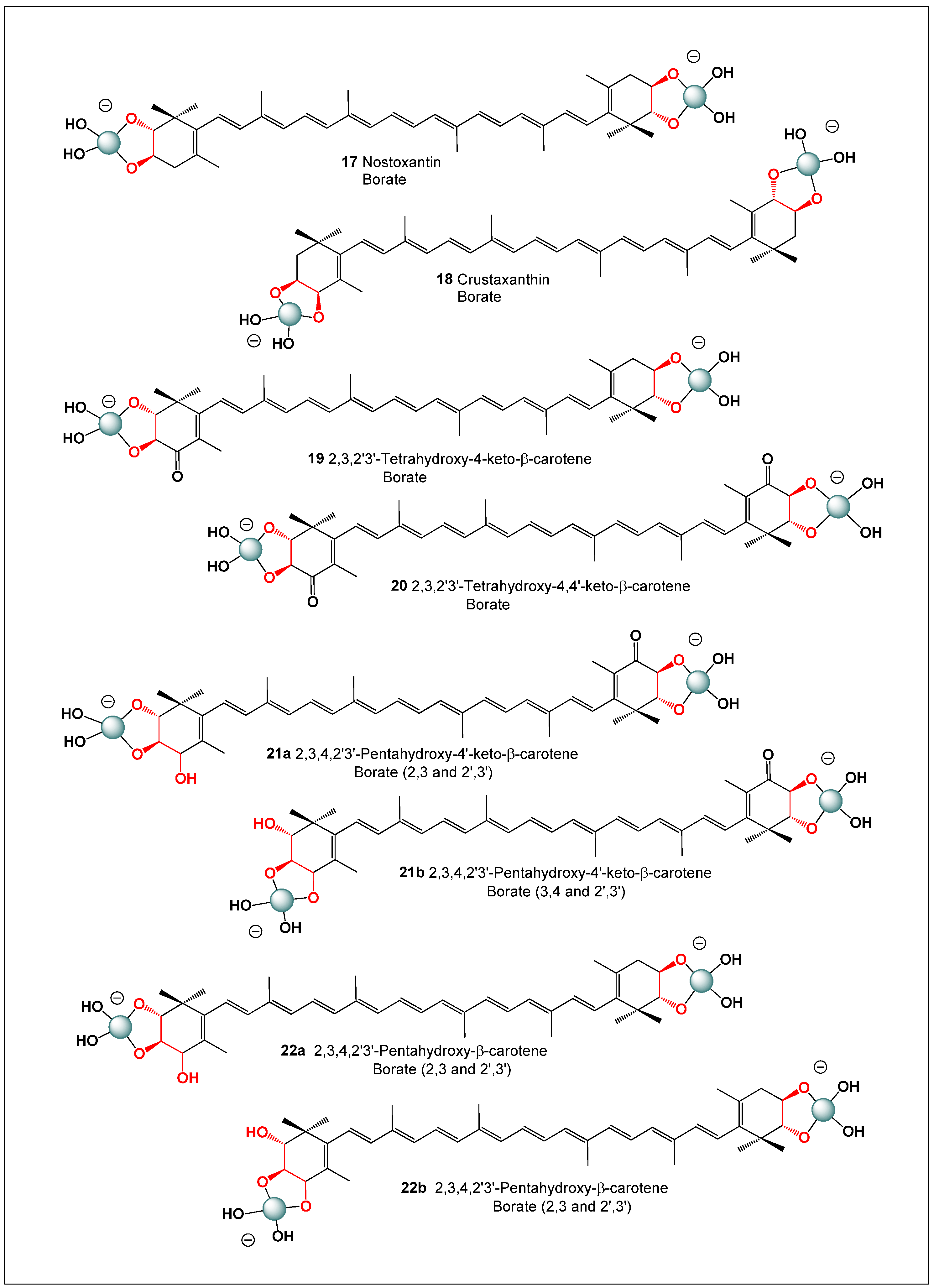
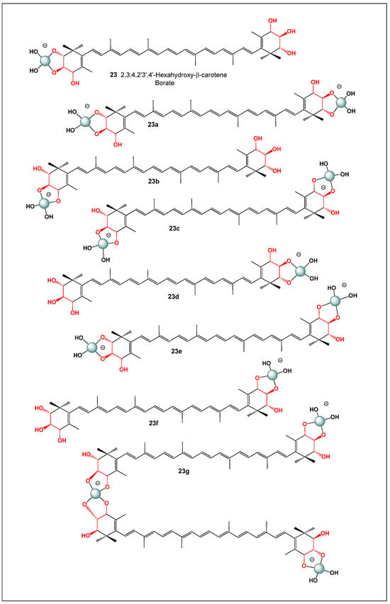
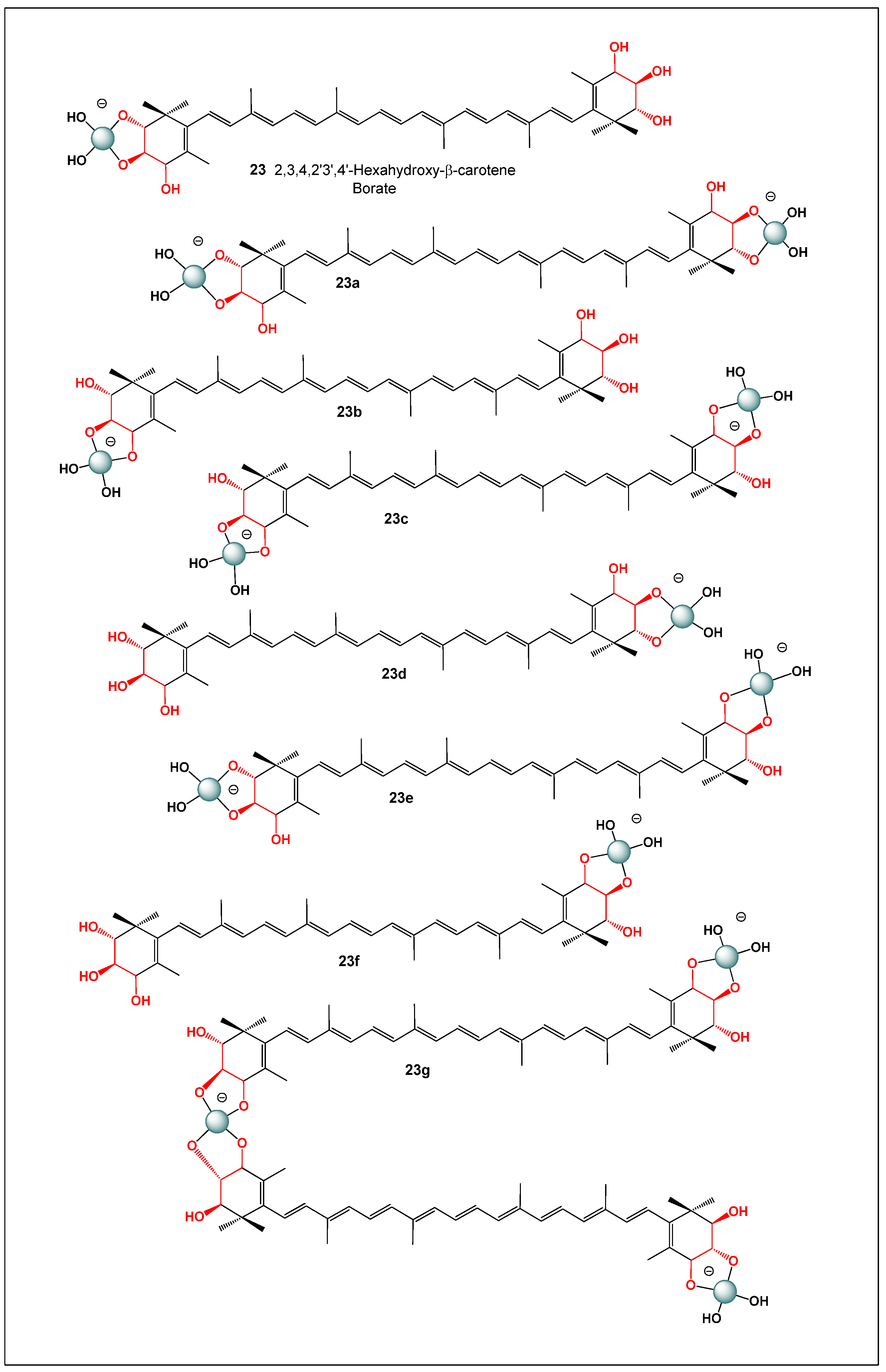
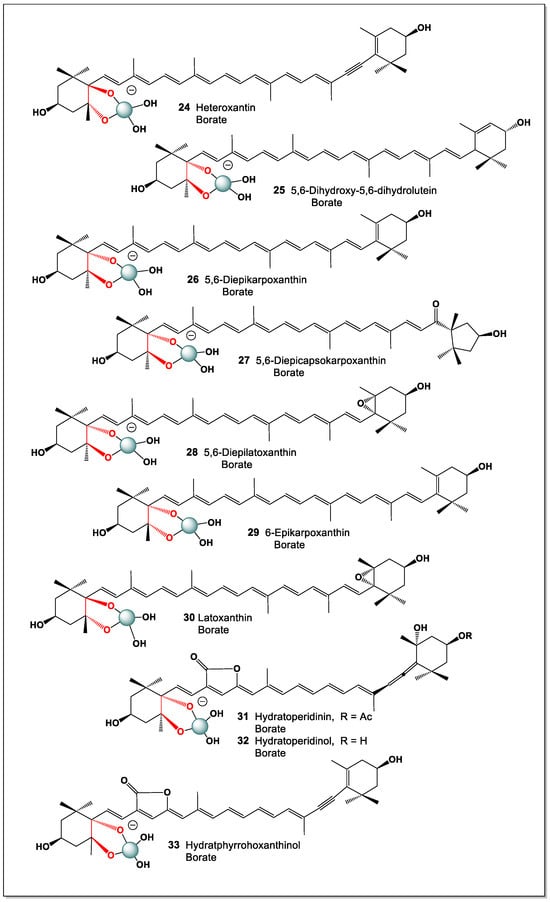
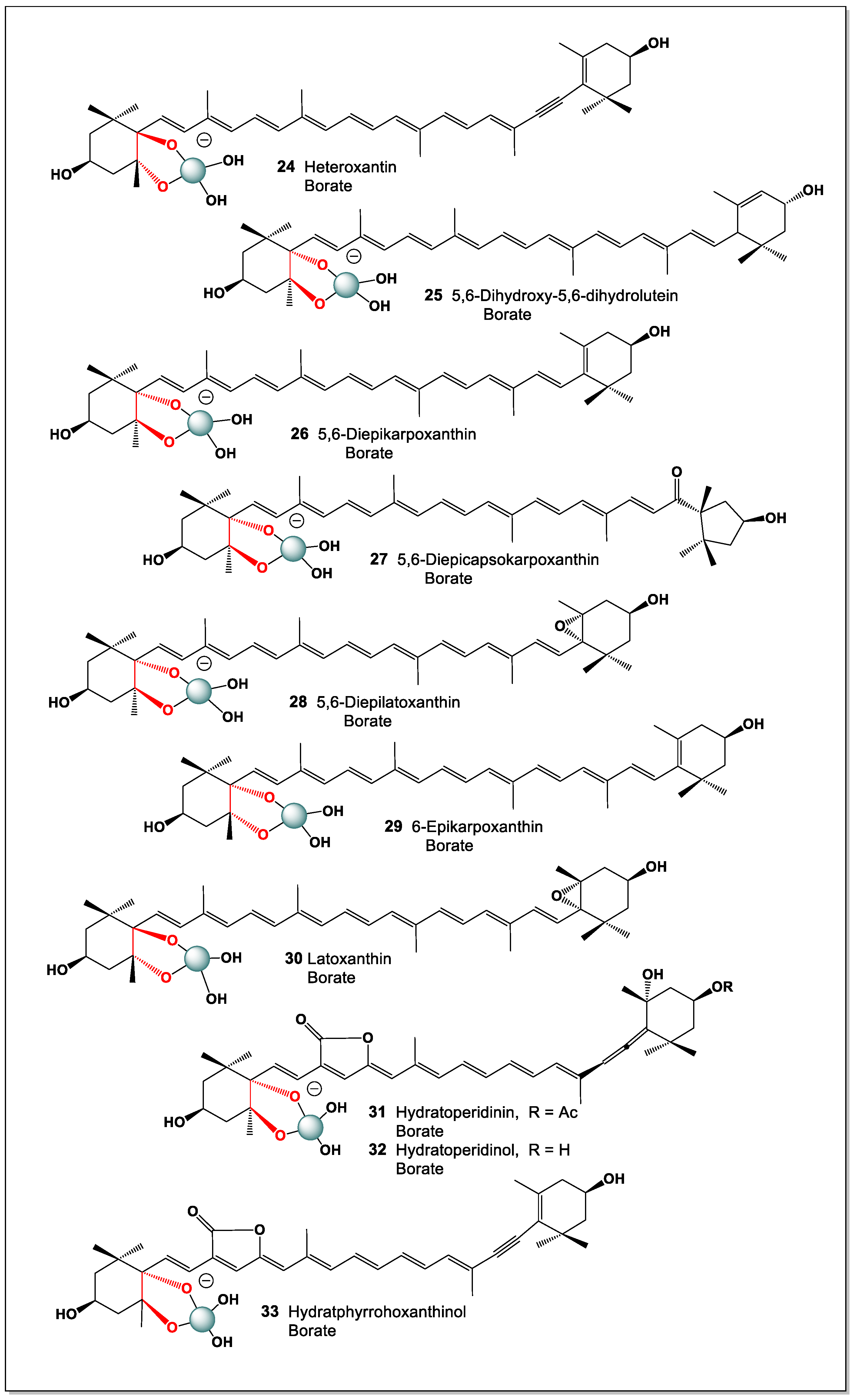
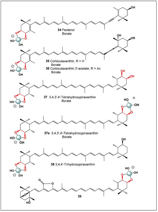
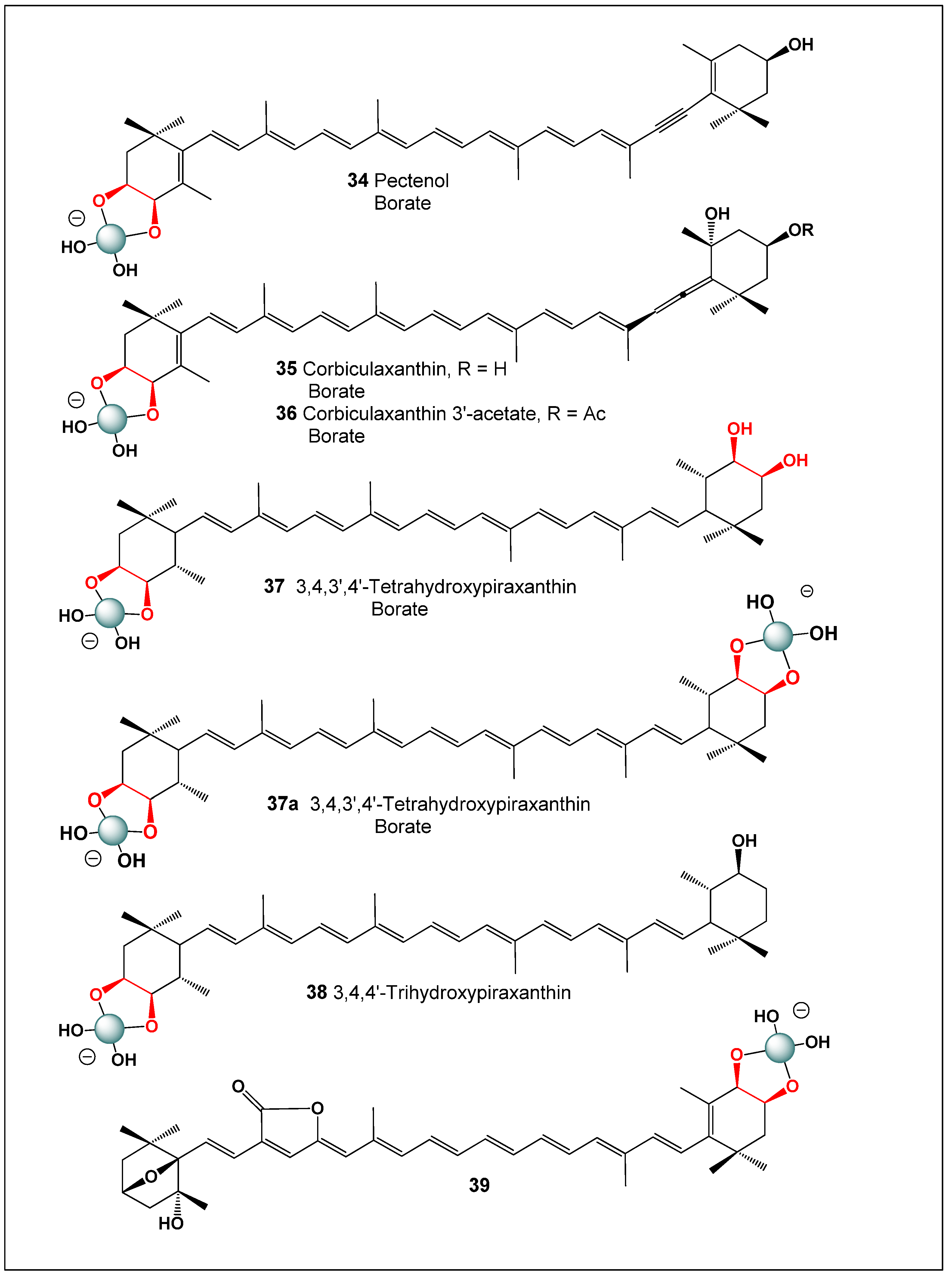
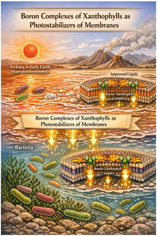
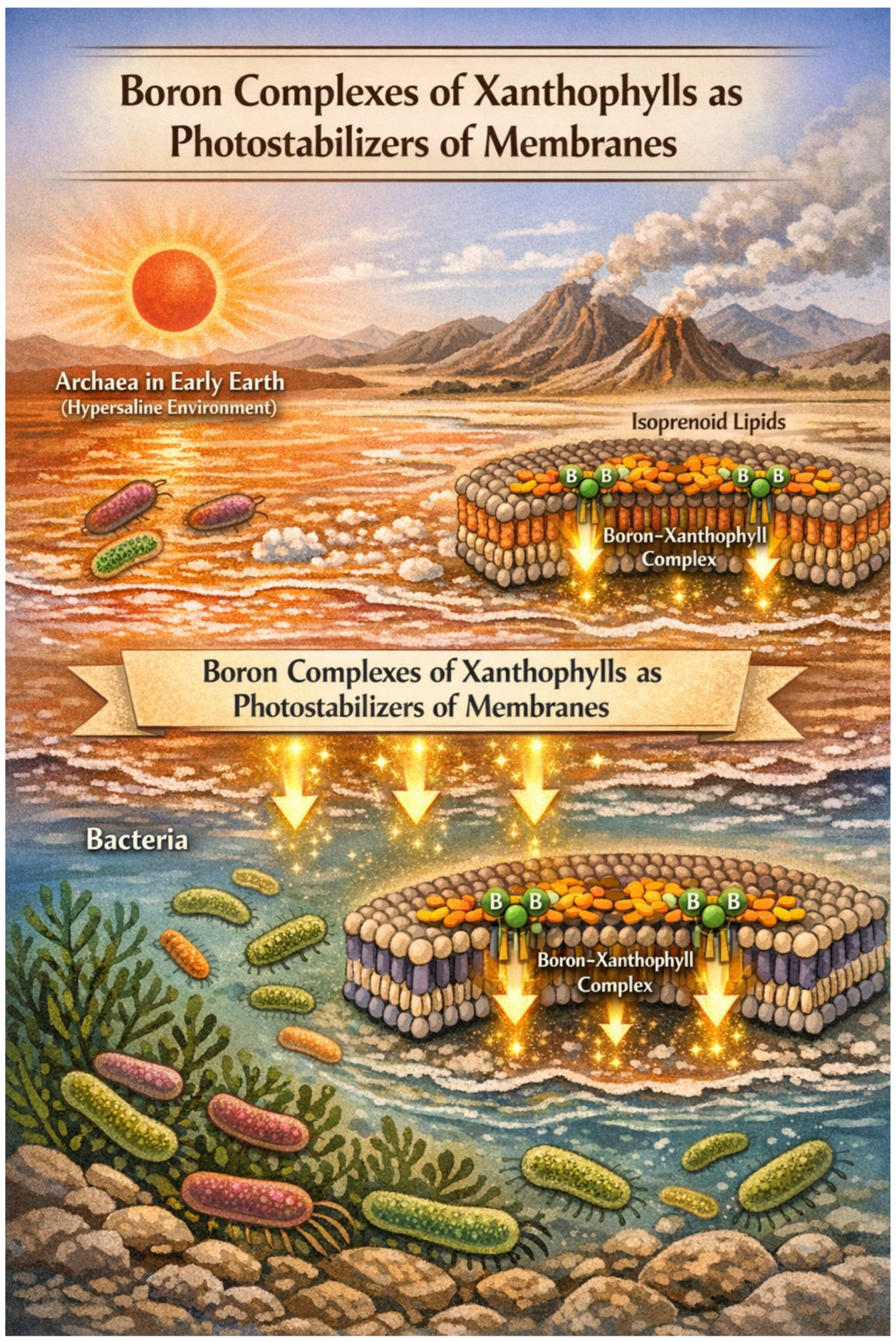

Disclaimer/Publisher’s Note: The statements, opinions and data contained in all publications are solely those of the individual author(s) and contributor(s) and not of MDPI and/or the editor(s). MDPI and/or the editor(s) disclaim responsibility for any injury to people or property resulting from any ideas, methods, instructions or products referred to in the content. |
© 2026 by the authors. Licensee MDPI, Basel, Switzerland. This article is an open access article distributed under the terms and conditions of the Creative Commons Attribution (CC BY) license.