Review

Unraveling the Potential of Photochemical Nanoplatforms in Tumor Microenvironments: Therapeutic Strategies for Gastrointestinal Malignancies

1
Department of Oncology, the Seventh Medical Center, Chinese PLA General Hospital, Beijing 100010, China
2
Medical School of Chinese PLA, Beijing 100853, China
3
Department of Oncology, the Fifth Medical Center, Chinese PLA General Hospital, Beijing 100071, China
*
Author to whom correspondence should be addressed.
Photochem 2026, 6(1), 10; https://doi.org/10.3390/photochem6010010
Submission received: 22 January 2026 / Revised: 10 February 2026 / Accepted: 13 February 2026 / Published: 4 March 2026

Abstract

Gastrointestinal (GI) malignancies have caused tremendous disease burden around the world; however, conventional therapy strategies, such as radiotherapy, chemotherapy, and immunotherapy, have achieved limited efficacy in the diagnosis and treatment. In further exploration of GI tumors, the complexity and heterogeneity of the tumor microenvironment (TME) have been increasingly recognized. Appropriate strategies to modulate the TME are necessary to enhance the therapeutic effect. Photosensitizers (PSs) are chemical substances that are activated at specific wavelengths of light to initiate photodynamic effects. Nanotechnology provides a platform for the targeted delivery of PSs and small-molecule drugs, enabling precise targeting and remodeling of the TME. In this review, we summarize the principles and mechanisms of photochemical reactions and elaborate on the effect of photochemical nanoplatforms in modulating the TME of GI tumors. Finally, we discuss the potential value of photochemical nanoplatforms for diagnosing GI malignancies.

1. Introduction

Gastrointestinal (GI) malignancies have represented a major health challenge, accounting for over a quarter of cancer incidence and one-third of cancer mortality [1,2]. Currently, therapeutic strategies for GI tumors include radiotherapy, chemotherapy, and immunotherapy, but the clinical benefits remain limited [3]. Increasing evidence suggests a complex tumor microenvironment (TME) in GI malignancies, where tumor cells constitute only 5–10%, and the remaining components include mesenchymal cells, immune cells, and a complex extracellular matrix (ECM) [4,5]. The cross-linking of the ECM induces fibrosis, which stiffens the tumor stroma and facilitates tumor cell growth, migration, and chemoresistance [6,7]. For example, it was reported that the TME of pancreatic adenocarcinoma (PDAC) contains over three times the collagen fiber density compared to breast cancer, creating a profound physical barrier to drug penetration [8]. In CRC, gut microbiota-derived metabolites (such as short-chain fatty acids) modulate immune cell repolarization, thereby exacerbating immunosuppression. Meanwhile, the unique sinusoidal vasculature of the HCC TME leads to rapid clearance of conventional nanocarriers by Kupffer cells (KCs) [9]. Furthermore, the immunosuppressive microenvironment and metabolic reprogramming of the TME also induce tumor cell malignant transformation and chemoresistance [10,11]. Thus, simply targeting tumor cells is insufficient for impeding GI malignancies’ progression, and modulating the TME of GI malignancies presents promising therapeutic prospects to enhance anti-tumor efficacy.
Currently, nanoplatforms for therapeutic drug delivery have achieved temporal and spatial control over drug release, significantly enhancing the efficacy of tumor treatment [12]. Nanotechnology enables the integration of multiple components, such as chemotherapy drugs, antibodies, and even photosensitizers (PSs) into a single delivery vehicle. The core advantage of photo-responsive nanoplatforms lies in their ability to precisely target tumors with photosensitizers and use light to controllably elicit diagnostic fluorescence or therapeutic photodynamic effects [13]. Photodynamic therapy (PDT) employs light and PSs to generate reactive oxygen species (ROS), which induce tumor-specific cytotoxicity and lead to tumor cell death. Utilizing PS-induced luminescence enables tumor-specific enrichment, thereby distinguishing tumor cells from normal cells. This facilitates photodiagnosis (PD) and extends its application to photoacoustic imaging, providing more comprehensive diagnostic information [14,15]. Photo-responsive nanoplatforms can be constructed by leveraging the tumor-accumulating properties of PSs. For example, human serum albumin-chlorin e6 nanoassemblies (HSA-Ce6 NAs) can achieve complete tumor elimination under 660 nm near-infrared (NIR) laser irradiation, with no recurrence or side effects. Furthermore, due to the inherent fluorescence, photoacoustic properties, and chelating capabilities of the porphyrin ring, HSA-Ce6 NAs enable tri-modal imaging—fluorescence (605 nm), photoacoustic (680 nm), and MRI—which facilitates real-time monitoring of tumor treatment efficacy [16].
In this review, we summarize the construction of photo-responsive nanoplatforms for PDT and PD. It systematically covers the evolution of PSs (1st- to 4th-generation), type I/II photochemical reactions, and the employed nanocarrier systems (such as polymers, liposomes, and inorganic nanoparticles (NPs)), along with their functional and targeting modifications. The main objective is to address the poor solubility of PSs and their insufficient tumor targeting capabilities in PDT. From the perspective of GI oncology, this review integrates a comprehensive TME framework (including angiogenesis, ECM remodeling, immunosuppression, metabolic reprogramming) with the practical demands of clinical translation.

2. Photochemical Nanoplatform Construction and Principles of Effect

With the development of nanoscience, it is possible to use photochemical nanoplatforms to achieve precise spatiotemporal control of drug release [17]. Typically, a photochemical system mainly relies on three key factors: PS, oxygen, and light. The primary steps are as follows: First, deliver specific PS to tumor regions via nanoplatforms to enable its selective accumulation. Next, apply certain wavelengths of light to fully activate the PS and perform optical treatment on the target area [12,18]. A deep understanding of photochemical nanotechnology, including nanoplatform assembly, specific PS, and mechanisms of action, is essential for constructing photochemical nanoplatforms and advancing applications in PDT and PD.

2.1. The Development and Evolution of PS

PSs are central components of photochemical nanoplatforms, and their properties primarily determine the effectiveness of treatment or diagnosis. PSs used in PDT typically exhibit optimal absorption peaks within the 600–800 nanometer range. Beyond this range, the penetration of PSs is limited, thereby reducing therapeutic efficacy. Furthermore, the foundation of PDT lies in combining the tumor-targeting accumulation of PSs with spatiotemporally controlled light exposure, which enables precise tumor cell destruction while sparing healthy tissues [13,19] (Table 1).
The first-generation PS was a mixture of water-soluble porphyrins, hematoporphyrin derivative (HPD) and purified photofrin that absorb wavelengths from 500 to 700 nm [20,21]. Photofrin enables the generation of singlet oxygen (1O2) and the PDT effect; however, it has several limitations, including prolonged skin photosensitivity and relatively low absorption at 630 nm, which influence therapeutic efficacy [22,23]. Designed to improve safety and efficacy over first-generation agents, second-generation photosensitizers include direct agents such as chlorin e6 and phthalocyanines, as well as prodrugs (e.g., methyl aminolevulinate (MAL) and 5-aminolevulinic acid (ALA)) that are converted in situ to the active protoporphyrin IX (PpIX) [24,25]. It is found that the second-generation PS absorbs longer wavelengths ranging from 600 nm to 800 nm, penetrates deeper, exhibits greater tissue selectivity, and metabolizes rapidly [26]. However, the limited water solubility and a tendency to self-aggregate in biological media limit intravenous administration, requiring alternative delivery methods [27]. To more effectively target tumor cells and reduce adverse effects, researchers developed third-generation PSs. Strategies include combining PSs with specific components such as amino acids, peptides, and antibodies, and encapsulating them in vehicles such as nanoparticles, cellular membranes, liposomes, and micelles [28,29]. By optimizing PS distribution and enhancing PDT efficacy, nanotechnology also stimulates immune cells to destroy tumor cells, creating a synergistic effect with immunotherapy. Recently, fourth-generation PSs have been developed that employ porous carriers, including mesoporous silica and metal–organic frameworks (MOFs) [30]. MOFs are porous structures composed of metal ions and organic ligands, enabling MOFs to adsorb or capture external molecules [31]. Studies indicated that integrating metal ions with organic PS ligands to form metal-PS complexes is a highly effective way to significantly improve the therapeutic outcomes of PDT [32]. It was found that PS-based MOFs have advantages in alleviating hypoxia, generating ROS, and functioning as contrast agents [33]. For example, loading the acridine fluorophore (ACF) into PS-based MOFs enhanced the PDT effect, which blocked HIF-1α/HIF-1β dimerization, thereby alleviating hypoxia and downregulating survival and metastasis-related genes (such as VEGF, BCL-2, and matrix metalloproteinase-9 (MMP9)) [34]. As noted above, the revolution in PSs mainly focuses on enhancing targeting capabilities and reducing adverse effects.
Table 1. Summary of representative PSs and characteristics.
GenerationRepresentative PSExcitation WavelengthΦΔPenetration Depth (µm)CharacteristicsReferences
FirstHematoporphyrin derivative (HPD)630 nm0.7–0.8500–1000Poor tissue penetration and skin photosensitivity[20,21]
Photofrin0.89500–1300
SecondPpIX600–800 nm0.3–0.61000–3000Limited water solubility and self-aggregation in biological media[24,25]
chlorin e6 (Ce6)0.6–0.752000–5000
ThirdMonoclonal antibodies, amino acids, and peptides conjugated with PS600–800 nmAs primary PSsIncrease by 20–40% compared to unmodified first- and second-generation PSHigher tissue selectivity and lower required dose[28,29]
FourthEmploy mesoporous silica and metal–organic frameworks (MOFs) as porous carriers600–800 nm0.6–0.728000–15,000High porosity, adjustable pore diameter, controllable composition, and multifunctional modification[30,34]
P.S.: Porphyrins and benzoporphyrin derivatives (ALA, MAL) are prodrugs that generate PpIX in PDT and PD.

2.2. The Light Sources

For activating the PSs, it is necessary to use a specific wavelength of light that matches the PSs’ absorption range. Typically, the selection of light sources should consider multiple factors, such as the absorption spectrum of photosensitizers (600–800 nm), disease type (including lesion location, size, and tissue characteristics), and cost. Additionally, light intensity, dose, exposure time, and delivery method (single exposure, fractionated exposure, and timed exposure) also influence the efficacy of PDT/PD [35]. At present, the most common light sources are light-emitting diodes (LEDs) and lasers [36,37]. Additionally, X-rays generate high-energy light that can penetrate deeply into tissue, which allows them to serve as a source of light for photochemical systems [38]. It shows promise in terms of tumor diagnosis and treatment, but has lower energy transmission efficiency.

2.3. Mechanism of Photochemical Action

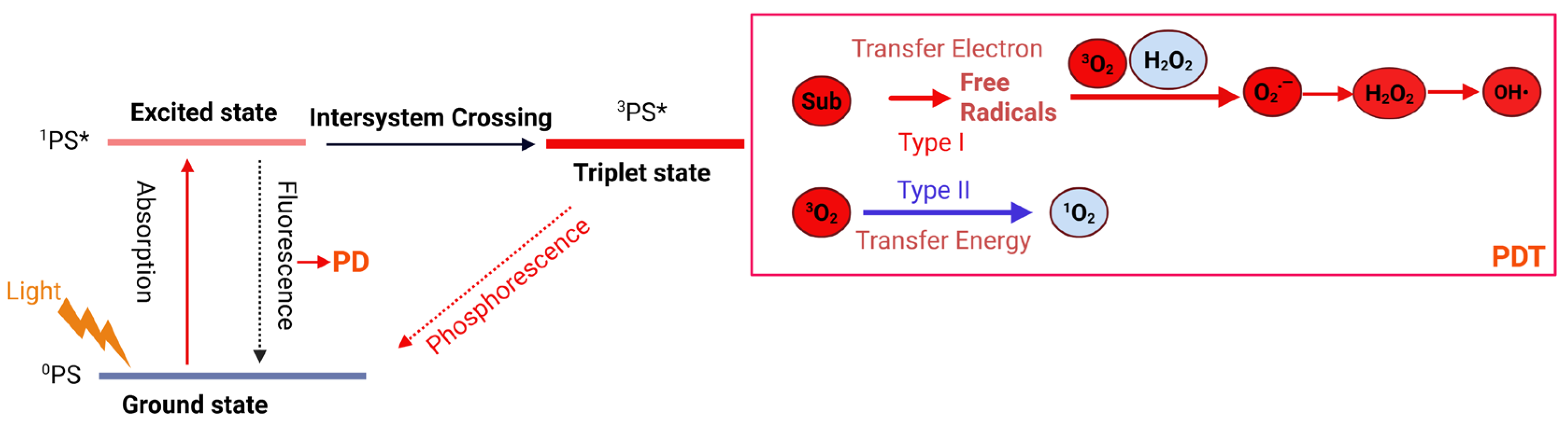
Photochemical mechanisms involve types I and II, with type II being the most common. Upon light excitation, the PS transitions from its initial ground state to the excited state (1PS*). Notably, the unstable 1PS* undergoes intersystem crossing (ISC) to form a more stable, longer-lived excited triplet state (3PS*). Although the 3PS* can relax to the ground state via phosphorescence, this process is forbidden by quantum selection rules. In type-II photodynamic reactions, the 3PS* transfers its energy to ground-state triplet oxygen (3O2), promoting its conversion to reactive singlet oxygen (1O2) while the PS returns to its ground state [39]. In type-I photodynamic reaction, the 3PS* undergoes electron or hydrogen-atom transfer with substrate molecules (such as biomembranes, proteins, and nucleic acids), generating free radicals [40]. These radicals undergo auto-oxidation to produce superoxide anion radicals (O2), which then form hydrogen peroxide (H2O2). Finally, H2O2 undergoes single-electron reduction to yield the highly reactive hydroxyl radical (HO•). The competition between Type I and Type II reactions depends on the PSs’ types, oxygen levels, and the substrate. This competition directly influences the ΦΔ value. Typically, Type II dominates when oxygen is abundant, whereas Type I is activated under hypoxia [41,42]. The underlying principle of PD is that the PS was internalized by tumor cells and then excited by short-wavelength light (330–400 nm), thereby emitting a detectable fluorescence (Figure 1).
The type II reaction follows conservation of angular momentum and selection rules, with the matching of electron spin states between PS and O2 serving as the core regulatory factor for energy transfer efficiency [43]. Porphyrins and phthalocyanines exhibit a high 1O2 quantum yield of 0.6–0.9 following ISC. In Type I reactions, BODIPY and ruthenium polypyridine complexes can enhance electron-transfer efficiency, sustaining effective ROS generation under hypoxic conditions [44]. The excitation wavelength and photon flux of PS are critical for determining PDT efficiency [45]. The PS absorption peak should be precisely aligned with the light source wavelength, preferably in the NIR I region (700–900 nm) or the NIR II region (1000–1700 nm) to improve tissue penetration depth. Additionally, the photon flux, typically 10–100 mW/cm2, is sufficient for PS excitation and ROS generation [46].
Notably, the unique characteristics of the TME may impair the PS excited-state lifetime and ROS production. The hypoxic microenvironment of solid tumors directly inhibits the energy transfer process, reducing 1O2 generation by 60–80%. High GSH in the TME not only reduces 3PS* via electron transfer but also degrades ROS, thereby compromising PDT efficacy [47]. Additionally, biomacromolecules (such as proteins and nucleic acids) in tumor tissues can bind to PS, which leads to PS aggregation and excited-state quenching.
PDT induces three primary pathways of cell death: apoptosis, necrosis, and autophagy-related cell death. Notably, apoptosis is the primary cell death mechanism, triggered by increased permeability of the mitochondrial outer membrane (MOMP) [48,49]. In brief, when PSs enriched in mitochondria are damaged, Bcl-2 proteins mainly signal to induce MOMP, which promotes the release of caspase activators such as cytochrome C, as well as other pro-apoptotic molecules, etc. Crucially, the rupture of the lysosomal membrane and the subsequent release of cathepsins further promote MOMP [50].

2.4. The Construction of Photo-Responsive Nanomaterials

Nanoparticles are engineered to vary in size, shape, and surface properties, allowing them to adjust to the pathophysiological characteristics of tumors [8,51]. Meanwhile, nanomaterials enable the delivery of therapeutic molecules, such as antibodies, chemotherapy drugs, and nucleic acids, to tumor tissue, enabling targeted treatment. Importantly, they employ both passive and active targeting mechanisms; when they reach a tumor, they can release therapeutic molecules there via the enhanced permeability and retention (EPR) effect [52,53]. Nanocarriers effectively suppress π-π stacking and hydrophobic aggregation among PSs via spatial confinement and host-guest interactions, thereby preventing excited-state quenching and increasing ΦΔ [54,55]. Studies have identified an optimal size window for organic nanocarriers to maximize ROS yield: for example, AIE-type nanoparticles reach a peak ROS yield (Φ = 0.78) at 45 nm. Meanwhile, the surface charge of nanocarriers influences ROS diffusion [56,57]. Cationic nanoparticles enhance electrostatic interactions with cell membranes, increasing ROS concentrations.
The application of nanomaterials enhances PDT effects and enables the controlled release of drugs. Because high-energy photons, such as ultraviolet and visible light, penetrate tissues poorly, photo-responsive nanomaterials, including liposomes, polymeric nanoparticles, and gold (Au) nanoparticles, were developed to respond to low-energy photons [58,59]. Liposomes are nanovesicles composed of bilayer phospholipid modules and exhibit excellent biocompatibility [60,61]. Importantly, liposomes serve as effective carriers to load small-molecule drugs. For example, the nanomedicine Ce6-SB3CT@Liposome (Lip-SC) was synthesized by co-encapsulating Ce6 and MMP inhibitor SB3CT within liposomes [62]. Additionally, modifying liposomes enhances their targeting abilities. Yi et al. [63] encapsulated indocyanine green (ICG) and the ROS-responsive doxorubicin prodrug (β-DOX) simultaneously in polyethylene glycol (PEG)-modified liposomes. This enabled passive targeting of tumor sites and demonstrated excellent anti-tumor effects. Polymers are one type of targeted delivery vehicle that exhibit compositional and structural diversity, as well as flexibility in particle size [64,65]. A recent study utilized heparin-conjugated pheophorbide-a (PHA) and folic acid to form nanoparticles through self-assembly [66]. This approach enhances passive targeting capabilities while maintaining PDT efficacy. To achieve stable NIR PDT for PDAC, researchers functionalized upconversion nanoparticles (UCPs) by coating them with poly [N, N-dimethylacrylamide-co-2-aminoethylacrylamide]-graft-poly (ethylene glycol) [P(DMA-AEM)-PEG-Ale] and loading them with mTHPC, thereby providing colloidal stability and PDT capability [67]. Furthermore, UCPs were coated with poly(methyl vinyl ether-alt-maleic acid) (PMVEMA) and temoporfin (THPC), which improved their stability in water and enabled them to produce 1O2 under NIR exposure [68]. Metal nanoparticles exhibit several advantages, including excellent biocompatibility, stable properties, and suitable surface modification, making them ideal carriers for PSs delivery [69]. Liu et al. [70] developed a multifunctional nanoplatform based on mesoporous silica-coated gold nanorods (named AuNR@MSN). The system was co-modified by loading ICG, attaching β-cyclodextrin (β-CD), and anchoring the mitochondrial-targeting peptide RLA ([RLARLAR]2). This pH-regulated nanoplatform enables PSs to accumulate specifically within mitochondria. Efficient cancer cell targeting can be achieved using MOFs. For example, MOFs with chlorinated compounds can be activated by long-wavelength light, and functionalizing their surface with chlorin improves targeting ability [71]. Another study demonstrated a novel approach using NH2-MOFs as templates to synthesize porous gold nanospheres (NH2-MOFs@Aushell). By encapsulating platinum nanozymes in NH2-MOFs, coating them with porous AuNSs, and loading Ce6, they developed the Pt@UiO-66-NH2-Aushell-Ce6 nanoplatform [72]. Subsequent experimental results confirmed that this system improved PDT efficacy.
Notably, studies indicate that modifying nanomaterials enables them to bind specifically to unique antigens or receptors expressed on the surface of tumor cells, such as the epidermal growth factor receptor (EGFR) or the low-density lipoprotein receptor [73,74]. This process facilitates the accumulation of drugs within tumor cells through receptor-mediated endocytosis. For instance, Li et al. [75] developed manganese dioxide/iridium dioxide (MnO2, IrO2) nanoplatforms via the redox reaction between potassium permanganate (KMnO4) and iridium chloride (IrCl3). After surface modification with polyvinylpyrrolidone (PVP) and Ce6 loading, they obtained nanoparticles with good colloidal stability and biocompatibility. Furthermore, coating nanoparticles with biocompatible polymers has been shown to enhance their stability and reduce toxicity. Jia et al. [76] developed SiO2@Au nanoparticles that were loaded with saponin (SAP) and coated with chitosan (CS). To further enhance biocompatibility and cellular uptake, the nanoparticles were functionalized with a cancer cell membrane (CCM) layer, thereby significantly improving the efficacy of PDT.

3. Photo-Responsive Nanoplatform for GI Malignancies Therapy

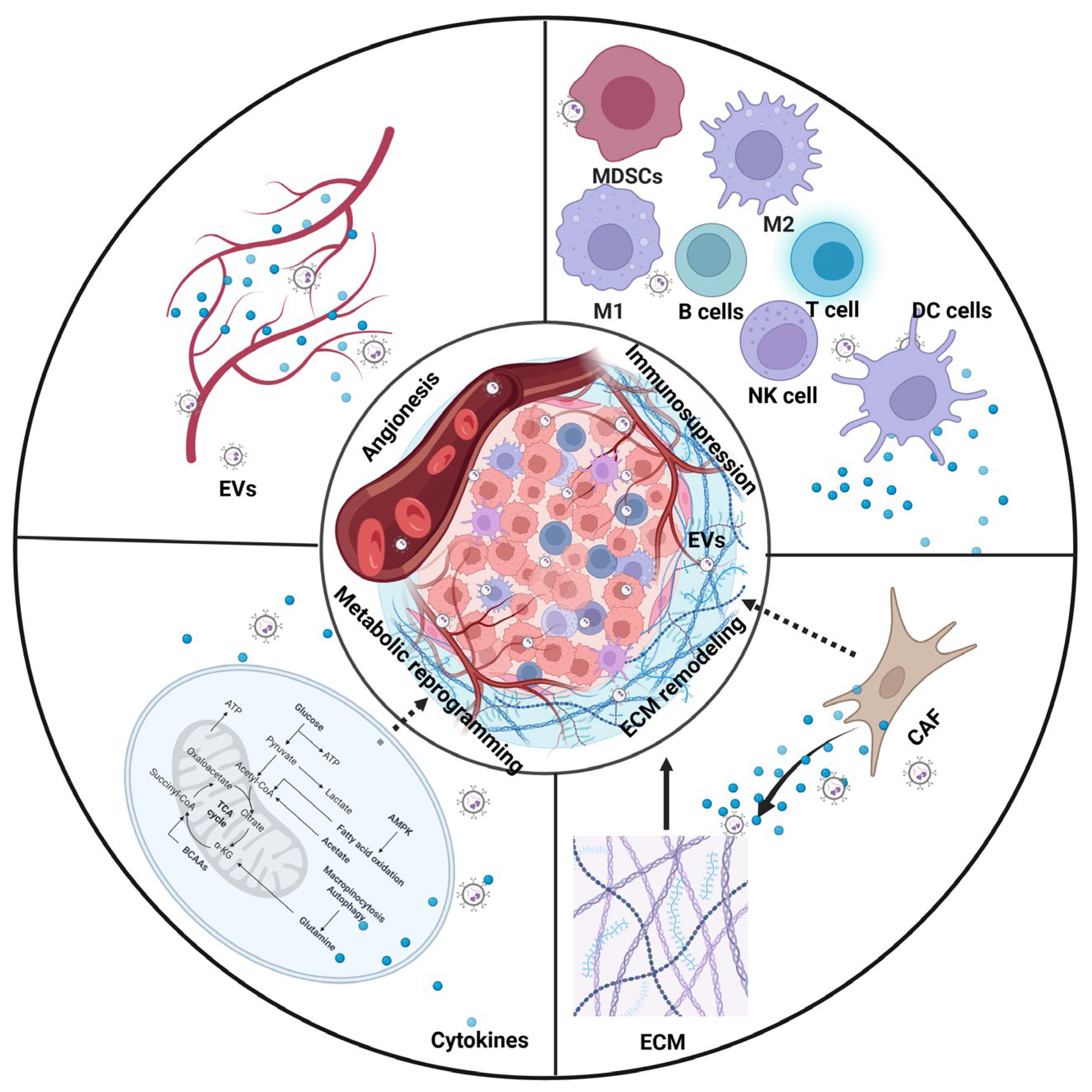
The TME of GI malignancies is a dynamic and complex system comprising multiple cell types, including tumor cells, immune cells, cancer-associated fibroblasts (CAFs), and endothelial cells (ECs), as well as non-cellular constituents such as the ECM [77]. The main characteristics of the TME are angiogenesis, an immunosuppressive microenvironment, ECM remodeling, and metabolic reprogramming. Notably, the TME exhibits high plasticity and is continuously remodeled through interactions among various cells and their bioactive products (Figure 2) [78,79]. Therefore, the strategy of targeting GI malignancies not only eliminates tumor cells, but remodels the TME (Table 2).
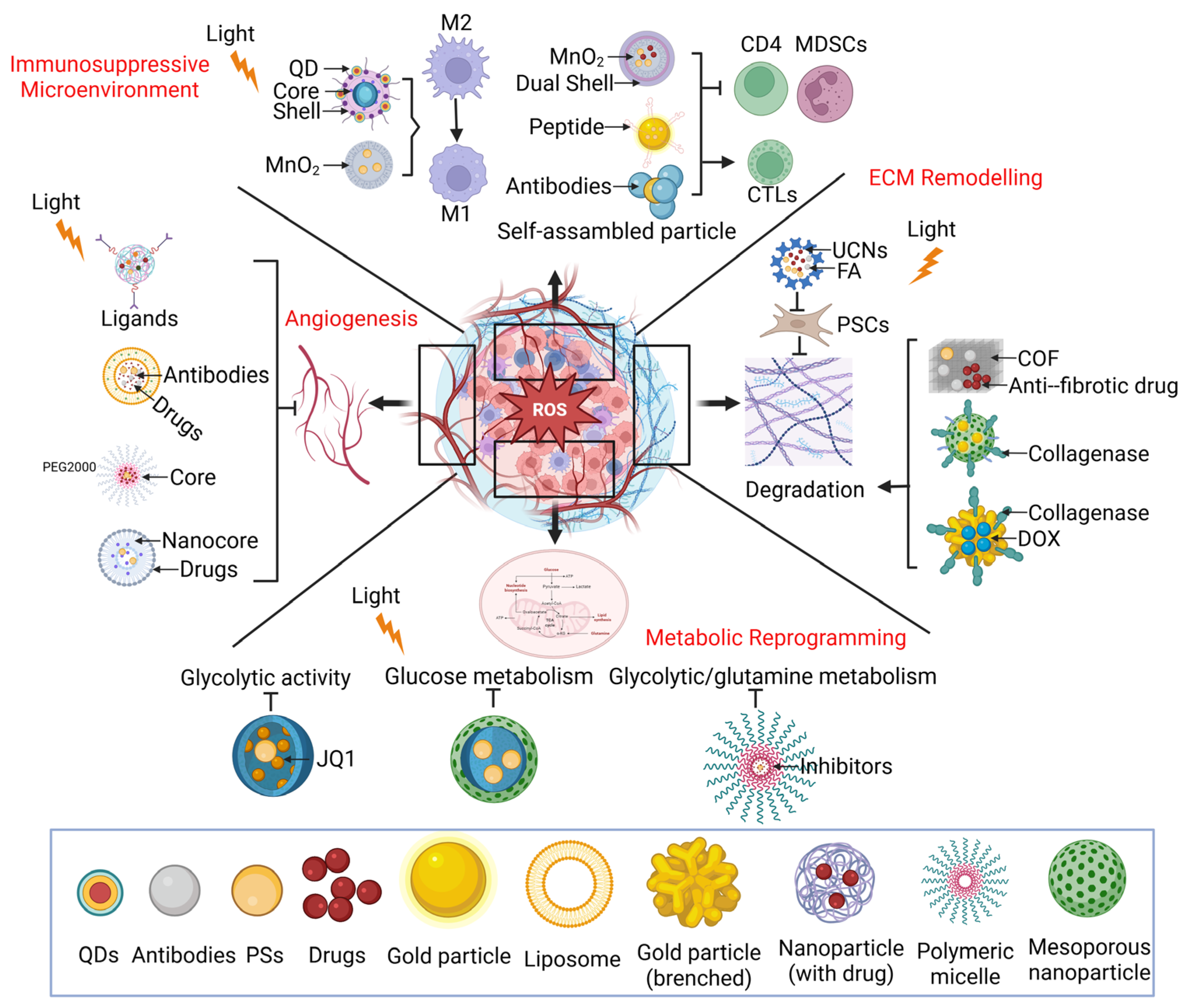
Photo-responsive nanoplatforms exhibit highly targeted properties and synergistic responsiveness to the TME, enabling effects such as O2 delivery, glutathione (GSH) depletion, and ICD induction, offering new possibilities for effectively treating cancer (Figure 3).

3.1. Inhibit Angiogenesis

The TME creates a favorable environment for tumor survival and growth by promoting pathological angiogenesis, thereby contributing to tumor progression [80,81]. Notably, the rapid proliferation of cancer cells and their aberrant angiogenesis exacerbate pathological hypoxia, which directly restricts the efficacy of oxygen-dependent type II photodynamic reactions [82]. However, there is evidence that angiogenesis inhibitors have not shown significant efficacy because of insufficient targeting and obstruction of the ECM [83]. Photo-responsive nanoplatforms leverage the sensitivity of PSs and the biocompatibility of nanoparticles to enhance targeting and promote anticancer activity. In addition, due to the excessive presence of H2O2 and GSH in the TME, after delivering catalase or other catalysts, photo-responsive nanoplatforms can promote O2 generation, alleviate tumor hypoxia, and enhance the PDT effect [84]. For example, Yuan et al. [85] synthesized iRGD-modified nanoplatform and co-encapsulated mTHPC and bufalin (BU) for precise CRC delivery. Experimental results demonstrated that mTHPC&BU@VES-CSO/TPGS-RGD nanoparticles (T-B@NP) significantly induced apoptosis and cell death. Mechanistically, BU targeted the SRC-3/HIF-1α pathway, suppressing their activation and vascular endothelial growth factor (VEGF)-stimulated angiogenesis. Another photo-responsive nanoplatform (SCF NPs) established by Ce6-PEG2000-FA and sunitinib was also found to suppress angiogenesis in HCC [86]. Further, the combination therapy of photo-responsive nanoparticles facilitates tumor killing and inhibits angiogenesis. Wang et al. [87] synthesized PDFI and PDFP nanoparticles by loading IR780 and paclitaxel, which were equipped with phospholipid/Pluronic F68 compound nanocores with pullulan shells. Results showed that the combination therapy effectively inhibited HCC angiogenesis.
Importantly, photo-responsive nanoparticles not only enhance targeting capabilities but also enable the simultaneous, spatiotemporal release of targeted drugs [88]. Nano-photosensitizing liposomes (nanoPALs) were utilized to deliver the photocytotoxic chromophore benzoporphyrin derivative monoacid A (BPD), as well as VEGF monoclonal antibody [89]. This achieved spatiotemporal co-delivery of bevacizumab to enhance PDT efficacy and inhibition of angiogenesis in PDAC by suppressing the VEGF signaling pathway.

3.2. Modulate the Immunosuppressive Microenvironment

Typically, the immunosuppressive TME is the most significant feature of solid tumors, characterized by infiltration of immunosuppressive cells, including myeloid-derived suppressor cells (MDSCs), T regulatory cells (Tregs), and tumor-associated macrophages (TAMs), as well as the activation of immunosuppressive pathways [90,91]. Furthermore, the low intrinsic immunogenicity of tumors leads to poor responses to immune checkpoint inhibitors in many patients. Nanotechnology enables precise tumor targeting and converts immunologically “cold” tumors into “hot” tumors. Photo-responsive nanoplatforms have been shown to induce immunogenic cell death (ICD), thereby enhancing anti-tumor immunity and reversing the immunosuppressive TME by secreting damage-associated molecular patterns (DAMPs), including ATP, high mobility group box 1 (HMGB1), and Calreticulin (CRT) [92,93,94]. Understanding the latest advancements of photo-responsive nanoparticles in modulating tumor immunity and reshaping the TME guides the next steps in PDT.
Photo-responsive nanoparticles suppress tumor progression and reprogram TAMs from a pro-tumor (M2-like) state to an anti-tumor (M1-like) state, thereby reversing the immunosuppressive tumor microenvironment. For example, Butkute A et al. [95] utilized the CdSe/ZnS core–shell structure, and loaded Ce6 and luminescent quantum dots (QDs) to synthesize QDs-Ce6 nanoparticles. Experimental results demonstrated that the QDs-Ce6 nanocomplex significantly reduced expression of the immunosuppressive factor IL-10 and repolarized M2 macrophages to M1, thereby reshaping the TME of CRC. Furthermore, progressive obstruction is a hallmark of EC, underscoring the need to inhibit tumor growth and restore esophageal lumen patency. Recently, a novel esophageal stent integrated with the Ce6-SiO2@MnO2 nanoplatform was developed to address this challenge [96]. This system not only alleviated tumor hypoxia by generating oxygen in situ to enhance the PDT effect but also reprogrammed TAMs induced by manganese ions (Mn2+), thereby activating anti-tumor immunity. Mechanistically, Mn2+ downregulates HMGB1 while enhancing the expression of signal transducer and activator of transcription 1 (STAT1), which induces TAM-mediated tumor suppression. Additionally, the nanoparticles inhibit TAM apoptosis, further sustaining their anti-tumor activity [96].
The combination of photo-responsive nanomaterials with small-molecule inhibitors or chemotherapy agents significantly promotes infiltration of cytotoxic T lymphocytes (CTLs). Researchers are adopting a two-step strategy to treat HCC [97]. First, they functionalized amino-modified mesoporous silica nanoplatforms (MSNs) with AEAA on the surface and loaded them with retinoic acid (RA) and gambogic acid (GA), yielding AEAA-MSNs@RA/GA. Second, PDT was applied using photo-responsive MSN@Ce6. This combined anti-tumor strategy not only improved tumor-specific accumulation of PS but also significantly enhanced the PDT-triggered immune response. Specifically, AEAA-MSNs@RA/GA modulated the immunosuppressive TME by promoting CD8+ T cell infiltration while reducing the presence of Tregs and MDSCs, thereby amplifying the effect of immunotherapy [97]. In the PDAC model, researchers developed a self-assembled nanoparticle (ISDN) comprising ICG and the Heat Shock Protein 90 (HSP90) inhibitor 17-Dimethylaminoethylamino-17-demethoxy-geldanamycin (17-DMAG) [98]. Studies showed that the ISDN significantly improved CTL infiltration and the activation of the major histocompatibility complex I (MHC I) and MHC II, demonstrating potent ICD effects and anti-tumor immune capabilities. Further, Li et al. [99] developed a novel nuclear-targeted dual-shell intelligent nanoplatform named HMCuS/Pt/ICG@MnO2@9R-P201 (HMCPIM9P). This system comprises a nanocore (HMCuS@MnO2) and is loaded with ICG and cisplatin (Pt). Experimental results showed that HMCPIM9P exhibited specific targeting and significantly enhanced the chemotherapeutic efficacy of Pt. Notably, Mn2+ released from this nanoplatform could activate the cGAS-STING pathway, leading to infiltration of CD8+ T cells into tumors [99]. Overall, this multifunctional platform synergistically combines CDT and immunotherapy, demonstrating markedly improved antitumor effects of photoreactive materials.
Photo-responsive nanoplatforms loaded with anti-programmed cell death ligand 1 (PD-L1) inhibitors also exhibit significant anti-tumor effects. The combination of CDT with anti-PD-L1 monoclonal antibodies has been shown to induce distal effects and establish long-term immune memory. For example, another novel photo-activated nanoplatform, named TB/PTX@RTK, which loads an aggregation-induced emission (AIE) photosensitizer and paclitaxel (PTX), was reported to inhibit tumor growth and induce ICD to stimulate antitumor immunity [100]. Mechanistically, TB/PTX@RTK promoted CRT translocation from the endoplasmic reticulum (ER) to the cell membrane. The CDT therapy also triggered extracellular release of HMGB1 and ATP, thereby enhancing HSP70 exposure. These factors recruited antigen-presenting cells (APCs) into the tumor area, leading to PD-L1 upregulation in HCC cells. Further, Yuan et al. [101] developed multifunctional nanoparticles (mTHPC@VeC/T-RGD) that encapsulated PS-mTHPC. Studies demonstrated that under NIR laser irradiation, mTHPC@VeC/T-RGD induced both apoptosis and necrosis in tumor cells, upregulated PD-L1 expression in CRC cells, and triggered a systemic immune response. Another study reported a porphyrin-based liposomal nanoplatform decorated with an anti-EGFR antibody (cetuximab), incorporating the NIR dye (IRDye800CW) and the MRI contrast DOTA-Gd to form EGFR-CPIG [102]. Subsequent experiments showed that, when combined with PD-L1 immunotherapy, EGFR-targeted nanoparticles significantly induced ICD and suppressed CRC growth.

3.3. Remodel the ECM

In solid tumors, the TME is characterized by a tightly structured and widely distributed ECM that influences tumor progression and is associated with poor prognosis [103]. The physical properties of the ECM, including stiffness and organization, play crucial roles in regulating cancer cell proliferation and invasion, promoting angiogenesis, and shaping the immunosuppressive microenvironment [104,105]. CAFs are the primary producers and key regulators of this matrix component. CAFs promote collagen cross-linking by expressing the lysyl oxidase (LOX) family, thereby increasing matrix stiffness [106]. ECM acts as a physical diffusion barrier, impeding the permeation of drugs and oxygen. Additionally, it confines and limits the oxidative damage inflicted by ROS on surrounding tumor cells. The photo-responsive nanoplatforms activate cascade reactions under light to degrade the ECM, providing a novel approach for targeted drug delivery and ECM remodeling, thereby enhancing the efficacy of PDT and immunotherapy [107]. For example, the poly-L-lysine (PLL), folic acid (FA), and protoporphyrin IX (PpIX) were covalently conjugated and coated onto the surface of upconversion nanoparticles (UCNs) to form UCNs@PPF [108]. Subsequent studies showed that UCNs@PPF inactivated pancreatic stellate cells (PSCs) and suppressed ECM secretion, thereby disrupting tumor-stromal crosstalk.
Further, photo-responsive nanoparticles not only remodel the ECM, but also realize deep tumor penetration. There was study revealed that PDT induced photodynamic matrix ablation (PSD) of PDAC [109]. For example, Zhang et al. [110] developed a size-adjustable porphyrin-based covalent organic framework (COF) modified with hyaluronic acid (HA) for combined chemotherapy and PDT. The larger COF (P-COF, ∼500 nm) was loaded with the anti-fibrotic drug losartan (LST) to form LST/P-COF@HA (LCH), which effectively remodeled ECM components. In contrast, the smaller one (p-COF, ∼200 nm) was loaded with DOX, enabling deep penetration and precise targeting of CRC. Furthermore, the nanoplatform can carry collagenase to create conditions for the subsequent penetration of PDT drugs. Researchers have tried to incorporate 2-(1-hexyloxyethyl)-2-devinylpyropheophorbide-a (HPPH) into a hollow mesoporous organosilicon nanoparticle (HMON) framework and have utilized the HMON cavity to immobilize ultrasmall gold nanoparticles. Next, they deposited a Cu2+-tannic acid complex onto the HMON surface and loaded collagenase (Col) into the system, thereby constructing HMON-Au@Cu-TA-PVP [111]. Notably, this nanocomposite remodels the ECM, enhances nanoparticle penetration, and improves the efficacy of PDT. Yang et al. [112] constructed a multifunctional therapeutic platform based on gold nanocores (AuNCs) loaded with Dox. In particular, the synthesized nanoparticles (Col-M@AuNCs/Dox) were encapsulated with PDAC cell membranes. Upon exposure to NIR light, these membrane-coated nanoparticles effectively facilitated PDT. Experimental results demonstrated that the nanosystem promoted deep tumor penetration by degrading the ECM, thereby achieving synergistic anti-tumor efficacy [112]. It remains essential to further investigate the development and evolution of CAFs and to elucidate the complexity and cellular heterogeneity within the TME. In brief, photo-responsive nanoparticles play significant roles in remodeling the ECM.

3.4. Reprogram the Metabolism

Metabolic reprogramming is widely recognized as a hallmark of malignant tumor cells. The TME contains a large amount of antioxidant GSH, and tumor cells prefer glycolysis over oxidative phosphorylation for energy [113]. Metabolic reprogramming support tumors cell survival under tough conditions, rapid proliferation, and resistance to therapies [114,115]. Notably, abnormally high levels of antioxidants, such as GSH and catalase, in the GI TME form a potent “antioxidant barrier,” significantly diminishing the therapeutic efficacy of ROS. Currently, the primary approach to targeting tumor cell metabolism involves the use of small-molecule inhibitors that modulate metabolic pathways [115]. However, therapeutic efficacy is often limited by inadequate targeting and low bioavailability. Photo-responsive nanoplatforms offer advantages in simplifying drug preparation and administration and can mimic the functions of various enzymes, thereby generating a large amount of ROS and effectively depleting intracellular GSH, ultimately inhibiting metabolic reprogramming.
It is reported that a prodrug nanoparticle (HCJSP) to regulate glucose metabolism of PDAC [116]. Researchers constructed a co-delivery system based on cyclodextrin-modified hyaluronic acid (HA-CD) to co-deliver pyropheophorbide a (PPa) and the BRD4 inhibitor JQ1. Upon exposure to endogenous GSH, JQ1 effectively suppressed c-Myc expression and glycolytic activity. Notably, JQ1 counteracted PDT-induced immune evasion by downregulating PD-L1 expression [116]. Further, Wu et al. [117] developed a multifunctional nanoplatform (LnNP@mSiO2-GC) consisting of a lanthanide-doped nanoparticle (LnNP) core loaded with 2-deoxy-D-glucose (2DG) and Ce6. Under 650 nm light irradiation, Ce6 generated 1O2, disrupting mitochondrial function and suppressing glucose metabolic reprogramming by downregulating expression of hexokinase 2 (HK2) and lactate dehydrogenase A (LDHA). Overall, the photo-responsive nanoplatform induces starvation therapy-enhanced tumor-targeted therapeutic efficacy. Another novel malfunctional nanoplatform was constructed by co-loading zinc phthalocyanine (ZnPc) with the PI3K/mTOR inhibitor BEZ235 (BEZ) and conjugating the MEK inhibitor selumetinib (SEL) to low-molecular-weight heparin via click chemistry, forming a ROS-responsive, PDAC-targeting nanomaterial named Z/B-PLS [118]. Further studies demonstrated that Z/B-PLS remodeled glycolytic and non-canonical glutamine metabolism. Importantly, the delivery-treatment feedback loop significantly enhances therapeutic efficacy.
Further, strategies for PDT to reprogram metabolism also include depleting intracellular GSH to enhance ROS generation, thereby improving therapeutic efficacy against GI malignancies. For example, by addressing the high GSH levels in HCC cells, conjugating GNPs with Ce6 restored both the fluorescence and ROS generation capabilities of Ce6, thereby enabling effective PDT [119]. Feng et al. [120] developed multifunctional UMCNs-PEG nanoparticles based on mesoporous manganese silicate-coated UCNPs for co-delivery of fluorescent g-C3N4 QDs, with PEG modification. These photo-responsive nanomaterials enhanced ROS generation and reduced GSH levels, showing great potential in GC therapy. Another study introduced a multifunctional nanoplatform (Fe-TCPP@MnO2@JUG@HA), constructed from an Fe-MOF core, a MnO2 shell loaded with juglone (JUG), and an HA coating. The system promoted ferroptosis by depleting GSH, inhibiting the biosynthesis of the lipid repair enzyme GPX4, and thereby collectively disrupting cellular redox homeostasis [121]. Overall, the platform demonstrates potent anti-tumor efficacy, excellent biocompatibility, and metabolism regulation, highlighting its promising PDT potential.
Table 2. Photo-responsive Nanoplatforms modulate the TME for GI malignancies therapy.
EffectsNanoplatformsNanoplatform TypePSExcitation WavelengthTarget TumorsAnti-Tumor EffectsReferences
Inhibit angiogenesisT-B@NPiRGD-modified nanoparticlesmTHPC652 nmCRCInhibit the SRC-3/HIF-1α pathway and reduce tumor growth by 84.2% in BALB/c mice.[85]
SCF NPsAmphiphilic polymerCe6650 nmHCCInhibit tumor growth and anti-angiogenesis; SCF NP treatment eradicated tumors, and no recurrence was observed. multimodal imaging capabilities in Hep-3B tumor-bearing nude mice.[86]
PDFI and PDFPPhospholipid/Pluronic F68 complex nanocores and pullulan shellsIR780780 nmHCCSynergistic effects on inhibiting tumor angiogenesis; delay local tumor recurrence; cell proliferation, and induce cell apoptosis and cell cycle arrest at G2/M phase in MHCC-97H tumor-bearing mice.[87]
nanoPALsNanophotoactivatable liposome (nanoPAL)BPDNot specifiedPDACAnti-angiogenesis and enhance tumor killing; 33% of mice achieved complete remission following nanoPAL treatment in PDAC-bearing mice.[89]
Modulate the immunosuppressive microenvironmentQDs-Ce6 nanocomplexQDs-based nanocomplexQDs and Ce6980/650 nmCRCInduce M2 changes to M0 and upregulate PD-L1 expression in vitro.[95]
Ce6-SiO2@MnO2Esophageal stent-loaded nanoparticlesCe6650 nmECReprogram TAMs and activate innate anti-tumor immunity, suppress the apoptosis of TAMs in VX2 rabbit.[96]
AEAA-MSNs@RA/GA and MSN@Ce6Mesoporous silica nanocarriers (MSNs)Ce6650 nmHCCIncrease immunostimulatory cells and regulate the immunosuppressive TME in ICR mice.[97]
ISDNSelf-assembled nanoparticlesICG808 nmPDACImprove cytotoxic T lymphocyte infiltration and MHC I and MHC II activation in C57BL/6 mice.[98]
HMCPIM9PDouble-shell multifunctional nanoparticlesICG808 nmHCCIncrease adaptive immune responses and elevate the level of CD8+ T cells.[99]
TB/PTX@RTKMicellesTB370 nmHCCUpregulate PD-L1 expression on the HCC surface in C57BL/6 mice.[100]
mTHPC@VeC/T-RGDMultifunctional nanoparticlesmTHPC652 nmCRCBlock the PD-L1 and build long-term host immunological memory in BALB/c mice.[101]
EGFR-CPIGPorphyrin-containing liposomal nanohybrid cerasomesIRDye800CW980 nmCRCIncrease PD-L1 immunotherapy in BALB/c mice.[102]
Remodel the ECMUCNs@PPFUCNs-based nanoparticlesprotoporphyrin IX (PpIX)980 nmPDACInterrupt the mutual support between cancer cells and stroma cells. Enhance the therapeutic effectiveness of gemcitabine for recurrent pancreatic tumors in BALB/c nude mice[108]
LST/P-COF@HA (LCH)COFsDCH550 nmCRCDownregulate ECM component levels and decrease collagen density, reducing tumor solid stress in BALB/c mice.[110]
HMON-Au@Cu-TA-PVPPolymerized hollow mesoporous organosilica nanoparticle (HMON) biocatalysisHPPH655 nmPDACDegrade the collagen I fiber in; suppress tumor growth in BALB/c nude mice.[111]
Col-M@AuNCs/DoxBiomimetic drug-loaded nanoplatformAuNCsNIRPDACDegrade dense ECM by collagenase, enable deep penetration of NPs into tumor parenchymal tissue; combine phototherapy and chemotherapy suppress tumor growth in BALB/c nude mice.[112]
Reprogram the metabolismHCJSPSupramolecular prodrug nanoplatformPPa365 nmPDACRegulate tumor glycolysis and inhibit immune evasion in C57BL/6 mice.[116]
LnNP@mSiO2-GCA multifunctional nano-platform comprising lanthanide-doped nanoparticle (LnNP) coresCe6670 nmCRCDamage mitochondrial function, inhibit hexokinase 2 and lactate dehydrogenase A expressions, and reprogram glucose metabolism. Inhibit CRC progression in BALB/c nude mice.[117]
Z/B-PLSROS-responsive and PDAC-targeted nanodrugZnPc694 nmPDACRemodel glycolysis and non-canonical glutamine metabolism in BALB/c nude mice.[118]
AQ4N-Cu (II)-AptCe6-GNPsSmart Cu (II)-aptamer complexesCe6670 nmHCCConsume GSH in tumors, and enhance chemotherapy efficacy
in BALB/c-nude mice.
[119]
UCNPs@MnSiO3@g-C3N4Multifunctional nanoparticles based on mesoporous manganese silicateg-C3N4QDs980 nmCRCConsume GSH in tumors and effectively suppress CRC progression in BALB/c mice.[120]
P.S.: EC: esophageal carcinoma, GC: gastric carcinoma, HCC: hepatocellular carcinoma, PDAC: pancreatic ductal adenocarcinoma, CRC: colorectal carcinoma.

4. Photo-Responsive Nanoplatform for GI Malignancies Diagnosis

It is estimated that most GI tumor patients are already at advanced stages when first diagnosed, which leads to poor prognosis [122]. Although conventional diagnostic strategies have made significant advances, they still struggle to detect GI malignancies effectively at an early stage. PD is a non-invasive technique that utilizes the ability of PS to passively accumulate in cancer cells [18,123]. When exposed to specific wavelengths of light, the PS emits fluorescence, enabling a clear distinction between malignant and healthy cells. For example, under NIR irradiation, the LnNP@mSiO2-GC nanoplatform enables real-time monitoring of its tumor accumulation [117]. The Col-M@AuNCs/Dox nanoplatform also exhibited significant X-ray attenuation, providing a means to guide and monitor PDAC treatment through CT imaging [112]. Importantly, pDT and PD are induced by PS, typically via blue or NIR activation, which enables strategies for selectively identifying and treating tumors collaboratively [124]. For example, the SCF NPs nanoplatform not only inhibited angiogenesis in HCC but also demonstrated multimodal imaging capabilities in vivo, which can be used for tumor diagnosis and operative navigation [86]. EGFR-targeted nanoparticles (EGFR-CPIG) combined with immunotherapy also showed great potential for guiding precise dual-modality imaging for PDT [102].
Notably, photo-responsive nanoparticles also play roles in multimodal imaging. For example, a photo-responsive nanoplatform was developed to target PDAC and used tumor-specific midkine nanobodies (Nbs) to achieve precise delivery to the TME [125]. Notably, this engineered nanoparticle supported multimodal imaging of PDAC tumors, including fluorescence imaging (FLI) and photoacoustic imaging (PAI). Furthermore, Dong et al. [126] created a theranostic nanosphere (PPR@LVN/IR780) for HCC by constructing it from PLGA-PEG, modifying it with a cRGD peptide to improve tumor targeting, and loading it with lenvatinib and IR780. This system successfully inhibited tumor growth, enabled multimodal imaging (FLI, PAI, PTI), and thus provided integrated diagnosis and therapy [126]. Another multifunctional nanoplatform, ICG/Mn-PDA-PEG-CXCR4 (IMPP-c), enhanced accumulation in HCC and enabled PA and magnetic resonance (MR) imaging [127]. Overall, photo-responsive nanotechnology offers high resolution and deep tissue penetration, allowing precise localization of early-stage small tumor lesions

5. Conclusions

GI malignancies have imposed a heavy disease burden globally, while traditional therapeutic strategies have shown limited success. In an in-depth exploration of the complexity of the TME, researchers have identified and targeted several key characteristics, including angiogenesis, an immunosuppressive microenvironment, ECM remodeling, and metabolic reprogramming, for therapeutic intervention.
In recent years, advances in nanotechnology have enabled precise targeting, allowing stable drug release and good biocompatibility. Fortunately, integrating PSs with nanotechnology has enabled the application of photochemical nanoplatforms in the diagnosis and treatment of GI malignancies. This review introduced the elements of photochemical nanoplatforms, summarized the development and evolution of PSs, and outlined the principles of the photochemical effect. Importantly, we elaborated on how photochemical nanoplatforms regulate the TME of gastrointestinal tumors, including inhibition of angiogenesis, ECM remodeling, modulation of the immunosuppressive microenvironment, and metabolic reprogramming. Finally, we discussed the potential value of photochemical nanoplatforms for diagnosing GI malignancies. Nanocarriers modulate PS properties by regulating molecular conformation and intermolecular interactions. It has been reported that PSs can undergo π-π stacking within nanocarriers, which shortens excited-state lifetimes [128]. Liposomal and polymeric nanocarriers reduce interactions between PS and biomacromolecules. MOFs enable precise control of PS dispersion by regulating their pore size [129]. These extend PS excited-state lifetimes and promote ROS generation.
Photochemical nanoplatforms hold great potential for treating GI tumors, but several challenges remain. The thin mucosal layer of the gastrointestinal tract (particularly the stomach and colon) is vulnerable to tumor infiltration, which compromises mucosal integrity and increases local tissue fragility. Furthermore, when ROS kill tumors, they also damage the capillary endothelium of normal mucosa, causing a sharp increase in vascular permeability. This leads to the extravasation of plasma proteins and fluid into the interstitial spaces, triggering leakage syndromes such as edema and ascites [130]. Given these risks, prudent management, including careful assessment of side effects and appropriate supportive care, is essential to reduce complications and optimize therapeutic outcomes. The limited biodegradability of polymeric nanodrugs and inorganic nanomaterials significantly restricts their clinical applicability. Small-molecule self-assembled nanodrugs offer greater potential for clinical translation. Furthermore, the mechanisms by which PDT triggers ICD to remodel the TME are not fully understood, necessitating studies to pinpoint the key signaling pathways. The clinical translation of photo-responsive nanoplatforms also remains a key challenge. These include biocompatibility (such as long-term metabolic toxicity), manufacturability, and reproducibility. Furthermore, the penetration depth of light is limited. Another problem is the lack of tools for quantifying ROS in hypoxic regions. To address these, future work must optimize nanocarrier pharmacokinetics and build quantitative dose–response-toxicity models to guide clinical translation. Mechanistic studies should elucidate the interplay between PDT and non-apoptotic cell death pathways, such as ferroptosis and pyroptosis, providing a theoretical basis for combination therapies.

Author Contributions

Conceptualization, validation, writing—original draft preparation, D.L.; revision, supervision, formal analysis, Y.C.; conceptualization, review and editing, X.L. All authors have read and agreed to the published version of the manuscript.

Funding

This research received no external funding.

Data Availability Statement

No new data were created or analyzed in this study. Data sharing is not applicable to this article.

Acknowledgments

All Figures were created in BioRender (www.biorender.com).

Conflicts of Interest

The authors declare no conflicts of interest.

Abbreviations

17-DMAGDimethylaminoethylamino-17-demethoxy-geldanamycin
2DG2-deoxy-D-glucose
AIEaggregation-induced emission
ALA5-aminolevulinic acid
APCsantigen-presenting cells
Augold
BRD4ibromodomain-containing protein 4 inhibitor
BUbufalin
CCAcholangiocarcinoma
CCMcancer cell membrane
CDcarbon dot
CDTchemodynamic therapy
Ce6chlorin e6
COFcovalent organic framework
Colcollagenase
CRCcolorectal carcinoma
CRTcalreticulin
CSchitosan
CTLscytotoxic T lymphocytes
ECesophageal carcinoma
ECMextracellular matrix
EGFRepidermal growth factor receptors
EPRenhanced permeability and retention
ERendoplasmic reticulum
FAfolic acid
FLIfluorescence imaging
GAgambogic acid
GCgastric carcinoma
GIgastrointestinal
GPX4glutathione peroxidase 4
GSHglutathione
HCChepatocellular carcinoma
HK2hexokinase 2
HMGB1high mobility group box 1
HPDhematoporphyrin derivative
HPPH2-(1-hexyloxyethyl)-2-devinylpyropheophorbide-a
HSP90Heat Shock Protein 90
ICDimmunogenic cell death
ICGindocyanine green
IrO2iridium dioxide
JUGjuglone
KMnO4potassium permanganate
LDHAlactate dehydrogenase A
LEDsLight-emitting diodes
LnNPlanthanide-doped nanoparticle
LOXlysyl oxidase
LVNlenvatinib
MALmethylamine levulinate hydrochloride
MDSCsmyeloid-derived suppressor cells
MHC Ihistocompatibility complex I
MnO2manganese dioxide
MOFsmetal–organic frameworks
MOMPmitochondrial outer membrane
MMPmatrix metalloproteinase
MRmagnetic resonance
MSNsmesoporous silica nanoplatforms
Nbsnanobodies
NIRnear-infrared
PAIphotoacoustic imaging
PDACpancreatic ductal adenocarcinoma
PDphotodiagnosis
PD-L1programmed cell death ligand 1
PDTphotodynamic therapy
PEGpolyethylene glycol
PHApheophorbide-a
PLLpoly-L-lysine
PPapyropheophorbide a
PpIXprotoporphyrin IX
PSCspancreatic stellate cells
PSsphotosensitizers
Ptcisplatin
PTXpaclitaxel
PVPpolyvinylpyrrolidone
QDsquantum dots
RAretinoic acid
ROSreactive oxygen species
SAPsaponin
STAT1signal transducer and activator of transcription 1
TAMstumor-associated macrophages
TMEtumor microenvironments
TregsT regulatory cells
UCNsupconversion nanoparticles
VEGFvascular endothelial growth factor
ZnPczinc phthalocyanine
β-CDβ-cyclodextrin
β-DOXdoxorubicin prodrug

References

  1. Siegel, R.L.; Kratzer, T.B.; Giaquinto, A.N.; Sung, H.; Jemal, A. Cancer statistics, 2025. CA Cancer J. Clin. 2025, 75, 10–45. [Google Scholar] [CrossRef] [PubMed]
  2. Huang, J.; Lucero-Prisno, D.E., 3rd; Zhang, L.; Xu, W.; Wong, S.H.; Ng, S.C.; Wong, M.C.S. Updated epidemiology of gastrointestinal cancers in East Asia. Nat. Rev. Gastroenterol. Hepatol. 2023, 20, 271–287. [Google Scholar] [CrossRef] [PubMed]
  3. Ho, J.; Fiocco, C.; Spencer, K. Treating Biliary Tract Cancers: New Targets and Therapies. Drugs 2022, 82, 1629–1647. [Google Scholar] [CrossRef]
  4. Romeo, E.; Caserta, C.A.; Rumio, C.; Marcucci, F. The Vicious Cross-Talk between Tumor Cells with an EMT Phenotype and Cells of the Immune System. Cells 2019, 8, 460. [Google Scholar] [CrossRef]
  5. Yamaguchi, R.; Perkins, G. Animal models for studying tumor microenvironment (TME) and resistance to lymphocytic infiltration. Cancer Biol. Ther. 2018, 19, 745–754. [Google Scholar] [CrossRef]
  6. Samuel, M.S.; Lopez, J.I.; McGhee, E.J.; Croft, D.R.; Strachan, D.; Timpson, P.; Munro, J.; Schröder, E.; Zhou, J.; Brunton, V.G.; et al. Actomyosin-mediated cellular tension drives increased tissue stiffness and beta-catenin activation to induce epidermal hyperplasia and tumor growth. Cancer Cell 2011, 19, 776–791. [Google Scholar] [CrossRef]
  7. Darvishi, B.; Eisavand, M.R.; Majidzadeh, A.K.; Farahmand, L. Matrix stiffening and acquired resistance to chemotherapy: Concepts and clinical significance. Br. J. Cancer 2022, 126, 1253–1263. [Google Scholar] [CrossRef]
  8. Yan, K.; Zhang, Y.; Mu, C.; Xu, Q.; Jing, X.; Wang, D.; Dang, D.; Meng, L.; Ma, J. Versatile Nanoplatforms with enhanced Photodynamic Therapy: Designs and Applications. Theranostics 2020, 10, 7287–7318. [Google Scholar] [CrossRef]
  9. Wu, P.; Han, J.; Gong, Y.; Liu, C.; Yu, H.; Xie, N. Nanoparticle-Based Drug Delivery Systems Targeting Tumor Microenvironment for Cancer Immunotherapy Resistance: Current Advances and Applications. Pharmaceutics 2022, 14, 1990. [Google Scholar] [CrossRef]
  10. Wang, D.; Yang, L.; Yu, W.; Wu, Q.; Lian, J.; Li, F.; Liu, S.; Li, A.; He, Z.; Liu, J.; et al. Colorectal cancer cell-derived CCL20 recruits regulatory T cells to promote chemoresistance via FOXO1/CEBPB/NF-kappaB signaling. J. Immunother. Cancer 2019, 7, 215. [Google Scholar] [CrossRef]
  11. Qin, C.; Yang, G.; Yang, J.; Ren, B.; Wang, H.; Chen, G.; Zhao, F.; You, L.; Wang, W.; Zhao, Y. Metabolism of pancreatic cancer: Paving the way to better anticancer strategies. Mol. Cancer 2020, 19, 50. [Google Scholar] [CrossRef]
  12. Cabral, F.V.; Alburquerque, J.Q.; Roberts, H.J.; Hasan, T. Shedding Light on Chemoresistance: The Perspective of Photodynamic Therapy in Cancer Management. Int. J. Mol. Sci. 2024, 25, 3811. [Google Scholar] [CrossRef] [PubMed]
  13. Baskaran, R.; Lee, J.; Yang, S.-G. Clinical development of photodynamic agents and therapeutic applications. Biomater. Res. 2018, 22, 25. [Google Scholar] [CrossRef] [PubMed]
  14. Repetowski, P.; Warszyńska, M.; Dąbrowski, J.M. NIR-activated multifunctional agents for the combined application in cancer imaging and therapy. Adv. Colloid Interface Sci. 2025, 336, 103356. [Google Scholar] [CrossRef]
  15. Grebinyk, A.; Chepurna, O.; Frohme, M.; Qu, J.; Patil, R.; Vretik, L.; Ohulchanskyy, T. Molecular and nanoparticulate agents for photodynamic therapy guided by near infrared imaging. J. Photochem. Photobiol. C Photochem. Rev. 2024, 58, 100652. [Google Scholar] [CrossRef]
  16. Hu, D.; Sheng, Z.; Gao, G.; Siu, F.; Liu, C.; Wan, Q.; Gong, P.; Zheng, H.; Ma, Y.; Cai, L. Activatable albumin-photosensitizer nanoassemblies for triple-modal imaging and thermal-modulated photodynamic therapy of cancer. Biomaterials 2016, 93, 10–19. [Google Scholar] [CrossRef]
  17. Truong, D.H.; Tran, P.T.T.; Tran, T.H. Nanoparticles as carriers of photosensitizers to improve photodynamic therapy in cancer. Pharm. Dev. Technol. 2024, 29, 221–235. [Google Scholar] [CrossRef]
  18. Dobson, J.; de Queiroz, G.F.; Golding, J.P. Photodynamic therapy and diagnosis: Principles and comparative aspects. Vet. J. 2018, 233, 8–18. [Google Scholar] [CrossRef]
  19. Allison, R.R.; Sibata, C.H. Oncologic photodynamic therapy photosensitizers: A clinical review. Photodiagnosis Photodyn. Ther. 2010, 7, 61–75. [Google Scholar] [CrossRef]
  20. Agostinis, P.; Berg, K.; Cengel, K.A.; Foster, T.H.; Girotti, A.W.; Gollnick, S.O.; Hahn, S.M.; Hamblin, M.R.; Juzeniene, A.; Kessel, D.; et al. Photodynamic therapy of cancer: An update. CA Cancer J. Clin. 2011, 61, 250–281. [Google Scholar] [CrossRef]
  21. Spikes, J.D. Chlorins as photosensitizers in biology and medicine. J. Photochem. Photobiol. B 1990, 6, 259–274. [Google Scholar] [CrossRef]
  22. Baas, P.; van Mansom, I.; van Tinteren, H.; Stewart, F.A.; van Zandwijk, N. Effect of N-acetylcysteine on Photofrin-induced skin photosensitivity in patients. Lasers Surg. Med. 1995, 16, 359–367. [Google Scholar] [CrossRef] [PubMed]
  23. Abrahamse, H.; Hamblin, M.R. New photosensitizers for photodynamic therapy. Biochem. J. 2016, 473, 347–364. [Google Scholar] [CrossRef] [PubMed]
  24. Beiki, D.; Eggleston, I.M.; Pourzand, C. Daylight-PDT: Everything under the sun. Biochem. Soc. Trans. 2022, 50, 975–985. [Google Scholar] [CrossRef]
  25. Biswas, R.; Moon, J.H.; Ahn, J.C. Chlorin e6 derivative radachlorin mainly accumulates in mitochondria, lysosome and endoplasmic reticulum and shows high affinity toward tumors in nude mice in photodynamic therapy. Photochem. Photobiol. 2014, 90, 1108–1118. [Google Scholar] [CrossRef]
  26. Chilakamarthi, U.; Giribabu, L. Photodynamic Therapy: Past, Present and Future. Chem. Rec. 2017, 17, 775–802. [Google Scholar] [CrossRef]
  27. Zhang, J.; Jiang, C.; Figueiró Longo, J.P.; Azevedo, R.B.; Zhang, H.; Muehlmann, L.A. An updated overview on the development of new photosensitizers for anticancer photodynamic therapy. Acta Pharm. Sin. B 2018, 8, 137–146. [Google Scholar] [CrossRef]
  28. Lan, M.; Zhao, S.; Liu, W.; Lee, C.-S.; Zhang, W.; Wang, P. Photosensitizers for Photodynamic Therapy. Adv. Healthc. Mater. 2019, 8, e1900132. [Google Scholar] [CrossRef]
  29. Ermakov, A.V.; Verkhovskii, R.A.; Babushkina, I.V.; Trushina, D.B.; Inozemtseva, O.A.; Lukyanets, E.A.; Ulyanov, V.J.; Gorin, D.A.; Belyakov, S.; Antipina, M.N. In Vitro Bioeffects of Polyelectrolyte Multilayer Microcapsules Post-Loaded with Water-Soluble Cationic Photosensitizer. Pharmaceutics 2020, 12, 610. [Google Scholar] [CrossRef]
  30. Bayir, S.; Barras, A.; Boukherroub, R.; Szunerits, S.; Raehm, L.; Richeter, S.; Durand, J.-O. Mesoporous silica nanoparticles in recent photodynamic therapy applications. Photochem. Photobiol. Sci. 2018, 17, 1651–1674. [Google Scholar] [CrossRef]
  31. Yang, J.; Yang, Y.W. Metal-Organic Frameworks for Biomedical Applications. Small 2020, 16, e1906846. [Google Scholar] [CrossRef] [PubMed]
  32. Song, Y.; Wang, L.; Xie, Z. Metal-Organic Frameworks for Photodynamic Therapy: Emerging Synergistic Cancer Therapy. Biotechnol. J. 2021, 16, e1900382. [Google Scholar] [CrossRef] [PubMed]
  33. Alves, S.R.; Calori, I.R.; Tedesco, A.C. Photosensitizer-based metal-organic frameworks for highly effective photodynamic therapy. Mater. Sci. Eng. C Mater. Biol. Appl. 2021, 131, 112514. [Google Scholar] [CrossRef]
  34. Cai, Z.; Xin, F.; Yao, C.; Liu, X.; Wu, M.; Lin, X.; Du, X.; Chen, G.; Zhang, D.; Zhang, Z.; et al. Photodynamic Therapy Combined with Antihypoxic Signaling and CpG Adjuvant as an In Situ Tumor Vaccine Based on Metal-Organic Framework Nanoparticles to Boost Cancer Immunotherapy. Adv. Healthc. Mater. 2020, 9, e1900996. [Google Scholar] [CrossRef]
  35. Torna, S.; Gkretsi, V.; Stylianou, A. Photodynamic Therapy and Tumor Microenvironment-Targeting Strategies: A Novel Synergy at the Frontier of Cancer Treatment. Int. J. Mol. Sci. 2025, 26, 8588. [Google Scholar] [CrossRef]
  36. Lim, H.J.; Oh, C.H. Indocyanine green-based photodynamic therapy with 785nm light emitting diode for oral squamous cancer cells. Photodiagnosis Photodyn. Ther. 2011, 8, 337–342. [Google Scholar] [CrossRef]
  37. Hsiao, C.Y.; Yang, S.C.; Alalaiwe, A.; Fang, J.Y. Laser ablation and topical drug delivery: A review of recent advances. Expert Opin. Drug Deliv. 2019, 16, 937–952. [Google Scholar] [CrossRef]
  38. Isikawa, M.M.; Muradova, Z.; Morris, T.; Lessa, J.V.V.; Borges, F.H.; Gonçalves, R.R.; Berbeco, R.I.; Guidelli, E.J. Enhanced Cancer Radiosensitization via Energy Transfer from Eu-Doped GdF3 Nanoparticles to Methylene Blue in X-ray Photodynamic Therapy. ACS Appl. Mater. Interfaces 2025, 17, 51620–51633. [Google Scholar] [CrossRef]
  39. Dash, B.S.; Das, S.; Chen, J.P. Photosensitizer-Functionalized Nanocomposites for Light-Activated Cancer Theranostics. Int. J. Mol. Sci. 2021, 22, 6658. [Google Scholar] [CrossRef]
  40. Jiang, M.; Liu, Y.; Dong, Y.; Wang, K.; Yuan, Y. Bioorthogonal chemistry and illumination controlled programmed size-changeable nanomedicine for synergistic photodynamic and hypoxia-activated therapy. Biomaterials 2022, 284, 121480. [Google Scholar] [CrossRef]
  41. Ding, H.; Yu, H.; Dong, Y.; Tian, R.; Huang, G.; Boothman, D.A.; Sumer, B.D.; Gao, J. Photoactivation switch from type II to type I reactions by electron-rich micelles for improved photodynamic therapy of cancer cells under hypoxia. J. Control. Release 2011, 156, 276–280. [Google Scholar] [CrossRef]
  42. Zhou, Q.; Huang, G.; Si, J.; Wu, Y.; Jin, S.; Ji, Y.; Ge, Z. Potent Covalent Organic Framework Nanophotosensitizers with Staggered Type I/II Motifs for Photodynamic Immunotherapy of Hypoxic Tumors. ACS Nano 2024, 18, 35671–35683. [Google Scholar] [CrossRef]
  43. Robbins, E.; Leroy-Lhez, S.; Villandier, N.; Samoć, M.; Matczyszyn, K. Prospects for More Efficient Multi-Photon Absorption Photosensitizers Exhibiting Both Reactive Oxygen Species Generation and Luminescence. Molecules 2021, 26, 6323. [Google Scholar] [CrossRef]
  44. Pati, S.; Maity, A.; Banerji, P.; Majumder, S.B. Qualitative and quantitative differentiation of gases using ZnO thin film gas sensors and pattern recognition analysis. Analyst 2014, 139, 1796–1800. [Google Scholar] [CrossRef] [PubMed]
  45. Allison, R.R.; Moghissi, K. Photodynamic Therapy (PDT): PDT Mechanisms. Clin. Endosc. 2013, 46, 24–29. [Google Scholar] [CrossRef] [PubMed]
  46. Chen, D.; Long, Z.; Zhong, C.; Chen, L.; Dang, Y.; Hu, J.-J.; Lou, X.; Xia, F. Highly Efficient Near-Infrared Photosensitizers with Aggregation-Induced Emission Characteristics: Rational Molecular Design and Photodynamic Cancer Cell Ablation. ACS Appl. Bio Mater. 2021, 4, 5231–5239. [Google Scholar] [CrossRef] [PubMed]
  47. Correia, J.H.; Rodrigues, J.A.; Pimenta, S.; Dong, T.; Yang, Z. Photodynamic Therapy Review: Principles, Photosensitizers, Applications, and Future Directions. Pharmaceutics 2021, 13, 1332. [Google Scholar] [CrossRef]
  48. Kessel, D.; Castelli, M. Evidence that bcl-2 is the target of three photosensitizers that induce a rapid apoptotic response. Photochem. Photobiol. 2001, 74, 318–322. [Google Scholar] [CrossRef]
  49. Maharjan, P.S.; Bhattarai, H.K. Singlet Oxygen, Photodynamic Therapy, and Mechanisms of Cancer Cell Death. J. Oncol. 2022, 2022, 7211485. [Google Scholar] [CrossRef]
  50. Reinheckel, T.; Tholen, M. Low-level lysosomal membrane permeabilization for limited release and sublethal functions of cathepsin proteases in the cytosol and nucleus. FEBS Open Bio 2022, 12, 694–707. [Google Scholar] [CrossRef]
  51. Mukhopadhyay, B.; Singh, S.; Singh, A. Utilizing nanomaterials for cancer treatment and diagnosis: An overview. Discov. Nano 2024, 19, 215. [Google Scholar] [CrossRef]
  52. Deng, W.; Zhong, Z.; Shang, H.; Tong, Y.; Huang, Q.; He, Y.; Wu, J.; Ba, X.; Chen, Z.; Chen, Y.; et al. Nanoparticles induced cuproptosis to enhance antitumor immunotherapy. J. Nanobiotechnol. 2025, 23, 544. [Google Scholar] [CrossRef]
  53. Ammar, M.M.; Ali, R.; Elaziz, N.A.A.; Habib, H.; Abbas, F.M.; Yassin, M.T.; Maniah, K.; Abdelaziz, R. Nanotechnology in oncology: Advances in biosynthesis, drug delivery, and theranostics. Discov. Oncol. 2025, 16, 1172. [Google Scholar] [CrossRef]
  54. Li, R.; Yang, T.; Peng, X.; Feng, Q.; Hou, Y.; Zhu, J.; Chu, D.; Duan, X.; Zhang, Y.; Zhang, M. Enhancing the Photosensitivity of Hypocrellin A by Perylene Diimide Metallacage-Based Host-Guest Complexation for Photodynamic Therapy. Nanomicro Lett. 2024, 16, 226. [Google Scholar] [CrossRef] [PubMed]
  55. Zhou, J.; Yu, G.; Huang, F. Supramolecular chemotherapy based on host-guest molecular recognition: A novel strategy in the battle against cancer with a bright future. Chem. Soc. Rev. 2017, 46, 7021–7053. [Google Scholar] [CrossRef]
  56. Saini, V.; Venkatesh, V. AIE material for photodynamic therapy. Prog. Mol. Biol. Transl. Sci. 2021, 185, 45–73. [Google Scholar] [PubMed]
  57. Jia, S.; Yuan, H.; Hu, R. Design and structural regulation of AIE photosensitizers for imaging-guided photodynamic anti-tumor application. Biomater. Sci. 2022, 10, 4443–4457. [Google Scholar] [CrossRef]
  58. Zhao, W.; Zhao, Y.; Wang, Q.; Liu, T.; Sun, J.; Zhang, R. Remote Light-Responsive Nanocarriers for Controlled Drug Delivery: Advances and Perspectives. Small 2019, 15, e1903060. [Google Scholar] [CrossRef]
  59. Li, X.; Lovell, J.F.; Yoon, J.; Chen, X. Clinical development and potential of photothermal and photodynamic therapies for cancer. Nat. Rev. Clin. Oncol. 2020, 17, 657–674. [Google Scholar] [CrossRef]
  60. Cheng, X.; Gao, J.; Ding, Y.; Lu, Y.; Wei, Q.; Cui, D.; Fan, J.; Li, X.; Zhu, E.; Lu, Y.; et al. Multi-Functional Liposome: A Powerful Theranostic Nano-Platform Enhancing Photodynamic Therapy. Adv. Sci. 2021, 8, e2100876. [Google Scholar] [CrossRef]
  61. Moghassemi, S.; Dadashzadeh, A.; Azevedo, R.B.; Feron, O.; Amorim, C.A. Photodynamic cancer therapy using liposomes as an advanced vesicular photosensitizer delivery system. J. Control. Release 2021, 339, 75–90. [Google Scholar] [CrossRef] [PubMed]
  62. Liu, H.; Yao, C.; Zhang, L.; Xin, J.; Zhang, Z.; Wang, S. Nanoliposomes co-encapsulating Ce6 and SB3CT against the proliferation and metastasis of melanoma with the integration of photodynamic therapy and NKG2D-related immunotherapy on A375 cells. Nanotechnology 2021, 32, 455102. [Google Scholar] [CrossRef]
  63. Yi, H.; Lu, W.; Liu, F.; Zhang, G.; Xie, F.; Liu, W.; Wang, L.; Zhou, W.; Cheng, Z. ROS-responsive liposomes with NIR light-triggered doxorubicin release for combinatorial therapy of breast cancer. J. Nanobiotechnol. 2021, 19, 134. [Google Scholar] [CrossRef] [PubMed]
  64. Avramovic, N.; Mandic, B.; Savic-Radojevic, A.; Simic, T. Polymeric Nanocarriers of Drug Delivery Systems in Cancer Therapy. Pharmaceutics 2020, 12, 298. [Google Scholar] [CrossRef]
  65. Karges, J.; Heinemann, F.; Jakubaszek, M.; Maschietto, F.; Subecz, C.; Dotou, M.; Vinck, R.; Blacque, O.; Tharaud, M.; Goud, B.; et al. Rationally Designed Long-Wavelength Absorbing Ru(II) Polypyridyl Complexes as Photosensitizers for Photodynamic Therapy. J. Am. Chem. Soc. 2020, 142, 6578–6587. [Google Scholar] [CrossRef]
  66. Tran, T.H.; Bae, B.-C.; Lee, Y.-K.; Na, K.; Huh, K.M. Heparin-folate-retinoic acid bioconjugates for targeted delivery of hydrophobic photosensitizers. Carbohydr. Polym. 2013, 92, 1615–1624. [Google Scholar] [CrossRef]
  67. Shapoval, O.; Patsula, V.; Větvička, D.; Engstová, H.; Oleksa, V.; Kabešová, M.; Vasylyshyn, T.; Poučková, P.; Horák, D. Temoporfin-Conjugated PEGylated Poly(N,N-dimethylacrylamide)-Coated Upconversion Colloid for NIR-Induced Photodynamic Therapy of Pancreatic Cancer. Biomacromolecules 2024, 25, 5771–5785. [Google Scholar] [CrossRef]
  68. Shapoval, O.; Větvička, D.; Patsula, V.; Engstová, H.; Kočková, O.; Konefał, M.; Kabešová, M.; Horák, D. Temoporfin-Conjugated Upconversion Nanoparticles for NIR-Induced Photodynamic Therapy: Studies with Pancreatic Adenocarcinoma Cells In Vitro and In Vivo. Pharmaceutics 2023, 15, 2694. [Google Scholar] [CrossRef]
  69. Neha, D.; Momin, M.; Khan, T.; Gharat, S.; Ningthoujam, R.S.; Omri, A. Metallic nanoparticles as drug delivery system for the treatment of cancer. Expert Opin. Drug Deliv. 2021, 18, 1261–1290. [Google Scholar] [CrossRef]
  70. Liu, J.; Liang, H.; Li, M.; Luo, Z.; Zhang, J.; Guo, X.; Cai, K. Tumor acidity activating multifunctional nanoplatform for NIR-mediated multiple enhanced photodynamic and photothermal tumor therapy. Biomaterials 2018, 157, 107–124. [Google Scholar] [CrossRef]
  71. Sakamaki, Y.; Ozdemir, J.; Heidrick, Z.; Azzun, A.; Watson, O.; Tsuji, M.; Salmon, C.; Sinha, A.; Batta-Mpouma, J.; McConnell, Z.; et al. A Bio-Conjugated Chlorin-Based Metal-Organic Framework for Targeted Photodynamic Therapy of Triple Negative Breast and Pancreatic Cancers. ACS Appl. Bio Mater. 2021, 4, 1432–1440. [Google Scholar] [CrossRef]
  72. Liu, C.; Luo, L.; Zeng, L.; Xing, J.; Xia, Y.; Sun, S.; Zhang, L.; Yu, Z.; Yao, J.; Yu, Z.; et al. Porous Gold Nanoshells on Functional NH2-MOFs: Facile Synthesis and Designable Platforms for Cancer Multiple Therapy. Small 2018, 14, e1801851. [Google Scholar] [CrossRef] [PubMed]
  73. Chen, W.H.; Luo, G.F.; Zhang, X.Z. Recent Advances in Subcellular Targeted Cancer Therapy Based on Functional Materials. Adv. Mater. 2019, 31, e1802725. [Google Scholar] [CrossRef] [PubMed]
  74. Liang, Y.; Wang, Y.; Wang, L.; Liang, Z.; Li, D.; Xu, X.; Chen, Y.; Yang, X.; Zhang, H.; Niu, H. Self-crosslinkable chitosan-hyaluronic acid dialdehyde nanoparticles for CD44-targeted siRNA delivery to treat bladder cancer. Bioact. Mater. 2021, 6, 433–446. [Google Scholar] [CrossRef]
  75. Li, J.; Zhou, C.; Zhang, J.; Xu, F.; Zheng, Y.; Wang, S.; Zou, D. Photo-induced tumor therapy using MnO2/IrO2-PVP nano-enzyme with TME-responsive behaviors. Colloids Surf. B Biointerfaces 2021, 205, 111852. [Google Scholar] [CrossRef]
  76. Jia, Y.; Guo, K.; Zhai, G.; Guo, Z.; Wang, E.; Zhao, X.; Guo, R. Bio-engineered cancer cell membrane and chitosan-coated core-shell SiO2@au nanoparticles co-loaded with sophoridine for targeted chemo-photodynamic therapy of gastric adenocarcinoma cells: In vitro and in vivo analysis. Int. J. Biol. Macromol. 2025, 320, 145665. [Google Scholar] [CrossRef]
  77. Zhang, A.; Zhang, Y.; Xu, J.; Zhu, R.; Liang, T.; Guo, L. Molecular Landscape of Colorectal Cancer Liver Metastasis: Tumor Microenvironment Heterogeneity and Driver Inference. Crit. Rev. Oncol. Hematol. 2025, 216, 104946. [Google Scholar] [CrossRef]
  78. Wu, X.; Yan, H.; Qiu, M.; Qu, X.; Wang, J.; Xu, S.; Zheng, Y.; Ge, M.; Yan, L.; Liang, L. Comprehensive characterization of tumor microenvironment in colorectal cancer via molecular analysis. eLife 2023, 12, e86032. [Google Scholar] [CrossRef]
  79. Pancione, M.; Giordano, G.; Remo, A.; Febbraro, A.; Sabatino, L.; Manfrin, E.; Ceccarelli, M.; Colantuoni, V. Immune escape mechanisms in colorectal cancer pathogenesis and liver metastasis. J. Immunol. Res. 2014, 2014, 686879. [Google Scholar] [CrossRef]
  80. Lugano, R.; Ramachandran, M.; Dimberg, A. Tumor angiogenesis: Causes, consequences, challenges and opportunities. Cell. Mol. Life Sci. 2020, 77, 1745–1770. [Google Scholar] [CrossRef]
  81. De Palma, M.; Biziato, D.; Petrova, T.V. Microenvironmental regulation of tumour angiogenesis. Nat. Rev. Cancer 2017, 17, 457–474. [Google Scholar] [CrossRef] [PubMed]
  82. Cai, Y.; Chai, T.; Nguyen, W.; Liu, J.; Xiao, E.; Ran, X.; Ran, Y.; Du, D.; Chen, W.; Chen, X. Phototherapy in cancer treatment: Strategies and challenges. Signal Transduct. Target. Ther. 2025, 10, 115. [Google Scholar] [CrossRef]
  83. Nghiem, E.; Friedman, B.; Srivastava, N.; Takchi, A.; Mohammadi, M.; Dedushi, D.; Edelmann, W.; Kuang, C.; Bteich, F. Emerging Strategies for Targeting Angiogenesis and the Tumor Microenvironment in Gastrointestinal Malignancies: A Comprehensive Review. Pharmaceuticals 2025, 18, 1160. [Google Scholar] [CrossRef]
  84. Liu, K.; Yao, Y.; Xue, S.; Zhang, M.; Li, D.; Xu, T.; Zhi, F.; Liu, Y.; Ding, D. Recent Advances of Tumor Microenvironment-Responsive Nanomedicines-Energized Combined Phototherapy of Cancers. Pharmaceutics 2023, 15, 2480. [Google Scholar] [CrossRef] [PubMed]
  85. Yuan, Z.; Liu, C.; Sun, Y.; Li, Y.; Wu, H.; Ma, S.; Shang, J.; Zhan, Y.; Yin, P.; Gao, F. Bufalin exacerbates Photodynamic therapy of colorectal cancer by targeting SRC-3/HIF-1alpha pathway. Int. J. Pharm. 2022, 624, 122018. [Google Scholar] [CrossRef] [PubMed]
  86. Xu, W.; Yang, M.; Du, X.; Peng, H.; Yang, Y.; Wang, J.; Zhang, Y. Multifunctional Nanoplatform Based on Sunitinib for Synergistic Phototherapy and Molecular Targeted Therapy of Hepatocellular Carcinoma. Micromachines 2023, 14, 613. [Google Scholar] [CrossRef]
  87. Wang, D.; Zhang, S.; Zhang, T.; Wan, G.; Chen, B.; Xiong, Q.; Zhang, J.; Zhang, W.; Wang, Y. Pullulan-coated phospholipid and Pluronic F68 complex nanoparticles for carrying IR780 and paclitaxel to treat hepatocellular carcinoma by combining photothermal therapy/photodynamic therapy and chemotherapy. Int. J. Nanomed. 2017, 12, 8649–8670. [Google Scholar] [CrossRef]
  88. Ichikawa, K.; Takeuchi, Y.; Yonezawa, S.; Hikita, T.; Kurohane, K.; Namba, Y.; Oku, N. Antiangiogenic photodynamic therapy (PDT) using Visudyne causes effective suppression of tumor growth. Cancer Lett. 2004, 205, 39–48. [Google Scholar] [CrossRef][Green Version]
  89. Tangutoori, S.; Spring, B.Q.; Mai, Z.; Palanisami, A.; Mensah, L.B.; Hasan, T. Simultaneous delivery of cytotoxic and biologic therapeutics using nanophotoactivatable liposomes enhances treatment efficacy in a mouse model of pancreatic cancer. Nanomedicine 2016, 12, 223–234. [Google Scholar] [CrossRef]
  90. Ai, L.; Mu, S.; Sun, C.; Fan, F.; Yan, H.; Qin, Y.; Cui, G.; Wang, Y.; Guo, T.; Mei, H.; et al. Myeloid-derived suppressor cells endow stem-like qualities to multiple myeloma cells by inducing piRNA-823 expression and DNMT3B activation. Mol. Cancer 2019, 18, 88. [Google Scholar] [CrossRef]
  91. Yang, L.; Li, A.; Lei, Q.; Zhang, Y. Tumor-intrinsic signaling pathways: Key roles in the regulation of the immunosuppressive tumor microenvironment. J. Hematol. Oncol. 2019, 12, 125. [Google Scholar] [CrossRef]
  92. Liu, Z.; Xie, Z.; Li, W.; Wu, X.; Jiang, X.; Li, G.; Cao, L.; Zhang, D.; Wang, Q.; Xue, P.; et al. Photodynamic immunotherapy of cancers based on nanotechnology: Recent advances and future challenges. J. Nanobiotechnol. 2021, 19, 160. [Google Scholar] [CrossRef] [PubMed]
  93. Li, G.; Wang, C.; Jin, B.; Sun, T.; Sun, K.; Wang, S.; Fan, Z. Advances in smart nanotechnology-supported photodynamic therapy for cancer. Cell Death Discov. 2024, 10, 466. [Google Scholar] [CrossRef]
  94. Warszyńska, M.; Repetowski, P.; Dąbrowski, J.M. Photodynamic therapy combined with immunotherapy: Recent advances and future research directions. Coord. Chem. Rev. 2023, 495, 215350. [Google Scholar] [CrossRef]
  95. Butkute, A.; Kazlauske, E.; Mlynska, A.; Peciukaityte, E.; Karabanovas, V.; Rotomskis, R.; Steponkiene, S. Unveiling Theranostics: Nanocomplex-Assisted Photodynamic Eradication of Aggressive Cancer Cells and Modulation of Tumor-Associated Macrophages. Int. J. Nanomed. 2025, 20, 9787–9806. [Google Scholar] [CrossRef] [PubMed]
  96. Xu, H.; Zhang, Y.; Guo, S.; Fang, H.; Wei, L.; He, G.; Cheng, Y.; Zhu, Y. Macrophage reprogramming combined with enhanced photodynamic therapy increases the patency of malignant esophageal obstruction after stenting. Nanoscale 2024, 16, 16035–16047. [Google Scholar] [CrossRef]
  97. Fei, B.; Mo, Z.; Yang, J.; Wang, Z.; Li, S. Nanodrugs Reprogram Cancer-Associated Fibroblasts and Normalize Tumor Vasculatures for Sequentially Enhancing Photodynamic Therapy of Hepatocellular Carcinoma. Int. J. Nanomed. 2023, 18, 6379–6391. [Google Scholar] [CrossRef]
  98. Yang, J.; Ren, B.; Yin, X.; Xiang, L.; Hua, Y.; Huang, X.; Wang, H.; Mao, Z.; Chen, W.; Deng, J. Expanded ROS Generation and Hypoxia Reversal: Excipient-free Self-assembled Nanotheranostics for Enhanced Cancer Photodynamic Immunotherapy. Adv. Mater. 2024, 36, e2402720. [Google Scholar] [CrossRef]
  99. Li, L.-S.; Chen, P.-W.; Zhao, X.-J.; Cheng, D.; Liu, B.-B.; Tang, X.-J.; Zhu, W.-Q.; Yang, X.; Zhao, M.-X. Nuclear-targeted smart nanoplatforms featuring double-shell hollow mesoporous copper sulfide coated with manganese dioxide synergistically potentiate chemotherapy and immunotherapy in hepatocellular carcinoma cells. J. Colloid Interface Sci. 2025, 680, 202–214. [Google Scholar] [CrossRef]
  100. Xu, J.; Zheng, Q.; Cheng, X.; Hu, S.; Zhang, C.; Zhou, X.; Sun, P.; Wang, W.; Su, Z.; Zou, T.; et al. Chemo-photodynamic therapy with light-triggered disassembly of theranostic nanoplatform in combination with checkpoint blockade for immunotherapy of hepatocellular carcinoma. J. Nanobiotechnol. 2021, 19, 355. [Google Scholar] [CrossRef]
  101. Yuan, Z.; Fan, G.; Wu, H.; Liu, C.; Zhan, Y.; Qiu, Y.; Shou, C.; Gao, F.; Zhang, J.; Yin, P.; et al. Photodynamic therapy synergizes with PD-L1 checkpoint blockade for immunotherapy of CRC by multifunctional nanoparticles. Mol. Ther. 2021, 29, 2931–2948. [Google Scholar] [CrossRef]
  102. Li, Y.; Du, Y.; Liang, X.; Sun, T.; Xue, H.; Tian, J.; Jin, Z. EGFR-targeted liposomal nanohybrid cerasomes: Theranostic function and immune checkpoint inhibition in a mouse model of colorectal cancer. Nanoscale 2018, 10, 16738–16749. [Google Scholar] [CrossRef] [PubMed]
  103. Ortiz, F.d.l.J.; Cimmino, C.; Ventre, M.; Cambi, A. Understanding and measuring mechanical signals in the tumor stroma. FEBS Open Bio 2024, 15, 1585–1598. [Google Scholar] [CrossRef]
  104. Mayer, S.; Milo, T.; Isaacson, A.; Halperin, C.; Miyara, S.; Stein, Y.; Lior, C.; Pevsner-Fischer, M.; Tzahor, E.; Mayo, A.; et al. The tumor microenvironment shows a hierarchy of cell-cell interactions dominated by fibroblasts. Nat. Commun. 2023, 14, 5810. [Google Scholar] [CrossRef]
  105. Sutherland, T.E.; Dyer, D.P.; Allen, J.E. The extracellular matrix and the immune system: A mutually dependent relationship. Science 2023, 379, eabp8964. [Google Scholar] [CrossRef] [PubMed]
  106. Liu, X.; Dai, K.; Zhang, X.; Huang, G.; Lynn, H.; Rabata, A.; Liang, J.; Noble, P.W.; Jiang, D. Multiple Fibroblast Subtypes Contribute to Matrix Deposition in Pulmonary Fibrosis. Am. J. Respir. Cell Mol. Biol. 2023, 69, 45–56. [Google Scholar] [CrossRef]
  107. Obaid, G.; Celli, J.P.; Broekgaarden, M.; Bulin, A.-L.; Uusimaa, P.; Pogue, B.; Hasan, T.; Huang, H.-C. Engineering photodynamics for treatment, priming and imaging. Nat. Rev. Bioeng. 2024, 2, 752–769. [Google Scholar] [CrossRef]
  108. Liu, Y.; Wu, X.; Chen, F.; Li, H.; Wang, T.; Liu, N.; Sun, K.; Zhou, G.; Tao, K. Modulating cancer-stroma crosstalk by a nanoparticle-based photodynamic method to pave the way for subsequent therapies. Biomaterials 2022, 289, 121813. [Google Scholar] [CrossRef] [PubMed]
  109. Karimnia, V.; Stanley, M.E.; Fitzgerald, C.T.; Rizvi, I.; Slack, F.J.; Celli, J.P. Photodynamic Stromal Depletion Enhances Therapeutic Nanoparticle Delivery in 3D Pancreatic Ductal Adenocarcinoma Tumor Models. Photochem. Photobiol. 2023, 99, 120–131. [Google Scholar] [CrossRef]
  110. Zhang, Y.; Zhang, M.; Hu, X.; Hao, H.; Quan, C.; Ren, T.; Gao, H.; Wang, J. Engineering a porphyrin COFs encapsulated by hyaluronic acid tumor-targeted nanoplatform for sequential chemo-photodynamic multimodal tumor therapy. Int. J. Biol. Macromol. 2024, 279, 135328. [Google Scholar] [CrossRef]
  111. Li, L.; Yang, Z.; Fan, W.; He, L.; Cui, C.; Zou, J.; Tang, W.; Jacobson, O.; Wang, Z.; Niu, G.; et al. In Situ Polymerized Hollow Mesoporous Organosilica Biocatalysis Nanoreactor for Enhancing ROS-Mediated Anticancer Therapy. Adv. Funct. Mater. 2020, 30, 1907716. [Google Scholar] [CrossRef]
  112. Yang, X.-Y.; Zhang, J.-G.; Zhou, Q.-M.; Yu, J.-N.; Lu, Y.-F.; Wang, X.-J.; Zhou, J.-P.; Ding, X.-F.; Du, Y.-Z.; Yu, R.-S. Extracellular matrix modulating enzyme functionalized biomimetic Au nanoplatform-mediated enhanced tumor penetration and synergistic antitumor therapy for pancreatic cancer. J. Nanobiotechnol. 2022, 20, 524. [Google Scholar] [CrossRef] [PubMed]
  113. Tufail, M.; Jiang, C.-H.; Li, N. Altered metabolism in cancer: Insights into energy pathways and therapeutic targets. Mol. Cancer 2024, 23, 203. [Google Scholar] [CrossRef] [PubMed]
  114. Finley, L.W.S. What is cancer metabolism? Cell 2023, 186, 1670–1688. [Google Scholar] [CrossRef]
  115. Zhu, Y.; Yan, W.; Tong, L.; Yang, J.; Ge, S.; Fan, J.; Jia, R.; Wen, X. Metabolic Reprogramming: A Crucial Contributor to Anticancer Drug Resistance. MedComm 2025, 6, e70358. [Google Scholar] [CrossRef]
  116. Sun, F.; Zhu, Q.; Li, T.; Saeed, M.; Xu, Z.; Zhong, F.; Song, R.; Huai, M.; Zheng, M.; Xie, C.; et al. Regulating Glucose Metabolism with Prodrug Nanoparticles for Promoting Photoimmunotherapy of Pancreatic Cancer. Adv. Sci. 2021, 8, 2002746. [Google Scholar] [CrossRef]
  117. Wu, X.; Fan, Y.; Wang, K.; Miao, Y.; Chang, Y.; Ming, J.; Wang, X.; Lu, S.; Liu, R.; Zhang, F.; et al. NIR-II imaging-guided precise photodynamic therapy for augmenting tumor-starvation therapy by glucose metabolism reprogramming interference. Sci. Bull. 2024, 69, 1263–1274. [Google Scholar] [CrossRef]
  118. Qiao, J.; Liu, S.; Huang, Y.; Zhu, X.; Xue, C.; Wang, Y.; Xiong, H.; Yao, J. Glycolysis-non-canonical glutamine dual-metabolism regulation nanodrug enhanced the phototherapy effect for pancreatic ductal adenocarcinoma treatment. J. Colloid Interface Sci. 2024, 665, 477–490. [Google Scholar] [CrossRef]
  119. Zhang, D.; Zheng, A.; Li, J.; Wu, M.; Wu, L.; Wei, Z.; Liao, N.; Zhang, X.; Cai, Z.; Yang, H.; et al. Smart Cu(II)-aptamer complexes based gold nanoplatform for tumor micro-environment triggered programmable intracellular prodrug release, photodynamic treatment and aggregation induced photothermal therapy of hepatocellular carcinoma. Theranostics 2017, 7, 164–179. [Google Scholar] [CrossRef]
  120. Feng, L.; He, F.; Dai, Y.; Gai, S.; Zhong, C.; Li, C.; Yang, P. Multifunctional UCNPs@MnSiO3@g-C3N4 nanoplatform: Improved ROS generation and reduced glutathione levels for highly efficient photodynamic therapy. Biomater. Sci. 2017, 5, 2456–2467. [Google Scholar] [CrossRef]
  121. Yang, P.; Zhang, J.; Chang, Y.; Tang, L.; Ma, G.; Liu, X.; Gao, F.; Ma, X.; Guo, Y. Tumor microenvironment responsive smart nanoplatform for synergistic tumor therapy through co-enhancement of GSH depletion and hypoxia relief. J. Inorg. Biochem. 2025, 272, 113005. [Google Scholar] [CrossRef] [PubMed]
  122. Vega, P.; Valentin, F.; Cubiella, J. Colorectal cancer diagnosis: Pitfalls and opportunities. World J. Gastrointest. Oncol. 2015, 7, 422–433. [Google Scholar] [CrossRef]
  123. Didamson, O.C.; Abrahamse, H. Targeted Photodynamic Diagnosis and Therapy for Esophageal Cancer: Potential Role of Functionalized Nanomedicine. Pharmaceutics 2021, 13, 1943. [Google Scholar] [CrossRef] [PubMed]
  124. Gomez, S.; Tsung, A.; Hu, Z. Current Targets and Bioconjugation Strategies in Photodynamic Diagnosis and Therapy of Cancer. Molecules 2020, 25, 4964. [Google Scholar] [CrossRef]
  125. Qu, C.; Yuan, H.; Tian, M.; Zhang, X.; Xia, P.; Shi, G.; Hou, R.; Li, J.; Jiang, H.; Yang, Z.; et al. Precise Photodynamic Therapy by Midkine Nanobody-Engineered Nanoparticles Remodels the Microenvironment of Pancreatic Ductal Adenocarcinoma and Potentiates the Immunotherapy. ACS Nano 2024, 18, 4019–4037. [Google Scholar] [CrossRef]
  126. Dong, M.; Liu, Y.; Xiao, Y.; Wu, Q.; Guan, M.; Xiao, Z.; Liu, J.; Cao, L.; Lu, Y. Tumor-Targeted PLGA Nanospheres Enhance Therapeutic Effect of Lenvatinib in Hepatocellular Carcinoma via Photothermal and Photodynamic Therapy. ACS Appl. Mater. Interfaces 2025, 17, 44922–44938. [Google Scholar] [CrossRef]
  127. Qi, S.; Zhou, L.; Liu, W.; Shen, L.; Yao, Y.; Tian, B.; Liu, C.; Cheng, W.; Chen, K. PA/MR imaging-guided precision phototherapy and efficacy evaluation of hepatocellular carcinoma utilizing a targeted multifunctional nanoprobe. Front. Immunol. 2025, 16, 1605048. [Google Scholar] [CrossRef]
  128. Ren, H.; Liu, J.; Su, F.; Ge, S.; Yuan, A.; Dai, W.; Wu, J.; Hu, Y. Relighting Photosensitizers by Synergistic Integration of Albumin and Perfluorocarbon for Enhanced Photodynamic Therapy. ACS Appl. Mater. Interfaces 2017, 9, 3463–3473. [Google Scholar] [CrossRef]
  129. Gan, S.; Wu, W.; Feng, G.; Wang, Z.; Liu, B.; Tang, B.Z. Size Optimization of Organic Nanoparticles with Aggregation-Induced Emission Characteristics for Improved ROS Generation and Photodynamic Cancer Cell Ablation. Small 2022, 18, e2202242. [Google Scholar] [CrossRef]
  130. Kim, H.I.; Wilson, B.C. Photodynamic Diagnosis and Therapy for Peritoneal Carcinomatosis from Gastrointestinal Cancers: Status, Opportunities, and Challenges. J. Gastric Cancer 2020, 20, 355–375. [Google Scholar] [CrossRef]
Figure 1. Principles of PDT and PD. When irradiated, the PS is excited from its ground singlet state (0PS) to an excited singlet state (1PS*), which can either emit fluorescence (used for PD) to return to the 0PS or undergo intersystem crossing (ISC) to form a long-lived triplet state (3PS*). The 3PS* can emit phosphorescence to deactivate, or initiate two cytotoxic pathways: Type I electron transfer generates free radicals (e.g., O2•, H2O2, OH•), while Type II energy transfer produces singlet oxygen (1O2). These ROS induce oxidative stress and cell death for tumor therapy. Created in BioRender. Li, D. (2026) https://BioRender.com/ss2yb0y (accessed on 12 February 2026).
Photochem 06 00010 g001
Figure 2. The schematic depicts the TME as a dynamically regulated network centered on four key processes. Angiogenesis is promoted by EVs; immunosuppression involves a suite of immune cells, EVs, and cytokines; metabolic reprogramming alters nutrient use and signaling; and ECM remodeling is carried out by CAFs through EV-mediated communication. These interconnected events collaboratively fuel tumor progression. Created in BioRender. Li, D. (2026) https://BioRender.com/ss2yb0y (accessed on 12 February 2026).
Photochem 06 00010 g002
Figure 3. This schematic depicts multi-functional photo-responsive nanoplatforms designed to remodel the GI tumor microenvironment (TME) by targeting four core processes: (1) reversing immunosuppression through modulation of immune cells (M1/M2 macrophages, MDSCs, CTLs); (2) inhibiting angiogenesis via nanocarrier-delivered anti-angiogenic drugs; (3) remodeling the extracellular matrix (ECM) through photo-triggered degradation and anti-fibrotic agents; and (4) reprogramming tumor metabolism. These coordinated strategies collectively enhance antitumor therapeutic efficacy. Created in BioRender. Li, D. (2026) https://BioRender.com/i4tcrik (accessed on 12 February 2026).
Photochem 06 00010 g003
Disclaimer/Publisher’s Note: The statements, opinions and data contained in all publications are solely those of the individual author(s) and contributor(s) and not of MDPI and/or the editor(s). MDPI and/or the editor(s) disclaim responsibility for any injury to people or property resulting from any ideas, methods, instructions or products referred to in the content.