Article

Stimuli-Responsive Luminescence of an Amphiphilic Flavin Derivative via Thermodynamic and Kinetic Aggregation in Water

Department of Chemistry, Graduate School of Engineering Science, The University of Osaka Machikaneyama, Toyonaka, Osaka 560-8531, Japan
*
Authors to whom correspondence should be addressed.
Photochem 2025, 5(3), 25; https://doi.org/10.3390/photochem5030025
Submission received: 5 August 2025 / Revised: 2 September 2025 / Accepted: 3 September 2025 / Published: 8 September 2025
(This article belongs to the Special Issue Photochemistry Directed Applications of Organic Fluorescent Materials)

Abstract

In this study, we investigated environmentally responsive photoluminescence color changes in water using an amphiphilic flavin derivative (1a) functionalized with an alkylsulfonate group. At low concentrations and room temperature, 1a exhibited a green emission. Upon increasing the concentration, thermodynamically stable micelle-like aggregates were formed, leading to a yellow emission. In contrast, under rapid freezing conditions, fibrous aggregates were formed under kinetic control, which also exhibited a yellow emission. These distinct aggregation modes are attributed to the cooperative effects of molecular design: the π-stacking ability of the tricyclic isoalloxazine core, flexible long alkyl chains, and the hydrophilic sulfonate moiety. This work demonstrates photoluminescent color switching based on aggregation-state control of a biogenic and potentially sustainable flavin luminophore, offering a new perspective for designing responsive and sustainable photofunctional materials.

1. Introduction

Stimuli-responsive luminescent molecules that exhibit changes in photophysical properties upon external stimuli have attracted significant attention across a wide range of fields, including materials science and life sciences, due to their potential applications in sensing, imaging, and multifunctional material design [1,2,3]. Molecules that undergo aggregation-state transitions in response to environmental changes and exhibit corresponding changes in emission behavior have emerged as a powerful strategy for controlling photophysical properties in a stimuli-responsive manner [4,5,6,7,8,9,10,11,12,13,14,15,16,17,18,19,20,21]. In such systems, aggregation-state changes modulate intermolecular interactions, including exciton coupling and excimer formation, thereby causing pronounced shifts in emission wavelength and intensity. A wide range of photoluminescent materials have been developed that exhibit aggregation-dependent emission behavior in response to external stimuli such as solvent polarity [11,12,16], temperature [4,5,7,9,20,21], concentration [4], chemical additives [5,7,9], mechanical force [6,8,10,11,14,15,17,18,19], and photoirradiation [13].
Flavins are well known as the fundamental framework of various flavoenzymes [22,23,24,25], where the characteristic isoalloxazine ring system serves as a rare class of organic catalysts that promote diverse molecular transformations through redox activity [26,27,28,29,30,31,32,33]. In addition, flavins have attracted considerable attention as functional luminescent materials due to their rigid tricyclic heteroaromatic structure, which exhibits strong fluorescence [34,35,36,37,38,39,40,41,42,43,44]. Flavins and flavoproteins typically display bright green emission under UV or visible (blue) light excitation at room temperature in aqueous solution, and their high molar absorptivity and excellent emission efficiency have been widely utilized for real-time visualization of biological processes as bioimaging agents [45,46,47,48,49,50,51,52]. Some synthetic flavin derivatives have been applied as stimuli-responsive sensors by tuning their photophysical properties [53,54,55,56,57]. For example, Yashima, Iida et al. reported amine vapor-induced quenching of flavin fluorescence through a covalent reaction [54], and Mishra et al. demonstrated selective chelation of metal ions such as Zn2+ that led to fluorescence quenching of flavins [56]. Most reported systems, however, rely on such ON/OFF-type emission switching, whereas examples of emission color control based on aggregation-state changes remain limited.
One possible reason is that aggregation-caused quenching tends to dominate when flavin derivatives aggregate, due to the high planarity of the isoalloxazine ring, the core structure of flavin. As a result, fluorescence quenching often occurs preferentially over detectable changes in emission wavelength. Therefore, the development of environmentally responsive flavin-based materials that exhibit visible emission color changes is of considerable interest, owing to the biogenic and potentially sustainable nature of flavin derivatives.
We previously developed π-conjugated vinylene-linked bis-riboflavin derivatives that represent a rare example of flavin-based compounds exhibiting distinct emission color changes in response to various chemical and physical stimuli [58]. In these systems, the bis-flavin architecture promoted supramolecular association through continuous hydrogen bonding while enabling the formation of J-aggregates, which led to a marked shift in the emission color. As a result, these compounds displayed unprecedented multi-stimuli-responsive photochromism among flavin derivatives. Furthermore, SEM observations confirmed the formation of fibrous aggregates, providing new insights into the relationship between the aggregation behavior and photophysical properties of flavin-based systems.
In this study, we propose a new approach to aggregation-induced flavin-based sensors by designing and synthesizing an amphiphilic lumiflavin derivative (1a) bearing a sodium sulfonate group on the side chain (Figure 1). By utilizing non-covalent self-assembly in aqueous media, we aimed to achieve environmentally responsive control over the emission color. The flavin derivative 1a exhibited a green emission at low concentrations and ambient temperature, while changes in concentration or temperature led to reversible alterations in its aggregation state, accompanied by a bathochromic shift to a yellow emission. Analysis of the aggregation behavior indicated the formation of either thermodynamically favored micelle-like structures or kinetically formed microcrystalline aggregates, depending on the external conditions. This gradual modulation of emission color, distinct from conventional binary ON/OFF systems, indicates the potential of flavin derivatives for advanced photofunctional applications.

2. Materials and Methods

General: Melting points were measured on a glass plate on a micro melting point apparatus (Yanagimoto Manufacturing Co., Ltd., Kyoto, Japan). IR spectroscopy was performed using a FT/IR-460 plus (JASCO Corporation, Tokyo, Japan) with an ATR PRO450-S accessory (JASCO Corporation, Tokyo, Japan). 1H and 13C NMR spectra were recorded on a Unity–Inova 500 spectrometer (Varian Inc., Palo Alto, CA, USA; 500 MHz for 1H, 125 MHz for 13C) and a ECZ-500 (JEOL Ltd., Tokyo, Japan; 500 MHz for 1H NMR, 125 MHz for 13C NMR). Chemical shifts are denoted in δ-unit (ppm) relative to tetramethylsilane. The splitting patterns are designated as follows: s (singlet), d (doublet), t (triplet), q (quartet), m (multiplet), and br (broad). High-resolution mass spectrometry (HRMS) was conducted using a micrOTOF II-OCU spectrometer (Bruker Daltonics, Billerica, MA, USA, APCI mode) and a JMS-DX 303 spectrometer (JEOL Ltd., Tokyo, Japan, FAB mode) Photoluminescence emission spectra were obtained using a fluorescence spectrophotometer FP-6500N (JASCO Corporation, Tokyo, Japan). Emission spectra were corrected for the wavelength-dependent sensitivity of the detector. UV–vis absorption spectra were obtained on a V-650 spectrophotometer (JASCO Corporation, Tokyo, Japan). Purification by column chromatography was performed with Merck silica gel 60 F254 (Art. 5714). FE-SEM images were obtained on a VE-7800 electron microscope (Keyence Corporation, Osaka, Japan) or a SU6600 electron microscope (Hitachi High-Technologies Corporation, Tokyo, Japan). DLS measurements were performed on a FPAR-1000 spectrophotometer (Otsuka Electronics Co., Ltd., Osaka, Japan).
Materials: K2CO3 (Nacalai Tesque, Kyoto, Japan), Na2SO3 (Nacalai Tesque), 1,12-dodecanediboronic acid (FUJIFILM Wako Pure Chemical Corporation, Osaka, Japan) and 1-bromododecane (Nacalai Tesque) were purchased from commercial suppliers and used as received. All solvents were of reagent grade and used without further purification. Lumiflavin was synthesized according to a previously reported procedure [59].
Synthesis of 3-(12-bromododecyl)lumiflavin (1a′). A solution of lumiflavin (0.311 g, 1.21 mmol) in DMF (100 mL) was treated with K2CO3 (0.670 g, 4.85 mmol) and 1,12-dibromododecane (7.96 g, 24.3 mmol), and the mixture was stirred at 80 °C for 18 h. After cooling, the reaction mixture was concentrated under reduced pressure. The resulting brown residue was dissolved in dichloromethane (200 mL) and extracted with water (2 × 200 mL) and brine (200 mL). The organic layer was dried over anhydrous Na2SO4, filtered, and concentrated under reduced pressure. The crude product was purified by column chromatography on silica gel (ethyl acetate/chloroform = 1:1) to obtain the pure compound as a yellow solid (0.275 g, 45%). M.p. = 125–126 °C; IR (ATR): 2970, 2920, 2850, 1737, 1647, 1541, 1365, 1217, 1016 cm−1; 1H NMR (CDCl3, 500 MHz) δ = 1.26–1.42 (m, 16H), 1.72 (tt, J = 6.8, 7.5, 2H), 1.85 (tt, J = 7.0, 7.5, 2H), 2.45 (s, 3H), 2.55 (s, 3H), 3.40 (t, J = 7.0, 2H), 4.10 (t, J = 7.5, 2H), 4.11 (s, 3H), 7.41 (s, 1H), 8.06 (s, 1H); 13C NMR (125 MHz, CDCl3): 19.5, 21.5, 26.9, 27.7, 28.2, 28.7, 29.3, 29.4, 29.45 (2 C), 29.48, 31.9, 32.8, 34.1, 42.1, 115.2, 131.7, 132.6, 134.6, 135.8, 136.6, 147.7, 149.0, 155.56, 159.8.; HRMS (APCI): m/z calcd for C25H35BrN4O2: 503.1989; found: 503.2016 [M + H]+.
Synthesis of 3-(12-dodecylsalfate)lumiflavin sodium salt (1a). A mixture of 3-(12-bromododecyl)lumiflavin (1a′) (0.100 g, 0.199 mmol) and Na2SO3 (0.0751 g, 0.596 mmol) was dissolved in H2O/EtOH (5 mL + 5 mL) and stirred at 60 °C under reflux for 18 h. After completion, the reaction mixture was concentrated under reduced pressure. The residue was washed with water, and the filtrate was again concentrated under reduced pressure. The crude product was purified by preparative layer chromatography (ethyl acetate, 100%) followed by column chromatography (acetonitrile: H2O = 4:1) to obtain the pure product as a yellow solid (0.010 g, 10%). The relatively low yield is mainly due to the formation of minor byproducts with similar polarity to the target compound, which makes purification difficult. M.p. = 246–247 °C; IR (ATR): 2920, 2850, 1716, 1647, 1583, 1550, 1458, 1176, 1049 cm−1; 1H NMR (D2O, 1.0 mM, 25 °C, 500 MHz) δ = 1.21–1.42 (m, 16H), 1.61 (tt, J = 7.8, 8.0, 2H), 1.66–1.73 (m, 2H), 2.46 (s, 3H), 2.57 (s, 3H), 2.80 (t, J = 8.0, 2H), 4.01 (t, J = 7.2, 2H), 4.11 (s, 3H), 7.78 (s, 1H), 7.92 (s, 1H); 13C NMR (D2O, ca. 28 mM, 80 °C, 125 MHz): 19.1, 21.2, 24.7, 26.5, 27.3, 28.4, 28.9, 29.0, 29.1, 29.16, 29.20, 33.2, 42.9, 51.8, 116.8, 131.3. At room temperature, the 13C NMR spectrum showed severe line broadening with almost no resolved signals due to aggregation in aqueous solution (Figure S4). Heating the sample to 80 °C improved the resolution (Figure S5), although some peaks remain broadened and not clearly observed. These missing signals are therefore attributed to aggregation-induced broadening. 13C NMR (DMSO-d6, ca. 28 mM, room temp., 125 MHz): 19.3, 21.2, 25.6, 27.0, 27.9, 29.0, 29.4, 29.5 (2C), 29.6, 32.3, 41.3, 52.1, 116.9, 131.4, 132.1, 134.4, 136.5, 136.7, 147.3, 149.6, 155.3, 160.0. A well-resolved spectrum was obtained in DMSO-d6 at room temperature (Figure S6).; HRMS (FAB): m/z calcd for C25H36N4O5SNa: 527.2304; found: 527.2301 [M]+.
Synthesis of 3-dodecyllumiflavin (1b). A solution of lumiflavin (0.256 g, 1.00 mmol) in DMF (100 mL) was treated with K2CO3 (0.553 g, 4.00 mmol) and 1-bromododecane (C12H25Br, 12.0 g, 48.0 mmol), and the mixture was stirred at 60 °C for 18 h. After completion, the reaction mixture was concentrated under reduced pressure while cooling the flask. The resulting brown solid was poured into chloroform (200 mL) and extracted with water (2 × 200 mL) and brine (200 mL). The organic layer was concentrated under reduced pressure, and the crude product was purified by column chromatography on silica gel (ethyl acetate, 100%) to afford the desired product as a yellow solid (0.266 g, 65%). M.p. = 194–195 °C; IR (ATR): 2918, 2850, 1714, 1653, 1583, 1550, 1456, 1338, 1192 cm−1; 1H NMR (CDCl3, 500 MHz) δ = 0.87 (t, J = 7.0 Hz, 3H), 1.25–1.42 (m, 18H), 1.72 (q, J = 7.6, 2H), 2.45 (s, 3H), 2.55 (s, 3H), 4.10 (t, J = 7.6, 2H), 4.11 (s, 3H), 7.40 (s, 1H), 8.06 (s, 1H); 13C NMR (125 MHz, CDCl3): 14.1, 19.5, 21.5, 22.7, 27.0, 27.8, 29.3, 29.4, 29.51, 29.59 (3C), 29.62, 31.9, 42.1, 115.2, 131.7, 134.6, 135.8, 136.6, 147.7, 149.0, 155.6, 159.8.; HRMS (APCI): m/z calcd for C25H36N4O2: 425.2889; found: 425.2911 [M + H]+.

3. Results and Discussion

3.1. Stimuli-Responsive Photoluminescence Properties

To investigate the external stimulus responsiveness of the photophysical properties of the amphiphilic flavin derivative 1a, photoluminescence measurements were conducted under various conditions. For comparison, analogous measurements were also performed on the hydrophobic derivative 1b, which has a terminal alkyl chain (Figure 2a). In aqueous solution, increasing the concentration of 1a from 0.1 mM to 5.0 mM resulted in a pronounced emission color change from green to yellow (Figure 2b, left). In contrast, 1b in CHCl3 exhibited no obvious change in emission color over the same concentration range (Figure 2b, right). These observations suggest that the amphiphilic structure of 1a promotes concentration-dependent intermolecular interactions in water, which significantly influence its emission behavior. As shown in Figure 2c, the emission maximum of 1a exhibited a bathochromic shift from 529 nm to 546 nm with increasing concentration. In contrast, the emission maximum of 1b varied only slightly between 534 nm and 537 nm, showing limited spectral change consistent with the absence of visible color variation. In parallel, the PLQY values of both compounds decreased with increasing concentration (for 1a: Φ = 0.22, 0.12 and 0.07 at 0.1, 1.0, and 5.0 mM; for 1b: Φ = 0.50, 0.34 and 0.29). As indicated by the partial spectral overlap between absorption and emission despite a moderate Stokes shift (Figure S9), this decrease can be attributed in part to self-absorption (inner-filter effects) at higher concentrations. However, the stronger decrease observed for 1a is consistent with its aggregation behavior, suggesting that weakly emissive aggregates formed at higher concentrations further contribute to fluorescence quenching. In addition, time-resolved measurements revealed that the emission lifetime of 1a decreased slightly with increasing concentration (Figure S10), accompanied by not only an increase in non-radiative rate constant (knr) but also a decrease in radiative rate constant kr. This dual trend suggests that the quenching behavior of 1a arises from both enhanced nonradiative pathways and a change in the exited state upon aggregation.
The impact of temperature on the aggregation behavior was further evaluated by examining the emission properties of 1a at a fixed concentration of 0.1 mM under ambient conditions (298 K) and after cooling in liquid nitrogen (77 K). In aqueous solution, 1a exhibited a clear emission color change from green to yellow in liquid nitrogen (Figure 2d, left). In contrast, 1b in CHCl3 showed no significant change in emission color under the same temperature conditions (Figure 2d, right). These observations suggest that 1a undergoes aggregation or enhanced intermolecular interactions even at cryogenic temperatures, leading to a distinct emissive state. As shown in the emission spectra (Figure 2e), the maximum emission wavelength of 1a shifted markedly from 529 nm at room temperature to 565 nm at 77 K. Meanwhile, 1b displayed only a minor shift from 533 nm to 534 nm, indicating minimal changes in its aggregation state or emission behavior.

3.2. Thermodynamic Self-Assembly: Micelle-like Aggregation

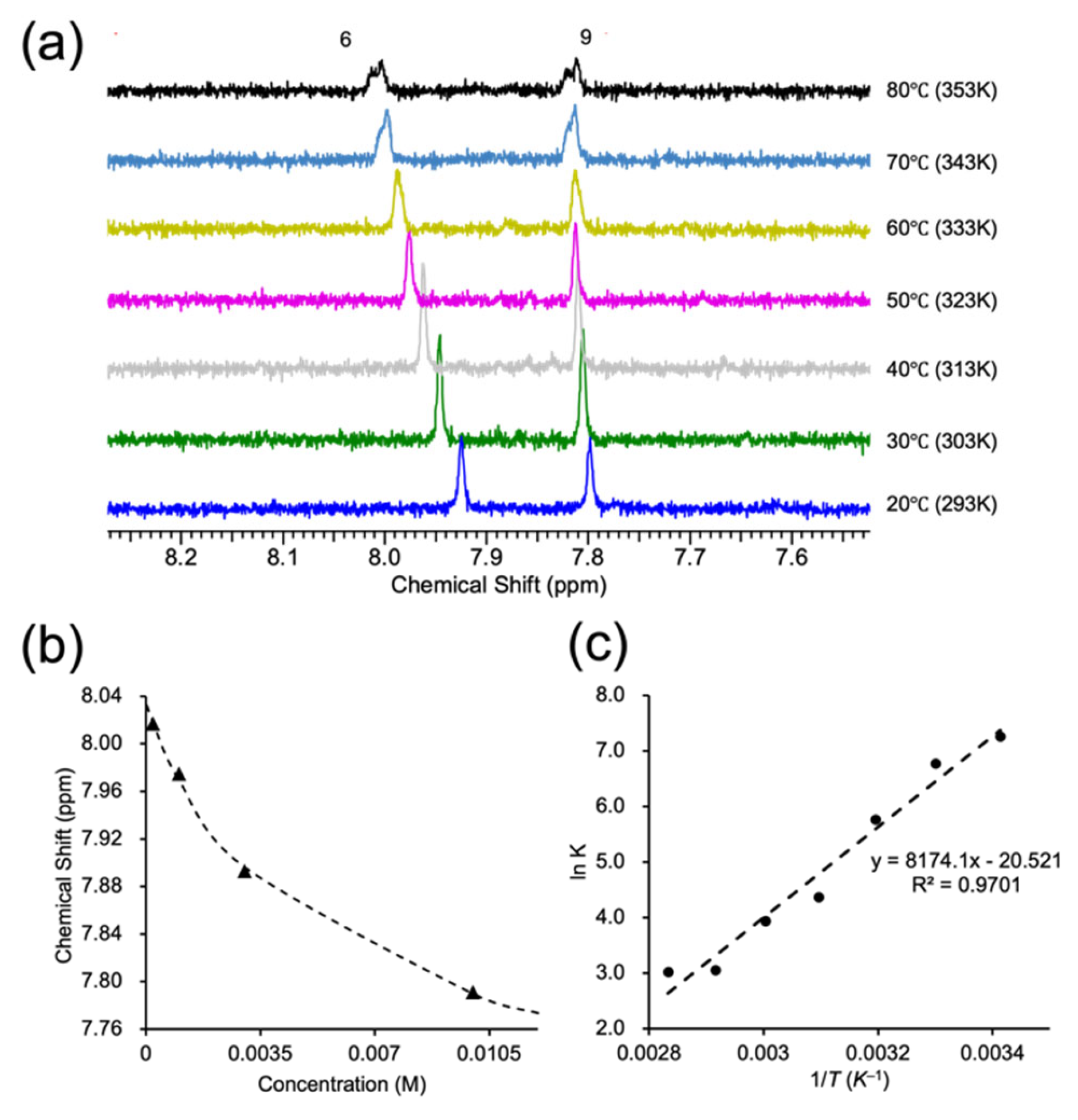
To evaluate whether the concentration-dependent emission behavior of 1a in water arises from molecular aggregation, variable-temperature 1H NMR measurements were conducted in D2O at different concentrations. Representative data at 1.0 mM are shown in Figure 3a; results at other concentrations are provided in Figure S11. As the temperature increased, continuous downfield shifts were observed for the protons at the 6- and 9-positions of the isoalloxazine core, indicating that self-association based on intermolecular interaction was occurring in the solution. To quantitatively assess the thermodynamic parameters of this self-association, chemical shift changes at each concentration and temperature were fitted using a dimerization model to derive association constants. The result at 313 K is shown in Figure 3b, while those at other temperatures are provided in Figure S12 [60]. A van’t Hoff plot constructed from these values (Figure 3c) showed good linearity, yielding thermodynamic parameters of ΔH° = −68.0 kJ mol−1, ΔS° = −171 J mol−1 K−1, and ΔG° = −17.0 kJ mol−1 at 298K. These results suggest an enthalpy-driven association process, likely governed by hydrophobic and π-π interactions. The association constant at 298K was determined to be 1037 M−1. Taken together, these findings indicate that 1a undergoes concentration-dependent self-association in aqueous media, and this aggregation behavior is responsible for modulating its emission properties.
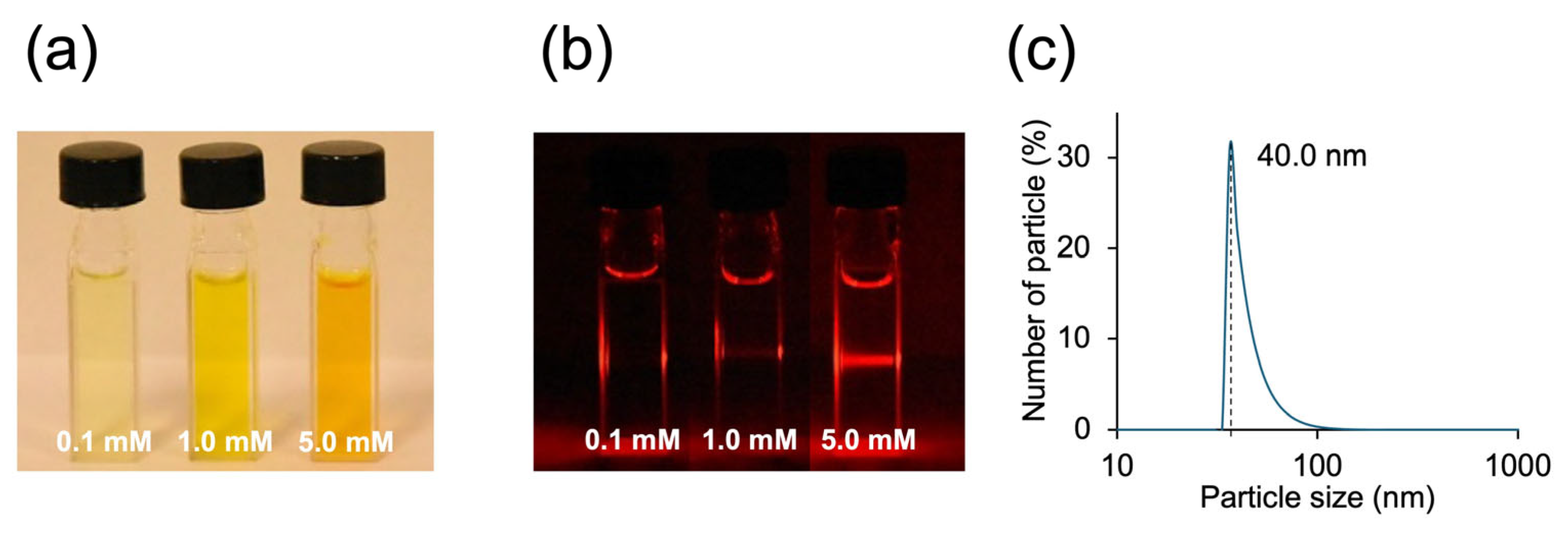
As visual evidence of concentration-dependent aggregation, Tyndall scattering was evaluated by irradiating aqueous solutions of 1a with a laser beam (Figure 4a,b). No scattering was observed at low concentration (0.1 mM), whereas clear Tyndall scattering was detected at 1.0 mM and 5.0 mM, indicating the presence of submicron-sized particles in the solution. To quantitatively assess the formation of these aggregates, dynamic light scattering (DLS) measurements were conducted at 5.0 mM (Figure 4c). The average hydrodynamic diameter was determined to be 40.0 nm, consistent with the formation of micelle-like aggregates at this concentration. Additional DLS results at lower concentrations are provided in the Supporting Information (Figure S13), where seemingly larger particle sizes were observed. This is likely due to the presence of loose, transient aggregates that yield hydrodynamic diameters larger than the actual micelle-like species [61]. Furthermore, the critical micelle concentration (CMC) of 1a was determined to be 0.6 ± 0.1 mM, based on the inflection points observed in both UV–vis absorption (0.7 mM) and fluorescence (0.5 mM) measurements (Figure S14).
To investigate the morphology of the aggregates, scanning electron microscopy (SEM) analysis was conducted by drop-casting aqueous solutions of 1a onto a glass plate, followed by natural drying (Figure 5a). The SEM images revealed the formation of numerous spherical particles with diameters ranging from several tens of to several hundred nanometers (Figure 5b), consistent with the size range suggested by DLS measurements. These results support the presence of micelle-like aggregates in aqueous solution. Taken together, these findings indicate that the amphiphilic structure of 1a promotes concentration-dependent micelle-like aggregation in water, which is responsible for the continuous modulation of its emission properties. All samples were prepared after sufficient standing time, suggesting that the observed aggregation states represent thermodynamically stable species.

3.3. Kinetic Self-Assembly: Fiber-like Aggregation

While previous experiments focused on aggregation states formed under thermodynamic equilibrium, we also investigated how 1a responds to abrupt environmental changes such as rapid freezing. Specifically, the emission behavior was examined under temperature variations at a fixed concentration (1.0 mM) using photoluminescence measurements (Figure 6). As the sample was cooled from room temperature to lower temperatures, the emission spectra did not exhibit a continuous shift. Instead, a pronounced discontinuity was observed near the freezing point of water. This result suggests that the modulation of emission is not simply due to gradual temperature change but is caused by the phase transition of the solvent during freezing.
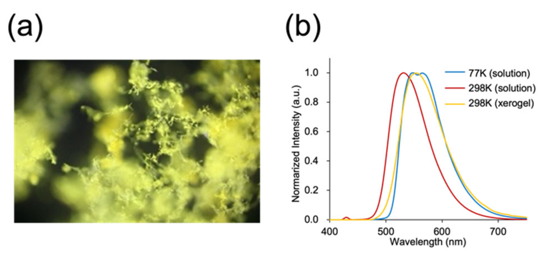
To verify whether the change in emission behavior at low temperatures arises from aggregate formation, an aqueous solution of 1a was frozen in liquid nitrogen and subsequently freeze-dried under vacuum. The resulting solid sample was analyzed by SEM (Figure 7). Compared to the room-temperature-dried sample, which showed spherical aggregates (Figure 5), the freeze-dried sample exhibited elongated, continuous fibrous structures (Figure 7b). Furthermore, the dried-xerogel sample emitted yellow fluorescence under 365 nm UV light (Figure 8a), and its emission spectrum closely matched that observed in aqueous solution at 77 K (Figure 8b). These findings strongly suggest that the yellow emission observed at low temperatures originates from the formation of kinetically controlled fibrous aggregates.
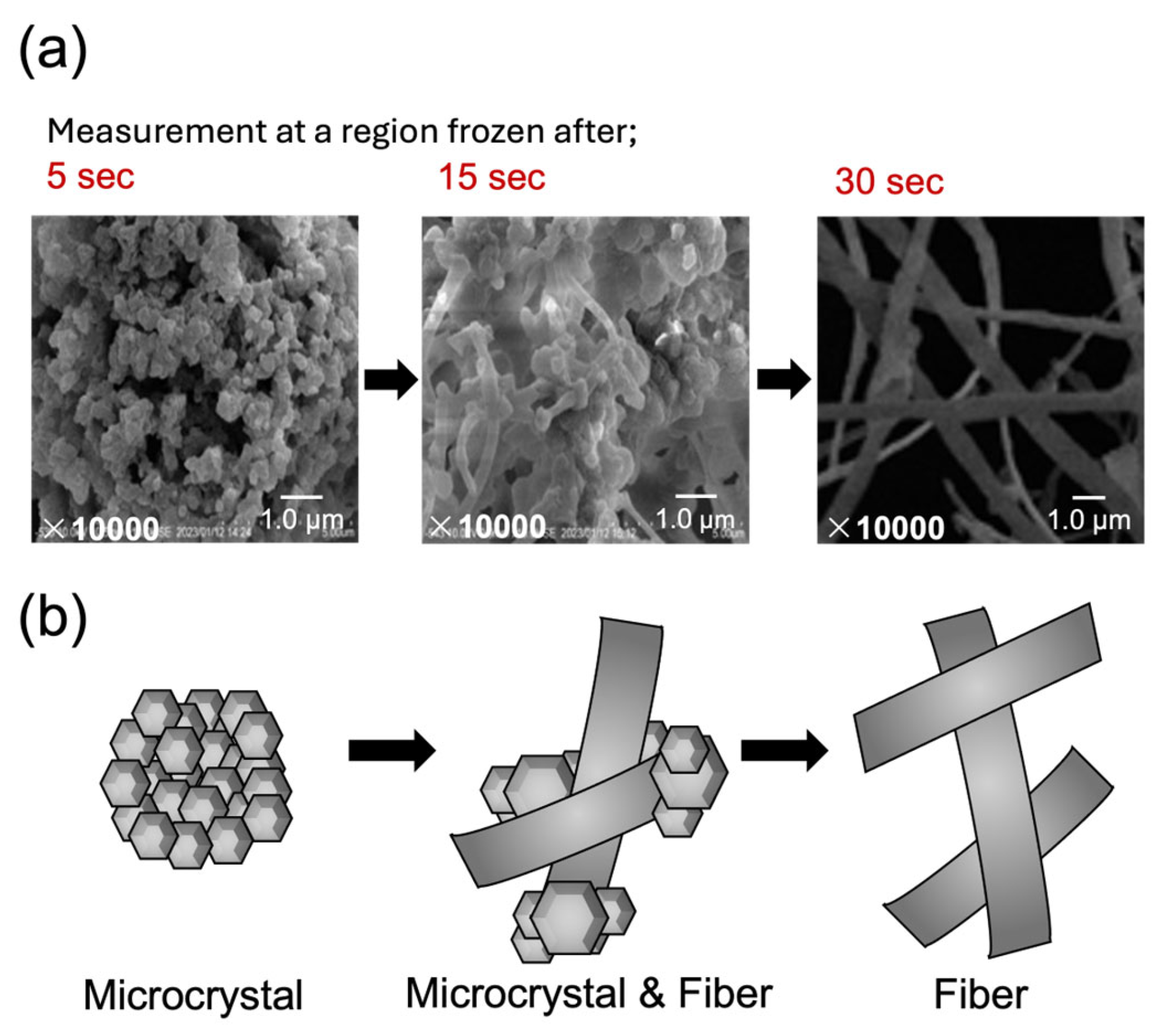
The formation process of the fibrous aggregates was examined under kinetically controlled conditions simulated by rapid freezing (Figure 9). An aqueous solution of 1a (1.0 mM) was drop-cast onto a glass substrate, rapidly frozen in liquid nitrogen, and subsequently freeze-dried. Due to the variation in local thickness between the edge and center of the droplet, the freezing time differed spatially (ca. 5 s at the edge and 30 s at the center), allowing partial spatial visualization of the aggregate growth pathway. SEM analysis (Figure 9) revealed that the outer edge predominantly contained microcrystalline domains, while progressing toward the center, fibrous extensions appeared to emanate from these microcrystals. Although real-time observation of fiber formation is rare, this spatial progression resembled the gel fiber growth we previously reported [62]. These findings indicate that under conditions of rapid cooling and limited growth time, fibrous structures are selectively formed as a kinetically favored, metastable state, and further suggest that this morphology is correlated with the yellow emission observed at low temperatures.

4. Conclusions

This study demonstrated environmentally responsive emission control in water using a water-soluble amphiphilic flavin derivative (1a). The results revealed that 1a forms two distinct types of aggregates in response to changes in external conditions such as concentration and temperature, each accompanied by characteristic changes in emission behavior. Specifically, thermodynamically driven self-association at higher concentrations led to the formation of micelle-like spherical aggregates, accompanied by a continuous bathochromic shift in emission. In contrast, rapid cooling induced the formation of fibrous aggregates under kinetic control, which exhibited a distinct yellow emission and a discontinuous spectral shift near the freezing point of water.
These dual aggregation modes are attributed to the synergistic effects of structural elements in 1a: the rigid, π-conjugated isoalloxazine core that favors stacking, the flexible long alkyl chain, and the sulfonate group that imparts water solubility. This molecular architecture provides a well-balanced combination of rigidity and flexibility, allowing for intermolecular interactions strong enough to induce emission color changes without causing excessive π-stacking that typically leads to fluorescence quenching.
Unlike most previously reported stimuli-responsive flavin systems that rely on redox or covalent reactions to achieve ON/OFF-type fluorescence switching, this work provides a rare and compelling example of reversible and continuous emission color modulation via aggregation states in flavin derivatives, thus expanding the design principles of flavin-based photofunctional materials.

Supplementary Materials

The following supporting information can be downloaded at https://www.mdpi.com/article/10.3390/photochem5030025/s1, Figure S1: 1H NMR spectrum of 1a′ in CDCl3; Figure S2: 13C NMR spectrum of 1a′ in CDCl3; Figure S3: 1H NMR spectrum of 1a in D2O (1.0 mM) at 25 °C; Figure S4: 13C NMR spectrum of 1a in D2O (ca. 28 mM) at room temperature; Figure S5: 13C NMR spectrum of 1a in D2O (ca. 28 mM) at 80 °C; Figure S6: 13C NMR spectrum of 1a in DMSO-d6(ca. 28 mM) at room temp; Figure S7: 1H NMR spectrum of 1b in CDCl3; Figure S8: 13C NMR spectrum of 1b in CDCl3; Figure S9: UV–vis absorption spectrum and normalized emission spectrum of 1a in aqueous solution; Figure S10: Time-resolved emission decay profiles of compound 1a in aqueous solution; Figure S11: Temperature-dependent 1H NMR spectra of 1a; Figure S12: Plots of the chemical shift of the H6 proton as a function of concentration; Table S1; Association constants for the self-aggregation of 1a in D2O. Figure S13: DLS measurements of aqueous solutions of 1a at concentrations of 0.1 and 1.0 mM; Figure S14: Determination of the critical micelle concentration (CMC) of compound 1a from UV–vis absorption and fluorescence measurements in aqueous solution.

Author Contributions

Conceptualization, K.O., S.K. and T.N.; methodology, K.O.; software, K.O. and S.K.; validation, S.K.; formal analysis, K.O., S.H. and S.K.; investigation, K.O.; data curation, S.K. and S.H.; writing—original draft preparation, S.K.; writing—review and editing, S.K.; visualization, K.O. and S.K.; supervision, T.N.; project administration, T.N.; funding acquisition, S.K. and T.N. All authors have read and agreed to the published version of the manuscript.

Funding

This research was supported by a Kakenhi Grant-in-Aid (No. JP20H02753) (T.N.) from the Japan Society for the Promotion of Science (JSPS), the Iketani Science and Technology Foundation (S.K.), the Toyota Physical and Chemical Research Institute (S.K.), and the Chubei Itoh Foundation (S.K.).

Data Availability Statement

The original contributions presented in this study are included in the article. Further inquiries can be directed to the corresponding authors.

Acknowledgments

We thank Masahiro Ikeshita (Nihon University) for providing measurements of the emission lifetime.

Conflicts of Interest

The authors declare no conflicts of interest.

References

  1. Tiwari, A.; Kobayashi, H. (Eds.) Responsive Materials and Methods: State-of-the-Art Stimuli-Responsive Materials and Their Applications; Advance Materials Series; Wiley: Beverly, MA, USA, 2014. [Google Scholar]
  2. Li, X.; Gao, X.; Shi, W.; Ma, H. Design Strategies for Water-Soluble Small Molecular Chromogenic and Fluorogenic Probes. Chem. Rev. 2014, 114, 590–659. [Google Scholar] [CrossRef]
  3. Gao, M.; Tang, B.Z. Fluorescent Sensors Based on Aggregation-Induced Emission: Recent Advances and Perspectives. ACS Sens. 2017, 2, 1382–1399. [Google Scholar] [CrossRef] [PubMed]
  4. Hoeben, F.J.M.; Herz, L.M.; Daniel, C.; Jonkheijm, P.; Schenning, A.P.H.J.; Silva, C.; Meskers, S.C.J.; Beljonne, D.; Phillips, R.T.; Friend, R.H.; et al. Efficient Energy Transfer in Mixed Columnar Stacks of Hydrogen-Bonded Oligo(p-phenylene Vinylene)s in Solution. Angew. Chem. Int. Ed. 2004, 43, 1976–1979. [Google Scholar] [CrossRef] [PubMed]
  5. Yagai, S.; Seki, T.; Karatsu, T.; Kitamura, A.; Würthner, F. Transformation from H- to J-Aggregated Perylene Bisimide Dyes by Complexation with Cyanurates. Angew. Chem. Int. Ed. 2008, 47, 3367–3371. [Google Scholar] [CrossRef] [PubMed]
  6. Chi, Z.; Zhang, X.; Xu, B.; Zhou, X.; Ma, C.; Zhang, Y.; Liu, S.; Xu, J. Recent Advances in Organic Mechanofluorochromic Materials. Chem. Soc. Rev. 2012, 41, 3878. [Google Scholar] [CrossRef]
  7. Kartha, K.K.; Babu, S.S.; Srinivasan, S.; Ajayaghosh, A. Attogram Sensing of Trinitrotoluene with a Self-Assembled Molecular Gelator. J. Am. Chem. Soc. 2012, 134, 4834–4841. [Google Scholar] [CrossRef]
  8. Basak, S.; Nanda, J.; Banerjee, A. Assembly of Naphthalenediimide Conjugated Peptides: Aggregation Induced Changes in Fluorescence. Chem. Commun. 2013, 49, 6891. [Google Scholar] [CrossRef]
  9. Samanta, S.K.; Bhattacharya, S. Excellent Chirality Transcription in Two-Component Photochromic Organogels Assembled through J-Aggregation. Chem. Commun. 2013, 49, 1425. [Google Scholar] [CrossRef]
  10. Praveen, V.K.; Ranjith, C.; Bandini, E.; Ajayaghosh, A.; Armaroli, N. Oligo(Phenylenevinylene) Hybrids and Self-Assemblies: Versatile Materials for Excitation Energy Transfer. Chem. Soc. Rev. 2014, 43, 4222–4242. [Google Scholar] [CrossRef]
  11. Yagai, S.; Okamura, S.; Nakano, Y.; Yamauchi, M.; Kishikawa, K.; Karatsu, T.; Kitamura, A.; Ueno, A.; Kuzuhara, D.; Yamada, H.; et al. Design Amphiphilic Dipolar π-Systems for Stimuli-Responsive Luminescent Materials Using Metastable States. Nat. Commun. 2014, 5, 4013. [Google Scholar] [CrossRef]
  12. Cao, X.; Meng, L.; Li, Z.; Mao, Y.; Lan, H.; Chen, L.; Fan, Y.; Yi, T. Large Red-Shifted Fluorescent Emission via Intermolecular π–π Stacking in 4-Ethynyl-1,8-Naphthalimide-Based Supramolecular Assemblies. Langmuir 2014, 30, 11753–11760. [Google Scholar] [CrossRef] [PubMed]
  13. Liu, J.; Guo, X.; Hu, R.; Xu, J.; Wang, S.; Li, S.; Li, Y.; Yang, G. Intracellular Fluorescent Temperature Probe Based on Triarylboron Substituted Poly N -Isopropylacrylamide and Energy Transfer. Anal. Chem. 2015, 87, 3694–3698. [Google Scholar] [CrossRef] [PubMed]
  14. Ikeda, T.; Masuda, T.; Takayama, M.; Adachi, H.; Haino, T. Solvent-Induced Emission of Organogels Based on Tris(Phenylisoxazolyl)Benzene. Org. Biomol. Chem. 2016, 14, 36–39. [Google Scholar] [CrossRef] [PubMed]
  15. Cui, J.; Kwon, J.E.; Kim, H.-J.; Whang, D.R.; Park, S.Y. Smart Fluorescent Nanoparticles in Water Showing Temperature-Dependent Ratiometric Fluorescence Color Change. ACS Appl. Mater. Interfaces 2017, 9, 2883–2890. [Google Scholar] [CrossRef]
  16. Choudhury, P.; Das, K.; Das, P.K. l-Phenylalanine-Tethered, Naphthalene Diimide-Based, Aggregation-Induced, Green-Emitting Organic Nanoparticles. Langmuir 2017, 33, 4500–4510. [Google Scholar] [CrossRef]
  17. Das, P.; Kumar, A.; Chowdhury, A.; Mukherjee, P.S. Aggregation-Induced Emission and White Luminescence from a Combination of π-Conjugated Donor–Acceptor Organic Luminogens. ACS Omega 2018, 3, 13757–13771. [Google Scholar] [CrossRef]
  18. Liang, S.; Wang, Y.; Wu, X.; Chen, M.; Mu, L.; She, G.; Shi, W. An Ultrasensitive Ratiometric Fluorescent Thermometer Based on Frustrated Static Excimers in the Physiological Temperature Range. Chem. Commun. 2019, 55, 3509–3512. [Google Scholar] [CrossRef]
  19. Yadav, P.; Gond, S.; Singh, A.K.; Singh, V.P. A Pyrene-Thiophene Based Probe for Aggregation Induced Emission Enhancement (AIEE) and Naked-Eye Detection of Fluoride Ions. J. Lumin. 2019, 215, 116704. [Google Scholar] [CrossRef]
  20. Xiao, T.; Ren, D.; Tang, L.; Wu, Z.; Wang, Q.; Li, Z.-Y.; Sun, X.-Q. A Temperature-Responsive Artificial Light-Harvesting System in Water with Tunable White-Light Emission. J. Mater. Chem. A 2023, 11, 18419–18425. [Google Scholar] [CrossRef]
  21. Tang, L.; Wu, Z.; Zhang, Q.; Hu, Q.; Dang, X.; Cui, F.; Tang, L.; Xiao, T. A Sequential Light-Harvesting System with Thermosensitive Colorimetric Emission in Both Aqueous Solution and Hydrogel. Chem. Commun. 2024, 60, 4719–4722. [Google Scholar] [CrossRef]
  22. Chapman, S.K.; Perham, R.N.; Scrutton, N.S. Flavins and flavoproteins 2002. In Proceedings of the Fourteenth International Symposium, St. John’s College, University of Cambridge, Cambridge, UK, 14–18 July 2002. [Google Scholar]
  23. Weber, S.; Schleicher, E. (Eds.) Flavins and Flavoproteins: Methods and Protocols; Humana Press: New York, NY, USA, 2014. [Google Scholar]
  24. Ghisla, S.; Massey, V. Mechanisms of Flavoprotein-catalyzed Reactions. Euro. J. Biochem. 1989, 181, 1–17. [Google Scholar] [CrossRef] [PubMed]
  25. Massey, V. The Chemical and Biological Versatility of Riboflavin. Biochem. Soc. Trans. 2000, 28, 283–296. [Google Scholar] [CrossRef] [PubMed]
  26. Imada, Y.; Naota, T. Flavins as Organocatalysts for Environmentally Benign Molecular Transformations. Chem. Rec. 2007, 7, 354–361. [Google Scholar] [CrossRef] [PubMed]
  27. Gelalcha, F.G. Heterocyclic Hydroperoxides in Selective Oxidations. Chem. Rev. 2007, 107, 3338–3361. [Google Scholar] [CrossRef]
  28. Imada, Y.; Iida, H.; Kitagawa, T.; Naota, T. Aerobic Reduction of Olefins by In Situ Generation of Diimide with Synthetic Flavin Catalysts. Chem. Euro. J. 2011, 17, 5908–5920. [Google Scholar] [CrossRef]
  29. de Gonzalo, G.; Fraaije, M.W. Recent Developments in Flavin-based Catalysis. ChemCatChem 2013, 5, 403–415. [Google Scholar] [CrossRef]
  30. Imada, Y.; Kugimiya, Y.; Iwata, S.; Komiya, N.; Naota, T. Non-Covalently Dendronized Flavins as Organocatalysts for Aerobic Reduction of Olefins. Tetrahedron 2013, 69, 8572–8578. [Google Scholar] [CrossRef]
  31. Cibulka, R. Artificial Flavin Systems for Chemoselective and Stereoselective Oxidations. Eur. J. Org. Chem. 2015, 2015, 915–932. [Google Scholar] [CrossRef]
  32. Imada, Y.; Osaki, M.; Noguchi, M.; Maeda, T.; Fujiki, M.; Kawamorita, S.; Komiya, N.; Naota, T. Flavin-Functionalized Gold Nanoparticles as an Efficient Catalyst for Aerobic Organic Transformations. ChemCatChem 2015, 7, 99–106. [Google Scholar] [CrossRef]
  33. Kawamorita, S.; Fujiki, M.; Li, Z.; Kitagawa, T.; Imada, Y.; Naota, T. Aggregation-induced Substrate Specificity in Aerobic Reduction of Olefins with Ultrasound Gel Catalyst of Synthetic Flavin. ChemCatChem 2019, 11, 878–884. [Google Scholar] [CrossRef]
  34. Heelis, P.F. The Photophysical and Photochemical Properties of Flavins (Isoalloxazines). Chem. Soc. Rev. 1982, 11, 15–39. [Google Scholar] [CrossRef]
  35. Manna, S.; Saha, A.; Nandi, A.K. A Two Component Thermoreversible Hydrogel of Riboflavin and Melamine: Enhancement of Photoluminescence in the Gel Form. Chem. Commun. 2006, 4285. [Google Scholar] [CrossRef]
  36. Daďová, J.; Kümmel, S.; Feldmeier, C.; Cibulková, J.; Pažout, R.; Maixner, J.; Gschwind, R.M.; König, B.; Cibulka, R. Aggregation Effects in Visible-Light Flavin Photocatalysts: Synthesis, Structure, and Catalytic Activity of 10-Arylflavins. Chem. Eur. J. 2013, 19, 1066–1075. [Google Scholar] [CrossRef] [PubMed]
  37. Mataranga-Popa, L.N.; Torje, I.; Ghosh, T.; Leitl, M.J.; Späth, A.; Novianti, M.L.; Webster, R.D.; König, B. Synthesis and Electronic Properties of π-Extended Flavins. Org. Biomol. Chem. 2015, 13, 10198–10204. [Google Scholar] [CrossRef] [PubMed]
  38. Suzuki, H.; Inoue, R.; Kawamorita, S.; Komiya, N.; Imada, Y.; Naota, T. Highly Fluorescent Flavins: Rational Molecular Design for Quenching Protection Based on Repulsive and Attractive Control of Molecular Alignment. Chem. Eur. J. 2015, 21, 9171–9178. [Google Scholar] [CrossRef] [PubMed]
  39. Reiffers, A.; Torres Ziegenbein, C.; Engelhardt, A.; Kühnemuth, R.; Gilch, P.; Czekelius, C. Impact of Mono-Fluorination on the Photophysics of the Flavin Chromophore. Photochem. Photobiol. 2018, 94, 667–676. [Google Scholar] [CrossRef]
  40. Vinod Mouli, M.S.S.; Mishra, A.K. Flavin Based Supramolecular Gel Displaying Multi-Stimuli Triggered Sol–Gel Transition. Org. Biomol. Chem. 2023, 21, 5622–5628. [Google Scholar] [CrossRef]
  41. Čubiňák, M.; Varma, N.; Oeser, P.; Pokluda, A.; Pavlovska, T.; Cibulka, R.; Sikorski, M.; Tobrman, T. Tuning the Photophysical Properties of Flavins by Attaching an Aryl Moiety via Direct C–C Bond Coupling. J. Org. Chem. 2023, 88, 218–229. [Google Scholar] [CrossRef]
  42. Guo, H.; Liu, S.; Liu, X.; Zhang, L. Lightening Flavin by Amination for Fluorescent Sensing. Phys. Chem. Chem. Phys. 2024, 26, 19554–19563. [Google Scholar] [CrossRef]
  43. Agrawal, H.G.; Raha, S.; Mishra, A.K. Subtle Chemical Modification around the Flavin Core to Create Diverse Morphological Assembly and Sensing Application. J. Mol. Struct. 2025, 1319, 139229. [Google Scholar] [CrossRef]
  44. Gopal Agrawal, H.; Dubey, A.; Mondal, D.; Kumar Mishra, A. Subtle Molecular Engineering around Flavin Core for Stimuli-Responsive Solid-State Luminophore. Chem. Asian J. 2025, 20, e202401099. [Google Scholar] [CrossRef]
  45. Shuttleworth, C.W.; Brennan, A.M.; Connor, J.A. NAD(P)H Fluorescence Imaging of Postsynaptic Neuronal Activation in Murine Hippocampal Slices. J. Neurosci. 2003, 23, 3196–3208. [Google Scholar] [CrossRef]
  46. Shibuki, K.; Hishida, R.; Murakami, H.; Kudoh, M.; Kawaguchi, T.; Watanabe, M.; Watanabe, S.; Kouuchi, T.; Tanaka, R. Dynamic Imaging of Somatosensory Cortical Activity in the Rat Visualized by Flavoprotein Autofluorescence. J. Physiol. 2003, 549, 919–927. [Google Scholar] [CrossRef]
  47. Murakami, H.; Kamatani, D.; Hishida, R.; Takao, T.; Kudoh, M.; Kawaguchi, T.; Tanaka, R.; Shibuki, K. Short-term Plasticity Visualized with Flavoprotein Autofluorescence in the Somatosensory Cortex of Anaesthetized Rats. Eur. J. Neurosci. 2004, 19, 1352–1360. [Google Scholar] [CrossRef]
  48. Weber, B.; Burger, C.; Wyss, M.T.; Von Schulthess, G.K.; Scheffold, F.; Buck, A. Optical Imaging of the Spatiotemporal Dynamics of Cerebral Blood Flow and Oxidative Metabolism in the Rat Barrel Cortex. Eur. J. Neurosci. 2004, 20, 2664–2670. [Google Scholar] [CrossRef] [PubMed]
  49. Takahashi, K.; Hishida, R.; Kubota, Y.; Kudoh, M.; Takahashi, S.; Shibuki, K. Transcranial Fluorescence Imaging of Auditory Cortical Plasticity Regulated by Acoustic Environments in Mice. Eur. J. Neurosci. 2006, 23, 1365–1376. [Google Scholar] [CrossRef] [PubMed]
  50. Drepper, T.; Eggert, T.; Circolone, F.; Heck, A.; Krauß, U.; Guterl, J.-K.; Wendorff, M.; Losi, A.; Gärtner, W.; Jaeger, K.-E. Reporter Proteins for in Vivo Fluorescence without Oxygen. Nat. Biotechnol. 2007, 25, 443–445. [Google Scholar] [CrossRef] [PubMed]
  51. Król, J.E.; Rogers, L.M.; Krone, S.M.; Top, E.M. Dual Reporter System for In Situ Detection of Plasmid Transfer under Aerobic and Anaerobic Conditions. Appl. Environ. Microbiol. 2010, 76, 4553–4556. [Google Scholar] [CrossRef]
  52. Landete, J.M.; Peirotén, Á.; Rodríguez, E.; Margolles, A.; Medina, M.; Arqués, J.L. Anaerobic Green Fluorescent Protein as a Marker of Bifidobacterium Strains. Int. J. Food Microbiol. 2014, 175, 6–13. [Google Scholar] [CrossRef]
  53. Iida, H.; Iwahana, S.; Mizoguchi, T.; Yashima, E. Main-Chain Optically Active Riboflavin Polymer for Asymmetric Catalysis and Its Vapochromic Behavior. J. Am. Chem. Soc. 2012, 134, 15103–15113. [Google Scholar] [CrossRef]
  54. Iida, H.; Miki, M.; Iwahana, S.; Yashima, E. Riboflavin-Based Fluorogenic Sensor for Chemo- and Enantioselective Detection of Amine Vapors. Chem. Eur. J. 2014, 20, 4257–4262. [Google Scholar] [CrossRef] [PubMed]
  55. Liu, J.; Han, S.; Hu, Y.; Pao, C.-W. Fabrication and Characterization of a Novel PMO Containing Riboflavin-5′-Phosphate Sodium Salt for Sensitive Detection of Pesticide Ferbam. Colloids Surf. A 2021, 617, 126375. [Google Scholar] [CrossRef]
  56. Mouli, M.S.S.V.; Mishra, A.K. Sequential Recognition Capability of a Novel Flavin-Dipicolyl Analogue toward Zinc and Phosphate Ion: A Model Capable of Selective Recognition of AMP over ADP/ATP. Dyes Pigm. 2023, 212, 111148. [Google Scholar] [CrossRef]
  57. Oka, M.; Kozako, R.; Teranishi, Y.; Yamada, Y.; Miyake, K.; Fujimura, T.; Sasai, R.; Ikeue, T.; Iida, H. Chiral Supramolecular Organogel Constructed Using Riboflavin and Melamine: Its Application in Photo-Catalyzed Colorimetric Chiral Sensing and Enantioselective Adsorption. Chem. Eur. J. 2024, 30, e202303353. [Google Scholar] [CrossRef]
  58. Kawamorita, S.; Li, Z.; Okamoto, K.; Naota, T. Multistimuli-Responsive Chromism of Vinylene-Linked Bisflavin Based on the Aggregation and Redox Properties. Chem. Eur. J. 2023, 29, e202202257. [Google Scholar] [CrossRef]
  59. Imada, Y.; Iida, H.; Ono, S.; Masui, Y.; Murahashi, S. Flavin-Catalyzed Oxidation of Amines and Sulfides with Molecular Oxygen: Biomimetic Green Oxidation. Chem. Asian J. 2006, 1, 136–147. [Google Scholar] [CrossRef]
  60. Horman, I.; Dreux, B. Estimation of Dimerisation Constants from Complexatin-Induced Displacements of1 H-NMR Chemical Shifts: Dimerisation of Caffeine. Helv. Chim. Acta 1984, 67, 754–764. [Google Scholar] [CrossRef]
  61. Filippov, S.K.; Khusnutdinov, R.; Murmiliuk, A.; Inam, W.; Zakharova, L.Y.; Zhang, H.; Khutoryanskiy, V.V. Dynamic Light Scattering and Transmission Electron Microscopy in Drug Delivery: A Roadmap for Correct Characterization of Nanoparticles and Interpretation of Results. Mater. Horiz. 2023, 10, 5354–5370. [Google Scholar] [CrossRef]
  62. Naito, M.; Inoue, R.; Iida, M.; Kuwajima, Y.; Kawamorita, S.; Komiya, N.; Naota, T. Control of Metal Arrays Based on Heterometallics Masquerading in Heterochiral Aggregations of Chiral Clothespin-Shaped Complexes. Chem. Eur. J. 2015, 21, 12927–12939. [Google Scholar] [CrossRef]
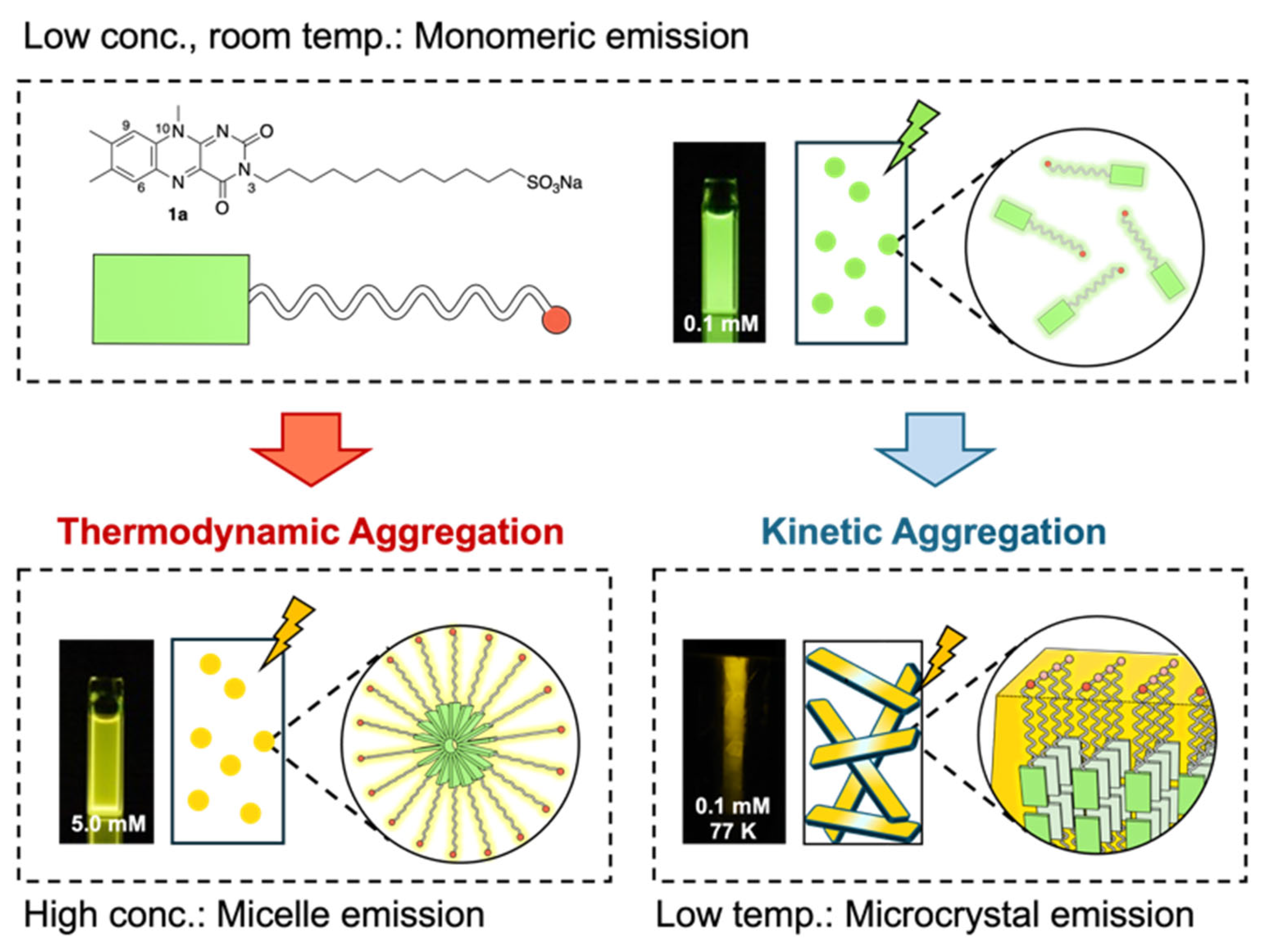
Figure 1. Schematic illustration of dual-mode self-assembly and stimuli-responsive photoluminescence behavior of amphiphilic flavin derivative 1a.
Photochem 05 00025 g001
Figure 2. (a) Molecular structures of 1a and 1b. (b) Photographs of aqueous solutions of 1a (left) and chloroform solutions of 1b (right) at different concentrations (0.1, 1.0, and 5.0 mM) under UV light (λex = 365 nm) at room temperature. (c) Corresponding emission spectra of the solutions shown in (b) (λex = 430 nm). (d) Photographs of 1a (left, in water) and 1b (right, in chloroform) at 0.1 mM under UV light (λex = 365 nm) at 298 and 77 K. (e) Emission spectra of the samples shown in (d) (λex = 430 nm).
Photochem 05 00025 g002
Figure 3. Thermodynamic analysis of the self-assembly behavior of 1a in D2O. (a) Variable-temperature 1H NMR spectra of 1a (1.0 mM, 293–353 K), showing temperature-dependent chemical shift changes of H6 and H9 protons in the isoalloxazine core. (b) Plot of the chemical shift of the H6 proton versus concentration at 313 K, fitted using a self-association model to obtain the equilibrium constant (K). (c) Van’t Hoff plot generated from K values at different temperatures, along with the linear fitting equation.
Photochem 05 00025 g003
Figure 4. Photographs of aqueous solutions of 1a at different concentrations (0.1, 1.0, and 5.0 mM) under (a) ambient light and (b) UV irradiation at 365 nm. (c) DLS measurements of aqueous solutions of 1a at concentrations of 5.0 mM. The most frequent particle diameter was 40.0 nm.
Photochem 05 00025 g004
Figure 5. (a) Schematic illustration of the sample preparation procedure for SEM observation: an aqueous solution of 1a (1.0 mM) was drop-cast onto a glass substrate and left to dry at room temperature for 24 h. (b) SEM image of the resulting dried solid.
Photochem 05 00025 g005
Figure 6. Variable-temperature photoluminescence spectra of an aqueous solution of 1a (1.0 mM) (λex = 430 nm).
Photochem 05 00025 g006
Figure 7. (a) Schematic illustration of the sample preparation procedure for SEM observation: an aqueous solution of 1a (1.0 mM) was drop-cast onto a glass substrate, rapidly frozen in liquid nitrogen, and freeze-dried. (b) SEM image of the resulting dried solid.
Photochem 05 00025 g007
Figure 8. (a) Optical microscopy image of the xerogel obtained by freeze-drying an aqueous solution of 1a (1.0 mM), taken under UV light irradiation at 365 nm. (b) Emission spectra of 1a in aqueous solution (0.1 mM) at 298 K and 77 K, and of the corresponding xerogel measured at 298 K.
Photochem 05 00025 g008
Figure 9. (a) SEM images of samples obtained by rapid freezing of aqueous solutions of 1a (1.0 mM) using liquid nitrogen. Images were taken from different regions of a single sample that experienced different freezing times (5, 15, or 30 s) due to variations in local thickness. (b) Schematic illustration of the morphological evolution from microcrystalline aggregates to fibrous structures.
Photochem 05 00025 g009
Disclaimer/Publisher’s Note: The statements, opinions and data contained in all publications are solely those of the individual author(s) and contributor(s) and not of MDPI and/or the editor(s). MDPI and/or the editor(s) disclaim responsibility for any injury to people or property resulting from any ideas, methods, instructions or products referred to in the content.