Review

Tetraphenylethylene (TPE)-Based AIE Luminogens: Recent Advances in Bioimaging Applications

by
Vanam Hariprasad
1,2,†,
Kavya S. Keremane
3,†,
Praveen Naik
1,2,*,
Dickson D. Babu
4 and
Sunitha M. Shivashankar
1,2
1
Department of Chemistry, Nitte Meenakshi Institute of Technology, Nitte (Deemed to be University), Bengaluru Campus, Bengaluru 560064, India
2
Visvesvaraya Technological University, Belagavi 590018, India
3
Department of Materials Science and Engineering, The Pennsylvania State University, University Park, PA 16802, USA
4
Department of Chemistry, St. Thomas College, Kozhencherry 689641, India
*
Author to whom correspondence should be addressed.
These authors contributed equally to this work.
Photochem 2025, 5(3), 23; https://doi.org/10.3390/photochem5030023
Submission received: 25 July 2025 / Revised: 22 August 2025 / Accepted: 29 August 2025 / Published: 4 September 2025
(This article belongs to the Special Issue Photochemistry Directed Applications of Organic Fluorescent Materials)

Abstract

Aggregation-induced emission (AIE) luminogens are materials that exhibit enhanced light emission in the aggregated state, primarily due to the restriction of intramolecular motions, which reduces energy loss through non-radiative pathways. Tetraphenylethylene (TPE) and its derivatives are prominent examples of AIE-active materials, owing to their ease of synthesis, tuneable photophysical properties, and strong aggregation tendencies. This review provides an overview of the fundamental AIE mechanisms in TPE-based systems, with a focus on the role of restricted intramolecular rotation (RIR) and π-twisting in governing their emission behaviour. It explores the influence of molecular structure, electronic configuration, and intermolecular interactions on fluorescence properties. Furthermore, recent advances in practical applications of TPE-based AIE luminogens are highlighted across a spectrum of biological imaging domains, including cellular imaging, tissue and in vivo imaging, and organelle-targeted imaging. Additionally, their integration into multifunctional and theranostic platforms, along with the development of stimuli-responsive and self-assembled systems, underscores their versatility and expanding potential in biomedical research and diagnostics. This review aims to offer valuable insights into the design principles and functional potential of TPE-based AIE luminogens, guiding the development of next-generation materials for advanced bioimaging technologies.

1. Introduction

Fluorescent organic materials have become indispensable in modern science and technology, with broad applications in chemical sensing, environmental monitoring, bioimaging, and optoelectronic devices [1,2,3,4,5,6]. Their high sensitivity, structural tunability, and versatile photophysical properties have made them vital tools for real-time detection and imaging [7,8,9,10,11,12,13,14]. However, traditional fluorophores often face a major limitation known as aggregation-caused quenching (ACQ). In the solid or aggregated state, strong π–π stacking interactions lead to non-radiative decay pathways, resulting in a significant reduction in fluorescence intensity [15,16,17,18,19]. This limitation poses challenges for their use in solid-state lighting, optoelectronics, and biological systems, where aggregation is often unavoidable [20].
The discovery of AIE by Tang and co-workers in 2001 revolutionized the field of luminescent materials [21,22,23]. In sharp contrast to ACQ, AIE luminogens (AIEgens) are non-emissive or weakly emissive in dilute solution but exhibit strong fluorescence when aggregated or in the solid state [24]. This discovery not only overcame the drawbacks of ACQ but also opened new avenues for developing highly efficient solid-state emitters, sensing probes, and bioimaging agents [25,26,27]. Among various AIE-active systems, Tetraphenylethylene (TPE) and its derivatives have emerged as particularly promising frameworks. The propeller-like molecular geometry of TPE naturally prevents detrimental π–π stacking, enabling intense fluorescence in the solid state [28,29,30,31]. Additionally, TPE-based luminogens offer synthetic flexibility, excellent photostability, large Stokes shifts, and low background interference, making them attractive for constructing robust AIE-based imaging and sensing platforms [32,33,34,35]. The unique structural features of TPE also allow for reversible mechanochromic luminescence, further expanding its potential in smart optoelectronic devices and responsive materials [36,37,38].
Recent studies have demonstrated that TPE-based AIEgens can be engineered into highly sensitive and selective fluorescent probes capable of detecting metal ions, nitroaromatic compounds, small biomolecules, and other biologically relevant targets [39,40,41,42,43]. Their high signal-to-noise ratios, excellent biocompatibility, and strong performance in aggregated or solid states make them particularly suitable for advanced bioimaging and diagnostic applications [44,45,46,47]. While previous reviews have discussed the general progress in AIE research, comprehensive research on TPE-based AIEgens for bioimaging applications remains limited. This review aims to address this gap by presenting an overview of TPE-based AIE systems, their design principles, and their role in biomedical imaging. In the next section, we discuss the general mechanism of AIE, which provides the foundational understanding of how molecular aggregation leads to enhanced luminescence and guides the rational design of high-performance AIEgens.

2. General Mechanism of AIE

AIE is a distinctive photophysical phenomenon wherein organic luminophores that are weakly or non-emissive in dilute solutions become highly fluorescent upon aggregation. This behaviour is predominantly explained by the restriction of intramolecular motion (RIM), which includes both RIR) [4,48,49] and restricted intramolecular vibration (RIV) (Figure 1). In a molecularly dispersed state, free intramolecular motions dissipate the excited-state energy through non-radiative pathways, leading to weak fluorescence. When the molecules aggregate, these dynamic motions are significantly hindered, suppressing non-radiative decay and facilitating radiative transitions, thereby producing intense emission [50,51,52].
To further enhance luminescence efficiency, molecular design strategies such as donor–acceptor (D–A) engineering, twisted molecular structures, π-conjugation extension, heavy atom incorporation, and steric hindrance have been employed. D–A engineering facilitates the formation of intramolecular charge transfer (ICT) states with red-shifted emission and improved quantum yields by aligning the HOMO–LUMO energy levels and minimizing non-radiative losses [53,54,55]. Twisted structures not only prevent detrimental π–π stacking but also promote twisted intramolecular charge transfer (TICT) states, which are highly sensitive to microenvironmental factors such as polarity and viscosity; upon aggregation, these twists are locked, enhancing fluorescence intensity [27,56,57,58]. Expanding π-conjugation pathways increases fluorescence efficiency, stabilizes excited states, and enables emission shifts toward the red or near-infrared (NIR) region, while strong π–π interactions enhance photothermal properties and further reduce non-radiative decay [59,60,61]. Additionally, incorporating heavy atoms (e.g., iodine or bromine) enhances spin–orbit coupling, promoting intersystem crossing (ISC) and facilitating the generation of reactive oxygen species (ROS), which is particularly beneficial for photodynamic therapy (PDT) [62,63,64,65]. Steric hindrance, on the other hand, suppresses excessive π–π stacking while maintaining molecular rigidity, improving both fluorescence stability and brightness.
Together, these photophysical principles not only explain the fundamental AIE mechanism but also provide a framework for designing advanced luminogens with high brightness, tuneable emission, excellent photostability, and biocompatibility. The ability of AIEgens to emit across the visible and NIR windows (NIR-I: 700–1000 nm; NIR-II: 1000–1500 nm) allows for deep-tissue penetration, high signal-to-noise ratios, and minimal background interference, making them powerful tools for bioimaging, sensing, and theranostic applications [66,67].
Figure 1. (a) Jablonski diagram of photophysical processes. Adapted with permission from Ref. [68] Copyright 2020, Wiley-VCH. (b) AIE mechanism via restriction of intramolecular motion (RIM): TPE shows RIR, and THBA shows RIV. Adapted with permission from Ref. [69]. Copyright 2014, Wiley-VCH.
Photochem 05 00023 g001

3. TPE-Based Systems for Bioimaging

Understanding molecular recognition, how biomolecules selectively interact through noncovalent forces, is essential. This knowledge has empowered chemists to design sophisticated biomimetic structures with remarkable functionalities in sensing, drug delivery, and synthetic biology [70]. TPE-derived luminogens are a prominent class of AIE-active materials. Unlike conventional fluorophores that suffer from ACQ, TPE-based luminogens exhibit strong fluorescence upon aggregation due to the Restriction of Intramolecular Motion (RIM), which suppresses non-radiative decay processes [71,72]. This unique property, combined with their high photostability, tunable optical properties, low cytotoxicity, and facile chemical modification, makes TPE-based luminogens highly suitable for diverse bioimaging applications. They are particularly valuable for functioning in crowded or heterogeneous biological environments [73,74].

3.1. TPE-Based AIEgens for Cellular Imaging

With advanced fluorescence microscopy techniques, cell imaging has become a cornerstone in studying cell biology and living systems. However, conventional fluorescent probes such as organic dyes or inorganic quantum dots have disadvantages; mainly, aromatic organic dyes usually suffer from ACQ, which quenches light emission in biological media, and most quantum dots are cell-toxic. AIE luminogens are just the best solution. Unlike traditional dyes, AIEgens emit more brightly when they cluster together, producing intensely high signal-to-background ratios, which is ideal for bioimaging. Organic AIE dots, for instance, are brighter and give better performance in biological imaging compared to inorganic quantum dots [75,76]. Since they can only emit when in an aggregated form, AIE probes reduce background fluorescence, making detection easier. TPE is the prototypic AIE molecule, its skeletal structure prevents intramolecular rotation in aggregates, suppressing non-radiative decay significantly and achieving high fluorescence quantum yields. In addition, TPE’s modular nature makes it easy to modify and tune for imaging applications in cellular imaging. Therefore, utilization of AIE-based probes, especially TPE derivatives, overcomes critical limitations of traditional fluorescent labels, exhibiting enhanced brightness, biocompatibility, and imaging sensitivity for emerging cell biology and biomedical applications [77,78].
Ding et al. (2016) [79] reported a series of three donor-acceptor-donor (D-A-D) luminogens (1–3) that used triphenylamine (TPA) donor groups with hydroxyl, ethoxy, or unsubstituted parts (Figure 2a). These structural changes enabled tunable emission properties and yielded very large two-photon absorption cross-sections (σ = 4230 GM for some derivatives). Dynamic light scattering (DLS) and scanning electron microscopy (SEM) revealed that the introduction of hydrophobic substituents facilitated the formation of larger aggregates, thereby enhancing the AIE property. With low cytotoxicity and strong fluorescence under both one- and two-photon excitation, these luminogens served well as dual-mode bioimaging probes. Mai et al. (2018) [80] synthesized TPE-based systems by designing tetraphenylethene-phenanthrene conjugates with notable AIE activity. They synthesized two derivatives, pTPEP and mTPEP, by connecting TPE units to the para- and meta-positions of the phenanthrene core, respectively (Figure 2b). Although non-emissive in pure tetrahydrofuran (THF), these compounds showed intense sky-blue fluorescence in THF/water mixtures with water fractions up to 90%. This was due to the restriction of intramolecular phenyl rotation when they were aggregated. For biological applications, both derivatives were encapsulated into monodisperse silica nanoparticles (~110 nm), which preserved their AIE in aqueous media and enabled successful visualization of HeLa cells (Figure 2c). The silica matrix acted as a protective barrier, ensuring fluorescence stability in the biological environment. Collectively, these studies highlight the design versatility and imaging potential of functionalized TPE derivatives for advanced bioimaging applications.
Chu and co-workers in 2018 reported the development of simple TPE–amino acid conjugates, namely TPE-Ser and TPE-Asp, which self-assemble into fluorescent supramolecular hydrogels with promising bioimaging potential [81]. The structural design, featuring amino acid side chains with functional groups (hydroxyl in Ser and carboxyl in Asp), facilitates hydrogen bonding and π–π stacking interactions that drive hydrogelation. TPE-Ser, having the lowest molecular weight, forms stable hydrogels under physiological pH, whereas TPE-Asp requires mildly acidic conditions (pH ~6.0) for self-assembly. Spectroscopic (UV-vis, IR, PL) and rheological studies confirmed the formation, stability, and fluorescence of the hydrogels, while TEM images revealed nanosheet-like morphologies. Importantly, in vitro studies using 3A6 cells showed that both TPE-Ser and TPE-Asp exhibit strong fluorescence signals in microcellular environments, highlighting their potential for bioimaging applications. These findings emphasize the relationship between molecular structure and hydrogelation behaviour, offering valuable design principles for single amino acid-based TPE luminogens.
Wang and co-workers in 2018 developed a near-infrared (NIR) emitting AIE fluorophore (TPE-PTZ-R) (Figure 3) by strategically integrating a phenothiazine (PTZ) electron-donating unit with a TPE core, forming a D–π–A architecture to effectively reduce the energy gap [82]. The fluorophore exhibits a large Stokes shift, pronounced AIE characteristics, and efficient NIR emission. To enhance its bioimaging applicability, uniform and stable nanoparticles (TPE-PTZ-R NPs) were fabricated using Pluronic F127 as a stabilizer. These nanoparticles demonstrated excellent photophysical properties, including strong fluorescence, high cellular uptake, low cytotoxicity, and good photostability. Density functional theory (DFT) calculations further validated the electronic design principles. The successful application of TPE-PTZ-R NPs in cellular bioimaging highlights their potential as promising NIR-active AIE-based probes for advanced biomedical applications.
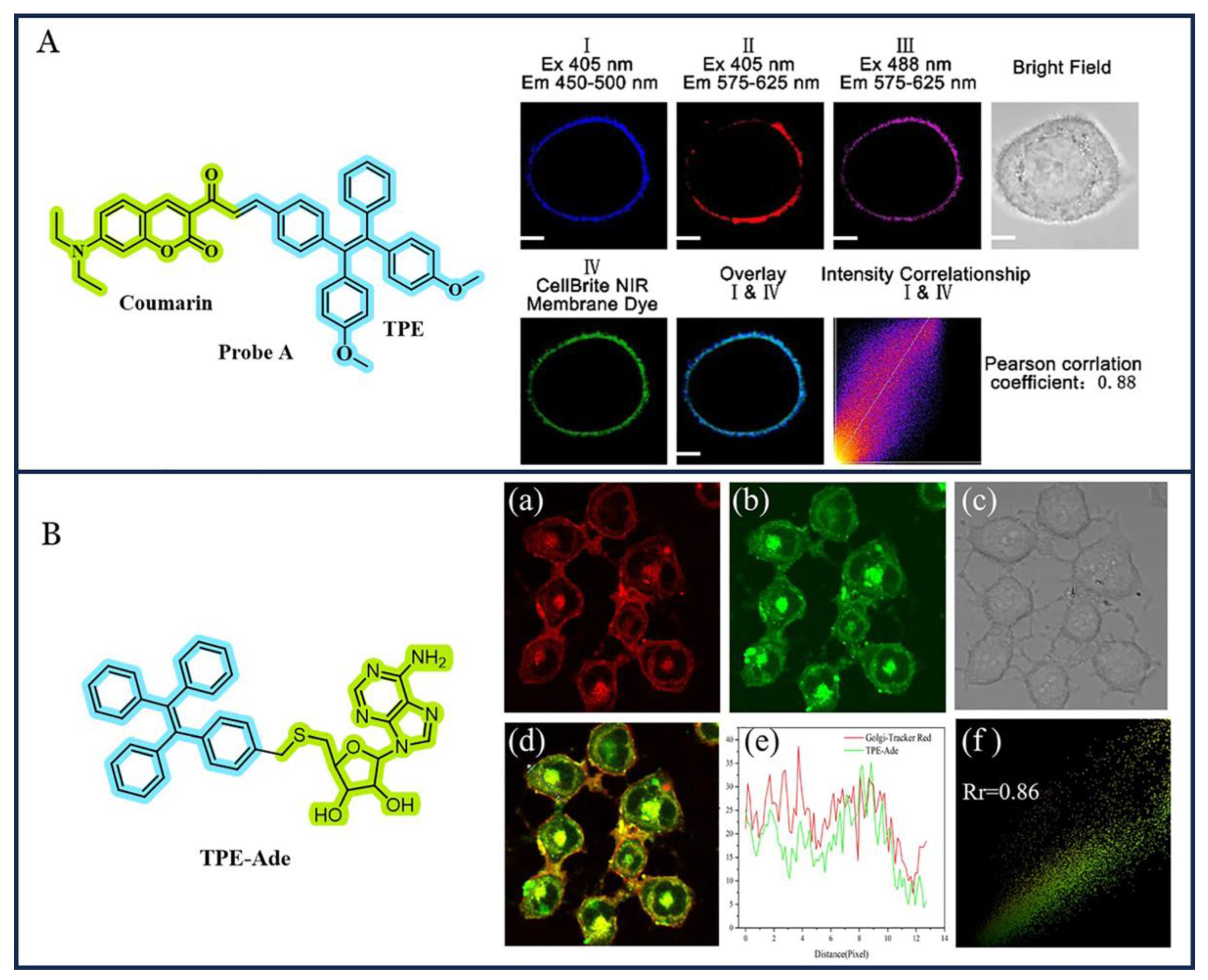
Zhang and co-workers (2020) [83] designed a novel cell membrane-specific fluorescent probe (Probe A) by integrating a coumarin fluorophore with a TPE unit through an α,β-unsaturated ketone linker (Figure 4A). This structural framework imparts strong AIE characteristics and dual-emission behaviour. In solution, Probe A exhibits absorption bands at 300 nm (TPE) and 458.5 nm (coumarin), with fluorescence at 470 nm in THF. Upon increasing the water content, a significant redshift and the emergence of a new emission peak at 591 nm are observed at 95% water content, indicating enhanced AIE activity. Confocal microscopy studies reveal that Probe A selectively targets cell membranes, as confirmed by its high colocalization with a commercial CellBrite NIR membrane dye (Pearson’s correlation coefficient = 0.93). Furthermore, the probe demonstrates excellent photostability, low cytotoxicity, and prolonged membrane retention, making it highly suitable for high-contrast, long-term live-cell membrane imaging. Its performance was further validated in Drosophila melanogaster larvae, emphasizing its utility in both cellular and small-animal bioimaging.
Xing et al. (2021) developed TPE-Ade, a Golgi-targeted probe obtained by conjugating tetraphenylethylene with 2′,3′-O-isopropylideneadenosine, which functions as a Golgi-localization unit [84]. As shown in Figure 4B, the chemical structure of TPE-Ade and its co-localization with the commercial Golgi-Tracker Red dye in HL-7402 cells demonstrate its high targeting specificity. The fluorescence signal from Golgi-Tracker Red, excited at 543 nm with emission collected between 562 and 662 nm, strongly overlaps with the signal from TPE-Ade, excited at 405 nm with emission collected between 425 and 485 nm. This co-localization, confirmed by the merged fluorescence image, intensity profile, and a Pearson’s correlation coefficient of 0.86, highlights the accuracy of TPE-Ade as a Golgi probe. Furthermore, TPE-Ade exhibits a remarkable 160-fold fluorescence enhancement upon aggregation, robust AIE behaviour, superior photostability compared to commercial Golgi trackers, low cytotoxicity, and resistance to quenching at high concentrations, making it a promising Golgi imaging tool.
Xiao and co-workers (2023) [85] reported the facile synthesis of two unprecedented tetraphenylethene (TPE)-based fluorophores, X-1 and X-2, derived from 4,5-bis(TPE)-1H-imidazole fused with pyridine or quinoline π-extended systems. Interestingly, these molecules exhibit completely opposite mechanofluorochromic (MFC) behaviours. MFC behaviour originates from the transformation between ordered crystalline and amorphous states, with further crystal structure and theoretical studies attributing the phenomena to distinct π–π stacking modes induced by the π-extended systems. Beyond mechanofluorochromism, both X-1 and X-2 demonstrated excellent biocompatibility, high fluorescence efficiency, and potential applications in bioimaging, writable data storage, and anti-counterfeiting. This study not only introduces a new class of opposite MFC materials but also provides a rational strategy for designing tunable stimuli-responsive fluorophores based on identical core units regulated by π-extension.

3.2. TPE-Based AIEgens for Tissue and In Vivo Imaging

Bioimaging platforms with properties such as cytocompatibility, photostability, red fluorescence, and optical nonlinearity are greatly in demand to investigate dynamic biological processes. New advances in imaging systems have enabled real-time visualization of cell events such as protein trafficking, organelle dynamics, and migration of cells. AIE systems, especially TPE-based AIE systems, have emerged as powerful probes for in vivo and tissue imaging due to their high sensitivity, high signal-to-background ratios, and high biocompatibility [28]. Unlike the conventional fluorophores plagued with ACQ behaviour, AIEgens are very bright upon aggregation, which improves imaging stability and resolution. Some AIEgens synthesized based on the intramolecular rotation (RIR) mechanism have successfully been engineered into nanoparticles to mark cells, detect sentinel lymph nodes, and diagnose cancer. Their strong luminescence, photostability, and biocompatibility make them especially promising for deep-tissue, long-duration imaging [86,87]. In particular, two-photon laser scanning microscopy (TPLSM) based on near-infrared excitation exhibits less scattering and deeper tissue penetration than one-photon systems, making it particularly well-suited for 3D in vivo imaging of vasculature and neural networks. Organic AIE dots, particularly TPE-based dots, are more biocompatible and more photostable than inorganic quantum dots because they do not blink and are perfectly suitable for live imaging [88,89]. This section summarises the recent advancements in TPE-based AIEgens tailored for tissue, and in vivo imaging applications [90,91]
Leung et al. (2012) designed and synthesized tetraphenylethene-triphenylphosphonium (TPE-TPP) [92], an AIE-active luminogen, for specific and stable mitochondrial imaging. The mitochondrial-targeting ability of triphenylphosphonium (TPP) and the AIE characteristics of the TPE core helped TPE-TPP show excellent photostability, environmental tolerance, and suitability for long-term tracking of mitochondrial shape changes [93]. This made it a flexible platform for further development in sensing reactive oxygen species (ROS), metal ions, or pH in mitochondria. Qian et al. in 2015 [94] used TPE-TPP for two-photon neuroimaging, where its nanoaggregates showed outstanding resistance to photobleaching during extended excitation. This allowed long-term staining and tracking of primary neurons and brain microglia both in vitro and in vivo, showing its potential in neuroscience research. Zhu et al. (2015) [95] broadened the use of TPE-TPP to three-photon microscopy with femtosecond laser excitation at 1020 nm. This reduced the photodamage and autofluorescence and provided a better spatial resolution and a higher signal-to-noise ratio compared to two-photon excitation. This technique enabled the stable, high-contrast imaging of HeLa cells and mouse brain tissues. In another biomedical study, Leung et al. (2015) [96] showed that TPE-TPP could act as a highly sensitive fluorescence probe for tracking the fibrillation process of α-synuclein (α-Syn), an important marker in Parkinson’s disease. Compared to thioflavin T (ThT), TPE-TPP had stronger fluorescence, greater sensitivity to α-Syn oligomers, and shorter detection times without changing the kinetics of fibrillation. Overall, these studies demonstrate the versatility of TPE-TPP as a strong, stable AIE luminogen with wide applications in organelle imaging, deep-tissue microscopy, and monitoring disease-related protein aggregation.
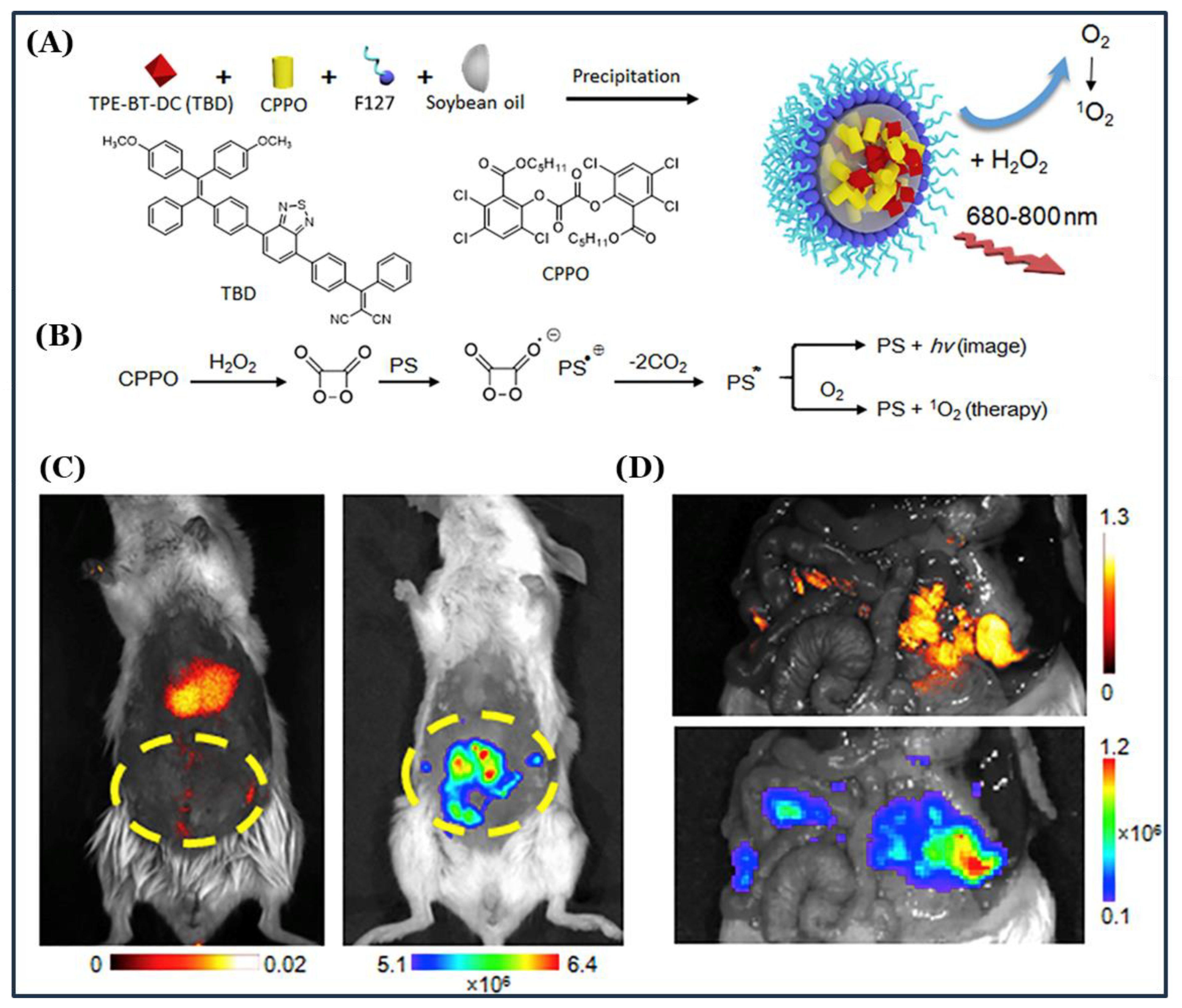
Mao et al. (2017) [97] developed a chemiluminescence-activated nanoplatform (C-TBD NPs) for precise image-guided cancer therapy (Figure 5). The nanoparticles were fabricated by co-encapsulating bis [2,4,5-trichloro-6-(pentyloxycarbonyl) phenyl] oxalate (CPPO) and an aggregation-induced emission photosensitizer (TBD) within a pluronic F-127/soybean oil matrix. These C-TBD NPs produce far-red/near-infrared (FR/NIR) chemiluminescence and singlet oxygen (1O2) upon activation by H2O2 present in the tumour microenvironment, enabling high-signal-to-noise ratio imaging and efficient photodynamic therapy. Primary and metastatic breast tumours in mouse models were clearly visualized through chemiluminescence imaging, with the therapeutic effect enhanced upon co-administration of the anti-tumour agent FEITC, which increases H2O2 generation. The synergistic action of tumour localization, imaging, and therapy establishes C-TBD NPs as a promising strategy for simultaneous tumour diagnosis and treatment.
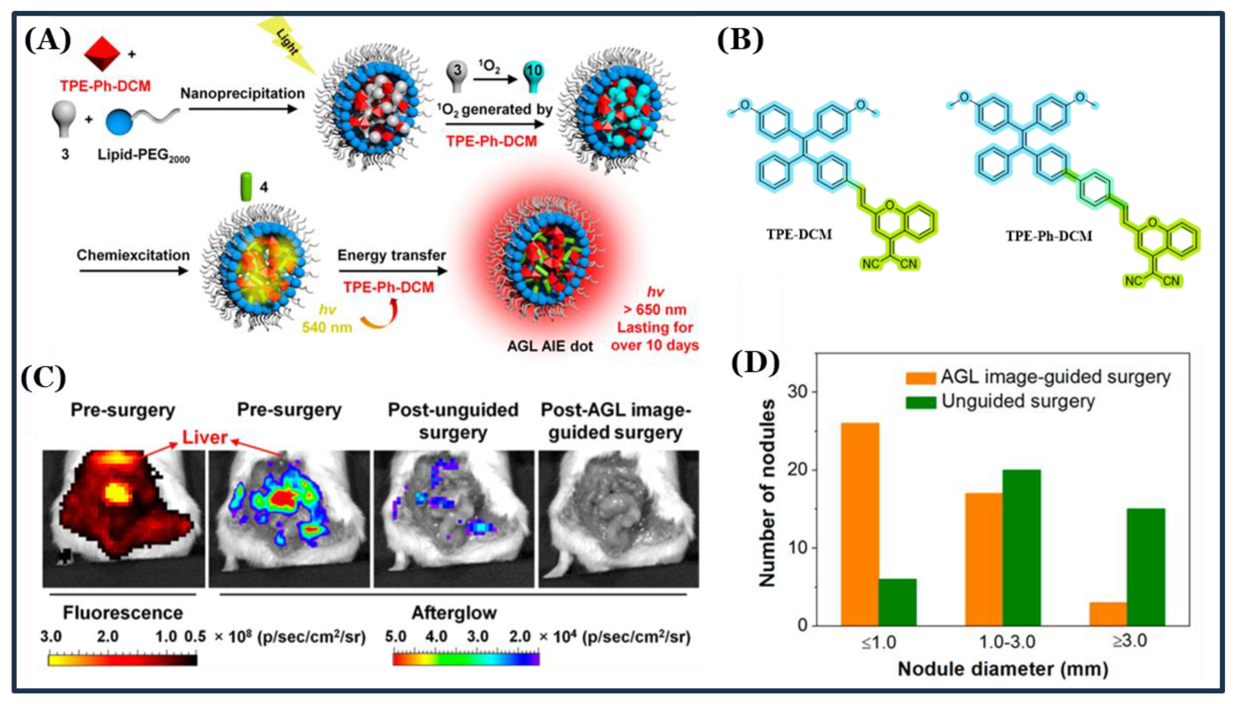
Ni et al. (2018) [98] developed near-infrared (NIR) afterglow luminescent nanoparticles with AIE properties, termed AGL AIE dots, for precise image-guided cancer surgery. These nanoparticles emit intense NIR afterglow signals persisting for over 10 days following a single excitation, enabled by a sequence of processes including singlet oxygen generation, Schaap’s dioxetane formation, chemiexcitation, and energy transfer to NIR-emitting AIEgens. Remarkably, AGL AIE dots exhibited rapid afterglow quenching in healthy organs (liver, spleen, kidney), leading to an ultrahigh tumour-to-liver signal ratio approximately 100-fold higher than conventional fluorescence imaging. In peritoneal carcinomatosis mouse models, this exceptional signal contrast significantly improved surgical outcomes, allowing for precise detection and removal of tumour nodules (Figure 6). These findings highlight the potential of AGL AIE dots for advanced surgical navigation with minimal background interference.
Zhang et al. (2024) [99] reported the design and synthesis of two TPE-based AIE fluorescent probes, TPE-Ma and TPE-Py, incorporating morpholine and pyrrolidone as lysosome-targeting groups. These probes exhibit typical AIE characteristics, a large Stokes shift, low cytotoxicity, and are suitable for two-photon fluorescence imaging. To improve water solubility and cellular uptake, the probes were encapsulated in a biocompatible polymer, DSPE-mPEG2000, forming DSPE@TPE-Ma and DSPE@TPE-Py nanoprobes. These nanoprobes demonstrated superior photostability, high cell permeability, and specific lysosome targeting in MCF-7 cells under two-photon femtosecond laser excitation. Their favourable hydrophilicity, biocompatibility, and strong fluorescence performance highlight their potential as advanced tools for lysosome imaging and cancer cell studies. This work underscores the importance of combining AIE properties with nanocarrier strategies for effective two-photon imaging applications.
Ma et al. (2024) [100] introduced a mash-up strategy of π-extension and deuteration to overcome energy gap law limitations and enhance luminescence efficiency in second near-infrared (NIR-II) AIEgens. By extending the π-conjugation and incorporating isotope effects, the designed AIEgen (NDA-PDTPE) achieved enhanced radiative decay (kr) and suppressed nonradiative decay (knr), leading to higher oscillator strength and superior excited-state stability. The resulting NIR-II emissive nanoparticles exhibited high brightness, large Stokes shift, and excellent photostability, making them suitable for dual-mode image-guided cancer surgery and sentinel lymph node (SLN) mapping.
Further, to address the persistent challenge of oxygen- and water-induced quenching of room-temperature phosphorescence (RTP), Gu et al. (2024) [101] proposed a rational molecular design strategy that integrates branched π-conjugated cores with multiple alkyl side chains. This architecture enhances molecular hydrophobicity, limiting water penetration into aggregates, while simultaneously promoting tight intermolecular packing via van der Waals interactions, thereby shielding excited triplet states from quenching by dissolved oxygen. The branched cores also restrict intramolecular motions, suppressing non-radiative decay and improving radiative efficiency (Figure 7A,B). The structures of four representative compounds namely TPM-4BT, SFlu-4BT, TPE-4BT, and DBC-4BT are shown in Figure 7C, highlighting the combination of extended conjugation with peripheral hydrophobic chains that facilitate compact, highly ordered aggregates with exceptionally bright NIR RTP emission. Comparative analysis of particle size and brightness (Figure 7D) demonstrated that TPE-4BT-based nanoparticles not only maintained dimensions suitable for biological delivery but also exhibited phosphorescence intensities surpassing previously reported RTP systems. In vivo imaging studies (Figure 7E) confirmed their capability for high-contrast, deep-tissue visualization of the liver, gastrointestinal tract, and lymph nodes in both small- and large-animal models, with strong signal-to-background ratios and minimal interference from tissue autofluorescence. Collectively, these findings establish hydrophobic branched architectures as an effective and generalizable design principle for enhancing RTP stability under physiological conditions, offering a promising platform for clinical bioimaging applications such as image-guided surgery, sentinel lymph node mapping, and non-invasive diagnostics.

3.3. TPE-Based AIEgens for Organelle-Targeted Imaging

Subcellular organelles such as mitochondria, lysosomes, lipid droplets, the endoplasmic reticulum (ER), and the nucleus are central to cell function and are increasingly dominant targets in precision medicine [102,103,104]. Targeting these compartments selectively with therapy is possible, enabling the induction of organelle-dependent cell death with reduced systemic side effects and diminished doses of therapy [105]. Concurrently, real-time imaging of organelles enhances our understanding of their dynamics and physiological activities [106,107]. AIE luminogens are particularly suitable for this purpose due to their bright fluorescence emission after aggregation and high photostability. AIE probes excel in this context, they are non-emissive in dilute solutions but glow brightly upon aggregation with high signal quality. Recent efforts have produced mono-organelle–targeted AIE probes (e.g., with triphenylphosphine for mitochondria, morpholine for lysosomes, glibenclamide for ER, etc.). Despite earlier efforts primarily focusing on single-organelle-targeted probes, the latest advances have led to dual-/multi-organelle-targeted AIE probes with accurate localization and ROS generation in target organelles critical in image-guided photodynamic therapy (PDT) and apoptosis tracing. This section documents the widening role of such AIE systems in technologies such as organelle-targeted imaging [108,109].
In a study by Lou et al., 2016 [110], a photostable AIE fluorogen, TPE-CA, was developed for lysosome-targetable imaging of living cells. The authors addressed a major limitation in traditional lysosome-targeted probes, such as LysoTracker Green DND-26 (LTG) and LysoTracker Red DND-99 (LTR), which suffer from the ACQ phenomenon and photobleaching under continuous excitation. Their synthesized TPE-CA, based on the TPE core, exhibited strong blue fluorescence in acidic conditions (pH ≈ 4), demonstrating high selectivity for lysosomal environments. Using NMR, HRMS, and crystallographic analyses, the pH-responsive fluorescence mechanism of TPE-CA was elucidated. The probe was shown to be biocompatible and cell-permeable in HeLa, MCF-7, and HLF cells. Co-localization studies with LTG and LTR confirmed its lysosome-targeting ability. Importantly, TPE-CA retained the AIE properties of TPE, allowing for intense fluorescence even at higher concentrations, and exhibited superior photostability compared to commercial dyes. The findings underscore the potential of TPE-CA in studying lysosomal pH dynamics and offer a robust platform for long-term fluorescence imaging in biological systems.
Wu and co-workers (2019) [111] developed a mitochondria-targeted AIE-active probe by integrating a TPE fluorogen with a triphenylphosphonium (TPP) cationic group. The TPE unit endowed the probe with strong aggregation-induced emission characteristics, while the TPP moiety facilitated efficient accumulation within mitochondria due to its lipophilic cationic nature. This dual functionality enabled selective mitochondrial localization, high photostability, and resistance to photobleaching, making it suitable for long-term imaging in living cells. Importantly, the probe demonstrated low cytotoxicity and strong fluorescence contrast between the mitochondrial region and surrounding cytoplasm, highlighting its potential as a reliable tool for organelle-specific imaging. The combination of AIE luminogens with targeting ligands such as TPP provides a versatile strategy for designing organelle-specific probes, expanding the application of TPE-based systems in bioimaging.
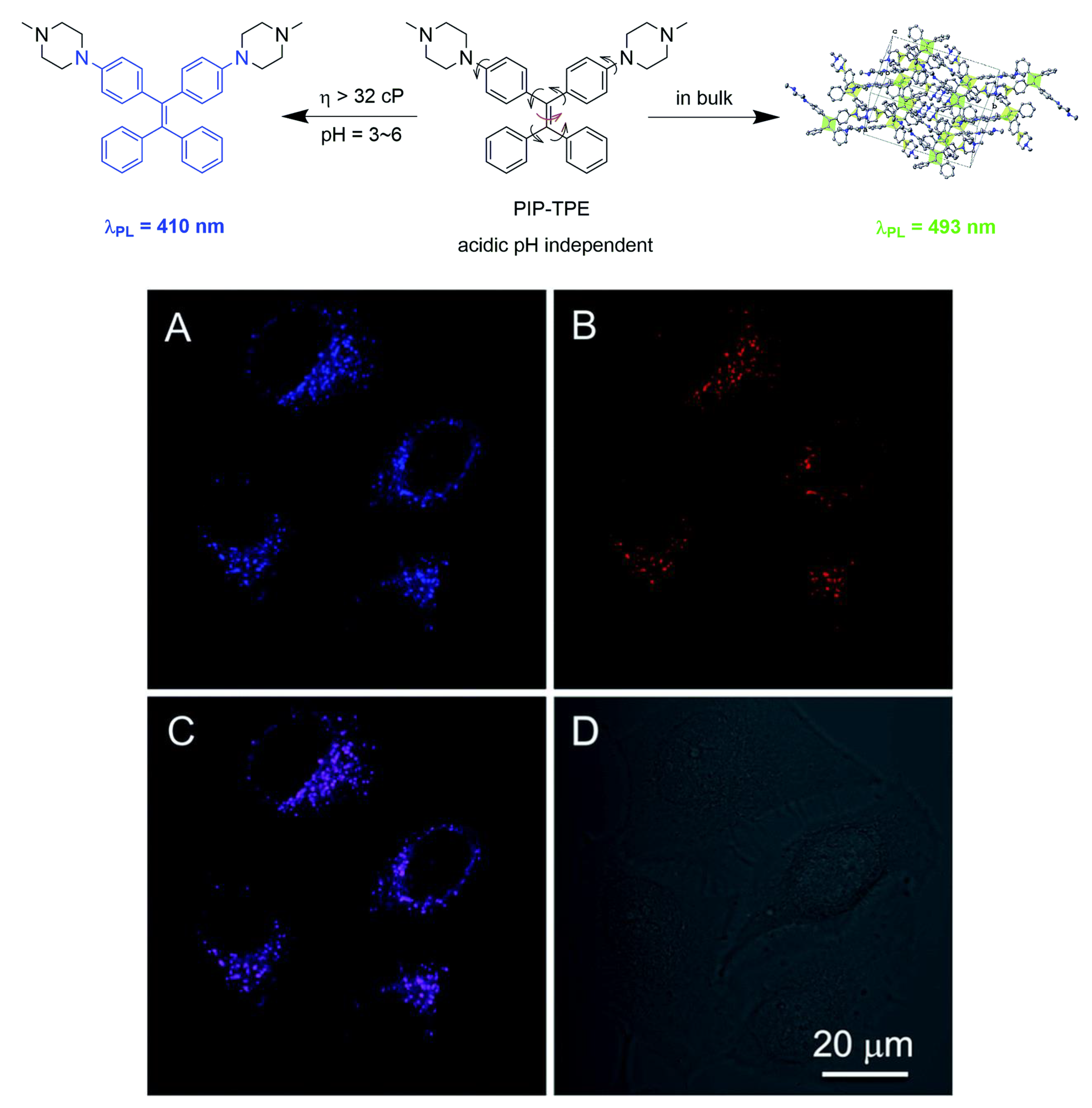
Lysosome-targeting probes were further advanced by Cai et al. (2017) [112]. who developed PIP–TPE, a pH-independent, AIE-active fluorescent probe specifically designed for lysosomal imaging (Figure 8). Unlike conventional lysosomal dyes that depend on acidic pH to suppress photoinduced electron transfer (PET), PIP–TPE activates fluorescence through restriction of intramolecular motion (RIM), triggered by the high viscosity of the lysosomal environment. This mechanism allows for reliable imaging under varying pH conditions. PIP–TPE exhibits weak blue emission (λ = 410 nm) in dilute solution, which intensifies significantly in the viscous lysosomal microenvironment. In the bulk aggregated state, its emission red-shifts to 493 nm, reflecting its sensitivity to local environmental changes. The piperazine-functionalized TPE structure provides an optimal balance of polarity and lipophilicity for selective lysosomal accumulation. Compared to commercial dyes such as LysoTracker Red, PIP–TPE offers enhanced photostability, a higher signal-to-noise ratio, and low cytotoxicity. Confocal imaging of HeLa cells co-stained with PIP–TPE and LysoTracker Red confirmed precise lysosomal targeting through strong colocalization of fluorescence signals (Figure 8A–C), while the bright-field image (Figure 8D) verified cell morphology. These features establish PIP–TPE as a robust tool for long-term, pH-independent lysosomal tracking.
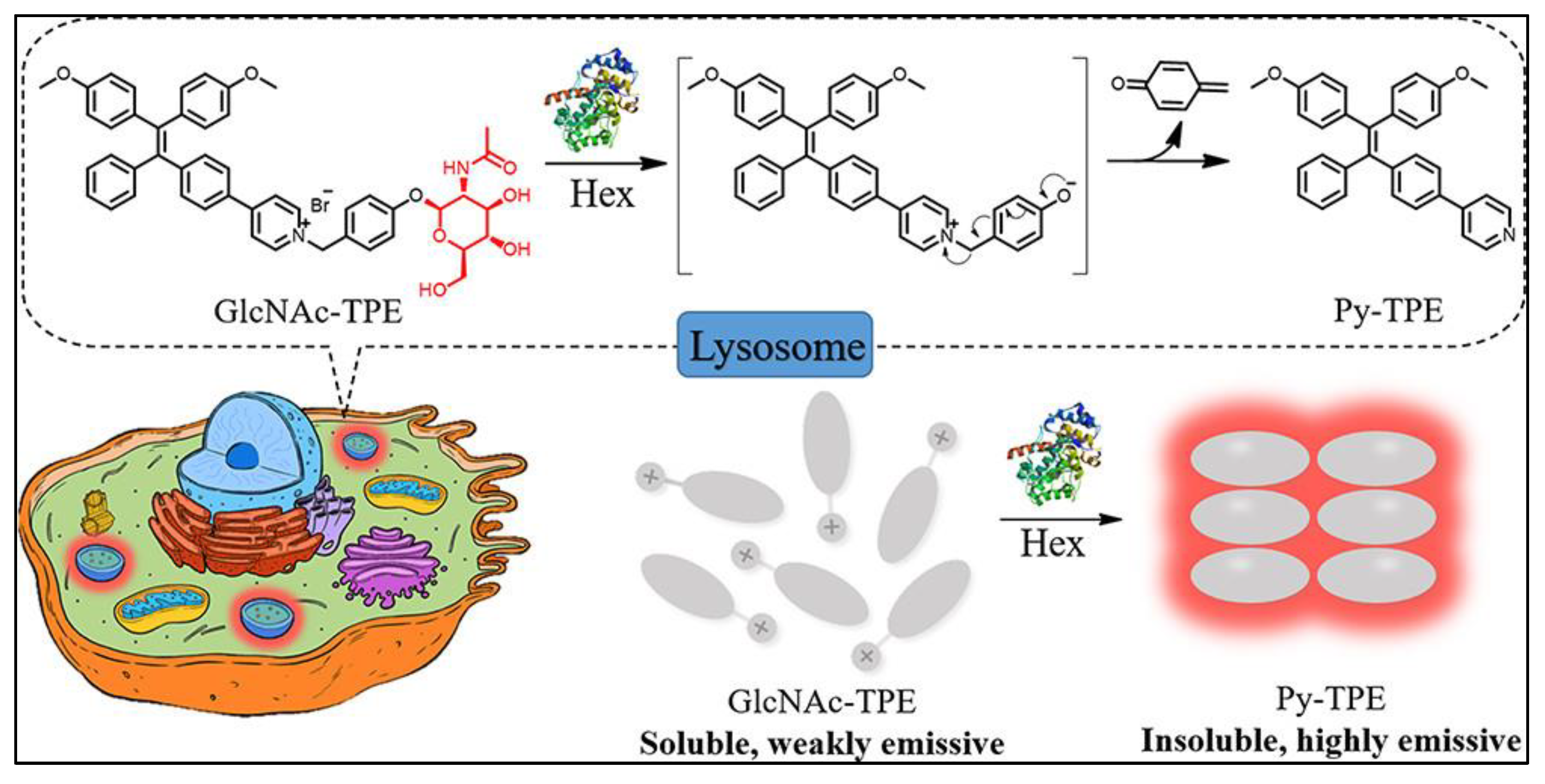
Enzyme-responsive imaging has seen remarkable progress with the contribution of Wang et al. (2019) [113] who developed GlcNAc-TPE, the first lysosome-targeted AIE-active fluorescent probe tailored for real-time imaging of β-N-acetylhexosaminidase (Hex) activity in live cells and animal models (Figure 9). Hex, a crucial enzyme implicated in neurodegenerative lipid storage disorders and various malignancies such as breast and colorectal cancers, serves as an important biomarker for early diagnosis and therapeutic monitoring. GlcNAc-TPE is non-emissive in aqueous media due to its hydrophilic N-acetyl-β-d-glucosaminide moiety but undergoes bright red fluorescence activation via the AIE process upon enzymatic cleavage, which generates a hydrophobic product that promotes molecular aggregation. The probe features a large Stokes shift (~252 nm), outstanding photostability, high sensitivity (limit of detection = 1.4 nM), and excellent selectivity toward Hex. Co-localization studies with LysoTracker™ Green DND-26 confirmed precise lysosomal targeting in HCT116 cancer cells, while in vivo imaging in live mice demonstrated its capability to monitor endogenous Hex activity with high spatial resolution. Collectively, these results highlight GlcNAc-TPE as a powerful tool for enzyme-responsive imaging and a promising platform for biomedical research and cancer diagnostics.
Li et al. (2024) [114] reported the development of QTrPEP, the first AIE-based mitochondria-targeting fluorescent probe designed for monitoring the fluctuation of endogenous hypochlorous acid (HOCl) during ferroptosis. The probe was rationally constructed by integrating a quinoline-conjugated triphenylethylene (QTrPE) AIE luminogen with a phenylborate ester moiety that undergoes selective cleavage upon reaction with HOCl, leading to the release of strongly emissive QTrPE. This HOCl-triggered activation endowed QTrPEP with high sensitivity, excellent selectivity, and favourable mitochondria-targeting ability. Furthermore, the probe could be extended into practical applications through QTrPEP-doped test strips combined with smartphone-based digital analysis, demonstrating its versatility in real-time HOCl detection. Importantly, QTrPEP successfully distinguished ferroptosis from other cell death pathways (apoptosis, pyroptosis, and autophagy) by selectively tracking HOCl fluctuations in ferroptosis models, with glutathione (GSH) serving as an effective inhibitor of this process. With negligible cytotoxicity and strong biocompatibility, this study establishes QTrPEP as a promising tool for probing ferroptosis-related mechanisms and provides new insights into the pathophysiological roles of HOCl in disease progression.

3.4. TPE-Based AIEgens for Multifunctional and Theranostic Applications

Theranostics, uniting diagnostic imaging and therapeutic intervention in a singular, spatially colocalized platform, has gained vast popularity as a research tool and clinical application [115,116,117]. This dual modality allows for simultaneous target detection, drug distribution monitoring, and assessment of therapeutic response, thereby enhancing the efficacy of treatment, maximizing drug safety, reducing pharmacokinetics complexity, and facilitating accelerated drug development. Different theranostic systems have been explored, including various imaging modalities (e.g., fluorescence imaging [FLI], photoacoustic imaging [PAI], MRI, CT, PET) and corresponding therapies (e.g., photodynamic therapy [PDT], photothermal therapy [PTT], radiotherapy, gene therapy, and chemotherapy) [118,119,120]. Among imaging tools, FLI has seen extensive development due to its high sensitivity, simplicity, cost-effectiveness, and biocompatibility. But most conventional fluorophores suffer from ACQ, which limits their brightness and utility in biological systems. AIE molecules offer a new hope. AIEgens are intensely fluorescent in the aggregated form and have essential advantages like the maintenance of intense fluorescence at high concentrations, large Stokes shifts, high photostability, and “light-up” activation upon aggregation that trump the deficiencies of traditional theranostic agents Their inherent biocompatibility, ease of synthesis and modification, and suitable nanoscale size for improved permeability and retention (EPR) make them especially attractive for constructing multifunctional theranostic agents [121]. Despite such advancements, however, challenges persist, such as the relatively high concentration of work required for AIEgen-based theranostic platforms, which should constrain clinical translation. Nevertheless, AIEgens are poised to be efficacious, multi-functional theranostic agents in spearheading innovation in personalized medicine in the years to come. This section endeavours to present a concerted overview of AIE-based theranostic technologies, focusing on their unique photophysical advantages, current breakthroughs, and future direction [122,123].
Zhao et al. (2013) [124] synthesized a multifunctional AIE-active luminogen, TPE-Py, by integrating a pyridinium group with the TPE moiety via a vinyl linkage. TPE-Py exhibited weak fluorescence in solution but showed strong AIE in nanoparticle suspensions and solid states. Interestingly, its crystalline aggregates emitted brighter and bluer light compared to amorphous ones, and the emission colour could be reversibly switched between green and yellow through grinding–fuming or grinding–heating processes. With a large Stokes shift and low optical loss coefficient (~0.032 dB μm−1), the crystalline microrods of TPE-Py were demonstrated as efficient optical waveguides. Furthermore, due to its cationic and hydrophobic nature, TPE-Py showed high photostability and selective staining of mitochondria in living cells, highlighting its potential in bioimaging applications. Expanding the utility of AIEgens, Yueyue Zhao et al. (2015) [125] developed TPE–Py–N3 and Cy–Py–N3, two azide-functionalized AIE probes designed for S-phase DNA synthesis and cell proliferation imaging via the EdU assay. These probes exhibited superior brightness, enhanced photostability, and high tolerance to concentration changes compared to commercial Alexa-azide dyes, establishing them as cost-effective and robust alternatives for long-term and high-throughput bioimaging studies
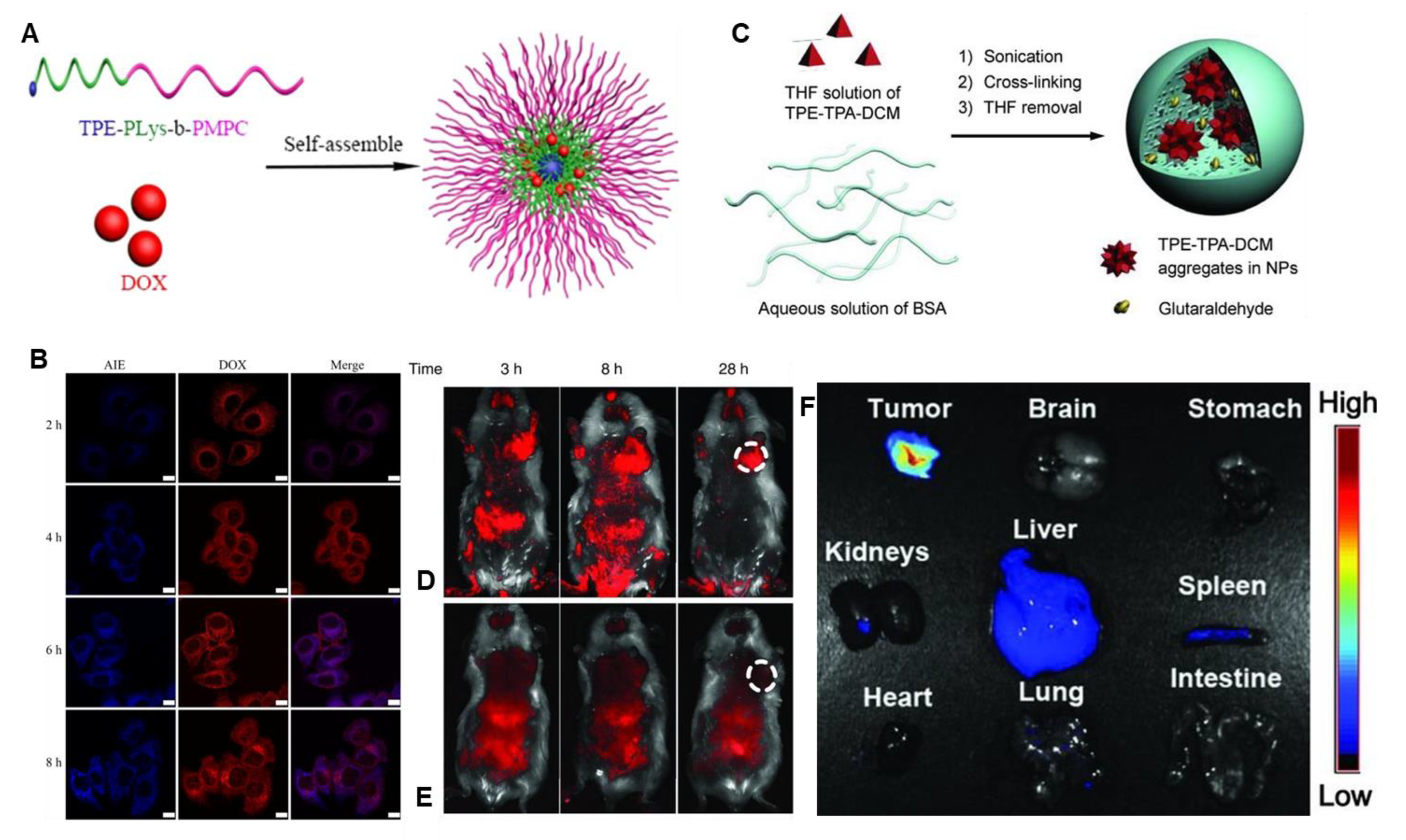
In 2017, Zhuang and colleagues [126] developed a smart theranostic nanocarrier based on an amphiphilic copolymer, TPE-PLys-b-PMPC, incorporating a TPE moiety as the AIE-active fluorogen (Figure 10A). Upon self-assembly, these micelles exhibited strong AIE, enabling real-time tracking of doxorubicin (DOX) release. Confocal laser scanning microscopy (CLSM) revealed rapid internalization into HeLa cells within 4 h, followed by gradual DOX release into the nucleus by 6–8 h, while the TPE-labelled micelles remained cytoplasmic (Figure 10B). This spatiotemporal separation enhanced the therapeutic efficacy against HeLa and 4T1 cancer cells while retaining excellent biocompatibility and biodegradability, underscoring their potential in combined cancer therapy and diagnostics.
Qin et al. in 2012 [127]. designed far-red/near-infrared fluorescent protein nanoparticles (FPNs) by encapsulating a TPE-based donor–acceptor dye (TPE-TPA-DCM) into bovine serum albumin (BSA) nanoparticles (Figure 10C). This formulation enhanced the water dispersibility and bioavailability of the hydrophobic dye. In vivo fluorescence imaging in H22 tumour-bearing mice revealed that the BSA-encapsulated nanoparticles achieved significantly stronger tumour localization than unmodified nanoparticles (Figure 10D,E), with signal retention up to 28 h. Ex vivo imaging at 24 h post-injection further confirmed predominant accumulation in tumour tissue over major organs (Figure 10F), validating the system’s promise for non-invasive tumour detection.
To advance phototheranostic strategies for treating deep-seated tumours, Xu and colleagues [128] in 2019 developed a multifunctional nanoplatform by integrating up conversion nanoparticles (UCNPs) with AIE-active photosensitizers (PSs), enabling NIR-light activation in vivo. In this system, the AIEgen TTD was co-encapsulated with UCNPs using DSPE-PEG and functionalized with a cRGD targeting moiety to yield UCNP@TTD-cRGD nanoparticles (Figure 11). The emission spectrum of the UCNPs was well-matched with the absorption of TTD, facilitating efficient energy transfer and reactive oxygen species (ROS) generation under 980 nm laser irradiation—even beneath 6 mm of tissue. These nanoparticles selectively accumulated in MDA-MB-231 tumour cells, inducing significant cytotoxicity in both 2D and 3D in vitro models upon light exposure, while exhibiting negligible dark toxicity. In vivo studies further demonstrated excellent tumour targeting and suppression following either intertumoral or intravenous administration and NIR illumination. The platform also displayed high photostability, retaining over 70% of its fluorescence after 30 days, making it a promising candidate for non-invasive, image-guided photodynamic therapy of deep-seated malignancies.
Tumour-associated macrophages (TAMs) have emerged as critical targets in cancer therapy due to their prominent role in tumour growth and immune suppression. Gao et al. (2019) [129] designed a mannose-functionalized tetraphenylethylene probe (TPE-Man) to achieve selective TAM recognition via mannose receptor (CD206) interactions. This red-emissive AIE probe combines high-contrast fluorescence imaging with photodynamic therapy (PDT), as it can generate reactive oxygen species (ROS) under light irradiation to ablate TAMs. A control analogue, TPE-Gal, was also synthesized to verify the sugar–receptor specificity of TPE-Man. As illustrated in Figure 1, the probe demonstrated precise targeting, efficient PDT ablation, and promising potential as a cost-effective and multifunctional theranostic platform for tumour microenvironment modulation (Figure 12).
In 2020, Yang and co-workers [130] designed two cationic TPE-based luminogens, TPE-QN and TPE-DQN, for mitochondria-targeted photodynamic therapy (PDT). By integrating a quinolinium moiety and extending the π-conjugation framework, TPE-DQN achieved near-infrared (NIR) emission at 650 nm with a large Stokes shift (~200 nm) and an impressive singlet oxygen quantum yield (Φ = 0.83). The cationic charge facilitated selective mitochondrial localization in cancer cells, enabling precise imaging and efficient light-triggered therapy. Both luminogens displayed high photostability and strong resistance to photobleaching, while their AIE characteristics allowed for high-contrast, wash-free imaging. Under white-light irradiation (25 mW cm−2), TPE-DQN exhibited excellent photodynamic activity, leading to significant cancer cell ablation and tumour growth inhibition in vitro and in vivo. These results underline the potential of cationic AIE luminogens as multifunctional platforms for image-guided theranostic applications.
Zheng and co-workers (2020) [131] developed a mitochondria-targeted AIE bio probe, termed TPN, for the non-invasive identification of viable tumour cells based on mitochondrial differences between cancer cells and leukocytes. Structurally, TPN consists of a TPE core and a pyridinium group, which together facilitate mitochondrial targeting and AIE behaviour (Figure 13a). Upon incubation with various cancer cell lines, TPN exhibited significantly stronger fluorescence in tumour cells compared to leukocytes, due to the elevated mitochondrial membrane potential and increased mitochondrial content characteristic of metabolically active cancer cells (Figure 13b). Flow cytometry data confirmed distinct separation between tumour and leukocyte populations based on TPN signal intensity (Figure 13c–e). Furthermore, spiking experiments demonstrated that TPN could effectively label rare tumour cells within leukocyte populations, as verified by co-staining with the leukocyte marker CD45 and a cell tracker dye (Figure 13f). This strategy provides an antibody-free, low-cost, and minimally invasive method for identifying circulating tumour cells (CTCs), enabling high-quality single-cell analysis for downstream genomic or transcriptomic applications. The mitochondrial-specific labelling capability of AIEgens like TPN highlights their potential for use in liquid biopsy-based cancer diagnostics.
Ding et al. in (2022) [132]. developed a series of AIE-active rhodamine-based probes, namely TPE-T-RS, TPE-T-RO, and TPE-T-RCN (Figure 14A). These luminogens were synthesized through a combination of McMurry coupling, Suzuki–Miyaura reaction, and Knoevenagel condensation. The design incorporated a twisted methoxy-substituted TPE core as an electron-donating unit, while rhodamine derivatives bearing O, S, or dicyanomethylene (–CN) substituents acted as strong electron acceptors. A thiophene (T) bridge was introduced between the donor and acceptor units to promote intramolecular charge transfer (ICT) and extend π-conjugation. Among the three probes, TPE-T-RCN exhibited the highest molar extinction coefficient (2.95 × 10⁴ L·mol−1·cm−1), superior charge transfer efficiency, and the largest photoluminescence quantum yield (18.9%), along with a strong red emission peak at 652 nm. When encapsulated with DSPE-PEG2000, the emission peak further shifted to 662 nm. To enhance target specificity, anti-CD47 antibodies were conjugated onto the nanoparticle surface, allowing the selective recognition of CD47 overexpression in AS plaques. These anti-CD47 nanoparticles (NPs) successfully detected both advanced and early-stage AS plaques in apolipoprotein E-deficient (apoE/) mice (Figure 14B,C) and freshly excised human carotid plaque specimens (Figure 14D,E). This fluorescence-based approach outperformed conventional CT and MRI, which often fail to visualize early plaques. Such AIE nanoprobes also demonstrated utility in monitoring the therapeutic response to anti-AS drugs, making them promising tools for early AS detection and drug.
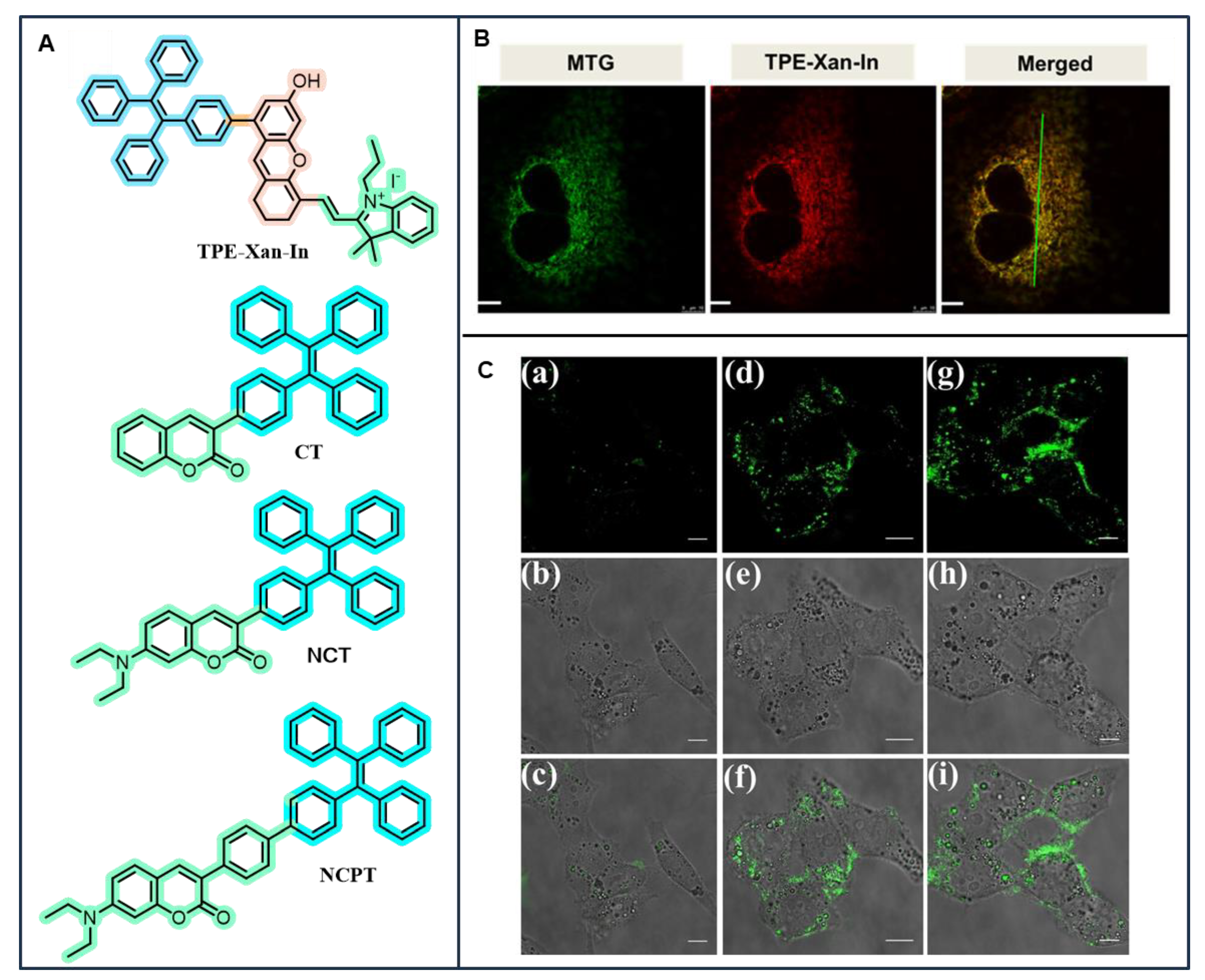
Zhao et al. [133] developed a hybrid probe, TPE–Xan–In, combining a TPE core with a xanthene unit. This molecule showed strong AIE at neutral pH, but fluorescence was quenched in acidic conditions due to phenolate ion formation (Figure 15A,B). TPE–Xan–In successfully monitored mitochondrial pH changes across a wide pH range (4.0–7.0), demonstrating low toxicity and high photostability, making it a promising candidate for real-time, dynamic bioimaging applications. Huang et al. (2023) [134] described the design of coumarin–tetraphenylethene (TPE) luminogens to achieve dual-state emission (DSE) with reversible mechanofluorochromism and live-cell imaging ability. By adjusting the molecular electronic structures and shapes, they synthesized three coumarin–TPE derivatives: a directly linked coumarin–TPE (CT) that shows typical AIE and two N,N-diethylamino-modified derivatives (NCT and NCPT) with strong DSE behaviour (Figure 15A). The DSE properties of NCT and NCPT come from effective intramolecular charge transfer in solution and highly rigid twisted shapes in the solid state, stabilized by multiple weak intermolecular forces that form 3D networks. These luminogens showed reversible mechanofluorochromism, allowing for possible uses in rewritable solid-state information storage. Confocal laser scanning microscopy showed successful uptake in cells, with NCT and NCPT shining bright green in the cytoplasm of HeLa cells, while CT displayed comparatively weak emission inside the cells (Figure 15C). This work showcases a clear strategy for molecular engineering to adjust electronic structures and shapes to achieve DSE and multifunctional AIE-based materials that can be used in both solid-state optical devices and bioimaging.
Liang et al. (2024) [135] designed a pH-responsive amphiphilic polymer (Bio-HA(TPE-CN)-mPEG), constructed by conjugating a hydrophobic AIE fluorophore (TPE-CN), methoxy poly(ethylene glycol) (mPEG), acid-labile imine bonds, and the tumour-targeting ligands biotin and hyaluronic acid (HA) to a single polymer backbone. These self-assembled micelles not only exhibited excellent AIE behaviour shifting from red fluorescence in physiological pH to blue in acidic tumour environments, but also enabled rapid drug release under acidic conditions due to imine bond hydrolysis. Extending this strategy, Liu et al. (2025) [136] synthesized BT-PGA-TPE-HNPE micelles by modifying γ-polyglutamic acid with biotin, an acid-sensitive imine bond, and a TPE-based fluorophore (O-TPE-HNPE). These micelles exhibited strong AIE properties and a unique fluorescence colour shift from yellow at neutral pH to blue in acidic tumour environments, enabling pH-triggered imaging. High PTX-loading capacity, pH-sensitive release, and efficient tumour suppression in vitro and in vivo highlighted their potential as dual-functional nanoplatforms for chemotherapy and bioimaging.
Shan et al. (2024) developed a red-emissive tetraphenylethylene-fused rhodamine dye (TPE-RhMe) with large Stokes shifts (58–199 nm) and long-wavelength emission (601–718 nm) [137]. The incorporation of a multi-rotor TPE unit endows TPE-RhMe with viscosity-responsive fluorescence, exhibiting significant emission enhancement in glycerol/water mixtures and selective response toward glycerol across a broad pH range (1.0–8.0). TPE-RhMe also enables viscosity sensing through fluorescence lifetime measurements, providing a sensitive platform for microenvironmental monitoring. Importantly, TPE-RhMe demonstrates excellent mitochondria-targeting capability, with a Pearson’s correlation coefficient (PCC) of 0.96, and shows enhanced fluorescence in tumour cells (4T1 and HeLa) compared to normal LO2 cells. This dual functionality—viscosity sensing and mitochondria-specific imaging—highlights the potential of TPE-RhMe as a versatile probe for bioimaging and cancer diagnostics.
The versatility of TPE-based AIE probes extends beyond mitochondria and lysosomes to target a range of other organelles and specific biological markers, including cell membranes. By integrating appropriate targeting moieties, these systems enable high-specificity imaging of various cellular compartments and disease-related biomarkers, broadening their utility in biological research and diagnostics. The limited water solubility of hydrophobic TPE derivatives was addressed by Liu et al. (2016) [138], synthesised and developed TPE-CMS luminescent polymeric nanoprobes (TPE-CMS LPNs) via a simple, one-pot, catalyst-free approach using TPE-CHO, carboxymethyl starch sodium (CMS), and 3-aminophenylboronic acid. These nanoprobes exhibited strong blue-green fluorescence, high water dispersibility, pH- and glucose-responsiveness, and excellent cytocompatibility in HepG2 cells. Their rapid and efficient synthesis, combined with environmental responsiveness, established them as promising candidates for smart imaging and drug delivery systems.
Zhang et al., 2024 [139] developed a TPE-based nanoprobe, TPE-4NMB, for the dual purpose of detecting and scavenging peroxynitrite (ONOO), a key oxidative mediator in inflammation and tumour metastasis (Figure 16A). The probe transforms AIE-active TPE-4NM upon reacting with ONOO, leading to a fluorescence “turn-on” response with intensity linearly correlated to ONOO concentrations in the 0.1–100 μM range and a low detection limit of 0.55 μM. Following surface modification with DSPE-PEG2000, TPE-4NMB nanoparticles (NPs) achieved selective imaging of ONOO in living 4T1 cells (Figure 16B), inflammation sites, and tumour wound regions post-resection (Figure 16C,D). Moreover, the ONOO depletion capability of TPE-4NMB NPs suppressed matrix metalloproteinase (MMP) activation and reduced the risk of postoperative tumour metastasis (Figure 16E). This strategy highlights the theranostic potential of AIE nanoprobes for both real-time imaging and therapeutic intervention.

3.5. TPE-Based AIEgens for Stimuli-Responsive and Self-Assembled TPE Systems

AIEgens effectively overcome the intrinsic ACQ limitations observed in traditional luminophores, allowing for efficient luminescence even in aggregated states or thin films [140,141,142]. Over the past two decades, the discovery of AIE has sparked rapid advancements across various application domains, including stimuli-responsive chemical sensing, biomedical imaging, optoelectronics, and other smart material systems [87]. Among these developments, multi-stimuli responsive materials have garnered increasing interest, particularly in biological imaging, as researchers continue to delve deeper into complex biological phenomena [143,144]. Within this context, TPE-based fluorophores stand out due to their strong emission in the aggregated or solid state, a result of restricted intramolecular motions. This section focuses on recent progress in the development of stimuli-responsive and self-assembled AIEgens incorporating the TPE moiety, highlighting their versatile functional potential in responsive and dynamic environments [121,145].
Xu et al., 2019 [146], introduced a novel strategy to design water-soluble aggregation-induced emission luminogens (AIEgens) for hypoxia detection, a key biomarker for tumour diagnosis. The team synthesized three AIE-active N-oxide derivatives (TPE-2M N-oxide, TPE-2E N-oxide, and TPE-2M2F N-oxide) by integrating a TPE core with zwitterionic N-oxide functionalities (Figure 17A). These molecules were non-emissive in aqueous solutions due to active intramolecular motions but exhibited strong fluorescence upon aggregation. Under hypoxic conditions, cellular reductases selectively reduce the N-oxide moieties, yielding hydrophobic TPE residues. These residues aggregate, restricting intramolecular motion and triggering a “turn-on” fluorescence response. Notably, TPE-2E N-oxide demonstrated excellent biocompatibility and an oxygen-dependent fluorescence enhancement in HeLa cells, with selective localization in lipid droplets. This hydrophilic-to-hydrophobic transformation under hypoxic environments enables real-time, high-contrast imaging of hypoxic regions in cells. The study thus presents a simple yet effective design for water-soluble AIE-based hypoxia probes with promising applications in tumour diagnostics and theranostics.
Later, the same group (Xu et al., 2021) [147] reported a dark pro-AIEgen for hypoxia detection, based on an azo-conjugated TPE (TPE-Azo) framework (Figure 17B). Here, the active intramolecular motion of the azo group effectively quenched fluorescence in both solution and solid states. Under hypoxic conditions, intracellular azoreductase triggered reductase-modulated derotation of TPE-Azo to its fluorescent amine derivative (TPE-Am), enabling selective visualization of hypoxic environments in living cells and those generated during photodynamic therapy (PDT). The probe, obtainable via a one-step azo coupling, demonstrated high sensitivity and specificity toward reductase activity. Given the overexpression of reductases in many reactive oxygen species (ROS)-related diseases, this work presents a versatile strategy for designing reductase-responsive pro-AIEgens with significant potential in early disease diagnosis and targeted bioimaging.
Stimuli-responsive AIE probes have emerged as powerful tools for advanced bioimaging. In 2018, Yang et al. [148] introduced two TPE centred luminogens TCPy and its ionic derivative TCPyP that displayed both AIE and mechanochromic (MC) properties. TCPy emitted strong green fluorescence (512 nm, Φs,o = 0.90), which slightly red-shifted under mechanical force. TCPyP exhibited a more significant shift in emission from bright green (527 nm) to deep red (617 nm) upon grinding, reversible by solvent fuming. This reversible switching resulted from a transformation in molecular packing from H- to J-aggregates. Both molecules effectively targeted mitochondria in A549 cells, showing high biocompatibility. TCPyP also enabled long-term tumour imaging in vivo due to a secondary aggregation-induced emission enhancement (AIEE) effect.
Zhang et al. (2021) [149] reported the molecular engineering of high-performance AIE photosensitizers integrated with tailored nanocarrier systems for advanced cancer phototheranostics. In their design, four homologous AIEgens with tunable donor–acceptor strengths were synthesized, among which TPE-TTMN-TPA displayed the most favourable properties, including bright near-infrared fluorescence, long-wavelength emission, and superior ROS generation efficiency, surpassing that of Rose Bengal. To enhance targeting, the nanoplatform was functionalized with a TAT peptide, enabling efficient delivery to tumour sites, where it became activated by lysosomal acidity and subsequently achieved nucleus-targeted imaging and therapy. Both in vitro and in vivo studies confirmed the potential of these AIE nanotheranostic systems, which exhibited excellent photostability, high tumour inhibition (up to 78%), and strong biocompatibility. This work exemplifies how the rational molecular engineering of AIE photosensitizers combined with smart nanovehicles can overcome limitations of conventional photosensitizers, thereby advancing fluorescence imaging-guided photodynamic therapy for precise cancer treatment.
Park and co-workers (2025) [150] reported a supramolecular micelle system co-assembled from amphiphilic TPE derivatives and tamoxifen (TMX) to address resistance in estrogen receptor-positive breast cancer. By rationally tuning oligoethylene glycol (OEG) side-chain lengths, micelles with distinct surface charges were obtained, enabling enhanced cellular uptake and preferential localization within lipid droplets (LDs) organelles implicated in drug sequestration and resistance (Figure 18). The intrinsic fluorescence of TPE allowed real-time tracking of intracellular drug distribution, providing direct mechanistic insight into LD-mediated TMX resistance. Importantly, co-treatment with orlistat, an LD biogenesis inhibitor, disrupted this resistance pathway and synergistically amplified TMX efficacy. This dual-functional strategy combining supramolecular self-assembly for drug delivery with organelle-level modulation of LDs demonstrates a powerful approach to overcoming endocrine therapy resistance. Moreover, the study highlights how fine-tuning supramolecular design parameters, such as OEG chain length, governs biological behaviour, including cellular uptake, organelle targeting, and therapeutic outcomes, thereby advancing the development of next-generation, organelle-specific nanomedicines.

4. Future Direction and Perspective

Building on recent progress in cellular, tissue, organelle-targeted, and multifunctional applications, the next step for TPE-based bioimaging is to create multifunctional [151] platforms. These systems aim to combine imaging, sensing, and therapeutic features. They will provide real-time diagnosis and targeted treatments while improving precision and reducing side effects. To move beyond traditional fluorescence methods, the focus is now on designing near-infrared II (NIR-II, 1000–1700 nm) emitters. These emitters allow for deeper tissue penetration and higher-resolution imaging by reducing light scattering and autofluorescence. Shen et al. (2023) [152] noted that donor–acceptor engineering of TPE luminogens can shift emission into the NIR-II region. This opens up opportunities for non-invasive vascular and deep-tissue imaging [149,153].
Despite this advancement, achieving high quantum yields in water and physiological environments is still a challenge for clinical use. The natural hydrophobicity of most TPE derivatives limits their fluorescence efficiency in biological systems. To tackle this issue, researchers are using surface engineering methods like PEGylation, polymer encapsulation, and bonding with hydrophilic biomolecules. Additionally, biomimetic carriers such as exosomes, liposomes, and protein-based nanoplatforms are becoming effective delivery vehicles. They offer better dispersibility, longer circulation, and targeted delivery in living organisms. Meanwhile, efforts are also focused on merging TPE-based probes with advanced imaging techniques. Their high stability, large Stokes shifts, and multiphoton absorption characteristics make them excellent candidates for super-resolution fluorescence microscopy, two- and three-photon excitation, and photoacoustic imaging [154,155]. Importantly, TPE-based NIR probes for photoacoustic imaging provide both optical and acoustic readings. This allows for real-time, high-resolution visualization of deep tissues [156].
Looking to the future, developing stimuli-responsive TPE probes will help monitor dynamic biological events like changes in pH, redox states, enzyme activities, and biomechanical forces [157,158,159,160]. When used with photodynamic or photothermal therapy, these responsive systems could lead to precise, image-guided treatments for cancer, cardiovascular diseases, and neurodegenerative disorders. Lastly, for clinical use to succeed, future research should focus on creating biodegradable, metabolizable, and regulatory-compliant AIE luminogens. Modular synthetic approaches will be important for adjusting photophysical properties, targeting abilities, and therapeutic prospects. These advances will help move TPE-based AIEgens from the lab to practical applications in precision medicine and integrated biomedical imaging. Beyond biomedical applications, the unique photophysical characteristics of TPE-based AIE luminogens also present opportunities in energy conversion and catalytic systems, where their light-harvesting capabilities and emission properties can be harnessed to enhance processes such as solar-to-hydrogen generation and fuel cell performance [161,162,163,164].

5. Conclusions

TPE core systems continue to be a key example of an AIE-active and mechanoresponsive luminogens. While many TPE-based systems show mechanochromic behaviour, only a few reveal significant colour changes when exposed to mechanical stimuli. It is important to study how molecular packing and solid-state interactions affect the mechanisms behind these mechanoresponsive emissions. This review covers the basic aspects of AIE in TPE systems, highlighting the roles of restricted intramolecular rotation (RIR) and twisted π-conjugation. It also emphasizes how structural and electronic factors, along with intermolecular interactions, impact their emission behaviour.
Looking ahead, careful molecular design and control of aggregation behaviour will be essential for broadening the use of TPE-based bioimaging technologies. These systems provide a flexible platform for addressing various diagnostic and therapeutic needs in biomedical science. We expect that the ongoing development of TPE-based luminogens will lead to new solutions in clinical imaging, targeted therapy, and integrated diagnostics.

Funding

This research received no external funding.

Acknowledgments

The authors would like to thank their colleagues and peers for valuable discussions and insights that contributed to this review.

Conflicts of Interest

The authors declare no conflicts of interest.

References

  1. Lu, P.; Ai, J. Fluorescence Probes and Their Sensing Applications in Nanomaterials System. Talanta Open 2023, 8, 100248. [Google Scholar] [CrossRef]
  2. Ha, J.M.; Hur, S.H.; Pathak, A.; Jeong, J.E.; Woo, H.Y. Recent Advances in Organic Luminescent Materials with Narrowband Emission. NPG Asia Mater. 2021, 13, 53. [Google Scholar] [CrossRef]
  3. Shi, Y.; Zhang, W.; Xue, Y.; Zhang, J. Fluorescent Sensors for Detecting and Imaging Metal Ions in Biological Systems: Recent Advances and Future Perspectives. Chemosensors 2023, 11, 226. [Google Scholar] [CrossRef]
  4. Keremane, K.S.; Acharya, M.G.; Naik, P.; Malakar, C.C.; Wang, K.; Poudel, B. Recent Advances in Aggregation-Induced Emission (AIE) Fluorescent Sensors for Biomolecule Detection. Chemosensors 2025, 13, 174. [Google Scholar] [CrossRef]
  5. Zhang, G.; Zhang, S.; Chen, F.; Ni, Y.; Wang, C.; Yang, L.; Kong, L.; Yang, J. Novel AIE-Active Tetraphenylethylene Derivatives as Multitask Smart Materials for Turn-on Mechanofluorochromism, Quantitative Sensing of Pressure and Picric Acid Detection. Dye. Pigment. 2022, 203, 110327. [Google Scholar] [CrossRef]
  6. Pilicode, N.; Naik, P.; Acharya, M.; Adhikari, A.V. Synthesis, Characterization and Electroluminescence Studies of Cyanopyridine-Based π-Conjugative Polymers Carrying Benzo[: C] [1,2,5]Thiadiazole and Naphtho [1,2- c:5,6- c ′]Bis([1,2,5]Thiadiazole) Units. New J. Chem. 2020, 44, 10796–10805. [Google Scholar] [CrossRef]
  7. Wang, Z.; Zhou, F.; Wang, J.; Zhao, Z.; Qin, A.; Yu, Z.; Tang, B.Z. Electronic Effect on the Optical Properties and Sensing Ability of AIEgens with ESIPT Process Based on Salicylaldehyde Azine. Sci. China Chem. 2018, 61, 76–87. [Google Scholar] [CrossRef]
  8. Blebea, N.M.; Pușcașu, C.; Vlad, R.A.; Hancu, G. Chitosan-Based Gel Development: Extraction, Gelation Mechanisms, and Biomedical Applications. Gels 2025, 11, 275. [Google Scholar] [CrossRef]
  9. Xu, Y.; Xu, P.; Hu, D.; Ma, Y. Recent Progress in Hot Exciton Materials for Organic Light-Emitting Diodes. Chem. Soc. Rev. 2021, 50, 1030–1069. [Google Scholar] [CrossRef]
  10. Huang, J.; Yang, X.; Li, X.; Chen, P.; Tang, R.; Li, F.; Lu, P.; Ma, Y.; Wang, L.; Qin, J.; et al. Bipolar AIE-Active Luminogens Comprised of an Oxadiazole Core and Terminal TPE Moieties as a New Type of Host for Doped Electroluminescence. Chem. Commun. 2012, 48, 9586–9588. [Google Scholar] [CrossRef]
  11. Naik, P.; Pilicode, N.; Keremane, K.S.; Acharya, M.; Adhikari, A.V. Synthesis, Optical, Electrochemical, and Computational Investigation of New Cyanopyridine-Centered Organic Dyads. Opt. Mater. (Amst.) 2023, 142, 114002. [Google Scholar] [CrossRef]
  12. Harish, K.K.; Nesaragi, A.R.; Kalagatur, N.K.; Naik, P.; Madegowda, M.; Pandith, A.; Dahlous, K.A.; Mohammad, S.; Shivarudrappa, H.P.; Sharanakumar, T.M.; et al. Imidazole-Centred Cupric Ions Sensor: Experimental Validation, Theoretical Understanding, and Zebrafish Bioimaging. J. Photochem. Photobiol. A Chem. 2024, 452, 115565. [Google Scholar] [CrossRef]
  13. Nesaragi, A.R.; Naik, P.; Mulla, B.B.A.; Al-Zaqri, N.; Vidyasagar, C.C.; Kalagatur, N.K.; Sharanakumar, T.M.; Guddappa, H.; Sidarai, A.H.; Shivarudrappa, H.P. Selective Al3+ and Fe3+ Detection Using Imidazole–Oxadiazole Sensors: Bioimaging Evidence from Zebrafish. New J. Chem. 2025, 49, 5729–5739. [Google Scholar] [CrossRef]
  14. Nesaragi, A.R.; Mulla, B.B.A.; Chapi, S.; Venuprasad, K.D.; Kalagatur, N.K.; Sharanakumar, T.M.; Vidyasagar, C.C.; Guddappa, H.; Al-Zaqri, N.; Warad, I.; et al. Imidazole-Centred Oxadiazole Sensor for Detecting Al3+ and Fe3+ Cations in Living Cells: A Zebrafish Bioimaging Approach. Appl. Organomet. Chem. 2025, 39, e70087. [Google Scholar] [CrossRef]
  15. Xu, Y.; Xu, R.; Wang, Z.; Zhou, Y.; Shen, Q.; Ji, W.; Dang, D.; Meng, L.; Tang, B.Z. Recent Advances in Luminescent Materials for Super-Resolution Imaging: Via Stimulated Emission Depletion Nanoscopy. Chem. Soc. Rev. 2021, 50, 667–690. [Google Scholar] [CrossRef] [PubMed]
  16. Hu, R.; Yang, X.; Qin, A.; Tang, B.Z. AIE Polymers in Sensing, Imaging and Theranostic Applications. Mater. Chem. Front. 2021, 5, 4073–4088. [Google Scholar] [CrossRef]
  17. Naik, P.; Elias, L.; Keremane, K.S.; Babu, D.D.; Abdellah, I.M. Metal-Free Organic Dyes for NiO-Based Dye-Sensitized Solar Cells: Recent Developments and Future Perspectives. Energy Technol. 2024, 12, 2301666. [Google Scholar] [CrossRef]
  18. Naik, P.; Babu, D.D.; Su, R.; El-Shafei, A.; Adhikari, A.V. Synthesis, Characterization and Performance Studies of a New Metal-Free Organic Sensitizer for DSSC Application. Mater. Today Proc. 2018, 5, 3150–3157. [Google Scholar] [CrossRef]
  19. Kim, T.; Baek, E.; Kim, H.; Han, J.; Lee, Y.; Oh, H.; Cho, K.H.; Kim, J. Tunable Emission Properties of Indolizine-Based Aggregation-Induced Emission Luminogens for White-Light Emission. JACS Au 2025, 5, 3262–3274. [Google Scholar] [CrossRef]
  20. Fan, W.; Li, Z.; Chen, K.; Wang, Z.; Yang, L. Aggregation-Induced Emission in Natural Product Research: Recent Advances and Future Perspectives. Coord. Chem. Rev. 2025, 542, 216843. [Google Scholar] [CrossRef]
  21. Peng, Q.; Shuai, Z. Molecular Mechanism of Aggregation-Induced Emission. Aggregate 2021, 2, e91. [Google Scholar] [CrossRef]
  22. Luo, J.; Xie, Z.; Xie, Z.; Lam, J.W.Y.; Cheng, L.; Chen, H.; Qiu, C.; Kwok, H.S.; Zhan, X.; Liu, Y.; et al. Aggregation-Induced Emission of 1-Methyl-1,2,3,4,5-Pentaphenylsilole. Chem. Commun. 2001, 18, 1740–1741. [Google Scholar] [CrossRef] [PubMed]
  23. Han, T.; Yan, D.; Wu, Q.; Song, N.; Zhang, H.; Wang, D. Aggregation-Induced Emission: A Rising Star in Chemistry and Materials Science. Chin. J. Chem. 2021, 39, 677–689. [Google Scholar] [CrossRef]
  24. Wang, H.; Zhao, E.; Lam, J.W.Y.; Tang, B.Z. AIE Luminogens: Emission Brightened by Aggregation. Mater. Today 2015, 18, 365–377. [Google Scholar] [CrossRef]
  25. Zhao, W.; He, Z.; Lam, J.W.Y.; Peng, Q.; Ma, H.; Shuai, Z.; Bai, G.; Hao, J.; Tang, B.Z. Rational Molecular Design for Achieving Persistent and Efficient Pure Organic Room-Temperature Phosphorescence. Chem 2016, 1, 592–602. [Google Scholar] [CrossRef]
  26. Chen, X.; Xu, C.; Wang, T.; Zhou, C.; Du, J.; Wang, Z.; Xu, H.; Xie, T.; Bi, G.; Jiang, J.; et al. Versatile Room-Temperature-Phosphorescent Materials Prepared from N-Substituted Naphthalimides: Emission Enhancement and Chemical Conjugation. Angew. Chem.-Int. Ed. 2016, 55, 9872–9876. [Google Scholar] [CrossRef] [PubMed]
  27. Debnath, S.; Mohanty, A.; Naik, P.; Salzner, U.; Dasgupta, J.; Patil, S. Deciphering Intramolecular Charge Transfer in Fluoranthene Derivatives. J. Mater. Chem. C Mater. 2024, 12, 9200–9209. [Google Scholar] [CrossRef]
  28. Sharath Kumar, K.S.; Girish, Y.R.; Ashrafizadeh, M.; Mirzaei, S.; Rakesh, K.P.; Hossein Gholami, M.; Zabolian, A.; Hushmandi, K.; Orive, G.; Kadumudi, F.B.; et al. AIE-Featured Tetraphenylethylene Nanoarchitectures in Biomedical Application: Bioimaging, Drug Delivery and Disease Treatment. Coord. Chem. Rev. 2021, 447, 214135. [Google Scholar] [CrossRef]
  29. Yan, Y.; Yin, G.Q.; Khalife, S.; He, Z.H.; Xu, C.; Li, X. Self-Assembly of Emissive Metallocycles with Tetraphenylethylene, BODIPY and Terpyridine in One System. Supramol. Chem. 2019, 31, 597. [Google Scholar] [CrossRef]
  30. Tanaka, Y.; Machida, T.; Noumi, T.; Sada, K.; Kokado, K. Emissive Tetraphenylethylene (TPE) Derivatives in a Dissolved State Tightly Fastened by a Short Oligo(Ethylene Glycol) Chain. Org. Chem. Front. 2020, 7, 2649–2656. [Google Scholar] [CrossRef]
  31. Bhardwaj, S.; Naik, P.; Palariya, A.K.; Panda, S.; Patil, S.A.; Avashti, S. Surface and Grain Boundary Passivation Using Tetraphenylethylene Derivative for High-Performance Perovskite Solar Cell. Mater. Chem. Front. 2025. [Google Scholar] [CrossRef]
  32. Lafzi, F.; Taskesenligil, Y.; Canlmkurbey, B.; Plravadlll, S.; Kilic, H.; Saracoglu, N. Four-Winged Propeller-Shaped Indole-Modified and Indole-Substituted Tetraphenylethylenes: Greenish-Blue Emitters with Aggregation-Induced Emission Features for Conventional Organic Light-Emitting Diodes. ACS Omega 2022, 7, 44322–44337. [Google Scholar] [CrossRef]
  33. Bhosale, R.S.; Aljabri, M.; La, D.D.; Bhosale, S.V.; Jones, L.A.; Bhosale, S.V. Tetraphenylethene Derivatives: A Promising Class of AIE Luminogens—Synthesis, Properties, and Applications. Princ. Appl. Aggreg.-Induc. Emiss. 2019, 223–264. [Google Scholar] [CrossRef]
  34. Duc La, D.; Anuradha, A.; Gupta, A.; Al Kobaisi, M.; Rananaware, A.; Bhosale, S.V. Supramolecular Chemistry of AIE-Active Tetraphenylethylene Luminophores. SPR Nanosci. 2017, 4, 75–107. [Google Scholar] [CrossRef]
  35. La, D.D.; Bhosale, S.V.; Jones, L.A.; Bhosale, S.V. Tetraphenylethylene-Based AIE-Active Probes for Sensing Applications. ACS Appl. Mater. Interfaces 2018, 10, 12189–12216. [Google Scholar] [CrossRef] [PubMed]
  36. Khan, F.; Mahmoudi, M.; Gupta, P.K.; Volyniuk, D.; Grazulevicius, J.V.; Misra, R. Mechanochromic Materials Based on Tetraphenylethylene-Substituted Phenothiazines: Substituent-Dependent Hypsochromic and Bathochromic Switching of Emission. J. Phys. Chem. C 2023, 127, 1643–1654. [Google Scholar] [CrossRef]
  37. Ma, Y.; Zhang, Y.; Kong, L.; Yang, J. Mechanoresponsive Material of AIE-Active 1,4-Dihydropyrrolo [3,2-b]Pyrrole Luminophores Bearing Tetraphenylethylene Group with Rewritable Data Storage. Molecules 2018, 23, 3255. [Google Scholar] [CrossRef] [PubMed]
  38. Yang, Z.; Chi, Z.; Mao, Z.; Zhang, Y.; Liu, S.; Zhao, J.; Aldred, M.P.; Chi, Z. Recent Advances in Mechano-Responsive Luminescence of Tetraphenylethylene Derivatives with Aggregation-Induced Emission Properties. Mater. Chem. Front. 2018, 2, 861–890. [Google Scholar] [CrossRef]
  39. Kalva, N.; Tran, C.H.; Lee, M.W.; Augustine, R.; Lee, S.J.; Kim, I. Aggregation-Induced Emission-Active Hyperbranched Polymers Conjugated with Tetraphenylethylene for Nitroaromatic Explosive Detection. Dye. Pigment. 2021, 194, 109617. [Google Scholar] [CrossRef]
  40. Kokado, K.; Sada, K. Consideration of Molecular Structure in the Excited State to Design New Luminogens with Aggregation-Induced Emission. Angew. Chem. 2019, 131, 8724–8731. [Google Scholar] [CrossRef]
  41. Chang, C.C.; Hsieh, M.C.; Lin, J.C.; Chang, T.C. Selective Photodynamic Therapy Based on Aggregation-Induced Emission Enhancement of Fluorescent Organic Nanoparticles. Biomaterials 2012, 33, 897–906. [Google Scholar] [CrossRef]
  42. Zhao, Z.; Chen, B.; Geng, J.; Chang, Z.; Aparicio-Ixta, L.; Nie, H.; Goh, C.C.; Ng, L.G.; Qin, A.; Ramos-Ortiz, G.; et al. Red Emissive Biocompatible Nanoparticles from Tetraphenylethene-Decorated BODIPY Luminogens for Two-Photon Excited Fluorescence Cellular Imaging and Mouse Brain Blood Vascular Visualization. Part. Part. Syst. Charact. 2014, 31, 481–491. [Google Scholar] [CrossRef]
  43. Li, Q.; Li, X.; Wu, Z.; Sun, Y.; Fang, J.; Chen, D. Highly Efficient Luminescent Side-Chain Polymers with Short-Spacer Attached Tetraphenylethylene AIEgens via RAFT Polymerization Capable of Naked Eye Explosive Detection. Polym. Chem. 2018, 9, 4150–4160. [Google Scholar] [CrossRef]
  44. Geng, J.; Li, K.; Ding, D.; Zhang, X.; Qin, W.; Liu, J.; Tang, B.Z.; Liu, B. Lipid-PEG-Folate Encapsulated Nanoparticles with Aggregation Induced Emission Characteristics: Cellular Uptake Mechanism and Two-Photon Fluorescence Imaging. Small 2012, 8, 3655–3663. [Google Scholar] [CrossRef]
  45. Gawade, V.K.; Jadhav, R.W.; Bhosale, S.V. AIE-Based & Organic Luminescent Materials: Nanoarchitectonics and Advanced Applications. Chem. Asian J. 2024, 19, e202400682. [Google Scholar] [CrossRef]
  46. Serhan, M.; Jackemeyer, D.; Long, M.; Sprowls, M.; Diez Perez, I.; Maret, W.; Chen, F.; Tao, N.; Forzani, E. Total Iron Measurement in Human Serum with a Novel Smartphone-Based Assay. IEEE J. Transl. Eng. Health Med. 2020, 8, 1–9. [Google Scholar] [CrossRef]
  47. Wang, M.; Zhang, G.; Zhang, D.; Zhu, D.; Tang, B.Z. Fluorescent Bio/Chemosensors Based on Silole and Tetraphenylethene Luminogens with Aggregation-Induced Emission Feature. J. Mater. Chem. 2010, 20, 1858–1867. [Google Scholar] [CrossRef]
  48. Zhang, Y.; Li, J.; Tang, B.Z.; Wong, K.S. Aggregation Enhancement on Two-Photon Optical Properties of AIE-Active D-TPE-A Molecules. J. Phys. Chem. C 2014, 118, 26981–26986. [Google Scholar] [CrossRef]
  49. Yang, J.; Fang, M.; Li, Z. Organic Luminescent Materials: The Concentration on Aggregates from Aggregation-Induced Emission. Aggregate 2020, 1, 6–18. [Google Scholar] [CrossRef]
  50. Liang, J.; Tang, B.Z.; Liu, B. Specific Light-up Bioprobes Based on AIEgen Conjugates. Chem. Soc. Rev. 2015, 44, 2798–2811. [Google Scholar] [CrossRef]
  51. Kwok, R.T.K.; Leung, C.W.T.; Lam, J.W.Y.; Tang, B.Z. Biosensing by Luminogens with Aggregation-Induced Emission Characteristics. Chem. Soc. Rev. 2015, 44, 4228–4238. [Google Scholar] [CrossRef]
  52. Mei, J.; Leung, N.L.C.; Kwok, R.T.K.; Lam, J.W.Y.; Tang, B.Z. Aggregation-Induced Emission: Together We Shine, United We Soar! Chem. Rev. 2015, 115, 11718–11940. [Google Scholar] [CrossRef]
  53. Xu, S.; Duan, Y.; Liu, B. Precise Molecular Design for High-Performance Luminogens with Aggregation-Induced Emission. Adv. Mater. 2020, 32, 1903530. [Google Scholar] [CrossRef]
  54. Madushani, B.; Mamada, M.; Goushi, K.; Nguyen, T.B.; Nakanotani, H.; Kaji, H.; Adachi, C. Multiple Donor–Acceptor Design for Highly Luminescent and Stable Thermally Activated Delayed Fluorescence Emitters. Sci. Rep. 2023, 13, 7644. [Google Scholar] [CrossRef]
  55. Barman, D.; Narang, K.; Parui, R.; Zehra, N.; Khatun, M.N.; Adil, L.R.; Iyer, P.K. Review on Recent Trends and Prospects in π-Conjugated Luminescent Aggregates for Biomedical Applications. Aggregate 2022, 3, e172. [Google Scholar] [CrossRef]
  56. El-Zohry, A.M.; Orabi, E.A.; Karlsson, M.; Zietz, B. Twisted Intramolecular Charge Transfer (TICT) Controlled by Dimerization: An Overlooked Piece of the TICT Puzzle. J. Phys. Chem. A 2021, 125, 2885–2894. [Google Scholar] [CrossRef]
  57. Sasaki, S.; Drummen, G.P.C.; Konishi, G.I. Recent Advances in Twisted Intramolecular Charge Transfer (TICT) Fluorescence and Related Phenomena in Materials Chemistry. J. Mater. Chem. C Mater. 2016, 4, 2731–2743. [Google Scholar] [CrossRef]
  58. Vesga-Hernández, C.; dos Santos Carvalho, R.; Barreto, A.; Pedrozo Peñafiel, M.J.; de Noronha, J.R.; Maqueira, L.; Back, D.; Mello Nascimento, D.; Zucolotto Cocca, L.H.; Aucélio, R.Q.; et al. Aggregation-Induced Enhanced Emission and Twisted Intramolecular Charge Transfer in a Pyridylcarbodinitrile with Tunable Photoluminescence in Solution, Films, and OLEDs. J. Photochem. Photobiol. A Chem. 2025, 464, 116312. [Google Scholar] [CrossRef]
  59. Kuila, S.; Miranda-Salinas, H.; Eng, J.; Li, C.; Bryce, M.R.; Penfold, T.J.; Monkman, A.P. Rigid and Planar π-Conjugated Molecules Leading to Long-Lived Intramolecular Charge-Transfer States Exhibiting Thermally Activated Delayed Fluorescence. Nat. Commun. 2024, 15, 9611. [Google Scholar] [CrossRef] [PubMed]
  60. Yamaguchi, Y.; Matsubara, Y.; Ochi, T.; Wakamiya, T.; Yoshida, Z.I. How the π Conjugation Length Affects the Fluorescence Emission Efficiency. J. Am. Chem. Soc. 2008, 130, 13867–13869. [Google Scholar] [CrossRef] [PubMed]
  61. Zhu, Y.; Lai, H.; Gu, Y.; Wei, Z.; Chen, L.; Lai, X.; Han, L.; Tan, P.; Pu, M.; Xiao, F.; et al. The Balance Effect of π–π Electronic Coupling on NIR-II Emission and Photodynamic Properties of Highly Hydrophobic Conjugated Photosensitizers. Adv. Sci. 2024, 11, 2307569. [Google Scholar] [CrossRef]
  62. Hua, Z.; Yu, H.; Gong, H.; Li, P.; Zhang, T.; Deng, Z.; Sun, W.; Tian, Y.; Fu, H. Heavy-Atom Effect Regulating Room-Temperature Phosphorescence of Organic-Inorganic Zn-Based Halides for White-Light Emission. J. Phys. Chem. Lett. 2024, 15, 4729–4736. [Google Scholar] [CrossRef]
  63. Hamzehpoor, E.; Ruchlin, C.; Tao, Y.; Liu, C.H.; Titi, H.M.; Perepichka, D.F. Efficient Room-Temperature Phosphorescence of Covalent Organic Frameworks through Covalent Halogen Doping. Nat. Chem. 2023, 15, 83–90. [Google Scholar] [CrossRef]
  64. Gu, Q.; Pandey, S.K.; Tiwari, R. A Computational Method to Estimate Spin–Orbital Interaction Strength in Solid State Systems. Comput. Mater. Sci. 2023, 221, 112090. [Google Scholar] [CrossRef]
  65. Fan, T.; Liu, Q.; Zhang, H.; Wang, X.; Zhang, D.; Duan, L. Enhancing Spin–Orbit Coupling in an Indolocarbazole Multiresonance Emitter by a Sulfur-Containing Peripheral Substituent for a Fast Reverse Intersystem Crossing. Adv. Mater. 2024, 36, 2408816. [Google Scholar] [CrossRef]
  66. Zhu, C.; Kwok, R.T.K.; Lam, J.W.Y.; Tang, B.Z. Aggregation-Induced Emission: A Trailblazing Journey to the Field of Biomedicine. ACS Appl. Bio Mater. 2018, 1, 1768–1786. [Google Scholar] [CrossRef]
  67. Ma, H.; Liu, C.; Hu, Z.; Yu, P.; Zhu, X.; Ma, R.; Sun, Z.; Zhang, C.H.; Sun, H.; Zhu, S.; et al. Propylenedioxy Thiophene Donor to Achieve NIR-II Molecular Fluorophores with Enhanced Brightness. Chem. Mater. 2020, 32, 2061–2069. [Google Scholar] [CrossRef]
  68. Zhao, Z.; Zhang, H.; Lam, J.W.Y.; Tang, B.Z. Aggregation-Induced Emission: New Vistas at the Aggregate Level. Angew. Chem. Int. Ed. 2020, 59, 9888–9907. [Google Scholar] [CrossRef] [PubMed]
  69. Mei, J.; Hong, Y.; Lam, J.W.Y.; Qin, A.; Tang, Y.; Tang, B.Z. Aggregation-Induced Emission: The Whole Is More Brilliant than the Parts. Adv. Mater. 2014, 26, 5429–5479. [Google Scholar] [CrossRef] [PubMed]
  70. Persch, E.; Dumele, O.; Diederich, F. Molecular Recognition in Chemical and Biological Systems. Angew. Chem.-Int. Ed. 2015, 54, 3290–3327. [Google Scholar] [CrossRef] [PubMed]
  71. Antoniou, A.I.; Pesenti, M.; Crespi, S.; Shenoy, D.S.; Penconi, M.; Bossi, A.; Pellegrino, S. Aggregation-Induced Enhanced Emission of Tetraphenylethene-Phenylalanine Hybrids: Synthesis and Characterization. J. Org. Chem. 2024, 89, 4733–4740. [Google Scholar] [CrossRef]
  72. Li, X.; Yin, Y.; Yan, H.; Lu, C. Aggregation-Induced Emission Characteristics of o-Carborane-Functionalized Tetraphenylethylene Luminogens: The Influence of Carborane Cages on Photoluminescence. Chem. Asian J. 2017, 12, 2207–2210. [Google Scholar] [CrossRef]
  73. Duo, Y.; Han, L.; Yang, Y.; Wang, Z.; Wang, L.; Chen, J.; Xiang, Z.; Yoon, J.; Luo, G.; Tang, B.Z. Aggregation-Induced Emission Luminogen: Role in Biopsy for Precision Medicine. Chem. Rev. 2024, 124, 11242–11347. [Google Scholar] [CrossRef]
  74. Li, W.; Ai, S.; Zhu, H.; Lin, W. Activatable Second-near-Infrared-Window Multimodal Luminogens with Aggregation-Induced-Emission and Aggregation-Caused-Quenching Properties for Step-Imaging Guided Tumor Therapy. Nat. Commun. 2025, 16, 2471. [Google Scholar] [CrossRef]
  75. Feng, G.; Tay, C.Y.; Chui, Q.X.; Liu, R.; Tomczak, N.; Liu, J.; Tang, B.Z.; Leong, D.T.; Liu, B. Ultrabright Organic Dots with Aggregation-Induced Emission Characteristics for Cell Tracking. Biomaterials 2014, 35, 8669–8677. [Google Scholar] [CrossRef] [PubMed]
  76. Li, K.; Zhu, Z.; Cai, P.; Liu, R.; Tomczak, N.; Ding, D.; Liu, J.; Qin, W.; Zhao, Z.; Hu, Y.; et al. Organic Dots with Aggregation-Induced Emission (AIE Dots) Characteristics for Dual-Color Cell Tracing. Chem. Mater. 2013, 25, 4181–4187. [Google Scholar] [CrossRef]
  77. Xu, J.; Yan, R.; Wang, H.; Du, Z.; Gu, J.; Cheng, X.; Xiong, J. A Novel Biocompatible Zwitterionic Polyurethane with AIE Effect for Cell Imaging in Living Cells. RSC Adv. 2018, 8, 6798–6804. [Google Scholar] [CrossRef] [PubMed]
  78. Jayaram, D.T.; Ramos-Romero, S.; Shankar, B.H.; Garrido, C.; Rubio, N.; Sanchez-Cid, L.; Gómez, S.B.; Blanco, J.; Ramaiah, D. In Vitro and in Vivo Demonstration of Photodynamic Activity and Cytoplasm Imaging through TPE Nanoparticles. ACS Chem. Biol. 2016, 11, 104–112. [Google Scholar] [CrossRef]
  79. Ding, A.X.; Hao, H.J.; Gao, Y.G.; Shi, Y.D.; Tang, Q.; Lu, Z.L. D-A-D Type Chromophores with Aggregation-Induced Emission and Two-Photon Absorption: Synthesis, Optical Characteristics and Cell Imaging. J. Mater. Chem. C Mater. 2016, 4, 5379–5389. [Google Scholar] [CrossRef]
  80. Mai, D.K.; Lee, J.; Min, I.; Vales, T.P.; Choi, K.H.; Park, B.J.; Cho, S.; Kim, H.J. Aggregation-Induced Emission of Tetraphenylethene-Conjugated Phenanthrene Derivatives and Their Bio-Imaging Applications. Nanomaterials 2018, 8, 728. [Google Scholar] [CrossRef]
  81. Chu, N.T.; Chakravarthy, R.D.; Shih, N.C.; Lin, Y.H.; Liu, Y.C.; Lin, J.H.; Lin, H.C. Fluorescent Supramolecular Hydrogels Self-Assembled from Tetraphenylethene (TPE)/Single Amino Acid Conjugates. RSC Adv. 2018, 8, 20922–20927. [Google Scholar] [CrossRef]
  82. Wang, L.; Xia, Q.; Zhang, Z.; Qu, J.; Liu, R. Precise Design and Synthesis of an AIE Fluorophore with Near-Infrared Emission for Cellular Bioimaging. Mater. Sci. Eng. C 2018, 93, 399–406. [Google Scholar] [CrossRef]
  83. Zhang, Y.; Yan, Y.; Xia, S.; Wan, S.; Steenwinkel, T.E.; Medford, J.; Durocher, E.; Luck, R.L.; Werner, T.; Liu, H. Cell Membrane-Specific Fluorescent Probe Featuring Dual and Aggregation-Induced Emissions. ACS Appl. Mater. Interfaces 2020, 12, 20172–20179. [Google Scholar] [CrossRef]
  84. Xing, X.; Jia, Y.; Zhang, J.; Wu, Z.; Qin, M.; Li, P.; Feng, X.; Sun, Y.; Zhao, G. A Novel Aggregation Induced Emission (AIE) Fluorescence Probe by Combining Tetraphenylethylene and 2′,3′-O-Isopropylideneadenosine for Localizing Golgi Apparatus. Sens. Actuators B Chem. 2021, 329, 129245. [Google Scholar] [CrossRef]
  85. Xiao, Y.; Zheng, K.; Zhang, N.; Wang, Y.; Yan, J.; Wang, D.; Liu, X. Facile Synthesis of Tetraphenylethene (TPE)-Based Fluorophores Derived by π-Extended Systems: Opposite Mechanofluorochromism, Anti-Counterfeiting and Bioimaging. Chem.-A Eur. J. 2023, 29, e202203772. [Google Scholar] [CrossRef]
  86. Feng, X.; Zhang, J.; Hu, Z.; Wang, Q.; Islam, M.M.; Ni, J.S.; Elsegood, M.R.J.; Lam, J.W.Y.; Zhou, E.; Tang, B.Z. Pyrene-Based Aggregation-Induced Emission Luminogens (AIEgen): Structure Correlated with Particle Size Distribution and Mechanochromism. J. Mater. Chem. C Mater. 2019, 7, 6932–6940. [Google Scholar] [CrossRef]
  87. Ren, K.; Zhang, B.; Guo, J.; Cao, H.; Cheng, J.; Guo, J.; Li, D. Aggregation-Induced Emission(AIE)for next-Generation Biosensing and Imaging: A Review. Biosens. Bioelectron. 2025, 271, 117067. [Google Scholar] [CrossRef]
  88. Li, K.; Qin, W.; Ding, D.; Tomczak, N.; Geng, J.; Liu, R.; Liu, J.; Zhang, X.; Liu, H.; Liu, B.; et al. Photostable Fluorescent Organic Dots with Aggregation-Induced Emission (AIE Dots) for Noninvasive Long-Term Cell Tracing. Sci. Rep. 2013, 3, 1150. [Google Scholar] [CrossRef]
  89. Feng, G.; Liu, B. Aggregation-Induced Emission (AIE) Dots: Emerging Theranostic Nanolights. Acc. Chem. Res. 2018, 51, 1404–1414. [Google Scholar] [CrossRef] [PubMed]
  90. Yang, L.; Xiong, L.H.; He, X. Applications of Aggregation-Induced Emission Materials in Immunology: From Diagnostics to Immunotherapy. Chemical and Biomedical Imaging 2025, 3, 499–521. [Google Scholar] [CrossRef] [PubMed]
  91. Wang, D.; Qian, J.; Qin, W.; Qin, A.; Tang, B.Z.; He, S. Biocompatible and Photostable AIE Dots with Red Emission for in Vivo Two-Photon Bioimaging. Sci. Rep. 2014, 4, 4279. [Google Scholar] [CrossRef]
  92. Leung, C.W.T.; Hong, Y.; Chen, S.; Zhao, E.; Lam, J.W.Y.; Tang, B.Z. A Photostable AIE Luminogen for Specific Mitochondrial Imaging and Tracking. J. Am. Chem. Soc. 2013, 135, 62–65. [Google Scholar] [CrossRef]
  93. Tang, S.; Ye, S.; Zhang, X. When Aggregation-Induced Emission Meets Protein Aggregates. Natl. Sci. Rev. 2021, 8, 13. [Google Scholar] [CrossRef] [PubMed]
  94. Leung, C.W.T.; Su, L.; Qian, J.; Zhu, Z.; Chen, G.; He, S.; Qin, A.; Xi, W.; Tang, B.Z. Long-Term Two-Photon Neuroimaging with a Photostable AIE Luminogen. Biomed. Opt. Express 2015, 6, 1477–1486. [Google Scholar] [CrossRef]
  95. Zhu, Z.; Leung, C.W.T.; Zhao, X.; Wang, Y.; Qian, J.; Tang, B.Z.; He, S. Using AIE Luminogen for Long-Term and Low-Background Three-Photon Microscopic Functional Bioimaging. Sci. Rep. 2015, 5, 15189. [Google Scholar] [CrossRef]
  96. Leung, C.W.T.; Guo, F.; Hong, Y.; Zhao, E.; Kwok, R.T.K.; Leung, N.L.C.; Chen, S.; Vaikath, N.N.; El-Agnaf, O.M.; Tang, Y.; et al. Detection of Oligomers and Fibrils of α-Synuclein by AIEgen with Strong Fluorescence. Chem. Commun. 2015, 51, 1866–1869. [Google Scholar] [CrossRef]
  97. Mao, D.; Wu, W.; Kong, D.; Ding, D.; Mao, D.; Wu, W.; Ji, S.; Chen, C.; Hu, F.; Kong, D.; et al. Chemiluminescence-Guided Cancer Therapy Using a Chemiexcited Photosensitizer Chemiluminescence-Guided Cancer Therapy Using a Chemiexcited Photosensitizer. Chem 2017, 3, 991–1007. [Google Scholar] [CrossRef]
  98. Ni, X.; Zhang, X.; Duan, X.; Zheng, H.L.; Xue, X.S.; Ding, D. Near-Infrared Afterglow Luminescent Aggregation-Induced Emission Dots with Ultrahigh Tumor-to-Liver Signal Ratio for Promoted Image-Guided Cancer Surgery. Nano Lett. 2019, 19, 318–330. [Google Scholar] [CrossRef]
  99. Zhang, T.; Huang, Y.; Chen, X.; Zheng, F.; Shen, Y.; Chen, G.; Ye, Q.; Chen, K.; Xiao, X.; Peng, Y. Tetraphenylethylene-Based AIE Nanoprobes for Labeling Lysosome by Two-Photon Imaging in Living Cells. Spectrochim. Acta A Mol. Biomol. Spectrosc. 2024, 306, 123630. [Google Scholar] [CrossRef] [PubMed]
  100. Ma, F.; Jia, Q.; Deng, Z.; Wang, B.; Zhang, S.; Jiang, J.; Xing, G.; Wang, Z.; Qiu, Z.; Zhao, Z.; et al. Boosting Luminescence Efficiency of Near-Infrared-II Aggregation-Induced Emission Luminogens via a Mash-Up Strategy of π-Extension and Deuteration for Dual-Model Image-Guided Surgery. ACS Nano 2024, 18, 9431–9442. [Google Scholar] [CrossRef] [PubMed]
  101. Gu, J.; Yuan, W.; Chang, K.; Zhong, C.; Yuan, Y.; Li, J.; Zhang, Y.; Deng, T.; Fan, Y.; Yuan, L.; et al. Organic Materials with Ultrabright Phosphorescence at Room Temperature under Physiological Conditions for Bioimaging. Angew. Chem.-Int. Ed. 2024, 64, e202415637. [Google Scholar] [CrossRef]
  102. Li, Z.; Zou, J.; Chen, X. In Response to Precision Medicine: Current Subcellular Targeting Strategies for Cancer Therapy. Adv. Mater. 2023, 35, 2209529. [Google Scholar] [CrossRef]
  103. Saminathan, A.; Zajac, M.; Anees, P.; Krishnan, Y. Organelle-Level Precision with next-Generation Targeting Technologies. Nat. Rev. Mater. 2021, 7, 355–371. [Google Scholar] [CrossRef]
  104. Liu, X.; Li, M.; Woo, S. Subcellular Drug Distribution: Exploring Organelle-Specific Characteristics for Enhanced Therapeutic Efficacy. Pharmaceutics 2024, 16, 1167. [Google Scholar] [CrossRef]
  105. Dąbrowski, J.M.; Arnaut, L.G. Photodynamic Therapy (PDT) of Cancer: From Local to Systemic Treatment. Photochem. Photobiol. Sci. 2015, 14, 1765–1780. [Google Scholar] [CrossRef] [PubMed]
  106. Guo, Y.; Li, D.; Zhang, S.; Yang, Y.; Liu, J.J.; Wang, X.; Liu, C.; Milkie, D.E.; Moore, R.P.; Tulu, U.S.; et al. Visualizing Intracellular Organelle and Cytoskeletal Interactions at Nanoscale Resolution on Millisecond Timescales. Cell 2018, 175, 1430–1442.e17. [Google Scholar] [CrossRef]
  107. Paris, J.L.; Baeza, A.; Vallet-Regí, M. Overcoming the Stability, Toxicity, and Biodegradation Challenges of Tumor Stimuli-Responsive Inorganic Nanoparticles for Delivery of Cancer Therapeutics. Expert. Opin. Drug Deliv. 2019, 16, 1095–1112. [Google Scholar] [CrossRef] [PubMed]
  108. Yang, J.; Griffin, A.; Qiang, Z.; Ren, J. Organelle-Targeted Therapies: A Comprehensive Review on System Design for Enabling Precision Oncology. Signal Transduct. Target. Ther. 2022, 7, 379. [Google Scholar] [CrossRef] [PubMed]
  109. You, Y.; Lin, S.; Tang, C.; Li, Y.; Yan, D.; Wang, D.; Chen, X. Dual-/Multi-Organelle-Targeted AIE Probes Associated with Oxidative Stress for Biomedical Applications. J. Mater. Chem. B 2024, 12, 8812–8824. [Google Scholar] [CrossRef]
  110. Lou, X.; Zhang, M.; Zhao, Z.; Min, X.; Hakeem, A.; Huang, F.; Gao, P.; Xia, F.; Tang, B.Z. A Photostable AIE Fluorogen for Lysosome-Targetable Imaging of Living Cells. J. Mater. Chem. B 2016, 4, 5412–5417. [Google Scholar] [CrossRef]
  111. Su, X.; Yu, Q.; Zhang, T.; Zhang, Y.M.; Yu, L.; Zhang, I.; Li, M.; Liu, Y.; Zhang, S.X.A. A Fluorescence Molecular Switch with High Contrast Multi-Emissions and ON/OFF States. RSC Adv. 2016, 6, 90305–90309. [Google Scholar] [CrossRef]
  112. Cai, Y.; Gui, C.; Samedov, K.; Su, H.; Gu, X.; Li, S.; Luo, W.; Sung, H.H.Y.; Lam, J.W.Y.; Kwok, R.T.K.; et al. An Acidic PH Independent Piperazine-TPE AIEgen as a Unique Bioprobe for Lysosome Tracing. Chem. Sci. 2017, 8, 7593–7603. [Google Scholar] [CrossRef]
  113. Wang, Q.; Li, C.; Chen, Q.; Zhang, P.; Wang, D. Lysosome-Targeting Red-Emitting Aggregation-Induced Emission Probe with Large Stokes Shift for Light-Up in Situ Visualization of β-N-Acetylhexosaminidase. Anal. Chem. 2019, 91, 12611–12614. [Google Scholar] [CrossRef]
  114. Li, J.; Yang, Z.; Yang, W.; Zhao, X.; Li, L.; Cao, Z.; Zhou, H.; Zheng, R.; Deng, Y.; Peng, C.; et al. A Novel AIE-Based Mitochondria-Targeting Fluorescent Probe for Monitoring of the Fluctuation of Endogenous Hypochlorous Acid in Ferroptosis Models. Anal Bioanal Chem 2024, 416, 4873–4885. [Google Scholar] [CrossRef]
  115. Cui, Y.; Zhang, Z.; Shi, Y.; Hu, Y. Chemical Imaging for Biological Systems: Techniques, AI-Driven Processing, and Applications. J. Mater. Chem. B 2025, 13, 6916–6948. [Google Scholar] [CrossRef] [PubMed]
  116. Kelkar, S.S.; Reineke, T.M. Theranostics: Combining Imaging and Therapy. Bioconjug Chem. 2011, 22, 1879–1903. [Google Scholar] [CrossRef] [PubMed]
  117. Suhag, D.; Kaushik, S.; Taxak, V.B. Theranostics: Combining Diagnosis and Therapy. In Handbook of Biomaterials for Medical Applications, Volume 1: Fundamentals; Springer Nature: Singapore, 2024; pp. 271–295. [Google Scholar] [CrossRef]
  118. Sarbadhikary, P.; George, B.P.; Abrahamse, H. Recent Advances in Photosensitizers as Multifunctional Theranostic Agents for Imaging-Guided Photodynamic Therapy of Cancer. Theranostics 2021, 11, 9054–9088. [Google Scholar] [CrossRef] [PubMed]
  119. Li, L.; Li, S.; Fan, Z.; Huang, G.; Tang, J.; Nie, L. Current Strategies of Photoacoustic Imaging Assisted Cancer Theragnostics toward Clinical Studies. ACS Photonics 2022, 9, 2555–2578. [Google Scholar] [CrossRef]
  120. Liu, Y.; Bhattarai, P.; Dai, Z.; Chen, X. Photothermal Therapy and Photoacoustic Imaging via Nanotheranostics in Fighting Cancer. Chem. Soc. Rev. 2019, 48, 2053–2108. [Google Scholar] [CrossRef]
  121. Wang, W.J.; Xin, Z.Y.; Su, X.; Hao, L.; Qiu, Z.; Li, K.; Luo, Y.; Cai, X.M.; Zhang, J.; Alam, P.; et al. Aggregation-Induced Emission Luminogens Realizing High-Contrast Bioimaging. ACS Nano 2025, 19, 281–306. [Google Scholar] [CrossRef]
  122. Shen, H.; Xu, C.; Sun, F.; Zhao, M.; Wu, Q.; Zhang, J.; Li, S.; Zhang, J.; Lam, J.W.Y.; Tang, B.Z. Metal-Based Aggregation-Induced Emission Theranostic Systems. ChemMedChem 2022, 17, e202100578. [Google Scholar] [CrossRef]
  123. Burkett, B.J.; Bartlett, D.J.; McGarrah, P.W.; Lewis, A.R.; Johnson, D.R.; Berberoğlu, K.; Pandey, M.K.; Packard, A.T.; Halfdanarson, T.R.; Hruska, C.B.; et al. A Review of Theranostics: Perspectives on Emerging Approaches and Clinical Advancements. Radiol. Imaging Cancer 2023, 5, e220157. [Google Scholar] [CrossRef]
  124. Zhao, N.; Li, M.; Yan, Y.; Lam, J.W.Y.; Zhang, Y.L.; Zhao, Y.S.; Wongd, K.S.; Tang, B.Z. A Tetraphenylethene-Substituted Pyridinium Salt with Multiple Functionalities: Synthesis, Stimuli-Responsive Emission, Optical Waveguide and Specific Mitochondrion Imaging. J. Mater. Chem. C Mater. 2013, 1, 4640–4646. [Google Scholar] [CrossRef]
  125. Zhao, Y.; Yu, C.Y.Y.; Kwok, R.T.K.; Chen, Y.; Chen, S.; Lam, J.W.Y.; Tang, B.Z. Photostable AIE Fluorogens for Accurate and Sensitive Detection of S-Phase DNA Synthesis and Cell Proliferation. J. Mater. Chem. B 2015, 3, 4993–4996. [Google Scholar] [CrossRef]
  126. Zhuang, W.; Ma, B.; Liu, G.; Li, G.; Wang, Y. TPE-Conjugated Biomimetic and Biodegradable Polymeric Micelle for AIE Active Cell Imaging and Cancer Therapy. J. Appl. Polym. Sci. 2018, 135, 45651. [Google Scholar] [CrossRef]
  127. Qin, W.; Ding, D.; Liu, J.; Yuan, W.Z.; Hu, Y.; Liu, B.; Tang, B.Z. Biocompatible Nanoparticles with Aggregation-Induced Emission Characteristics as Far-Red/Near-Infrared Fluorescent Bioprobes for In Vitro and In Vivo Imaging Applications. Adv. Funct. Mater. 2012, 22, 771–779. [Google Scholar] [CrossRef]
  128. Jin, G.; He, R.; Liu, Q.; Lin, M.; Dong, Y.; Li, K.; Tang, B.Z.; Liu, B.; Xu, F. Near-Infrared Light-Regulated Cancer Theranostic Nanoplatform Based on Aggregation-Induced Emission Luminogen Encapsulated Upconversion Nanoparticles. Theranostics 2019, 9, 246–264. [Google Scholar] [CrossRef]
  129. Gao, X.; Mao, D.; Zuo, X.; Hu, F.; Cao, J.; Zhang, P.; Sun, J.Z.; Liu, J.; Liu, B.; Tang, B.Z. Specific Targeting, Imaging, and Ablation of Tumor-Associated Macrophages by Theranostic Mannose-AIEgen Conjugates. Anal. Chem. 2019, 91, 6836–6843. [Google Scholar] [CrossRef] [PubMed]
  130. Yang, H.; Zhuang, J.; Li, N.; Li, Y.; Zhu, S.; Hao, J.; Xin, J.; Zhao, N. Efficient Near-Infrared Photosensitizer with Aggregation-Induced Emission Characteristics for Mitochondria-Targeted and Image-Guided Photodynamic Cancer Therapy. Mater. Chem. Front. 2020, 4, 2064–2071. [Google Scholar] [CrossRef]
  131. Situ, B.; Ye, X.; Zhao, Q.; Mai, L.; Huang, Y.; Wang, S.; Chen, J.; Li, B.; He, B.; Zhang, Y.; et al. Identification and Single-Cell Analysis of Viable Circulating Tumor Cells by a Mitochondrion-Specific AIE Bioprobe. Adv. Sci. 2020, 7, 1902760. [Google Scholar] [CrossRef]
  132. Wang, K.; Gao, H.; Zhang, Y.; Yan, H.; Si, J.; Mi, X.; Xia, S.; Feng, X.; Liu, D.; Kong, D.; et al. Highly Bright AIE Nanoparticles by Regulating the Substituent of Rhodanine for Precise Early Detection of Atherosclerosis and Drug Screening. Adv. Mater. 2022, 34, 2106994. [Google Scholar] [CrossRef]
  133. Zhao, X.; Chen, Y.; Niu, G.; Gu, D.; Wang, J.; Cao, Y.; Yin, Y.; Li, X.; Ding, D.; Xi, R.; et al. Photostable PH-Sensitive Near-Infrared Aggregation-Induced Emission Luminogen for Long-Term Mitochondrial Tracking. ACS Appl. Mater. Interfaces 2019, 11, 13134–13139. [Google Scholar] [CrossRef]
  134. Huang, Z.; Ding, A.; Yang, J.; Wang, C.; Tang, F. Conjugating Coumarin with Tetraphenylethylene to Achieve Dual-State Emission for Reversible Mechanofluorochromism and Live Cell Imaging. Chem.–A Eur. J. 2023, 29, e202203628. [Google Scholar] [CrossRef] [PubMed]
  135. Liang, N.; Zhao, W.; Li, S.; Li, X.; Liu, Z.; Jiang, K.; Sun, S. Tumor Targeting PH-Triggered Fluorescence-Switchable Hyaluronic Acid-Based Micelles with Aggregation-Induced Emission Activity for Tracing Drug Release and Intelligent Drug Delivery. Int. J. Biol. Macromol. 2024, 277, 134386. [Google Scholar] [CrossRef]
  136. Liu, Z.; Zong, Z.; Li, X.; Sun, S. Smart Polymeric Micelles with Aggregation-Induced Emission and PH-Responsive Fluorescence Color Change Behavior for Bioimaging and Cancer Therapy. Int. J. Mol. Sci. 2025, 26, 6654. [Google Scholar] [CrossRef] [PubMed]
  137. Shan, X.; Ren, J.; Han, X.; Wang, Q.; Huang, K.; Du, B.; Liang, L. A Red Emission Tetraphenyl Ethylene-Fused Rhodamine Dye for Viscosity Sensing and Mitochondria-Targeted Cell Imaging. J. Mol. Struct. 2024, 1310, 138280. [Google Scholar] [CrossRef]
  138. Liu, M.; Huang, H.; Wang, K.; Xu, D.; Wan, Q.; Tian, J.; Huang, Q.; Deng, F.; Zhang, X.; Wei, Y. Fabrication and Biological Imaging Application of AIE-Active Luminescent Starch Based Nanoprobes. Carbohydr. Polym. 2016, 142, 38–44. [Google Scholar] [CrossRef]
  139. Zhang, Z.; Yu, Y.; Liu, S.; Li, J.; Zhao, B.; Wang, F.; Zhao, Z.; Ni, Q.; Liu, F.; Xue, J. Simultaneous Visualization and Depletion of Peroxynitrite by a Simple Aggregation-Induced Emission Nanoprobe for Preventing Breast Cancer Metastasis after Surgery. Anal. Chem. 2024, 96, 4180–4189. [Google Scholar] [CrossRef]
  140. Zhu, W.; Huang, L.; Wu, C.; Liu, L.; Li, H. Reviewing the Evolutive ACQ-to-AIE Transformation of Photosensitizers for Phototheranostics. Luminescence 2024, 39, e4655. [Google Scholar] [CrossRef]
  141. Xu, L.; Jiang, X.; Liang, K.; Gao, M.; Kong, B. Frontier Luminous Strategy of Functional Silica Nanohybrids in Sensing and Bioimaging: From ACQ to AIE. Aggregate 2022, 3, e121. [Google Scholar] [CrossRef]
  142. Gao, J.; Tang, Y.; Martella, D.; Guo, J.; Wiersma, D.S.; Li, Q. Stimuli-Responsive Photonic Actuators for Integrated Biomimetic and Intelligent Systems. Responsive Mater. 2023, 1, e20230008. [Google Scholar] [CrossRef]
  143. Khalil, A.K.A.; Teow, Y.H.; Takriff, M.S.; Ahmad, A.L.; Atieh, M.A. Recent Developments in Stimuli-Responsive Polymer for Emerging Applications: A Review. Results Eng. 2025, 25, 103900. [Google Scholar] [CrossRef]
  144. Dolui, S.; Sahu, B.; Banerjee, S. Stimuli-Responsive Functional Polymeric Materials: Recent Advances and Future Perspectives. Macromol. Chem. Phys. 2025, 226, 2400472. [Google Scholar] [CrossRef]
  145. Heo, J.; Seo, S.; Yun, H.; Ku, K.H. Stimuli-Responsive Nanoparticle Self-Assembly at Complex Fluid Interfaces: A New Insight into Dynamic Surface Chemistry. Nanoscale 2024, 16, 3951–3968. [Google Scholar] [CrossRef]
  146. Xu, C.; Zou, H.; Zhao, Z.; Zhang, P.; Kwok, R.T.K.; Lam, J.W.Y.; Sung, H.H.Y.; Williams, I.D.; Tang, B.Z. A New Strategy toward “Simple” Water-Soluble AIE Probes for Hypoxia Detection. Adv. Funct. Mater. 2019, 29, 1903278. [Google Scholar] [CrossRef]
  147. Xu, C.; Zou, H.; Zhao, Z.; Zheng, Z.; Kwok, R.T.K.; Lam, J.W.Y.; Sung, H.H.Y.; Williams, I.D.; Chen, S.; Zheng, L.; et al. Turning on Light Emission of a Dark Pro-Aggregation-Induced Emission Luminogen in Aqueous Media Through Reductase-Modulated Derotation. Adv. Nanobiomed Res. 2021, 1, 2000080. [Google Scholar] [CrossRef]
  148. Yang, X.; Wang, Q.; Hu, P.; Xu, C.; Guo, W.; Wang, Z.; Mao, Z.; Yang, Z.; Liu, C.; Shi, G.; et al. Achieving Remarkable and Reversible Mechanochromism from a Bright Ionic AIEgen with High Specificity for Mitochondrial Imaging and Secondary Aggregation Emission Enhancement for Long-Term Tracking of Tumors. Mater. Chem. Front. 2020, 4, 941–949. [Google Scholar] [CrossRef]
  149. Zhang, Z.; Xu, W.; Xiao, P.; Kang, M.; Yan, D.; Wen, H.; Song, N.; Wang, D.; Tang, B.Z. Molecular Engineering of High-Performance Aggregation-Induced Emission Photosensitizers to Boost Cancer Theranostics Mediated by Acid-Triggered Nucleus-Targeted Nanovectors. ACS Nano 2021, 15, 10689–10699. [Google Scholar] [CrossRef]
  150. Park, J.Y.; Lee, J.; Kim, Y.Y.; Lee, S.; Kim, Y. Synergistic Combination of Tamoxifen–Tetraphenylethylene Co-Assembled Micelles and Orlistat-Induced Lipid Droplet Inhibition to Overcome Tamoxifen Resistance. Adv. Heal. Mater. 2025, e02060. [Google Scholar] [CrossRef]
  151. Cen, P.; Huang, J.; Jin, C.; Wang, J.; Wei, Y.; Zhang, H.; Tian, M. Aggregation-Induced Emission Luminogens for in Vivo Molecular Imaging and Theranostics in Cancer. Aggregate 2023, 4, e352. [Google Scholar] [CrossRef]
  152. Shen, H.; Xu, C.; Ye, R.; Liu, T.M.; Zhang, J.; Kwok, R.T.K.; Lam, J.W.Y.; Guo, Z.; Sun, J.; Tang, B.Z. Water-Soluble Aggregation-Induced Emission Luminogens with Near-Infrared Emission for Advanced Phototheranostics. Small Sci. 2023, 3, 2300052. [Google Scholar] [CrossRef]
  153. Lan, K.; Zhang, C.; Li, Y.; Hu, C.; Su, Z.; Cheng, C. Strain Energy Prompted Tunable Aggregation-Induced Emission Property of Tetraphenylethylene. Angew. Chem.-Int. Ed. 2025, e202507763. [Google Scholar] [CrossRef]
  154. Zhu, F.Y.; Mei, L.J.; Tian, R.; Li, C.; Wang, Y.L.; Xiang, S.L.; Zhu, M.Q.; Tang, B.Z. Recent Advances in Super-Resolution Optical Imaging Based on Aggregation-Induced Emission. Chem. Soc. Rev. 2024, 53, 3350–3383. [Google Scholar] [CrossRef]
  155. Bin, Y.; Huang, L.; Qin, J.; Zhao, S.; Tian, J.; Zhang, L. Exceptional Near-Infrared II Organic Small Molecule Nanoagent for Photoacoustic/Photothermal Imaging-Guided Highly Efficient Therapy in Cancer. Bioconjugate Chem. 2025, 36, 803–814. [Google Scholar] [CrossRef]
  156. Huang, G.; Jiang, Y.; Yang, S.; Li, B.S.; Tang, B.Z. Multistimuli Response and Polymorphism of a Novel Tetraphenylethylene Derivative. Adv. Funct. Mater. 2019, 29, 1900516. [Google Scholar] [CrossRef]
  157. Qayyum, M.; Bushra, T.; Khan, Z.A.; Gul, H.; Majeed, S.; Yu, C.; Farooq, U.; Shaikh, A.J.; Shahzad, S.A. Synthesis and Tetraphenylethylene-Based Aggregation-Induced Emission Probe for Rapid Detection of Nitroaromatic Compounds in Aqueous Media. ACS Omega 2021, 6, 25447–25460. [Google Scholar] [CrossRef]
  158. Wang, L.; Zhang, R.; Huang, Z.; Guo, S.; Yang, J.X.; Kong, L. A Multi-Stimuli-Responsive Tetraphenylethene Derivative with High Fluorescent Emission in Solid State. Dye. Pigment. 2022, 197, 109909. [Google Scholar] [CrossRef]
  159. Gao, R.; Guo, X.; Wang, L.; Yang, W. Highly Differentiated Multi-Stimuli-Responsive Fluorescence Performance of Tetraphenylethylene-Containing Styrene–Maleic Acid Copolymers Induced by Macromolecular Architecture Control. Polym. Chem. 2023, 14, 5178–5190. [Google Scholar] [CrossRef]
  160. Wang, X.; Zhou, J.; Cheng, Y.; Wu, H.; Su, X.; Wang, R.; Xiao, K.; Zhang, W.; Chen, Z.; Xu, B.; et al. Hydrazone-Based Photocages for Precise Sub-Organelle Visualization and Drug Release. Commun. Chem. 2025, 8, 181. [Google Scholar] [CrossRef] [PubMed]
  161. Huang, Y.; Babu, D.D.; Wu, M.; Wang, Y. Synergistic Supports Beyond Carbon Black for Polymer Electrolyte Fuel Cell Anodes. ChemCatChem 2018, 10, 4497–4508. [Google Scholar] [CrossRef]
  162. Wang, Y.; Zhou, S.; Pan, S.; Sun, X.; Zhou, J.; Li, H. Color-Tunable Carbon Dots with Aggregation-Induced Emission Constructed by FRET between Surface Luminescence Centers. Adv. Opt. Mater. 2024, 12, 2301486. [Google Scholar] [CrossRef]
  163. Yang, J.C.; Chen, K.; Zhang, G.L.; Qi, C.; Feng, H.T.; Tang, B.Z. Novel Supramolecular Artificial Light-Harvesting Systems Based on AIE-Active Macrocycles for Efficient White-Light Photocatalysis in Water. Chem. Sci. 2025, 16, 4741–4748. [Google Scholar] [CrossRef] [PubMed]
  164. Abbas, S.C.; Peng, Z.; Wu, J.; Anandhababu, G.; Babu, D.D.; Huang, Y.; Ghausi, M.A.; Wu, M.; Wang, Y. Novel N-Mo2C Active Sites for Efficient Solar-to-Hydrogen Generation. ChemElectroChem 2018, 5, 1186–1190. [Google Scholar] [CrossRef]
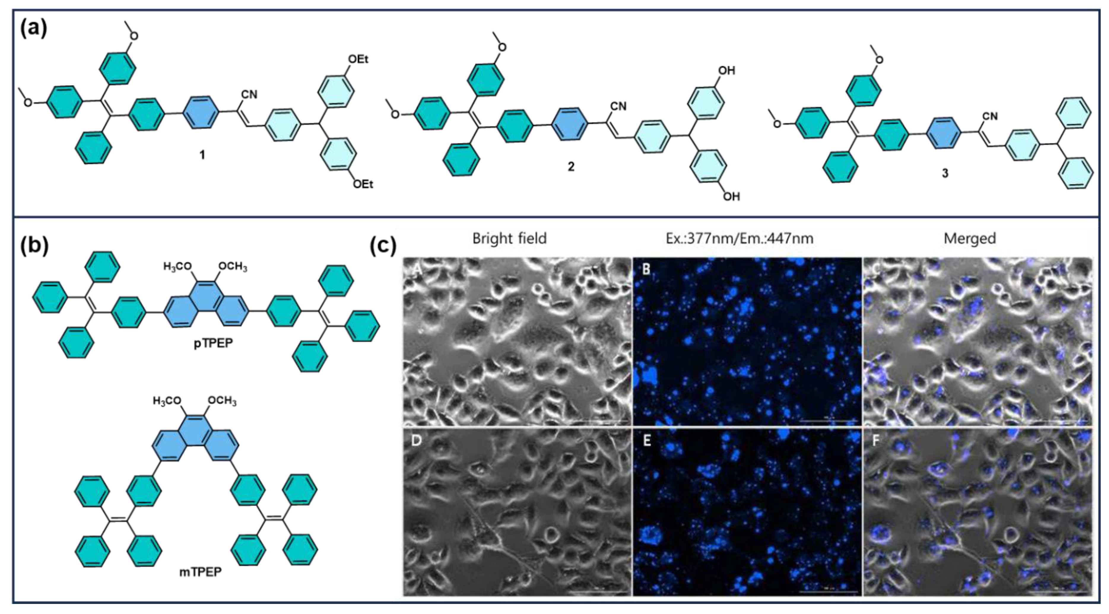
Figure 2. (a) Molecular structure of TPA-based AIEgens (structure 1–3) [79], (b) chemical structure of pTPEP and mTPEP (c) luorescence images of HeLa cells treated with pTPEP–SiO2 (AC) and mTPEP–SiO2 (DF) nanoparticles (40 μg/mL, 2 h). Excitation: 377 nm; emission: 447 nm. Scale bar: 100 μm [80].
Photochem 05 00023 g002
Figure 3. (a) Schematic illustration of the fabrication process of TPE-PTZ-R nanoparticles (NPs). Confocal images of HeLa cells co-stained with (b) DAPI and (c) TPE-PTZ-R NPs, and (d) the merged image. TPE-PTZ-R NP concentration: 30 μg/mL. Scale bar: 20 μm. Reprinted with permission from Ref. [82]. Copyright 2018, Elsevier.
Photochem 05 00023 g003
Figure 4. (A) Structure of Probe A (Coumarin–TPE) and confocal images of cells showing strong colocalization with CellBrite NIR membrane dye (Pearson’s coefficient = 0.93). Adapted from Ref. [83]. Copyright 2020, American Chemical Society. (B). Chemical structure of TPE-Ade and its co-localization with Golgi-Tracker Red in HL-7402 cells. Panels (ad) show fluorescence, bright-field, and merged images; (e) fluorescence intensity profile; (f) Pearson’s correlation coefficient (Rr = 0.86). Adapted with permission from Ref. [84]. Copyright 2021, Elsevier.
Photochem 05 00023 g004
Figure 5. Schematic of TPE-BT-DC(TBD)/CPPO-based nanoparticles for chemiluminescence-activated imaging and photodynamic therapy. (A) Preparation and H2O2-triggered luminescence/1O2 generation mechanism. (B,C) In vivo fluorescence and chemiluminescence imaging of tumour-bearing mice. (D) Combination therapy strategy with FEITC and H2O2. Adapted with permission from [97]. Copyright 2017, Elsevier.
Photochem 05 00023 g005
Figure 6. (A) Mechanism of afterglow luminescence in AGL AIE dots. (B) NIR fluorescence and afterglow imaging of tumour-bearing mice pre- and post-surgery. (C) Tumour nodule removal efficiency. Adapted with permission from Ref. [98]. Copyright 2019, American Chemical Society.
Photochem 05 00023 g006
Figure 7. Design and bioimaging applications of ultrabright NIR RTP nanoparticles. (A) Strategy to reduce quenching in water for strong room-temperature phosphorescence. (B) Molecular design using branched structures and alkyl chains. (C) Structures of TPE-4BT along with other studied molecules. (D) Size and phosphorescence intensity comparison with literature data. (E) In vivo imaging of liver, GI tract (mice), and lymph nodes (rabbits) using red/NIR RTP nanoparticles. Adapted with permission from ref. [101]. Copyright 2024, Wiley-VCH.
Photochem 05 00023 g007
Figure 8. Lysosome-specific fluorescence activation of PIP–TPE via viscosity-induced AIE. PIP–TPE shows blue fluorescence (λPL = 410 nm) in viscous lysosomal environments and shifts to green (λPL = 493 nm) in the bulk state (top). (A) Confocal image of HeLa cells stained with PIP–TPE (1 µM, 15 min); (B) LysoTracker Red (200 nM) staining; (C) merged image confirming lysosomal colocalization; (D) bright-field image. Excitation: 405 nm for PIP–TPE, 561 nm for LysoTracker Red; scale bar: 20 µm [112].
Photochem 05 00023 g008
Figure 9. Illustration of the detection mechanism of GlcNAc-TPE towards Hex for membrane-targeted fluorescence imaging. Reprinted with permission from Ref. [113]. Copyright 2019, American Chemical Society.
Photochem 05 00023 g009
Figure 10. TPE-based nanoplatforms for drug delivery and tumour imaging. (A) Amphiphilic block copolymer TPE-PLys-b-PMPC self-assembles into micelles for doxorubicin (DOX) encapsulation. (B) Confocal images reveal time-dependent cellular uptake, with AIE fluorescence (blue) and DOX (red) signals increasing over 2–8 h. Adapted from Ref. [126], Copyright 2017, Wiley-VCH. (C) TPE-TPA-DCM nanoparticles are prepared via BSA-assisted encapsulation, glutaraldehyde crosslinking, and THF removal. (D,E) In vivo fluorescence imaging of tumour-bearing mice demonstrates nanoparticle accumulation at 3, 8, and 28 h post-injection. (F) Ex vivo analysis shows biodistribution in tumours and major organs, with intensity mapped by the fluorescence scale bar. Adapted with permission from Ref. [127]. Copyright 2011, Wiley-VCH.
Photochem 05 00023 g010
Figure 11. NIR-triggered theranostic platform based on UCNP@TTD-cRGD nanoparticles for targeted imaging and photodynamic therapy (PDT) of deep-seated tumours. The schematic includes the AIEgen structure, nanoparticle design, and in vitro/in vivo applications under 980 nm laser. Adapted from Jin et al., 2019 [128].
Photochem 05 00023 g011
Figure 12. Schematic illustration of TPE-Man for TAM targeting and photodynamic ablation via ROS generation. Adapted with permission from Ref. [129]. Copyright 2019, American Chemical Society.
Photochem 05 00023 g012
Figure 13. Mitochondria-targeting AIEgen TPN with TPE and pyridinium units for tumour cell identification. (a) Chemical structure. (be) Enhanced fluorescence and uptake in cancer cells vs. leukocytes. (f) Confocal images of stained cell mixtures confirming tumour cell selectivity. Scale bars: 50 µm [131].
Photochem 05 00023 g013
Figure 14. AIEgen-based strategy for ALS tissue biopsy. (A) Molecular Structures of TPE-T-RO, TPE-T-RS and TPE-T-RCN. (B) Fluorescence imaging-based diagnosis of advanced atherosclerotic plaques using the as-prepared nanoparticles. (C) Comparative diagnostic results for early ALS obtained using different techniques. (D) Cross-sectional fluorescence imaging and Oil Red O staining of freshly resected human carotid plaque specimens. (E) Oil Red O staining and confocal fluorescence images of representative cross-sections of human carotid plaques treated with various nanoparticles. Adapted with permission from ref. [132]. Copyright 2022, Wiley-VCH.
Photochem 05 00023 g014
Figure 15. (A) Molecular structure of TPE–Xan–In, CT, NCT, and NCPT. (B) Confocal imaging using TPE–Xan–In and MitoTracker. Adapted with permission from Ref. [133]. Copyright 2019, American Chemical Society. (C) Confocal laser scanning microscopy images of HeLa cells incubated with CT, NCT, and NCPT (10 μM, λ_ex = 405 nm). Panels (a,d,g) show fluorescence images, (b,e,h) bright-field images, and (c,f,i) merged images. Scale bars represent 10 μm. Adapted with permission from Ref. [134]. Copyright 2023, Wiley.
Photochem 05 00023 g015
Figure 16. (A) Proposed mechanism of ONOO detection by TPE-4NMB. (B) Fluorescence imaging of ONOO in 4T1 cells. (C,D) In vivo fluorescence imaging of tumour resection models. (E) Monitoring ONOO levels during treatment. Adapted with permission from Ref. [139]. Copyright 2024, American Chemical Society.
Photochem 05 00023 g016
Figure 17. (A) Schematic illustration of the intracellular reductase-triggered transformation of non-emissive TPE-2E N-oxide into fluorescent TPE-2E under hypoxic conditions, driven by hydrophilic-to-hydrophobic conversion and subsequent aggregation. Adapted with permission from Ref. [146]. Copyright 2019, Wiley. (B) Schematic illustration of TPE-Azo for hypoxia detection in living cells. Adapted from Xu et al., 2021 [147].
Photochem 05 00023 g017
Figure 18. Design and mechanism of tamoxifen (TMX)-based supramolecular micelles. (a) Formation of fluorescent micelles via co-assembly of TMX and TPE derivatives with variable ethylene glycol chains. (b) TMX alone shows limited uptake and mild ER stress, whereas TMX-loaded micelles enhance uptake, induce strong ER stress, and regulate lipid droplets (LDs), restoring drug sensitivity with orlistat [150].
Photochem 05 00023 g018
Disclaimer/Publisher’s Note: The statements, opinions and data contained in all publications are solely those of the individual author(s) and contributor(s) and not of MDPI and/or the editor(s). MDPI and/or the editor(s) disclaim responsibility for any injury to people or property resulting from any ideas, methods, instructions or products referred to in the content.