Review

Phototoxicity of Quinolones and Fluoroquinolones: A Mechanistic Review About Photophysical and Photochemical Pathways

by
Elisa Leyva
1,*,
Silvia E. Loredo-Carrillo
1,
Irving R. Rodríguez-Gutiérrez
1,
Denisse de Loera
1,
Gabriela Navarro-Tovar
1 and
Lluvia I. López
2
1
Facultad de Ciencias Químicas, Universidad Autónoma de San Luis Potosí, Manuel Nava No. 6, Zona Universitaria, San Luis Potosí 78290, Mexico
2
Facultad de Ingeniería, Universidad Autónoma de San Luis Potosí, Manuel Nava No. 6, Zona Universitaria, San Luis Potosí 78290, Mexico
*
Author to whom correspondence should be addressed.
Photochem 2025, 5(3), 17; https://doi.org/10.3390/photochem5030017
Submission received: 16 May 2025 / Revised: 18 June 2025 / Accepted: 22 June 2025 / Published: 1 July 2025
(This article belongs to the Special Issue Feature Review Papers in Photochemistry)

Abstract

Quinolones and fluoroquinolones are heterocyclic compounds with important antibacterial properties, and they have been extensively used in medicinal chemistry to treat diverse bacterial infections. However, their clinical applications have been limited by several factors. On one side, there is an increasing number of resistant bacterial strains. On the other side, some of these heterocyclic compounds have shown several adverse effects such as photocarcinogenic cutaneous reactions, with the development of skin tumors. These adverse properties have motivated a large number of studies on the photophysical, photochemical and phototoxic properties of these compounds. In this review, several important chemical aspects about quinolones and fluoroquinolones are discussed. In the first sections, their basic structure is presented, along with some important physicochemical properties. In the next sections, their photochemical and photophysical processes are discussed. Upon photolysis in aqueous neutral conditions, these heterocyclic compounds generate several highly reactive intermediates that could initiate diverse reactions with molecules. In a biological environment, quinolones and fluoroquinolones are known to associate with biomolecules and generate complexes. Within these complexes, photophysical and photochemical processes generate intermediates, accelerating diverse reactions between biomolecules and these heterocyclic compounds. For several decades, diverse fluoroquinolones have been prepared for the treatment of a variety of bacterial infections. However, their prescription has been restricted due to the associated severe side effects. In the last decade, new derivatives have been developed and are already in use. Their introduction into actual practice extends the number of antibiotics and provides new options for difficult-to-treat infections. Thus, for new pharmaceutical compounds to be used in medicinal practice, it is important to investigate their biological activity, as well as other biological properties and adverse effects, such as phototoxicity.

1. Introduction

Quinolones (Qs) and fluoroquinolones (FQs) are an important class of antibacterial agents available for the treatment of infectious diseases. However, their clinical utility has been limited by the growing number of resistant bacterial strains. The most common resistance mechanism is induced by specific mutations in DNA gyrase and topoisomerase IV. To extend the clinical use of quinolones into the future, it is important to discover new compounds with improved activity against wild-type and mutant gyrase and topoisomerase IV [1,2,3].
Fluorination in pharmaceutical compounds generally improves their bioactivity and pharmacokinetic properties [4]. The introduction of a fluorine atom in the quinolone structure is based on several aspects. Fluorine is the smallest atom after hydrogen; thus, the replacement of H by F leads to a similar or improved biological activity without steric hindrance. Fluorinated derivatives mimic non-fluorinated ones and interact with molecules in the same manner. Due to strong inductive, resonance and electrostatic effects, F modifies drugs’ metabolism at adjacent or distant sites; thus it prevents oxidative metabolism. F modifies the lipophilicity/hydrophilicity balance of parent quinolone and reduces its basicity, improving membrane permeation and bioavailability. The substitution of H by F improves gyrase–complex binding and gyrase activity. Moreover, F can act as a hydrogen bond acceptor, since it is a bioisostere of a hydroxyl group, modifies molecular conformation and increases the binding affinity with a protein, either by direct interaction or by modifying the bond polarity of other functional groups that actually interact with proteins. All of these aspects may tune biological properties such as absorption, distribution, metabolism and excretion of FQs.
The introduction of new Qs and FQs in clinical practice has increased the number of reports on phototoxicity [5]. The human symptoms due to phototoxicity include sunburn, erythema, edema, hyperpigmentation, among others. Some quinolones have adverse photoallergic, photomutagenic and photocarcinogenic properties and induce several cutaneous photoreactions, with the development of skin tumors. These adverse effects have resulted in a large number of studies to understand the photochemical and photophysical properties of Qs and FQs [6].
In this review, some basic concepts and details on the photophysical and photochemical processes of Qs and FQs are discussed. The formation of highly reactive intermediates generated in these processes explain some important biological side effects such as phototoxicity. In the last section, the photosensitized reactions of FQs with biomolecules such as DNA are also discussed. The understanding of the photochemical and photophysical properties of Qs and FQs is an important issue to explain their photobiological properties and phototoxicity.

2. Structure and Properties

The basic chemical structure of quinolone and naphthyridone antibiotics contains two rings (Scheme 1). A quinolone has a carbon in X and contains a substituted benzene ring B. A naphthyridone has a nitrogen in X and contains a substituted pyridine ring B. In position one of ring A, different alkyl and aryl substituents have been introduced. A carboxylic acid (COOH) and a carbonyl (C=O) group are present in positions three and four, respectively. In addition to this, two important features are present in the structure of more recently developed and clinically useful quinolones. A fluorine atom is present in position six, and a cyclic amine is present in position seven, since these two substituents have been shown to enhance biological activity [7].
Since Qs and FQs have many applications in human and animal medicine, there are many articles about their synthesis [8,9,10,11,12] and their diverse biological and clinical properties [13,14,15,16]. Their structure–activity relationship has been investigated by many researchers throughout the years. Taking in account their antimicrobial spectrum of activity, quinolones have been classified into several generations [17]. The purpose of each generation was to obtain a broader spectrum of activity. To achieve this, several substituents have been introduced into different positions of the basic pharmacophore. The structure of some representative Qs and FQs, along with their generations, discussed in this review is presented in Scheme 2.
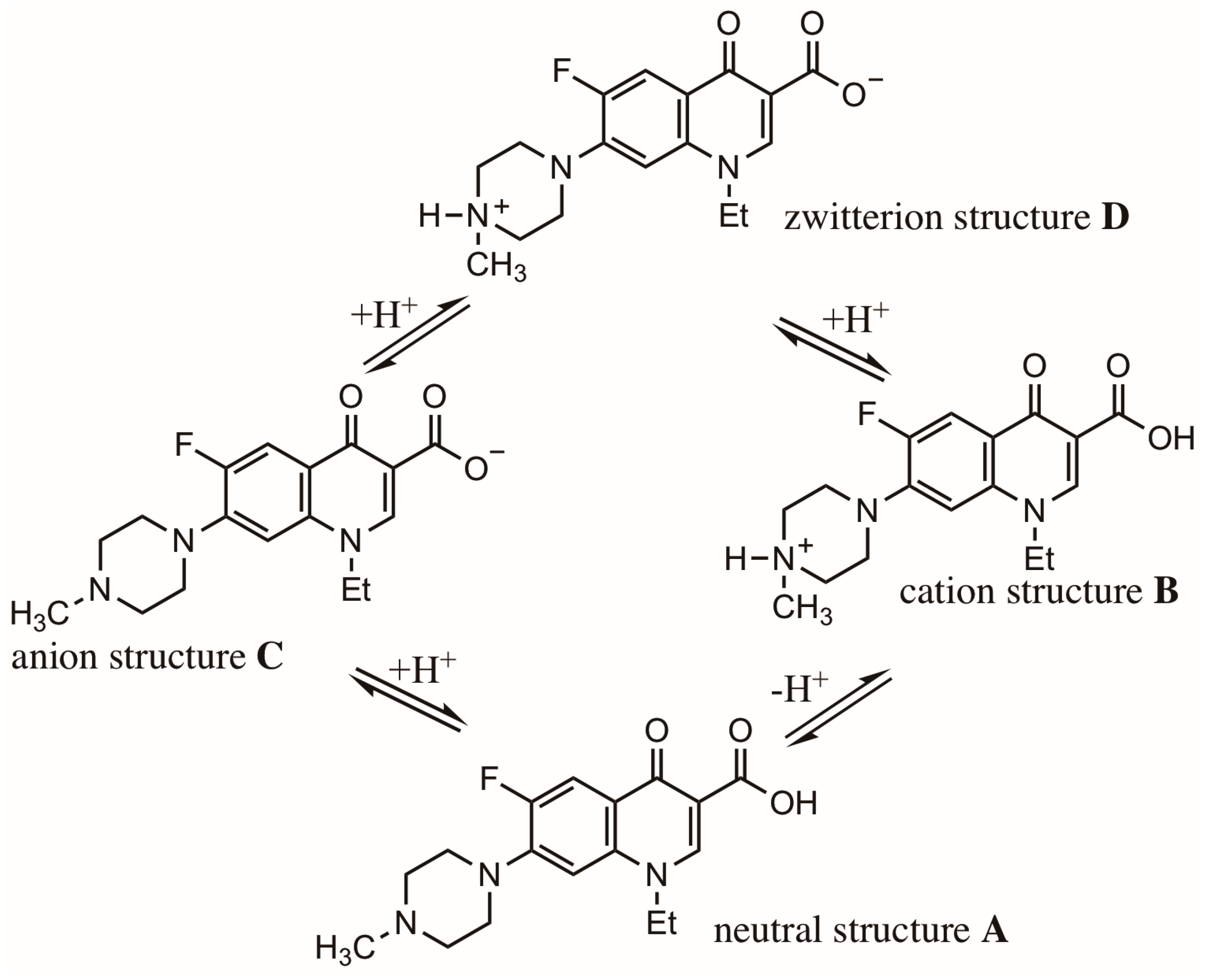
Due to their structure, FQs contain multiple proton binding sites, and they have complex acid-base equilibria (Scheme 3). Studies using different analytical techniques such as potentiometry, UV–Vis, NMR and spectrofluorometry [18,19] have shown that two groups, carboxylate and 4′-amino in piperazine, are the most important binding sites in a biological system (pka in the pH range 5 to 9). In aqueous media, four species are in equilibrium for a given FQ. The ratio of concentration of these species changes with different derivatives. However, at a neutral pH the prevailing species is always the zwitterion. But, the cation form predominates in an acidic medium and the anionic form in a basic one. Therefore, in a particular FQ, different species in a given solution contribute to the cellular uptake by a combination of mechanisms. There are many studies about these mechanisms and their interplay with the equilibrium of different species in a FQ in terms of pKa and pH [20].
After the development of a new Q or FQ, several physicochemical properties must be determined, like their diffusion coefficient and lipophilicity [21]. Furthermore, FQs are known to form insoluble salts with different buffer solutions; thus the nature of the buffer to use in a quinolone preparation must be taken into consideration [22].

2.1. Photophysical and Photochemical Processes in Qs and FQs

To explain the phototoxicity of several Qs and FQs, the processes induced on them by light have been extensively investigated for several decades [23,24,25,26,27,28,29,30,31,32,33,34,35,36,37,38,39,40,41,42,43,44,45,46,47,48,49,50,51,52,53,54,55,56,57,58,59,60]. In some studies, the products generated upon exposure to light have been determined. In other studies, the intermediates and their reactions have been investigated by time-resolved and steady-state spectroscopic techniques. By means of these techniques, it has been feasible to obtain information about transient intermediates and excited states generated upon the photolysis of an heterocyclic quinolone.
Diverse photochemical and photophysical processes take place upon the photolysis of a FQ. In the ground state (FQ, Scheme 4), it absorbs energy and generates an excited singlet state 1FQ* that undergoes intersystem crossing (ISC) very fast to an excited triplet state 3FQ*. The ISC mechanism is an interesting process in which a singlet electronic state makes a spin transition to a triplet state at a point where the potential energy curves for singlet and triplet cross. On the other hand, the 1FQ* could go back to FQ by fluorescence (F). Once the triplet state (3FQ*) is generated, it could undergo different pathways. It could go back to FQ by phosphorescence (P), or it could undergo triplet–triplet energy transfer (TTET) with certain molecules (MOL) to eventually generate dimers. It could also undergo diverse physical and chemical processes with oxygen, with the subsequent generation of highly reactive oxygen species (ROS) like singlet oxygen (1O2*) and superoxide radical anion (O2•−). These ROS could eventually oxidize a molecule (MOLs). Both excited intermediates (singlet and triplet) could undergo several mechanisms and generate other highly reactive intermediates (radicals, anions, cations and carbenes), which react very fast with cell tissue or biological molecules. Evidence of the formation of these intermediates was obtained in several investigations [1,6,23,24,25,26,27,28,29,30,31,32,33,34,35,36,37,38,39].
The triplet state of several Qs and FQs has been characterized by phosphorescence spectroscopy [25,26,27,28,29,30,31,32]. At low-temperature photolysis, norfloxacin and enoxacin gave a strong phosphorescence emission, with a λmax at 500 nm and 450 nm, respectively. Based on the position of the phosphorescence band, the energy for these triplet intermediates was estimated (260 to 280 KJ mol−1). Triplet intermediates have been further characterized by laser flash photolysis under different buffer solutions at close to neutral conditions (a pH of 7.0 to 7.4), and their main characteristics are given in Table 1. The lifetimes of the triplet intermediates were strongly dependent on the experimental conditions and structure of the Qs. A quinolone without an amino substituent in position seven, such as nalidixic acid, gave a relatively large triplet lifetime of approximately 100 μs. In contrast, FQs with an amino substituent in the same position (norfloxacin, enoxacin and ofloxacin) gave a rather small lifetime.
Most of the Qs and FQs investigated have a fast ISC [26,27,28,29]. At pH 9.2, nalidixic acid, the first-generation quinolone, gave an ISC for anionic carboxylate (NAL) with a high quantum yield (0.9). At pH 7.0, The zwitterionic form of several subsequent-generation quinolone derivatives like ofloxacin gave an ISC with a lower quantum yield (0.33). High concentrations of phosphate buffer salt led to a substantial reduction in the ISC for lomefloxacin due to interactions of the salt with the quinolone. In this particular case, the formation of a ground state complex (Scheme 5) was proposed [29]. This complex could inhibit a fluorescence pathway by several mechanisms, either channeling energy into a radiationless mechanism or inducing proton release towards a phosphate dianion. Since a methylated quinolone like ofloxacin could not form such a ground state complex, reduction in its ISC was not observed [29].
Once an excited triplet state of a quinolone is generated (3FQ*, Scheme 4), it is easily quenched with oxygen by three different mechanisms: energy transfer (ET, Equation (1)) with the formation of singlet oxygen, physical quenching (PQ, Equation (2)) or electron transfer (eT, Equation (3)) to generate superoxide anion (O2•−) and a quinolone radical cation (FQ•+). The quenching of 3FQ* by oxygen has a rate constant (2 to 3 × 10⁹ M−1 s−1) similar to the one observed for aromatic molecules [6].
FQ * 3 + O 2   E n e r g y   T r a n f e r   >   FQ + O 2 * 1
FQ * 3 + O 2   P h y s i c a l   Q u e n c h i n g   >   FQ + O 2 * 3
FQ * 3 + O 2   E l e c t r o n   T r a n s f e r   >   FQ + + O 2
In addition to oxygen, other compounds have been found to be good quenchers for different quinolone triplet states [25,26,27,28,29,30,31,32,33]. Tyrosine and tryptophan quenched the triplet states of flumequine and nalidixic acid by an electron transfer mechanism. Compounds like acetonaphthone and b-caroteno quenched the triplet states either by energy transfer or charge transfer.
The UVA irradiation of several Qs induces the generation of some highly reactive oxygen species (ROS), including hydrogen peroxide (H2O2), hydroxyl radical (HO•) and superoxide radical anion (O2•−) [34,35,36]. Their phototoxicity has been related to the formation of these species. Energy transfer from Qs to molecular oxygen has been investigated and corroborated with oxolinic and nalidixic acid [6]. In a subsequent study, Martinez et al. investigated the generation of singlet oxygen (1O2*) by Qs upon exposure to UVA light [34]. Measuring the luminescence at 1270 nm, the yield of singlet oxygen was determined, and the quenching rate of this highly reactive intermediate was also measured in a number of Qs [26,27,34]. The results of several investigations are summarized in Table 2. The quantum yield for the generation of 1O2* for the Qs and FQs investigated was rather low, with values between 0.06 to 0.09 in phosphate buffer at pH 7.5. The rate constant for singlet oxygen quenching by the ground state of several Qs was measured. Nalidixic acid was a good singlet oxygen sensitizer with an unusually high quantum yield both in deprotonated (0.15) and protonated form (0.24). Other FQs, like flumequine (0.34) and rufloxacin (0.32), also gave good quantum yields. The rest of the FQs had quantum yields that were rather low (<0.1) at a neutral pH. Under these experimental conditions, the zwitterionic species of FQs predominate, and their triplet state has a very low ability to transfer energy to molecular oxygen. Therefore, electron transfer (Equation (3)) must be the favored mechanism in these later compounds.
The photogeneration of superoxide by several Qs has been investigated by two procedures [33]. Under steady-state irradiation, it was measured by the detection of H2O2, generated upon the disproportionation of superoxide (O2•−), with its conjugated acid form (HO2) that induced the reduction of citocrome c. In a more recent investigation, the photogeneration was evaluated using electron paramagnetic resonance, performing spin trapping with 5,5-dimethyl-1-pyrroline N-oxide (DMPO). The rates of superoxide production for some quinolones at a neutral pH are also given in Table 2. The best rate for superoxide generation was obtained with naphthyridones; nalidixic acid gave the highest rate (10), followed by enoxacin (1.7). Other FQs like lomefloxacin, ciprofloxacin, norfloxacin and fleroxacin had a rather low rate (0.17 to 0.36). An usually low rate was also observed with ofloxacin. However, these experimental studies on ROS generation by Qs do not explain the phototoxicity observed in humans, which has been reported to present approximately the following order: fleroxacin > lomefloxacin, pefloxacin > ciprofloxacin > enoxacin, norfloxacin and ofloxacin [35]. Therefore, in the case of some Qs the phototoxicity must be due to a combination of effects. Not only the generation of ROS but also the generation of other highly reactive species like carbenes [34].
The triplet state of a given Q could also be quenched by electron transfer reactions inducing the generation of reduced species [6]. For example, the reduced form of nalidixate anion, NA2•− radical anion, gave an strong absorption at 650 nm. The reaction of this radical dianion induced the formation of superoxide anion, which has a characteristic reaction with ferricytochrome c [30,31]. Laser flash photolysis and pulse radiolysis experiments with ofloxacin (OFL) were used to investigate the excited states and the free radicals generated in aqueous solutions [26]. The primary photophysical and photochemical process for OFL have been summarized (Equations (4)–(12)).
OFL                                           h v                             >   OFL * 1
OFL * 1                                                                         >   OFL + F
OFL * 1                               I S C                             >   OFL * 3
OFL * 1                                                                         >   OFL + + e ( H 2 O )
OFL   + e   ( H 2 O )                                                                       >   O F L
O F L + O 2                                                                                 >     OFL + O 2
OFL * 3   + O 2                                           E T                           >   O F L + O 2 * 1
OFL * 3   + O 2                                           P Q                           >   O F L + O 2 * 3
OFL * 3   + O 2                                             e T                             >   O F L + + O 2
Upon the photolysis of OFL (Equation (4)), a high energy or excited singlet state intermediate (1OFL*) is generated. This intermediate gives out its excess energy through several mechanisms. It can go back to ground state by light emission (Equation (5)), it can undergo intersystem crossing (ISC) (Equation (6)) to an excited triplet state intermediate (3OFL*) or generate hydrated electrons e(H2O) in aqueous solution (Equation (7)). These hydrated electrons react (Equation (8)) with ground state ofloxacin OFL to generate the corresponding anion (OFL•−), which in a subsequent reaction (Equation (9)) generates superoxide anion (O2•−). As it was mentioned before, the excited triplet state intermediate (3OFL*) can undergo at least three possible mechanisms. An energy transfer (ET, Equation (10)) with the formation of singlet oxygen (1O2*), physical quenching (PQ) of oxygen (Equation (11)) and electron transfer (eT, Equation (12)), with the concomitant formation of O2•− and a quinolone cation radical (OFL•+). This latter intermediate could actually act as a hapten, inducing some photoallergic reactions associated with quinolones [36].
Several studies have clearly indicated that Qs react with oxygen and generate both singlet oxygen and superoxide anion [6]. The total (physical and chemical) rate constants for the quenching of singlet oxygen were in the range of 106–107 M−1 s−1 in aqueous media, while they were 107 M−1 s−1 in methanol, and these values varied very little with structure [34]. The observed values were very close to the ones obtained with anilines, which have a similar conjugated p system [1].

2.2. Structure and Photochemistry of Qs and FQs

In neutral aqueous medium where the zwitterionic form predominates, compounds containing the 4-quinolone or 4-naphthyridone structure absorb light in the range between 200 to 400 nm [1]. Since the predominant species change with pH, the absorption spectra of most these compounds also changes with pH. Therefore, the intermediates and products generated upon photolysis depend on the quinolone structure, environment and source of irradiation. The several processes induced on different Qs and FQs by light will be discussed in this section. Upon irradiation, they can undergo different photochemical reactions depending on the functional groups present in the main structure (Scheme 6). Depending on their substituents and structure, quinolones experience different photochemical reactions [6]. They can undergo decarboxylation (a), cleavage of C-N (b and c), side chain oxidative degradation by ROS (d) and dehalogenations with C-F or C-X cleavage by different mechanisms (e, e′ and f). These reactions will be briefly discussed in this section, along with some investigations about phototoxicity.

2.2.1. Decarboxylation

Early Qs with a similar structure to nalidixic acid containing an alkyl substituent on position seven, with no C-6 fluorine substituent or a piperazinyl moiety at C-7, undergo decarboxylation upon photolysis in water [1]. After decarboxylation, they generate several highly reactive quinolone intermediates (radicals and anions) and ROS species (1O2 and O2•−). The different mechanisms involved in the formation of these intermediates are presented in subsequent sections. As a consequence, the phototoxicity associated with these Qs has been related to the generation of highly reactive species that could react with cell tissue or biomolecules [31].
For FQs, a decarboxylation mechanism is only observed in cases where a strong electron donating substituent at C8 inhibits C-6 defluorination, like in rufloxacin [38], or when there is an alkyl or alkoxy substituent in position eight [1]. However, this is a very minor process in quinolones containing amino electron donating groups in C7 like ciprofloxacin [32].

2.2.2. Side Chain Oxidation and C-N Cleavage in FQs

A photochemical fluorine elimination (C6-F) reaction takes place very easily for most FQs under neutral conditions [1]. However, Qs containing a fluorine atom in position six and an electron donating group (OR) in position eight, as with ofloxacin, levofloxacin, and moxifloxacin, do not undergo this fluorine elimination process. Instead, they present inefficient alkylamino side chain oxidation, leaving the basic heterocyclic fluoroquinolone structure intact.
Several studies have demonstrated that photoreaction involving side chain oxidation is a more efficient process in the presence of oxygen, indicating the participation of reactive oxygen species or ROS [1]. Furthermore, some heterocyclic compounds, like pyrrole and furan (present in sitafloxacin), are easily oxidized by reaction with 1O2* [23,61]. Experimental studies suggest the presence of a radical intermediate rather than an aryl cation in the degradation mechanism involved in this side chain oxidation. A Q containing a F in C6 and an O-S group in C8, such as rufloxacin, gave side chain degradation by means of a radical cation intermediate (RUF.+) generated by singlet oxygen (1O2*) in solution [28].

2.2.3. C6-F Cleavage in FQs and Phototoxicity

Miolo et al. investigated the effect of C6 and C7 substituents on the phototoxicity of several quinolones [62]. They reported that a C6 substituent plays an important role on phototoxicity with the following order (F > H >> NH2). Thus an amino group in this position exerts the lesser effect. They also demonstrated that the nature of the substituent on C7 also modulates phototoxicity. Changing pyperidinyl (present in ciprofloxacin) by a more hydrophobic substituent such as tetrahydroisoquinolinyl increases phototoxicity. The capacity of several Qs to generate singlet oxygen upon UVA irradiation was poor and comparable with the reference compounds, ciprofloxacin and lomefloxacin. Another interesting feature of non-fluorinated derivatives was their lack of plasmid DNA photocleavage activity, which was observed in reference compounds.
Most of the antibacterial Qs contain at least one fluorine atom in position six. Under neutral conditions, their main photochemical reaction involves the elimination of fluorine. The quantum yield for this particular reaction depends on the structure and varies considerably (0.55 to 0.001). In addition, different mechanisms and intermediates can be involved in this process [6]. For instance, ciprofloxacin contains a fluorine atom in position six and undergoes photochemical degradation in water by several mechanisms [32]. The main mechanism consists of reductive defluorination with the formation of a radical anion (CIP•−) and subsequent loss of fluorine anion to give a radical (CIP).
Several Qs (norfloxacin, enoxacin and ciprofloxacin) have one fluorine in position six, and they undergo photosubstitution of the halogen by a hydroxyl group [29,32,33]. Two mechanisms have been proposed for this photochemical process. One involves the generation of an excited state with the subsequent fragmentation of the C-F bond. Another mechanism, could be the addition of a hydroxyl OH group with the loss of fluoride anion via a triplet state, similar to the route involved in the photosubstitution of some aromatic compounds. This latter process occurs by an addition–elimination mechanism (SN2Ar) induced by the stronger electrophilicity of the triplet with respect to the ground state. Flash photolysis studies for norfloxacin and enoxacin corroborated their transformation by this latter mechanism involving a long-lived cyclohexadienyl anion intermediate with a strong absorption band at 700 nm [29].
Further evidence for this mechanism was provided by the value of the quantum yield for defluorination (in position six), which was actually proportional to the electrophilicity of the heterocyclic ring. This physicochemical property was modulated by the character of the substituent in position eight [27,29,33,37]. Enoxacin with an electron withdrawing substituent had the highest quantum yield (0.13), ciprofloxacin and norfloxacin with no substituent had an intermediate quantum yield (0.6–0.007), and ofloxacin and rufloxacin with an electron donating substituent had a rather low quantum yield (>0.001). These latter Qs did not give a hydroxylated derivative upon photolysis; instead they presented photochemical degradation involving decarboxylation and radical side chain oxidative reactions [27,37,38,39].

2.2.4. C8-F Cleavage in FQs and Phototoxicity

Dermatologic adverse effects have been reported with the use of fluoroquinolones [63]. Two types of photosensitivity reactions have received a lot of attention. Photoallergic reactions are not common, and they manifest after several days of exposure to a pharmaceutical compound. In contrast, phototoxic reactions are more common. They usually occur within hours, upon initial exposure of an individual to an FQ and enough UV light. The mechanism for phototoxicity has been related to the photochemical degradation of FQs and their property of generating highly reactive intermediates. These species attack cellular tissue or biomolecular compounds. Persons with fair skin are more prone to these reactions than those with darker skin. Photosensitivity reactions have been reported for most FQs, but they differ in incidence and severity. Therefore, upon administration it is safer to cover skin with clothing and avoid sunlight exposure.
The phototoxicity of several FQs has been investigated by different procedures in vitro, measuring cellular damage [64] or DNA damage [65]. It has also been investigated using in vivo mouse models [66]. This later methodology became the preferred one since it takes into account all aspects of phototoxic response such as compound photoreactivity, half-life, skin penetration and toxicity to skin. Experimental results from several studies using in vivo methodology [67,68,69,70,71] gave an approximate order for FQs’ phototoxicity: lomefloxacin, fleroxacin > clinafloxacin > sparfloxacin > enoxacin > pefloxacin > ciprofloxacin > grepafloxacin > norfloxacin > ofloxacin > levofloxacin and trovafloxacin. Photoreactivity and consequently phototoxicity is strongly influenced by the substituent in position eight. Taking in account this substituent, the order of phototoxicity observed is CF >> CCl > N > CH > CF3 > COR. Therefore, lomefloxacin (with a CF) is more phototoxic than clinafloxacin (with a CCl). In the same line, enoxacin (with a N) is more phototoxic than norfloxacin (with a C-H). Ofloxacin (with a COR) gives a higher phototoxicity than ciprofloxacin (with a C-H). Interestingly, sparfloxacin containing a NH2 substituent in position five shows a lower phototoxicity than expected for a compound with a C-F in position eight [69]. This effect has been explained to be due to singlet oxygen quenching by the amino group. Qs containing a bulky side chain in N1 like fleroxacin also show a reduced phototoxicity due to long half-lives or a high skin penetration. Since Qs containing a halogen in the eight position have shown increased potency, many new quinolones with this particular substituent have been developed. As a consequence, phototoxicity has become an important issue in clinical practice. For example, a high incidence of phototoxicity has been reported for fleroxacin and lomefloxacin [66].
FQs with a second halogen in position eight have shown an increase in photosensitivity; this physicochemical property has been previously reported [29,33,72,73,74,75,76,77,78]. The C-F bond in this particular position is easily fragmented for lomefloxacin, fleroxacin, sparfloxacin and orbifloxacin. Lomefloxacin gave the highest quantum yield for defluorination in water, and the other quinolones reacted at a rate only slightly slower. For these FQs, the products generated and the intermediates observed also changed. With lomefloxacin, a transient intermediate was detected; it was attributed to the excited triplet state 3FQ*, and it was weak and short-lived [29]. Therefore in this case, the excited singlet state 1FQ* reacted very fast in competition with the ISC to triplet. If the 3FQ* was formed, it reacted more efficiently than the corresponding excited triplet state generated with the other FQs.
The mechanism for photoinduced fragmentation for 6-fluoro and 6,8-difluoroquinolone derivatives has been explained in detail [6,37,73]. The cleavage of the C-F bond is not an homolytic process, since its energy (477 kJ mol−1) is larger than the energy associated with the enoxacin excited singlet state (~330 kJ mol−1) and its corresponding excited triplet state (~278 kJ mol−1). Fasani et al. investigated the photochemical reactivity of four FQs. In the case of norfloxacin, enoxacin, lomefloxacin and ofloxacin, the charge density is transferred into the aromatic ring upon photochemical excitation, and the C-F bond breaks in an heterolytic manner to give a carbocation [37]. The mechanism for intermediate cation formation for a 2,6-fluoroquinolone is presented in Scheme 7. Several pieces of evidence support this mode of reaction. The efficient defluorination observed in position six (norfloxacin and enoxacin) or position eight (lomefloxacin) is explained by the better stabilization of the resulting aryl cations. These cations are highly reactive intermediates; although they have not been characterized by laser flash photolysis in solution, they have been detected by low-temperature EPR and UV–Vis spectroscopy [79]. For these aryl cations, the most important stabilization is the one arising from p-conjugated mesomers with the charge in the neighboring amino group. For example, for the cation in position eight (1a), three different mesomeric structures (1b, 1c and 1d) contribute to its stability. Some of these mesomeric structures have one carbon, with a carbene character similar to the carbene generated upon the photochemical dehalogenation of 4-halogenated phenols. This carbene intermediate has been previously characterized by the laser flash photolysis of several halogenated phenols [80]. In this photochemical process, Qs generate two highly reactive intermediates aryl cations and carbenes, which may react fast with other molecules in the environment. In another study by Dichirante et al., it was demonstrated that having a terminal NH2 in the heterocyclic aniline favors defluorination, with the subsequent formation of an aryl cation [81].
Sparfloxacin with an amino group in C5 and two fluorine atoms in C6 and C8 gave a product resulting from defluorination in C8, indicating this position is very labile. In this case, a mechanism involving a radical intermediate has been proposed [78]. As expected, a Q containing a chlorine atom in C8 is more easily cleaved than one containing a fluorine in C8. This was corroborated for the chlorine derivative of lomefloxacin [72]. Orbifloxacin containing three fluorine atoms (C5, C6 and C8) gave products from defluorination in C5 and C8, indicating that for this quinolone C6-F is less labile [76,77].
Performing photochemical reactions of several quinolones in a buffer solution induces reductive defluorination [6,82]. For derivatives containing a fluorine in C6 (norfloxacin, ciprofloxacin and enoxacin), this is an important degradation route. Under such experimental conditions, the triplet excited state (3FQ*) is quenched by the salt to generate a radical anion intermediate. This later intermediate generates a radical by the loss of fluorine anion. For lomefloxacin, this reductive mechanisms occurs at C8 [73].

2.2.5. C5 Substitution in FQs and Phototoxicity

Phototoxic effects have been observed by the presence of diverse substituents in C5. A Q containing a bulky side or a methyl group on C5, such as grepafloxacin, has a lower phototoxicity due to a long half-life and high skin penetration [83].
As was mentioned, quinolones with an NH2 in C5 present less phototoxicity [69]. For example, sparfloxacin containing this particular substituent is less phototoxic than the same Q without it. Levofloxacin is a third-generation FQ, and it has been used in medicine to treat bacterial infections. Photochemically, it behaves very similarly to ofloxacin, but it presents several adverse effects in mice and humans, such as phototoxicity [84]. Antofloxacin is an improved version of levofloxacin, containing an NH2 in the C5 position [85]. Both FQs, present a quite comparable spectrum of antibacterial activities in vitro. However, antofloxacin presents a longer elimination half-life than levofloxacin, and it is usually eliminated in the urine. Interestingly, a recent study in rats did not present any toxicity for antofloxacin, making this a very useful pharmaceutical compound. It has been previously indicated that a NH2 substituent in this particular position reduces phototoxicity [67]. This reduced antofloxacin phototoxicity was explained in terms of the several alternative intermediates and reaction pathways involved in its photochemistry. As was mentioned, a quinolone like levofloxacin without an amino group undergoes intersystem crossing (1FQ* to 3FQ)* more efficiently. In the case of antofloxacin, a singlet state is generated, and it is stabilized by an intramolecular charge transfer induced by NH2, giving diverse stable resonance structures, which eventually undergo ISC to triplet or go back to the ground state GSFQ. By means of these routes, antofloxacin is deactivated, inducing photostability in the FQ.
Phototoxic reactions are initiated by the absorption of light by an FQ, distributed into the skin, which then induces several reactions in the body [86]. The mechanisms for these reactions are explained in section four of this review. On one side, several intermediates (excited states, carbenes, radicals or ROS) are generated within biomolecules and induce direct non-specific degradation reactions. On the other side, photogenerated intermediates bind to different cellular components (DNA or proteins) and initiate several degradation reactions in situ.
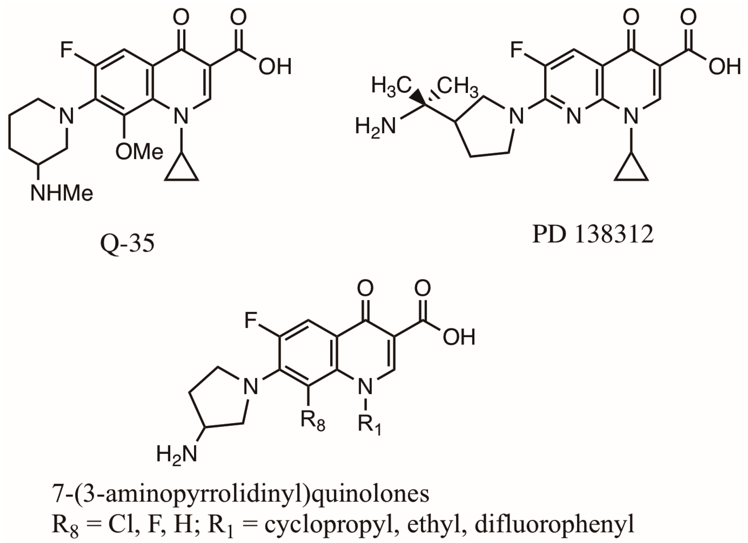
FQs with reduced phototoxicity have been synthesized. In 1993, Marutani et al. reported the synthesis of fluoroquinolone Q-35 (Scheme 8), containing a methoxy group in C8 [87]. They claimed this compound presented a lower phototoxicity than lomefloxacin, enoxacin, norfloxacin, ciprofloxacin and ofloxacin. Hagen et al. reported the synthesis of several FQ derivatives with a very low degree of phototoxicity [88]. R and S isomers of 7-[3-(1-amino-1-methylethyl)-1-pyrrolidinyl]-1,4-dihydro-4-oxoquinoline and 1,8-naphthyridine-3-carboxylic acids were prepared. All of the isomers presented excellent antibacterial activity and a low potential for phototoxicity. One isomer in particular, PD-138312 (Scheme 8), gave the best results. Several experimental and clinical studies have demonstrated that an FQ containing a halogenated atom at C8 induces severe photoxicity [69]. In contrast an FQ containing a hydrogen atom at C8 induces lower phototoxicity effects [69]. Furthermore, a methoxy in C8 does not induce significant phototoxicity [87]. In 2004, Hayashi et al. investigated the structure–phototoxicity relationship of a series of quinolones (Scheme 8) containing a C7 substituent (3-aminopyrolidinyl), a C8 substituent (hydrogen or halogen) and various substituents in N1 [89]. The degree of phototoxicity from having a difluorophenyl in N1 changed from mild to severe when a hydrogen atom was replaced by a halogen one at C8. On the other hand, severe phototoxicity was observed from having a cyclopropyl or ethyl in N1, regardless of the substituent in position C8. These results indicate that phototoxicity is modified by substituents at position C8 and N1.
In conclusion, photochemical processes are modulated by substituents in different quinolone positions (C5, C6, C7, C8 and N1) demonstrating the need to develop novel quinolones with a modified structure in such a way that they still keep their biological activity in addition to a resistance to photodegradation.
Several in vitro model systems have been developed to investigate the phototoxicity of novel pharmaceutical compounds [90,91,92]. Studies using mouse 3T3 fibroblast and the neutral red (NR) endpoint have demonstrated a good correlation with reported in vivo data. There are only a couple of reports about the correlation between FQ phototoxicity studies in vitro and in vivo clinical trials. However, these studies present contradicting results. Thus, for a new Q or FQ candidate to be used in medicinal practice, it is a requirement to determine its phototoxic potential using a combination of techniques (in vitro and in vivo) [92].

2.3. Structure and Biological Properties of Qs and FQs

The first-generation quinolones (nalidixic acid and oxolinic acid) showed activity only against Gram-negative organisms, excluding Pseudomona species (Scheme 2). Shortly after their clinical introduction, they were found to cause rapid resistance in many microorganisms [16,17]. These findings led to many investigations to discover new naphthyridone analogues with improved physicochemical and bioactivity properties.
An important discovery was observed upon the synthesis of second-generation Qs (enoxacin, norfloxacin and ciprofloxacin) [93]. They exemplified two key modifications in their basic structure; a fluorine atom at C6 (Scheme 2) increased their spectrum of activity, and a piperazine in C7 enhanced their potency [16,17]. In the case of ciprofloxacin, the cyclopropyl group in N1 improved the overall activity, and it became the most active and effective FQ at the moment [93]. Another two analogues, lomefloxacin containing an alkylated piperazine in C7 and a fluorine in C6 and ofloxacin with a methyl piperazine in C7 and an OR in C8, were also introduced [93]. These later derivatives presented good activity against several Gram-negative and Gram-positive bacteria like Staphylococcus aureus and some atypical organisms like Mycoplasma pneumoniae and Chlamydia pneumoniae. However, ofloxacin has been shown to have a higher potency against Gram-positive microorganisms, and it has been extensively used for clinical treatment.
Qs entered the third generation with the development of fleroxacin, sparfloxacin, grepafloxacin, clinafloxacin and gatifloxacin [93]. Structurally, these quinolones contained an alkylated piperazine or pyrrolidine group in position seven (Scheme 2). An NH2, OH or CH3 group was introduced in position five of the pharmacophore. A couple of derivatives retained the cyclopropyl substituent on nitrogen, like ciprofloxacin, while fleroxacin contained a fluoroethyl in this position. Some of the derivatives contained a fluoro, chloro or methoxy in position eight; this particular modification enhanced the activity and spectrum of the quinolones. All of these newer derivatives retained the activity of second-generation quinolones but had a stronger activity against atypical pathogens. They also showed expanded activity against Gram-positive microorganisms, including penicillin-sensitive and penicillin-resistant S. pneumoniae. Among these quinolones, clinafloxacin was shown to have the strongest activity [93].
Fourth-generation Qs still had the activity of the previous-generation analogues [94]. In addition to this, they presented activity against anaerobic microorganisms [17]. Several structural features are noticeable in these derivatives. Some of them still contained the cyclopropyl on nitrogen, like ciprofloxacin. However, in the case of trovafloxacin, a 2,4-difluorophenyl was introduced in this nitrogen position. This modification resulted in an improved activity against anaerobic bacteria [17]. Two naphthyridones with a pyridine ring similar to oxolinic acid were introduced. Gemifloxacin and trovafloxacin, having a nitrogen in the eight position, gave strong activity against anaerobic microorganisms. This particular activity was also favored by an alkoxy substituent on the eight position like in gatifloxacin. In addition, an aza bicyclic group on the seven position was shown to enhanced the potency against Gram-positive bacteria [17].
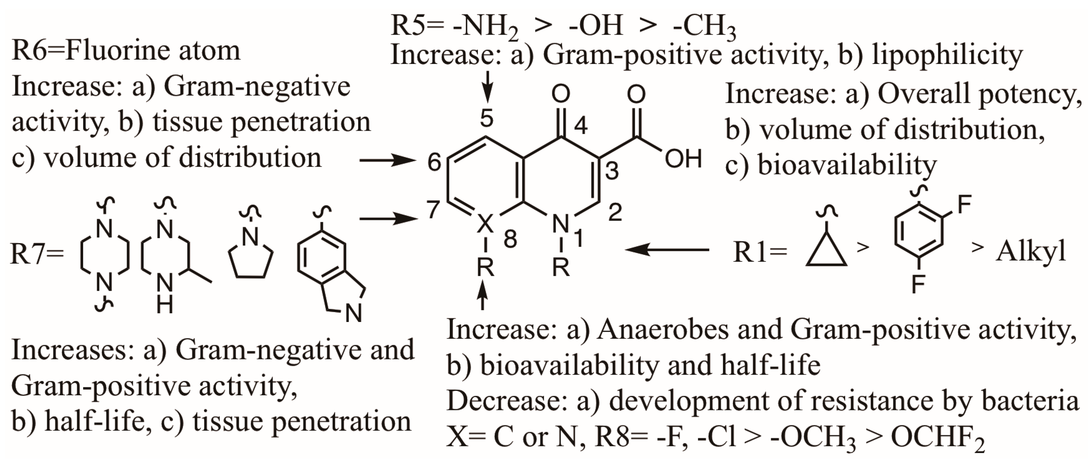
A summary of the structural modifications and their relationship with the biological activity and pharmacokinetic properties of Qs and FQs is presented in Scheme 9. The different changes in the structure of quinolones had the purpose of modifying their physicochemical and bioactivity properties to improve their metabolism, elimination and transportation [17]. The first two Qs (nalidixic acid and oxolinic acid) gave relatively low serum concentrations and were only used in the treatment of urinary tract infections. The several modifications performed in the basic structure of these Qs led to analogues with an improved oral absorption and a higher serum concentration. These modifications also improved their half-life of elimination, thus allowing a once-a-day dose. Furthermore, the FQs developed had a broader spectrum of activity and enhanced potency. Structural modifications (Scheme 9) with different functional groups in several positions (R5, R6, R7 and R8) of the Q resulted in a higher tissue penetration, increased volume distribution and enhanced bioavailability [9]. With the development of sparfloxacin (SPA), containing an amino group in position five, an increase in lipophilicity was observed [17]. In general, the introduction of a fluorine atom in position six enhanced the penetration into the bacterial cell and improved the volume of distribution in the microorganism. This improvement was observed upon the development of second-generation FQs. The addition of cyclic amines (piperazine and azabicyclic groups) in position seven improved the half-life and bacterial tissue penetration [95]. However, the substitution with pyrrolidine resulted in an unfavorable water solubility and oral bioavailability [96]. To correct these adverse physicochemical properties, methyl substituents were added in the cyclic amine. Qs containing a methyl piperazine group in position seven, like ofloxacin, lomefloxacin, sparfloxacin, grepafloxacin and gatifloxacin, gave an increased elimination half-life [17]. The presence of alkoxy groups at the eight position gave very interesting properties. It increased the elimination half-life and tissue penetration, but it lowered the development of resistance to quinolones by bacteria [97,98,99]. Several investigations indicated that a chlorine substituent in C-8 confers potent activity against DNA gyrase of mutant bacteria [97,98,99].

2.4. Qs and FQs Developed in Recent Decades

Since 2000, new antibacterial quinolones have been introduced in therapy with diverse structural characteristics [100,101,102,103,104,105,106,107,108,109,110]. Taking in account their basic structure (Scheme 10), some of these have a quinolone nucleus (besifloxacin, delafloxacin, finafloxacin, lascufloxacin, nemonoxacin and avarofloxacin), a quinolone nucleus with a tricyclic ring (nadifloxacin and levonadifloxacin) or a naphthyridine nucleus (zabofloxacin).
There are some particularly important considerations concerning the structural and biological properties of these new quinolones:
(1).
They have a broader spectrum of activity, and they are active against resistant bacteria.
(2).
Delafloxacin and finafloxacin are very active in an acidic pH.
(3).
Due to a high binding capacity to phosphatidylserine, lascufloxacin has a high tissue penetration.
(4).
With the exception of nemonoxacin, they contain a F in C6, which increases the biological activity against Gram-negative bacteria and the degree of penetration into bacterial cells.
(5).
They have an alkyl, aryl or cyclopropyl substituent on N1. This increases the bacterial activity, volume of distribution and bioavailability.
(6).
They contain a cyclic amine in C7, different to the piperazine present in earlier FQs.
(7).
They do not contain a F atom on C8, which is known to cause severe adverse effects, such as phototoxicity. Some of them contain a methoxy on C8, which is known to reduce phototoxicity.
(8).
They all present mild adverse effects, including phototoxicity.

3. Relationship Between Photophysics, Photochemistry and Phototoxicity of FQs

FQs have shown excellent antibacterial activity, a broader spectrum and better pharmacokinetic properties compared with earlier non-fluorinated Qs. Since they have been extensively used in medicine, some of their biological properties, phototoxicity and other adverse effects, have become important research issues [111,112]. In recent decades, some clinically approved FQs have been withdrawn due to diverse severe adverse effects in humans. Fleroxacin, lomefloxacin, sparfloxacin and clinafloxacin were withdrawn after several years of approval due to phototoxicity [113]. Several FQs have shown photosensitization, photoallergenic or even photomutagenic properties [114,115,116,117,118,119,120]. To explain the phototoxic properties of FQs, their photophysical and photochemistry processes have been investigated. Using product studies as well as conventional and advanced spectroscopic methodologies, mechanistic pathways and intermediates have been elucidated [6].
The photochemistry of FQs in water is quite complex, with the presence of several intermediates and reaction routes. This complexity is partially due to the amino acid and donor–acceptor character of the basic quinolone structure. In aqueous solution, most quinolones actually exist in four different species (neutral, zwitterion, anion and cation, Scheme 3), which modify their photophysical and photochemical properties. In addition, the photochemical excitation of an acceptor FQ induces a variety of reactions. For several FQs, photodehalogenation is an important process associated with some phototoxic effects. With FQs where this pathway is a minor process, their photosensitizing properties have been associated with the generation of ROS [111,112].
The photophysical and photochemical properties of a particular Q or FQ are modulated by different factors: the nature and position of the substituents and several structural features on the basic quinolone. For zwitterionic Qs, the pH of the reaction mixture and the presence of inorganic salts are important issues [6]. These important physicochemical properties are discussed in the following sections.

3.1. Photophysical and Photochemical Pathways Involved with Monohalogenated FQs

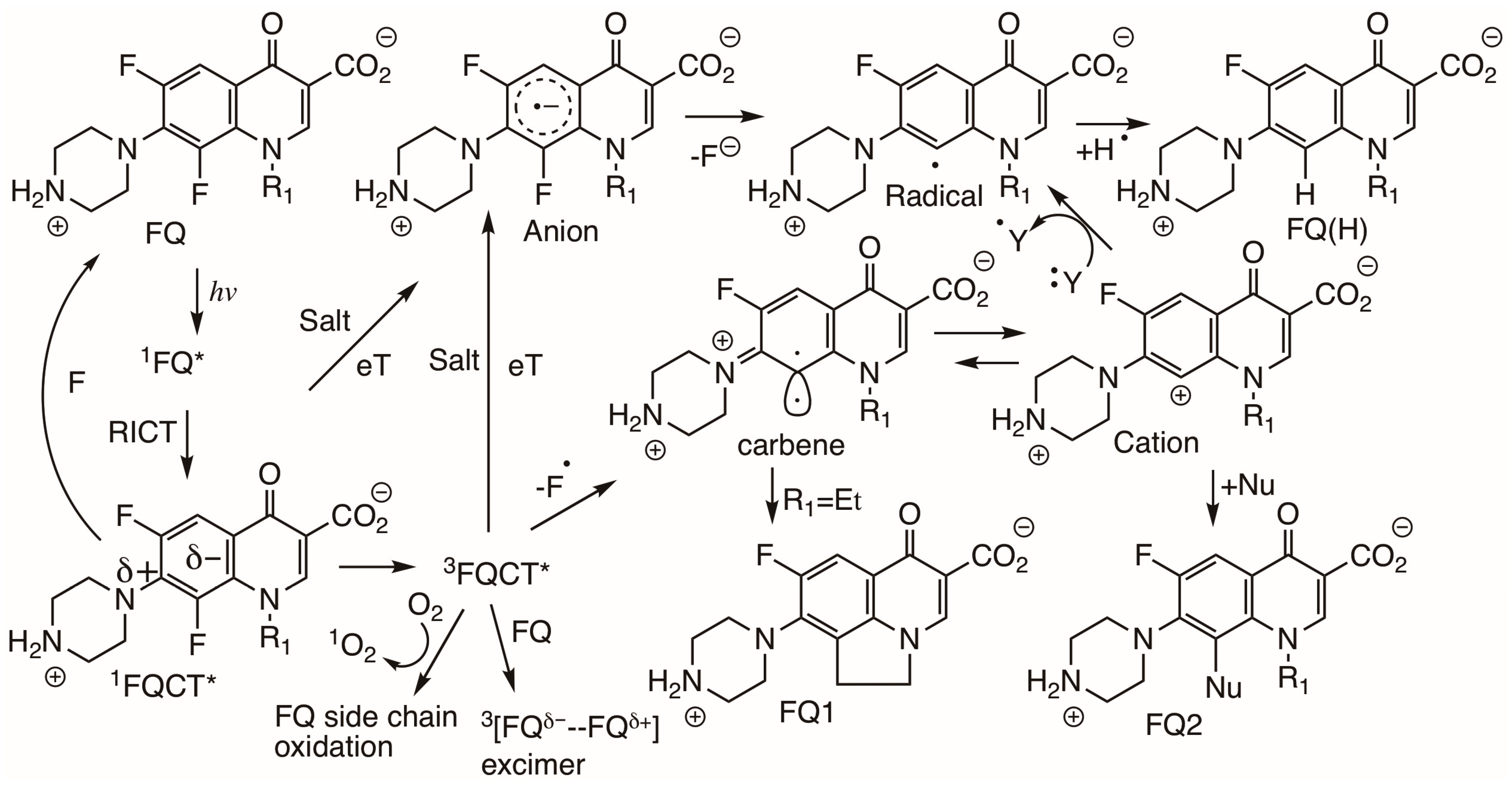
The intermediates generated in the main photophysical and photochemical processes in monohalogenated FQs are presented in Scheme 11. Upon photolysis, an FQ in the ground state generates an excited singlet state 1FQ*. This intermediate generates a charge transfer singlet excited state 1FQCT* by an intramolecular rehybridization charge transfer mechanism (RICT) [6,44]. This mechanism has been investigated in detail by the fluorescence spectroscopy of two quinolones containing a piperazine in their structure, ofloxacin and norfloxacin, in micelles [121]. A 1FQCT* could deactivate and go back to a ground state FQ by emitting the excess energy by fluorescence [29,82]. Several quinolones investigated gave fast intersystem crossing (ISC) (Table 1) under neutral conditions [6]. The quantum yield for triplet formation oscillated considerably (0.2 to 0.9), and quinolones with zwitterionic structures gave a particularly lower value (0.2). A high concentration of phosphate buffer (H3PO4) induced a reduction in ISC due to the formation of a complex of the ground state FQ with the salt (Scheme 5). The formation of this complex also explains the significant effect of the salt on the fluorescence quantum yield [6,29]. An FQ in the singlet state, free or in the form of a complex, easily undergoes electron transfer (eT) to generate an aryl anion, which upon subsequent defluorination generates an aryl radical. 1FQCT* undergoes ISC to 3FQCT*. This later transient, containing highly electronegative substituents, is quite prone to interact with hydroxyl anions to generate the corresponding phenols [45]. FQs in a triplet state 3FQCT* have lifetimes in the microsecond range; they react by electron transfer with surrounding salts to generated radical anions [6].
In addition, a 3FQCT* reacts with the remaining FQ to give secondary transient species 3[FQd−- -FQd+] with an excimer nature. In fact, the generation of secondary species on the photochemistry of FQs and electron donor compounds like tryptophan has been previously reported [59,122]. 3FQCT* also reacts by triplet energy transfer to oxygen with the production of singlet oxygen (1O2), a high-energy molecule that could easily oxidize an organic compound and/another FQ. FQs containing electron donor substituents (O or N) in the main ring (ofloxacin and rufloxacin) are more photostable and do not present C-F cleavage. Their main photodegradation pathway is the oxidation of the N-alkyl groups by ROS [28,39,120].

3.2. Photophysical and Photochemical Pathways Involved with Dihalogenated FQs

The photochemical and photophysical properties of dihalogenated quinolones (C6 and C8) have been investigated in many studies [6,51,54,123,124,125]. As was mentioned before, FQs containing a NH2 in an amino substituent (C7) like a piperazinyl group also present an association with phosphate salt, generating the corresponding complex molecule (Scheme 5). This effect is very small with ofloxacin and rufloxacin, since these two FQs contain a N-CH3 in the piperazinyl group and do not associate with phosphate salt [6].
Studies on these FQs (Scheme 12) indicated the unusual photochemical dehalogenation of the strong C8-halogen bond. This involves an electron transfer reaction between the quinolone main ring and the lone electron pair on piperazinyl [92] or an electron donor buffer [6]. On the other side, the triplet state of an FQ can easily undergo heterolysis of the C8-F bond. These photochemical processes involve the generation of an aryl radical cation and an aryl radical. Both intermediates are very reactive and have alkylating properties [51,54,93,94,95]. Aryl cations have been detected by means of nanosecond laser flash photolysis (LFP) in the photochemistry of FQs [54,124]. Several studies by this technique, along with fluorescence studies and photoproducts analysis, indicated there is an equilibrium between the triplet and singlet state of the aryl cation leading to the formation of both FQ1 and FQ2 products [54]. As previously described, an excited triplet state 3FQCT* with two fluorine atoms and enough energy could also undergo reactions with other FQs or with singlet oxygen [111].

3.3. Photophysical and Photochemical Pathways for Nonhalogenated Qs Like Nalidixic Acid

Early studies on nalidixic acid photochemical degradation indicated that this process is modulated by reaction conditions, with the degradation favored under a neutral pH [1,31]. Subsequent experimental and theoretical studies gave a more complete view of this process [126,127]. Based on several laser flash photolysis (LFP) studies, fundamental pathways for primary photo-conversions (Scheme 13) for nalidixic acidic anions in neutral conditions have been presented. Upon photolysis of the quinolone in the ground state (Q), a singlet excited state is generated (1Q*). This intermediate could return to ground state by emitting fluorescence light (F). By intersystem crossing (ISC), it could transform into a triplet excited state (3Q*). This latter triplet intermediate could undergo three different photo-conversion pathways. On one side, it could react with the proton in water to give a neutral triplet excited state 3Q* capable of dimerization. On the other side, it could give out a solvated electron e(H2O) to generate a radical anion in the triplet state. Eventually, this radical anion could generate another radical and several photoproducts. Furthermore, 3Q* could also react with oxygen to generate ROS. These three routes for 3Q* are consistent with its lifetime (100 ms) estimated from the phosphorescence spectra of nalidixic acid in different buffer solutions.
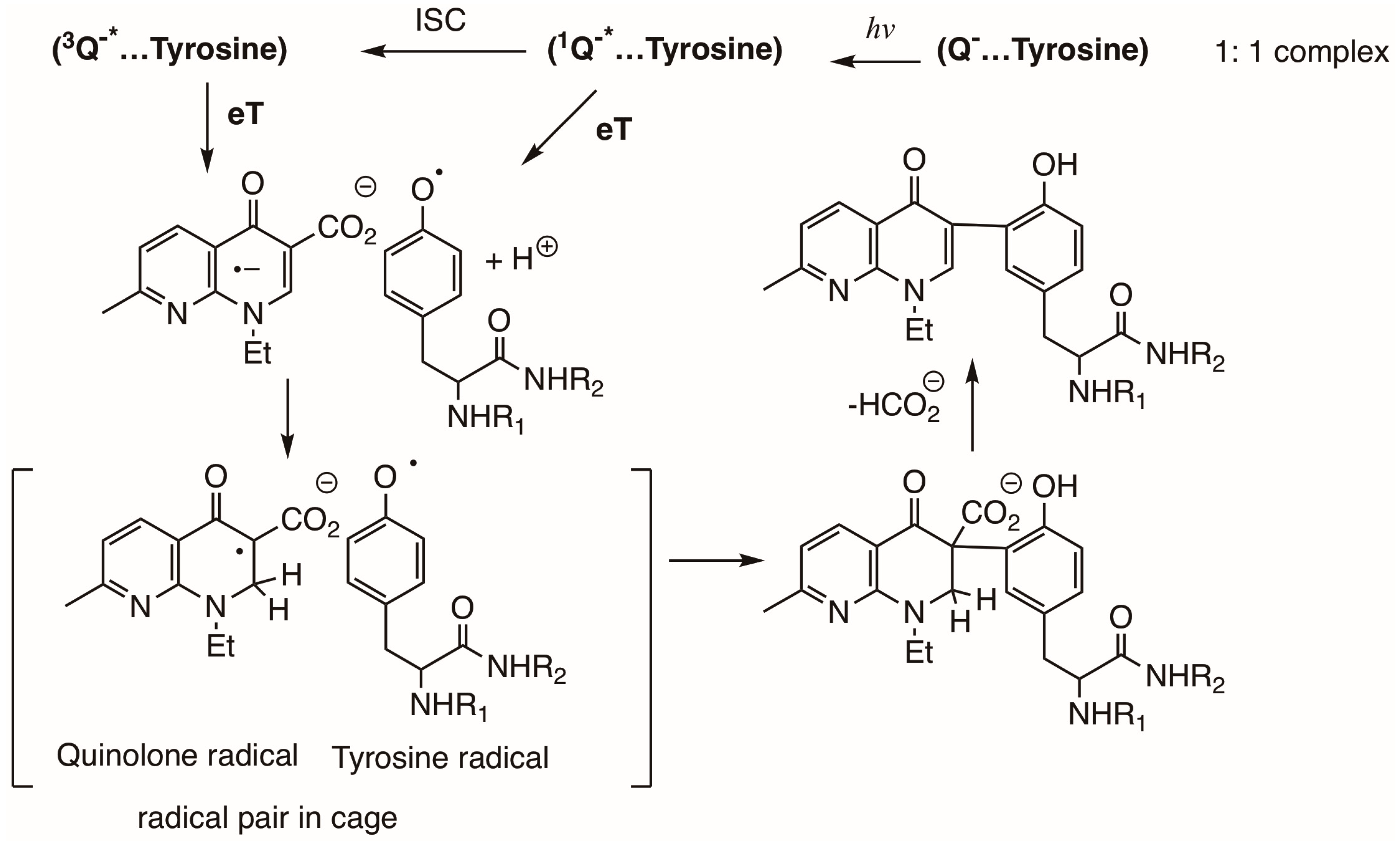
Monti et al. performed a detailed investigation of the basic photochemistry of the nalidixic anion Q in a protein environment by LFP [127]. Upon analysis of transient spectra of the triplet–triplet of a complex (1:1) of [3Q*…tyrosine], they demonstrated that a fast electron transfer (eT) from tyrosine to Q* leads to the formation of a radical pair (Scheme 14). This radical pair consisted of a tyrosine radical (λmax = 410 nm) and a quinolone radical (λmax = 640). No precursor excited state was detected, but this electron transfer process could take place from either excited state (singlet or triplet). The degradation of the radical pair by first-order kinetics and the nature of the photoproducts obtained indicated a cage combination reaction. Furthermore, the photoinduced covalent binding of the quinolone to the biomolecule of tyrosine was demonstrated by HPLC. These experimental LFP studies presented strong evidence of photochemically induced reactions of a quinolone (Q) with a biomolecule (BIOM). Other experimental LFP studies have also demonstrated that a Q in the form of a radical or a triplet excited state (Scheme 13) could easily generate oxygen superoxide upon an electron transfer reaction (eT) with oxygen [127].

3.4. Photosensitized Biological Damage by Qs and FQs

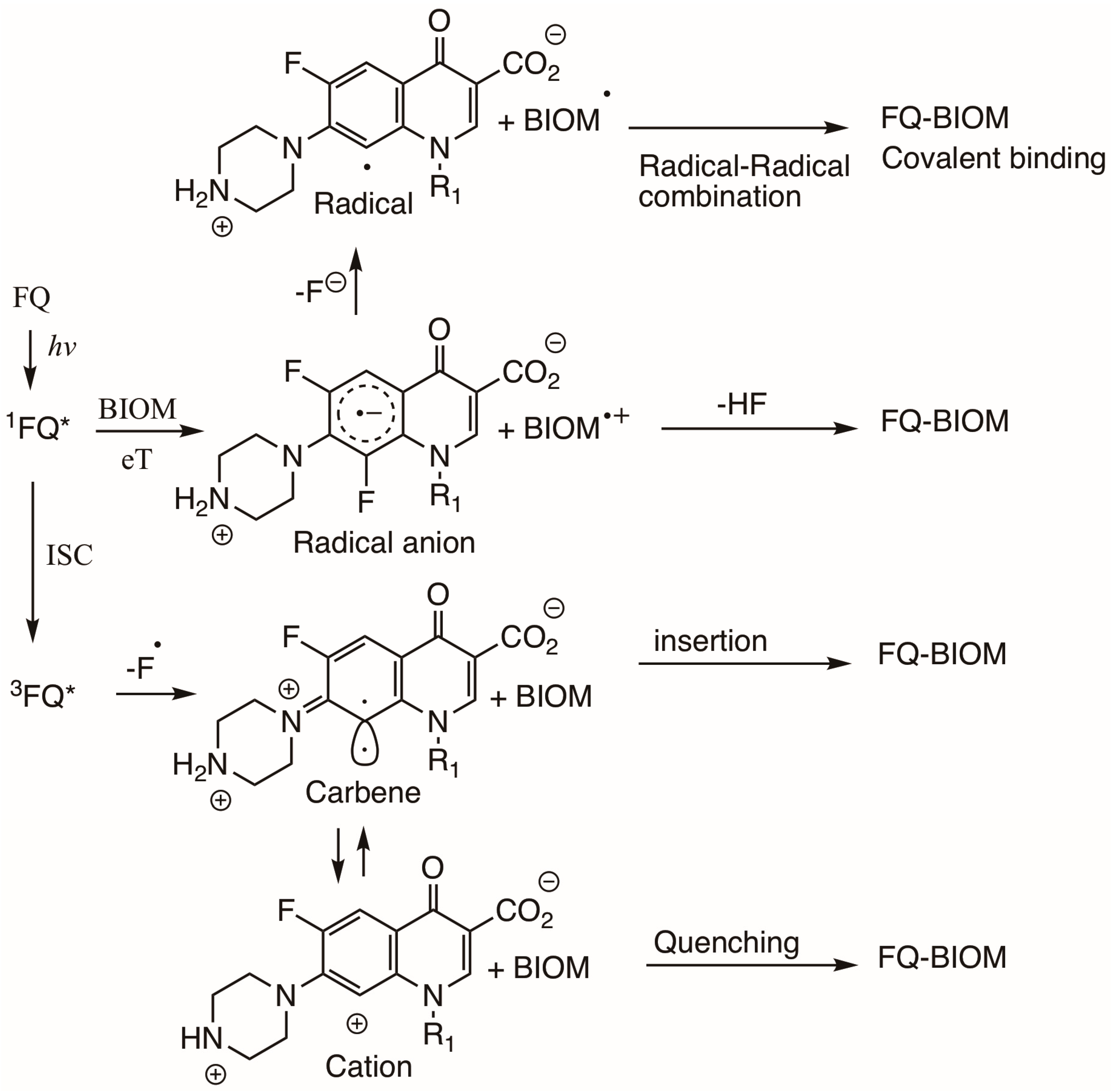
In neutral aqueous solution, the photochemical processes of Qs and FQs involve a variety of reactions such as dehalogenation, oxidation of amino substituents, decarboxylation and production of highly reactive organic and oxygen species (ROS) [128]. However, since the production of ROS by Qs and FQs does not correlate with their phototoxicity effects [111,112], these adverse effects have been associated with different physicochemical properties such as their photosensitized reactions with biomolecules (Scheme 15) [129]. The first step in these processes, involves the absorption of light by an FQ sensitizer, with the production of a singlet excited state 1FQ*. This latter intermediate undergoes intersystem crossing (ISC) very fast to generate a triplet excited state 3FQ*. Any of these two highly reactive intermediates (singlet or triplet) could react with a biomolecule (BIOM) by means of direct reaction, involving hydrogen atom or electron transfer to give radicals and radical anions. They could also react to give carbenes, radicals, anions and cations. The presence of several intermediate species in the photochemistry of FQs explains their different direct reactions with a BIOM such as photobinding, photooxidation, photocrosslinking [49,111,112,129] and pyrimidine dimerization in DNA [130]. All of these reactions, observed with diverse BIOM, have been classified as Type I reactions. On the other side, Type II reactions involve indirect oxidation of a BIOM by the triplet photosensitizer 3FQ*. First, there occurs a transfer of energy from the excited triplet 3FQ* to oxygen, inducing the formation of highly reactive singlet oxygen (1O2) with the subsequent oxidation of a biomolecule.
Some studies on cell membrane damage have shown that Qs and FQs induce the photooxidation of lipids, since they generate ROS, which trigger lipid peroxidation chain reactions [49]. Investigations of the photochemical reactions of Qs and FQs with proteins and DNA indicate these processes could induce damage in proteins. They could induce protein–protein photocrosslinking, protein photooxidation and protein–FQ photobinding [49,56,111,112,129,130]. Furthermore, FQs in the presence of light could also induce DNA damage by two mechanisms; photooxidation or the generation of thymine dimers [39,48,49,119,131,132,133]. These dimers have been directly detected by HPLC coupled with a particular technique (electrochemistry, UV–Vis, fluorescence or mass spectrometry) or by indirect detection upon the formation of strand breaks or single strand breaks (SBs or SSBs) of DNA [134].

3.5. Photochemical Reactions Between Q Intermediates and Monomers of Biomolecules

Nalidixic acid was the first Q used as an antimicrobial agent. However, its application in medicine was limited by skin photosensitization reactions [31]. Under NAL treatment of urinary tract infections, skin eruptions appeared after the exposure of patients to sunlight. Several studies on NAL indicated the photochemistry of a Q is actually a quite complex process. It involves the formation of several excited states and intermediates that could react with biomolecules by different mechanisms [126,127]. It was actually demonstrated that NAL associated with tyrosine to generate a radical pair and a tyrosine derivative. These pioneering studies on NAL have been followed by many studies on fluoroquinolone damage in proteins and DNA [112].
The formation of a highly reactive aryl cation, upon unusual C8–halogen bond photochemical heterolysis in some FQs, was originally proposed as the most important cause of FQs adverse side effects [34,35]. In the case of lomefloxacin (LOM), this highly reactive aryl cation was quenched by guanosine monophosphate, and it was possible to detect the formation of the resulting covalently bound compound (LOM-dGMP) [125]. However, the triplet excited states of monohalogenated and dihalogenated quinolones have also been shown to be very reactive species. They react with diverse biomolecules like tyrosine, tryptophan and deoxyguanosine [122,126]. Furthermore, FQ irradiation induces very complex photophysical and photochemical pathways that involve the generation of several excited states and intermediates. These highly reactive species could also react with a biomolecules (BIOM) by different mechanisms (Scheme 16).

3.6. Photosensitized Reactions with Proteins by Qs and FQs

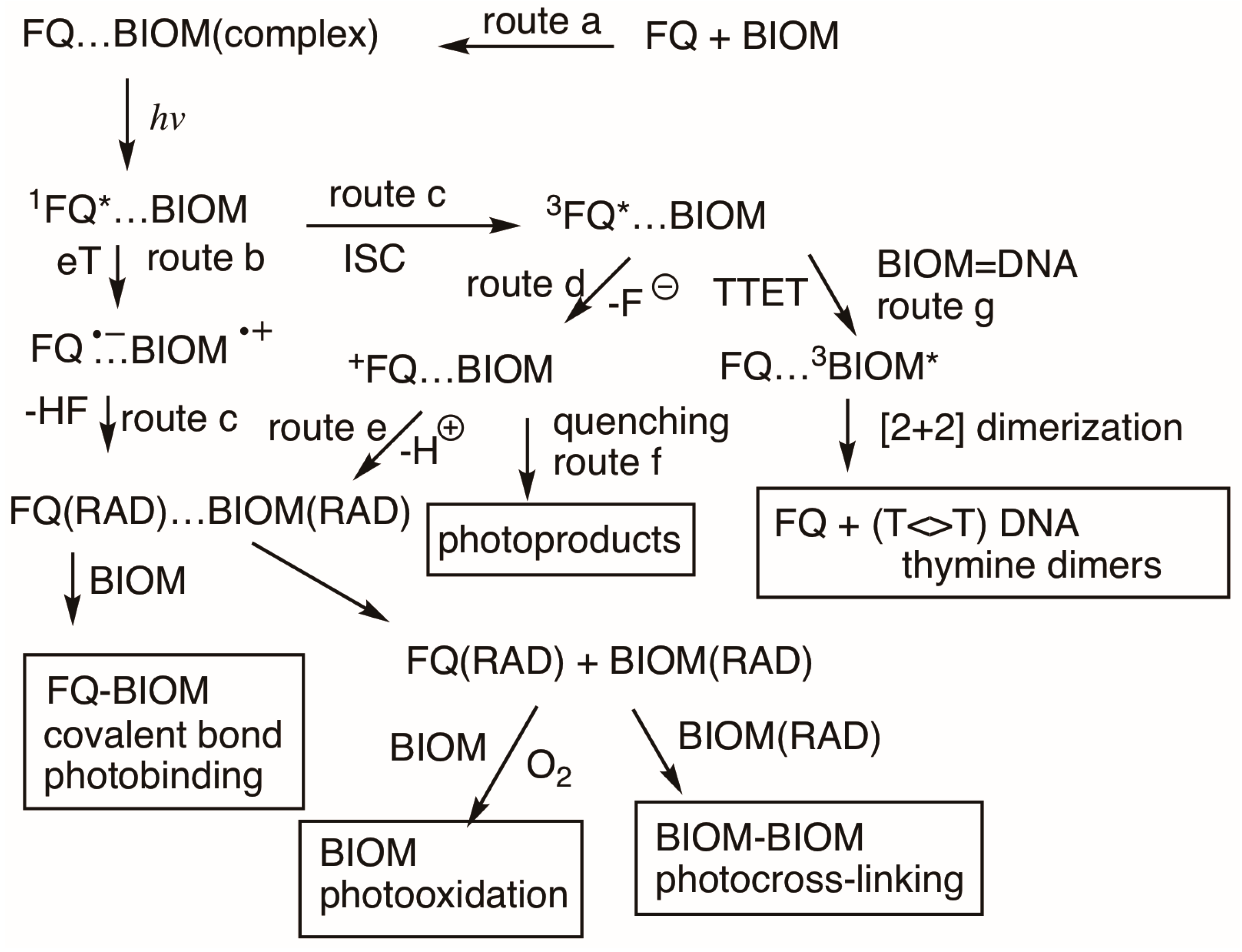
It has been demonstrated that FQs’ excited states behave very different when they are generated in an aqueous environment or when they are associated with proteins [55,129] Therefore, another important physicochemical property to consider in FQs’ photosensitized reactions with biomolecules is the formation of complexes (FQ-BIOM) that will favor fast reactions [111]. The binding constants for albumin protein and different FQs has been calculated using different techniques [135,136]. It was observed that most FQs gave small Ka values, indicating a low percentage of association under biological conditions. The main photophysical and photochemical pathways for the photoinduced damage of a biomolecule (BIOM) by FQs after the formation of complexes are presented in Scheme 17. Photoproduct detection and laser flash photolysis (LFP) studies provided evidence that the main mechanism in FQ protein damage (route a) involved the formation of a complex (1FQ*…BIOM) [129]. An electron transfer (route b) from an amino acid or thiol group from the BIOM to the 1FQ* is a key step in the formation of radicals and photobinding. The formation (routes c and e) of biomolecular radical BIOM(RAD) eventually results in the photocrosslinking and photooxidation of abiomolecule. This latter process also results from the reaction of singlet oxygen (1O2) generated by energy transfer (ET) from 3FQ* to molecular oxygen. The photobinding and photoproducts observed with difluoro quinolones (routes c, d, e and f) correlate well with the heterogeneous dehalogenation properties of (3FQ*-BIOM) that generate a highly reactive cation complex (FQ+…BIOM). In general, photobinding is a process with higher yields for dihalogenated FQs [111].
Several processes are involved in DNA damage by FQs. BIOM photooxidation is eventually induced by radicals generated from the complex (1FQ*…BIOM). This oxidation also occurs by means of singlet oxygen generated in situ. Furthermore, thymine cyclobutene dimers are generated, by triplet–triplet energy transfer (TTET, route g), from 3FQ* to BIOMs. This later mechanism is explained in the following section.

3.7. Photosensitized Reactions in DNA by Qs and FQs

Ultraviolet solar radiation involves UVB light with a short wavelength (290 to 320 nm) and UVA light with a longer wavelength (320 to 400 nm) [119]. Photocarcinogenesis has been mostly associated with oxidative stress [137,138]. Oxidation by singlet oxygen (Type I), and to a lesser extent radical production (Type II), has been associated with the oxidation of guanine in DNA upon exposure to UVA light. However, cyclobutene pyrimidine dimers (CPDs) could also be produced by the UVB irradiation of DNA, and their yields are usually higher. The mechanism involved in this latter process has been a matter of discussion for several years [139,140]. It could be induced by two different procedures, the direct UVA irradiation of DNA or the photosensitization of endogenous agents. Since DNA hardly absorbs UVA light, a photosensitization mechanism involving different types of biomolecules (psoralen, porphyrins, flavins, steroids and quinones) has been proposed [141].
CPDs’ formation by photosensitization has been investigated by theoretical and experimental studies, and the mechanism (Scheme 18) for this process has been elucidated [119]. It takes place by a [2 + 2] cycloaddition between two pyrimidines. First an FQ photosensitizer undergoes UVA light absorption and generates a singlet excited state 1FQ*. This singlet intermediate undergoes intersystem crossing to a triplet excited state 3FQ*. At this point, through a triplet–triplet energy transfer TTET, the 3FQ* transfers its excess energy to a pyrimidine base, giving rise to a base excited state (3Pyr*). This latter excited state has enough energy to react with a ground state (Pyr) to generate cyclobutene dimers. Several physicochemical properties should be fulfilled for the photosensitizer to participate in this reaction. For selective excitation, it should absorb light at a longer wavelength than pyrimidine. Thermodynamically, it should have a triplet with a higher energy than pyrimidine (Scheme 18). To increase the probability of energy transfer to pyrimidine, it should have a good intersystem crossing quantum yield (θISC) and a long triplet lifetime (τT). To increase the possibility of collisions, it should be close to pyrimidine.
Pyrimidine triplet excited states have been associated with DNA damage, but only recently have they been analyzed by theoretical studies [142,143,144]. After the excitation of a photosensitizer, such as an FQ, diverse processes may take place. The first step is the formation of a singlet excited state. But this latter intermediate can go back to the ground state by fluorescence or internal conversion. On the other side, ISC leads to an excited triplet state. Therefore, a good photosensitizer should have a quantum yield close to one for the ISC to be favored. The triplet energy of the donor (3FQ*) must be at least 12 KJ/mol higher than that of the triplet (3Pyr*) for an irreversible triplet–triplet energy transfer (TTET).
In spite of several adverse effects, FQs are an important class of pharmaceutical compounds with a broad biological activity as antibacterial, antimalarial, antiviral, antifungal and anticancer drugs. Investigations about different synthetic methodologies to prepare new FQs are contributing to the advancement of new derivatives with diverse physicochemical properties [145,146,147,148,149,150].

4. Conclusions

In order to obtain quinolones with a broader spectrum of activity and better physicochemical properties, new generations have been developed. Chemical modifications on the basic quinolone structure modulate their biological activity. A fluorine atom in position six enhances bacterial cell penetration and improves distribution in the microorganism. A second halogen atom in position eight gives a broader spectrum of activity. An amino in position five increases the overall potency and modifies several physicochemical properties.
Irradiation of a given Q or FQ under neutral conditions induces diverse reactions. Upon light excitation of a nonhalogenated quinolone such as nalidixic acid, a singlet excited state is generated. This intermediate easily undergoes ISC to a triplet excited state, which undergoes several reactions. Upon decarboxylation, this latter triplet generates radicals that react with diverse biomolecules or generate dimers. This triplet also reacts with other Qs, oxygen or water to generate other reactive intermediates (singlet oxygen or anions) capable of inducing oxidation or electron transfer reactions.
In a neutral aqueous solution, irradiation of an FQ initiates diverse chemical reactions such as dehalogenation, decarboxylation, the oxidation of amino substituents, and the production of highly reactive organic or oxygen species (ROS). However, since phototoxicity does not correlate with the production of ROS, phototoxicity has been associated with diverse photosensitized reactions of FQs. Upon light absorption, an FQ sensitizer is transformed into two species, excited singlet and triplet FQs. These two species easily generate other intermediates such as carbenes, radicals, anions and cations. The presence of all of these intermediates explains the direct photoreactions of FQs with biomolecules, inducing photobinding, photooxidation, photocrosslinking and pyrimidine dimerization in DNA. All direct photochemical reactions of FQs with biomolecules have been classified as Type I reactions, while indirect FQs reactions with biomolecules are known as Type II reactions. Indirect reactions involve an energy transfer from the excited triplet state 3FQ* to oxygen, with the generation of highly reactive singlet oxygen and the subsequent oxidation of biomolecules.
Since Qs and FQs bind to biomolecules, generating complexes, their photochemical behavior is quite different in an aqueous solution from their behavior in a biological environment. Due to close contact within a complex, reactions between an FQ and a biomolecule are accelerated. Upon photolysis, singlet and triplet excited FQs bound to biomolecules are generated and react very fast, inducing photobinding, photooxidation, protocrosslinking and dimerization. Lomefloxacin, fleroxacin, enoxacin and norfloxacin are highly photoreactive, and their major mechanisms of reaction come from the photochemistry of their complexes. On the other side, ofloxacin and rufloxacin are molecules with a low-energy triplet (below 269 kcal mol−1) and a low quantum yield for photodegradation; thus the same intermediates are generated under an aqueous or biological environment. These intermediates are responsible for biological damage to cells and tissues.
In recent decades, new Qs and FQs have been developed in order to obtain derivatives with adequate biological activity and mild adverse effects. With the exception of nemonoxacin, all of these derivatives contain a F in C6, since this substituent confers adequate biological and physicochemical properties. They also have either a cyclic amine or cyclic alkane in C7, with a structure different from the piperazine present in earlier Qs. They do not have a halogen on C8; instead, some of they have a methoxy in this position. This latter substituent has been reported to lower phototoxicity. All of these recently developed derivatives present mild adverse effects, including phototoxicity.
FQs are an important class of pharmaceutical compounds with a broad biological activity as antibacterial, antimalarial, antiviral, antifungal and anticancer drugs. In general, their medicinal applications have been limited due to several adverse effects in humans, like phototoxicity. Investigations about different synthetic methodologies to prepare new FQs are contributing to the advancement of new derivatives with improved physicochemical properties. However, for these new derivatives to be used in medical practice, it is important to investigate their photophysical and photochemical properties and adverse human effects, such as phototoxicity.

Funding

This research was funded by L′oréal, UNESCO and the Mexican Academy of Science Grant (G 2022).

Acknowledgments

Elisa Leyva gratefully acknowledges L’oréal, UNESCO and the Mexican Academy of Science. Irving R. Rodríguez-Gutierrez acknowledges CONAHCYT a research fellowship (CVU 513493).

Conflicts of Interest

The authors declare no conflict of interest.

References

  1. Mitscher, L.A. Bacterial Topoisomerase Inhibitors: Quinolone and Pyridone Antibacterial Agents. Chem. Rev. 2005, 105, 559–592. [Google Scholar] [CrossRef] [PubMed]
  2. Alfred, K.J.; Kerns, R.J.; Osheroff, N. Mechanim of Quinolone Action and Resistance. Biochemistry 2014, 53, 1565–1574. [Google Scholar] [CrossRef]
  3. Anderson, V.E.; Osheroff, N. Type II Topoisomerases as targets for Quinolone Antibacterials: Turning Dr. Jeryll into Mr Hyde. Curr. Pharm. Des. 2001, 7, 337–353. [Google Scholar] [CrossRef] [PubMed]
  4. Sheikhi, N.; Bahraminejad, M.; Saeedi, M.; Mirfazli, S.S. A review: DFA-approved fluorine-containing small molecules from 2015 to 2022. Eur. J. Med. Chem. 2023, 260, 115758. [Google Scholar] [CrossRef]
  5. Zhanel, G.G.; Ennis, K.; Vercaigne, L.; Walkty, A.; Gin, A.S.; Embil, J.; Smith, H.; Hoban, D.J. A Critical Review of the Fluoroquinolones. Focus on Respiratory Tract Infections. Drugs 2002, 62, 13–59. [Google Scholar] [CrossRef]
  6. Albini, A.; Monti, A. Photophysics and photochemistry of fluoroquinolonas. Chem. Soc. Rev. 2003, 32, 238–250. [Google Scholar] [CrossRef] [PubMed]
  7. Bisacchi, G.S. Origins of the Quinolone Class of Antibacterials: An Expanded Discovery Story. J. Med. Chem. 2015, 58, 4874–4882. [Google Scholar] [CrossRef] [PubMed]
  8. Naeem, A.; Badshah, S.L.; Muska, M.; Ahmad, N.; Khan, K. The Current Case of Quinolones: Synthetic Approaches and antibacterial Activity. Molecules 2016, 21, 268. [Google Scholar] [CrossRef]
  9. Kumar, A.; Saxena, N.; Mehrotra, A.; Srivastava, N. Review: Studies on the Synthesis of Quinolone Derivatives with their Antibacterial Activity (Part 1). Curr. Org. Chem. 2020, 24, 817–854. [Google Scholar] [CrossRef]
  10. Saxena, N.; Shankhdhar, S.; Kumar, A.; Srivastava, N. Part 2. Studies on the Synthesis of Quinolone Derivatives with Their Biological Activity. Curr. Org. Chem. 2024, 28, 185–212. [Google Scholar] [CrossRef]
  11. Leyva, S.; Leyva, E. Thermochemical reaction of 7-azide-1-ethyl-6,8-difluoroquinolone-3-carboxylate with heterocyclic amines. An expeditious synthesis of novel fluoroquinolone derivatives. Tetrahedron 2007, 63, 2093–2097. [Google Scholar] [CrossRef]
  12. Leyva, E.; de Loera, D.; Leyva, S. Photochemistry of 7-azide-1-ethyl-3-carboxylate-6,8-difluoroquinolone: A novel reagent for photoaffinity labeling. Tett. Lett. 2008, 49, 6759–6761. [Google Scholar] [CrossRef]
  13. Anwar, A.I.; Lu, L.; Plaisance, C.J.; Daniel, C.P.; Flanagan, C.J.; Wenger, D.M.; McGregor, D.; Varrassi, G.; Kaye, A.M.; Ahmadzadeh, S.; et al. Fluoroquinolones: Neurobiological Complications and Side Effets in Clinical Practice. Cureus 2024, 16, e54565. [Google Scholar] [CrossRef] [PubMed]
  14. Owens, R.C.; Ambrose, P.G. Antimicrobial Safety: Focus on Fluoroquinolones. Clin. Infect. Dis. 2005, 41, S144–S157. [Google Scholar] [CrossRef]
  15. Carbon, C. Comparison of Side Effects of Levofloxacin versus Other Fluoroquinolones. Chemotheraphy 2001, 47, 9–14. [Google Scholar] [CrossRef]
  16. Bush, N.G.; Diez-Santos, I.; Abbot, L.R.; Maxwell, A. Quinolones: Mechanism, Lethality and Their Contributions to Antibiotic Resistance. Molecules 2020, 25, 5662. [Google Scholar] [CrossRef]
  17. Zhang, G.F.; Zhang, S.; Pan, B.; Liu, X.; Feng, L.S. 4-Quinolone derivatives and their activities against Gram positive Pathogens. Eur. J. Chem. 2017, 143, 710–723. [Google Scholar] [CrossRef] [PubMed]
  18. Takács-Novák, K.; Noszál, B.; Hermecz, I.; Keresztúri, G.; Podányi, B.; Szász, G. Protonation Equilibria of Quinolone Antibacterials. J. Pharm. Sci. 1990, 79, 1025–1028. [Google Scholar] [CrossRef]
  19. Rusu, A.; Tóth, G.; Azocs, L.; Kökósi, J.; Kraszni, M.; Gyéresi, A.; Noszál, B. Triprotic site-specific acid-base equilibria and related properties of fluoroquinolone antibacterials. J. Pharm. Biomed. Anal. 2012, 66, 50–57. [Google Scholar] [CrossRef]
  20. Fresta, M.; Guccione, S.; Beccari, A.R.; Furneri, P.M.; Puglisi, G. Combining molecular modeling with experimental methodologies: Mechanism of membrane permeation and accumulation of ofloxacin. Bioorg. Med. Chem. 2020, 10, 3871–3889. [Google Scholar] [CrossRef]
  21. Barret, R. Importance and Evaluation of Liphophilicity. In Therapeutical Chemistry; Elsevier: Amsterdam, The Netherlands, 2018; pp. 53–78. [Google Scholar] [CrossRef]
  22. Cuprys, A.; Pulicharla, R.; Brar, S.K.; Drogui, P.; Verma, M.; Surampalli, R. Y: Fluoroquinolones metal complexation and its environmental impacts. Coord. Chem. Rev. 2018, 376, 46–61. [Google Scholar] [CrossRef]
  23. Araki, T.; Ohta, Y.K.I.; Kitaoka, H. Photochemical behavior of Sitafloxacin, Fluoroquinolone Antibiotic, in an Aqueous Solution. Chem. Pharm. Bull. 2002, 50, 229–234. [Google Scholar] [CrossRef]
  24. Bano, R.; Anwar, Z.; Mustn, N.; Ahmad, I. Photochemistry of Fluoroquinolones: A Review. Baqai J. Health Sci. 2017, 1, 1–22. [Google Scholar]
  25. Sortino, S.; De Guidi, G.; Giuffrida, S.; Monti, S.; Velardita, A. pH Effects on the Spectroscopic and photochemical Behavior of Enoxacin: A Steady-State and Time-Resolved Study. Photochem. Photobiol. 1998, 67, 167–173. [Google Scholar] [CrossRef]
  26. Navaratnam, S.; Claridge, J. Primary Photophysical Properties of Ofloxacin. Photochem. Photobiol. 2000, 72, 283–290. [Google Scholar] [CrossRef] [PubMed]
  27. Sortino, S.; Marconi, G.; Giuffrida, S.; De Guidi, G.; Monti, S. Photophysical Properties of Rubofloxacin in Neutral Aqueous Solution. Photochem. Photobiol. 1999, 70, 731–736. [Google Scholar] [CrossRef]
  28. Belvedere, A.; Boscá, F.; Cuquerella, M.C.; De Guidi, G.; Miranda, M.A. Photoinduced N-Demethylation of Rufloxacin and its Methyl Ester Under Aerobic Conditions. Photochem. Photobiol. 2002, 76, 252–258. [Google Scholar] [CrossRef]
  29. Monti, S.; Sortino, S.; Fasani, E.; Albini, A. Multifaceted Photoreactivity of 6-Fluoro-7-aminoquinolones from the Lowest Excited States in Aqueous Media: A Study of Nanosecond and Picosecond Spectroscopic Techniques. Chem. Eur. J. 2001, 7, 2185–2196. [Google Scholar] [CrossRef]
  30. Bazin, M.; Bosca, F.; Marin, M.L.; Miranda, M.A.; Patterson, L.K.; Santus, R.; Laser, A. Flash Study and Pulse Radiolysis Study of Primary Photochemical Processes of Flumequine. Photochem. Photobiol. 2000, 72, 451–457. [Google Scholar] [CrossRef]
  31. Vermeersch, G.; Ronfard-Haret, J.C.; Bazin, M.; Carillet, V.; Morliere, P.; Santus, R. Type I and Type II Photosensitization by the Antibacterial Drug Nalidixic Acid. A laser Flash Photolysis Study. Photochem. Photobiol. 1991, 54, 661–666. [Google Scholar] [CrossRef]
  32. Mella, M.; Fasani, E.; Albini, A. Photochemistry of 1-Cyclopropyl-6-fluoro-1,4-dihydro-4-oxo-7-(piperazin-1-yl)quinoline-3-carboxylic Acid(=Ciprofloxacin) in Aqueous Solutions. Helv. Chim. Acta 2001, 84, 2508–2519. [Google Scholar] [CrossRef]
  33. Fasani, E.; Barberis Negra, F.F.; Mella, M.; Monti, S.; Albani, A. Photoinduced C-F Cleavage in Some Fluorinated 7-Amino-4-quinolone-3-carboxylic Acids. J. Org. Chem. 1999, 64, 5388–5395. [Google Scholar] [CrossRef] [PubMed]
  34. Martínez, L.J.; Sik, R.H.; Chignell, C.F. Fluoroquinolone Antimicrobials: Singlet Oxygen, Superoxide and Phototoxicity. Photochem. Photobiol. 1998, 67, 399–403. [Google Scholar] [CrossRef]
  35. Scheife, R.T.; Cramer, W.R.; Decker, E.L. Photosensitizing potential of ofloxacin. Int. J. Dermatol. 1993, 32, 413–416. [Google Scholar] [CrossRef] [PubMed]
  36. Tokura, Y.; Nishijima, T.; Yagi, H.; Furukawa, F.; Takigawa, M. Photohaptenic Properties of Fluoroquinolones. Photochem. Photobiol. 1996, 64, 838–842. [Google Scholar] [CrossRef]
  37. Fasani, E.; Profumo, A.; Albini, A. Structure and Medium-Dependent Photodecomposition of Fluoroquinolone Antibiotics. Photochem. Photobiol. 1998, 68, 666–674. [Google Scholar]
  38. Condorelli, G.; De Guidi, G.; Giuffrida, S.; Sortino, S.; Chillemi, R.; Sciuto, S. Molecular Mechanisms of Photosensitization Induced by Drugs XII. Photochemistry and Photosensitization of Rufloxacin. Photochem. Photobiol. 1999, 70, 280–286. [Google Scholar] [PubMed]
  39. Belvedere, A.; Boscá, F.; Catalfo, A.; Cuquerella, M.C.; de Guidi, G.; Miranda, M.A. Type II Guanine Oxidation Photoinduced by the Antibacterial Fluoroquinolone Rufloxacin in Isolated DNA and in 2′Deoxyguanosine. Chem. Res. Toxicol. 2002, 5, 1142–1149. [Google Scholar] [CrossRef] [PubMed]
  40. Cosa, G.; Scaiano, J.C. Laser Techniques in the Study of Drug Photochemistry. Photochem. Photobiol. 2004, 80, 159–174. [Google Scholar] [CrossRef]
  41. Shields, D.J.; Chakraborty, M.; Abdelaziz, N.; Duley, A.; Gudmundsdottir, A.D. Review of laser flash photolysis of organic molecules (2015–2018). Photochemistry 2020, 47, 70–121. [Google Scholar] [CrossRef]
  42. Xu, Y.; Liu, Y.; Zhao, J.; Li, H.; Zhang, P.; Wang, W. Photochemical properties of Gemifloxacin: A laser flash photolysis study. J. Photochem. Photobiol. B. 2015, 143, 30–37. [Google Scholar] [CrossRef] [PubMed]
  43. Li, H.; Zhang, P.; Li, Y.; Tang, R.; Tang, Z.; Xing, Z.; Yao, S.; Fu, H.; Wang, W. Photophysical properties of gatifloxacin in aquous solution by laser photolysis and pulse radiolysis. Radiat. Phys. Chem. 2012, 81, 40–45. [Google Scholar] [CrossRef]
  44. Cuquerella, M.C.; Miranda, M.A.; Boscá, F. Role of Singlet Intramolecular Charge Transfer in the photophysical Properties of Norfloxacin and Its Derivatives. J. Phys. Chem. A 2006, 110, 2607–2612. [Google Scholar] [CrossRef]
  45. Cuquerella, M.C.; Boscá, F.; Miranda, M.A. Photonucleic Aromatic Substitution of 6-fluoroquinolones in Basic Media: Triplet Quenching of Hydroxide Anion. J. Org. Chem. 2004, 69, 7256–7261. [Google Scholar] [CrossRef] [PubMed]
  46. Fasani, E.; Mella, M.; Albani, A. Photochemistry of the Phototoxic Drug Lomefloxacin: Paths Observed in the presence of Amines or NaOH and from the Methyl Ester. Eur. J. Org. Chem. 2004, 2004, 5075–5082. [Google Scholar] [CrossRef]
  47. Luo, T.; Chen, J.; Song, B.; Ma, H.; Fu, Z.; Peijnenburg, W.J.G.M. Time-gated luminescence imaging of singlet oxygen photoinduced by fluoroquinolones and functionalized graphenes in Daphnia magna. Aquatic Toxicol. 2017, 191, 105–112. [Google Scholar] [CrossRef]
  48. Bosca, F.; Lhiaubet-Vallet, V.; Cuquerella, M.C.; Castell, J.V.; Miranda, M.A. The Triplet Energy of Thymine in DNA. J. Am. Chem. Soc. 2006, 128, 6318–6319. [Google Scholar] [CrossRef]
  49. Viola, G.; Facciolo, L.; Canton, M.; Vedaldi, D.; Dall’Acqua, F.; Aloisi, G.G.; Amelia, M.; Barbafina, A.; Elisei, F.; Latterini, L. Photophysical and phototoxic properties of the antibacterial fluoroquinolones levofloxacin and moxifloxacin. Chem. Biodivers. 2004, 1, 782–801. [Google Scholar] [CrossRef]
  50. Lovdahl, M.J.; Priebe, S.R. Characterization of clinafloxacin photodegradation products by LC-MS/MS and NMR. J. Pharm. Biomed. Anal. 2000, 23, 521–534. [Google Scholar] [CrossRef]
  51. Fasani, E.; Monti, S.; Manet, I.; Tilocca, F.; Petali, L. Inter- and Intramolecular Photochemical Reactions of Fleroxacin. Org. Lett. 2009, 11, 1875–1878. [Google Scholar] [CrossRef]
  52. Lorenzo, F.; Navaratnam, S.; Edge, R.; Allen, N.S. Primary Photophysical Properties of Moxifloxacin. A Fluoroquinolone Antibiotic. Photochem. Photobiol. 2008, 84, 1118–1125. [Google Scholar] [CrossRef] [PubMed]
  53. Monti, S.; Sortino, S. Laser flash photolysis study for Photoionization in fluoroquinolones. Photochem. Photobiol. Sci. 2002, 1, 877–881. [Google Scholar] [CrossRef]
  54. Soldevilla, S.; Bosca, F. Photoreactivity of Fluoroquinolones: Nature of Aryl cations Generated in Water. Org. Lett. 2012, 14, 3940–3943. [Google Scholar] [CrossRef]
  55. Soldevilla, S.; Cuquerella, M.C.; Lhiaubert-Vallet, V.; Edge, R.; Bosca, F. Seeking the mechanism responsible for fluoroquinolone photomutagenicity: A pulse radiolysis, steady-state, and laser flash photolysis study. Free. Radic. Biol. Med. 2014, 67, 417–425. [Google Scholar] [CrossRef] [PubMed]
  56. Lorenzo, F.; Navaratnam, S.; Allen, N.S. Formation of Secondary Triplet Species after Excitation of Fluoroquinolones in the Presence of Relative Strong Bases. J. Am. Chem. Soc. 2008, 130, 12238–12239. [Google Scholar] [CrossRef]
  57. Anaya-Gonzalez, C.; Soldevilla, S.; Garcia-Lainez, G.; Bosca, F. Chemical tuning for potential antitumor fluoroquinolonas. Free Radical Biol. Med. 2019, 141, 150–158. [Google Scholar] [CrossRef]
  58. Soldevilla, S.; Bosca, F. Assessing physical properties of amphoteric fluoroquinolones using phosphorescence spectroscopy. Spectrochim. Acta A Mol. Biomol. Spectrosc. 2020, 227, 117569. [Google Scholar] [CrossRef]
  59. Cuquerella, M.C.; Andreu, I.; Soldevilla, S.; Bosca, F. Triplet Excimers of Fluoroquinolones in Aquous Media. J. Phys. Chem. A 2012, 116, 5030–5038. [Google Scholar] [CrossRef] [PubMed]
  60. Peng, Z.; HaiXia, L.; SiDe, Y.; WenFeng, W. Effects of pH and polarity on the excited states of norfloxacin and its 4′-N-acetyk derivative: A steady-state and time-resolved study. Sci. China Chem. 2014, 57, 409–416. [Google Scholar] [CrossRef]
  61. Iesce, M.R.; Cermola, F.; Termussi, F. Photooxygenation of Heterocycles. Curr. Org. Chem. 2005, 9, 109–139. [Google Scholar] [CrossRef]
  62. Miolo, G.; Viola, G.; Vedaldi, D.; Dall’Acqua, F.; Fravolini, A.; Tabarrini, O. In vitro phototoxic properties of new 6-des-fluoro and 6-fluoro-8-methylquinolones. Toxicol. Vitr. 2002, 16, 683–693. [Google Scholar] [CrossRef] [PubMed]
  63. Lipsky, B.A.; Baker, C.A. Fluoroquinolone Toxicity Profiles: A Review Focusing on Newer Agents. Clin. Infect. Dis. 1999, 28, 352–364. [Google Scholar] [CrossRef]
  64. Wammer, K.H.; Korte, A.R.; Lundeen, R.A.; Sundberg, J.E.; McNeill, K.; Arold, W.A. Direct photochemistry of three fluoroquinolone antibacterial: Norfloxacin, ofloxacin, and enrofloxacin. Water Res. 2013, 47, 439–448. [Google Scholar] [CrossRef]
  65. Sanchez, G.; Hidalgo, M.E.; Vivanco, J.M.; Escobar, J. Induces and Photoinduced DNA Damage by Quinolones: Ciprofloxacin, Ofloxacin and Nalidixic Acid Determined by Comet Assay. Photochem. Photobiol. 2005, 81, 819–822. [Google Scholar] [CrossRef] [PubMed]
  66. Domagala, J.M.; Hagen, S.E.; Joannides, T.; Kiely, J.S.; Laborde, E.; Schroeder, M.C.; Sesnie, J.A.; Shapiro, M.A.; Suto, M.J.; Vanderroest, S. Quinolone antibacterials containing the new 7-[3-(1-aminoethyl)-1-pyrrolidinyl] side chain: The effects of the 1-aminoethyl moiety and its stereochemical configurations on potency and in vivo efficacy. J. Med. Chem. 1993, 36, 871–882. [Google Scholar] [CrossRef]
  67. Domagala, J.M.; Bridges, A.J.; Culbertson, T.P.; Gambino, L.; Hagen, S.E.; Karrick, G.; Porter, K.; Sanchez, J.P.; Sesnie, J.A.; Spense, F.G.; et al. Synthesis and Biological Activity of 5-Amino and 5-Hydroxyquinolones, and the Overwhelming Influence of the Remote N1-Substituent in Determining the Structure-Activity Relationship. J. Med. Chem. 1991, 34, 1142–1154. [Google Scholar] [CrossRef]
  68. Paton, J.H.; Reeves, D.S. Clinical features and Management of Adverse Effects of Quinolone Antibacterials. Drug Saf. 1991, 6, 8–27. [Google Scholar] [CrossRef]
  69. Domagala, J.M. Structure-activity and structure-side-effect relationships for the quinolone antibacterial. J. Antimicrob. Chemother. 1994, 33, 685–706. [Google Scholar] [CrossRef] [PubMed]
  70. Yabe, K.; Goto, K.; Jindo, T.; Sekiguchi, M.; Furuhama, K. Structure-phototoxicity relationship in Balb/c mice treated with fluoroquinolone derivatives, followed by ultraviolet-A irradiation. Toxicol. Lett. 2005, 157, 203–210. [Google Scholar] [CrossRef]
  71. Mandel, L.; Tillotson, G. Safety of Fluoroquinolones: An Update. Can. J. Infect. Dis. 2002, 131, 54–61. [Google Scholar] [CrossRef]
  72. Fasani, E.; Rampi, M.; Albini, A. Photochemistry of some fluoroquinolones: Effect of pH and chloride ion. J. Chem. Soc. Perkin Trans. 2 1999, 9, 1901–1907. [Google Scholar] [CrossRef]
  73. Fasani, E.; Mella, M.; Caccia, D.; Tassi, S.; Fagnoni, M.; Albini, A. The photochemistry of Lomefloxacin. An aromatic carbene as the key intermediate in photodecomposition. Chem. Commun. 1997, 14, 1329–1330. [Google Scholar] [CrossRef]
  74. Martinez, L.J.; Li, G.; Chignell, C.F. Photogeneration of Fluoride by the Fluoroquinolone Antimicrobial Agents Lomefloxacion and Fleroxacin. Photochem. Photobiol. 1997, 65, 599–602. [Google Scholar] [CrossRef] [PubMed]
  75. Morimura, T.; Ohno, T.; Matsukura, H.; Nobuhara, Y. Photodegradation Kinetics of the New Antibacterial Fluoroquinolone Derivative, Orbifloxacin, in Aqueous Solution. Chem. Pharm. Bull. 1995, 43, 1000–1004. [Google Scholar] [CrossRef]
  76. Morimura, T.; Nobuhara, Y.; Matsukura, H. Photodegradation Products of a New Antibacterial Fluoroquinolone Derivative, Orbifloxacin, in Aqueous Solution. Chem. Pharm. Bull. 1997, 45, 373–377. [Google Scholar] [CrossRef]
  77. Morimura, T.; Kohno, K.; Nobuhara, Y.; Matsukura, H. Photoreaction and Active Oxygen Generation by Photosensitization of a New Fluoroquinolone Derivative, Orbifloxacin, in the Presence of Chloride Ion. Chem. Pharm. Bull. 1997, 45, 1828–1832. [Google Scholar] [CrossRef]
  78. Engler, M.; Ruesing, G.; Soergel, F.; Holzgrabe, U. Defluorinated Sparfloxacin as a New Product Identified by Liquid Chromathography Coupled with UV Detection and Tandem Mass Spectrometry. Antimicrob. Agent. Chemother. 1998, 42, 1151–1159. [Google Scholar] [CrossRef]
  79. Ambroz, H.B.; Kempt, T.J.; Przybytniak, G.K. Substituent effects on the production and electronic spectra of intermediates in the photodecomposition of ArN2+. J. Photochem. Photobiol. A Chem. 1992, 68, 85–95. [Google Scholar] [CrossRef]
  80. Grabner, G.; Richard, C.; Köhler, G. Formation and reactivity of 4-oxocyclohexa-2,5-dienylidene in the photolysis of 4-chlorophenol in aqueous solution at ambient temperature. J. Am. Chem. Soc. 1994, 116, 11470–11480. [Google Scholar] [CrossRef]
  81. Dichiarante, V.; Pretali, L.; Fasani, E.; Albani, A. Photochemistry of some nonzwitterionic fluoroquinolones. J. Photochem. Photobiol. A Chem. 2013, 265, 41–48. [Google Scholar] [CrossRef]
  82. Fasani, E.; Mella, M.; Monti, S.; Albani, A.A. Unexpected Photoreactions of Some 7-Amino-6-fluoroquinolones in Phosphate Buffer. Eur. J. Org. Chem. 2001, 2001, 391–397. [Google Scholar] [CrossRef]
  83. Owen, K. Comparative grepafloxacin photoxicity in mouse skin. J. Antimicrob. Chemother. 1998, 42, 261–264. [Google Scholar] [CrossRef] [PubMed]
  84. Dwivedi, A.; Mujtaba, S.F.; Kushwawa, H.N.; Ali, D.; Yadav, N.; Singh, S.K.; Ray, R.S. Photosensitizing mechanism and identification of of levofloxacin photoproducts at ambient UV radiation. Photochem. Photobiol. 2012, 88, 344–355. [Google Scholar] [CrossRef] [PubMed]
  85. Zhao, J.; Liu, Y.; Jiang, X.; Guo, P.; Xu, Y.; Zhang, P.; Ji, T.; Lin, Z.; Wang, W. Effect of C-5 position on the photochemical properties and phototoxicity of antofloxacin and levofloxacin: A stable and transient stydy. J. Photochem. Photobiol. B. Biol. 2016, 155, 122–129. [Google Scholar] [CrossRef] [PubMed]
  86. Davis, A.E.; Kennelley, G.E.; Amaye-Obu, T.; Jowdy, P.F.; Ghadersohi, S.; Nasir-Moin, M.; Paragh, G.; Berman, A.A.; Huss, W.J. The phenomenon of phototoxicity and long-term risks of commonly prescribed and structurally diverse drugs. J. Photochem. Photobiol. 2024, 19, 100221. [Google Scholar] [CrossRef] [PubMed]
  87. Marutani, K.; Matsumoto, M.; Otabe, Y.; Nagamuta, M.; Tanaka, K.; Miyoshi, A.; Hasegawa, T.; Nagano, H.; Matsubara, S.; Kamide, R.; et al. Reduced Phototoxicity of a Fluoroquinolone Antibacterial Agent with a Methoxy Group at the 8 Position in Mice Irradiated with Long-Wavelenght UV Light. Antimicrob. Agents Chemother. 1993, 37, 2217–2223. [Google Scholar] [CrossRef]
  88. Hagen, S.E.; Domagala, J.M.; Gracheck, S.J.; Sesnie, J.A.; Stier, M.A.; Suto, M.J. Synthesis and Antibacterial Activity of New Quinolones Containing a 7-[3-(1-amino-1-methylethyl)-1-pyrrolidinyl] Moiety. Gram-Positive Agents with Excellent Oral Activity and Low Side Effect Potential. J. Med. Chem. 1994, 37, 733–738. [Google Scholar] [CrossRef]
  89. Hayashi, N.; Nakata, Y.; Yazaki, A. New Finding on the Structure-phototoxicity Relationship and Photostability of Fluoroquinolones with Various Substituents at Position 1. Antimicrob. Agents Chemother. 2004, 48, 799–803. [Google Scholar] [CrossRef]
  90. Traynor, N.J.; Barratt, M.D.; Lovell, W.W.; Ferguson, J.; Gibbs, N.K. Comparison of an In Vitro Cellular Photoxicity Model Against Controlled Clinical Trials of Fluoroquinolone Skin Phototoxicity. Toxicol. Vitr. 2000, 14, 275–283. [Google Scholar] [CrossRef]
  91. Seto, Y.; Inoue, R.; Ochi, M.; Gandy, G.; Yamada, S.; Onoue, S. Combined Use of In Vitro Phototoxic Assessments and cassete Dosing Pharmacokinetics Study for Photoxicity Caracterizacion of Fluoroquinolones. AAPS J. 2011, 30, 482–492. [Google Scholar] [CrossRef]
  92. Ferguson, J. Phototoxicity Due to Fluoroquinolones. In Quinolone Antimicrobial Agents; Hooper, D.C., Rubinstein, E., Eds.; Wiley: Hoboken, NJ, USA, 2003; ISBN 978-1-119-73865-7. [Google Scholar] [CrossRef]
  93. Appelbaum, P.C.; Hunter, P.A. The fluoroquinolone antibacterials: Past, present and future perspectives. Int. J. Antimicrob. Agents 2000, 16, 5–15. [Google Scholar] [CrossRef] [PubMed]
  94. Peterson, L.R. Quinolone Molecular Structure-Activity Relationships: What We Have Learned about Improving antimicrobial Activity. Clin. Infect. Dis. 2001, 33, S180–S186. [Google Scholar] [CrossRef]
  95. Brighty, K.E.; Gootz, T.D. The chemistry and biological profile of trovafloxacin. J. Antimicrob. Agent Chemother. 1997, 39 (Suppl. B), 1–14. [Google Scholar] [CrossRef]
  96. Lowe, M.N.; Lamb, H.M. Gemifloxacin. Drugs 2000, 59, 1137–1147. [Google Scholar] [CrossRef] [PubMed]
  97. Dong, Y.; Xu, C.; Zhao, X.; Domagala, J.; Drlica, K. Fluoroquinolone Action against Mycobacteria: Effect of C-8 Substituents on Growth, Survival, and Resistance. Antimicrob. Agents Chemother. 1998, 42, 2978–2984. [Google Scholar] [CrossRef] [PubMed]
  98. Kitamura, A.; Hoshino, K.; Kimura, Y.; Hayakawa, I.; Sato, K. Contribution of the C-8 Substituent of DU-6859, A new Potent Fluoroquinolone, to Its Activity against DNA Gyrase Mutants of Pseudomonas aeruginosa. Antimicrob. Agents Chemother. 1995, 39, 1467–1471. [Google Scholar] [CrossRef]
  99. Ito, T.; Matsumoto, M.; Nishino, T. Improved Bactericidal Activity of Q-35 against Quinolone-Resistant Staphylococci. Antimicrob. Agents Chemother. 1995, 39, 1522–1525. [Google Scholar] [CrossRef]
  100. Kocsis, B.; Gulyás, D.; Szabó, D. Delafloxacin, Finafloxacin, and Zabofloxacin: Novel Fluoroquinolones un the Antibiotic Pipeline. Antibiotics 2021, 10, 1506. [Google Scholar] [CrossRef]
  101. Rusu, A.; Lungu, I.-A.; Moldovan, O.L.; Tanase, C.; Hancu, G. Structural characterization of the Millenian Fluoroquinolones-Shaping the Fifth Generation. Pharmaceutics 2021, 13, 1289. [Google Scholar] [CrossRef]
  102. Kocsis, B.; Domokos, J.; Szabo, D. Chemical structure and pharmacokinetics of novel quinolone agents represented by avarofloxacion, delafloxacin, finafloxacin, zabofloxacin and nemonoxacin. Ann. Clin. Microbiol. Antimicrob. 2016, 15, 2–8. [Google Scholar] [CrossRef]
  103. Morrow, B.J.; He, W.; Amsler, K.M.; Foleno, B.D.; Macielag, M.J.; Lynch, A.S.; Bush, K. In Vitro Antibacterial Activities of JNJ-Q2, a New Broad-Spectrum Fluoroquinolone. Antimicrob. Agents Chemother. 2010, 54, 1955–1964. [Google Scholar] [CrossRef] [PubMed]
  104. Candel, F.J.; Peñuelas, M. Delafloxacin: Design, development and potential place in therapy. Drug Des. Dev. Ther. 2017, 20, 881–891. [Google Scholar] [CrossRef]
  105. Jorgensen, S.C.J.; Mercuro, N.N.J.; Davis, S.L.; Rybak, M.J. Delafloxacin,: Place in Therapy and Review of Microbiologic, Clinical and Pharmacologic Properties. Infec. Dis. Ther. 2018, 7, 197–217. [Google Scholar] [CrossRef]
  106. Maris, A.S.; Mody, P.; Brewer, D.J.; Humphries, R.M. The Fluoroquinolones: An Update for the Clinical Microbiologist. Clin. Mic. News. 2021, 43, 97–107. [Google Scholar] [CrossRef]
  107. Coba-Males, M.A.; Lavecchia, M.J.; Alcívar-León, C.D.; Santamaría-Aguirre, J. Novel Fluoroquinolones with Possible Antibacterial Activity in Gram-Negative Resistant Pathogens: In Silico Drug Discovery. Molecules 2023, 28, 6929. [Google Scholar] [CrossRef]
  108. Qin, X.; Huang, H. Review of nemonoxacin with special focus on clinical development. Drug Des. Dev. Ther. 2014, 8, 765–774. [Google Scholar] [CrossRef]
  109. Pham, T.D.M.; Ziora, Z.M.; Blaskovich, M.A.T. Quinolone Antibiotics. Med. Chem. Commun. 2019, 10, 1719–1739. [Google Scholar] [CrossRef]
  110. Turban, A.; Guérin, F.; Dinh, A.; Cattoir, V. Updated Review on Clinically-Relevant Properties of Delafloxacin. Antibiotics 2023, 12, 1241. [Google Scholar] [CrossRef]
  111. Bosca, F. A Photochemical Aproach to Fluoroquinolones Toxicity. In Advances in Molecular Toxicology; Elsevier: Alpharetta, GA, USA, 2016; Volume 9, pp. 259–280. [Google Scholar]
  112. de Guidi, G.; Bracchitta, G.; Catalfo, A. Photosensitization reactions of fluoroquinolones and their biological consequences. Photochem. Photobiol. 2011, 87, 1214–1229. [Google Scholar] [CrossRef]
  113. Rusu, A.; Munteanu, A.-C.; Arbvanasi, E.-M.; Uivarosi, V. Overview of Side-Effects on Antibacterial Fluoroquinolones. Pharmaceuticals 2023, 15, 804. [Google Scholar] [CrossRef]
  114. Geddes, A.M. Safety of Fleroxacin in Clinical Trials. Am. J. Med. 1993, 94, 201S–203S. [Google Scholar] [CrossRef] [PubMed]
  115. Young, A.R.; Fakouhi, T.D.; Harrison, G.I.; Roniker, B.; Swabb, E.A.; Hawk, J.L.M. The UVR wavelength dependence for lomefloxacin photosensitization of human skin. J. Photochem. Photobiol. B 1996, 32, 165–170. [Google Scholar] [CrossRef]
  116. Lipsky, B.A.; Door, M.B.; Magner, D.J.; Talbot, G.H. Safety profile of sparfloxacin, a new fluoroquinolone antibiotic. Clin. Ther. 1999, 21, 148–159. [Google Scholar] [CrossRef]
  117. Agrawal, N.; Ray, R.S.; Farooq, M.; Pant, A.B.; Hans, R.K. Photosensitizing potential of ciprofloxacin at ambient level of UV radiation. Photochem. Photobiol. 2007, 83, 1226–1236. [Google Scholar] [CrossRef]
  118. Reus, A.A.; Usta, M.; Kenny, J.D.; Clements, P.J.; Priumboom-Bress, I.; Aylott, M.; Lunch, A.M.; Krul, C.A.M. The in vivo rat skin photomicronucleous assay: Phototoxicity and photogenotoxicity evaluation of six fluoroquinolones. Mutagenesis 2012, 27, 721–729. [Google Scholar] [CrossRef] [PubMed]
  119. Cuquerella, M.C.; Lhiuaubet-Vallet, V.; Bosca, F.; Miranda, M.A. Photosensitised pyrimidine dimerization in DNA. Chem. Sci. 2011, 2, 1219. [Google Scholar] [CrossRef]
  120. Marrot, L.; Agapakis-Causse, C. Differences in the photogenotoxicity potential of two fluoroquinolones as shown in diploid yeast strain (Saccharomyces cerevisae) and supercoiled plasmid DNA. Mutat. Res. 2000, 468, 1–9. [Google Scholar] [CrossRef]
  121. Park, H.-R.; Lee, H.-C.; Kim, T.H.; Lee, J.-K.; Yang, K.; Bark, K.M. Spectroscopic Properties of Fuoroquinolone Antibiotics and Nanosecond Solvation Dynamics in Aerosol-OT Reverse Micelles. Photochem. Photobiol. 2000, 71, 281–289. [Google Scholar] [CrossRef]
  122. Bosca, F. Seeking to Shed Some Light on the Binding of Fluoroquinolones to Albumins. J. Phys. Chem. B 2012, 116, 3504–3511. [Google Scholar] [CrossRef] [PubMed]
  123. Freccero, M.; Fasani, E.; Mella, M.; Manet, I.; Monti, S.; Albani, A. Modeling the Photochemistry of the Reference Phototoxic Drug Lomefloxacin by Steady-State and Time Resolved Experiments, and DFT and Post H-F Calculations. Chem. Eur. J. 2008, 14, 653–663. [Google Scholar] [CrossRef]
  124. Cuquerella, M.C.; Miranda, M.A.; Boscá, F. Generation of Detectable Singlet Aryl Cations by Photodehalogenation of Fluoroquinolonas. J. Phys. Chem. B. 2006, 110, 6441–6443. [Google Scholar] [CrossRef]
  125. Fasani, E.; Manet, I.; Capobianco, M.L.; Monti, S.; Preti, L.; Albani, A. Fluoroquinolones as potential photochemotherapeutic agents: Covalent addition to guanosine monophosphate. Org. Biomol. Chem. 2010, 8, 3621–3623. [Google Scholar] [CrossRef] [PubMed]
  126. Polischuck, A.V.; Emelina, T.B.; Cramariuc, O.; Chukharev, V.I.; Karaseva, T.E.; Karasev, V.E. Photolysis and Quantum Chemical calculations of the Nalidixic Acid Radical States. Russ. J. Gen. Chem. 2012, 82, 328–333. [Google Scholar] [CrossRef]
  127. Monti, S.; Manet, I.; Manoli, F.; Capobianco, M.L.; Marconi, G. Gaining and Insight Into the Photoreactivity of a Drug in a Protein Environment: A Case Study of Nalidixic Acid and Serum Albumin. J. Phys. Chem. B 2008, 112, 5742–5754. [Google Scholar] [CrossRef]
  128. Zhao, B.; Chignell, C.F.; Rammal, M.; Smith, F.; Hmilton, M.G.; Andley, U.P.; Roberts, J.E. Detection and Prevention of Ocular Phototoxicity of Ciprofloxacin and Other Fluoroquinolone Antibiotics. Photochem. Photobiol. 2010, 86, 798–805. [Google Scholar] [CrossRef]
  129. Soldevilla, S.; Cuquerella, M.C.; Bosca, F. Understanding of the Photoallergic Properties of Fluoroquinolones: Photoreactivity of Lomefloxacin with Aminoacids and Albumin. Chem. Res. Toxicol. 2014, 27, 514–523. [Google Scholar] [CrossRef]
  130. Sauvaigo, S.; Douki, T.; Odin, F.; Caillat, S.; Ravanat, J.-L.; Cadet, J. Analysis of Fluoroquinolone-mediated Photosensitization of 2′-Deoxiguanosine, Calf Thymus and Cellular DNA: Determination of Type I, Type II and Triplet-Triplet Energy Transfer Mechanism Contribution. Photochem. Photobiol. 2001, 73, 230–237. [Google Scholar] [CrossRef]
  131. Lhiaubet-Vallet, V.; Bosca, F.; Miranda, M.A. Photosensitized DNA Damage: The case of Fluoroquinolones. Photochem. Photobiol. 2009, 85, 861–868. [Google Scholar] [CrossRef]
  132. Cuquerella, M.C.; Boscá, F.; Miranda, M.A.; Belvedere, A.; Catalfo, A.; de Guido, G. Photochemical Properties of Ofloxacin involved in Oxidative DNA Damage: A comparison with Rufloxacin. Chem. Res. Toxicol. 2003, 16, 562–570. [Google Scholar] [CrossRef] [PubMed]
  133. Lhiaubet-Vallet, V.; Cuquerella, M.C.; Castell, J.V.; Bosca, F.; Miranda, M.A. Triplet excited fluoroquinolonas as mediators for thymine cyclobutane dimer formation. J. Phys. Chem. B 2007, 111, 7409–7414. [Google Scholar] [CrossRef]
  134. Dall’Acqua, F.; Viola, G.; Vedaldi, D.; Aloisi, G.G.; Elsei, F.; Latterini, L.; Passeri, R. Photoinduced modifications of fluoroquinolone drugs in bovine serum albumin (BSA) and ribonuclease a (RNAse) as model proteins. ARKIVOC 2007, 8, 231–244. [Google Scholar] [CrossRef]
  135. Sortino, S.; Condorelli, G. Complexes between fluoroquinolones and calf thymus DNA: Binding mode and photochemical reactivity. New J. Chem. 2002, 26, 250–258. [Google Scholar] [CrossRef]
  136. Son, G.S.; Yeo, J.A.; Kim, M.S.; Kim, S.K.; Holmén, A.; Akerman, B.; Nordén, B. Binding mode of norfloxacin to calf thymus DNA. J. Am. Chem. Soc. 1998, 120, 6451–6457. [Google Scholar] [CrossRef]
  137. Douki, T.; Reynaud-Angelin, A.; Cadet, J.; Sage, E. Bipyridine photoproducts rather than oxidative lesions are the main type of DNA damage involved in the genotoxic effect of solar UVA radiation. Biochemistry 2003, 42, 9221–9226. [Google Scholar] [CrossRef]
  138. Ravanat, J.L.; Saint-Pierre, C.; DiMascio, P.; Martinez, G.R.; Medeiros, M.H.G.; Cadet, J. Damage to isolated DNA mediated by singlet oxygen. Helv. Chim. Acta 2001, 84, 3702–3709. [Google Scholar] [CrossRef]
  139. Mouret, S.; Philippe, C.; Gracia-Chantegrel, J.; Banyasz, A.; Karpati, S. UVA-induced cyclobutale pyrimidibe dimers in DNA: A direct photochemical mechanism? Org. Biomol. Chem. 2010, 8, 1706–1711. [Google Scholar] [CrossRef] [PubMed]
  140. Cadet, J.; Courdavault, S.; Ravanat, J.L.; Douki, T. UVB and UVA radiation mediated damage to isolated and cellular DMA. Pure Appl. Chem. 2005, 77, 947–961. [Google Scholar] [CrossRef]
  141. Moysan, A.; Viari, A.; Vigny, P.; Voituriez, L.; Cadet, J.; Moustacchi, E.; Sage, E. Formation of cyclobutene thymine dimers photosensitized by pyridopsoralens: Quantitative and qualitative distribution within DNA. Biochemistry 1991, 30, 7080–7088. [Google Scholar] [CrossRef]
  142. Roca-Sanjuán, D.; Olaso-González, G.; González-Ramírez, I.; Serrano-Andrés, L.; Merchant, M. Molecular Basis of DNA photodimerization: Intrinsic production of cyclobutane cytosine dimers. J. Am. Chem. Soc. 2008, 130, 10768–10779. [Google Scholar] [CrossRef]
  143. Climent, T.; González-Luque, R.; Merchán, M.; Serrano-Adrés, L. On the intrinsic population of the lowest triplet state of uracil. Chem. Phys. Lett. 2007, 441, 327–331. [Google Scholar] [CrossRef]
  144. Etinski, M.; Fleig, T.; Marian, C.M. Intersystem crossing and characterization of dark states in the pyrimidine nucleobases uracil, thymine, and 1-methylthymine. J. Phys. Chem. 2009, 113, 11809–11816. [Google Scholar] [CrossRef] [PubMed]
  145. Dine, I.; Mulugeta, E.; Melaku, Y.; Belete, M. Recent advances in the synthesis of pharmaceutically active 4-quinolone and its analogues: A review. RSV Adv. 2023, 13, 8657–8682. [Google Scholar] [CrossRef] [PubMed]
  146. Hu, Y.; Zhang, S.; Xu, Z.; Lv, Z.; Liu, M. 4-Quinolone hybrids and their antibacterial activities. Eur. J. Med. Chem. 2017, 9, 335–345. [Google Scholar] [CrossRef] [PubMed]
  147. Shen, C.; Wang, A.; Xu, J.; An, Z.; Loh, K.Y.; Zhang, P.; Liu, X. Recent Advances in the catalytic synthesis of 4-Quinolones. Chem 2019, 5, 1059–1107. [Google Scholar] [CrossRef]
  148. Dhiman, P.; Arora, N.; Thanikachalam, P.V.; Monga, V. Recent advances in the synthetic and medicinal perspective of quinolones: A review. Bioorg. Chem. 2019, 92, 103291. [Google Scholar] [CrossRef]
  149. Hryhoriv, H.; Kovalenko, S.M.; Georgiyants, M.; Sidorenko, L.; Georgiyants, V. Comprehensive review on chemical synthesis and chemotherapeutic potential of 3-heteroaryl fluoroquinolone hybrids. Antibiotics 2023, 12, 625. [Google Scholar] [CrossRef]
  150. Mohammed, H.H.H.; Abuo-Rahma, G.E.A.A.; Abbas, S.H.; Abdelhafez, E.M.N. Current Trends and Future Directions of Fluoroquinolones. Curr. Med. Chem. 2019, 26, 3132–3149. [Google Scholar] [CrossRef]
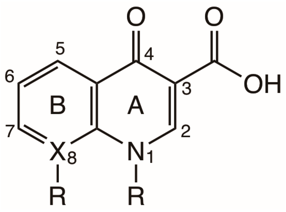
Scheme 1. Basic structure of quinolone (X=C) and naphthyridone (X=N) antibiotics.
Photochem 05 00017 sch001
Scheme 2. Structure of some representative Qs and FQs and their generations.
Photochem 05 00017 sch002
Scheme 3. Structures in equilibrium in norfloxacin in solution.
Photochem 05 00017 sch003
Scheme 4. Photophysical and photochemical processes characteristic of an FQ.
Photochem 05 00017 sch004
Scheme 5. Proposed structure of ground state complex for LOM and phosphate buffer.
Photochem 05 00017 sch005
Scheme 6. Photochemical reactions in Qs and FQs.
Photochem 05 00017 sch006
Scheme 7. Photolysis of 2,6-difluoroquinolone FQ with the generation of an aryl cation (1a) and its mesomeric structures (1b, 1c and 1d).
Photochem 05 00017 sch007
Scheme 8. FQs with reduced phototoxicity.
Photochem 05 00017 sch008
Scheme 9. Structural modifications and their relationship to biological activity and pharmacokinetic properties of Qs and FQs.
Photochem 05 00017 sch009
Scheme 10. Chemical structure of recently developed quinolones.
Photochem 05 00017 sch010
Scheme 11. Main photophysical and photochemical pathways for monohalogenated FQs in neutral aqueous media.
Photochem 05 00017 sch011
Scheme 12. Main photophysical and photochemical pathways for dihalogenated FQs in neutral aqueous media.
Photochem 05 00017 sch012
Scheme 13. Main photophysical and photochemical pathways for nonhalogenated quinolones in neutral aqueous media.
Photochem 05 00017 sch013
Scheme 14. Radical–radical cage reaction between Q nalidixic acid (Quinolone radical) and an amino acid (Tyrosine radical).
Photochem 05 00017 sch014
Scheme 15. Different photophysical, photochemical and phototoxic processes involved with a fluoroquinolones (FQ) and a biomolecule (BIOM).
Photochem 05 00017 sch015
Scheme 16. Different intermediates and mechanisms involved in the reaction of a biomolecule (BIOM) with an FQ upon irradiation.
Photochem 05 00017 sch016
Scheme 17. Main photophysical and photochemical pathways for photoinduced damage of a biomolecule (BIOM) by FQs after the formation of a complex (FQ…BIOM).
Photochem 05 00017 sch017
Scheme 18. Photophysical and photochemical processes involved in pyrimidine dimerization by FQ photosensitizers.
Photochem 05 00017 sch018
Table 1. Triplet state properties of neutral FQs in various media.
CompoundMediumT–T Absorption
λmax nm
Quantum Yield Triplet Formation
ΦT
εmaxTΦTTriplet Lifetime
τT/μs
Ref
NorfloxacinNaHCO3 0.001 M620≥0.534001.3[29]
EnoxacinNaHCO3 0.001 M520≥0.535000.85[29]
PB 0.01 M520 35000.09[25]
RufloxacinPB 0.01 M640≥0.4280010[27,29]
6400.3621007[28]
OfloxacinPB 0.01 M620≥0.323001.8[29]
6100.33360040[26]
LomefloxacinNaHCO3 0.001 M500≤0.29000.1[29]
FlumequinePB 0.02 M pH 85750.912,60010[30]
Nalidixic acidBuffer H2O pH 9.2620≥ 0.65400100[31]
CiprofloxacinNaHCO3 0.001 M610 1.5[32]
PB: phosphate buffer, pH 7–7.4.
Table 2. Singlet oxygen quantum yield (Φ), rate constant of singlet oxygen quenching by Qs and FQs (kq/106 M−1 s−1) in neutral aqueous medium and rate of DMPO/•OOH generation in arbitrary units a.u./min) in DMSO for Qs and FQs.
CompoundΦ akq/106
M−1 s−1
Rate of Generation of DMPO/•OOH
a.u./min b
Ref
Norfloxacin0.0811.8 (0.1)0.25[34]
Pefloxacin0.04512 (1) [34]
Enoxacin0.0611.4 (0.2)1.7[34]
Rufloxacin0.32 [27]
Ofloxacin0.0765.6 (0.6)0.06 c[34]
0.13 [26]
Lomefloxacin0.07218 (3)0.17[34]
Fleroxacin0.0296.9 (0.7)0.36[34]
Ciprofloxacin 0.0925.2 (0.2)0.20[34]
Nalidixic acid0.1525010.1[34]
Flumequine0.343.4 (0.4) [34]
a Data in neutral phosphate buffer, 50 mM in D2O, 0.01 M 90/10 D2O/H2O, 0.01M in D2O. b In these EPR experiments the spin trap DMPO was used; c the initial rates of generation of the DMPO/OOH adduct signal were measured and corrected for the number of photons absorbed.
Disclaimer/Publisher’s Note: The statements, opinions and data contained in all publications are solely those of the individual author(s) and contributor(s) and not of MDPI and/or the editor(s). MDPI and/or the editor(s) disclaim responsibility for any injury to people or property resulting from any ideas, methods, instructions or products referred to in the content.