Review

Application of Textile Technology in Vascular Tissue Engineering

1
Winner Institute for Innovation Research, Winner Medical Co., Ltd., Wuhan 430400, China
2
School of Bioengineering and Health, Wuhan Textile University, Wuhan 430200, China
*
Author to whom correspondence should be addressed.
Textiles 2025, 5(3), 38; https://doi.org/10.3390/textiles5030038
Submission received: 23 July 2025 / Revised: 15 August 2025 / Accepted: 21 August 2025 / Published: 3 September 2025

Abstract

Cardiovascular diseases pose a significant global health burden, driving the need for artificial vascular grafts to address limitations of autologous and allogeneic vessels. This review examines the integration of fiber materials and textile technologies in vascular tissue engineering, focusing on structural mimicry and functional regeneration of native blood vessels. Traditional textile techniques (weaving, knitting, and braiding) and advanced methods (electrospinning, melt electrowriting, wet spinning, and gel spinning) enable the fabrication of fibrous scaffolds with hierarchical architectures resembling the extracellular matrix. The convergence of textile technology and fiber materials holds promise for next-generation grafts that integrate seamlessly with host tissue, addressing unmet clinical needs in vascular tissue regeneration.

1. Introduction

Cardiovascular diseases (CVDs) impose a substantial burden on global healthcare systems, affecting millions of people annually and accounting for a large proportion of global mortality [1,2,3]. These conditions, including coronary heart disease, stroke, and peripheral artery disease, typically involve vascular occlusion or dysfunction [4,5,6]. Traditional treatments for such conditions may involve surgical interventions such as bypass grafting or angioplasty. However, the limited availability of autologous (patients’ own) vessels and the potential complications associated with allografts (donated vessels) have necessitated the development of artificial blood vessels [7,8]. Artificial blood vessels, or vascular grafts, offer a viable alternative that can be tailored to meet the specific needs of patients, reducing the reliance on donor tissues and potentially mitigating some of the risks associated with surgical procedures.
In the field of vascular tissue engineering, fiber materials play a pivotal role in the design and fabrication of artificial blood vessels [9,10,11,12]. These materials are critical in mimicking the structural and functional properties of native blood vessels, ensuring effective blood flow, and promoting integration with the host tissue [13,14]. Fiber-based scaffolds can provide the necessary mechanical support while allowing for cell infiltration, proliferation, and differentiation, ultimately leading to the formation of a functional, living tissue [15,16,17]. The choice of fiber material, its orientation, and porosity can significantly influence the performance of an artificial blood vessel, affecting factors such as compliance, permeability, and thrombosis resistance [18,19,20,21]. Consequently, the ongoing research and development of advanced fiber materials are essential for enhancing the biocompatibility, durability, and overall success of artificial blood vessels in clinical applications.
Textile technology serves as a transformative bridge between fiber materials and functional vascular grafts, enabling precise control over scaffold architecture, porosity, and mechanical anisotropy. Textile technology offers a paradigm shift in artificial blood vessels, leveraging millennia-old fabrication methods (e.g., weaving, knitting, braiding) [22,23] and modern innovations (e.g., electrospinning, melt electrowriting (MEW)) [24,25] to create biomimetic vascular scaffolds. The discipline’s core advantage lies in its ability to engineer hierarchical fiber architectures (from nanometers to millimeters) that mirror the extracellular matrix (ECM), which consists of 50–500 nm diameter collagen/elastin fibers [26,27]. This structural mimicry facilitates cell adhesion, migration, and phenotype maintenance critical for preventing smooth muscle cell (SMC) dedifferentiation and promoting endothelial cell (EC) alignment [28]. The integration of textile technology in vascular tissue engineering presents transformative opportunities to address unmet clinical needs.
The purpose of this review is to provide a comprehensive overview of the current advancements and challenges in the use of fiber materials for the tissue engineering of artificial blood vessels. By addressing both technological innovations and translational challenges, we aim to highlight textile-based scaffolds as a transformative approach for treating CVDs. Furthermore, we will discuss the latest research trends and innovations in the field, highlighting promising developments that could pave the way for more effective and durable artificial blood vessel solutions. The scope of this review will encompass both traditional textile technologies and modern innovations, as well as hybrid systems that combine the best attributes of both. By synthesizing this information, we hope to offer insights that can guide future research efforts and contribute to the ongoing improvement of artificial blood vessel technology.

2. Architecture of Native Vessels

To advance the development of functional vascular grafts, a foundational step is to elucidate the architecture, compositional characteristics, mechanical properties of native blood vessels and the interrelationships among these attributes.
Blood vessels serve as vascular conduits responsible for delivering oxygen and nutrients to tissues and eliminating carbon dioxide and metabolic wastes from them. In terms of inner diameter (ID), blood vessels are generally classified into microvessels (ID < 1 mm), small vessels (ID ranging from 1 to 6 mm), medium vessels (ID ranging from 6 to 10 mm), and large vessels (ID > 10 mm) [29,30].
Natural blood vessels are categorized into three types with distinct structural and functional specializations: arteries, veins, and capillaries. Arteries, characterized by thick, muscular walls, transport oxygenated blood away from the heart under high pressure; in contrast, veins—with thinner walls and valves to prevent backflow—return deoxygenated blood to the heart. Small-diameter arteries (arterioles) and veins (venules) function as regulatory segments, modulating blood flow through vasoconstriction and vasodilation. Capillaries, the smallest vessels, form a vast network with single-layered endothelial walls, enabling efficient diffusion of gases (e.g., oxygen and carbon dioxide), nutrients, and waste products between blood and surrounding tissues. This exchange is facilitated by their thin walls and extensive surface area, which maximize contact with interstitial fluid [31].
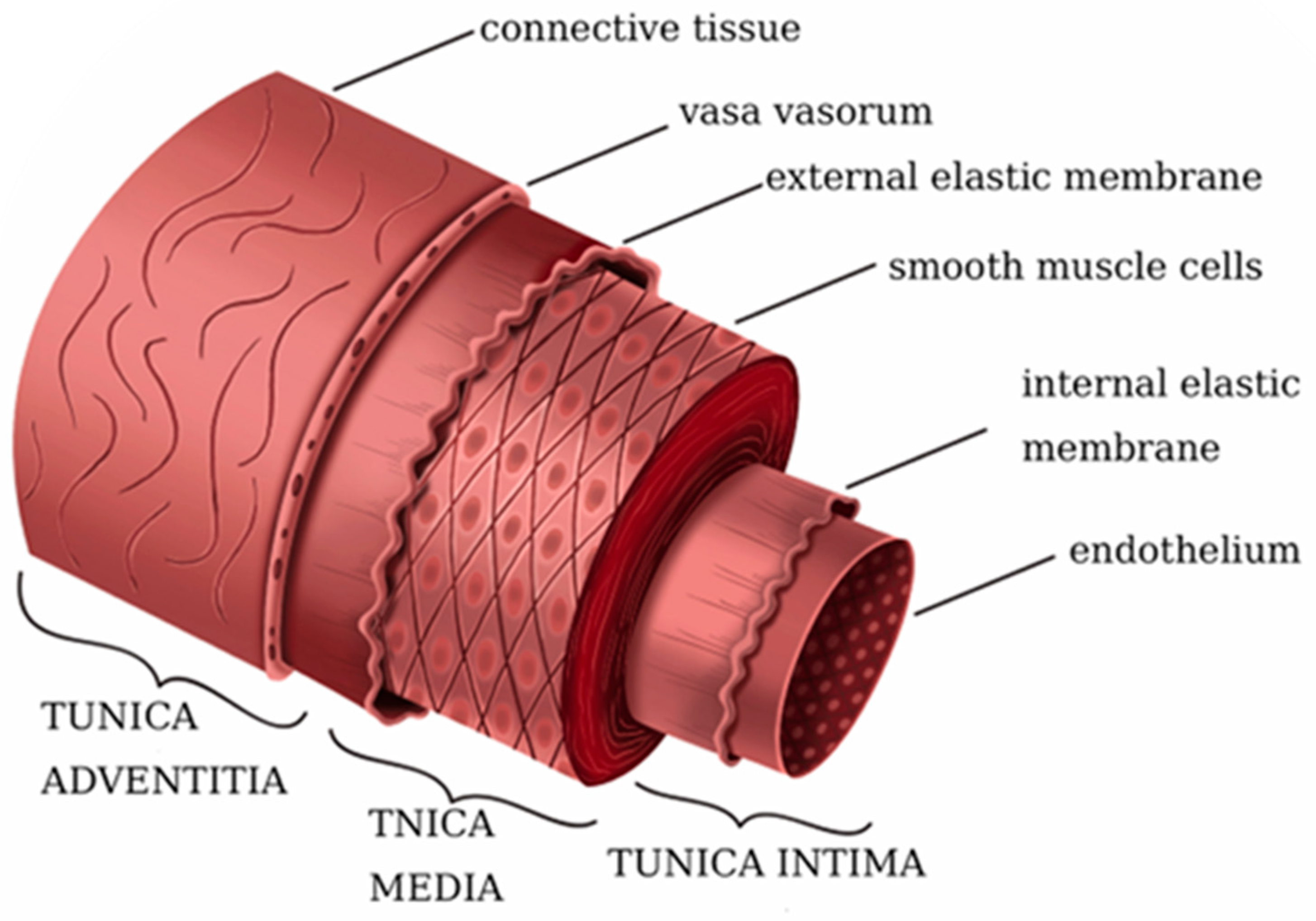
The native blood vessels in the human body exhibit hierarchical complexity, with arterial walls exemplifying the most elaborate structure. The arterial wall is composed of three distinct concentric layers: (i) intima, (ii) media, and (iii) adventitia (Figure 1), each with specialized molecular compositions and mechanical roles. The intima, the innermost layer, consists of a continuous monolayer of ECs anchored to a basal lamina rich in laminin, fibronectin, and type IV collagen. This basal lamina provides a scaffold for EC adhesion and separates the intima from the underlying media. The intimal ECs express cell-surface molecules like vascular endothelial cadherin to maintain monolayer integrity and secrete anticoagulant factors (e.g., heparan sulfate) to prevent thrombosis [29,32].
The media, the middle layer, is a dynamic mechanical barrier composed of circumferentially arranged SMCs embedded in a matrix of elastic fibers, collagen fibrils, and proteoglycans. The elastic fibers, primarily composed of elastin and fibrillin-1, form periodic lamellae that endow the vessel with recoil capacity, allowing arteries to withstand pulsatile blood pressure. Interspersed among the elastic lamellae are type I collagen fibers, which provide tensile strength and limit excessive expansion. SMCs in the media exhibit a contractile phenotype, expressing proteins like α-smooth muscle actin and calponin, enabling them to regulate vessel diameter in response to neural, hormonal, or paracrine signals [30,33].
Figure 1. Three-layer structure of blood vessels. The arterial wall is composed of three primary layers: the adventitia, media, and intima. Endothelial cells line the luminal surface, while smooth muscle cells and fibroblasts are present in the outer layers [34].
Textiles 05 00038 g001
The adventitia, the outermost layer, consists of a collagenous ECM dominated by type I collagen fibers arranged in wavy patterns to resist tensile forces. This layer contains fibroblasts, which synthesize and remodel the ECM, as well as perivascular nerve endings and the vasa vasorum (a network of small blood vessels supplying the outer vessel wall). The adventitia interfaces with the surrounding connective tissues, providing structural support and anchoring the vessel within the tissue microenvironment [35,36].
Thus, the design of tissue-engineered blood vessels must recapitulate not only the layered architecture but also the molecular composition (e.g., ECM protein gradients), cellular phenotypes (e.g., EC and SMC spatial alignment), and mechanobiological interactions that underlie native vessel function [37]. Understanding how the concentric ECM layers, cell–matrix interactions, and mechanical properties synergize to enable vasoreactivity and long-term durability is essential for translating vascular tissue engineering from bench to clinic.
Table 1 presents the mechanical properties of commonly used autologous vascular grafts and the performance criteria for artificial blood vessels.
Burst pressure is the maximum fluid pressure a vascular graft can withstand before it ruptures, tears, or fails (e.g., at the anastomosis or in the graft body). It is a critical measure of the graft’s mechanical integrity, reflecting its ability to tolerate physiological blood pressure (typically 80–120 mmHg systolic in humans). Grafts with insufficient burst pressure risk acute failure, leading to hemorrhage [38].
Compliance refers to the ability of a vascular graft to distend (expand) and recoil in response to changes in intravascular pressure, mimicking the natural elasticity of native blood vessels. It is defined as the fractional change in volume or diameter per unit change in pressure (ΔV/V0 per mmHg or ΔD/D0 per mmHg). Natural vessels (e.g., arteries) are compliant, expanding during systole (high pressure) and contracting during diastole (low pressure) to maintain steady blood flow. A graft with compliance mismatched to the native vessel can cause turbulence, increased stress at the anastomosis, or impaired blood flow, leading to long-term complications like intimal hyperplasia or graft occlusion [39].
Suture retention refers to the ability of a vascular graft material to securely hold sutures in place at the anastomotic site (where the graft is sewn to native blood vessels) without suture pullout, tearing, or loosening. This property ensures the integrity of the surgical connection, preventing leaks, dehiscence (separation), and blood loss during and after implantation [40].
The development of next-generation grafts poses a complex, multifaceted challenge that integrates elements across multiple fields, including mechanical engineering, vascular biology, and immunoregulation. From a mechanical standpoint, there is broad consensus that the goal is to accurately mimic the human internal mammary artery—the gold standard for bypass grafts.
Table 1. Mechanical properties of natural blood vessels and artificial blood vessels.
Mechanical PropertySaphenous VeinInternal Mammary ArteryArtificial Blood Vessel Benchmarking
Young’s modulus (MPa)4.2 (circ) [37,41]
23.7 (long) [37,41]
8.0 (circ) [37,41]
16.8 (long) [37,41]
>1 (circ) [41,42]
Burst pressure (mmHg)1599 ± 877 [20,43]3196 ± 1264 [20,44]>1000 [20,42]
Compliance (%/100 mmHg)4.4 [37,45]11.5 [20,37]10–20 [20,42]
Wall thickness (µm)180 to 650 [43,46]180 to 430 [43,46]500 [43]
Suture retention (g)196 ± 2 [43,47]138 ± 50 [20,43]>100 [20,42]
Circ: circumferential; long: longitudinal.

3. Application of Textile Technology in Artificial Blood Vessels

Textiles are flexible materials constituted by interlaced networks of fibers. They can be categorized into fiber-based and yarn-based types. For fiber-based textiles, fabric formation occurs concurrently with fiber production; examples include various nonwoven fabrics and electrospun mats. In contrast, yarn-based textiles involve an additional procedure: fibers or filaments are first spun into yarns, which are then used to produce fabrics such as woven, weft-knitted, warp-knitted, and braided ones [48].
The effectiveness of a fiber-based vascular graft is determined by a suite of key variables that collectively influence its patency, mechanical stability, host integration, and long-term functionality. These include material composition (dictating biocompatibility and degradation), fiber diameter (affecting cell adhesion and surface area), porosity and pore size (regulating nutrient diffusion and tissue infiltration), fiber orientation (governing mechanical anisotropy to match native vessel properties), mechanical attributes like compliance and burst pressure (ensuring tolerance of physiological pressures), biocompatibility (minimizing immune responses), surface properties (reducing thrombosis and promoting endothelialization), biodegradability (for resorbable grafts and matching tissue regeneration rates), and suture retention (securing anastomotic integrity). Balancing these variables is critical to developing effective grafts, particularly for small-diameter applications, where they mitigate risks like stenosis and thrombosis.

3.1. Electrospinning for Vascular Grafts

Electrospinning, first patented in 1934 [2], has evolved as a versatile fabrication technique with broad utility across disciplines. Within vascular tissue engineering—particularly for developing small-diameter vascular grafts (SDVGs)—its significance has surged, owing to its capacity to generate biomimetic fibrous scaffolds that recapitulate the nanoscale topography and architectural features of native ECM [26,49].
The electrospinning process leverages high-voltage electrostatic fields to draw ultrafine polymeric fibers (ranging from nanometers to micrometers in diameter) [50,51]. A typical electrospinning platform comprises a high-voltage power supply, a syringe pump for precise control of the polymeric dope flow rate, a spinneret (injection needle), and a conductive collector for nanofiber deposition. Mechanistically, application of a direct current voltage to the polymeric dope induces electrostatic repulsion within the charged liquid, elongating the meniscus into a conical configuration (Taylor cone). Once the applied voltage exceeds the surface tension of the Taylor cone, a charged jet is ejected and accelerates toward the collector. During transit, solvent evaporation occurs, solidifying the jet into continuous nanofibers [52].
Notably, these electrospun nanofibrous matrices mimic the fibrous microarchitecture and mechanical cues of the native ECM, making them highly attractive for fabricating scaffolds tailored to SDVG applications.

3.1.1. Mimicking the Multilayered Architecture of Native Vessels

Multilayered electrospun vascular grafts represent a promising candidate, as they can recapitulate the native multilayered architecture of natural blood vessels. Such a design enables customization of diverse properties via polymer selection, tuning of electrospinning parameters, or incorporation of bioactive molecules, thereby optimally accommodating the phenotypic demands of cells in each layer.
Shi et al. [53] constructed a biomimetic tri-layered SDVGs by electrospinning poly (l-lactide-co-caprolactone) (PLCL) with decellularized extracellular matrix (dECM) powders from porcine thoracic aorta intima and media and loaded salidroside (Sal) into the inner layer to inhibit thrombosis. The inner layer (PLCL/intima-dECM/Sal), middle layer (PLCL/media-dECM), and outer layer (pure PLCL) were sequentially electrospun onto a stainless-steel rod (Figure 2). In vitro tests showed that the graft promoted human umbilical vein endothelial cell (HUVEC) adhesion, migration, and tube formation, with salidroside releasing sustainably (45% in 20 h). The graft had a burst pressure exceeding 1000 mmHg, a water contact angle of 71.66° ± 12.8°, and good mechanical properties (tensile strength: 4.46 ± 0.77 MPa). In rat abdominal aorta replacement models, the PLCL-dECM-Sal graft showed 100% patency at 3 and 6 weeks, promoting endothelialization, smooth muscle regeneration, and ECM deposition (collagen, elastin), with reduced platelet adhesion and thrombosis. This study provides a promising strategy for SDVG design by integrating a dECM and salidroside to mimic native artery structure, enhancing hemocompatibility and tissue regeneration.
Han et al. [54] developed a tri-layer vascular graft engineered for the spatiotemporal delivery of vascular endothelial growth factor (VEGF) and platelet-derived growth factor (PDGF). The inner layer, fabricated from 75:25 PLCL, was designed for rapid VEGF release to accelerate endothelialization, whereas the middle layer—composed of 75:25 poly (lactic-co-glycolic acid) (PLGA)—enabled sustained PDGF release to support vascular smooth muscle cell (VSMC) layer formation. The outer polycaprolactone (PCL) layer enhanced graft mechanical strength while delaying PDGF release. Mechanical characterization revealed that the tri-layer graft outperformed porcine coronary arteries used as controls, with superior tensile strength (5.2 ± 0.7 MPa vs. 2.6 MPa), Young’s modulus (35.9 ± 7.7 MPa vs. 1 MPa), and elongation at break (146.7 ± 0.6% vs. 100%). However, its burst pressure and suture retention strength were comparable to those of native vessels. While not observed in their 8-week in vivo study in New Zealand white rabbits, compliance mismatch remains a concern, as it could induce intimal hyperplasia and restenosis.
Wu et al. [55] constructed a tri-layer graft with inner two layers tailored to ECs and VSMCs. The inner layer consisted of circumferentially aligned PLCL/collagen nanofibers. HUVECs exhibited robust proliferation on these aligned fibers, with no significant difference compared to random fibers; notably, HUVECs aligned along the nanofibers, a feature hypothesized to promote the formation of an organized, functional endothelium. The middle layer comprised PLGA/silk fibroin (SF) electrospun yarns, fabricated by collecting PLGA/SF nanofibers on a rotating funnel to twist them into yarns, which were then wrapped around the aligned PLCL layer. VSMCs proliferated significantly better on these yarns than on PLGA/SF fibers, attributed to the yarns’ porous 3D architecture, and also aligned along the yarns—an arrangement conducive to VSMC function in the media. Finally, an outer layer of random PLCL/collagen nanofibers provided structural integrity to the entire construct.
To mimic native arterial architecture, Ma et al. [56] introduced a novel wet-electrospinning technique combined with conventional electrospinning to fabricate a tri-layer bionic vascular scaffold. The inner layer (PCL/gelatin/heparin) and outer layer (PCL/PLGA) were prepared via rotating-rod electrospinning, while the middle PCL layer was fabricated using Wet Vertical Magnetic Rod Electrospinning. The scaffold was characterized based on morphology, mechanical performance, degradation behavior, wettability, and biocompatibility; heparin crosslinking was employed to enhance hemocompatibility. Results showed porosity ranging from 71.51% to 78.44%, with the inner layer exhibiting excellent hydrophilicity (water contact angle decreasing from 87.28° to 69.20° within 60 s) and appropriate degradability (24.73% weight loss over 14 days). The middle layer displayed anisotropic mechanical properties (axial Young’s modulus: 5.13 ± 10.02 MPa; radial: 62.09 ± 550.40 MPa), and the tri-layer scaffold demonstrated good biocompatibility, supporting HUVEC adhesion and proliferation. This study successfully developed a small-diameter tri-layer tubular scaffold with bionic structure and mechanical properties, offering a promising strategy for tissue-engineered SDVGs.

3.1.2. Recapitulating Native Fibrous ECM: Fiber Morphology and Alignment

Electrospun fiber morphology and mechanical properties play a pivotal role in regulating cell behavior, including growth and proliferation. For most electrospinning applications, bead-free, non-adherent fibers are preferred. Fiber diameter, in particular, exerts a profound influence on cellular responses. In one study, Han D. et al. [57] investigated how increasing fiber diameter affects VSMC proliferation, infiltration, and phenotypic modulation. PCL fibers (0.5–10 μm in diameter) were fabricated by tuning PCL concentration, solvent composition, and applied voltage. Over a 10-day period, fiber diameter had no impact on VSMC survival but correlated with slower proliferation as diameter increased. Conversely, larger diameters enhanced VSMC infiltration into the PCL scaffold, attributed to increased pore size associated with thicker fibers. Notably, after 10 days of culture, the proportion of VSMCs with a synthetic phenotype (relative to total VSMCs) increased with fiber diameter—a trend mirrored in in vivo studies at 7, 14, and 28 days post-implantation in mice. This finding is intriguing, as the synthetic phenotype is linked to VSMC proliferation, potentially driven by enhanced infiltration in scaffolds with larger fibers. Additionally, scaffolds with larger fibers exhibited more activated macrophages, a factor critical for scaffold remodeling.
Shen et al. [44] further investigated the impact of fiber diameter on cellular resistance to shear stress, utilizing electrospun zein (a primary corn protein) with aligned fibers of three diameters: 112 ± 31 nm, 513 ± 80 nm, and 959 ± 147 nm. Under 15 dynes/cm2 shear stress for 4 h, human endothelial cells showed similar retention on randomly oriented fibers regardless of diameter. However, on aligned fibers, medium-diameter fibers significantly improved cell retention at 1, 2, and 4 h under both parallel and perpendicular flow, outperforming small and large diameters (with small diameters also superior to large ones). Following 2 days of adhesion, human endothelial cells on medium-diameter fibers resisted shear stress in both flow directions and elongated parallel to the flow. In contrast, cells on large-diameter fibers—initially aligned with the fibers—adopted a rounded morphology and were ultimately washed away under both flow conditions.
Fiber morphology also modulates the mechanical properties of electrospun constructs. In native tissues, collagen fibers exhibit a crimped structure that contributes to elasticity [58]. Chao et al. [59] replicated this crimped morphology in polylactic acid (PLA) nanofibers: heating PLA at 85 °C for 15 min induced stable crimping, hypothesized to stem from crystallinity changes (transitioning from amorphous to semi-crystalline). A similar effect was achieved by soaking PLA nanofibers in 95% ethanol at 37 °C for 2 days—a method applicable to PCL and PLGA nanofibers as well. Heat-treated (362.5 ± 110.8 MPa) and ethanol-soaked (441.6 ± 46.1 MPa) fibers showed a reduced elastic modulus compared to as-spun fibers (478.3 ± 77.8 MPa), though not significantly. They also exhibited significantly increased transition strain, with stress–strain curves displaying a J-shaped response—characteristic of native vessels—attributed to reduced “crimpness” under high strain. Crimp retention was maintained under dynamic loading. Anterior cruciate ligament fibroblasts aligned along crimped fibers, adopting an undulating morphology, and showed significantly upregulated type I collagen expression—further enhanced under dynamic loading.
Nanofiber alignment is another key regulator of cell behavior on electrospun scaffolds. In native blood vessels, cellular alignment is functionally critical: ECs align longitudinally with blood flow, while VSMCs orient circumferentially to enable vessel contraction. For tubular scaffolds, circumferentially aligned fibers are achieved by rotating the mandrel at sufficient speed, where mandrel surface velocity must exceed fiber deposition velocity. He et al. [60] found that a 4 mm-diameter mandrel rotating at 1000 rpm produced aligned nanofibers, whereas higher or lower speeds resulted in random orientation. Given setup dependence, reporting either mandrel diameter with rotation speed or linear surface velocity is recommended. Fusaro et al. [61] demonstrated that circumferentially aligned nanofibers enhanced EC proliferation and function. Longitudinally aligned fibers, by contrast, are obtained using two parallel plates as collectors—fibers bridge the gap, aligning perpendicularly to the plates—and can be transferred to a mandrel by rolling, orienting them along the mandrel length. Tan et al. [62] showed that longitudinally aligned PLCL nanofibers promoted HUVEC adhesion and proliferation more effectively than random PLCL fibers.
Modifying mandrel geometry is a strategy to tailor the mechanical behavior of electrospun grafts. Of particular interest are grafts with wavy cross-sections, which enable circumferential expansion to mimic the nonlinear mechanics of native vessels. The simplest approach uses a wavy cross-section mandrel; collecting PU fibers directly on such a mandrel forms a dense wall, with the wavy shape retained after removal and 100 cycles of cyclic loading, yielding stress–strain curves with native-like nonlinearity [63].
Yu et al. and Mi et al. [64,65] developed a novel approach using a round mandrel surrounded by thin (0.8 mm-diameter) rods. At sufficient rotational speed, the rods flexed like jump ropes; as nanofibers collected on these “satellite rods,” they constricted and retracted toward the mandrel, forming a wavy structure. Satellite rods also facilitated graft removal without damage. Fibers in contact with rod exteriors formed dense, flat regions, while inter-rod areas had loose, wavy regions. Polymer selection influenced wavy structure retention: elastic PU (mimicking elastin) blended with stiffer polymers (SF for Yu et al. and PCL for Mi et al.—mimicking collagen) yielded optimal shape retention. Pure stiff polymers failed to constrict satellite rods, while pure elastic polymers contracted to a round shape post-removal.
Fiber orientation can also be used to delineate layers in a multilayered graft [62,66]. Guo et al. [67] proposed a sequential electrospinning strategy combined with folding and rolling manipulation to fabricate a gelatin-based three-layer biomimetic vascular scaffold. The inner and middle layers with spatially perpendicular alignments were prepared by electrospinning at different rotating speeds, while the outer layer was a random-oriented structure (Figure 3). The scaffold’s morphology, mechanical properties, and cell behaviors were characterized, and in vitro biomimetic reconstruction and subcutaneous implantation in Beagle dogs were conducted. The results showed that the scaffold had an inner diameter of 5 mm, with the inner and middle layers featuring aligned fibers (diameters of 0.4–0.6 μm and 0.6–0.8 μm, respectively) and the outer layer showing random fibers (diameter of 0.8–1.2 μm). The tensile strength of the dry scaffold was 3.24 ± 0.75 MPa, and the wet scaffold showed an ultimate strain of 118.92 ± 49.59%. HUVECs showed good proliferation and orientation on the corresponding layers, and the in vitro reconstructed scaffold exhibited a three-layer cell distribution similar to native blood vessels. Subcutaneous implantation indicated that the scaffold promoted cell migration and maintained the three-layer structure. This study provides a feasible approach to fabricate a three-layer vascular scaffold with spatial alignment, which has great potential for guiding vascular tissue regeneration and reconstruction.

3.2. Controlled Fabrication of Vascular Grafts

3.2.1. Wet Spinning and Gel Spinning for Vascular Grafts

Wet spinning is a straightforward method for fabricating fibrous scaffolds with both random and aligned nanofibrous morphologies. Fibers produced via wet spinning have been prepared from various materials and applied in multiple tissue engineering fields, including cartilage, tendon, ligament, and blood vessel engineering. This technique entails a non-solvent-induced phase separation process, in which a polymer solution is extruded into a coagulation bath. Zhang et al. [68] developed a wet-spinning system to fabricate oriented PCL microfiber scaffolds using a blended solution of edible oil and hexane as a non-solvent coagulation bath. The process involved extruding a 10% PCL solution into the bath at varying flow rates (1, 4, 8 mL/h) and collecting the fibers on a rotating rod, resulting in scaffolds with controllable fiber diameters (7–27 μm) and porosities (68–82%). SMCs seeded on the scaffolds grew oriented along the fibers, infiltrated the scaffold interior, and maintained their phenotype as confirmed by α-SMA staining. Tensile testing showed that cell-seeded scaffolds had enhanced yield strength in the fiber-aligned direction. This study demonstrates that the wet-spun PCL microfiber scaffolds can guide both oriented SMC growth and infiltration, holding promise for vascular tissue engineering and regeneration of other fibrous tissues like tendons and ligaments.
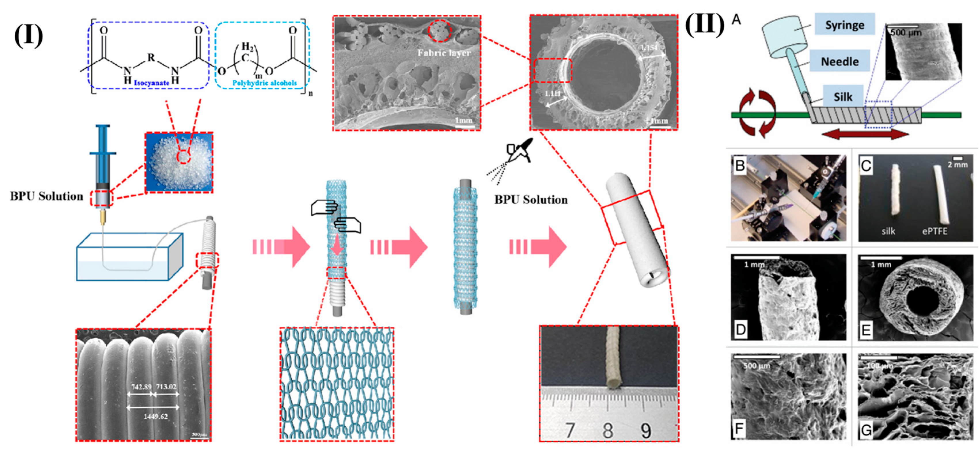
Similarly, Zhang et al. [69] designed and fabricated a three-layer small-diameter polyurethane vascular graft with high strength and excellent compliance using wet spinning, knitting, and spraying techniques (Figure 4I). The inner layer was made by wet spinning a 20% biomedical polyurethane solution, the middle layer was a spandex tubular fabric knitted with different fineness yarns, and the outer layer was formed by spraying a 5% BPU solution. The graft’s morphology, dynamic compliance, mechanical properties, water permeability, and strength after repeated puncture were characterized. The results showed that the graft had a uniform wall thickness of about 0.54 mm, a dynamic compliance significantly higher than that of human arteries and expanded polytetrafluoroethylene (ePTFE) grafts (up to 11.5% vs. 1%), a radial fracture strength of 30–50 N/cm (higher than human femoral artery’s 11 N/cm), low water permeability, and a puncture strength of >15 N/cm after 24 punctures. This study provides an effective approach for preparing polyurethane vascular grafts with balanced strength and compliance, which has potential clinical applications in replacing traditional PET and ePTFE vascular grafts.
Gel-spinning requires the solution concentration to exceed a minimum viscosity threshold, ensuring that the formed gel adheres to the collecting mandrel during continuous rotation. However, extruding such highly concentrated solutions from the deposition needle becomes challenging when they are excessively viscous. For example, Lovett et al. [70] proposed silk fibroin as a material for SDVG and developed tubular silk scaffolds using an aqueous gel spinning technique (Figure 4II). They evaluated the scaffolds in vitro for thrombogenicity (thrombin and fibrinogen adsorption, platelet adhesion) and vascular cell responses (EC and SMC attachment and proliferation), comparing them with polytetrafluoroethylene (PTFE), and then implanted them into the abdominal aortas of Sprague Dawley rats to assess patency and tissue remodeling. The silk tubes had an elastic modulus of 2.20 ± 0.90 MPa and ultimate tensile strength of 0.273 ± 0.11 MPa, closely matching the rat aorta’s mechanical properties. They showed lower thrombin and fibrinogen adsorption than PTFE, with platelets adhering in a non-activated morphology. In vitro, endothelial and smooth muscle cells attached and proliferated better on silk than on PTFE. In vivo, silk grafts remained patent for up to 4 weeks, with host cells forming a confluent endothelial lining and populating the graft walls. This study demonstrates silk fibroin’s potential as a biocompatible, biodegradable material for small-diameter vascular grafts, offering advantages over synthetic materials like PTFE in terms of mechanical compliance and cell integration.
Rodriguez et al. [71] aimed to enhance the porosity of gel-spun silk vascular grafts to facilitate faster degradation and cellular colonization while maintaining mechanical properties. They prepared silk solutions with varying molecular weights by adjusting extraction times (5–30 min, MB), leading to different concentrations and viscosities, and used a custom gel-spinning device to fabricate tubes, which were lyophilized and characterized for porosity, degradation, mechanics, and in vivo performance in rat abdominal aorta models. The results showed that lower concentration and higher molecular weight solutions yielded more porous tubes (porosity: 52–86.7%; pore size: 0.02–293.8 μm), with faster in vitro degradation (52% mass loss in 14 days for 5 MB vs. 25% for 20 MB). Mechanical tests indicated that higher porosity correlated with a lower elastic modulus (1–10 MPa) and suture retention strength (0.25–1 N), but compliance (3.3–6 mm Hg−1 × 10⁻²) approached that of the saphenous vein. In vivo, highly porous grafts (5–10 MB) showed early cell infiltration but failed at 6 months due to mechanical issues, while less porous 20 MB grafts had delayed remodeling. This study highlights the trade-off between porosity, mechanics, and degradation in silk grafts, providing a basis for developing compliant, biocompatible vascular substitutes.

3.2.2. Melt Electrowriting (MEW) for Vascular Grafts

Advances in understanding the electrohydrodynamics of electrospinning, combined with principles of conventional additive manufacturing, have positioned MEW as a highly controllable scaffold fabrication technology for tissue engineering applications [72]. By using polymer melts rather than solutions, MEW overcomes key limitations of traditional electrospinning, enabling the production of scaffolds with precisely controlled geometries. These scaffolds can mimic the ECM framework of native tissues through biomimetic architectures, surface modifications, and biofunctionalization [73,74].
MEW, a heat-driven, direct-write evolution of electrospinning, is entirely solvent-free and offers exceptional precision in fiber deposition. Polymer melts possess unique properties—including lower electrical conductivity and higher viscosity compared to polymer solutions—which reduce surface charge density on the molten jet, dampening the extrusion instability typical of solution electrospinning. In MEW, a pressurized, stable viscoelastic molten polymer jet is extruded through a high-voltage-connected nozzle, forming a Taylor cone that draws the fiber toward a grounded collector (either a flat plate or, for tubular constructs, a cylindrical mandrel) [75,76].
As comprehensively reviewed by Paxton et al. [77], tuning polymer melt temperature, applied voltage, and collector translational speed enables highly reproducible regulation of scaffold architecture (including pore size/shape, fiber laydown angles, and height) and fiber diameters spanning nanometers to micrometers. Like conventional 3D printing, the collector’s translation and rotation are programmed via G-Code [77,78]. Optimization of G-Code facilitates the fabrication of highly ordered porous scaffolds with high surface-to-volume ratios, thereby enhancing cell adhesion and proliferation. Furthermore, architectural refinement—achieved through perpendicular deposition of curved and straight fibers—enables the production of mechanically anisotropic scaffolds, featuring fully tunable deformation characteristics and biomimetic nonlinear stress–strain responses [79]. This capability is particularly advantageous for tissue-engineered vascular grafts (TEVGs), as it allows controlled manufacture of tubular scaffolds with predefined, biomimetic radial compliance—potentially overcoming anastomotic complications linked to non-compliant grafts [80].
While MEW excels at precise scaffold fabrication, its material scope is largely restricted to synthetic thermoplastics with melting points below ~200 °C. The printability of a polymer in modern MEW processes is strongly dependent on its viscosity and electrical conductivity [81,82]. PCL, a widely utilized biodegradable and biocompatible synthetic polymer in tissue engineering scaffolds, is prized for its tailorable degradation kinetics and favorable mechanical properties [83,84]; it is approved for medical devices by both the FDA and TGA [85]. Recent work by Diaz et al. [86] demonstrated that thermal pretreatment improves the melt viscosity of PLCL (previously limited to electrospinning), enabling successful MEW of this highly elastic, biocompatible polymer. Other polymers successfully used in MEW for microscale fiber fabrication include non-degradable polypropylene, electroactive poly (vinylidene fluoride), and PLA, as reviewed in detail by Kade and Dalton [87].
While electrospinning is widely applied to fabricate small-diameter TEVGs, MEW use remains limited due to its relative novelty and the scarcity of (often custom-manufactured) MEW equipment. Nevertheless, recent key studies highlight MEW’s progression as a scaffold fabrication technique and its suitability for vascular tissue engineering.
Jungst et al. [88] proposed an integral scaffold design (inner diameter of 3 mm) that enables the formation of an intraluminal endothelial cell monolayer, with an outer layer of oriented VSMCs. Facilitated by a bi-layered tubular scaffold, this design comprises an inner layer of randomly oriented electrospun fibers and an outer layer of MEW linear microfibers with controlled winding angles. The heterotypic architecture directs physiological cell organization without requiring soluble factors or scaffold bioactivation. Building on this work, Bartolf-Kopp et al. [89] adjusted the PCL-to-poly (ester-urethane) ratio during the electrospinning step of hybrid constructs to recapitulate the native vessel’s J-shaped stress–strain response (Young’s modulus: 0.9 ± 0.7 kPa for cell-seeded hybrid scaffolds vs. 0.5 ± 0.6 kPa for the internal mammary artery). Federici et al. [90] also investigated the effects of fiber winding angle inspired by the extracellular matrix orientation in the tunica media using a MEW scaffold alone. This biomimetic fiber arrangement promoted neo-tissue formation along the MEW fibers with extracellular matrix deposition preferentially oriented along the pore’s long axis and enabled a biomimetic stress–strain characteristic in the physiological range (up to 10% strain).
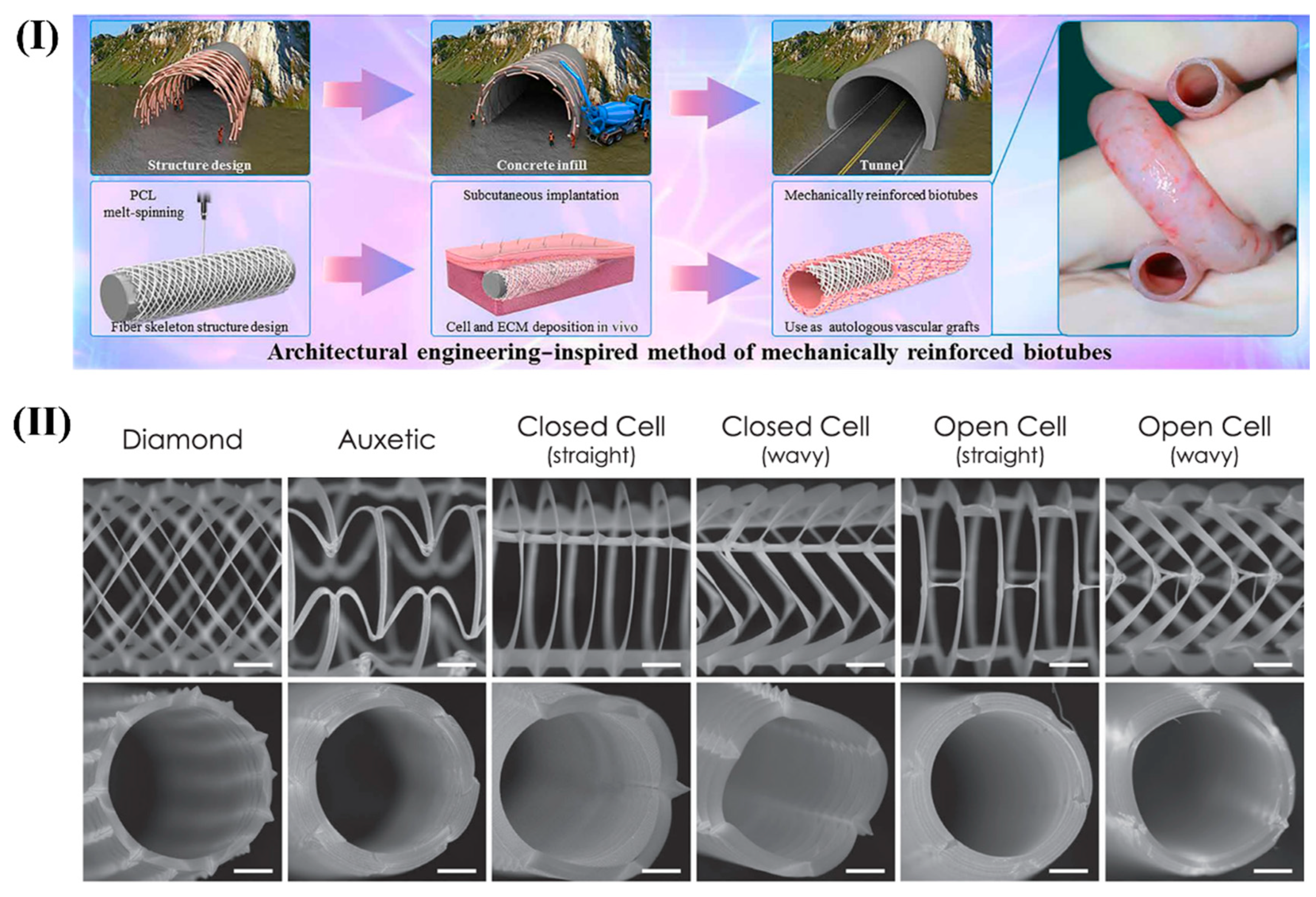
Following the in vivo tissue engineering strategy, Zhi et al. [91] subcutaneously implanted MEW tubular scaffolds in rats to leverage the foreign body response, which induces fibrous encapsulation (Figure 5I). These biohybrid constructs exhibited favorable performance in in vitro evaluations and as abdominal artery replacements in rats. Successful translation to larger animal models (canines and sheep) highlights this as a promising approach for future alternatives to autologous vessel replacements. While the MEW fiber architecture in these studies was a simple diamond pattern, other research has reported tubular auxetic and nonlinear designs [92] that could potentially enable combined longitudinal and radial growth (Figure 5II). Additionally, serpentine fiber patterns have been shown to impart tubular scaffolds with a compliance of 12.9 ± 0.6% (100 mmHg)−1 [93], within the physiological range of the internal mammary artery (IMA), which is 11.5 ± 3.9% (100 mmHg)−1 [20]. Conducting fiber deposition on patient-specific water-soluble polyvinyl alcohol molds will further facilitate the fabrication of anatomically relevant tubular constructs [94].
MEW scaffolds are predominantly macroporous, as entrapped charge carriers in fibers hinder precise fiber placement below an interfiber distance dependent on fiber diameter [95,96]. Consequently, MEW scaffolds are typically used as mechanical reinforcements, requiring combination with a secondary biomaterial to provide the microporosity necessary for cellular infiltration under the in situ tissue engineering paradigm. To address this, Mueller et al. [97] developed a design strategy that directly yields microporous MEW scaffolds, enabling tailored directional anisotropy for various cardiovascular tissues. Furthermore, this approach decouples fiber diameter from pore size (in contrast to electrospinning, where they are correlated) and is applicable to both flat and tubular scaffold architectures. Additionally, covered stents can be fabricated using this method. Initial efforts to develop purely MEW stents involved fabricating PCL stents mechanically reinforced with reduced graphene oxide to enhance flexural stiffness [85]. However, how their mechanical properties evolve with PCL degradation remains to be investigated.
Benno Neuhaus et al. [98] conducted a study on the fiber deviation phenomenon in MEW during the fabrication of tubular scaffolds for tissue engineering, which had not been previously reported in planar scaffolds. They utilized a novel automated optical scanning system integrated into a four-axis bioprinter to measure fiber deviation precisely and developed a mathematical model based on geometric analysis to predict deviation using parameters like jet characteristics and printing conditions. The research revealed that the deviation often exceeded 100 µm, compromising scaffold integrity, and evaluated four optimized toolpath strategies, with some reducing mean fiber spacing variation to ±4 µm. Key data showed that winding angles of 0° and 90° eliminated deviation, while 45° yielded the highest deviation, and printing speed and mandrel radius significantly influenced deviation magnitude. This study is significant as it uncovers the origin of fiber deviation in tubular MEW, provides a predictive model, and offers effective toolpath strategies to enhance the precision of tubular scaffold fabrication, thereby advancing the development of high-quality vascular grafts and other tissue engineering constructs.

3.3. Traditional Textile Technologies for Vascular Tissue Engineering

Textile manufacturing technology, a specialized engineering field, utilizes diverse natural and synthetic filament materials in specific interlaced structures for fabricating artificial blood vessels. Traditional yarn-based textile techniques include weaving, braiding, and knitting, where yarns are interconnected via undulation (weaving and braiding) or stitch formation (knitting). Table 2 summarizes the advantages and disadvantages of these traditional textile techniques for vascular graft fabrication. The artificial blood vessels produced through these methods exhibit advantages such as favorable elasticity, appropriate porosity, and ease of suturing, remaining in clinical use today.
Woven vascular grafts are distinguished by their stable structure and uniform texture (Figure 6A). By tuning the density and arrangement of warp and weft yarns, surface structures with varying porosity and elasticity can be constructed, facilitating effective cell adhesion and proliferation [103]. In contrast to woven grafts, knitted artificial blood vessels offer superior elasticity and compliance (Figure 6B). Braiding involves interlacing multiple filaments in a defined pattern to fabricate vascular grafts (Figure 6C) [104], with variations in braiding parameters exerting a significant impact on graft performance.

3.3.1. Weaving: Structurally Stable Vascular Grafts with Tailored Permeability and Mechanical Strength

In weaving, textiles are formed by interlacing weft yarns with warp yarns at a 90° angle. Warp yarns align longitudinally along the textile, while weft yarns run transversely. The three most common weave patterns are plain, twill, and satin, distinguished by the undulation patterns of their weft and warp systems. Beyond 2D flat textiles, 3D structures can be woven into tubular forms or constructed by interlacing multiple weft layers with warp yarns [11,48].
Woven-based vascular grafts are under development [105,106]. Numerous comprehensive studies across diverse research areas have been performed, and their findings will be elaborated in subsequent sections.
Zekun Liu et al. [107] conducted a study to develop a heparin-functionalized bifurcated stent graft (BSG) using textile weaving technology, with SF and heparin surface modification via steam/air treatment to enhance anti-occlusion properties (Figure 7I). The BSG showed a thickness of 0.085 ± 0.004 mm, water permeability resistance of 1.154 ± 0.854 mL/(cm2 × min), and prolonged heparin release due to SF β-sheet structure induction by steam treatment, with 60% heparin released over 200 h compared to 100% within 50 h for air-dried samples. The modified BSG significantly prolonged coagulation times and inhibited human vascular smooth muscle cell proliferation, demonstrating its potential for treating vascular diseases by combining thinness, low permeability, and anticoagulant function in a textile-based stent graft design.
Gaëtan Roudier et al. [108] investigated the effects of weaving parameters (warp count, weft ribbon width, weft tension) and cell-assembled extracellular matrix (CAM) sheet strength on the properties of completely biological TEVGs (Figure 7II). By optimizing these parameters, they reduced burst pressure by 35% to 3492 mmHg and wall thickness by 38% to 0.69 mm and increased compliance by 269% to 4.8%/100 mmHg, while maintaining suture retention strength (4.7 N) and low transmural permeability (24 mL·min−1·cm−2). Using decellularized CAM threads increased permeability but enhanced burst pressure. This study demonstrates that tuning weaving parameters can tailor TEVG properties to mimic native blood vessels, addressing mechanical mismatch and improving potential for long-term patency, with plans for in vivo testing in a sheep model.
Laure Magnan et al. [109] developed a novel approach to produce completely biological human textiles using CAM synthesized by human fibroblasts. They cultured fibroblasts for 6–12 weeks to form CAM sheets, which were cut into ribbons and twisted into yarns at different rotation rates (2.5–7.5 rev/cm). The yarns were characterized based on diameter, tensile strength, and ultrastructure, showing that twisting at 5 rev/cm increased ultimate tensile stress to ~40 MPa and strain at failure to ~20%. Using weaving, they created TEVGs with a burst pressure of 5968 ± 732 mmHg, suture retention strength of 566 ± 17 gf, and low permeability (10 ± 8 mL·min−1·cm−2). The team also demonstrated the yarn’s usability as a suture in nude rats, with minimal inflammation at 1 month. This study highlights the potential of CAM-based textiles to create implantable, mechanically robust tissues that mimic the native ECM, offering a biocompatible alternative to synthetic materials for vascular grafts and other tissue engineering applications.
Due to its ability to provide adequate mechanical properties, this method has historically been used primarily for the production of artificial blood vessels.

3.3.2. Knitting: Elastic and Compliant Vascular Grafts with Controllable Loop Architectures

Knitting, a traditional textile technique, constructs intricate 2D and 3D structures by interlocking yarns into structured loops. The process involves drawing yarns through pre-formed loops to create a continuous network of interconnected loops [110]. Characterized by well-defined loop architectures, knitted fabrics exhibit notable elasticity; their versatility allows tailoring of porosity and mechanical properties via tuning yarn parameters and knitting configurations.
In biomaterial applications—particularly cardiovascular ones—knitting techniques offer distinct advantages stemming from their unique structural attributes [111,112,113,114,115]. Knitted structures are valued for their porosity-related permeability, flexibility, and conformability, making them well-suited for biomedical devices such as cardiac patches and vascular grafts [99]. Their porous nature facilitates efficient transport of therapeutic agents, enhancing drug delivery capabilities. Furthermore, facile customization via advanced knitting technologies enables production of patient-specific biomedical devices, optimizing treatment efficacy. Such advantages highlight the growing significance of knitted biomaterials in cardiovascular applications.
Zhang et al. [116] aimed to fabricate small-caliber vascular grafts by circular knitting electrochemically aligned collagen filaments and electrospinning collagen nanofibers to form a bilayer structure (Figure 8I,II). They characterized the mechanical properties (bursting strength, suture retention strength, radial compliance) and biological performance (cell adhesion and proliferation) of the grafts under dry and hydrated conditions. The results showed that the collagen grafts had significantly higher bursting strength (dry: 1.66–2.11 MPa; hydrated: 0.66–0.70 MPa) than the human internal mammary artery and saphenous vein, comparable radial compliance to the human saphenous vein (2.81–3.06%/100 mmHg), and superior endothelial cell adhesion and proliferation compared to PLA grafts. This study demonstrates the feasibility of using collagen filaments and nanofibers to engineer vascular grafts with mechanical properties similar to those of native vessels and enhanced biological performance, providing a promising approach for small-caliber vascular graft development.
Lou et al. [117] investigated how yarn types (PET/spandex combinations) and fabric structures (braids, warp knits, weft knits) affect the compliance and bursting strength of vascular grafts, preparing tubular fabrics from wrapped yarns and incorporating them with PVA via freeze–thaw cycles (2–3 times). They evaluated mechanical properties using SEM, pressure–diameter tests, and bursting strength assays, comparing results with porcine carotid arteries. Key findings showed that weft/warp knits had higher compliance than braids, with spandex-containing yarns enhancing deformation (diameter variation up to 1.191 at 200 mmHg) and compliance (up to 12%/100 mmHg), while three freeze–thaw cycles increased bursting strength to >700 mmHg. This study highlights the potential of PVA–fabric composites to mimic native vessel mechanics, addressing compliance mismatch in small-diameter grafts and paving the way for improved tissue-engineered vascular substitutes.
Figure 8. Fabrication of knitted vascular grafts. (I). Collagen filaments were directly knitted into a tubular structure (B) on a lamb circular weft knitting machine (A) [116]. (II). Microscopic images of COL-K (a,b), COL-KE (c,d), and PLA-K (e,f) [116]. (III). The photo shows an SF vascular graft (A) and an ePTFE vascular graft (B). A gross view of the SF graft (C) and ePTFE graft (D) used to replace a rat inferior vena cava [118]. (IV). A schematic diagram of the double-raschel knitting pattern (A). SEM images of the surface (B) and cross-section (C) of SF grafts coated with SF sponges [118].
Textiles 05 00038 g008
Kiritani et al. [118] aimed to develop a tissue-engineered vascular graft for abdominal venous system replacement by fabricating double-raschel knitted SF grafts coated with an SF sponge, which were implanted into the inferior vena cava of rats (10 mm long, 3 mm diameter; n = 19) and compared with ePTFE grafts (n = 10) in terms of patency rates and histologic reactions (Figure 8III,IV). The SF grafts showed 100.0% and 94.7% patency rates at 1 and 4 weeks, respectively, while ePTFE grafts had 100.0% and 80.0% (p = 0.36), with SF grafts exhibiting complete luminal surface coverage by CD31-positive endothelial cells at 4 weeks, indicating superior endothelialization. Mechanically, SF grafts had a longitudinal suture retention strength of 6.4 ± 0.6 N and 82% porosity, absorbing blood rapidly post-recanalization. This study highlights SF grafts as a promising scaffold for abdominal venous replacement due to their favorable patency, endothelialization, and biocompatibility, addressing the limitations of autologous and synthetic grafts in contaminated surgical fields.

3.3.3. Braiding: Flexible, Anisotropic Vascular Grafts with Tunable Porosity and Regeneration Potential

Braiding, a versatile and widely adopted technique in medical device fabrication, offers distinct advantages alongside inherent limitations [119]. A key benefit is its ability to enhance structural integrity [120], enabling the creation of robust yet flexible frameworks—critical for applications such as vascular grafts and stents, where durability and adaptability to bodily movements are paramount [121,122,123,124].
Braided structures are further distinguished by superior flexibility paired with robust structural stability. Their flexibility and conformability allow adaptation to complex anatomical geometries, ensuring optimal fit within the vascular system and reducing complication risks. Additionally, the braiding process enables precise tuning of porosity—a feature particularly valuable for vascular grafts [124]. This tunable permeability facilitates blood flow regulation, thrombus prevention, and integration with native tissues.
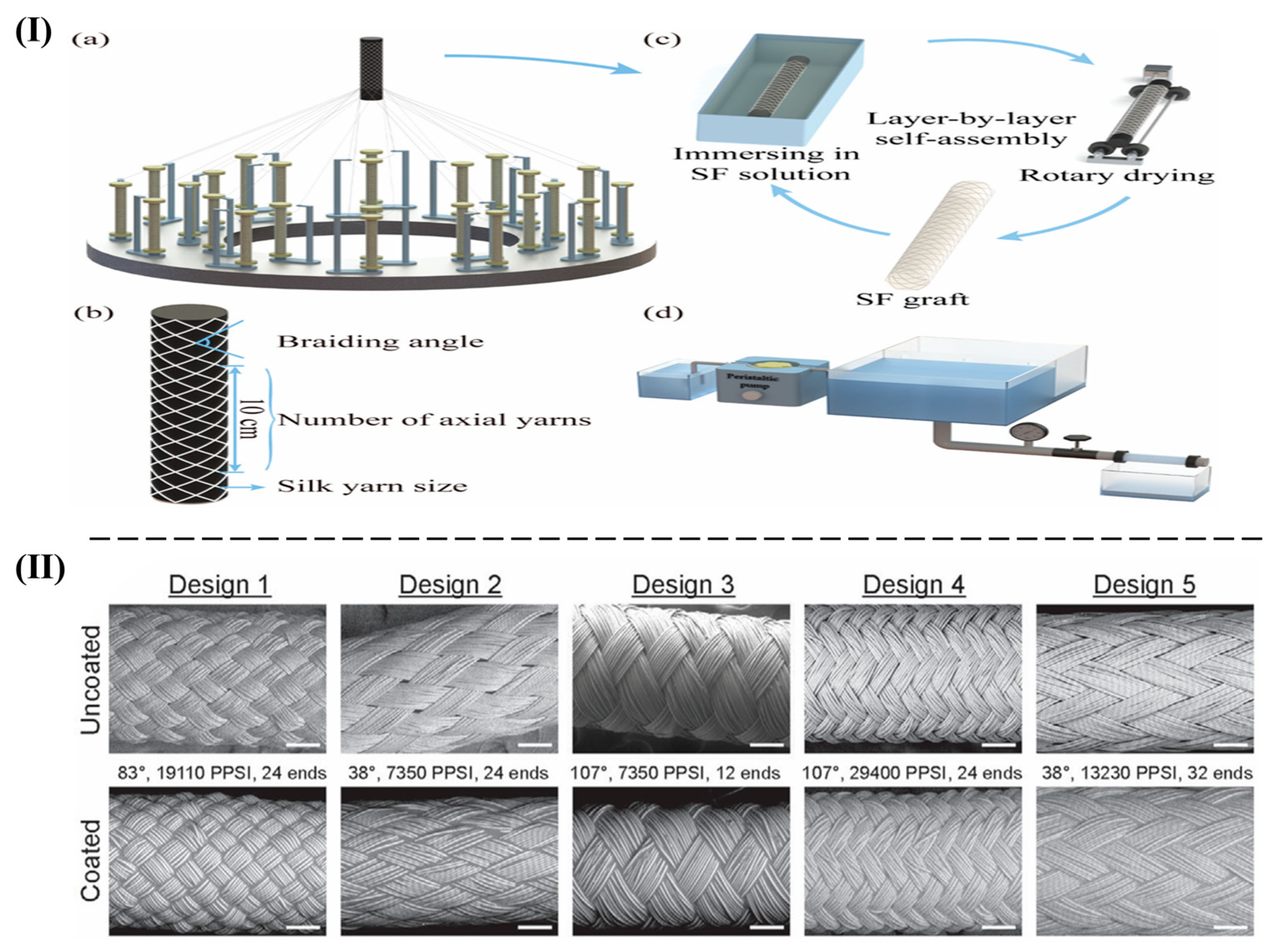
Yu et al. [125] developed SF/silk braiding fabric composite grafts using braiding technology and layer-by-layer (LBL) self-assembly, investigating their morphology, water permeability, and cytotoxicity. They prepared regenerated SF (RSF) solutions, braided silk tubular fabrics with varying parameters (braiding angles, axial yarn counts, yarn sizes), and coated them via LBL self-assembly with rotary drying to form uniform RSF films (Figure 9I). Key results showed that grafts with a 90° braiding angle and ≥60 axial yarns/10 cm had low water permeability, with 1 × 2 yarn-based grafts exhibiting 70–80 μm thickness and 8.8 mL/min·cm2 permeability. L929 fibroblast tests revealed no significant cytotoxicity, with relative growth rates >90% after LBL rinsing. This study highlights SF composite grafts as promising biomaterials for vascular repair, combining low water permeability, uniform thickness, and cytocompatibility to address long-term patency challenges in stent grafts.
Zbinden et al. [126] developed TEVGs using braided PGA fibers with or without poly (glycerol sebacate) (PGS) coating, investigating the impact of braiding parameters (angle, density) and coating on neotissue formation in a Beige mouse model. They manufactured scaffolds with different braiding patterns, implanted them in the infrarenal abdominal aorta, and evaluated them via 4D ultrasound, biaxial mechanical testing, and histology (Figure 9II). Key results showed 12-week survival rates ranged from 29% to 93%, with the dense 1 × 1 braided, PGS-coated Design 1 performing best (93% survival). Ultrasound revealed diameter increases between weeks 4 and 8, while mechanical tests indicated that all grafts had lower distensibility than the native aorta. Histology showed that Design 2 had elastin density similar to native vessels, and regression analysis linked higher braiding density to reduced elastin and increased collagen. This study highlights how braiding parameters and PGS coating interdependently affect inflammation, matrix production, and graft survival, providing critical insights for rational TEVG design to balance mechanical properties and neotissue regeneration.
Ding et al. [127] conducted a study to explore how braided silk fiber skeletons with varying porosities influence in vivo vascular tissue regeneration and long-term patency. Using finite element analysis, they designed low-, medium-, and high-porosity braided silk fiber skeletons, which were then coated with hemocompatible sulfated silk fibroin sponges to create vascular grafts. Mechanical property tests showed that high-porosity grafts exhibited higher elastic moduli and compliance but lower suture retention strength, whereas medium-porosity grafts struck an optimal balance in mechanical properties, featuring a compliance of 5.0 ± 0.7%/(mmHg × 10−2) and a suture retention strength of 4.1 ± 0.4 N. In vivo experiments in rats demonstrated that medium-porosity grafts facilitated the regeneration of the vascular smooth muscle layer, polarization of M2 macrophages, and synchronous pulsation with native arteries. Notably, these grafts maintained a 75% patency rate after 24 months and displayed contractile function in response to vasoactive agents. This research underscores the pivotal role of porosity in regulating graft performance, offering valuable guidance for the design of next-generation vascular grafts that enable long-term patency and functional tissue regeneration.
Additionally, Table 3 offers valuable information about the categories of the materials used for preparing vascular grafts by each textile technology including natural biomaterials, degradable synthetic materials, and non-degradable synthetic materials.

4. Summary and Prospects

The integration of textile technology in vascular tissue engineering has revolutionized the design of artificial blood vessels, bridging structural mimicry of native vessels with functional regeneration. Traditional textile techniques (weaving, knitting, braiding) and modern innovations (electrospinning, MEW, wet spinning) have enabled the fabrication of scaffolds with hierarchical fiber architectures, mechanical anisotropy, and biomimetic ECM structures. For instance, electrospun nanofibers have shown promise in mimicking the nano-scale ECM, promoting EC alignment and SMC phenotype maintenance, while MEW allows precise control over scaffold mechanics to replicate the nonlinear stress–strain behavior of native vessels. Tri-layered scaffolds, inspired by the intima–media–adventitia structure of arteries, have demonstrated enhanced hemocompatibility and tissue regeneration in preclinical models.
However, challenges remain for clinical translation. Mechanical mismatch between grafts and native vessels (e.g., compliance and burst pressure) can lead to intimal hyperplasia and restenosis, as seen in some MEW and electrospun grafts. Additionally, long-term durability and resistance to thrombosis require further optimization, particularly for small-diameter grafts (<6 mm). The trade-off between porosity for cell infiltration and mechanical strength in silk-based or hybrid scaffolds also necessitates careful design. Despite these hurdles, textile-based approaches have advanced significantly, offering biocompatible, tunable, and scalable solutions that surpass traditional synthetic grafts like ePTFE.

Author Contributions

Conceptualization, H.J. and H.Y.; methodology, Z.L.; software, H.J.; validation, H.J., H.Y. and Z.L.; formal analysis, H.J.; investigation, H.J.; resources, H.J.; data curation, H.Y.; writing—original draft preparation, H.Y.; writing—review and editing, Z.L.; visualization, H.J.; supervision, Z.L.; project administration, Z.L.; funding acquisition, H.J. All authors have read and agreed to the published version of the manuscript.

Funding

This research was funded by the financial support of the National Key Research and Development Program of China (No. 2023YFC2412400 and No. 2023YFC2412405) and the Hubei Provincial Major Science and Technology Projects (No. 2022ACA002).

Institutional Review Board Statement

Not applicable.

Data Availability Statement

Not applicable.

Conflicts of Interest

The authors Hua Ji and Zehao Li were employed by the company Winner Medical Co., Ltd. The remaining authors declare that this research was conducted in the absence of any commercial or financial relationships that could be construed as potential conflicts of interest.

References

  1. West-Livingston, L.; Lim, J.W.; Lee, S.J. Translational tissue-engineered vascular grafts: From bench to bedside. Biomaterials 2023, 302, 122322. [Google Scholar] [CrossRef]
  2. Hernandez-Sanchez, D.; Comtois-Bona, M.; Munoz, M.; Ruel, M.; Suuronen, E.J.; Alarcon, E.I. Manufacturing and validation of small-diameter vascular grafts: A mini review. iScience 2024, 27, 109845. [Google Scholar] [CrossRef] [PubMed]
  3. Tsao, C.W.; Aday, A.W.; Almarzooq, Z.I.; Alonso, A.; Beaton, A.Z.; Bittencourt, M.S.; Boehme, A.K.; Buxton, A.E.; Carson, A.P.; Commodore-Mensah, Y.; et al. Heart Disease and Stroke Statistics-2022 Update: A Report from the American Heart Association. Circulation 2022, 145, e153–e639. [Google Scholar] [CrossRef] [PubMed]
  4. Shahriari-Khalaji, M.; Shafiq, M.; Cui, H.; Cao, R.; Zhu, M. Advancements in the fabrication technologies and biomaterials for small diameter vascular grafts: A fine-tuning of physicochemical and biological properties. Appl. Mater. Today 2023, 31, 101778. [Google Scholar] [CrossRef]
  5. Weekes, A.; Bartnikowski, N.; Pinto, N.; Jenkins, J.; Meinert, C.; Klein, T.J. Biofabrication of small diameter tissue-engineered vascular grafts. Acta Biomater. 2022, 138, 92–111. [Google Scholar] [CrossRef] [PubMed]
  6. Benjamin, E.J.; Muntner, P.; Alonso, A.; Bittencourt, M.S.; Callaway, C.W.; Carson, A.P.; Chamberlain, A.M.; Chang, A.R.; Cheng, S.; Das, S.R.; et al. Heart Disease and Stroke Statistics-2019 Update: A Report from the American Heart Association. Circulation 2019, 139, e56–e528. [Google Scholar] [CrossRef]
  7. Skovrind, I.; Harvald, E.B.; Juul Belling, H.; Jorgensen, C.D.; Lindholt, J.S.; Andersen, D.C. Concise Review: Patency of Small-Diameter Tissue-Engineered Vascular Grafts: A Meta-Analysis of Preclinical Trials. Stem Cells Transl. Med. 2019, 8, 671–680. [Google Scholar] [CrossRef]
  8. Saito, J.; Kaneko, M.; Ishikawa, Y.; Yokoyama, U. Challenges and Possibilities of Cell-Based Tissue-Engineered Vascular Grafts. Cyborg Bionic Syst. 2021, 2021, 1532103. [Google Scholar] [CrossRef]
  9. Bakare, A.; Mohanadas, H.P.; Tucker, N.; Ahmed, W.; Manikandan, A.; Faudzi, A.A.M.; Mohamaddan, S.; Jaganathan, S.K. Advancements in textile techniques for cardiovascular tissue replacement and repair. APL Bioeng. 2024, 8, 041503. [Google Scholar] [CrossRef]
  10. Jiang, C.; Wang, K.; Liu, Y.; Zhang, C.; Wang, B. Application of textile technology in tissue engineering: A review. Acta Biomater. 2021, 128, 60–76. [Google Scholar] [CrossRef]
  11. Doersam, A.; Tsigkou, O.; Jones, C. A Review: Textile Technologies for Single and Multi-Layer Tubular Soft Tissue Engineering. Adv. Mater. Technol. 2022, 7, 2101720. [Google Scholar] [CrossRef]
  12. Liu, C.; Dai, J.; Wang, X.; Hu, X. The Influence of Textile Structure Characteristics on the Performance of Artificial Blood Vessels. Polymers 2023, 15, 3003. [Google Scholar] [CrossRef]
  13. Hasan, A.; Memic, A.; Annabi, N.; Hossain, M.; Paul, A.; Dokmeci, M.R.; Dehghani, F.; Khademhosseini, A. Electrospun scaffolds for tissue engineering of vascular grafts. Acta Biomater. 2014, 10, 11–25. [Google Scholar] [CrossRef]
  14. Rickel, A.P.; Deng, X.; Engebretson, D.; Hong, Z. Electrospun nanofiber scaffold for vascular tissue engineering. Mater. Sci. Eng. C Mater. Biol. Appl. 2021, 129, 112373. [Google Scholar] [CrossRef]
  15. Kawecki, F.; L’Heureux, N. Current biofabrication methods for vascular tissue engineering and an introduction to biological textiles. Biofabrication 2023, 15, 022004. [Google Scholar] [CrossRef]
  16. Yuan, Q.; Ma, C.; Ma, M.-G. Biotextile-based scaffolds in tissue engineering. In Medical Textiles from Natural Resources; Woodhead: Cambridge, UK, 2022; pp. 285–313. [Google Scholar]
  17. Di Francesco, D.; Pigliafreddo, A.; Casarella, S.; Di Nunno, L.; Mantovani, D.; Boccafoschi, F. Biological Materials for Tissue-Engineered Vascular Grafts: Overview of Recent Advancements. Biomolecules 2023, 13, 1389. [Google Scholar] [CrossRef]
  18. Jockenhoevel, S.; Zund, G.; Hoerstrup, S.P.; Chalabi, K.; Sachweh, J.S.; Demircan, L.; Messmer, B.J.; Turina, M. Fibrin gel– advantages of a new scaffold in cardiovascular tissue engineering. Eur. J. Cardiothorac. Surg. 2001, 19, 424–430. [Google Scholar] [CrossRef] [PubMed]
  19. Hajjaji, R.; Abdessalem, S.B.; Ganghoffer, J.F. The influence of textile vascular prosthesis crimping on graft longitudinal elasticity and flexibility. J. Mech. Behav. Biomed. Mater. 2012, 16, 73–80. [Google Scholar] [CrossRef]
  20. Konig, G.; McAllister, T.N.; Dusserre, N.; Garrido, S.A.; Iyican, C.; Marini, A.; Fiorillo, A.; Avila, H.; Wystrychowski, W.; Zagalski, K.; et al. Mechanical properties of completely autologous human tissue engineered blood vessels compared to human saphenous vein and mammary artery. Biomaterials 2009, 30, 1542–1550. [Google Scholar] [CrossRef]
  21. Wang, D.; Xu, Y.; Li, Q.; Turng, L.S. Artificial small-diameter blood vessels: Materials, fabrication, surface modification, mechanical properties, and bioactive functionalities. J. Mater. Chem. B 2020, 8, 1801–1822. [Google Scholar] [CrossRef]
  22. Zhang, T.; Zhang, R.; Zhang, Y.; Kannan, P.R.; Li, Y.; Lv, Y.; Zhao, R.; Kong, X. Silk-based biomaterials for tissue engineering. Adv. Colloid. Interface Sci. 2025, 338, 103413. [Google Scholar] [CrossRef] [PubMed]
  23. Gupta, P.; Mandal, B.B. Silk biomaterials for vascular tissue engineering applications. Acta Biomater. 2021, 134, 79–106. [Google Scholar] [CrossRef]
  24. Coimbra, P.; Santos, P.; Alves, P.; Miguel, S.P.; Carvalho, M.P.; de Sa, K.D.; Correia, I.J.; Ferreira, P. Coaxial electrospun PCL/Gelatin-MA fibers as scaffolds for vascular tissue engineering. Colloids Surf. B Biointerfaces 2017, 159, 7–15. [Google Scholar] [CrossRef]
  25. Hyun, G.W.; Park, S.H. 3D freeform fabrication of fiber-reinforced artificial vascular grafts with biomimetic softness and enhanced mechanical strength. J. Manuf. Process. 2025, 133, 1237–1248. [Google Scholar] [CrossRef]
  26. Xie, X.; Chen, Y.; Wang, X.; Xu, X.; Shen, Y.; Khan, A.U.R.; Aldalbahi, A.; Fetz, A.E.; Bowlin, G.L.; El-Newehy, M.; et al. Electrospinning nanofiber scaffolds for soft and hard tissue regeneration. J. Mater. Sci. Technol. 2020, 59, 243–261. [Google Scholar] [CrossRef]
  27. Barnes, C.P.; Sell, S.A.; Boland, E.D.; Simpson, D.G.; Bowlin, G.L. Nanofiber technology: Designing the next generation of tissue engineering scaffolds. Adv. Drug Deliv. Rev. 2007, 59, 1413–1433. [Google Scholar] [CrossRef]
  28. Wu, W.; Allen, R.A.; Wang, Y. Fast-degrading elastomer enables rapid remodeling of a cell-free synthetic graft into a neoartery. Nat. Med. 2012, 18, 1148–1153. [Google Scholar] [CrossRef]
  29. Guo, Y.; Wusiman, H.; Zhao, L.; James-ocloo, O.s.; Han, X.; Miao, Y.; Nie, J.; Wang, L.; Du, J.; Wei, Y.; et al. Engineering endothelialized small-diameter artificial blood vessels: Strategies, advances and applications. Compos. Part B Eng. 2025, 301, 112505. [Google Scholar] [CrossRef]
  30. Cao, X.; Maharjan, S.; Ashfaq, R.; Shin, J.; Zhang, Y.S. Bioprinting of Small-Diameter Blood Vessels. Engineering 2021, 7, 832–844. [Google Scholar] [CrossRef]
  31. Nwokoye, P.N.; Abilez, O.J. Bioengineering methods for vascularizing organoids. Cell Rep. Methods 2024, 4, 100779. [Google Scholar] [CrossRef] [PubMed]
  32. Bouten, C.V.; Dankers, P.Y.; Driessen-Mol, A.; Pedron, S.; Brizard, A.M.; Baaijens, F.P. Substrates for cardiovascular tissue engineering. Adv. Drug Deliv. Rev. 2011, 63, 221–241. [Google Scholar] [CrossRef] [PubMed]
  33. Jiang, S.; Wise, S.G.; Kovacic, J.C.; Rnjak-Kovacina, J.; Lord, M.S. Biomaterials containing extracellular matrix molecules as biomimetic next-generation vascular grafts. Trends Biotechnol. 2024, 42, 369–381. [Google Scholar] [CrossRef]
  34. Wang, X.; Li, K.; Yuan, Y.; Zhang, N.; Zou, Z.; Wang, Y.; Yan, S.; Li, X.; Zhao, P.; Li, Q. Nonlinear Elasticity of Blood Vessels and Vascular Grafts. ACS Biomater. Sci. Eng. 2024, 10, 3631–3654. [Google Scholar] [CrossRef]
  35. Breuer, T.; Jimenez, M.; Humphrey, J.D.; Shinoka, T.; Breuer, C.K. Tissue Engineering of Vascular Grafts: A Case Report from Bench to Bedside and Back. Arterioscler. Thromb. Vasc. Biol. 2023, 43, 399–409. [Google Scholar] [CrossRef]
  36. Krawiec, J.T.; Vorp, D.A. Adult stem cell-based tissue engineered blood vessels: A review. Biomaterials 2012, 33, 3388–3400. [Google Scholar] [CrossRef]
  37. Camasao, D.B.; Mantovani, D. The mechanical characterization of blood vessels and their substitutes in the continuous quest for physiological-relevant performances. A critical review. Mater. Today Bio 2021, 10, 100106. [Google Scholar] [CrossRef] [PubMed]
  38. Isella, B.; Sallustio, F.; Acosta, S.; Andre, D.; Jockenhovel, S.; Fernandez-Colino, A.; Rodriguez-Cabello, J.C.; Vaughan, T.J.; Kopp, A. A new approach for small-diameter vascular grafts using combined dip-coating of silk fibroin and elastin-like recombinamers. Biomater. Adv. 2025, 174, 214312. [Google Scholar] [CrossRef]
  39. Weekes, A.; Davern, J.W.; Pinto, N.; Jenkins, J.; Li, Z.; Meinert, C.; Klein, T.J. Enhancing compliance and extracellular matrix properties of tissue-engineered vascular grafts through pulsatile bioreactor culture. Biomater. Adv. 2025, 175, 214346. [Google Scholar] [CrossRef] [PubMed]
  40. Carrabba, M.; Fagnano, M.; Ghorbel, M.T.; Rapetto, F.; Su, B.; De Maria, C.; Vozzi, G.; Biglino, G.; Perriman, A.W.; Caputo, M.; et al. Development of a Novel Hierarchically Biofabricated Blood Vessel Mimic Decorated with Three Vascular Cell Populations for the Reconstruction of Small-Diameter Arteries. Adv. Funct. Mater. 2024, 34, 2300621. [Google Scholar] [CrossRef]
  41. Stekelenburg, M.; Rutten, M.C.M.; Snoeckx, L.H.E.H.; Baaijens, F.P.T. Dynamic straining combined with fibrin gel cell seeding improves strength of tissue-engineered small diameter vascular grafts. Tissue Eng. Part A 2009, 15, 1081–1089. [Google Scholar] [CrossRef]
  42. Moore, M.J.; Tan, R.P.; Yang, N.; Rnjak-Kovacina, J.; Wise, S.G. Bioengineering artificial blood vessels from natural materials. Trends Biotechnol. 2022, 40, 693–707. [Google Scholar] [CrossRef]
  43. Nash, K.M.; Boe, B.A.; Carrillo, S.A.; Harrison, A.; Iwaki, R.; Kelly, J.; Kirkton, R.D.; Krishnamurthy, R.; Lawson, J.H.; Matsuzaki, Y.; et al. Evaluation of tissue-engineered human acellular vessels as a Blalock-Taussig-Thomas shunt in a juvenile primate model. JTCVS Open 2023, 15, 433–445. [Google Scholar] [CrossRef]
  44. Shen, N.; Zhang, Y.; Raza, A.; Chang, L.; Wang, J.Y. Effects of the micro/nanostructure of electrospun zein fibres on cells in simulated blood flow environment. Mater. Sci. Eng. C Mater. Biol. Appl. 2021, 122, 111900. [Google Scholar] [CrossRef]
  45. Walden, R.; L’Italien, G.J.; Megerman, J.; Abbott, W.M. Matched elastic properties and successful arterial grafting. Arch. Surg. 1980, 115, 1166–1169. [Google Scholar] [CrossRef]
  46. Canham, P.B.; Finlay, H.M.; Boughner, D.R. Contrasting structure of the saphenous vein and internal mammary artery used as coronary bypass vessels. Cardiovasc. Res. 1997, 34, 557–567. [Google Scholar] [CrossRef] [PubMed]
  47. L’Heureux, N.; Dusserre, N.; Konig, G.; Victor, B.; Keire, P.; Wight, T.N.; Chronos, N.A.F.; Kyles, A.E.; Gregory, C.R.; Hoyt, G.; et al. Human Tissue Engineered Blood Vessel For Adult Arterial Revascularization. Nat. Med. 2006, 12, 361–365. [Google Scholar] [CrossRef]
  48. Scholpp, S.; Hoffmann, L.A.; Schatzlein, E.; Gries, T.; Emonts, C.; Blaeser, A. Interlacing biology and engineering: An introduction to textiles and their application in tissue engineering. Mater. Today Bio 2025, 31, 101617. [Google Scholar] [CrossRef]
  49. Mohammadalizadeh, Z.; Bahremandi-Toloue, E.; Karbasi, S. Recent advances in modification strategies of pre- and post-electrospinning of nanofiber scaffolds in tissue engineering. React. Funct. Polym. 2022, 172, 105202. [Google Scholar] [CrossRef]
  50. Keerthii, R.; Vinotha Sre, V.; Khan, S.S. Biopolymer-based electrospinning nanoarchitectonics for advancement in tissue regeneration. Surf. Interfaces 2025, 72, 107031. [Google Scholar] [CrossRef]
  51. Rahmati, M.; Mills, D.K.; Urbanska, A.M.; Saeb, M.R.; Venugopal, J.R.; Ramakrishna, S.; Mozafari, M. Electrospinning for tissue engineering applications. Prog. Mater. Sci. 2021, 117, 100721. [Google Scholar] [CrossRef]
  52. Karkan, S.F.; Davaran, S.; Rahbarghazi, R.; Salehi, R.; Akbarzadeh, A. Electrospun nanofibers for the fabrication of engineered vascular grafts. J. Biol. Eng. 2019, 13, 83. [Google Scholar] [CrossRef]
  53. Shi, J.; Teng, Y.; Li, D.; He, J.; Midgley, A.C.; Guo, X.; Wang, X.; Yang, X.; Wang, S.; Feng, Y.; et al. Biomimetic tri-layered small-diameter vascular grafts with decellularized extracellular matrix promoting vascular regeneration and inhibiting thrombosis with the salidroside. Mater. Today Bio 2023, 21, 100709. [Google Scholar] [CrossRef]
  54. Han, F.; Jia, X.; Dai, D.; Yang, X.; Zhao, J.; Zhao, Y.; Fan, Y.; Yuan, X. Performance of a multilayered small-diameter vascular scaffold dual-loaded with VEGF and PDGF. Biomaterials 2013, 34, 7302–7313. [Google Scholar] [CrossRef]
  55. Wu, T.; Zhang, J.; Wang, Y.; Li, D.; Sun, B.; El-Hamshary, H.; Yin, M.; Mo, X. Fabrication and preliminary study of a biomimetic tri-layer tubular graft based on fibers and fiber yarns for vascular tissue engineering. Mater. Sci. Eng. C Mater. Biol. Appl. 2018, 82, 121–129. [Google Scholar] [CrossRef]
  56. Ma, F.; Huang, X.; Wang, Y. Fabrication of a Triple-Layer Bionic Vascular Scaffold via Hybrid Electrospinning. J. Funct. Biomater. 2024, 15, 140. [Google Scholar] [CrossRef]
  57. Han, D.G.; Ahn, C.B.; Lee, J.H.; Hwang, Y.; Kim, J.H.; Park, K.Y.; Lee, J.W.; Son, K.H. Optimization of Electrospun Poly(caprolactone) Fiber Diameter for Vascular Scaffolds to Maximize Smooth Muscle Cell Infiltration and Phenotype Modulation. Polymers 2019, 11, 643. [Google Scholar] [CrossRef]
  58. Kew, S.J.; Gwynne, J.H.; Enea, D.; Abu-Rub, M.; Pandit, A.; Zeugolis, D.; Brooks, R.A.; Rushton, N.; Best, S.M.; Cameron, R.E. Regeneration and repair of tendon and ligament tissue using collagen fibre biomaterials. Acta Biomater. 2011, 7, 3237–3247. [Google Scholar] [CrossRef] [PubMed]
  59. Grace Chao, P.H.; Hsu, H.Y.; Tseng, H.Y. Electrospun microcrimped fibers with nonlinear mechanical properties enhance ligament fibroblast phenotype. Biofabrication 2014, 6, 035008. [Google Scholar] [CrossRef]
  60. He, W.; Hu, Z.; Xu, A.; Liu, R.; Yin, H.; Wang, J.; Wang, S. The preparation and performance of a new polyurethane vascular prosthesis. Cell Biochem. Biophys. 2013, 66, 855–866. [Google Scholar] [CrossRef]
  61. Fusaro, L.; Gualandi, C.; Antonioli, D.; Soccio, M.; Liguori, A.; Laus, M.; Lotti, N.; Boccafoschi, F.; Focarete, M.L. Elastomeric Electrospun Scaffolds of a Biodegradable Aliphatic Copolyester Containing PEG-Like Sequences for Dynamic Culture of Human Endothelial Cells. Biomolecules 2020, 10, 1620. [Google Scholar] [CrossRef]
  62. Tan, Z.; Gao, X.; Liu, T.; Yang, Y.; Zhong, J.; Tong, C.; Tan, Y. Electrospun vein grafts with high cell infiltration for vascular tissue engineering. Mater. Sci. Eng. C Mater. Biol. Appl. 2017, 81, 407–415. [Google Scholar] [CrossRef] [PubMed]
  63. Yan, S.; Napiwocki, B.; Xu, Y.; Zhang, J.; Zhang, X.; Wang, X.; Crone, W.C.; Li, Q.; Turng, L.S. Wavy small-diameter vascular graft made of eggshell membrane and thermoplastic polyurethane. Mater. Sci. Eng. C Mater. Biol. Appl. 2020, 107, 110311. [Google Scholar] [CrossRef] [PubMed]
  64. Mi, H.Y.; Jing, X.; Yu, E.; Wang, X.; Li, Q.; Turng, L.S. Manipulating the structure and mechanical properties of thermoplastic polyurethane/polycaprolactone hybrid small diameter vascular scaffolds fabricated via electrospinning using an assembled rotating collector. J. Mech. Behav. Biomed. Mater. 2018, 78, 433–441. [Google Scholar] [CrossRef]
  65. Yu, E.; Mi, H.Y.; Zhang, J.; Thomson, J.A.; Turng, L.S. Development of biomimetic thermoplastic polyurethane/fibroin small-diameter vascular grafts via a novel electrospinning approach. J. Biomed. Mater. Res. A 2018, 106, 985–996. [Google Scholar] [CrossRef]
  66. Elsayed, Y.; Lekakou, C.; Labeed, F.; Tomlins, P. Fabrication and characterisation of biomimetic, electrospun gelatin fibre scaffolds for tunica media-equivalent, tissue engineered vascular grafts. Mater. Sci. Eng. C Mater. Biol. Appl. 2016, 61, 473–483. [Google Scholar] [CrossRef]
  67. Guo, S.; Jiang, Y.; Jiao, J.; Shi, Y.; Zhu, T.; Li, L. Electrospun gelatin-based biomimetic scaffold with spatially aligned and three-layer architectures for vascular tissue engineering. Int. J. Biol. Macromol. 2023, 242 Pt 3, 125039. [Google Scholar] [CrossRef]
  68. Zhang, J.; Wang, L.; Zhu, M.; Wang, L.; Xiao, N.; Kong, D. Wet-spun poly(ε-caprolactone) microfiber scaffolds for oriented growth and infiltration of smooth muscle cells. Mater. Lett. 2014, 132, 59–62. [Google Scholar] [CrossRef]
  69. Zhang, B.; Xu, Y.; Ma, S.; Wang, L.; Liu, C.; Xu, W.; Shi, J.; Qiao, W.; Yang, H. Small-diameter polyurethane vascular graft with high strength and excellent compliance. J. Mech. Behav. Biomed. Mater. 2021, 121, 104614. [Google Scholar] [CrossRef]
  70. Lovett, M.; Eng, G.; Kluge, J.A.; Cannizzaro, C.; Vunjak-Novakovic, G. Tubular silk scaffolds for small diameter vascular grafts. Organogenesis 2010, 6, 217–224. [Google Scholar] [CrossRef]
  71. Rodriguez, M.; Kluge, J.A.; Smoot, D.; Kluge, M.A.; Schmidt, D.F.; Paetsch, C.R.; Kim, P.S.; Kaplan, D.L. Fabricating mechanically improved silk-based vascular grafts by solution control of the gel-spinning process. Biomaterials 2020, 230, 119567. [Google Scholar] [CrossRef] [PubMed]
  72. Dalton, P.D. Melt electrowriting with additive manufacturing principles. Curr. Opin. Biomed. Eng. 2017, 2, 49–57. [Google Scholar] [CrossRef]
  73. Fleischer, S.; Tavakol, D.N.; Vunjak-Novakovic, G. From Arteries to Capillaries: Approaches to Engineering Human Vasculature. Adv. Funct. Mater. 2020, 30, 1910811. [Google Scholar] [CrossRef]
  74. Mueller, K.M.A.; Mansi, S.; De-Juan-Pardo, E.M.; Mela, P. Advances in melt electrowriting for cardiovascular applications. Front. Bioeng. Biotechnol. 2024, 12, 1425073. [Google Scholar] [CrossRef]
  75. Raeisdasteh Hokmabad, V.; Davaran, S.; Ramazani, A.; Salehi, R. Design and fabrication of porous biodegradable scaffolds: A strategy for tissue engineering. J. Biomater. Sci. Polym. Ed. 2017, 28, 1797–1825. [Google Scholar] [CrossRef]
  76. Saidy, N.T.; Shabab, T.; Bas, O.; Rojas-Gonzalez, D.M.; Menne, M.; Henry, T.; Hutmacher, D.W.; Mela, P.; De-Juan-Pardo, E.M. Melt Electrowriting of Complex 3D Anatomically Relevant Scaffolds. Front. Bioeng. Biotechnol. 2020, 8, 793. [Google Scholar] [CrossRef]
  77. Paxton, N.C.; Lanaro, M.; Bo, A.; Crooks, N.; Ross, M.T.; Green, N.; Tetsworth, K.; Allenby, M.C.; Gu, Y.; Wong, C.S.; et al. Design tools for patient specific and highly controlled melt electrowritten scaffolds. J. Mech. Behav. Biomed. Mater. 2020, 105, 103695. [Google Scholar] [CrossRef] [PubMed]
  78. Paxton, N.C.; Daley, R.; Forrestal, D.P.; Allenby, M.C.; Woodruff, M.A. Auxetic tubular scaffolds via melt electrowriting. Mater. Des. 2020, 193, 108787. [Google Scholar] [CrossRef]
  79. Bas, O.; D’Angella, D.; Baldwin, J.G.; Castro, N.J.; Wunner, F.M.; Saidy, N.T.; Kollmannsberger, S.; Reali, A.; Rank, E.; De-Juan-Pardo, E.M.; et al. An Integrated Design, Material, and Fabrication Platform for Engineering Biomechanically and Biologically Functional Soft Tissues. ACS Appl. Mater. Interfaces 2017, 9, 29430–29437. [Google Scholar] [CrossRef] [PubMed]
  80. Jeong, Y.; Yao, Y.; Yim, E.K.F. Current understanding of intimal hyperplasia and effect of compliance in synthetic small diameter vascular grafts. Biomater. Sci. 2020, 8, 4383–4395. [Google Scholar] [CrossRef]
  81. Brown, T.D.; Dalton, P.D.; Hutmacher, D.W. Direct writing by way of melt electrospinning. Adv. Mater. 2011, 23, 5651–5657. [Google Scholar] [CrossRef]
  82. Xue, J.; Wu, T.; Dai, Y.; Xia, Y. Electrospinning and Electrospun Nanofibers: Methods, Materials, and Applications. Chem. Rev. 2019, 119, 5298–5415. [Google Scholar] [CrossRef] [PubMed]
  83. Woodruff, M.A.; Hutmacher, D.W. The return of a forgotten polymer—Polycaprolactone in the 21st century. Prog. Polym. Sci. 2010, 35, 1217–1256. [Google Scholar] [CrossRef]
  84. Bartnikowski, M.; Dargaville, T.R.; Ivanovski, S.; Hutmacher, D.W. Degradation mechanisms of polycaprolactone in the context of chemistry, geometry and environment. Prog. Polym. Sci. 2019, 96, 1–20. [Google Scholar] [CrossRef]
  85. Somszor, K.; Bas, O.; Karimi, F.; Shabab, T.; Saidy, N.T.; O’Connor, A.J.; Ellis, A.V.; Hutmacher, D.; Heath, D.E. Personalized, Mechanically Strong, and Biodegradable Coronary Artery Stents via Melt Electrowriting. ACS Macro Lett. 2020, 9, 1732–1739. [Google Scholar] [CrossRef]
  86. Sanchez Diaz, R.; Park, J.R.; Rodrigues, L.L.; Dalton, P.D.; De-Juan-Pardo, E.M.; Dargaville, T.R. Highly Elastic Scaffolds Produced by Melt Electrowriting of Poly(L-lactide-co-ε-caprolactone). Adv. Funct. Mater. 2021, 7, 2100508. [Google Scholar] [CrossRef]
  87. Kade, J.C.; Dalton, P.D. Polymers for Melt Electrowriting. Adv. Healthc. Mater. 2021, 10, e2001232. [Google Scholar] [CrossRef]
  88. Jungst, T.; Pennings, I.; Schmitz, M.; Rosenberg, A.J.W.P.; Groll, J.; Gawlitta, D. Heterotypic Scaffold Design Orchestrates Primary Cell Organization and Phenotypes in Cocultured Small Diameter Vascular Grafts. Adv. Funct. Mater. 2019, 29, 1905987. [Google Scholar] [CrossRef]
  89. Bartolf-Kopp, M.; de Silva, L.; Rosenberg, A.J.W.P.; Groll, J.; Gawlitta, D.; Jungst, T. Hybrid Co-Spinning and Melt Electrowriting Approach Enables Fabrication of Heterotypic Tubular Scaffolds Resembling the Non-Linear Mechanical Properties of Human Blood Vessels. Adv. Funct. Mater. 2024, 34, 2311797. [Google Scholar] [CrossRef]
  90. Federici, A.S.; Tornifoglio, B.; Lally, C.; Garcia, O.; Kelly, D.J.; Hoey, D.A. Melt electrowritten scaffold architectures to mimic tissue mechanics and guide neo-tissue orientation. J. Mech. Behav. Biomed. Mater. 2024, 150, 106292. [Google Scholar]
  91. Zhi, D.; Cheng, Q.; Midgley, A.C.; Zhang, Q.; Wei, T.; Li, Y. Mechanically reinforced biotubes for arterial replacement and arteriovenous grafting inspired by architectural engineering. Sci. Adv. 2022, 8, eabl3888. [Google Scholar] [CrossRef]
  92. McCosker, A.B.; Snowdon, M.E.; Lamont, R.; Woodruff, M.A.; Paxton, N.C. Exploiting Nonlinear Fiber Patterning to Control Tubular Scaffold Mechanical Behavior. Adv. Mater. Technol. 2022, 7, 2200259. [Google Scholar] [CrossRef]
  93. Weekes, A.; Wehr, G.; Pinto, N.; Jenkins, J.; Li, Z.; Meinert, C.; Klein, T.J. Highly compliant biomimetic scaffolds for small diameter tissue-engineered vascular grafts (TEVGs) produced via melt electrowriting (MEW). Biofabrication 2023, 16, 015017. [Google Scholar] [CrossRef] [PubMed]
  94. Brooks-Richards, T.L.; Paxton, N.C.; Allenby, M.C.; Woodruff, M.A. Dissolvable 3D printed PVA moulds for melt electrowriting tubular scaffolds with patient-specific geometry. Mater. Des. 2022, 215, 110466. [Google Scholar] [CrossRef]
  95. Ding, H.; Cao, K.; Zhang, F.; Boettcher, W.; Chang, R.C. A Fundamental Study of Charge Effects on Melt Electrowritten Polymer Fibers. Mater. Des. 2019, 178, 107857. [Google Scholar] [CrossRef]
  96. Kim, J.; Bakirci, E.; O’Neill, K.L.; Hrynevich, A.; Dalton, P.D. Fiber Bridging during Melt Electrowriting of Poly(ε-Caprolactone) and the Influence of Fiber Diameter and Wall Height. Macromol. Mater. Eng. 2021, 306, 2000685. [Google Scholar] [CrossRef]
  97. Mueller, K.M.A.; Unterrainer, A.; Rojas-González, D.M.; De-Juan-Pardo, E.; Willner, M.S.; Herzen, J.; Mela, P. Introducing Controlled Microporosity in Melt Electrowriting. Adv. Mater. Technol. 2023, 8, 2201158. [Google Scholar] [CrossRef]
  98. Neuhaus, B.; Loewner, S.; Heymann, H.; Webering, F.; Synofzik, J.; Blume, H.; Blume, C. Fiber deviation and optimized toolpath strategies in melt electrowriting of tubular scaffolds. Mater. Des. 2025, 254, 114147. [Google Scholar] [CrossRef]
  99. Jiao, Y.; Li, C.; Liu, L.; Wang, F.; Liu, X.; Mao, J.; Wang, L. Construction and application of textile-based tissue engineering scaffolds: A review. Biomater. Sci. 2020, 8, 3574–3600. [Google Scholar] [CrossRef]
  100. Fernández-Colino, A.; Jockenhoevel, S. Textile-Reinforced Scaffolds for Vascular Tissue Engineering. In Tissue-Engineered Vascular Grafts; Springer International: Cham, Switzerland, 2020; pp. 339–363. [Google Scholar]
  101. Akbari, M.; Tamayol, A.; Bagherifard, S.; Serex, L.; Mostafalu, P.; Faramarzi, N.; Mohammadi, M.H.; Khademhosseini, A. Textile Technologies and Tissue Engineering: A Path Toward Organ Weaving. Adv. Healthc. Mater. 2016, 5, 751–766. [Google Scholar] [CrossRef]
  102. Kun, M.; Chan, C.; Ramakrishna, S. Textile-based scaffolds for tissue engineering. In Advanced Textiles for Wound Care; Woodhead: Cambridge, UK, 2009; pp. 289–321. [Google Scholar]
  103. Hu, X.; Hu, T.; Guan, G.; Yu, S.; Wu, Y.; Wang, L. Control of weft yarn or density improves biocompatibility of PET small diameter artificial blood vessels. J. Biomed. Mater. Res. B Appl. Biomater. 2018, 106, 954–964. [Google Scholar] [CrossRef]
  104. Hu, K.; Li, Y.; Ke, Z.; Yang, H.; Lu, C.; Li, Y.; Guo, Y.; Wang, W. History, progress and future challenges of artificial blood vessels: A narrative review. Biomater. Transl. 2022, 3, 81–98. [Google Scholar]
  105. Joseph, J.; Krishnan, A.G.; Cherian, A.M.; Rajagopalan, B.; Jose, R.; Varma, P.; Maniyal, V.; Balakrishnan, S.; Nair, S.V.; Menon, D. Transforming Nanofibers into Woven Nanotextiles for Vascular Application. ACS Appl. Mater. Interfaces 2018, 10, 19449–19458. [Google Scholar] [CrossRef]
  106. Guan, G.; Yu, C.; Fang, X.; Guidoin, R.; King, M.W.; Wang, H.; Wang, L. Exploration into practical significance of integral water permeability of textile vascular grafts. J. Appl. Biomater. Funct. Mater. 2021, 19, 22808000211014007. [Google Scholar] [CrossRef]
  107. Liu, Z.; Zheng, Z.; Chen, K.; Li, Y.; Wang, X.; Li, G. A heparin-functionalized woven stent graft for endovascular exclusion. Colloids Surf. B Biointerfaces 2019, 180, 118–126. [Google Scholar] [CrossRef]
  108. Roudier, G.; Hourques, M.; Da Silva, N.; Gluais, M.; Binyet, E.; Olive, J.M.; L’Heureux, N. Effects of weaving parameters on the properties of completely biological tissue-engineered vascular grafts. Biofabrication 2023, 16, 015015. [Google Scholar] [CrossRef]
  109. Magnan, L.; Labrunie, G.; Fenelon, M.; Dusserre, N.; Foulc, M.P.; Lafourcade, M.; Svahn, I.; Gontier, E.; Velez, V.J.; McAllister, T.N.; et al. Human textiles: A cell-synthesized yarn as a truly “bio” material for tissue engineering applications. Acta Biomater. 2020, 105, 111–120. [Google Scholar] [CrossRef] [PubMed]
  110. Mishra, R.K. Advances in Textile Structural Composites II. Polymers 2025, 17, 1603. [Google Scholar] [CrossRef]
  111. Zhang, F.; Bambharoliya, T.; Xie, Y.; Liu, L.; Celik, H.; Wang, L.; Akkus, O.; King, M.W. A hybrid vascular graft harnessing the superior mechanical properties of synthetic fibers and the biological performance of collagen filaments. Mater. Sci. Eng. C Mater. Biol. Appl. 2021, 118, 111418. [Google Scholar] [CrossRef] [PubMed]
  112. Zhang, L.; Ao, Q.; Wang, A.; Lu, G.; Kong, L.; Gong, Y.; Zhao, N.; Zhang, X. A sandwich tubular scaffold derived from chitosan for blood vessel tissue engineering. J. Biomed. Mater. Res. A 2006, 77, 277–284. [Google Scholar] [CrossRef]
  113. Torikai, K.; Ichikawa, H.; Hirakawa, K.; Matsumiya, G.; Kuratani, T.; Iwai, S.; Saito, A.; Kawaguchi, N.; Matsuura, N.; Sawa, Y. A self-renewing, tissue-engineered vascular graft for arterial reconstruction. J. Thorac. Cardiovasc. Surg. 2008, 136, 37–45.e1. [Google Scholar] [CrossRef]
  114. Rama, E.; Mohapatra, S.R.; Sugimura, Y.; Suzuki, T.; Siebert, S.; Barmin, R.; Hermann, J.; Baier, J.; Rix, A.; Lemainque, T.; et al. In vitro and in vivo evaluation of biohybrid tissue-engineered vascular grafts with transformative 1H/19F MRI traceable scaffolds. Biomaterials 2024, 311, 122669. [Google Scholar] [CrossRef]
  115. Lou, C.-W.; Huang, C.-L.; Hsieh, C.-T.; Lu, P.-C.; Hsieh, C.-T.; Lin, J.-H. Polylactic acid tubular knits used as vascular grafts: Mechanical property evaluation. Fiber Polym. 2016, 16, 2593–2600. [Google Scholar] [CrossRef]
  116. Zhang, F.; Xie, Y.; Celik, H.; Akkus, O.; Bernacki, S.H.; King, M.W. Engineering small-caliber vascular grafts from collagen filaments and nanofibers with comparable mechanical properties to native vessels. Biofabrication 2019, 11, 035020. [Google Scholar] [CrossRef] [PubMed]
  117. Lou, C.W.; Lu, P.C.; Hu, J.J.; Lin, J.H. Effects of yarn types and fabric types on the compliance and bursting strength of vascular grafts. J. Mech. Behav. Biomed. Mater. 2016, 59, 474–483. [Google Scholar] [CrossRef] [PubMed]
  118. Kiritani, S.; Kaneko, J.; Ito, D.; Morito, M.; Ishizawa, T.; Akamatsu, N.; Tanaka, M.; Iida, T.; Tanaka, T.; Tanaka, R.; et al. Silk fibroin vascular graft: A promising tissue-engineered scaffold material for abdominal venous system replacement. Sci. Rep. 2020, 10, 21041. [Google Scholar] [CrossRef] [PubMed]
  119. Wang, F.; Liang, M.; Zhang, B.; Li, W.; Huang, X.; Zhang, X.; Chen, K.; Li, G. Advances in artificial blood vessels: Exploring materials, preparation, and functionality. J. Mater. Sci. Technol. 2025, 219, 225–256. [Google Scholar] [CrossRef]
  120. Emonts, C.; Grigat, N.; Merkord, F.; Vollbrecht, B.; Idrissi, A.; Sackmann, J.; Gries, T. Innovation in 3D Braiding Technology and Its Applications. Textiles 2021, 1, 185–205. [Google Scholar] [CrossRef]
  121. Lin, M.C.; Lou, C.W.; Lin, J.Y.; Lin, T.A.; Chen, Y.S.; Lin, J.H. Biodegradable Polyvinyl Alcohol Vascular Stents: Structural Model and Mechanical and Biological Property Evaluation. Mater. Sci. Eng. C Mater. Biol. Appl. 2018, 91, 404–413. [Google Scholar] [CrossRef]
  122. Mi, H.Y.; Jiang, Y.; Jing, X.; Enriquez, E.; Li, H.; Li, Q.; Turng, L.S. Fabrication of triple-layered vascular grafts composed of silk fibers, polyacrylamide hydrogel, and polyurethane nanofibers with biomimetic mechanical properties. Mater. Sci. Eng. C Mater. Biol. Appl. 2019, 98, 241–249. [Google Scholar] [CrossRef]
  123. Fukunishi, T.; Ong, C.S.; Lui, C.; Pitaktong, I.; Smoot, C.; Harris, J.; Gabriele, P.; Vricella, L.; Santhanam, L.; Lu, S.; et al. Formation of Neoarteries with Optimal Remodeling Using Rapidly Degrading Textile Vascular Grafts. Tissue Eng. Part A 2019, 25, 632–641. [Google Scholar] [CrossRef]
  124. Guan, G.; Yu, C.; Xing, M.; Wu, Y.; Hu, X.; Wang, H.; Wang, L. Hydrogel Small-Diameter Vascular Graft Reinforced with a Braided Fiber Strut with Improved Mechanical Properties. Polymers 2019, 11, 810. [Google Scholar] [CrossRef]
  125. Yu, Y.; Song, G.; Dai, M.; Meng, L.; Xu, J.; Fan, Q.; Dong, F.; Yin, Y.; Wang, J. Silk Fibroin/Silk Braiding Fabric Composite Grafts via Layer-by-Layer Self-Assembly: Water Permeability and Cytocompatibility. J. Nat. Fibers 2023, 20, 2163028. [Google Scholar] [CrossRef]
  126. Zbinden, J.C.; Blum, K.M.; Berman, A.G.; Ramachandra, A.B.; Szafron, J.M.; Kerr, K.E.; Anderson, J.L.; Sangha, G.S.; Earl, C.C.; Nigh, N.R.; et al. Effects of Braiding Parameters on Tissue Engineered Vascular Graft Development. Adv. Healthc. Mater. 2020, 9, e2001093. [Google Scholar] [CrossRef] [PubMed]
  127. Ding, X.; Zhang, W.; Xu, P.; Feng, W.; Tang, X.; Yang, X.; Wang, L.; Li, L.; Huang, Y.; Ji, J.; et al. The Regulatory Effect of Braided Silk Fiber Skeletons with Differential Porosities on In Vivo Vascular Tissue Regeneration and Long-Term Patency. Research 2022, 2022, 9825237. [Google Scholar] [CrossRef] [PubMed]
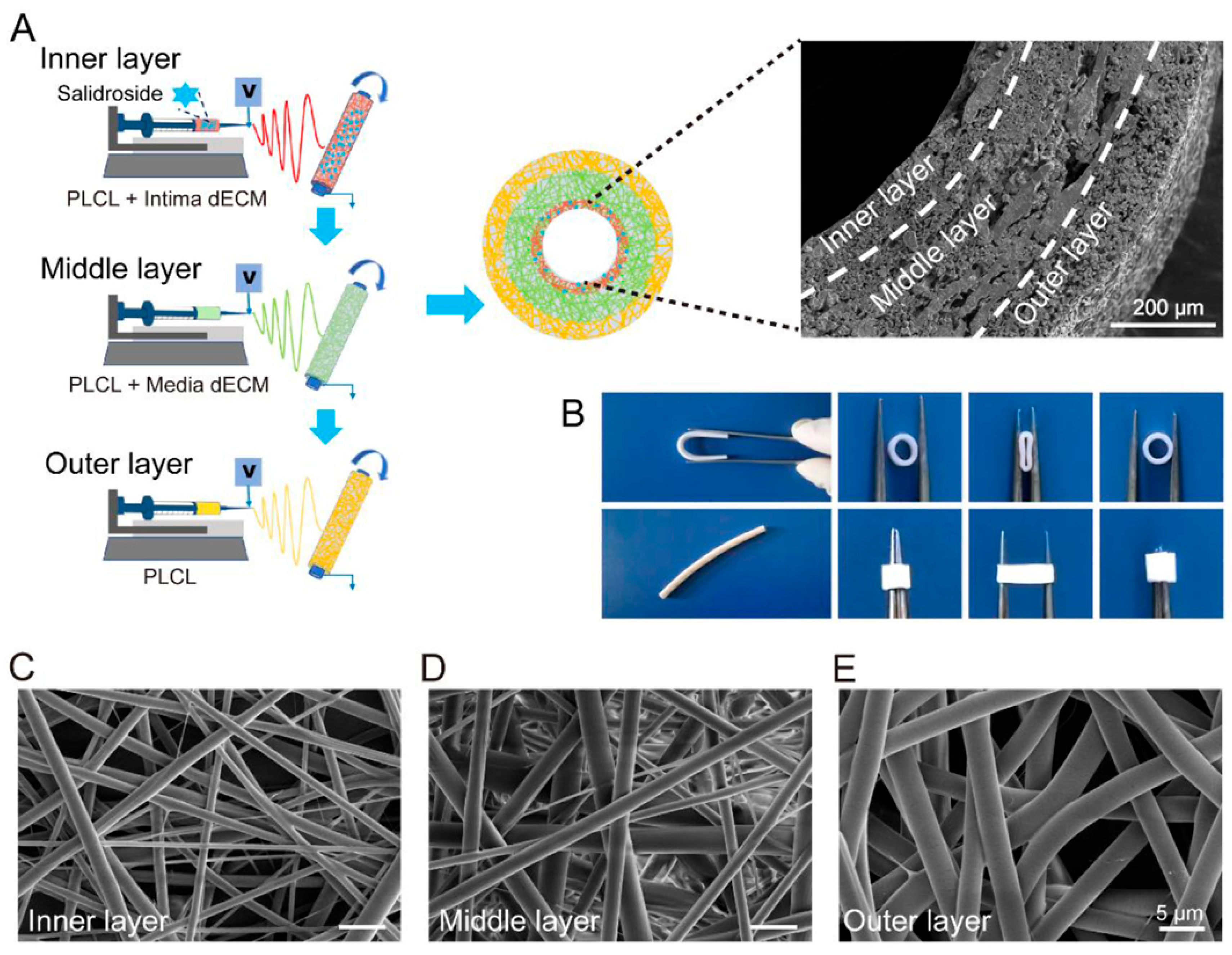
Figure 2. Characterization of the tri-layer scaffold. (A) Preparation and sectional morphology of PLCL-dECM-Sal vascular grafts. (B) Optical photograph of PLCL-dECM-Sal vascular grafts. SEM of PLCL-dECM-Sal vascular grafts: (C) inner layer, (D) middle layer, and (E) outer layer [53].
Textiles 05 00038 g002
Figure 3. Electrospinning of vascular grafts. (I). (a,b) Preparation of the outer layer; (c,d) preparation of the middle layer; (e,f) preparation process of the inner layer; (g) a fibrous membrane; (h) obtaining a vascular graft via rolling manipulation. (II). (a) Schematic illustration of the distribution of the inner, middle, and outer layers before rolling. (bd) SEM images of inner, middle, and outer fibers, respectively [67].
Textiles 05 00038 g003
Figure 4. Wet spinning and gel spinning techniques for TEVG fabrication. (I) Schematic showing artificial blood vessel prepared by wet spinning [69]. (II) Silk fibroin vascular grafts prepared by gel spinning. (A) Schematics of gel spinning process used to produce silk tubes where a concentrated silk solution is expelled through a small gauge needle and wound onto a rotating reciprocating mandrel. (B) Image of the gel spinning apparatus; (C) images of grafts compared in this study, silk and ePTFE; SEM images of silk tubes both longitudinally (D,F) and in cross-section (E,G) [70].
Textiles 05 00038 g004
Figure 5. MEW techniques for TEVG fabrication. (I) Schematic diagram of the architectural engineering-inspired generation of mechanically reinforced biotubes [91]. (II) Micrographs of printed tubular scaffolds for each of the six patterns developed in one study. Scale bar = 2 mm [92].
Textiles 05 00038 g005
Figure 6. Traditional textile techniques, including (A) weaving, (B) knitting, and (C) braiding [12].
Textiles 05 00038 g006
Figure 7. Fabrication of vascular grafts via weaving. (I). (A) Fabric structure diagrams of plain (a), 2/2 twill (b), and 3/1 twill weave (c). (BD) Photographs depicting the weaving process, a finished sample, and the surface of a bifurcated stent graft (BSG) [107]. (II). (a) Cell-assembled extracellular matrix (CAM) sheets were cut to produce two different types of threads. (bd) The warp consisted of groups of 5 mm-wide ribbons, cut along the sheet’s long axis. (eg) The weft consisted of 2 long ribbons, spiral-cut from a CAM sheet dried on a plastic sheet. (h) The warp was tensioned along the longitudinal axis around a 4.2 mm mandrel in a custom circular loom [108].
Textiles 05 00038 g007
Figure 9. Fabrication of braided vascular grafts. (I). Schematic diagram of SF tubular graft preparation and performance testing. (a) Fabrication of silk tubular fabric. (b) Parameters of the silk tubular fabric. (c) Preparation of SF tubular grafts via layer-by-layer self-assembly. (d) Water permeability testing [125]. (II). Representative SEM images of five graft designs with listed braiding parameters, depicting both poly (glycerol sebacate)-coated and uncoated cohorts [126].
Textiles 05 00038 g009
Table 2. Summary of advantages and disadvantages of traditional textile techniques for artificial blood vessels.
Textile
Technology
Mechanical StrengthMechanical StretchAdvantagesDisadvantagesRefs.
WeavingHighLowStable structure and excellent mechanical propertiesPoor compliance[9,16,99]
KnittingHighLowExcellent elasticity and complianceLarge pores and poor mechanical properties[11,100,101]
BraidingLowHighGood ductility, flexibility, and complianceLarge pore structure, which can cause serious blood leakage[10,15,102]
Table 3. List of materials used for preparing vascular grafts by each textile technology.
CategoryMaterialESWSGSMEWWeavingKnittingBraiding
Natural
biomaterials
Collagen
Elastin
Fibrin
Chitosan
Silk and silk fibroin
Bacterial cellulose
Gelatin
Degradable
synthetic
materials
Polycaprolactone (PCL)
Polylactic acid (PLA)
Poly(L-lactide-co-ε-caprolactone) (PLCL)
Polyglycolic acid (PGA)
Poly(lactic-co-glycolic) acid (PLGA)
Polyvinyl alcohol (PVA)
Polyglycerol sebacate (PGS)
Non-degradable
synthetic
materials
Polyurethanes (PU)
Polyethylene terephthalate (PET)
Thermoplastic polyurethane (TPU)
ES: electrospinning; WS: wet spinning; GS: gel spinning; MEW: melt electrowriting.
Disclaimer/Publisher’s Note: The statements, opinions and data contained in all publications are solely those of the individual author(s) and contributor(s) and not of MDPI and/or the editor(s). MDPI and/or the editor(s) disclaim responsibility for any injury to people or property resulting from any ideas, methods, instructions or products referred to in the content.