1. Introduction
Reflective display technologies utilize ambient light to present information or images by controlling, scattering, or generating interference with light [
1,
2,
3]. Consequently, they have attracted attention as energy-efficient devices [
4,
5]. Various reflection-controlling technologies have been reported, such as electrophoretic displays, reflective liquid-crystal displays, and structural color displays that employ photonic crystals or multilayer thin films. These technologies have been applied to devices such as smart windows and electronic paper (e-paper) [
6,
7], thereby contributing to the development of diverse optical display systems that can effectively exploit environmental light.
Among these, structural color has attracted significant attention as a coloration concept for next-generation reflective displays and optical devices [
8,
9,
10]. The fundamental principle is outlined below. Structural color originates from physical phenomena such as light interference, diffraction, and scattering caused by periodic microstructures. This can be achieved using self-assembled structures of spherical polystyrene (PS) or silica particles. Periodic arrangement of various nanoparticles formed through self-assembly or templating techniques have been extensively studied as structural color materials. In particular, template-assisted fabrication of periodic arrays of metal nanoparticles were pioneered by Van Duyne and co-workers around 2000, providing a foundation for the controlled assembly of ordered colloidal structures with well-defined optical properties [
11]. Unlike coloration using dyes or pigments, structural color is not based on light absorption. Consequently, it enables high optical stability and durability, appearing vivid and highly visible compared to light-absorption systems. Furthermore, the reflection wavelength can be precisely controlled by tuning the particle size and periodicity, enabling its application in advanced optical patterning, such as for anti-counterfeiting technologies [
12,
13,
14].
Optical anti-counterfeiting technologies have gained attention owing to their ability to provide immediate visual authentication, offering convenience and security. However, most existing technologies are limited to single-color changes or static pattern displays, making them relatively easy to replicate [
15,
16,
17].
Consequently, we sought to enhance the photofunctionality of structural coloration by integrating multiple coloration mechanisms. We focused on electrochromism (EC), which refers to the reversible modulation of optical properties in materials through electrochemical redox reactions [
18,
19,
20,
21,
22,
23,
24]. This phenomenon facilitates the appearance and disappearance of color via changes in visible-light absorption; additionally, it can be coupled with structural coloration. Viologens are known to form strongly colored radical cation states via electrochemical reduction, with clear transitions between the colored and bleached states, making them widely used as EC materials.
Integrating these two photonic phenomena, namely static structural color and dynamic EC behavior, within a single device facilitates the simultaneous and independent control of multiple visual parameters such as hue, visibility, and concealment. Unlike conventional single-functional display technologies, this approach enables dynamic environmental responsiveness and can control multiple states, thereby advancing the development of novel tunable optical devices. In previous studies, the EC materials used in devices combining structural colors typically comprised solid conductive polymer films deposited on electrodes. Conductive polymers such as poly(3,4-ethylenedioxythiophene) (PEDOT) and polyaniline exhibit reversible color changes via redox reactions and have been utilized alongside structural color materials to modulate visibility and background contrast. In previous studies, the EC polymer was placed behind photonic crystals, which affected the transmitted and reflected light, consequently controlling the visibility of the structural color [
25,
26].
Although solid-state EC materials offer superior film-forming ability and stability, they inherently restrict the device architecture because the electrochromic reactions occur only on the electrodes where the polymers are fixed. Consequently, the location of coloration within the device cannot be changed. If this type of spatial control of EC reactions could be achieved, it would enable unprecedented representations of reflection and light absorption in structurally colored systems.
To achieve this novel representation, we combined solution-based EC systems with structural coloration. Small-molecule-based EC compounds, such as viologens, exhibit obvious color changes between the transparent and colored states via reversible redox reactions. Moreover, because the dissolved molecules can easily diffuse into the solution, the electrode with EC coloration can be easily switched in the solution-based EC device. This flexibility enables a more adaptable design of structural color devices, potentially enhancing properties such as visibility and concealability.
Thus, solution-based electrochromic systems offer greater versatility in achievable display colors compared with conventional solid-state electrochromic systems.
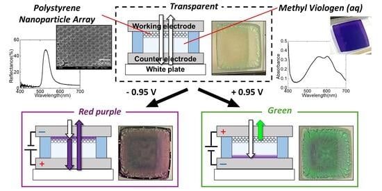
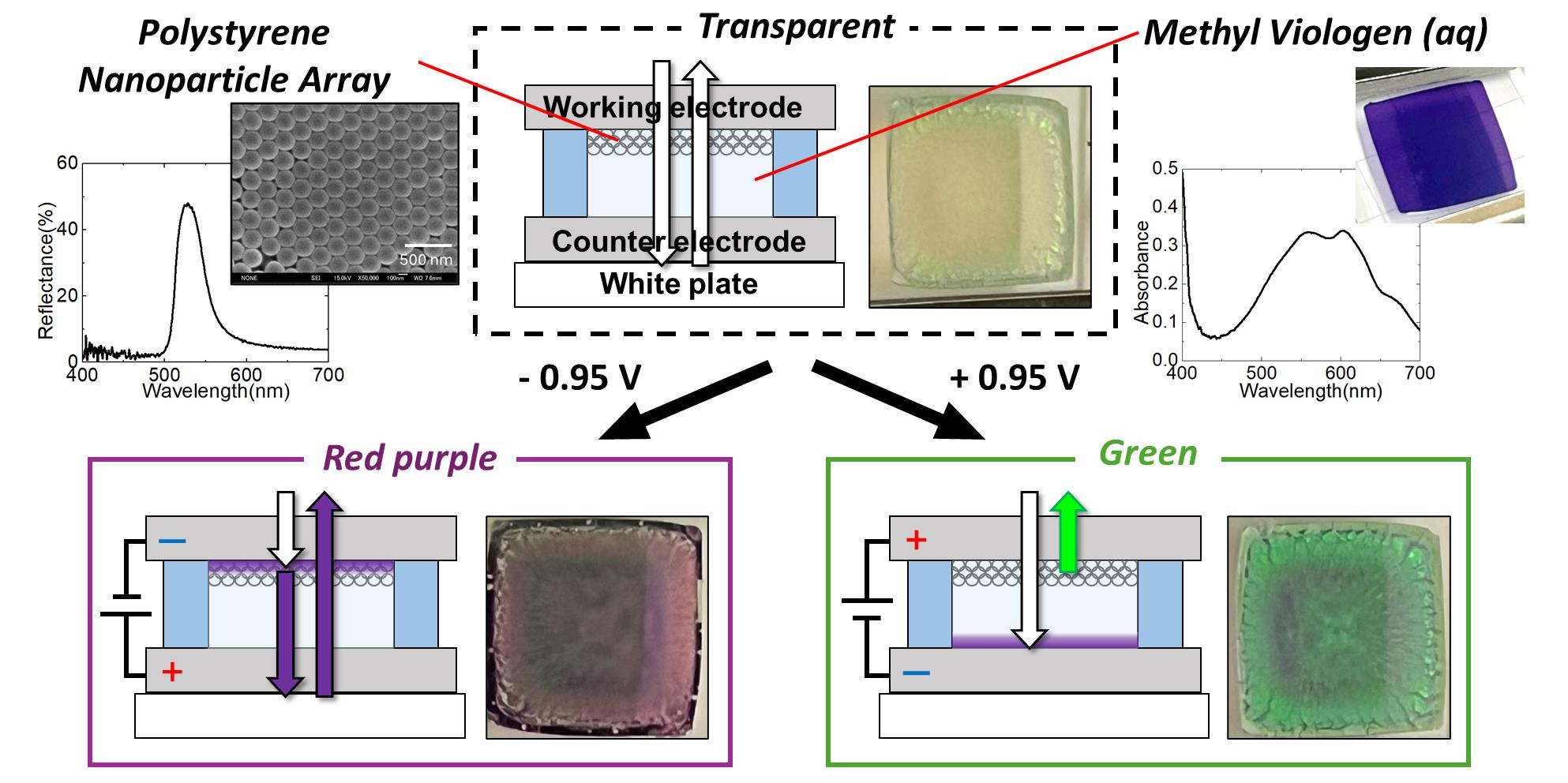
In this study, we developed a hybrid optical modulation device by utilizing a PS/polymethacrylate (PA) particle-modified ITO electrode as the structural color component and methyl viologen (MV
2+) as the solution-based EC material, as shown in
Figure 1.
Methylviologen has been widely employed as an electrochromic material because of its clear and reversible one-electron redox behavior, and numerous electrochromic devices based on methylviologen have been reported [
27,
28,
29,
30,
31].
In conventional methylviologen-based electrochromic devices, optical modulation has primarily been discussed in terms of transmittance changes or uniform coloration. In contrast, the present device combines solution-based electrochromism with a structurally colored colloidal photonic electrode, enabling modulation of the reflected appearance depending on the electrode on which electrochromic coloration occurs.
We evaluated the optical and electrochemical responses of this device to investigate how EC reactions occurring on the electrode influence the visibility and concealment of the structural color, as well as the overall color appearance of the device.
2. Experimental Section
2.1. Material
Styrene (FUJIFILM Wako Pure Chemical Corporation, Osaka, Japan), ethanol (Ueno Chemical Co., Ltd., Osaka, Japan), potassium peroxydisulfate (FUJIFILM Wako Pure Chemical Corporation, Osaka, Japan), 1,4-benzenediol (Tokyo Chemical Industry Co., Ltd., Tokyo, Japan), 1,1′-dimethyl-4,4′-bipyridinium dichloride (methyl viologen; Tokyo Chemical Industry Co., Ltd., Tokyo, Japan), lithium chloride (Kanto Chemical Co., Inc., Tokyo, Japan), sodium ferrocyanide (sodium hexacyanoferrate(II); Kanto Chemical Co., Inc., Tokyo, Japan), methyl methacrylate (Kanto Chemical Co., Inc., Tokyo, Japan), butyl acrylate (Sigma-Aldrich, St. Louis, MO, USA), and sodium dodecyl sulfate (FUJIFILM Wako Pure Chemical Corporation, Osaka, Japan) were used as received without further purification.
2.2. Preparation of PS/PA Composite Film and EC Solution
PS particles were synthesized via soap-free emulsion polymerization as described in the literature [
32]. Styrene (2 mol/L) was polymerized using an initiating agent of potassium persulfate (3 mmol/L) in water/EtOH (volume ratio 7:3) under a nitrogen atmosphere at 60 °C for approximately 3 h with continuous stirring. As the reaction progressed, the PS nanoparticles were generated and dispersed, making the solution turbid. The dispersion was concentrated under reduced pressure and washed several times via centrifugation. The resultant PS nanoparticles were weighed and redispersed in water to prepare a 1.3 wt.% aqueous dispersion.
To improve the mechanical stability of the particles, 0.15 wt.% PA composed of methyl methacrylate and butyl acrylate was added to the PS dispersion (relative to total dispersion weight); subsequently, ultrasonication was conducted to achieve a uniform dispersion. The PA particles were synthesized as described in previous literature [
33].
To fabricate the structural color film, a silicon spacer (300 μm thickness) with an active area of 1 cm × 1 cm was placed on an ITO electrode. Then, a 200 μL PS/PA dispersion was cast into the hole and dried on a 60 °C hotplate for approximately 1 h to yield a periodic structure exhibiting structural color. The resultant PS/PA-modified ITO electrode was used as an opal-structured electrode.
An aqueous EC solution was prepared by dissolving methyl viologen (MV2+; 30 mM) and sodium ferrocyanide (50 mmol/L) in pure water. The sodium ferrocyanide acted as both a counter-electrode reactant and a redox mediator for the EC reaction of the viologen molecules. Lithium chloride (200 mmol/L) was added as the supporting electrolyte. To reduce surface tension and enhance infiltration between the particles, 10 vol% ethanol was added.
2.3. Fabrication of Structural Color-EC Hybrid Electrochemical Cell
A three-electrode cell system was constructed using an ITO electrode as the working electrode, a platinum wire as the counter electrode, and an Ag/Ag+ electrode as the reference electrode. A hybrid electrochromic device was fabricated by sandwiching the EC solution between a PS/PA-modified ITO electrode and a flat ITO electrode. The inter-electrode distances of 300 μm were maintained using silicon spacers. The effective electrode area of the device was 1.0 × 1.0 cm2.
For comparison, an EC device comprising two unmodified ITO electrodes was fabricated using the same procedure described above.
2.4. Measurement Apparatus
Microscopic observations were conducted using a field-emission scanning electron microscope (FE-SEM; JSM-6700F, JEOL Ltd., Tokyo, Japan). The SEM images were analyzed using the ImageJ 1.54 image analysis software. Specular reflectance spectra were measured using a spectrophotometer (V-770, JASCO Corp., Tokyo, Japan). Cyclic voltammetry (CV) was performed using a potentiostat (Model 600A; ALS Co., Ltd., Tokyo, Japan). Transmittance measurements were performed using a multichannel CCD analyzer (USB4000, Ocean Optics, Orlando, FL, USA) in parallel with the CV analysis. Additionally, diffuse reflectance spectra (including specular components) were measured using a spectrophotometer (CM-600d, KONICA MINOLTA, Tokyo, Japan) to analyze the optical behavior in both the colored and bleached states. Changes in color appearance during the EC reactions were also observed. To measure the diffuse reflectance spectra, the device was placed on a white reflection plate to observe the actual appearance of the device.
3. Results and Discussion
3.1. Characterization of the PS Particle-Modified Electrode
PS particle and PS/PA composite dispersions were cast onto ITO electrodes to fabricate PS particle-modified and PS/PA composite films, respectively. After slow evaporation of the water solvent, a periodic arrangement of PS particles exhibiting structural color was formed via the self-organization of the particles. The particle arrangement and structural color properties of the films were characterized by FE-SEM and reflection spectroscopy.
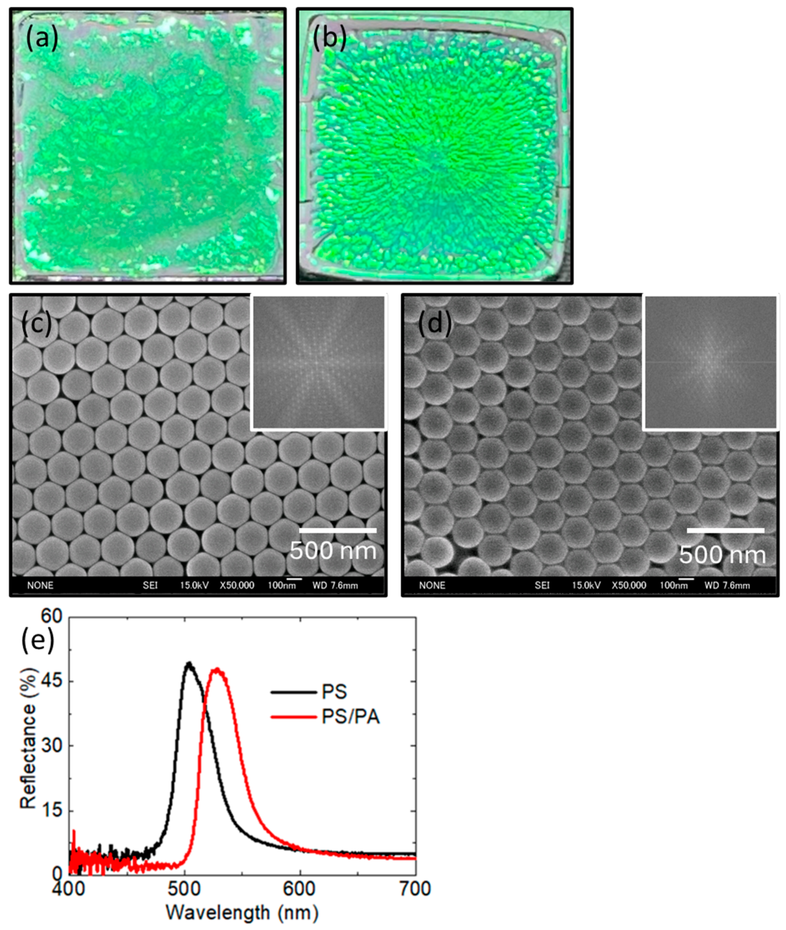
As shown in
Figure 2a,b, both the PS particle film and the PS/PA composite film exhibited a greenish structural color, indicating the formation of a periodic structure based on self-assembly.
FE-SEM observations of both films were conducted. The SEM image in
Figure 2c displays well-ordered close-packed PS particles with an average diameter of 210 ± 2 nm, calculated from ten randomly selected particles. Monodisperse PS nanoparticles were confirmed to be well ordered.
Furthermore, a two-dimensional Fourier transform (2D-FFT) analysis performed using ImageJ, ver. 1.54p (
Figure 2c, inset) revealed six-fold symmetric spots in the Fourier domain, indicating a highly ordered hexagonal arrangement of PS particles in real space.
The FE-SEM image of the PS/PA composite film is shown in
Figure 2d. The diameter of the PS/PA particles was 210 ± 2 nm, consistent with that of the PS-particle-modified film. This result indicated that the addition of PA did not affect the particle size. The 2D-FFT pattern also exhibited six-fold symmetric diffraction spots, confirming the presence of a highly ordered hexagonal arrangement in the PS/PA composite film.
To investigate the structural coloration of the films, the specular reflection spectra of both films were measured (incident angle: 5°). As shown in
Figure 2e, the reflection peak of the PS particle film appeared at 504 nm, corresponding to the optical interference responsible for the observed structural color.
The peak wavelength for the structural reflection was estimated using Bragg’s law (Equation (1)), where
l,
d,
neff, and
q are the reflection wavelength, particle diameter, effective refractive index, and incident and observation angles, respectively.
From the SEM measurements, the particle diameter, , was found to be 210 nm. Assuming a hexagonal close-packed arrangement from the FET results, was calculated using a particle filling fraction of 74%. Consequently, the peak reflection wavelength of the PS particle film was calculated to be 500 nm, which agrees well with the experimental results. This result further supports the idea that the PS-particle film forms a highly ordered, hexagonal, close-packed structure.
In contrast, the PS/PA composite film exhibited a red-shifted reflection peak at 528 nm, indicating modulation of the structural color. This shift is attributed to partial infiltration of PA (refractive index ≈ 1.5) into the interparticle voids originally filled with air (refractive index ≈ 1.0). Consequently, the reflection wavelength was redshifted according to Bragg’s law because the value of increased.
3.2. Characterization of the MV2+ Solution System
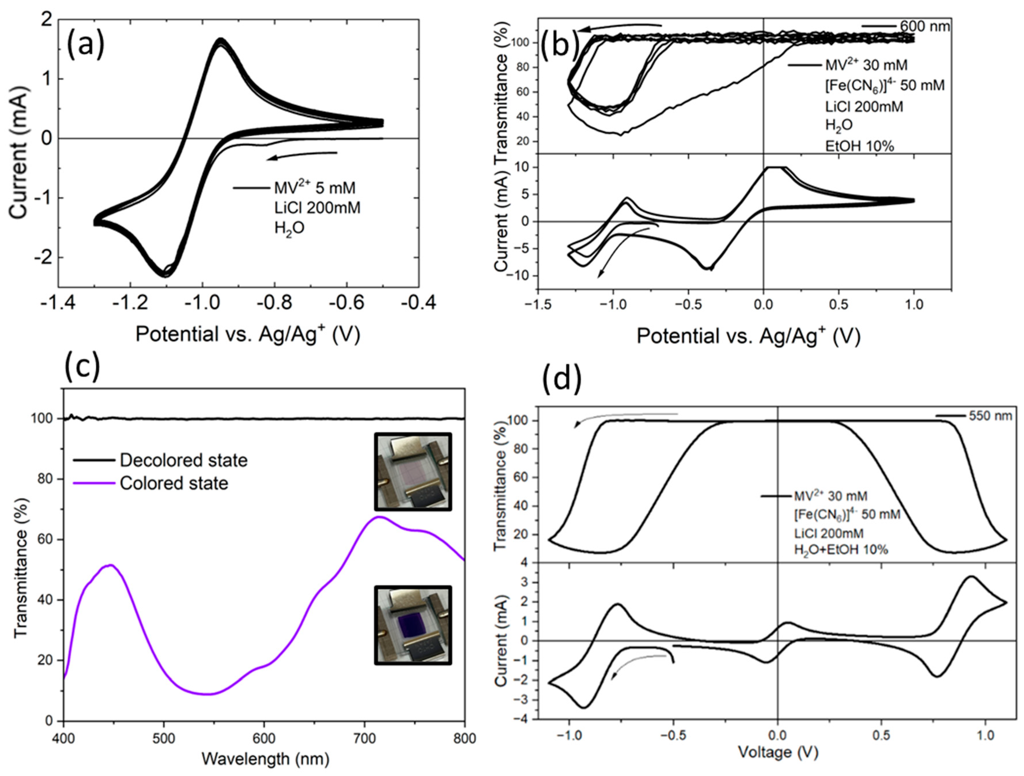
Next, the electrochemical characteristics and coloration/bleaching behavior of the EC material to be combined with the structural color were evaluated. To prevent the dissolution or degradation of the periodic structure of the PS particle film by an organic solvent, water was used as the EC solvent. Accordingly, water-soluble MV2+ was employed as the electrochromic molecule as it exhibits fast and vivid electrochromic reactions and possesses an absorption band in the same wavelength region as the reflection peak of the structural color.
Figure 3a shows the CV of the MV
2+ solution measured in a three-electrode system. The measurements were performed at a scan rate of 50 mV/s. A clear reduction peak was observed at approximately −1.1 V vs. Ag/Ag
+, indicating the reduction of MV
2+ to MV
▪+. The reverse oxidation process occurred near −0.95 V, regenerating MV
2+. These redox features indicate a reversible one-electron redox reaction between MV
2+ and MV
▪+.
Next, CV and transmittance measurements were conducted for a mixed aqueous solution containing both MV
2+ and sodium ferrocyanide. As shown in
Figure 3b, redox currents corresponding to both species were observed. A comparison between the first and subsequent CV cycles revealed that the bleaching of MV
▪+ was slower during the first cycle, whereas it proceeded much faster in later cycles, indicating the accelerated oxidation of MV
▪+. This improvement in bleaching behavior suggests that electrochemical mediation occurs between the reduced form of MV
▪+ and Fe
3+ ions. With repeated electrolysis, the concentration of sodium ferricyanide (Na
3[Fe(CN)
6]) generated in the solution gradually increased, consequently enhancing the oxidation (bleaching) of MV
▪+ through a mediated redox process.
Figure 3c depicts the transmittance spectra of the MV
2+ solution in a two-electrode system, taking the initial state of the device as 100% transmittance. The measurement was conducted by applying a voltage of –0.95 V to the device within 10 s. In the colored state (MV
▪+), the transmittance decreased in the 500–600 nm region, coinciding well with the reflection wavelength of the structural color shown in
Figure 2e. This decrease is induced by the absorption of MV
▪+. The inset photographs present the appearance of the two-electrode device before and after the application of a reduction voltage, illustrating a visible color change from transparent to purple, confirming the electrochemical generation of MV
▪+.
Finally, the CV of the EC solution and the corresponding transmittance change at 550 nm in a two-electrode system were simultaneously measured. As shown in
Figure 3d, the transmittance decreased as the reduction proceeded, reaching a minimum near the reduction peak at −0.95 V. During the reverse scan, an oxidation peak appeared around −0.8 V, accompanied by a recovery in transmittance. A comparison of the reduction and oxidation currents in the CV shows that the reduction current is larger than the oxidation current. This asymmetry suggests that a portion of the oxidation of MV
▪+ does not occur directly at the electrode surface, but instead proceeds via electron transfer form MV
▪+ to ferricyanide ions in the solution. The presence of sodium ferrocyanide improves the decolorization characteristics and reversibility. These results confirm that sodium ferrocyanide effectively functions as a counter redox species for charge compensation and reversibility-improving material in the two-electrode electrochromic system.
3.3. Analysis of Electrochemical and Optical Properties in the Hybrid Device
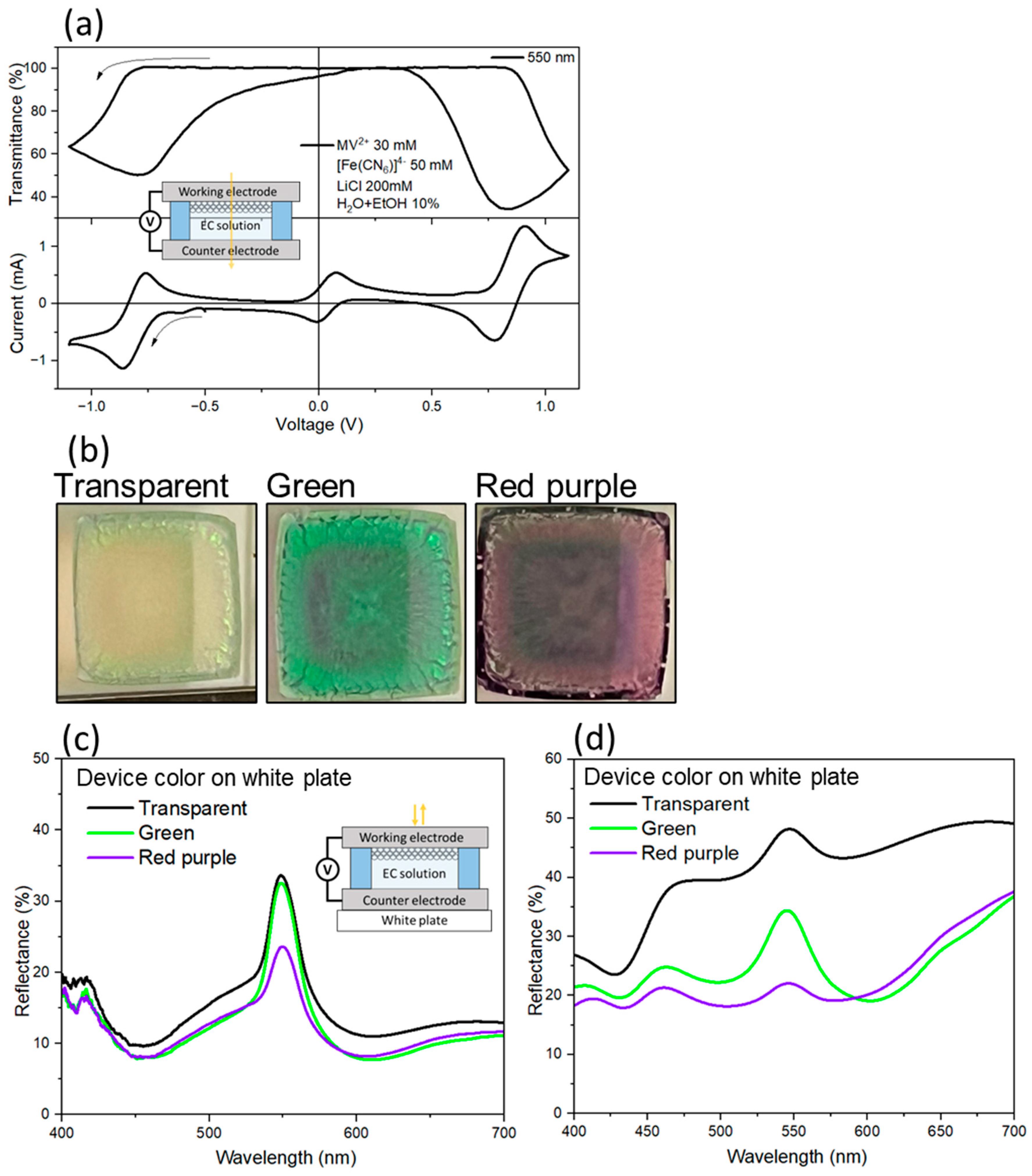
Next, a hybrid two-electrode device was assembled using the PS/PA-modified electrode (working electrode) and a flat ITO electrode (counter electrode).
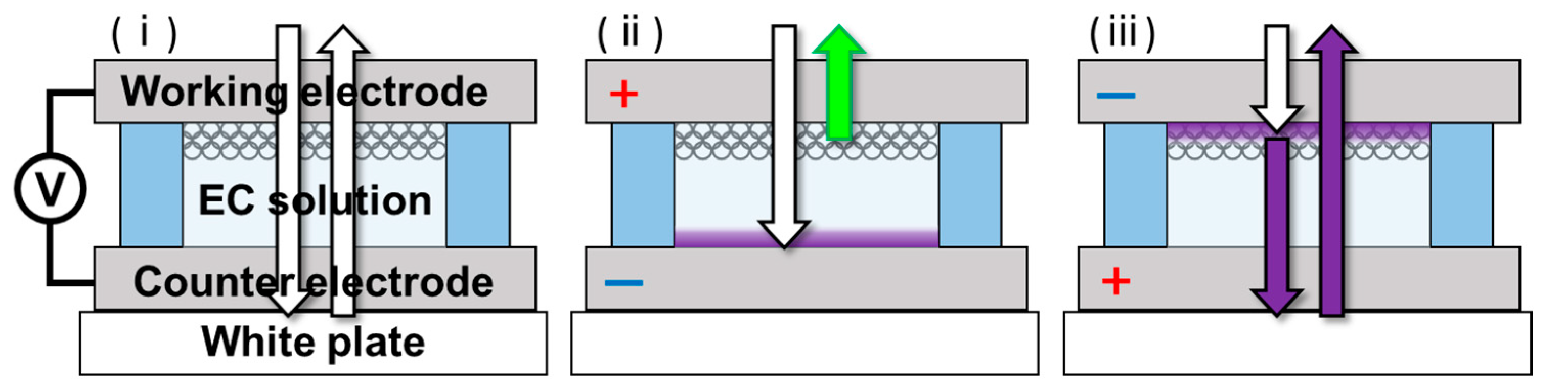
Figure 4a presents a schematic of the hybrid device, the CV curve, and the transmittance change at 550 nm for the hybrid device using the PS/PA-modified ITO electrode as the working electrode. The as-prepared state of the device (viologen was not colored) was used as the reference for the transmittance (550 nm) measurements. During the CV sweep, reduction currents were observed at approximately ±0.8 V, accompanied by a decrease in transmittance at 550 nm. This behavior is attributed to the generation of colored MV
▪+ species via the reduction of MV
2+. Upon reversing the voltage sweep, the oxidation reaction of MV
▪+ re-generated transparent MV
2+ states and recovered the transmittance to the initial states. These results indicate that the MV
2+/MV
▪+ redox reactions proceeded reversibly within this hybrid device, and that sodium ferrocyanide could suitably compensate for reaction charge at the counter electrode.
The optical appearance of the device was observed in the three states. Photographs of the device in each state are shown in
Figure 4b. When the MV
2+ layer was not colored, the device was highly transparent, and the reflection from the white plate under the device was visible. When the MV
2+ layer was colored on the counter-electrode side, the background white reflection was suppressed, making the device reflection appear greenish by enhancing the structural coloration. When the MV
2+ layer was colored on the working electrode side, part of the incident light was absorbed by the colored MV
2+, making the device appear red-purple. In this case, because the reflection wavelength of the structural color (550 nm) overlapped well with the absorption peak of MV
2+, the green structural reflection was suppressed. These images clearly reveal that the redox state of methylviologen modulates the apparent visibility and color appearance of the device through spectral overlap between electrochromic absorption and structural color reflection. These results indicate that using solution-based EC materials enables both the emphasis and suppression of the structural color through modulation of surface coloration. This enables the realization of three distinct optical states (transparent, structural color, and EC color) within a single device.
Next, the specular reflection spectra of the device in three states on a white plate were measured.
Figure 4c presents the specular reflection spectra for the transparent, red-purple, and green states of the device. The transparent and green states maintained a reflection peak at approximately 550 nm, corresponding to the structural color, indicating that changes in the background color negligibly affected the specular reflection of the structural color. In contrast, in the red-purple state, the reflection peak intensity at approximately 550 nm was significantly reduced owing to the suppression of the structural reflection by light absorption of MV
▪+.
Finally, to discuss the appearance of the actual device, the diffuse reflectance spectra, including the specular component, were measured for the device in three states on a white plate (
Figure 4d). In the transparent state, high reflectance was observed over a wide wavelength range owing to the white reflector placed under the device. In contrast, in the green state, background reflection was well suppressed by the absorption of colored MV
▪+ formed on the counter electrode, emphasizing the structural color peak and improving its visibility. In the red-purple state, the coloration of MV
▪+ within the particle layer suppressed the 550 nm structural reflection, inducing an overall decrease in reflectance.
In the hybrid device, coloration and bleaching reactions proceeded within several seconds to several tens of seconds after voltage application. This behavior can be attributed to redox reactions of electrochemically active species in the electrolyte. Within the measurement conditions and timescale of this study, the electrochromic response was reversible and no significant performance degradation was observed.