Article

Developing High-Efficiency PCR Mini-Barcoding to Enforce Conservation Efforts Against Illegal Trade and Habitat Loss of Endangered Taxus L. in the Himalayas

1
Center for Plant Sciences and Biodiversity, University of Swat, Charbagh 19120, Pakistan
2
Research Center for Tourism, Sustainability and Well-being, Campus de Gambelas, University of Algarve, 8000 Faro, Portugal
3
Department of Botany, Hazara University, Mansehra 21300, Pakistan
*
Author to whom correspondence should be addressed.
Conservation 2025, 5(4), 62; https://doi.org/10.3390/conservation5040062
Submission received: 28 August 2025 / Revised: 4 October 2025 / Accepted: 24 October 2025 / Published: 29 October 2025

Abstract

Environmental and ancient DNA are mostly present in degraded forms in nature. Plant forensics is necessary for plants like Taxus (Taxaceae), which is a medicinal, as well as poisonous, endangered plant. We designed a study to develop high-efficiency PCR mini-barcoding primers for the identification of Taxus. We collected environmental materials, fresh and old Taxus specimens from natural habitats, herbaria, and ex situ propagation sites. Taxon-specific mini-barcoding primers were prepared through primer3. All the primers were amplified onto Taxus specimens and environmental samples having Taxus DNA, while no amplification on fresh and herbarium specimens other than Taxus was noted. DNA sequencing of amplified regions of matK, ITS, and rbcL yielded lengths of 117, 175, and 200 bp. Blast taxonomy showed 100% identification power at the genus level, while 75–93% at the species level, and identified a total of 30 taxa within the genus Taxus, comprising 16 species, 5 varieties, 2 hybrids, and 7 variants. ITS was the most specific for genus identification, followed by matK and rbcL. Environmental, trade, socio-economic, and toxicological crimes were also identified. Our high-efficiency PCR mini-barcoding method can be useful in the prevention of Taxus illegal trade and habitat degradation to mitigate climate change in the Himalayan region of Pakistan.

1. Introduction

Wildlife forensics, a specialized branch of forensic science, applies advanced scientific methods to address legal issues involving wildlife, significantly contributing to law enforcement and biodiversity conservation [1]. Evolving from early techniques like fingerprinting in China during the 700s to modern high-tech instrumentation, forensic science has progressed to meet law enforcement demands, with disciplines like forensic genetics, chemistry, and morphology drawing from robust scientific foundations [2,3,4,5,6,7]. Central to forensic investigations is the “forensic triangle,” linking suspects, victims, and crime scenes, a principle increasingly vital in wildlife crime investigations driven by landmark laws such as the Lacey Act [8], Migratory Bird Treaty Act [9], Bald and Golden Eagle Protection Act [10], Marine Mammal Protection Act [11], CITES [12], and the Endangered Species Act [13,14,15,16,17]. The rise in global wildlife trafficking has underscored the importance of forensic sciences, enabled precise species identification, and supported legal proceedings to combat illegal trade and protect endangered species [15].
Taxus is distributed from temperate North America into subtropical Central America, and from temperate Eurasia to subtropical Southeast Asia [18]. Ecologically, it plays a vital role by supporting wildlife, preventing soil erosion, and maintaining forest ecosystem dynamics [19,20,21]. Despite these ecological contributions, the genus faces severe threats from overexploitation for traditional and modern medicine, fodder and fuel wood use, agricultural expansion, logging, climate change, high inbreeding and low genetic diversity, inadequate protection outside conservation areas, bark removal for Taxol extraction, timber harvesting, heavy grazing, infrastructure development, weak law enforcement, and poor community awareness [22,23,24,25,26]. Consequently, the two main Himalayan species, Taxus contorta (EN A2acd) and Taxus wallichiana (EN A2cd), are both assessed as endangered under the IUCN Red List assessment [22]. In Pakistan, the Trade Control of Wild Fauna and Flora Act, 2012 (Act No. XIV), Section 5 (a–b), enforces a complete ban on the use, harvest, and export of Taxus. Violations are prosecutable under Section 3, with penalties applied in accordance with Section 15 of the Act. Pakistan has the second-highest deforestation rate in Asia [27]. Although the Billion Tree Tsunami Afforestation Project [28] initiated by the Government of Khyber Pakhtunkhwa, Pakistan, and the Ten Billion Trees Tsunami Programme [29] launched by the Federal Government of Pakistan represent major reforestation efforts, Taxus has not been included in these initiatives due to the high cost of propagation and its extremely low survival rate under ex situ conditions. At present, there is no nursery in Pakistan engaged in the propagation of Taxus, nor are there any privately managed Taxus forests, primarily because of the species’ specific ecological amplitude.
The genus Taxus is valued for its medicinal properties, producing Taxol (paclitaxel) for cancer treatment and bioactive compounds with antimicrobial and antioxidant effects, while also being used traditionally for ailments like fever [30,31,32,33,34]. Conversely, Taxus is highly toxic due to taxine alkaloids, taxanes, and glycosides, causing severe cardiotoxic and neurotoxic symptoms, with fatalities reported in humans and animals [35,36,37]. Diagnosis of poisoning is difficult, relying on GC–MS and biomarkers such as 3,5-dimethoxyphenol, whereas DNA barcoding could provide an alternative but remains poorly validated [38,39,40].
DNA carries extensive genetic information and is recoverable from aged or limited material due to its stability and abundance [41,42]. Environmental DNA (eDNA), extracted from sources like soil, water, or air, enables non-invasive species monitoring by detecting intracellular and extracellular DNA [43,44,45,46,47,48,49,50,51,52]. Ancient DNA (aDNA), a subset of eDNA, is obtained from preserved plant remains in conditions like cold, dry, or low-oxygen environments, minimizing degradation [53]. Ancient DNA complements archaeobotanical methods, resolving taxonomic ambiguities, and provides insights into historical biodiversity, agriculture, domestication, and extinct cultivars with unique temporal and spatial resolution [54]. Despite challenges like incomplete reference libraries, eDNA, and aDNA, markers such as rbcL, trnH–psbA, matK, and ITS can be utilized for precise plant identification in forensic and conservation applications [55].
Degraded DNA is crucial in forensic science for identifying species from compromised samples, such as processed plant products, despite challenges posed by enzymatic activity, oxidative stress, hydrolysis, and environmental factors like temperature, humidity, pH, and microbial presence [53,56,57,58]. Traditional DNA barcoding effectively identifies fresh plant materials but “struggles” with processed plant products due to DNA degradation, PCR inhibition by additives, and insufficient species-specificity of standard markers like rbcL or matK [59]. DNA mini-barcoding, using shorter sequences (≤200 bp), overcomes these limitations by amplifying degraded DNA more efficiently with specially designed primers, enabling accurate species identification in forensic and conservation contexts [60,61].
This study aims to develop a high-efficiency PCR mini-barcoding protocol for the molecular identification of Taxus species in Pakistan from both environmental and fresh DNA sources, by targeting short, informative loci. The study optimizes PCR conditions to amplify degraded DNA extracted from non-traditional and compromised sources, including dung, droppings, feces, grave wood, powdered herbs, and market samples, alongside fresh plant material. Ultimately, the study evaluates the forensic applicability of mini-barcoding for detecting Taxus in seized materials, supporting CITES enforcement and conservation efforts against illegal trade and habitat loss.

2. Materials and Methods

2.1. Sampling of Intact and Environmental Materials

Fresh samples were collected from Miandam, and herbarium samples were borrowed from Hazara University, Mansehra, and Karachi University, Herbarium, Karachi, Pakistan. Cow dung, goat droppings, leaves from logging roads, unknown bird/animal feces, market raw and powdered samples, digested fodder from animal rumen in the Muslim Festival of Sacrifice (Eid ul Azha-2022), leaves from 3-years-propagated Taxus, a sample of Taxus from an Indigenous person selling Skimmia laureola (smoke use for treatment of the evil eye) and vomiting samples were collected in different parts of Himalayas in Swat, Pakistan. To support comprehensive forensic comparisons, approximately 250 species representing a broad diversity of angiosperms, gymnosperms, bryophytes, and ferns were collected from different parts of the Himalaya (see Supplementary Materials) and were PCR amplified to check for mini-barcoding amplifications. The entire specimens were deposited at the University of Swat Herbarium for further processing.

2.2. Primer Selection

A total of 4 cpDNA markers (including two coding genes: matK, rbcL, and non-coding regions: trnS-trnQ, trnL-F), along with two nuclear regions (ITS, TS), were used for the forensic identification of Taxus. For taxon-specific primers, we took matK and rbcL gene sequences of Taxus canadensis from NCBI, as the plant is monoecious, to cover male and female bias, and the first result of primer pair was selected in primer3 (Table 1). Every primer was checked for its 100% identity with Taxus spp. Full-length rbcL primers were used from Moller et al. [62]. Primers were synthesized by Genomics (BGI Genomics China, Shenzhen, China).

2.3. DNA Extraction Protocol

Natural Taxus samples were used without any prior treatment, while environmental samples were dried, ground, and put into water to differentiate between different layers, and layers having plant parts/debris were selected. DNA was extracted using a modified CTAB protocol. Stock solutions of CTAB isolation buffer (2% w/v CTAB, 1.4 M NaCl, 20 mM EDTA, 100 mM Tris-HCl, pH 8.0, 1% PVP 40,000, 0.2% β-mercaptoethanol), TE buffer (10 mM Tris-HCl, 1 mM EDTA, pH 8.0), Chloroform–isoamyl alcohol (24 + 1; v/v), 100% Isopropanol, and 70% Ethanol and 1 x TAE (40 mM Tris, 20 mM acetic acid, 1 mM EDTA) were prepared using Bio Base Electronic balance BE10002 (High-tech Zone, Jinan, Shandong, China), Eppendorf pipettes (Eppendorf AG, Barkhausenweg, Hamburg, Germany) and Jenway 3505 pH Meter (Keison Products, Chelmsford, Essex, England).
A total of 5 grams of fresh and 0.5 grams of dried plant material were ground to a fine powder using liquid nitrogen and a mortar and pestle. Ground tissue was transferred into 700 μL of prewarmed (60 °C) CTAB isolation buffer in a 1.5 mL Eppendorf tube. Isolation buffer and plant material were mixed with a Vortex mixer SCI-FS (SCILOGEX, Cromwell Avenue, Rocky Hill, CT, USA). Tubes were incubated for 90 min at 60 °C in a Drybath Stdrd 2blck (Thermo Fisher Scientific, Third Avenue Waltham, MA, USA) and mixed every 7 min with a vortex mixer. Samples were centrifuged for 10 min (13,000× g, room temperature) in an Eppendorf Centrifuge 5425 (Eppendorf AG, Barkhausenweg, Hamburg, Germany). The upper aqueous phase was transferred to a new Eppendorf tube, and one volume of chloroform–isoamyl alcohol was added to the tube and vortexed. The sample was centrifuged at 10,000 rpm for 5 min; the upper phase was transferred to a new tube, and 0.6 volume of 100% chilled isopropanol was added. Samples were stored overnight at −20 in Nihon Freezer NF-75SF3 (Gyeongchun-ro, Beon-gil, Hwado-eup, Namyangju-si, Gyeonggi-do, Republic of Korea). Samples were vortexed and centrifuged again for 10 min (5000× g, room temperature). The supernatant was discarded, and the white pellet at the bottom was obtained. A volume of 500 μL of 70% ethanol was added and centrifuged 2 times for 3 min each (5000× g, room temperature). Supernatant was discarded, and the tubes were inverted for five hours to let ethanol traces in the DNA evaporate. The DNA was diluted with 100 µL TE buffer to a final concentration of 30–50 ng/µL for PCR amplification.

2.4. PCR Optimization and DNA Sequencing

The PCRs for these markers were performed in 25 µL reactions with 30–50 ng template DNA, 0.25 µL U AmpliTaq DNA polymerase (Thermo Fisher Scientific, Third Avenue, Waltham, MA, USA), and final concentrations of 1 × PCR buffer, 1.5 mmol/L MgCl2, 0.2 mmol/L dNTP, and 0.3 µmol/L primer each. Fast Gene master mix (NIPPON Genetics EUROPE, Düren, Germany) was also used.
PCR amplifications were carried out on an Optimus 96G Gradient Thermal Cycler (Quality Lab Systems Wimborne, Dorset, England). The PCR profiles included an initial denaturation step at 94 °C for 3 min, followed by 35 cycles of 1 min at 94 °C, 1 min at the optimal annealing temperature of 54 °C for every DNA region, 1 min at 72 °C, and finished with an extension step of 10 min at 72 °C. The PCR products were inspected on 1% TAE agarose gels with 2 µL ethidium bromide in MS Mini-300 Gel electrophoresis (MS Major Science, Sea Gull Way, Saratoga, CA, USA). Gel electrophoresis results were analyzed through UV Transilluminator CSLUVTS312L equipped with CSL-MICRODOC (CLEAVER SCIENTIFIC LTD, Somers Road Industrial Estate, Rugby, UK). PCR products were sequenced in both directions with the same primers used for PCR. The PCR products were analyzed in the Biotech laboratory, Lahore, Pakistan, following the manufacturer’s instructions.

2.5. BLAST Search Criteria

Blast taxonomy results were taken according to three criteria: a search with 5000 matches with somewhat similarity (S.S) and an expect threshold (E.) value of 0.05, somewhat similarity with E., a value of 100, and a default search with 100 matches with similarity (S.) and E., value of 0.05. Blast+ 2.17.0 was used for secquence analysis.

3. Results

3.1. Sampling Distribution and Statistical Analysis

Taxus sampling for forensic identification was conducted across the Himalayan range in Swat, Pakistan. Five herbarium specimens of Taxus, from the Hazara University Herbarium (Figure 1G), representing the Swat region, and one sample from Karachi University, representing the Swat state era (before merging with Pakistan, 1953 AD) (Figure 1F), from Karachi University herbarium, were selected as reference material. Fresh samples of T. contorta leaves (Figure 1A), cow dung (Figure 1A,H,L), old leaves from logging roads (Figure 1M) and unidentified bird/animal feces (Figure 1C), clay oven fuel samples (Figure 1J), and burned tress leaves (Figure 1B) were collected from Miandam, while goat droppings were obtained from Mankyal. In Amankot, sacrificial animal rumen waste samples were obtained from five animals (Figure 1L) claimed to be raised in moist temperate mountains. In addition, ten Taxus samples were obtained from local herbal medicine stores located in Mingora, Madyan, Kabal, and Matta (Figure 1K) to assess the authenticity and source of marketed plant material. A sample was collected from the roadside (Figure 1E) in the green road of Mingora, selling Skimmia laureola. Five samples were collected from Hakims to investigate adulteration (Figure 1I). Samples from T. contorta propagated plants (Figure 1N) at the University of Swat, and food poisoning samples were obtained from the Saidu Group of Teaching Hospital, Swat, for forensic toxicological examination. The distribution map showed the presence of sampled materials at different localities in Swat, Himalaya (Figure 1D).

3.2. PCR Amplification of Taxus DNA Samples

Amplification was conducted on Taxus samples, associated environmental material, and selected non-Taxus species to enable accurate detection, cross-species comparison, and verification of assay specificity.
Pearson correlation (r) between samples and amplified samples was very low (non-significant). The statistics showed that amplification success is solely based on the quality of environmental material rather than the quantity of sampling. Such low correlation is one of the main qualities in checking environmental materials for DNA presence. Kruskal–Wallis test showed significant results of unequal medians across both samples and amplified samples.

3.2.1. Initial Amplification of Custom-Designed Primers for Taxus Samples

All six primer pairs were checked on a fresh Taxus specimen collected from Maidam, with a single annealing temperature, in a single PCR to determine their amplification. Out of the six genes, five were successfully amplified on a single annealing temperature of 54 °C, showing an 83% success rate on the first attempt. The TS gene showed amplification at 60 °C annealing temperature in further PCR assays. The amplified fragments showed very low bands on gel electrophoresis as compared to the full-length barcode of the rbcL gene, which were amplified at a 600−800 base pair region (see Supplementary Material). Mini-barcoding showed universality for Taxus fresh, herbarium, and environmental samples in PCR conditioning and amplification.

3.2.2. Diverse Sample Amplification

All the samples were run in a PCR with 100% success rate on matK, ITS primers, and rbcL (see Supplementary Material). The entire sample was amplified at a 54 °C annealing temperature. Primers also worked from 49−60 °C annealing temperatures on different samples with 30−45 cycles. Different plant species were PCR analyzed on current primers and universal primers. Universal rbcL was amplified only in fresh and herbal store tissues, while there were no amplifications for the rest of the degraded tissues. Our primers of ITS, matK, and rbcL did not amplify in plants other than Taxus.
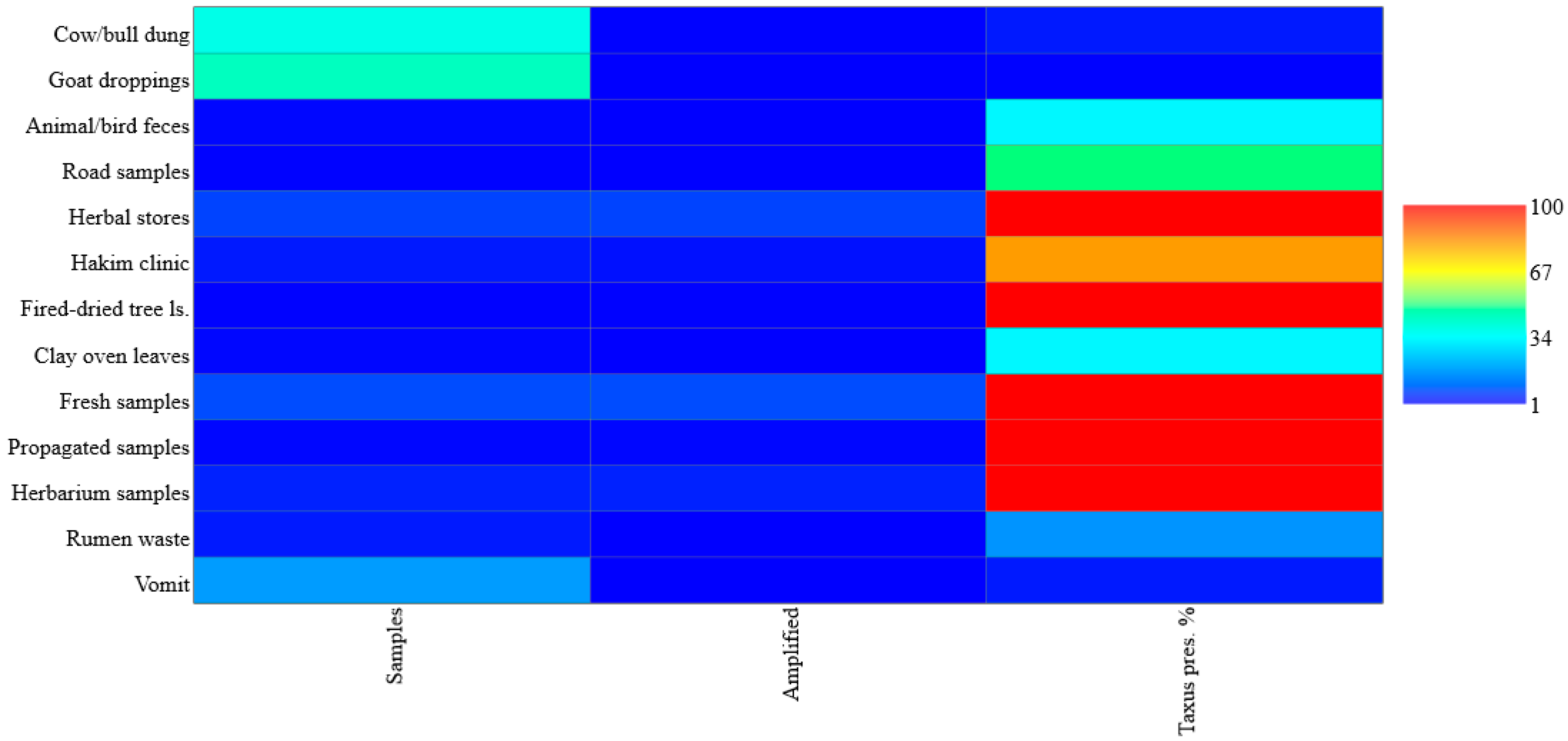
Amplification success varied considerably across sample sources. Among herbal stores, fresh samples, propagated material, fire-dried tree leaves, and herbarium specimens, amplification success was consistently high, with 100% of samples yielding successful results. Similarly, material obtained from Hakim clinics also showed a high amplification rate (80%). In contrast, animal-derived sources such as cow/bull dung (5%), goat droppings (2%), sacrificial animal rumen waste (20%), and vomit (5%) showed very low Taxus presence. Intermediate success rates were observed for unknown animal/bird feces (33%), leaves from clay ovens (33%), and leaves collected on secondary logging paths (50%). Overall, plant-derived samples, particularly those with direct or preserved Taxus presence, demonstrated markedly higher amplification efficiency compared to animal and environmental-derived materials, which were characterized by degraded or trace levels of target DNA (Figure 2).

3.3. BLAST Analysis of Amplified Sequences

One amplified PCR product each from matK, rbcL, and ITS was sequenced. The results were blasted (blastn), and taxonomy results were taken. The highest number of query length was observed in rbcL (200), followed by ITS (175) and matK (117). Three criteria were set: A search with 5000 matches with somewhat similarity and an expect threshold (E.) value of 0.05, which gives significant results for matK and ITS. A search with 5000 matches with somewhat similarity and an E. value of 100 gives significant results for matK and ITS. In these two searches, matK showed similarity with other genera of Taxaceae (Cephalotaxus), while ITS was confined most of the time to Taxus. A default search with 100 matches with similarity and an expect threshold of 0.05 gives significant results for matK, ITS, and rbcL, and shows only the Taxus genus (Table 2). The hits count of Taxus in every search denotes its universality across the Taxus genus.
The BLAST search identifies Eukaryota and Taxus as the highest taxonomic levels, with scores reaching 7637 for Eukaryota and 1030 for Taxus across the columns. These high scores suggest robust alignment of the amplified short sequences to broad taxonomic groups, providing a foundational layer for forensic analysis. The consistent presence of Taxus (scores ranging from 382 to 1030) across all columns validates the targeting of this genus, which is critical due to its toxic and medicinal taxine alkaloids, a key focus in forensic investigations. The detection of less common species, such as Taxus globosa, Taxus contorta, and Taxus phytonii, underscores the sensitivity of the BLAST approach with short fragments. These findings suggest the presence of underrepresented Taxus species, expanding the forensic profile of species potentially involved in illegal trade and poisoning incidents.

3.4. Recognized Species Diversity

Combining BLAST taxonomy searches under relaxed criteria, allowing partial similarity across 5000 searches with an E. value of 100, yielded a total of 30 taxa within the genus Taxus L., comprising 16 species, 5 varieties, 2 hybrids, and 7 variants. On average, species-level matches received 97 hits, while varieties and variants showed moderate representation with 25 and 35 hits, respectively. Hybrids were minimally represented, reflecting limited molecular data. The dataset included hits ranging from entire genomes to individual gene sequences (Table 3).
The method reliably detected genus-level identity across all fragments and captured the key Himalayan diversity, including T. wallichiana, T. contorta, and T. fuana. Intraspecific variation was also evident, as two varieties of T. wallichiana alone received 102 hits. Differences in sequence representation among taxa likely reflect variations in species abundance, detectability, or database coverage, offering valuable insights into their ecological distribution and forensic significance in Pakistan.
Further analysis identified intraspecific variation through the detection of Taxus varieties and subspecies, such as Taxus wallichiana var. wallichiana and Taxus cuspidata var. nana, highlighting the method’s sensitivity to taxonomic diversity within the genus. Additionally, the presence of less common variants, including Taxus wallichiana var. yunnanensis and Taxus cuspidata var. latifolia, pointed to potential unexplored diversity, suggesting the occurrence of rare taxa that could expand the forensic profile of Taxus in the region. The two hybrids, T. x hunnewelliana and T. x media, confirm the application of short-sequence DNA barcoding in forensic analysis in horticulture and agriculture industries (Table 3).

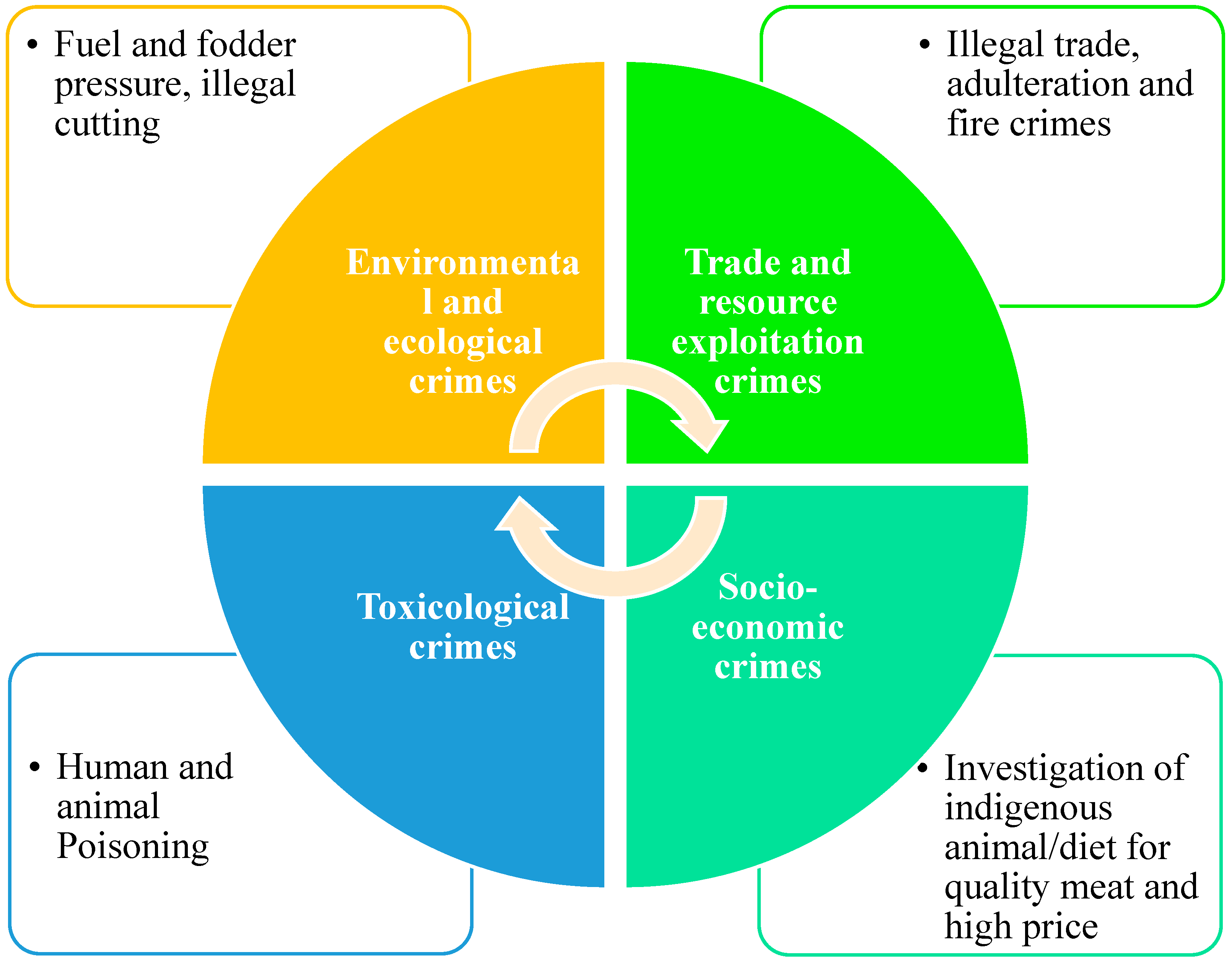
3.5. Mini-Barcoding-Based DNA Amplification Success and Detection of Crimes

Samples collected from cow dung and goat droppings revealed low presence of Taxus in their diet (2 out of 37 and 1 out of 42, respectively), but their successful amplification in a few cases indicates that these livestock occasionally consume Taxus foliage. Similarly, feces from unknown animals/birds yielded one positive amplification out of three, suggesting a possible wildlife foraging pressure, though it remains limited and needs further identification of the animal/bird species involved. DNA from a fresh logging path yielded one positive result out of two samples, clearly suggesting ongoing illegal cutting activities. Even more conclusive were the results from herbal markets and traditional medicine sources. All 10 samples of branches, leaves, and powdered forms from herbal store samples showed successful amplification, indicating active illegal trade in Taxus plant parts. The Hakim clinic samples also showed that four out of five were amplified, reinforcing concerns about possible adulteration in traditional medicine, as we collected only Taxus specimens.
Samples taken from sites impacted by fire and fuel use, such as fire-damaged tree leaves and leaves from clay ovens, also showed successful amplification (2/2 and 1/3, respectively). These findings highlight additional threats, including arson forest fires and unsustainable fuel wood harvesting, putting pressure on already vulnerable Taxus populations. All 11 fresh Taxus samples and all 6 herbarium samples amplified successfully, confirming the robustness of the barcoding method for taxonomic work. On the other hand, none of the 250 other plant samples (fresh and herbarium) amplified, validating the primer specificity of the barcoding method for Taxus but also indicating its limitations for broader plant detection. Of five rumen waste samples from sacrificial animals, only one yielded amplification. The purpose here was to assess dietary patterns in relation to livestock pricing and meat quality. Samples from propagated Taxus also showed 100% amplification. Finally, Taxus DNA was successfully amplified from one of 21 vomit samples. Although a low success rate, this one positive result is highly significant; it directly links ingestion of Taxus with poisoning cases, which may involve either accidental or intentional toxicological crime (Figure 3).

4. Discussion

4.1. Forensic Utility of Taxus Identification in Pakistan

Forensic Botany is a branch of botany where plants are used as forensic evidence to help solve criminal cases [63]. It involves analyzing plant fragments, residues, or materials collected from crime scenes to address legal and investigative questions [64]. A landmark case in 1992 marked the first use of molecular plant DNA analysis in forensics, where seed pods from a Palo Verde tree were used to place a suspect at the crime scene [65]. The genus Taxus altogether is a medicinal and poisonous plant, listed in CITIES appendix II. DNA mini-barcoding can identify its modified products, trace illegal trade, and identify health risks in poisoned patients in the moist and dry temperate regions of the Himalayas in Pakistan. Additionally, it can be applied in archaeobotany to reconstruct past plant communities and to identify lost species and varieties, owing to its ability to amplify DNA from degraded environmental materials.

4.1.1. Application to Poisoning and Illegal Trade

Some Medicinal plants act as a double-edged sword, serving therapeutic purposes under the umbrella of alternative medicine, while also being misused for malicious intent, such as poisoning, inducing fatal injuries, or incapacitating individuals through controlled doses of toxic compounds [66]. In South Asia, the use of poisonous plants in warfare, homicide, or suicide is historically significant, involving either acute or prolonged administration of plant toxins [66,67]. Taxus mini-barcoding can detect cases of poisoning, homicide, or suicide, as seen with plants like Nerium oleander and Peganum harmala [66,68]. As in our vomit samples from a patient from Banjot, Manglawar, Swat, plant materials were successfully amplified with our short DNA sequencing. Taxus powder was also given to a female child instead of Mentha powder for a stomachache. In advanced laboratories, Taxus mini-barcoding will be a useful tool to counteract Taxus-derived toxins. Rapid and accurate screening methods are necessary for diagnosing plant-induced toxicity in both medical and legal investigations [69].
Conservation and environmental management frequently depend on official statistics produced by government bodies. However, these data are often unreliable, leading to ineffective or even harmful policy decisions [70]. This issue is particularly evident in tracking the illegal harvest and trade of wild species. Numerous studies have shown that underreporting or failure to report such activities has obscured serious biodiversity threats [71,72,73]. In Pakistan, from 2001 to 2024, 7.9% of tree cover loss occurred in areas where the dominant drivers of loss resulted in deforestation. Logging accounts for 6.87 kha; Taxus only grows in Pakistan’s natural forests and is the most sort out plant for construction use. Our PCR amplification from leaf samples on logging roads confirms the illegal trade of Taxus.

4.1.2. Meeting Regional Forensic Demands

Forensic science in Pakistan has become a cornerstone of the criminal justice system, especially amid rising crime, leading to major institutional developments such as the establishment of the National Forensic Science Agency (NFSA) in 2002; the Punjab Forensic Science Agency (PFSA) in 2012 [74]; the National Forensic Science Agency, n.d.; and the Punjab Forensic Science Agency, n.d. These agencies offer multidisciplinary forensic services including DNA profiling, toxicology, and digital forensics. Despite progress, provinces like Sindh and Balochistan still lack fully operational labs, and growing public and judicial reliance on forensic evidence has led to significant backlogs, underscoring the need to expand infrastructure (Population Welfare Department, Government of Punjab, n.d.). DNA technologies, including barcoding, are gaining legal acceptance in Pakistan, as affirmed by the Supreme Court’s 2021 ruling (PLD 2021 SC 362), which highlighted the judiciary’s obligation to stay informed about scientific advancements [75,76]. Article 164 of the Qanun-e-Shahdat Order, 1984, and its 2017 amendment support the admissibility of modern forensic techniques, aligning with international standards [77,78,79]. DNA barcoding is especially useful in botanical forensics, offering rapid and accurate species identification in cases involving plant toxins, drug evidence, or ecological crimes [80,81,82,83]. While NFSA and PFSA have elevated forensic capabilities, further investment is required to bridge geographic and resource gaps, as emphasized by Steadman [84] and Lappas [85]. Moreover, forensic education and inter-disciplinary collaboration remain critical. Scholars argue that judicial officers and investigators require scientific literacy to interpret forensic data effectively, particularly in emerging areas like forensic botany [86,87,88,89]. Connor [90] and Platt [91] highlight that forensic science thrives through the dual forces of technological innovation and its seamless integration into legal systems. Mini-barcoding, for example, extends forensic science’s evidentiary scope, enabling high-precision identification of botanical traces to support investigations.

4.2. Effectiveness of Mini-Barcoding and Meta Barcoding for Taxus Identification

DNA mini-barcoding and meta-barcoding are two molecular techniques increasingly applied in the authentication of plant materials and forensic studies. DNA meta-barcoding, using universal PCR primers and next-generation sequencing (NGS), allows for the simultaneous identification of multiple species from complex environmental or herbal samples [92]. In contrast, DNA mini-barcoding targets shorter regions (200–300 bp) of DNA and is better suited for identifying individual species, especially in degraded or processed materials.
Meta-barcoding is particularly valuable in analyzing complex traditional Chinese proprietary medicines (CPMs), where markers such as ITS2 and trnL have shown high species-discrimination potential [93]. However, meta-barcoding is limited by sequencing errors introduced during PCR and NGS, especially in GC-rich regions or in degraded DNA samples, which can lead to false identifications [94,95]. For example, Coghlan et al. [96] found unknown or even endangered species such as the snow leopard in 50% of tested CPMs. Srivastava and Manjunath [97] demonstrated that the ITS region is the most effective DNA barcode for identifying endangered Indian orchids, based on analyses of 35 species using four loci. In a related study, Jiao et al. [98] used cpDNA barcoding to distinguish Santalum album from adulterants, finding that trnK had the highest recovery rate, and that a combination of psbA-trnH and trnK provided 100% species-level discrimination. These studies highlight the value of DNA barcoding in plant forensics and conservation, especially for rare and endangered species. Our analysis showed ITS and matK are more specific for minibarcoding than rbcl. A combination of ITS and matK was found suitable for Taxus identification. Our DNA mini-barcoding gives 100% success in Taxus genus-level identifications, with a 75−93% species discriminatory power. Full-length barcoding showed 100% species and genus discriminatory power but did not amplify in ancient, degraded, and environmental DNA.
The genus Taxus L., in Pakistan, has different species names; still, Taxus wallichiana, T. fuana, T. contorta, and T. baccata are in common use. The genetic diversity of Taxus contorta in Pakistan is very low as compared to other species [22,99]. One of the major factors was the dioecious nature of populations in fragmented habitats with reduced gene flow. PCR amplification on gel electrophoresis showed all genetic amplification of a single gene at the same position throughout the samples.

4.2.1. Management of Degraded Samples

DNA degradation is pivotal in forensic science, aiding in estimating post mortem interval (PMI) and time since deposition (TsD) through degraded biological material analysis [100,101]. In forensic archeology, it reveals skeletal remains’ age, sex, and origin [102]. Environmental forensics uses DNA damage to detect pollutant exposure [103], while genomic studies clarify degradation mechanisms [104]. Environmental factors like temperature and pH affect DNA stability [105], with recent models exploring degradation in various samples [106,107]. Mini-barcoding overcomes many of these degradation challenges. By targeting short chloroplast DNA (cpDNA) regions like rbcL and trnK, which are more stable than nuclear DNA, this technique allows for reliable plant identification even from highly degraded tissues such as bark, leaves, or wood shavings [41]. This makes mini-barcoding especially valuable in forensic investigations involving plant material like Taxus species, where precise identification is critical despite compromised sample quality.

4.2.2. Comparison with International Methods

A diverse array of techniques is employed to investigate DNA degradation across forensic and scientific domains, each offering insights into the extent and mechanisms of DNA damage. Gel electrophoresis remains a foundational method to assess DNA fragmentation by separating DNA based on size, where degraded DNA appears as smears compared to intact standards [108], and is particularly useful for analyzing nuclease-mediated degradation in nanostructures [109]. The comet assay, or single-cell gel electrophoresis, detects single- and double-strand breaks in individual cells by visualizing “comet tails” formed during electrophoresis, offering a sensitive method to assess damage and deposition time in forensic cases [101,110,111,112], with the MIRCA framework aiding standardization [62]. Flow cytometry enables high-throughput, fluorescently labeled quantification of DNA fragmentation in varied conditions, often paired with other techniques [111,113,114]. When combined with Annexin V labeling, it distinguishes apoptosis, necrosis, and autolysis through phosphatidylserine binding [115,116,117,118,119]. The TUNEL assay labels broken DNA ends with fluorescent dUTPs to detect strand breaks across applications, including cancer and plant biology [120,121,122,123,124,125]. Real-time quantitative PCR (RT-qPCR) is highly sensitive for quantifying degraded DNA using specific primers and is effective in forensic blood sample analysis [111,126,127,128], with melting curve analysis enhancing specificity [129]. Fluorescence in situ hybridization (FISH) uses fluorescent probes to identify specific sequences and has broad cytogenetic and diagnostic relevance [130,131,132]. Next-generation sequencing (NGS) provides comprehensive profiling of degraded DNA, with applications ranging from ancient DNA to forensic genomics [133,134,135]. Fragment length analysis is conducted using RFLP, where restriction enzyme cleavage reveals degradation through smearing [127,136], and capillary electrophoresis (CE), which enables precise separation of fluorescently labeled fragments [137]. SNP genotyping targets short, stable sequences, enhancing analysis in degraded samples [138]. DNA repair enzyme assays can quantify damage by measuring repair activity [110], while degraded DNA library preparation involves whole-genome amplification and enrichment protocols to facilitate downstream analysis [139]. Chemiluminescence-based methods such as ELISA [123,140], immunohistochemistry (IHC) [141], and immunoslot blot [142] detect degradation-related proteins and oxidized bases. Analytical techniques like HPLC–MS and GC–MS allow the detection of oxidized and UV-damaged bases with high precision [143,144,145,146,147,148], while electrochemical methods (EMs) measure charge transport disruption caused by lesions [149,150,151]. Collectively, these methods provide a comprehensive toolkit for assessing DNA degradation, and mini-barcoding targeting shorter amplicons is particularly suitable for degraded samples in resource-limited and climatically diverse settings like Pakistan.

4.3. Contribution to Global Databases

The study enhances global DNA barcode repositories by characterizing diverse Taxus taxa using high-efficiency PCR mini-barcoding, retrieving 1547 barcode hits for 16 species, including T. mairei (548 hits), T. phytonii (369), T. chinensis (171), and T. baccata (101), averaging 97 hits per taxon. Mini-barcoding effectively discriminates species, notably forensically significant taxa like T. wallichiana (68 hits) and T. contorta (26 hits) in Pakistan. Limited varietal representation (124 hits across five varieties, e.g., T. wallichiana var. wallichiana with 90 hits) indicates a need for further intra-specific sequencing. Hybrids like T. x hunnewelliana and T. x media had low representation (13 hits), reflecting their rarity and barcode recovery challenges. Additionally, 245 hits across seven variant taxa (e.g., T. sp. “Qinling type”) suggest uncharacterized diversity, highlighting mini-barcoding’s potential for uncovering cryptic lineages in herbarium, environmental, or degraded forensic samples. The study supports species-level resolution, exposes taxonomic ambiguities in databases like GenBank and BOLD, and advocates for expanded, curated reference libraries. Incorporating Pakistani Taxus populations, particularly T. contorta, strengthens global CITES enforcement, illegal trade tracking, and biodiversity monitoring in Asia.

4.4. Policy Recommendations and Future Research

The integration of modern technologies into conservation forensics has significantly enhanced efforts to detect, deter, and monitor illegal activities threatening conservation status species [152]. A future scheme was prepared by Haines et al. [152], including conservation measures, tools, and research needed for the conservation of Taxus in Pakistan. Tools such as UAVs and drones [153] enable rapid, high-resolution surveys of remote and inaccessible habitats, while thermal cameras [154] and night vision devices [155] facilitate the observation of nocturnal and cryptic species without disturbance. GPS technology [156], combined with smart devices [157] and camera systems [158], supports precise location tracking and behavioral documentation. Acoustic monitoring [159] expands the ability to detect vocal species over large areas, and tracking devices such as RFID tags [160] and GPS collars [161] provide insights into movement patterns and habitat use. Advanced sensors, including accelerometers [162], temperature loggers [163], pressure pads [164], and break-beam systems [165], further improve the capacity to collect fine-scale ecological data.
On a broader scale, satellite imagery [166] and prediction maps [167] allow for large-scale habitat monitoring and forecasting of biodiversity hotspots, while software tools [168] and cloud-based applications [169] streamline data analysis and sharing. Finally, artificial intelligence and machine learning [170] offer powerful means to process complex datasets, automate species identification, and predict ecological trends, thereby enabling proactive conservation measures. These tools combined application ensures not only timely identification and prevention of illegal activities but also contributes to long-term conservation goals by reinforcing habitat protection and law enforcement.

4.5. Botanical Forensics and Wildlife Protection: Legal and Conservation Challenges in Pakistan

Advances in molecular phylogenetics, environmental DNA (eDNA), and biodiversity databases now enable more precise species identification, comparable to human forensic systems like CODIS or GEDMatch [171,172]. These innovations raise new opportunities to identify ecosystem components, monitor ecological change, and detect keystone species through forensic and ecological data [173,174].
Wildlife crime driven by markets for traditional medicine, luxury goods, and exotic pets remains a significant global threat, worsened by internet-facilitated illegal trade [1,175,176]. Illustrative cases include the forensic tracking of ambiguous reptile specimens [177] and the exposure of illegal species substitution in the caviar trade [178,179]. In Pakistan, where plant-based crimes are understudied, there is a pressing need to integrate forensic botany, mini-barcoding, and biodiversity databases into wildlife crime investigations. Educational institutions should foster interdisciplinary training to bridge conservation science, law enforcement, and forensic practice [77,87]. Drawing from green and conservation criminology frameworks [180,181], a national strategy should promote collaboration between forensic experts, botanists, and policymakers. Ultimately, forensic botany not only enhances the evidentiary strength in environmental crime cases but also supports broader biodiversity preservation efforts. As a science policy interface, it is central to creating data-driven, legally sound conservation outcomes.

5. Conclusions

The forensic identification of Taxus L. in Pakistan using high-efficiency PCR mini-barcoding represents a significant advancement in both forensic science and biodiversity conservation. As a medicinal yet toxic genus protected under CITES Appendix II, Taxus species are frequently involved in illegal trade and poisoning cases. The application of mini-barcoding enables accurate identification even from degraded samples, such as leaves, powders, or partially decomposed tissues, making it an invaluable tool in legal investigations involving environmental crimes, poisoning incidents, and conservation violations. Pakistan’s moist and dry temperate zones, key habitats for Taxus L., have also been hotspots for illegal logging and biodiversity loss. This makes the deployment of forensic botany especially relevant. Despite challenges in DNA extraction from wood, successful amplification from leaf samples on logging routes indicates ongoing illegal trade. Additionally, real-world cases like accidental poisoning by Taxus powder underscore the urgent need for rapid molecular diagnostics. Institutional frameworks such as the NFSA and PFSA, along with Supreme Court rulings and legal provisions (PLD 2021 SC 362; QSO Article 164), now support the integration of modern forensic techniques like DNA barcoding into Pakistan’s legal system. However, gaps in forensic infrastructure and training persist, particularly in under-resourced provinces. Mini-barcoding offers a low-cost, scalable solution, especially for cases involving degraded plant materials, enhancing both criminal justice and ecological monitoring. The incorporation of mini-barcoding into a broader conservation–forensics framework supported by surveillance tools, ecological data, and international barcode databases can significantly improve enforcement against Taxus crimes. By identifying illegal harvests, tracing geographic origins, and monitoring trade patterns, this molecular method empowers authorities to respond proactively and with precision. High-efficiency PCR mini-barcoding not only strengthens forensic investigations involving Taxus but also supports policy, enforcement, and biodiversity protection in Pakistan. As an interdisciplinary bridge between botany, law, and conservation, it plays a vital role in building a scientifically informed and legally robust response to environmental crime.

Supplementary Materials

The following supporting information can be downloaded at: https://www.mdpi.com/article/10.3390/conservation5040062/s1, File S1: DNA sequences for ITS, matK, and rbcL.

Author Contributions

Conceptualization, S.D., H.A. and T.P.; methodology, S.D., H.A. and T.P.; software, S.D.; validation, S.D. and H.A.; investigation: S.D., H.A., J.A. and S.M.; resources, S.D. and H.A.; data curation, S.D., H.A., H.S. and T.P.; writing—original draft preparation, S.D., H.A. and T.P.; writing—review and editing, S.D., H.A. and T.P.; visualization, S.D., H.A. and T.P.; supervision, H.A.; project administration, S.D. and H.A.; funding acquisition, H.A. All authors have read and agreed to the published version of the manuscript.

Funding

Research was performed under HEC-NRPU-funded project No. 7303.

Institutional Review Board Statement

The study was conducted in accordance with the Declaration of Helsinki and approved by the Institutional Review Board of the University of Swat, Pakistan (protocol code Pakistan UoS/DASR/177 and approved on 7 October 2020).

Informed Consent Statement

Informed consent for participation was obtained from all subjects involved in the study.

Data Availability Statement

The original contributions presented in this study are included in the article/Supplementary Materials. Further inquiries can be directed to the corresponding authors.

Conflicts of Interest

The authors declare no conflicts of interest.

References

  1. Huffman, J.E.; Wallace, J.R. Wildlife Forensics: Methods and Applications; John Wiley & Sons: Hoboken, NJ, USA, 2011. [Google Scholar] [CrossRef]
  2. Galton, F. Finger Prints; MacMillan and Co.: London, UK; New York, NY, USA, 1892. [Google Scholar]
  3. Ashbaugh, D.R. Quantitative-Qualitative Friction Ridge Analysis: An Introduction to Basic and Advanced Ridgeology; CRC Press: Boca Raton, FL, USA, 1999. [Google Scholar]
  4. Caplan, R.M. How fingerprints came into use for personal identification. J. Am. Acad. Dermatol. 1990, 23, 109–114. [Google Scholar] [CrossRef]
  5. Ulery, B.T.; Hicklin, R.A.; Buscaglia, J.; Roberts, M.A. Accuracy and reliability of forensic latent fingerprint decisions. Proc. Natl. Acad. Sci. USA 2011, 108, 7733–7738. [Google Scholar] [CrossRef]
  6. Ulery, B.T.; Hicklin, R.A.; Buscaglia, J.; Roberts, M.A. Repeatability and reproducibility of decisions by latent fingerprint examiners. PLoS ONE 2012, 7, e32800. [Google Scholar] [CrossRef]
  7. Bell, S.; Sah, S.; Albright, T.D.; Gates, S.J., Jr.; Denton, M.B.; Casadevall, A. A call for more science in forensic science. Proc. Natl. Acad. Sci. USA 2018, 115, 4541–4544. [Google Scholar] [CrossRef] [PubMed]
  8. Lacey Act (1900 et seq) 16 U.S.C; U.S. GPO: Washington, DC, USA, 1990; pp. 3371–3378.
  9. Migratory Bird Treaty Act (1918 et seq) 16 U.S.C; U.S. GPO: Washington, DC, USA, 1918; pp. 703–712.
  10. Bald and Golden Eagle Protection Act (1940 et seq) 16 U.S.C; 668a–d; U.S. GPO: Washington, DC, USA, 1940.
  11. Marine Mammal Protection Act (1972) 16 U.S.C; §§1361–1383b, 1401–1406; U.S. GPO: Washington, DC, USA, 1972; pp. 1411h–1421h.
  12. CITES. Convention on International Trade in Endangered Species of Wild Fauna and Flora. 993 U.N.T.S. 243, 3 March 1973. [Google Scholar]
  13. Endangered Species Act of 1973 (1973 et seq); U.S. GPO: Washington, DC, USA, 1973. [CrossRef]
  14. Anderson, R.S. The Lacey Act: America’s premier weapon in the fight against unlawful wildlife trafficking. Pub. Land L. Rev. 1995, 16, 27. [Google Scholar]
  15. Burnham-Curtis, M.K.; Trail, P.W.; Kagan, R.; Moore, M.K. Wildlife forensics: An overview and update for the prosecutor. US Att’ys Bull. 2015, 63, 53. [Google Scholar]
  16. Breyer, S. Science in the courtroom. Issues Sci. Technol. 2000, 16, 52–56. [Google Scholar]
  17. Spjut, R.W. Taxonomy and nomenclature of Taxus (Taxaceae). J. Bot. Res. Inst. Tex. 2007, 1, 203–289. [Google Scholar]
  18. Cope, E.A. Taxaceae: The genera and cultivated species. Bot. Rev. 1998, 64, 291–322. [Google Scholar] [CrossRef]
  19. Shaheen, H.; Attique, A.; Riaz, M.T.; Manzoor, M.; Khan, R.W.A.; Riaz, M.T. From biodiversity hotspot to conservation hotspot: Assessing distribution, population structure, associated flora and habitat geography of threatened Himalayan Yew in temperate forest ecosystems of Kashmir. Biodivers. Conserv. 2024, 33, 553–577. [Google Scholar] [CrossRef]
  20. Bangelesa, F.; Abel, D.; Pollinger, F.; Rai, P.; Ziegler, K.; Ebengo, D.; Tshimanga, R.M.; Ali, M.M.; Knight, J.; Paeth, H. Projected changes in rainfall amount and distribution in the Democratic Republic of Congo–Evidence from an ensemble of high-resolution climate simulations. Weather Clim. Extrem. 2023, 42, 100620. [Google Scholar] [CrossRef]
  21. Tiwari, O.P.; Sharma, C.M. Anthropogenic disturbance impact on forest composition and dominance-diversity: A case of an ecosensitive region of Garhwal Himalaya, India. Acta Ecol. Sin. 2023, 43, 662–673. [Google Scholar] [CrossRef]
  22. Poudel, R.C.; Moeller, M.; Gao, L.M.; Ahrends, A.; Baral, S.R.; Liu, J.; Thomas, P.; Li, D.Z. Using morphological, molecular and climatic data to delimitate yews along the Hindu Kush-Himalaya and adjacent regions. PLoS ONE 2012, 7, e46873. [Google Scholar] [CrossRef] [PubMed]
  23. Bhardwaj, V. Taxus wallichiana Zucc. (Himalayan yew): A medicinal plant exhibiting antibacterial properties. Adv. Microbiol. Infect. Dis. Public Health 2023, 17, 145–153. [Google Scholar] [CrossRef]
  24. Gajurel, J.P.; Werth, S.; Shrestha, K.K.; Scheidegger, C. Species distribution modeling of Taxus wallichiana (Himalayan yew) in Nepal Himalaya. Asian J. Conserv. Biol. 2014, 3, 127–134. [Google Scholar]
  25. Mehta, P.; Sekar, K.C.; Bhatt, D.; Tewari, A.; Bisht, K.; Upadhyay, S.; Negi, V.S.; Soragi, B. Conservation and prioritization of threatened plants in Indian Himalayan Region. Biodivers. Conserv. 2020, 29, 1723. [Google Scholar] [CrossRef]
  26. Din, S.; Ali, H.; Panagopoulos, T.; Alam, J.; Malik, S.; Sher, H. AI-Driven Conservation of the Endangered Twisted Yew (Taxus contorta Griff.) in the Western Himalaya. Sustainability 2025, 17, 8541. [Google Scholar] [CrossRef]
  27. WWF-Pakistan. Annual Report 2020; Nawaz, R., Shahzad, S.M., Eds.; WWF-Pakistan: Lahore, Pakistan, 2020. [Google Scholar]
  28. Government of Khyber Pakhtunkhwa. Billion Tree Tsunami Afforestation Project; Climate Change, Forestry, Environment and Wildlife Department: Peshawar, Pakistan, 2014.
  29. Government of Pakistan. Ten Billion Trees Tsunami Programme—Phase-I: Up-Scaling of Green Pakistan Programme (Revised); Ministry of Climate Change and Environmental Coordination: Islamabad, Pakistan, 2019.
  30. Ali, H.A.I.D.A.R.; Ahmad, H.; Marwat, K.B.; Yousaf, M.; Gul, B.; Khan, I. Trade potential and conservation issues of medicinal plants in District Swat, Pakistan. Pak. J. Bot. 2012, 44, 1905–1912. [Google Scholar]
  31. Iqbal, J.; Meilan, R.; Khan, B. Assessment of risk, extinction, and threats to Himalayan yew in Pakistan. Saudi J. Biol. Sci. 2020, 27, 762–767. [Google Scholar] [CrossRef]
  32. Gillani, S.W.; Ahmad, M.; Zafar, M.; Haq, S.M.; Waheed, M.; Manzoor, M.; Shaheen, H.; Sultana, S.; Rehman, F.U.; Makhkamov, T. An insight into indigenous ethnobotanical knowledge of medicinal and aromatic plants from Kashmir Himalayan region. Ethnobot. Res. Appl. 2024, 28, 1–21. [Google Scholar] [CrossRef]
  33. Dhyani, S. Are Himalayan ecosystems facing hidden collapse? Assessing the drivers and impacts of change to aid conservation, restoration and conflict resolution challenges. Biodivers. Conserv. 2023, 32, 3731–3764. [Google Scholar] [CrossRef]
  34. Kumar, B.; Singh, K.; Sharma, J.; Gairola, S. A comprehensive review of fuelwood resources and their use pattern in rural villages of Western Himalaya, India. Plant Arch. 2020, 20, 1949–1958. [Google Scholar] [CrossRef]
  35. Ratsch, C. Encyclopedia of Psychoactive Plants: Botany, Ethnopharmacology, and Application AT Verlag; Park Street Press: Paris, France, 1998. [Google Scholar]
  36. Wilson, C.R.; Sauer, J.M.; Hooser, S.B. Taxines: A review of the mechanism and toxicity of yew (Taxus spp.) alkaloids. Toxicon 2001, 39, 175–185. [Google Scholar] [CrossRef]
  37. Frohne, D.; Pribilla, O. Fatal poisoning with Taxus baccata. Archiv für Toxikologie 1965, 21, 150–162. [Google Scholar] [CrossRef]
  38. Beike, J.; Karger, B.; Meiners, T.; Brinkmann, B.; Köhler, H. LC-MS determination of Taxus alkaloids in biological specimens. Int. J. Leg. Med. 2003, 117, 335–339. [Google Scholar] [CrossRef]
  39. Mroczek, T.; Glowniak, K. Solid-phase extraction and simplified high-performance liquid chromatographic determination of 10-deacetylbaccatin III and related taxoids in yew species. J. Pharm. Biomed. Anal. 2001, 26, 89–102. [Google Scholar] [CrossRef]
  40. Mußhoff, F.; Jacob, B.; Fowinkel, C.; Daldrup, T. Suicidal yew leave ingestion—Phloroglucindimethylether (3,5-dimethoxyphenol) as a marker for poisoning from Taxus baccata. Int. J. Leg. Med. 1993, 106, 45–50. [Google Scholar] [CrossRef]
  41. Li, R. Forensic Biology: Identification and DNA Analysis of Biological Evidence; CRC Press: Boca Raton, FL, USA, 2008. [Google Scholar] [CrossRef]
  42. Holland, M.M.; Parsons, T.J. Mitochondrial DNA Sequence Analysis-Validation and Use for Forensic Casework. Forensic Sci. Rev. 1999, 11, 21–50. [Google Scholar] [PubMed]
  43. Levy-Booth, D.J.; Campbell, R.G.; Gulden, R.H.; Hart, M.M.; Powell, J.R.; Klironomos, J.N.; Pauls, K.P.; Swanton, C.J.; Trevors, J.T.; Dunfield, K.E. Cycling of extracellular DNA in the soil environment. Soil Biol. Biochem. 2007, 39, 2977–2991. [Google Scholar] [CrossRef]
  44. Minamoto, T.; Yamanaka, H.; Takahara, T.; Honjo, M.N.; Kawabata, Z.I. Surveillance of fish species composition using environmental DNA. Limnology 2012, 13, 193–197. [Google Scholar] [CrossRef]
  45. Miya, M.; Sato, Y.; Fukunaga, T.; Sado, T.; Poulsen, J.Y.; Sato, K.; Minamoto, T.; Yamamoto, S.; Yamanaka, H.; Araki, H.; et al. MiFish, a set of universal PCR primers for metabarcoding environmental DNA from fishes: Detection of more than 230 subtropical marine species. R. Soc. Open Sci. 2015, 2, 150088. [Google Scholar] [CrossRef]
  46. Deiner, K.; Bik, H.M.; Mächler, E.; Seymour, M.; Lacoursière-Roussel, A.; Altermatt, F.; Creer, S.; Bista, I.; Lodge, D.M.; De Vere, N.; et al. Environmental DNA metabarcoding: Transforming how we survey animal and plant communities. Mol. Ecol. 2017, 26, 5872–5895. [Google Scholar] [CrossRef]
  47. Yamamoto, S.; Masuda, R.; Sato, Y.; Sado, T.; Araki, H.; Kondoh, M.; Minamoto, T.; Miya, M. Environmental DNA metabarcoding reveals local fish communities in a species-rich coastal sea. Sci. Rep. 2017, 7, 40368. [Google Scholar] [CrossRef]
  48. Cristescu, M.E.; Hebert, P.D. Uses and misuses of environmental DNA in biodiversity science and conservation. Annu. Rev. Ecol. Evol. Syst. 2018, 49, 209–230. [Google Scholar] [CrossRef]
  49. Taberlet, P.; Bonin, A.; Zinger, L.; Coissac, E. Environmental DNA: For Biodiversity Research and Monitoring; Oxford University Press: Oxford, UK, 2018. [Google Scholar]
  50. Ruppert, K.M.; Kline, R.J.; Rahman, M.S. Past, present, and future perspectives of environmental DNA (eDNA) metabarcoding: A systematic review in methods, monitoring, and applications of global eDNA. Glob. Ecol. Conserv. 2019, 17, e00547. [Google Scholar] [CrossRef]
  51. Calderón-Sanou, I.; Münkemüller, T.; Boyer, F.; Zinger, L.; Thuiller, W. From environmental DNA sequences to ecological conclusions: How strong is the influence of methodological choices? J. Biogeogr. 2020, 47, 193–206. [Google Scholar] [CrossRef]
  52. Banerjee, P.; Dey, G.; Antognazza, C.M.; Sharma, R.K.; Maity, J.P.; Chan, M.W.; Huang, Y.H.; Lin, P.Y.; Chao, H.C.; Lu, C.M.; et al. Reinforcement of environmental DNA based methods (sensu stricto) in biodiversity monitoring and conservation: A review. Biology 2021, 10, 1223. [Google Scholar] [CrossRef] [PubMed]
  53. Pääbo, S.; Poinar, H.; Serre, D.; Jaenicke-Després, V.; Hebler, J.; Rohland, N.; Kuch, M.; Krause, J.; Vigilant, L.; Hofreiter, M. Genetic analyses from ancient DNA. Annu. Rev. Genet. 2004, 38, 645–679. [Google Scholar] [CrossRef]
  54. Hansen, A.J.; Mitchell, D.L.; Wiuf, C.; Paniker, L.; Brand, T.B.; Binladen, J.; Gilichinsky, D.A.; Rønn, R.; Willerslev, E. Crosslinks rather than strand breaks determine access to ancient DNA sequences from frozen sediments. Genetics 2006, 173, 1175–1179. [Google Scholar] [CrossRef]
  55. Kress, W.J. Plant DNA barcodes: Applications today and in the future. J. Syst. Evol. 2017, 55, 291–307. [Google Scholar] [CrossRef]
  56. Alaeddini, R.; Walsh, S.J.; Abbas, A. Forensic implications of genetic analyses from degraded DNA—A review. Forensic Sci. Int. Genet. 2010, 4, 148–157. [Google Scholar] [CrossRef]
  57. Latham, K.E.; Miller, J.J. DNA recovery and analysis from skeletal material in modern forensic contexts. Forensic Sci. Res. 2019, 4, 51–59. [Google Scholar] [CrossRef]
  58. Bhoyar, L.; Mehar, P.; Chavali, K. Assessing the forensic implications of DNA degradation for PMI estimation using comet assay: A systematic review. J. Forensic Leg. Med. 2025, 109, 102801. [Google Scholar] [CrossRef] [PubMed]
  59. Parveen, I.; Gafner, S.; Techen, N.; Murch, S.J.; Khan, I.A. DNA barcoding for the identification of botanicals in herbal medicine and dietary supplements: Strengths and limitations. Planta Medica 2016, 82, 1225–1235. [Google Scholar] [CrossRef] [PubMed]
  60. Meusnier, I.; Singer, G.A.; Landry, J.F.; Hickey, D.A.; Hebert, P.D.; Hajibabaei, M. A universal DNA mini-barcode for biodiversity analysis. BMC Genom. 2008, 9, 214. [Google Scholar] [CrossRef] [PubMed]
  61. Srirama, R.; Gurumurthy, B.R.; Senthilkumar, U.; Ravikanth, G.; Shaanker, R.U.; Shivanna, M.B. Are mini DNA-barcodes sufficiently informative to resolve species identities? An in silico analysis using Phyllanthus. J. Genet. 2014, 93, 823–829. [Google Scholar] [CrossRef]
  62. Møller, P.; Azqueta, A.; Boutet-Robinet, E.; Koppen, G.; Bonassi, S.; Milić, M.; Gajski, G.; Costa, S.; Teixeira, J.P.; Costa Pereira, C.; et al. Minimum Information for Reporting on the Comet Assay (MIRCA): Recommendations for describing comet assay procedures and results. Nat. Protoc. 2020, 15, 3817–3826. [Google Scholar] [CrossRef]
  63. Coyle, H.M.; Ladd, C.; Palmbach, T.; Lee, H.C. The green revolution: Botanical contributions to forensics and drug enforcement. Croat. Med. J. 2001, 42, 340–345. [Google Scholar]
  64. Chandra, R.; Sharma, V. Forensic botany: An emerging discipline of plant sciences. Indian Bot. Blog-O-J. 2014, 1–8. [Google Scholar]
  65. Yoon, C.K. Botanical witness for the prosecution. Science 1993, 260, 894–895. [Google Scholar] [CrossRef]
  66. Pillay, V.V.; Sasidharan, A. Oleander and datura poisoning: An update. Indian J. Crit. Care Med. 2019, 23 (Suppl. S4), S250. [Google Scholar] [CrossRef]
  67. Khajja, B.S.; Sharma, M.; Singh, R.; Mathur, G.K. Forensic study of Indian toxicological plants as botanical weapon (BW): A review. J. Environ. Anal. Toxicol. 2011, 1, 112. [Google Scholar]
  68. Mahmoudian, M.; Salehian, P.; Jalilpour, H. Toxicity of Peganum harmala: Review and a case report. Iran. J. Pharmacol. Ther. (IJPT) 2002, 1, 1–4. [Google Scholar]
  69. Dubey, N.K.; Dwivedy, A.K.; Chaudhari, A.K.; Das, S. Common toxic plants and their forensic significance. In Natural Products and Drug Discovery; Elsevier: Amsterdam, The Netherlands, 2018; pp. 349–374. [Google Scholar]
  70. Friess, D.A.; Webb, E.L. Bad data equals bad policy: How to trust estimates of ecosystem loss when there is so much uncertainty? Environ. Conserv. 2011, 38, 1–5. [Google Scholar] [CrossRef]
  71. Flores-Palacios, A.; Valencia-Díaz, S. Local illegal trade reveals unknown diversity and involves a high species richness of wild vascular epiphytes. Biol. Conserv. 2007, 136, 372–387. [Google Scholar] [CrossRef]
  72. Challender, D.W.; Hywood, L. African pangolins under increased pressure from poaching and intercontinental trade. Traffic Bull. 2012, 24, 53–55. [Google Scholar]
  73. Caillabet, O.S. The Trade in Tokay Geckos Gekko Gecko in South-East Asia: With a Case Study on Novel Medicinal Claims in Peninsular Malaysia; TRAFFIC: Petaling Jaya, Malaysia, 2013. [Google Scholar]
  74. Mateen, R.M.; Tariq, A.; Rasool, N. Forensic science in Pakistan; present and future. Egypt J. Forensic. Sci. 2018, 8, 45. [Google Scholar] [CrossRef]
  75. Julian, R.; Kelty, S. Forensic Science and Justice: From Crime Scene to Court and Beyond. Curr. Issues Crim. Justice 2012, 24, 1–6. [Google Scholar] [CrossRef]
  76. Garland, N.M.; Stuckey, G.B. Criminal Evidence for the Law Enforcement Officer; Glencoe/McGraw-Hill Post Secondary: Columbus, OH, USA, 2000. [Google Scholar]
  77. Giannelli, P.C. The abuse of scientific evidence in criminal cases: The need for independent crime laboratories. Va. J. Soc. Pol’y L. 1996, 4, 439. [Google Scholar]
  78. Lindquist, C.A. Criminalistics education and the role of the criminalistics educator. Forensic Sci. Rev. 1995, 7, 61–75. [Google Scholar]
  79. Margot, P. Forensic science on trial-What is the law of the land? Aust. J. Forensic Sci. 2011, 43, 89–103. [Google Scholar] [CrossRef]
  80. Berghaus, G. DNA-Technology and Its Forensic Application; Springer: New York, NY, USA, 1991. [Google Scholar]
  81. DeForest, P.; Petraco, N.; Kobilinsky, L. Chapter 5 in Chemistry and Crime—From Sherlock Holmes to Today’s Courtroom. In Chemistry and the Challenge of Crime; Gerber, S.M., Ed.; ACS Symposium Series; American Chemical Society: Washington, DC, USA, 1983; pp. 45–63. [Google Scholar]
  82. Berg, B.; Horgan, J. Criminal Investigation. In Forensic Economics in Competition Law Enforcement; Centre for Law and Economics: Amsterdam, The Netherlands, 1998. [Google Scholar]
  83. Weston, P.; Lushbaugh, C. Criminal Investigation, Basic Perspectives, 9th ed.; Prentice Hall: Upper Saddle River, NJ, USA, 2003. [Google Scholar]
  84. Steadman, G.W. Survey of DNA Crime Laboratories, 1998; US Department of Justice, Office of Justice Programs, Bureau of Justice Statistics: Washington, DC, USA, 2000.
  85. Lappas, N.T. Forensic Science Laboratories in the United States: A Survey. J. Forensic Sci. Soc. 1978, 18, 171–180. [Google Scholar] [CrossRef]
  86. Fradella, H.F.; Owen, S.S.; Burke, T.W. Building bridges between criminal justice and the forensic sciences to create forensic studies programs. J. Crim. Justice Educ. 2007, 18, 261–282. [Google Scholar] [CrossRef]
  87. Tilstone, W.J. Education, training and assessment in forensic science. J. Forensic Sci. Soc. 1991, 31, 95–100. [Google Scholar] [CrossRef]
  88. Grover, N.; Tyagi, I. Development of Forensic Science and Criminal Prosecution-India. Nature 1910, 23, 76. [Google Scholar]
  89. Riaz, Z. Forensic DNA Testing and Its Conduct Problems in Sindh Police: Evaluations and Suggestions. Master’s Thesis, Department of Criminology, University of Sindh, Jamshoro, Pakistan, 2008. [Google Scholar]
  90. Connor, J.M. Forensic economics: An introduction with special emphasis on price fixing. Joclec 2008, 4, 31–59. [Google Scholar] [CrossRef]
  91. Platt, R. Crime Scene: The Ultimate Guide to Forensic Science; DK Publishing: London, UK, 2003. [Google Scholar]
  92. Taberlet, P.; Prud’Homme, S.M.; Campione, E.; Roy, J.; Miquel, C.; Shehzad, W.; Gielly, L.; Rioux, D.; Choler, P.; Clément, J.C.; et al. Soil sampling and isolation of extracellular DNA from large amount of starting material suitable for metabarcoding studies. Mol. Ecol. 2012, 21, 1816–1820. [Google Scholar] [CrossRef]
  93. Taberlet, P.; Coissac, E.; Pompanon, F.; Gielly, L.; Miquel, C.; Valentini, A.; Vermat, T.; Corthier, G.; Brochmann, C.; Willerslev, E. Power and limitations of the chloroplast trnL (UAA) intron for plant DNA barcoding. Nucleic Acids Res. 2007, 35, e14. [Google Scholar] [CrossRef]
  94. Dohm, J.C.; Lottaz, C.; Borodina, T.; Himmelbauer, H. Substantial biases in ultra-short read data sets from high-throughput DNA sequencing. Nucleic Acids Res. 2008, 36, e105. [Google Scholar] [CrossRef] [PubMed]
  95. Nakamura, K.; Oshima, T.; Morimoto, T.; Ikeda, S.; Yoshikawa, H.; Shiwa, Y.; Ishikawa, S.; Linak, M.C.; Hirai, A.; Takahashi, H.; et al. Sequence-specific error profile of Illumina sequencers. Nucleic Acids Res. 2011, 39, e90. [Google Scholar] [CrossRef]
  96. Coghlan, M.L.; Maker, G.; Crighton, E.; Haile, J.; Murray, D.C.; White, N.E.; Byard, R.W.; Bellgard, M.I.; Mullaney, I.; Trengove, R.; et al. Combined DNA, toxicological and heavy metal analyses provides an auditing toolkit to improve pharmacovigilance of traditional Chinese medicine (TCM). Sci. Rep. 2015, 5, 17475. [Google Scholar] [CrossRef]
  97. Srivastava, D.; Manjunath, K. DNA barcoding of endemic and endangered orchids of India: A molecular method of species identification. Pharmacogn. Mag. 2020, 16, 290–299. [Google Scholar] [CrossRef]
  98. Jiao, L.; He, T.; Dormontt, E.E.; Zhang, Y.; Lowe, A.J.; Yin, Y. Applicability of chloroplast DNA barcodes for wood identification between Santalum album and its adulterants. Holzforschung 2019, 73, 209–218. [Google Scholar] [CrossRef]
  99. Shah, A.; Li, D.Z.; Möller, M.; Gao, L.M.; Hollingsworth, M.L.; Gibby, M. Delimitation of Taxus fuana Nan Li & RR Mill (Taxaceae) based on morphological and molecular data. Taxon 2008, 57, 211–222. [Google Scholar] [CrossRef]
  100. Liu, L.; Shu, X.; Ren, L.; Zhou, H.; Li, Y.; Liu, W.; Zhu, C.; Liu, L. Determination of the early time of death by computerized image analysis of DNA degradation: Which is the best quantitative indicator of DNA degradation? J. Huazhong Univ. Sci. Technol. 2007, 27, 362–366. [Google Scholar] [CrossRef] [PubMed]
  101. Shukla, R.K. Forensic application of comet assay: An emerging technique. Forensic Sci. Res. 2017, 2, 180–184. [Google Scholar] [CrossRef] [PubMed]
  102. Gaudio, D.; Fernandes, D.M.; Schmidt, R.; Cheronet, O.; Mazzarelli, D.; Mattia, M.; O’Keeffe, T.; Feeney, R.N.; Cattaneo, C.; Pinhasi, R. Genome-wide DNA from degraded petrous bones and the assessment of sex and probable geographic origins of forensic cases. Sci. Rep. 2019, 9, 8226. [Google Scholar] [CrossRef]
  103. de Lapuente, J.; Lourenço, J.; Mendo, S.A.; Borràs, M.; Martins, M.G.; Costa, P.M.; Pacheco, M. The Comet Assay and its applications in the field of ecotoxicology: A mature tool that continues to expand its perspectives. Front. Genet. 2015, 6, 180. [Google Scholar] [CrossRef]
  104. Massey, S.E. Comparative microbial genomics and forensics. Microbiol. Spectr. 2016, 4, 237–276. [Google Scholar] [CrossRef] [PubMed]
  105. Meyers, R.A. (Ed.) Molecular Biology and Biotechnology: A Comprehensive Desk Reference; John Wiley & Sons: Hoboken, NJ, USA, 2021. [Google Scholar]
  106. Bannick, K. Mechanisms to Combat DNA Degradation. Honors Project No. 566. 2021. Available online: https://scholarworks.bgsu.edu/honorsprojects/566 (accessed on 17 July 2025).
  107. Bonfigli, A.; Cesare, P.; Volpe, A.R.; Colafarina, S.; Forgione, A.; Aloisi, M.; Zarivi, O.; Poma, A.M. Estimation of DNA degradation in archaeological human remains. Genes 2023, 14, 1238. [Google Scholar] [CrossRef]
  108. Mohamed, M.A.; El Sherbeny, M.; Farag, D.A. A comparative study of two gel-based techniques to detect the relationship between post-mortem interval and nuclear DNA degradation in different tissues. Egypt. Dent. J. 2020, 66, 175–186. [Google Scholar] [CrossRef]
  109. Chandrasekaran, A.R.; Halvorsen, K. Nuclease degradation analysis of DNA nanostructures using gel electrophoresis. Curr. Protoc. Nucleic Acid Chem. 2020, 82, e115. [Google Scholar] [CrossRef]
  110. Figueroa-González, G.; Pérez-Plasencia, C. Strategies for the evaluation of DNA damage and repair mechanisms in cancer. Oncol. Lett. 2017, 13, 3982–3988. [Google Scholar] [CrossRef] [PubMed]
  111. Tozzo, P.; Scrivano, S.; Sanavio, M.; Caenazzo, L. The role of DNA degradation in the estimation of post-mortem interval: A systematic review of the current literature. Int. J. Mol. Sci. 2020, 21, 3540. [Google Scholar] [CrossRef]
  112. Langie, S.A.; Azqueta, A.; Collins, A.R. The comet assay: Past, present, and future. Front. Genet. 2015, 6, 266. [Google Scholar] [CrossRef]
  113. Williams, T.; Soni, S.; White, J.; Can, G.; Javan, G.T. Evaluation of DNA degradation using flow cytometry: Promising tool for postmortem interval determination. Am. J. Forensic Med. Pathol. 2015, 36, 104–110. [Google Scholar] [CrossRef]
  114. Cina, S.J. Flow cytometric evaluation of DNA degradation: A predictor of postmortem interval? Am. J. Forensic Med. Pathol. 1994, 15, 300–302. [Google Scholar] [CrossRef]
  115. Henderson, D.S. DNA Repair Protocols Mammalian Systems, 2nd ed.; Humana Press: Totowa, NJ, USA, 2006. [Google Scholar]
  116. Huerta, S.; Goulet, E.J.; Huerta-Yepez, S.; Livingston, E.H. Screening and detection of apoptosis. J. Surg. Res. 2007, 139, 143–156. [Google Scholar] [CrossRef] [PubMed]
  117. Vermes, I.; Haanen, C.; Steffens-Nakken, H.; Reutellingsperger, C. A novel assay for apoptosis flow cytometric detection of phosphatidylserine expression on early apoptotic cells using fluorescein labelled annexin V. J. Immunol. Methods 1995, 184, 39–51. [Google Scholar] [CrossRef] [PubMed]
  118. Pietkiewicz, S.; Schmidt, J.H.; Lavrik, I.N. Quantification of apoptosis and necroptosis at the single cell level by a combination of Imaging Flow Cytometry with classical Annexin V/propidium iodide staining. J. Immunol. Methods 2015, 423, 99–103. [Google Scholar] [CrossRef]
  119. Basiji, D.; O’Gorman, M.R. Imaging flow cytometry. J. Immunol. Methods 2015, 423, 1–2. [Google Scholar] [CrossRef] [PubMed]
  120. Gavrieli, Y.; Sherman, Y.; Ben-Sasson, S.A. Identification of programmed cell death in situ via specific labeling of nuclear DNA fragmentation. J. Cell Biol. 1992, 119, 493–501. [Google Scholar] [CrossRef] [PubMed]
  121. Walker, P.R.; Carson, C.; Leblanc, J.; Sikorska, M. Labeling DNA damage with terminal transferase: Applicability, specificity, and limitations. In In Situ Detection of DNA Damage: Methods and Protocols; Humana Press: Totowa, NJ, USA, 2002; pp. 3–19. [Google Scholar] [CrossRef]
  122. Loo, D.T. TUNEL assay: An overview of techniques. In In Situ Detection of DNA Damage: Methods Protocols; Humana Press: Totowa, NJ, USA, 2002; pp. 21–30. [Google Scholar] [CrossRef]
  123. Kumari, S.; Rastogi, R.P.; Singh, K.L.; Singh, S.P.; Sinha, R.P. DNA damage: Detection strategies. EXCLI J 2008, 7, 44–62. [Google Scholar] [CrossRef]
  124. Hwang, J.S.; Kobayashi, C.; Agata, K.; Ikeo, K.; Gojobori, T. Detection of apoptosis during planarian regeneration by the expression of apoptosis-related genes and TUNEL assay. Gene 2004, 333, 15–25. [Google Scholar] [CrossRef]
  125. Tripathi, A.K.; Pareek, A.; Singla-Pareek, S.L. TUNEL assay to assess extent of DNA fragmentation and programmed cell death in root cells under various stress conditions. Bio-Protocol 2017, 7, e2502. [Google Scholar] [CrossRef]
  126. Bustin, S.A.; Benes, V.; Garson, J.A.; Hellemans, J.; Huggett, J.; Kubista, M.; Wittwer, C.T. The MIQE guidelines: Minimum information for publication of Quantitative real-time PCR experiments. Clin. Chem. 2009, 55, 611–622. [Google Scholar] [CrossRef]
  127. Lindahl, T. Instability and decay of the primary structure of DNA. Nature 1993, 362, 709–715. [Google Scholar] [CrossRef]
  128. Schulze Johann, K.; Bauer, H.; Wiegand, P.; Pfeiffer, H.; Vennemann, M. Detecting DNA damage in stored blood samples. Forensic Sci. Med. Pathol. 2023, 19, 50–59. [Google Scholar] [CrossRef]
  129. McCord, B.; Opel, K.; Funes, M.; Zoppis, S.; Meadows Jantz, L. An investigation of the effect of DNA degradation and inhibition on PCR amplification of single source and mixed forensic samples. In National Archive of Criminal Justice Data; US Department of Justice: Washington, DC, USA, 2011; pp. 1–65. [Google Scholar]
  130. Gall, J.G.; Pardue, M.L. Formation and detection of RNA-DNA hybrid molecules in cytological preparations. Proc. Natl. Acad. Sci. USA 1969, 63, 378–383. [Google Scholar] [CrossRef]
  131. Levsky, J.M.; Singer, R.H. Fluorescence in situ hybridization: Past, present and future. J. Cell Sci. 2003, 116, 2833–2838. [Google Scholar] [CrossRef] [PubMed]
  132. Halling, K.C.; Kipp, B.R. Fluorescence in situ hybridization in diagnostic cytology. Hum. Pathol. 2007, 38, 1137–1144. [Google Scholar] [CrossRef]
  133. Mardis, E.R. Next-generation DNA sequencing methods. Annu. Rev. Genom. Hum. Genet. 2008, 9, 387–402. [Google Scholar] [CrossRef]
  134. Briggs, A.W.; Good, J.M.; Green, R.E.; Krause, J.; Maricic, T.; Stenzel, U.; Lalueza-Fox, C.; Rudan, P.; Brajković, D.; Kućan, Ž.; et al. Targeted retrieval and analysis of five Neandertal mtDNA genomes. Science 2009, 325, 318–321. [Google Scholar] [CrossRef]
  135. Overballe-Petersen, S.; Orlando, L.; Willerslev, E. Next-generation sequencing offers new insights into DNA degradation. Trends Biotechnol. 2012, 30, 364–368. [Google Scholar] [CrossRef]
  136. Botstein, D.; White, R.L.; Skolnick, M.; Davis, R.W. Construction of a genetic linkage map in man using restriction fragment length polymorphisms. Am. J. Hum. Genet. 1980, 32, 314. [Google Scholar] [PubMed]
  137. Karger, B.L.; Snyder, L.R.; Horvath, C. An Introduction to Separation Science; Wiley: New York, NY, USA, 1973. [Google Scholar]
  138. Hughes-Stamm, S.R.; Ashton, K.J.; van Daal, A. Assessment of DNA degradation and the genotyping success of highly degraded samples. Int. J. Leg. Med. 2011, 125, 341–348. [Google Scholar] [CrossRef] [PubMed]
  139. Rohland, N.; Hofreiter, M. Ancient DNA extraction from bones and teeth. Nat. Protoc. 2007, 2, 1756–1762. [Google Scholar] [CrossRef]
  140. Santella, R.M. Immunological methods for detection of carcinogen-DNA damage in humans. Cancer Epidemiol. Biomark. Prev. 1999, 8, 733–739. [Google Scholar]
  141. Yatabe, Y. ALK FISH and IHC: You cannot have one without the other. J. Thorac. Oncol. 2015, 10, 548–550. [Google Scholar] [CrossRef]
  142. Kriste, A.G.; Martincigh, B.S.; Salter, L.F. A sensitive immunoassay technique for thymine dimer quantitation in UV-irradiated DNA. J. Photochem. Photobiol. A Chem. 1996, 93, 185–192. [Google Scholar] [CrossRef]
  143. Rindgen, D.; Turesky, R.J.; Vouros, P. Determination of in vitro formed DNA adducts of 2-amino-1-methyl-6-phenylimidazo [4,5-b] pyridine using capillary liquid chromatography/electrospray ionization/tandem mass spectrometry. Chem. Res. Toxicol. 1995, 8, 1005–1013. [Google Scholar] [CrossRef]
  144. Mullins, E.A.; Rubinson, E.H.; Pereira, K.N.; Calcutt, M.W.; Christov, P.P.; Eichman, B.F. An HPLC–tandem mass spectrometry method for simultaneous detection of alkylated base excision repair products. Methods 2013, 64, 59–66. [Google Scholar] [CrossRef]
  145. Toyokuni, S. Oxidative stress as an iceberg in carcinogenesis and cancer biology. Arch. Biochem. Biophys. 2016, 595, 46–49. [Google Scholar] [CrossRef] [PubMed]
  146. Dizdaroglu, M.İ.R.A.L.; Coskun, E.; Jaruga, P. Measurement of oxidatively induced DNA damage and its repair, by mass spectrometric techniques. Free Radic. Res. 2015, 49, 525–548. [Google Scholar] [CrossRef]
  147. Sato, K.; Greenberg, M.M. Selective detection of 2-deoxyribonolactone in DNA. J. Am. Chem. Soc. 2005, 127, 2806–2807. [Google Scholar] [CrossRef]
  148. Reddy, P.T.; Jaruga, P.; Nelson, B.C.; Lowenthal, M.; Dizdaroglu, M. Stable isotope-labeling of DNA repair proteins, and their purification and characterization. Protein Expr. Purif. 2011, 78, 94–101. [Google Scholar] [CrossRef]
  149. Fojta, M.; Daňhel, A.; Havran, L.; Vyskočil, V. Recent progress in electrochemical sensors and assays for DNA damage and repair. TrAC Trends Anal. Chem. 2016, 79, 160–167. [Google Scholar] [CrossRef]
  150. Boon, E.M.; Ceres, D.M.; Drummond, T.G.; Hill, M.G.; Barton, J.K. Mutation detection by electrocatalysis at DNA-modified electrodes. Nat. Biotechnol. 2000, 18, 1096–1100. [Google Scholar] [CrossRef] [PubMed]
  151. Boal, A.K.; Barton, J.K. Electrochemical detection of lesions in DNA. Bioconjugate Chem. 2005, 16, 312–321. [Google Scholar] [CrossRef]
  152. Haines, A.M.; Webb, S.L.; Wallace, J.R. Conservation forensics: The intersection of wildlife crime, forensics, and conservation. In Wildlife Biodiversity Conservation: Multidisciplinary and Forensic Approaches; Springer International Publishing: Cham, Switzerland, 2021; pp. 125–146. [Google Scholar] [CrossRef]
  153. Díaz-Delgado, R.; Mücher, S. Editorial of special issue “Drones for biodiversity conservation and ecological monitoring”. Drones 2019, 3, 47. [Google Scholar] [CrossRef]
  154. Augusteyn, J.; Pople, A.; Rich, M. Evaluating the use of thermal imaging cameras to monitor the endangered greater bilby at Astrebla Downs National Park. Aust. Mammal. 2020, 42, 329–340. [Google Scholar] [CrossRef]
  155. Darras, K.F.; Balle, M.; Xu, W.; Yan, Y.; Zakka, V.G.; Toledo-Hernández, M.; Sheng, D.; Lin, W.; Zhang, B.; Lan, Z.; et al. Eyes on nature: Embedded vision cameras for terrestrial biodiversity monitoring. Methods Ecol. Evol. 2024, 15, 2262–2275. [Google Scholar] [CrossRef]
  156. Dominy, N.J.; Duncan, B. GPS and GIS methods in an African rain forest: Applications to tropical ecology and conservation. Conserv. Ecol. 2002, 5, 6. [Google Scholar] [CrossRef]
  157. Hill, A.P.; Prince, P.; Piña Covarrubias, E.; Doncaster, C.P.; Snaddon, J.L.; Rogers, A. AudioMoth: Evaluation of a smart open acoustic device for monitoring biodiversity and the environment. Methods Ecol. Evol. 2018, 9, 1199–1211. [Google Scholar] [CrossRef]
  158. Hanisch, E.; Johnston, R.; Longnecker, N. Cameras for conservation: Wildlife photography and emotional engagement with biodiversity and nature. Hum. Dimens. Wildl. 2019, 24, 267–284. [Google Scholar] [CrossRef]
  159. Buxton, R.T.; McKenna, M.F.; Clapp, M.; Meyer, E.; Stabenau, E.; Angeloni, L.M.; Crooks, K.; Wittemyer, G. Efficacy of extracting indices from large-scale acoustic recordings to monitor biodiversity. Conserv. Biol. 2018, 32, 1174–1184. [Google Scholar] [CrossRef]
  160. Ross, R.; Anderson, B.; Bienvenu, B.; Scicluna, E.L.; Robert, K.A. WildTrack: An IoT system for tracking passive-RFID microchipped wildlife for ecology research. Automation 2022, 3, 426–438. [Google Scholar] [CrossRef]
  161. Foley, C.J.; Sillero-Zubiri, C. Open-source, low-cost modular GPS collars for monitoring and tracking wildlife. Methods Ecol. Evol. 2020, 11, 553–558. [Google Scholar] [CrossRef]
  162. Brown, D.D.; LaPoint, S.; Kays, R.; Heidrich, W.; Kümmeth, F.; Wikelski, M. Accelerometer-informed GPS telemetry: Reducing the trade-off between resolution and longevity. Wildl. Soc. Bull. 2012, 36, 139–146. [Google Scholar] [CrossRef]
  163. Turner, W. Sensing biodiversity. Science 2014, 346, 301–302. [Google Scholar] [CrossRef]
  164. Urbano, F.; Viterbi, R.; Pedrotti, L.; Vettorazzo, E.; Movalli, C.; Corlatti, L. Enhancing biodiversity conservation and monitoring in protected areas through efficient data management. Environ. Monit. Assess. 2024, 196, 12. [Google Scholar] [CrossRef]
  165. Dijk, N. Advanced Wildlife Camera Trapping Using Embedded AI Machine Vision. Bachelor’s Thesis, University of Twente, Enschede, The Netherlands, 2023. [Google Scholar]
  166. Pettorelli, N.; Williams, J.; Schulte to Bühne, H.; Crowson, M. Deep learning and satellite remote sensing for biodiversity monitoring and conservation. Remote Sens. Ecol. Conserv. 2025, 11, 123–132. [Google Scholar] [CrossRef]
  167. Rodríguez, J.P.; Brotons, L.; Bustamante, J.; Seoane, J. The application of predictive modelling of species distribution to biodiversity conservation. Divers. Distrib. 2007, 13, 243–251. [Google Scholar] [CrossRef]
  168. Sarkar, S.; Pressey, R.L.; Faith, D.P.; Margules, C.R.; Fuller, T.; Stoms, D.M.; Moffett, A.; Wilson, K.A.; Williams, K.J.; Williams, P.H.; et al. Biodiversity conservation planning tools: Present status and challenges for the future. Annu. Rev. Environ. Resour. 2006, 31, 123–159. [Google Scholar] [CrossRef]
  169. Raghav, Y.Y.; Vyas, V. Harnessing Cloud Computing and IoT for Wildlife Conservation. In Integration of Cloud Computing and IoT: Trends, Case Studies and Applications; CRC Press: Boca Raton, FL, USA, 2024; Volume 142. [Google Scholar]
  170. Ullah, F.; Saqib, S.; Xiong, Y.C. Integrating artificial intelligence in biodiversity conservation: Bridging classical and modern approaches. Biodivers. Conserv. 2025, 34, 45–65. [Google Scholar] [CrossRef]
  171. Hebert, P.D.; Cywinska, A.; Ball, S.L.; DeWaard, J.R. Biological identifications through DNA barcodes. Proc. R. Soc. London. Ser. B Biol. Sci. 2003, 270, 313–321. [Google Scholar] [CrossRef]
  172. Caporaso, J.G.; Lauber, C.L.; Walters, W.A.; Berg-Lyons, D.; Lozupone, C.A.; Turnbaugh, P.J.; Fierer, N.; Knight, R. Global patterns of 16S rRNA diversity at a depth of millions of sequences per sample. Proc. Natl. Acad. Sci. USA 2011, 108, 4516–4522. [Google Scholar] [CrossRef] [PubMed]
  173. Fahner, N.A.; Shokralla, S.; Baird, D.J.; Hajibabaei, M. Large-scale monitoring of plants through environmental DNA metabarcoding of soil: Recovery, resolution, and annotation of four DNA markers. PLoS ONE 2016, 11, e0157505. [Google Scholar] [CrossRef]
  174. Hunter, M.E.; Oyler-McCance, S.J.; Dorazio, R.M.; Fike, J.A.; Smith, B.J.; Hunter, C.T.; Reed, R.N.; Hart, K.M. Environmental DNA (eDNA) sampling improves occurrence and detection estimates of invasive Burmese pythons. PLoS ONE 2015, 10, e0121655. [Google Scholar] [CrossRef]
  175. TRAFFIC. Combating Wildlife Crime Linked to the Internet: Global Trends and China’s Experiences; TRAFFIC: Cambridge, UK, 2019. [Google Scholar]
  176. WWF. Over 100 Wildlife Rangers Died on Duty in Past Year. 2018. Available online: https://phys.org/news/2018-07-wildlife-rangers-died-duty-year.html (accessed on 17 July 2025).
  177. Ogden, R.; Dawnay, N.; McEwing, R. Wildlife DNA forensics—Bridging the gap between conservation genetics and law enforcement. Endanger. Species Res. 2009, 9, 179–195. [Google Scholar] [CrossRef]
  178. Harris, L.; Shiraishi, H. Understanding the Global Caviar Market. Results of a Rapid Assessment of Trade in Sturgeon Caviar. TRAFFIC and WWF Joint Report. 2018. Available online: https://www.traffic.org/site/assets/files/9805/global_caviar_market-1.pdf (accessed on 17 July 2025).
  179. Williamson, D.F. Caviar and Conservation: Status, Management and Trade of North American Sturgeon and Paddlefish; TRAFFIC North America; World Wildlife Fund: Washington, DC, USA, 2003. [Google Scholar]
  180. Gore, M.L. Global risks, conservation, and criminology. In Conservation Criminology; John Wiley & Sons Ltd.: Hoboken, NJ, USA, 2017; pp. 1–23. [Google Scholar] [CrossRef]
  181. Kahler, J.S.; Gore, M.L. Conservation crime science. In Conservation Criminology; John Wiley & Sons Ltd.: Hoboken, NJ, USA, 2017; pp. 25–43. [Google Scholar] [CrossRef]
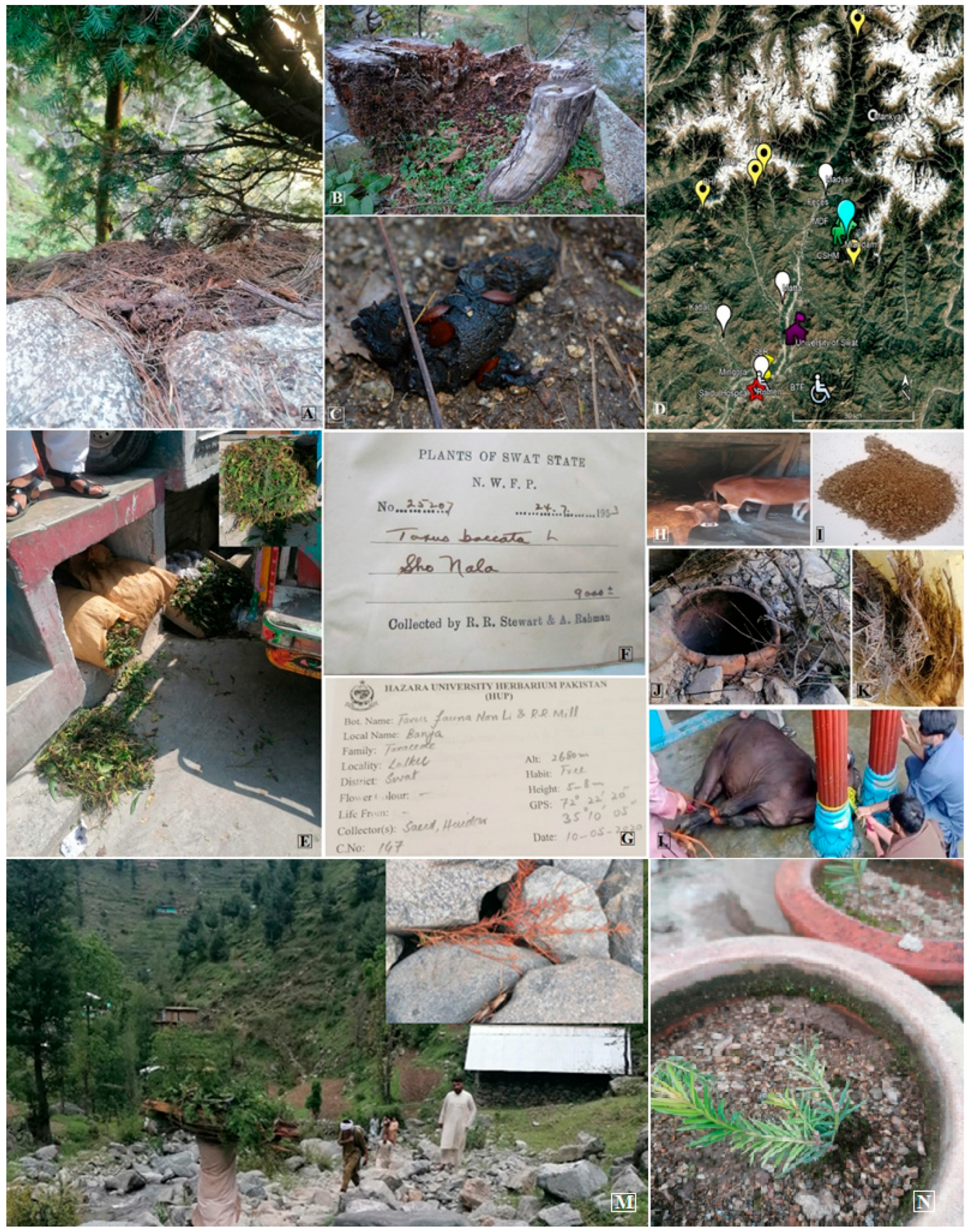
Figure 1. Fresh and environmental DNA sources collected for the forensic identification of Taxus. (A) cow dung with Taxus tree; (B) fired tree; (C) unknown animal/bird feces; (D) collection of samples locations (yellow points:herbarium materials locations, white: hakims and herbal stores, red: animal rumen, purple: Swat university Herberium, white chair: vomit samples, bluish-green pin; Taxus natural population, burned trees, clay oven, green animal sign: cow dung fresh and old, white circle: goat droppings); (E) road side samples; (F) Karachi University Taxus herbarium specimen from Swat state era; (G) Hazara university Taxus specimen; (H) fresh cow dung; (I) Taxus powder from hakims; (J) clay oven; (K) Taxus from herbal stores; (L) animal rumen; (M) logging road; (N) propagated Taxus. (Copyright is retained by the authors).
Conservation 05 00062 g001
Figure 2. Amplification success and Taxus DNA detection across different sample sources.
Conservation 05 00062 g002
Figure 3. Mini-barcoding-based crime investigation categories and collection purposes.
Conservation 05 00062 g003
Table 1. Taxon-specific primers, along with their lengths, were used in the study.
TaxonGenePrimerSecquenceLength
T. canadensismatKFGCAGAAAGCTCTGGTTCCAC20
RCGATCGGGAAAAGATCCATA
RbclFTTTGGATTCAAAGCCCTACG
RAGTCCACCACGGAGACATTC
T. wallichianaITSFGCGGTAGGATCATTGTCGTT
RGGCACTCGCCCTTGTAATAA
T. contortatrnL-FFATTTTGAATGGGCAATCCTG
RGGCAAGCCTAGAACAACTCAA21
TSFGCAGATGAGCTGGTTGTGAA20
RATTCGATACCCCATGATCCA
trnS-trnQFCTTGCCAAGCAAAGGCTAAG
RCGGAGAATCAAAATGCCAGT
Table 2. Blast taxonomy results of mini-barcoding.
CriteriaS. S. (5000) E. (0.05)S.S. (5000) E. (100)S. (100) E. (0.05)Query Length
HitsTaxon Eukaryota/Taxus L.Hits
matK270Taxaceae1396/382Taxus L. (100%)117
rbcL2454Gymnosperm/fern7637/557200
ITS159Taxus L.1209/1030175
Table 3. Taxus species diversity recognized by mini-barcoding BLAST taxonomy results.
GroupTaxonHitsTaxaA/hits
SpeciesT. baccata (101), T. brevifolia (27), T. calcicola (11), T. canadensis (45), T. celebica (1), T. chinensis (171), T. contorta (26), T. cuspidata (70), T. floridana (33), T. florinii (13), T. fuana (43), T. globosa (46), T. mairei (548), T. phytonii (369), T. sumatrana (15), T. wallichiana (68)15471697
VarietyT. cuspidata var. cuspidata (6), T. cuspidata var. latifolia (6), T. cuspidata var. nana (10), T. wallichiana var. wallichiana (90), T. wallichiana var. yunnanensis (12)124525
HybridT. x hunnewelliana (4), T. x media (9)1326.5
VariantT. sp. ‘Emei type’ (9), T. sp. ‘Huangshan type’ (17), T. sp. ‘Mandiya type’ (2), T. sp. ‘Qinling type’ (148), T. sp. ‘Yunman type’ (2), T. sp. SW-2025a (62), Emb. envir. sample (5)245735
Disclaimer/Publisher’s Note: The statements, opinions and data contained in all publications are solely those of the individual author(s) and contributor(s) and not of MDPI and/or the editor(s). MDPI and/or the editor(s) disclaim responsibility for any injury to people or property resulting from any ideas, methods, instructions or products referred to in the content.