Abstract
Microplastic pollution has attracted significant attention in recent years due to evidence that these particles can accumulate in organisms’ tissues and organs and induce adverse health effects, with oxidative stress being a key underlying mechanism of toxicity. The present study investigated the effects of polystyrene microplastics (0.1 μm in diameter) administered at a dose of 0.1 mg/day/animal for 4 weeks, followed by a 2-week recovery period without exposure, on oxidative stress markers in the liver, kidney, and spleen and on hematological and blood biochemical parameters in mice. The results showed a statistically significant increase in white blood cell counts, including lymphocytes, granulocytes, and monocytes, at week 5, indicating the development of an inflammatory response. During the last week of the recovery period (week 6), values returned to levels that approached baseline. Changes in lipid peroxidation demonstrated an induction of oxidative stress, accompanied by alterations in glutathione levels and antioxidant enzyme activities, with a tendency toward recovery after cessation of polystyrene microplastic exposure. In conclusion, these findings demonstrated that even short-term exposure to low doses of polystyrene microplastics could trigger oxidative stress and inflammatory responses, highlighting their potential health risks and the need for further investigation into their long-term biological effects.
1. Introduction
Microplastics (MPs) are synthetic polymer particles ranging in size from 0.1 to 5000 µm, which are either intentionally manufactured (primary MPs) for use in various products or generated through the degradation of larger plastic materials (secondary MPs) [1]. They are ubiquitously distributed in the environment, occurring in soil, water, and air [2]. MPs have been detected not only in urbanized regions but also in remote areas such as the Arctic and Antarctic, Mount Everest, and the Mariana Trench [3,4,5,6].
From the environment, MPs can enter living organisms, including humans, primarily via inhalation, ingestion of contaminated food and water, and, to a lesser extent, dermal contact [7]. Increasing evidence indicates that MPs can accumulate in various tissues and organs. In humans, they have been identified in multiple organ systems, including the cardiovascular, digestive, endocrine, integumentary, lymphatic, respiratory, reproductive, and urinary systems, as well as in biological samples such as breast milk, meconium, semen, stool, sputum, and urine [8].
Accumulating data suggest that MPs exert adverse biological effects in exposed organisms. Both in vitro and in vivo studies, as well as investigations in wild invertebrates and vertebrates, have demonstrated that MP exposure can lead to impaired growth, reduced body size, reproductive toxicity, developmental abnormalities, and shortened lifespan [7,9,10,11]. One of the key mechanisms underlying MP-induced toxicity is oxidative stress (OS) [7,12,13,14,15]. Excessive production of reactive oxygen species (ROS) and/or impairment of antioxidant defense systems disrupt redox homeostasis, resulting in lipid peroxidation, protein oxidation, DNA damage, and ultimately cellular dysfunction [16,17].
The mechanisms of ROS generation following MP exposure are complex and often interconnected. They include mitochondrial dysfunction, activation of NADPH oxidases, and inflammatory signaling pathways [15,18]. Mitochondria play a central role in this process, as MPs have been shown to disrupt mitochondrial membrane potential, structure, function, and dynamics [19]. Although larger MPs are unlikely to penetrate mitochondria directly, they can still damage mitochondrial membranes indirectly. The outer membrane (monoamine oxidase), along with the inner membrane (Complexes I–III) and the matrix (dehydrogenases) are the primary ROS producers in mitochondria [20]. The dysfunctional mitochondria become major sources of ROS, promoting OS and apoptosis [21].
Particle characteristics significantly influence toxicity. Factors such as polymer type, size, shape, surface properties, and exposure duration determine the extent of ROS production and cellular damage [18,22,23]. Cellular uptake is strongly size-dependent, with smaller particles exhibiting higher internalization rates; for example, uptake decreases from 49% to 30% as particle size increases from 1 µm to 6 µm [24,25]. Smaller MPs, and particularly nanoplastics, are more likely to accumulate intracellularly and induce more severe oxidative damage. Surface charge also plays a critical role, as positively charged particles have been shown to be more toxic than unmodified MPs [19,26]. Furthermore, modulation of antioxidant defense systems appears to be an important event in the cellular response to exposure to MPs and nanoplastics across multiple experimental models [27,28,29,30].
Several studies have demonstrated that MPs can induce OS in vitro and in vivo [19,31,32,33,34,35,36,37]. However, limited data are available regarding the temporal dynamics of blood parameters and redox alterations in specific organs following oral exposure. Understanding time-dependent changes in OS biomarkers is essential for elucidating the progression of tissue damage and the balance between injury and compensatory antioxidant responses. Furthermore, the changes in hematological parameters, blood biochemical markers, and organ-specific oxidative stress levels following cessation of MP exposure remain insufficiently understood. Such data could provide important insight into the reversibility or persistence of MP-induced effects. They help determine whether changes in hematological, biochemical, and oxidative stress parameters are temporary and adaptive or indicative of long-term damage. Such findings could contribute to understanding the organism’s recovery capacity, distinguishing between acute and chronic toxicity, and identifying sensitive biomarkers. Overall, they may improve our understanding of MPs’ toxicity mechanisms and support more accurate health risk assessments.
Polystyrene microplastics (PS-MPs) are among the most widespread environmental pollutants, largely due to their extensive use in packaging materials, food containers, and various industrial applications [38]. Moreover, several studies have demonstrated that PS-MPs can elicit adverse biological responses in the body [38,39,40]. In addition, polystyrene is one of the most commonly used polymers in experimental studies investigating the cellular effects of MPs. Metabolically active organs, such as the liver, spleen, and kidneys, are of particular interest in toxicological studies due to their central roles in detoxification, immune regulation, and excretion, which also make them primary targets for xenobiotic-induced damage.
Therefore, the present study aimed to investigate the time-dependent effects of orally administered PS-MPs during a four-week exposure period, followed by a two-week recovery phase, on hematological parameters and oxidative stress levels in the liver, spleen, and kidneys of mice. Particular emphasis was placed on lipid peroxidation (LPO) levels and key antioxidant markers, including total glutathione (GSH) and superoxide dismutase (SOD). We hypothesized that repeated oral exposure to PS-MPs would induce a time-dependent increase in oxidative stress with partial or incomplete recovery following cessation of exposure. Elucidating these mechanisms may contribute to a better understanding of MP-induced systemic toxicity and its potential health implications.
2. Results
2.1. Mouse Body and Organ Weight, Water Consumption, and Organ Indexes
Data on body weight, water consumption, and the absolute and relative weights of key organs (liver, kidney, and spleen), as well as the calculated organ index in mice from the control and experimental groups during PS-MPs exposure (W1–W4) and the subsequent recovery period (W5–W6), are presented in Table 1.
Table 1.
Weight, water consumption, organ weight, and organ indexes of control mice and mice administered 1 μm PS-MPs in drinking water at a dose of 0.1 mg/day for four weeks (W1–W4), followed by a two-week recovery period (W5–W6) without exposure.
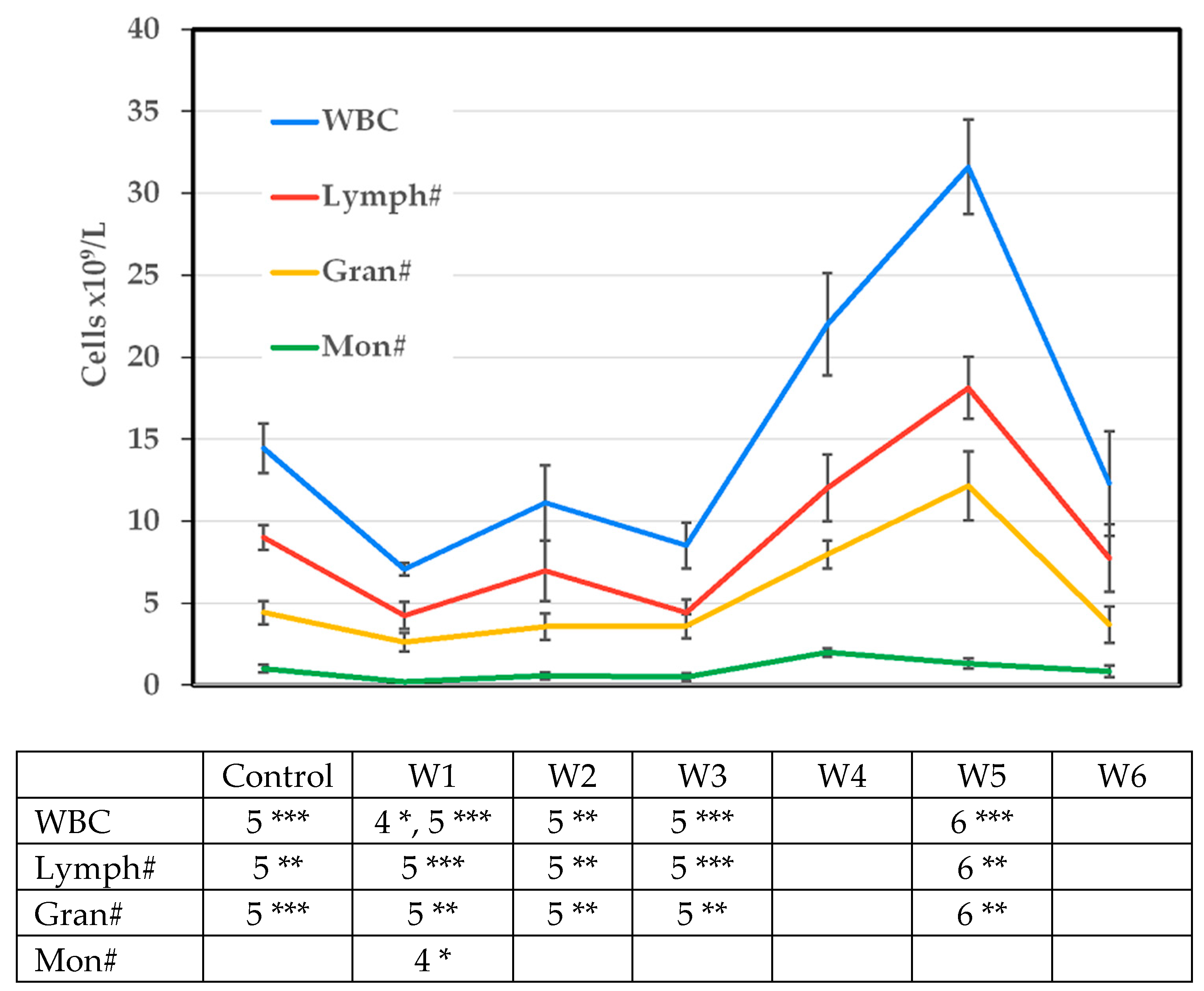
The mean body weight of mice in the experimental groups (W1–W6) did not differ significantly from that of the control group. During the 4-week PS-MP exposure period, slight, non-significant fluctuations in body weight were observed, with the highest values recorded at the end of exposure (W4). Body weight remained stable during the recovery period (W6). Water intake remained relatively constant throughout the experiment, indicating that the animals were normally hydrated and that fluid intake did not affect the parameters studied. No significant changes were observed in the absolute or relative weights of the liver and kidney compared to the control group. In contrast, both absolute and relative spleen weights were significantly increased in W4 and W5. By W6, the relative spleen weight had returned to baseline. Notably, the increase and subsequent decrease in spleen weight in W4 and W5 coincided with corresponding changes in the mean white blood cell count observed during the same period (see Figure 1).
Figure 1.
Changes in the main white blood cell parameters of control mice and mice treated with 1 μm PS-MP in drinking water at a dose of 0.1 mg/day for four weeks (W1–W4), followed by a two-week (W5–W6) recovery period without exposure. WBC—white blood cell, Lymph#—lymphocytes, Gran#—granulocytes, Mon#—monocytes; Numbers in the table cells indicate a statistically significant difference compared to the corresponding group (4—Week 4, 5—Week 5, 6—Week 6); Differences were considered significant at * p < 0.05, ** p < 0.01, and *** p < 0.001.
2.2. Results of Histological Examination
Microscopic examination of the stomach and small intestine contents revealed fluorescent PS-MPs in numbers comparable to those detected in the drinking water, whereas substantially higher amounts were observed in the contents of the large intestine. In contrast, no fluorescent PS-MPs were detected in cryosections of any examined organ at any time point during the study.
2.3. White Blood Cell Parameters
The data presented in Figure 1 illustrate the dynamics of total white blood cell (WBC) counts and their fractions (lymphocytes (Lymph#), granulocytes (Gran#), and monocytes (Mon#)), which exhibited statistically significant changes throughout the experiment.
The overall response exhibited a triphasic pattern, characterized by an initial insignificant decline during the first week, followed by a significant increase at the end of the exposure period (W4) and immediately after its cessation (W5), when WBC, Lymph#, and Gran# reached peak levels. During the final recovery week (W6), these parameters returned to baseline values. Monocyte count peaked earlier, at W4, preceding the maxima observed for Gran# and Lymph#, which is consistent with their role in the recognition and phagocytosis of foreign particles.
2.4. Red Blood Cell Parameters
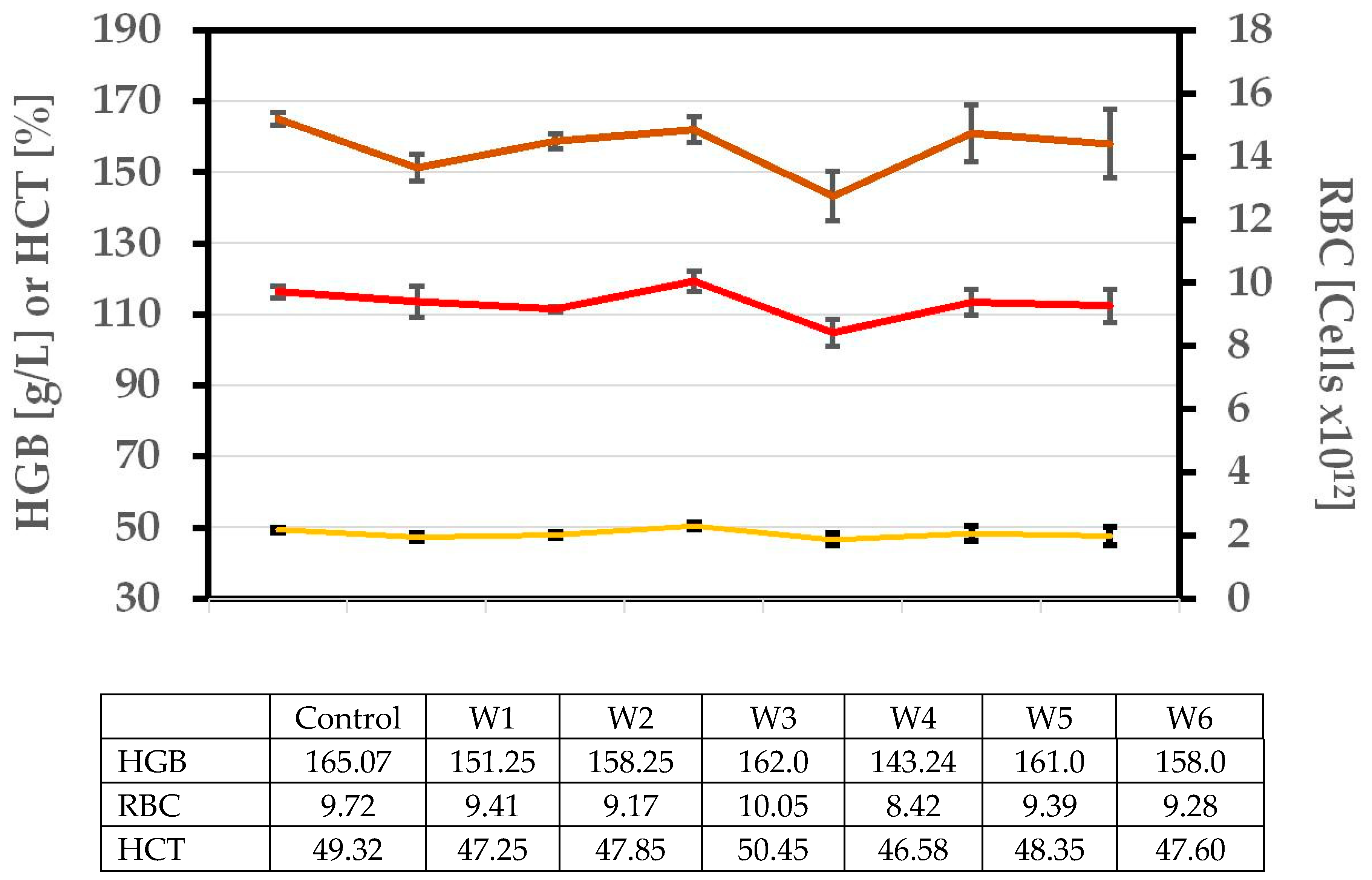
The data in Figure 2 describe the dynamics of the main red blood cell parameters (RBC, HGB, and HCT) in mice during the experiment. The values remained within a narrow range, with no statistically significant changes. The derived RBC parameters, including MCV, MCH, MCHC, and RDW, as well as PLT and its associated parameters (MPV, PDW, and PCT), also did not show significant alterations.
Figure 2.
Changes in the main red blood cell parameters of control mice and mice treated with 1 μm PS-MP in drinking water at a dose of 0.1 mg/day for four weeks (W1–W4), followed by a two-week (W5–W6) recovery period without exposure. The mean values of the parameters are presented in the table. RBC—red blood cell count, HGB—hemoglobin concentration; HCT—hematocrit concentration.
2.5. Blood Biochemical Parameters
The results presented in Table 2 reflect the changes in blood biochemical parameters in mice treated with PS-MPs over a 4-week exposure (W1–W4) and 2-week recovery period (W5–W6). Statistically significant changes were observed only in albumin (ALB), alanine aminotransferase (ALT), and glucose (GLU). As a parameter of protein profile, ALB concentration significantly decreased in week 1 (W1), week 4 (W4), and week 5 (W5) compared to the controls. In W6, the levels were normalized. Alanine aminotransferase activity was significantly elevated by more than 30% in week 5 (W5), the first week after cessation of PS-MP intake, compared to the controls. In week 6 (W6), the ALT levels began to decline. Regarding GLU, a significant decrease in levels was observed in week 4 (W4) compared with baseline. During the recovery period, levels returned to the initial ones, albeit slightly lower.
Table 2.
Changes in biochemical parameters of control mice and mice treated with 1 μm PS-MPs in drinking water at a dose of 0.1 mg/day for four weeks, followed by a two-week recovery period without exposure.
2.6. Oxidative Stress Parameters
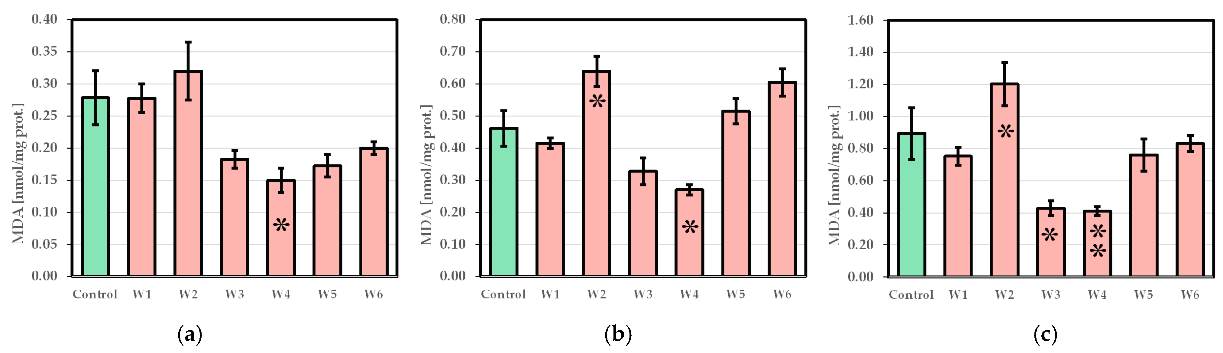
The data presented in Figure 3 illustrate the extent of OS, as indicated by malondialdehyde (MDA) levels (a marker of LPO) across the three examined organs: liver, kidney, and spleen. The LPO in the liver showed a slight increase in week 2 (W2) compared to the initial level (Figure 3a). Thereafter, MDA levels decreased and remained relatively low. In the kidney and spleen (Figure 3b,c), a statistically significant increase in LPO was observed in the second week (W2) compared to baseline, followed by decreases in weeks 3 and 4 and a rise during the recovery period, returning to initial levels.
Figure 3.
Lipid peroxidation in (a) liver, (b) kidney, and (c) spleen of control mice and mice treated with 1 μm PS-MP in drinking water at a dose of 0.1 mg/day for four weeks, followed by a two-week recovery period without exposure. (W1–W6—experimental weeks 1–6; significant differences at: * p < 0.05 vs. Co; ** p < 0.01 vs. Co).
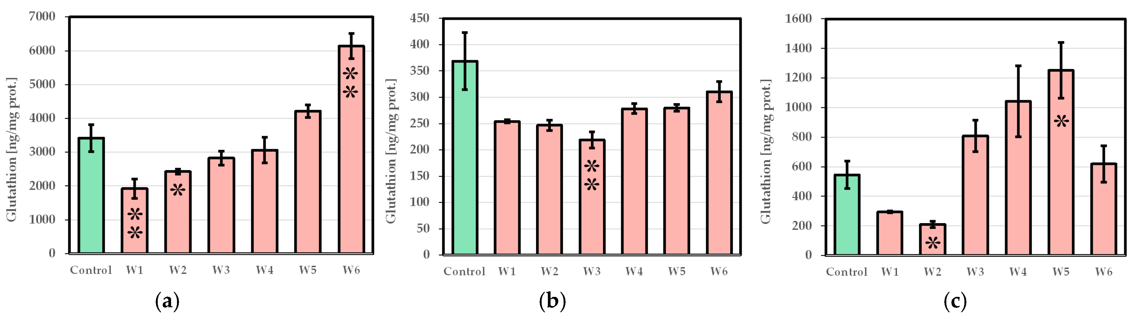
Glutathione levels varied across the organs studied during and after PS-MP exposure. In the liver, GSH decreased significantly during the first two weeks, followed by a gradual recovery from W3 to W5 and an overcompensation at W6, when concentrations nearly doubled the initial level (Figure 4a). In contrast, the kidneys exhibited a sustained decrease, with a significant drop at W3 to approximately half of baseline. Although a slight recovery was observed by W6, levels remained below the initial values throughout the experiment (Figure 4b). The spleen showed an intermediate pattern, with an initial decline in W1–W2, followed by a sharp increase from W3 to W5, peaking significantly at W5, and returning to baseline by W6 (Figure 4c). These results highlight organ-specific differences in non-enzymatic antioxidant response.
Figure 4.
Glutathione concentration in (a) liver, (b) kidney, and (c) spleen of control mice and mice treated with 1 μm PS-MP in drinking water at a dose of 0.1 mg/day for four weeks, followed by a two-week recovery period without exposure (W1–W6—experimental weeks 1–6; significant differences at: * p < 0.05 vs. Co; ** p < 0.01 vs. Co).
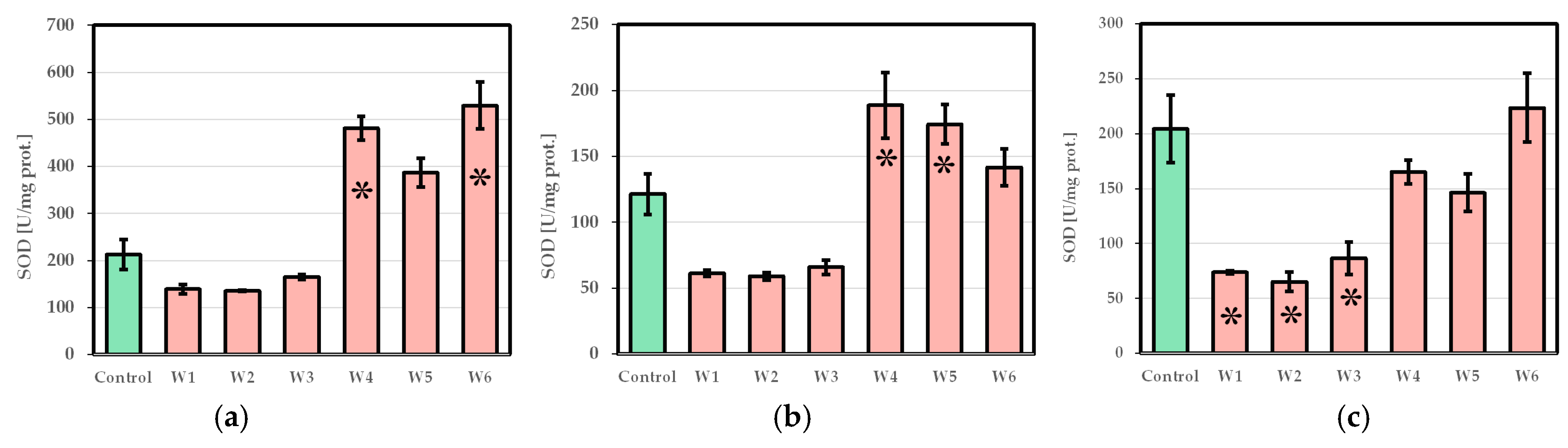
Superoxide dismutase activity across the studied organs, exhibiting similar dynamics: an initial decrease in W1 to W3 and recovery in W4 to W6. In the liver, SOD suppression during W1–W3 dropped nearly twofold, and there was a statistically significant increase at W4, exceeding control levels by more than twofold. During the recovery phase at W5–W6, SOD levels remained high and increased significantly at W6 (Figure 5a). The kidneys followed a similar pattern. SOD activities fell by almost half in W1–W3, peaked significantly at W4 and W5, and returned near baseline by W6 (Figure 5b). The spleen showed a critical drop in SOD activity of nearly threefold in W1–W3, followed by a recovery during W4–W6 (Figure 5c).
Figure 5.
Superoxide dismutase activity in (a) liver, (b) kidney, and (c) spleen of control mice and mice treated with 1 μm PS-MP in drinking water at a dose of 0.1 mg/day for four weeks, followed by a two-week recovery period without exposure (W1–W6—experimental weeks 1–6; significant differences at: * p < 0.05 vs. Co).
3. Discussion
This study aimed to investigate the effects of sub-chronic (4-week) MP exposure in a simplified murine model using single-sized PS-MPs administered at a fixed dose. In addition, a two-week post-exposure period was included to monitor potential recovery and the restoration of homeostasis following cessation of MP intake. The study focused on OS as a key mechanism through which MPs may impair organismal health, including potential implications for humans. The accumulation, distribution, and translocation of MPs across organs, as well as their systemic effects relevant to the human organism, can be most reliably assessed using in vivo animal models [41]. Such approaches enable a comprehensive evaluation of toxicokinetic and toxicodynamic processes without posing risks to human health. In contrast, in vitro systems, including cell cultures and isolated organs, are limited in their capacity to replicate the complex interactions and integrated whole-organism responses associated with MP exposure.
The results of this study indicated that PS-MP intake did not lead to changes in body weight, consistent with previous findings [42]. The absence of changes in body weight and water consumption indicates that the animals maintained normal metabolic balance and hydration status, excluding dehydration or nutritional stress as confounding factors. Therefore, any observed alterations in hematological, biochemical, and oxidative stress parameters are likely attributable to the direct biological effects of PS-MP exposure rather than secondary physiological disturbances. In addition, the animals’ normal water intake ensured they received the intended daily dose of PS-MPs as designed in the experiment.
However, at the dose used in this study, no accumulation of PS-MPs was detected in the livers, kidneys, and spleens analyzed. Confocal fluorescence microscopy of cryosections from these three organs revealed no fluorescence in any section. Previous reports of particle accumulation in organs [43] involved a single oral gavage of a dose five times higher (0.5 g/kg body weight). As expected, in this study, particles were detected in the stomach and intestines at concentrations close to the administered dose, with much higher concentrations observed in the colon. This is likely due to water reabsorption in the large intestine. It is also possible that this higher particle concentration in the colon may contribute to irritation of the terminal digestive tract. These findings indicate that if MP absorption occurs in the gastrointestinal tract, it is likely to be very limited.
Ingested MPs are resistant to digestion and can persist in the gastrointestinal tract, leading to a variety of disorders. Animal studies have shown that they may physically interact with the gut lining, causing mechanical injury and potentially triggering inflammatory responses. MPs may also increase intestinal permeability (“leaky gut”), facilitating the passage of harmful particles and pathogens into the bloodstream [44]. Furthermore, the gut microbiome, which is critical for digestion, metabolism, immune function, and overall health, is particularly vulnerable to MPs. The exposure to MPs is linked to dysbiosis, an imbalance between beneficial and pathogenic bacteria, leading to impaired gut function, weakened immunity, and increased risk of gastrointestinal disorders [45]. MP-induced microbiome alterations can also trigger systemic inflammation, contributing to chronic disorders [46]. From the results obtained herein, it can be assumed that the observed increase in spleen and white blood cells is a response to a local (in the digestive tract) or general inflammatory process.
In this study, with PS-MP intake, significant changes were observed in the WBC count in mouse blood, whereas RBC, HGB, and HTC remained largely unchanged. This suggests that PS-MP exposure does not directly affect red bone marrow, and the observed leukocytosis is unlikely to result from hemoconcentration, which would have increased the counts of all blood cell types. The coincident increases in spleen weight and circulating white blood cell counts observed during weeks 4 and 5 may reflect a coordinated immunological response to prolonged exposure to MPs [47]. It is plausible that MPs accumulate within lymphoid organs, including the spleen, where they may promote OS and low-grade inflammatory signaling, thereby modulating immune homeostasis rather than inducing an overt pathological response [48]. Such processes could contribute to a reactive enlargement of the spleen, potentially driven by increased immune cell retention, redistribution, or limited local proliferation within splenic compartments. In parallel, the observed elevation in leukocyte counts may indicate systemic immune activation or enhanced mobilization of immune cells into the circulation. Given the established role of the spleen as a reservoir and regulatory site for myeloid cells during inflammatory conditions, a contribution of splenic activity to peripheral leukocyte dynamics cannot be excluded [49]. The delayed onset of these changes suggests a time-dependent effect, possibly reflecting cumulative exposure or the gradual engagement of compensatory immune mechanisms. Taken together, the data are consistent with the interpretation that changes in spleen mass and leukocyte numbers represent interconnected aspects of an adaptive immune response to MPs, although alternative explanations, including stress-related or indirect systemic effects, warrant further investigation. At week 6 (W6), the relative spleen weight normalized in parallel with the white blood cell count.
The presence of colonic inflammation, accompanied by leukocytosis and splenomegaly, may exert secondary effects on liver function via systemic inflammatory mechanisms rather than direct hepatic injury. Inflammatory processes in the colon are known to disrupt intestinal barrier integrity, facilitating the translocation of microbial products into the portal circulation and thereby engaging the gut–liver axis [47]. Such signals can activate hepatic immune cells, including Kupffer cells, leading to mild hepatocellular stress that may manifest as a modest elevation in ALT without overt structural liver damage [48]. In parallel, systemic inflammation is characterized by a coordinated acute-phase response in the liver, during which albumin synthesis is downregulated in favor of positive acute-phase proteins, rendering albumin a negative marker of inflammatory burden rather than a specific indicator of hepatic dysfunction [50]. The observed reduction in ALB may therefore reflect inflammation-driven metabolic reprioritization rather than impaired synthetic capacity per se. Additionally, the spleen has been shown to act as an immunological reservoir during colitis, contributing to leukocyte mobilization and sustained cytokine release, which can further influence hepatic metabolism and protein synthesis [49]. Experimental models of colitis support the notion that intestinal inflammation alone is sufficient to induce mild alterations in liver biochemistry as part of a systemic response [51]. Importantly, the absence of pronounced cholestatic markers or severe transaminase elevations would argue against primary hepatotoxicity. Collectively, these findings are consistent with a model in which colonic inflammation, splenic immune activation, and systemic inflammatory signaling converge to produce mild functional changes in liver parameters. Further studies incorporating histological and molecular analyses would be required to distinguish transient inflammatory effects from direct hepatic injury. Furthermore, the decrease in blood GLU levels observed at the end of the PS-MP exposure period (Table 2) may be associated with impaired hepatic metabolic function, particularly disruption of gluconeogenesis [52]. In addition, this effect could be linked to oxidative stress-induced alterations in hepatocellular function, as ROS are known to interfere with key metabolic pathways and enzyme activity in the liver.
The results of this study indicated that exposure of mice to PS-MPs induced an inflammatory response, as evidenced by the aforementioned leukocytosis and splenomegaly. Such systemic immune activation can promote oxidative stress in organs, even in the absence of direct particle accumulation. Activated immune cells release ROS, while pro-inflammatory cytokines such as TNF-α, IL-1β, and IL-6 can stimulate ROS production in hepatocytes, renal cells, and splenic tissue. This mechanism provides a plausible explanation for organ-level OS observed in other studies following MP exposure [15,43,53], highlighting the interplay between systemic inflammation and oxidative stress as a key pathway of MP-induced toxicity. For the assessment of OS in this study, the liver, kidney, and spleen were selected due to their key roles in metabolism, detoxification, and immune function, as well as being sensitive indicators of systemic oxidative and inflammatory responses to MP exposure [15,33,43,54].
The early increase in MDA levels observed herein in the liver, kidneys, and spleen of mice after PS-MP exposure (Figure 3) may reflect enhanced LPO during the initial phase of OS, when ROS overwhelm membrane-bound antioxidant defenses [55]. Under these conditions, polyunsaturated fatty acids (PUFAs) are particularly susceptible to oxidative damage, leading to elevated MDA, a stable end product of LPO. With prolonged exposure, however, the availability of substrate (PUFAs) for lipid peroxidation decreases, while tissues appear to activate adaptive antioxidant mechanisms that limit further membrane oxidation. Induction of enzymatic and non-enzymatic antioxidant systems, including SOD- and GSH-dependent pathways, reduces the propagation of lipid radicals and MDA formation [56,57]. Consequently, MDA concentrations progressively decline despite ongoing oxidative challenge. The decrease in MDA below baseline levels at later time points is consistent with a hypercompensatory, hormetic antioxidant response that enhances membrane protection beyond the initial physiological state.
In contrast to the pro-oxidant (LPO) changes observed in the organs of mice following PS-MP exposure, the antioxidant defense system initially decreased, then recovered during the post-exposure phase after cessation of intake. The early decrease in total GSH levels and SOD activity observed in the liver, kidney, and spleen (Figure 4 and Figure 5) likely indicates early antioxidant depletion and functional impairment under conditions of acute OS, when ROS generation exceeds the capacity of endogenous defense systems [57,58]. Under OS conditions, cellular reduced glutathione (GSH) is rapidly consumed through direct reactions with ROS, conjugation to metabolites, protein S-glutathionylation, and as a co-substrate in antioxidant enzymatic reactions [59]. A major proportion of GSH involved in antioxidant defense is utilized by glutathione peroxidases, resulting in the formation of oxidized glutathione (GSSG) [59,60,61], which in turn can be regenerated to GSH by glutathione reductase (GR). Maintenance of an intracellular GSH:GSSG ratio, typically around 100:1, is essential for preserving the cellular reducing environment. However, when the OS extent exceeds the capacity of GR to regenerate GSH from GSSG, the reduction of GSSG becomes insufficient, resulting in its intracellular accumulation. Excess intracellular GSSG is deleterious because it disrupts cellular redox homeostasis and promotes further oxidative damage. In order to preserve intracellular redox balance, cells have evolved mechanisms for the active export of GSSG through ATP-dependent transporters [62]. Therefore, the decrease in total glutathione observed in the present study can be interpreted as an indicator of OS, induced by MP exposure.
Concomitantly, acute OS may transiently overwhelm antioxidant defenses, resulting in a functional decline in SOD activity. This reduction is not necessarily attributable to decreased enzyme abundance, but rather to oxidative inactivation and redox-dependent impairment of enzymatic function under conditions of sustained ROS exposure [63]. With continued exposure, however, tissues appear to activate compensatory redox-regulatory mechanisms to restore homeostasis. In particular, activation of the Nrf2–antioxidant response element (ARE) pathway promotes the upregulation of key antioxidant enzymes involved in GSH biosynthesis and recycling, such as glutamate–cysteine ligase and glutathione reductase, as well as SOD [56,64,65]. This adaptive response enables the gradual replenishment of intracellular GSH levels and the recovery and subsequent increase in SOD activity. Consequently, the normalization of GSH levels and the late elevation of SOD activity are consistent with a time-dependent antioxidant adaptation. This biphasic response, characterized by an initial suppression followed by compensatory upregulation, represents a classical hormetic adaptation to sustain but non-lethal oxidative challenge [66].
4. Materials and Methods
4.1. Experimental Animals
In this study, 60 sexually mature male Swiss albino mice (3 months old) were used. They were obtained from a specialized facility, Experimental Breeding Base for Experimental Animals, Slivnitsa, Bulgaria. Upon arrival, the animals were housed in specialized rooms under a 12-h light/dark cycle at 22 ± 1 °C, with ventilation and a relative humidity of 50 ± 5%. They were placed in cages with free access to food and water. At the start of the experiment, the mice had body weights of 30.25 ± 5.62 g.
4.2. Microplastics
Fluorescent polystyrene microplastics (PS-MPs) with a diameter of 1 µm (Cat. No. PSFR001UM) were obtained from Magsphere Inc. (Pasadena, CA, USA). Their excitation and emission wavelengths were 526 nm and 570 nm, respectively. The particles were administered to mice via drinking water. Because the relative density of PS-MPs (1.04 g/cm3) is slightly higher than that of water, particle aggregation may promote sedimentation and lead to uneven intake or administration of a higher dose than intended. To minimize aggregation and ensure a homogeneous and stable dispersion, PS-MPs suspended in purified water were ultrasonicated (20 kHz for 30 min) [43,67,68]. The ultrasound power and exposure duration were maintained at the minimum levels necessary to disperse aggregates while avoiding alterations to particle characteristics. The extent of particle aggregation was monitored microscopically. Fresh PS-MP suspensions were prepared twice weekly.
4.3. Experimental Design
After a week-long acclimatization period, the animals (n = 60) were randomly divided into two groups: (1) Control (n = 24) and (2) Experimental (n = 36). The duration of the experiment was 6 weeks. For four consecutive weeks, the experimental group mice received water containing 1 μm PS-MPs at a dose of 0.1 mg per 24 h, and the control mice received tap water. During the last two weeks, all animals received tap water (recovery period). Mice’s water consumption was measured twice weekly, and body weight was recorded once weekly. At the end of every week, six animals from each experimental group and four controls were sacrificed. Therefore, six experimental groups were established, designated Week 1 to Week 6 according to the study timeline. As no statistically significant differences were observed in the measured parameters among control mice across the experimental timeline (week 0, i.e., prior to treatment, and weeks 1–6), all control groups were pooled into a single control group (to increase statistical power and reduce variability without obscuring biologically relevant effects. The relatively low variability and standard error within the combined control group supported the homogeneity of the controls over time.
Each week, from each mouse, blood was undrawn, and liver, kidneys, and spleen were extracted. Hematological and blood biochemical parameters were analyzed in whole blood and plasma, respectively. The organs were processed appropriately, and the main oxidative stress parameters were measured. The experimental procedures strictly followed the requirements of the European Communities Council Directive (86/609/EEC) and were approved by the institutional Ethics Committee (Bioethics Committee at the Institute of Neurobiology, Bulgarian Academy of Sciences, Bulgaria) and the Bulgarian Food Safety Agency (Approval for working with laboratory animals No. 424/24.02.2025).
4.4. Histological Examination
Contents from the stomach, small intestine, and large intestine, as well as unstained cryosections from the liver, kidneys, and spleen, were examined for the presence of fluorescent PS-MPs using a Leica TCS SPE confocal microscope (Leica Microsystems GmbH, Wetzlar, Germany) with Leica Application Suite X (LAS X). Tissues were rinsed in cold phosphate-buffered saline (pH 7.4–7.6), embedded in cryostat sectioning medium, and frozen in liquid nitrogen. Sections (10 μm thick) were fixed and permeabilized in ice-cold methanol for 5 min, then allowed to dry at room temperature and stored at −20 °C. For the localization analysis of fluorescent PS-MPs, cryostat sections were washed three times with Tris-buffered saline (TBS), pH 7.6, and incubated with Hoechst solution (to stain nuclei) for 10 min at room temperature. After washing three times with TBS, sections were coverslipped with flash-drying medium to obtain permanent preparations. They were analyzed at the respective λ to visualize the fluorescence of PS-MPs and nuclei.
4.5. Hematological Analyses
Blood from each mouse was collected into a single EDTA Vacutainer. It was obtained from the vena temporalis superficialis in the zygomatic region.
Complete blood count (CBC) analysis of whole blood was performed using a Mindray BC-2800Vet hematology analyzer (Mindray Bio-Medical Electronics Co., Ltd., Shenzhen, China). The following parameters were measured: white blood cells (WBC, ×109 cells/L), lymphocyte count (Lymph#, ×109 cells/L), monocyte count (Mono#, ×109 cells/L), granulocyte count (Gran#, ×109 cells/L), lymphocyte percentage (Lymph, %), monocyte percentage (Mon, %), granulocyte percentage (Gran, %), red blood cells (RBC, ×1012 cells/L), hemoglobin (HGB, g/L), hematocrit (HCT, %), mean corpuscular volume (MCV, fL), mean corpuscular hemoglobin (MCH, pg), mean corpuscular hemoglobin concentration (MCHC, g/L), red cell distribution width (RDW, %), platelets (PLT, ×109 cells/L), mean platelet volume (MPV, fL), platelet distribution width (PDW, %), and plateletcrit (PCT, %).
Blood biochemical analyses were performed using a Celercare V5 chemistry analyzer (Tianjin MNCHIP Technologies Co., Ltd., Tianjin, China). The following parameters were measured: total protein (TP, g/L), albumin (ALB, g/L), globulin (GLO, g/L), albumin to globulin ratio (ALB/GLO), total bilirubin (TBIL, µmol/L), alanine aminotransferase (ALT, U/L), aspartate aminotransferase (AST, U/L), aspartate aminotransferase to alanine aminotransferase ratio (AST/ALT), gamma-glutamyl transferase (GGT, U/L), urea (mmol/L), creatinine (CRE, µmol/L), blood urea nitrogen-to-creatinine ratio (BUN/CRE), and glucose (GLU, mmol/L).
4.6. Tissue Preparation for Biochemical Analyses
Mice were sacrificed under light anesthesia, and the liver, kidneys, and spleen were removed. The organs were cut into small pieces, washed with cold 0.15 M KCl, homogenized, and centrifuged at 3000 rpm for 10 min at 4 °C. The post-nuclear fraction was used to determine lipid peroxidation (LPO) and total glutathione (GSH) levels. A part of the post-nuclear fraction was separated and re-centrifuged at 12,000 rpm for 20 min at 4 °C to get a post-mitochondrial supernatant, in which the activities of antioxidant enzyme superoxide dismutase (SOD) were measured.
4.7. Biochemical Analyses
The main oxidative stress parameters in the liver, kidneys, and spleen were assessed via commercially available kits, strictly following the relevant manufacturing protocols.
The protein concentrations of the studied fractions were determined via the Bradford Protein Colorimetric Assay Kit (Cat. No: E-BC-K168-M, Elabscience Ltd., Houston, TX, USA).
Lipid peroxidation and the concentration of the total glutathione were measured with the MDA Assay Kit, Cat. No. MAK085 and Glutathione Assay Kit Cat. No. CS0260, respectively, as both kits were purchased from Sigma–Aldrich Co., LLC (St. Louis, MO, USA). The antioxidant enzyme activities were determined using the Superoxide Dismutase Assay Kit (Cat. No. E-BC-K020-M, Elabscience Ltd., Houston, TX, USA). Each sample was performed in triplicate.
4.8. Statistical Analysis
Experimental data were analyzed by the Shapiro–Wilks test for distribution normality, followed by one-way analysis of variance (ANOVA) and Tukey’s post hoc test in GraphPad Prism 9.0. In the tables, the results were presented as mean ± SD, and in graphics as mean ± SE, with significance levels: * p < 0.05, ** p < 0.01, and *** p < 0.001.
5. Conclusions
The present study shows that four weeks of sub-chronic exposure to low doses of PS-MPs can cause measurable biological effects in a mouse model. Specifically, PS-MP exposure triggered a short-term inflammatory response, indicated by increased white blood cell counts, along with signs of oxidative stress (LPO) and alterations in antioxidant defense (GSH and SOD), likely as an adaptive antioxidant response to increased ROS production in the liver, kidney, and spleen. The biphasic response of the studied indicators of redox status, with an initial imbalance followed by partial normalization during recovery, suggests the activation of adaptive antioxidant mechanisms. Similarly, the reversal of blood-related changes after stopping exposure shows some physiological resilience and capacity to recover. Importantly, the lack of changes in body weight and water intake, along with no detectable microplastic accumulation in the organs examined, indicates that the effects are likely driven by systemic inflammatory and oxidative pathways rather than direct particle deposition. Overall, these findings highlight that even short-term exposure to environmentally relevant doses of PS-MPs can upset redox balance and immune function. This emphasizes the potential health risks of MP exposure and calls for further studies to investigate long-term effects, underlying mechanisms, and implications for human health. While the findings are relevant to human health, direct extrapolation should be made with caution. The murine model, while widely used, does not fully replicate human physiology and exposure patterns to MPs, particularly in terms of dose, duration, and routes of exposure, which limits direct translation of the findings. In addition, the relatively small group sizes and use only of male animals may contribute to imprecision in some outcome measures and reduce the ability to detect subtle effects.
Author Contributions
Conceptualization, L.P. and A.A.; methodology, L.P.; formal analysis, E.T., A.G., M.A., H.K. and K.D.; investigation, E.T., A.G., M.A., H.K. and K.D.; resources, M.A.; data curation, K.D. and L.P.; writing—original draft preparation, A.A. and L.P.; writing—review and editing, A.A., L.P. and M.M.; visualization, L.P.; supervision, A.A., L.P. and M.M.; project administration, A.A.; funding acquisition, A.A. and M.M. All authors have read and agreed to the published version of the manuscript.
Funding
This work was supported by Grant № КP-06-Н81/2, Bulgarian National Science Fund, Sofia, Bulgaria.
Institutional Review Board Statement
The animal study protocol was approved by the Animal Ethics Committee of the Bulgarian Food Safety Agency (Approval No. 424/24.02.2025, approved on 24 February 2025) and was conducted in strict accordance with internationally accepted standards and regulations.
Informed Consent Statement
Not applicable.
Data Availability Statement
The data that support the findings of this study are available from the corresponding author, A.A., upon reasonable request.
Conflicts of Interest
The authors declare no conflicts of interest. The funders had no role in the design of the study; in the collection, analyses, or interpretation of data; in the writing of the manuscript; or in the decision to publish the results.
Abbreviations
The following abbreviations are used in this manuscript:
| ALB | Albumin |
| ALB/GLO | Albumin to Globulin Ratio |
| ALT | Alanine Aminotransferase |
| AST | Aspartate Aminotransferase |
| AST/ALT | Aspartate Aminotransferase to Alanine Aminotransferase Ratio |
| BUN/CRE | Blood Urea Nitrogen to Creatinine Ratio |
| CBC | Complete Blood Count |
| CRE | Creatinine |
| GGT | Gamma-Glutamyl Transferase |
| GLO | Globulin |
| GLU | Glucose |
| GSH | Total Glutathione |
| Gran# | Granulocyte Count |
| Gran% | Granulocyte Percentage |
| HCT | Hematocrit |
| HGB | Hemoglobin |
| IL | Interleukin |
| LPO | Lipid Peroxidation |
| Lymph# | Lymphocyte Count |
| Lymph% | Lymphocyte Percentage |
| Mon# | Monocyte Count |
| Mon% | Monocyte Percentage |
| MCH | Mean Corpuscular Hemoglobin |
| MCHC | Mean Corpuscular Hemoglobin Concentration |
| MCV | Mean Corpuscular Volume |
| MDA | Malondialdehyde |
| MPs | Microplastics |
| MPV | Mean Platelet Volume |
| OS | Oxidative Stress |
| PCT | Plateletcrit |
| PDW | Platelet Distribution |
| PLT | Platelet |
| PS-MPs | Polystyrene Microplastics |
| RBC | Red Blood Cells |
| RDW | Red Cell Distribution Width |
| ROS | Reactive Oxygen Species |
| SOD | Superoxide Dismutase |
| TBIL | Total Bilirubin |
| TBS | Tris-Buffered Saline |
| TP | Total Protein |
| TNF | Tumor Necrosis Factor |
| W | Week |
| WBC | White Blood Cell |
References
- Lamichhane, G.; Acharya, A.; Marahatha, R.; Modi, B.; Paudel, R.; Adhikari, A.; Raut, B.K.; Aryal, S.; Parajuli, N. Microplastics in Environment: Global Concern, Challenges, and Controlling Measures. Int. J. Environ. Sci. Technol. 2023, 20, 4673–4694. [Google Scholar] [CrossRef] [Scilit]
- Mangla, S.; Pathak, G.; Bhan, V. Microplastics Contamination in Soil, Water, Air, and Environmental Health Risk Assessment. In Microplastics; Singh, B., Upadhyay, S.K., Eds.; Elsevier: Amsterdam, The Netherlands, 2025; pp. 3–30. [Google Scholar]
- Aves, A.R.; Revell, L.E.; Gaw, S.; Ruffell, H.; Schuddeboom, A.; Wotherspoon, N.E.; LaRue, M.; McDonald, A.J. First Evidence of Microplastics in Antarctic Snow. Cryosphere 2022, 16, 2127–2145. [Google Scholar] [CrossRef] [Scilit]
- Bergmann, M.; Collard, F.; Fabres, J.; Gabrielsen, G.W.; Provencher, J.F.; Rochman, C.M.; van Sebille, E.; Tekman, M.B. Plastic Pollution in the Arctic. Nat. Rev. Earth Environ. 2022, 3, 323–337, Correction in Nat. Rev. Earth Environ. 2022, 3, 420. [Google Scholar] [CrossRef] [Scilit]
- Napper, I.E.; Davies, B.F.R.; Clifford, H.; Elvin, S.; Koldewey, H.J.; Mayewski, P.A.; Miner, K.R.; Potocki, M.; Elmore, A.C.; Gajurel, A.P.; et al. Reaching New Heights in Plastic Pollution—Microplastics on Mount Everest. One Earth 2020, 3, 621–630. [Google Scholar] [CrossRef] [Scilit]
- Peng, X.; Chen, M.; Chen, S.; Dasgupta, S.; Xu, H.; Ta, K.; Du, M.; Li, J.; Guo, Z.; Bai, S. Microplastics contaminate the deepest part of the world’s ocean. Geochem. Perspect. Lett. 2018, 9, 1–5. [Google Scholar] [CrossRef] [Scilit]
- Li, Y.; Tao, L.; Wang, Q.; Wang, F.; Li, G.; Song, M. Potential Health Impact of Microplastics. Environ. Health 2023, 1, 249–257. [Google Scholar] [CrossRef] [Scilit]
- Roslan, N.S.; Lee, Y.Y.; Ibrahim, Y.S.; Anuar, S.T.; Yusof, K.M.K.K.; Lai, L.A.; Brentnall, T. Detection of Microplastics in Human Tissues. J. Glob. Health 2024, 14, 04179. [Google Scholar] [CrossRef] [Scilit]
- Zolotova, N.; Kosyreva, A.; Dzhalilova, D.; Fokichev, N.; Makarova, O. Harmful Effects of Microplastics on Animal Health. PeerJ 2022, 10, e13503. [Google Scholar] [CrossRef] [Scilit]
- Marcharla, E.; Vinayagam, S.; Gnanasekaran, L.; Soto-Moscoso, M.; Chen, W.-H.; Thanigaivel, S.; Ganesan, S. Microplastics in Marine Ecosystems. Environ. Res. 2024, 256, 119181. [Google Scholar] [CrossRef] [Scilit] [PubMed]
- Grattagliano, A.; Grattagliano, Z.; Manfra, L.; Libralato, G.; Biandolino, F.; Prato, E. An Overview on Microplastics Hazards to the Marine Ecosystem and Humans’ Health. Water 2025, 17, 916. [Google Scholar] [CrossRef] [Scilit]
- Kadac-Czapska, K.; Ośko, J.; Knez, E.; Grembecka, M. Microplastics and Oxidative Stress. Antioxidants 2024, 13, 579. [Google Scholar] [CrossRef] [Scilit]
- Hu, M.; Palić, D. Micro- and Nanoplastics Activation of Oxidative Pathways. Redox Biol. 2020, 37, 101620. [Google Scholar] [CrossRef] [Scilit]
- Ding, R.; Ma, Y.; Li, T.; Sun, M.; Sun, Z.; Duan, J. The Detrimental Effects of Micro-and Nano-Plastics on Digestive System: An Overview of Oxidative Stress-Related Adverse Outcome Pathway. Sci. Total Environ. 2023, 878, 163144. [Google Scholar] [CrossRef] [Scilit] [PubMed]
- Das, A. The emerging role of microplastics in systemic toxicity: Involvement of reactive oxygen species (ROS). Sci. Total Environ. 2023, 895, 165076. [Google Scholar] [CrossRef] [Scilit] [PubMed]
- Sies, H.; Berndt, C.; Jones, D.P. Oxidative stress. Annu. Rev. Biochem. 2017, 86, 715–748. [Google Scholar] [CrossRef] [Scilit]
- Chandimali, N.; Bak, S.G.; Park, E.H.; Lim, H.-J.; Won, Y.-S.; Kim, E.-K.; Park, S.-I. Free radicals and their impact on health and antioxidant defenses: A review. Cell Death Discov. 2025, 11, 19. [Google Scholar] [CrossRef] [Scilit]
- Lin, R.-H.; Chen, H.-T.; Lee, I.-T.; Vo, T.-T.-T.; Wang, Y.-L. Microplastics and Nanoplastics in Health Concerning Cellular Toxicity Mechanisms, Exposure Pathways, and Global Mitigation Strategies. Life 2025, 15, 1449. [Google Scholar] [CrossRef] [Scilit] [PubMed]
- Liu, T.; Hou, B.; Wang, Z.; Yang, Y. Polystyrene Microplastics Induce Mitochondrial Damage. Ecotoxicol. Environ. Saf. 2022, 237, 113520. [Google Scholar] [CrossRef] [Scilit]
- Zorov, D.B.; Juhaszova, M.; Sollott, S.J. Mitochondrial ROS-Induced ROS Release: An Update and Review. Biochim. Biophys. Acta (BBA) —Bioenerg. 2006, 1757, 509–517. [Google Scholar] [CrossRef] [Scilit]
- Lee, S.E.; Yi, Y.; Moon, S.; Yoon, H.; Park, Y.S. Impact of Micro- and Nanoplastics on mitochondria. Metabolites 2022, 12, 897. [Google Scholar] [CrossRef] [Scilit]
- Manabe, S.; Haga, Y.; Tsujino, H.; Ikuno, Y.; Asahara, H.; Higashisaka, K.; Tsutsumi, Y. Treatment of polyethylene microplastics degraded by ultraviolet light irradiation causes lysosome-deregulated cell death. Sci. Rep. 2024, 14, 24008. [Google Scholar] [CrossRef] [Scilit]
- Jeong, C.B.; Won, E.J.; Kang, H.M.; Lee, M.C.; Hwang, D.S.; Hwang, U.K.; Kim, H.S.; Shim, W.J.; Lee, J.S. Microplastic size-dependent toxicity, oxidative stress induction, and p-JNK and p-p38 activation in the monogonont rotifer (Brachionus koreanus). Environ. Sci. Technol. 2016, 50, 8849–8857. [Google Scholar] [CrossRef] [Scilit]
- Liu, L.; Xu, K.; Zhang, B.; Ye, Y.; Zhang, Q.; Jiang, W. Cellular internalization and release of polystyrene microplastics and nanoplastics. Sci. Total Environ. 2021, 779, 146523. [Google Scholar] [CrossRef] [Scilit] [PubMed]
- Wang, Q.; Bai, J.; Ning, B.; Fan, L.; Sun, T.; Fang, Y.; Wu, J.; Li, S.; Duan, C.; Zhang, Y.; et al. Effects of bisphenol A and nanoscale and microscale polystyrene plastic exposure on particle uptake and toxicity in human Caco-2 cells. Chemosphere 2020, 254, 126788. [Google Scholar] [CrossRef] [Scilit]
- Lin, S.; Zhang, H.; Wang, C.; Su, X.-L.; Song, Y.; Wu, P.; Yang, Z.; Wong, M.-H.; Cai, Z.; Zheng, C. Metabolomics reveal Nanoplastic-induced mitochondrial damage in human liver and lung cells. Environ. Sci. Technol. 2022, 56, 12483–12493. [Google Scholar] [CrossRef] [Scilit] [PubMed]
- Hamed, M.; Soliman, H.A.M.; Osman, A.G.M.; Sayed, A.E.-D.H. Antioxidants and Molecular Damage in Nile Tilapia (Oreochromis niloticus) after Exposure to Microplastics. Environ. Sci. Pollut. Res. 2020, 27, 14581–14588. [Google Scholar] [CrossRef] [Scilit]
- Huang, J.-N.; Wen, B.; Meng, L.-J.; Li, X.-X.; Wang, M.-H.; Gao, J.-Z.; Chen, Z.-Z. Integrated response of growth, antioxidant defense and isotopic composition to microplastics in juvenile guppy (Poecilia reticulata). J. Hazard. Mater. 2020, 399, 123044. [Google Scholar] [CrossRef] [Scilit]
- Jeong, C.-B.; Kang, H.-M.; Lee, M.-C.; Kim, D.-H.; Han, J.; Hwang, D.-S.; Souissi, S.; Lee, S.-J.; Shin, K.-H.; Park, H.G.; et al. Adverse Effects of Microplastics and Oxidative Stress-Induced MAPK/Nrf2 Pathway-Mediated Defense Mechanisms in the Marine Copepod Paracyclopina Nana. Sci. Rep. 2017, 7, 41323. [Google Scholar] [CrossRef] [Scilit] [PubMed]
- Rangasamy, B.; Malafaia, G.; Maheswaran, R. Evaluation of antioxidant response and Na+/K+-ATPase activity in zebrafish exposed to polyethylene microplastics: Shedding light on a physiological adaptation. J. Hazard. Mater. 2022, 426, 127789. [Google Scholar] [CrossRef] [Scilit]
- Zeng, G.; Li, J.; Wang, Y.; Su, J.; Lu, Z.; Zhang, F.; Ding, W. Polystyrene microplastic-induced oxidative stress triggers intestinal barrier dysfunction via the NF-κB/NLRP3/IL-1β/MCLK pathway. Environ. Pollut. 2024, 345, 123473. [Google Scholar] [CrossRef] [Scilit]
- Zhao, B.; Liu, R.; Guo, S.; Li, S.; Huang, Z.; Wang, Y.; Yu, C.; Hou, Z.; Zhang, Y.; Zhang, Y.; et al. Large-sized polystyrene microplastics induce oxidative stress in AML12 cells. Sci. Rep. 2025, 15, 26616. [Google Scholar] [CrossRef] [Scilit] [PubMed]
- Meng, X.; Yin, K.; Zhang, Y.; Wang, D.; Lu, H.; Hou, L.; Zhao, H.; Xing, M. Polystyrene Microplastics Induced Oxidative Stress, Inflammation and Necroptosis via NF-κB and RIP1/RIP3/MLKL Pathway in Chicken Kidney. Toxicology 2022, 478, 153296. [Google Scholar] [CrossRef] [Scilit] [PubMed]
- Pan, L.; Yu, D.; Zhang, Y.; Zhu, C.; Yin, Q.; Hu, Y.; Zhang, X.; Yue, R.; Xiong, X. Polystyrene Microplastics-Triggered Mitophagy and Oxidative Burst via Activation of PERK Pathway. Sci. Total Environ. 2021, 781, 146753. [Google Scholar] [CrossRef] [Scilit]
- Wang, W.; Guan, J.; Feng, Y.; Nie, L.; Xu, Y.; Xu, H.; Fu, F. Polystyrene Microplastics Induced Nephrotoxicity Associated with Oxidative Stress, Inflammation, and Endoplasmic Reticulum Stress in Juvenile Rats. Front. Nutr. 2022, 9, 1059660. [Google Scholar] [CrossRef] [Scilit]
- Wang, F.; Zhang, Q.; Cui, J.; Bao, B.; Deng, X.; Liu, L.; Guo, M. Polystyrene Microplastics Induce Endoplasmic Reticulum Stress, Apoptosis and Inflammation by Disrupting the Gut Microbiota in Carp Intestines. Environ. Pollut. 2023, 323, 121233. [Google Scholar] [CrossRef] [Scilit] [PubMed]
- Wang, W.; Guan, J.; Feng, Y.; Liu, S.; Zhao, Y.; Xu, Y.; Xu, H.; Fu, F. Polystyrene Microplastics Induced Ovarian Toxicity in Juvenile Rats Associated with Oxidative Stress and Activation of the PERK-EIF2α-ATF4-CHOP Signaling Pathway. Toxics 2023, 11, 225. [Google Scholar] [CrossRef] [Scilit]
- Dilip, B.S. Exploring the impact of polystyrene microplastics on human health: Unravelling the health implications of polystyrene microplastics (PS-MPs): A comprehensive study on cytotoxicity, reproductive health, human exposure, and exposure assessment. Toxicol. Res. 2024, 13, tfae063. [Google Scholar] [CrossRef] [Scilit]
- Jiang, J.; Shu, Z.; Qiu, L. Adverse effects and potential mechanisms of polystyrene microplastics (PS-MPs) on the blood-testis barrier. Environ. Geochem. Health 2024, 46, 238. [Google Scholar] [CrossRef] [Scilit]
- Li, Q.; Zhu, K.; Huang, L.; Niu, X.; Li, L.; Gao, L.; Xia, Z. Polystyrene microplastics induce liver fibrosis and lipid deposition in mice through three hub genes revealed by the RNA-seq. Sci. Rep. 2025, 15, 2583. [Google Scholar] [CrossRef] [Scilit]
- Mukherjee, P.; Roy, S.; Ghosh, D.; Nandi, S.K. Role of animal models in biomedical research: A review. Lab. Anim. Res. 2022, 38, 18. [Google Scholar] [CrossRef] [Scilit]
- Xu, M.; Niu, H.; Wu, L.; Xing, M.; Mo, Z.; Chen, Z.; Li, X.; Lou, X. Impact of Microplastic Exposure on Blood Glucose Levels and Gut Microbiota: Differential Effects under Normal or High-Fat Diet Conditions. Metabolites 2024, 14, 504. [Google Scholar] [CrossRef] [Scilit] [PubMed]
- Deng, Y.; Zhang, Y.; Lemos, B.; Ren, H. Tissue accumulation of microplastics in mice and biomarker responses suggest widespread health risks of exposure. Sci. Rep. 2017, 7, 46687. [Google Scholar] [CrossRef] [Scilit]
- Turroni, S.; Wright, S.; Rampelli, S.; Brigidi, P.; Zinzani, P.L.; Candela, M. Microplastics shape the ecology of the human gastrointestinal tract. Curr. Opin. Toxicol. 2021, 28, 32–37. [Google Scholar] [CrossRef] [Scilit]
- Fackelmann, G.; Sommer, S. Microplastics and the gut microbiome: How chronically exposed species may suffer from gut dysbiosis. Mar. Pollut. Bull. 2019, 143, 193–203. [Google Scholar] [CrossRef] [Scilit]
- Bora, S.S.; Gogoi, R.; Sharma, M.R.; Chanderwal, A.; Borah, M.P.; Deka, P.; Bora, J.; Naorem, R.S.; Das, J.; Teli, A.B. Microplastics and human health: Unveiling the gut microbiome disruption and chronic disease risks. Front. Cell. Infect. Microbiol. 2024, 14, 1492759. [Google Scholar] [CrossRef] [Scilit]
- Yang, W.; Jannatun, N.; Zeng, Y.; Liu, T.; Zhang, G.; Chen, C.; Li, Y. Impacts of microplastics on immunity. Front. Toxicol. 2022, 4, 956885. [Google Scholar] [CrossRef] [Scilit]
- Cheng, Y.; Yang, Y.; Bai, L.; Cui, J. Microplastics: An often-overlooked issue in the transition from chronic inflammation to cancer. J. Transl. Med. 2024, 22, 959. [Google Scholar] [CrossRef] [Scilit]
- Ferreira, A.V.; Uijen, R.F.; Bulut, O.; de Jonge, M.I.; Domínguez-Andrés, J.; Netea, M.G. Limited role of the spleen in a mouse model of trained immunity: Impact on neutrophilia. J. Leukoc. Biol. 2022, 111, 9–17. [Google Scholar] [CrossRef] [Scilit]
- Cameron, K.; Nguyen, A.L.; Gibson, D.J.; Ward, M.G.; Sparrow, M.P.; Gibson, P.R. Albumin and its role in inflammatory bowel disease: The old, the new, and the future. J. Gastroenterol. Hepatol. 2025, 40, 16895. [Google Scholar] [CrossRef] [Scilit]
- Bénard, A.; Mittelstädt, A.; Klösch, B.; Glanz, K.; Müller, J.; Schoen, J.; Nüse, B.; Brunner, M.; Naschberger, E.; Stürzl, M.; et al. IL-3 orchestrates ulcerative colitis pathogenesis by controlling the development and recruitment of splenic reservoir neutrophils. Cell Rep. 2023, 42, 112637. [Google Scholar] [CrossRef] [Scilit] [PubMed]
- Zhang, R.; Feng, Y.; Nie, P.; Wang, W.; Wu, H.; Wan, X.; Xu, H.; Fu, F. Polystyrene microplastics disturb maternal glucose homeostasis and induce adverse pregnancy outcomes. Ecotoxicol. Environ. Saf. 2024, 279, 116492. [Google Scholar] [CrossRef] [Scilit]
- Yong, C.Q.Y.; Valiyaveettil, S.; Tang, B.L. Toxicity of microplastics and nanoplastics in mammalian systems. Int. J. Environ. Res. Public Health 2020, 17, 1509. [Google Scholar] [CrossRef] [Scilit]
- Zhang, Y.; Yuan, J.; Mao, T. Impact of microplastics exposure on liver health: A comprehensive meta-analysis. Comp. Biochem. Physiol. C Toxicol. Pharmacol. 2025, 288, 110080. [Google Scholar] [CrossRef] [Scilit]
- Cordiano, R.; Di Gioacchino, M.; Mangifesta, R.; Panzera, C.; Gangemi, S.; Minciullo, P.L. Malondialdehyde as a potential oxidative stress marker for allergy-oriented diseases: An update. Molecules 2023, 28, 5979. [Google Scholar] [CrossRef] [Scilit]
- Nguyen, T.; Nioi, P.; Pickett, C.B. The Nrf2–antioxidant response element signaling pathway and its activation by oxidative stress. J. Biol. Chem. 2009, 284, 13291–13295. [Google Scholar] [CrossRef] [Scilit] [PubMed]
- Aquilano, K.; Baldelli, S.; Ciriolo, M.R. Glutathione: New roles in redox signaling for an old antioxidant. Front. Pharmacol. 2014, 5, 196. [Google Scholar] [CrossRef] [Scilit]
- Vaziri, N.D.; Dicus, M.; Ho, N.D.; Boroujerdi-Rad, L.; Sindhu, R.K. Oxidative stress and dysregulation of superoxide dismutase and NADPH oxidase in renal insufficiency. Kidney Int. 2003, 63, 179–185. [Google Scholar] [CrossRef] [Scilit] [PubMed]
- Musaogullari, A.; Chai, Y.-C. Redox Regulation by Protein S-Glutathionylation: From Molecular Mechanisms to Implications in Health and Disease. Int. J. Mol. Sci. 2020, 21, 8113. [Google Scholar] [CrossRef] [Scilit] [PubMed]
- Molavian, H.; Madani Tonekaboni, A.; Kohandel, M.; Sivaloganathan, S. The Synergetic Coupling among the Cellular Antioxidants Glutathione Peroxidase/Peroxiredoxin and Other Antioxidants and its Effect on the Concentration of H2O2. Sci. Rep. 2015, 5, 13620. [Google Scholar] [CrossRef] [Scilit]
- Forman, H.J.; Zhang, H.; Rinna, A. Glutathione: Overview of its protective roles, measurement, and biosynthesis. Mol. Asp. Med. 2009, 30, 1–12. [Google Scholar] [CrossRef] [Scilit]
- Ballatori, N.; Krance, S.M.; Notenboom, S.; Shi, S.; Tieu, K.; Hammond, C.L. Glutathione dysregulation and the etiology and progression of human diseases. Biol. Chem. 2009, 390, 191–214. [Google Scholar] [CrossRef] [Scilit]
- Miguel, F.; Augusto, A.C.; Gurgueira, S.A. Effect of acute vs chronic H2O2-induced oxidative stress on antioxidant enzyme activities. Free Radic. Res. 2009, 43, 340–347. [Google Scholar] [CrossRef] [Scilit]
- Hammad, M.; Raftari, M.; Cesário, R.; Salma, R.; Godoy, P.; Emami, S.N.; Haghdoost, S. Roles of oxidative stress and Nrf2 signaling in pathogenic and non-pathogenic cells. Antioxidants 2023, 12, 1371. [Google Scholar] [CrossRef] [Scilit] [PubMed]
- He, J.; Hewett, S.J. Nrf2 regulates basal glutathione production in astrocytes. Int. J. Mol. Sci. 2025, 26, 687. [Google Scholar] [CrossRef] [Scilit]
- Calabrese, E.J.; Baldwin, L.A. Defining hormesis. Hum. Exp. Toxicol. 2002, 21, 91–97. [Google Scholar] [CrossRef] [Scilit]
- Sun, R.; Xu, K.; Yu, L.; Pu, Y.; Xiong, F.; He, Y.; Huang, Q.; Tang, M.; Chen, M.; Yin, L.; et al. Preliminary study on impacts of polystyrene microplastics on the hematological system and gene expression in bone marrow cells of mice. Ecotoxicol. Environ. Saf. 2021, 218, 112296. [Google Scholar] [CrossRef] [Scilit] [PubMed]
- Choi, Y.J.; Park, J.W.; Kim, J.E.; Lee, S.J.; Gong, J.E.; Jung, Y.S.; Seo, S.; Hwang, D.Y. Oral administration of polystyrene microplastics for 2 weeks induces chronic constipation in ICR mice. Int. J. Mol. Sci. 2021, 22, 5845. [Google Scholar] [CrossRef] [Scilit]
Disclaimer/Publisher’s Note: The statements, opinions and data contained in all publications are solely those of the individual author(s) and contributor(s) and not of MDPI and/or the editor(s). MDPI and/or the editor(s) disclaim responsibility for any injury to people or property resulting from any ideas, methods, instructions or products referred to in the content. |
© 2026 by the authors. Licensee MDPI, Basel, Switzerland. This article is an open access article distributed under the terms and conditions of the Creative Commons Attribution (CC BY) license.