Article

Unravelling the Potentials of Managing Metabolic Diabetes and Related Oxidative Stresses with Extracts from Five South African Hypoxis Species

Department of Pharmaceutical Sciences, School of Pharmacy, Sefako Makgatho Health Sciences University, Pretoria 0204, South Africa
*
Author to whom correspondence should be addressed.
Stresses 2025, 5(3), 53; https://doi.org/10.3390/stresses5030053
Submission received: 7 May 2025 / Revised: 22 July 2025 / Accepted: 1 August 2025 / Published: 19 August 2025
(This article belongs to the Collection Feature Papers in Human and Animal Stresses)

Abstract

Hypoxis species (Hypoxidaceae) comprises twenty-nine species, but the research spotlight is on Hypoxis hemerocalldea (H. hemerocallidea). This study focused on the determination of phytochemical variations, total phenolic content, and antioxidant and antidiabetic potentials of five Hypoxis species from South Africa with the aim of averting over-harvesting and extinction of H. hemerocallidea. Standard protocols were used to determine six classes of phytochemicals, their variations, and antidiabetic and antioxidant potentials. Results obtained included variable phytochemicals (tannins, terpenoids, saponins, and deoxy sugar) content. All five Hypoxis species tested positive for antioxidants with 0.2 mM 1,1-diphenyl-2-picrylhydrazyl solution. In terms of quantitative antioxidant activity, Hypoxis obtusa displayed the best inhibition of 96.79% (IC50 = 0.15 mg/mL) for 1,1-diphenyl-2-picrylhydrazyl and 96.93% (IC50 = 0.04 mg/mL) for hydrogen peroxide, while Hypoxis colchicifolia attained the lowest inhibition of 81.43% (IC50 = 0.23 mg/mL) for 1,1-diphenyl-2-picrylhydrazyl and 81.25% (IC50 = 0.05 mg/mL) for hydrogen peroxide. Furthermore, Hypoxis obtusa and Hypoxis hemerocallidea afforded the best antioxidant activity of 65.64% (IC50 = 0.32 mg/mL) and 65.23% (IC50 0.81 mg/mL) for the ferric reducing antioxidant power assay. The antidiabetic potentials were similar with Hypoxis hemerocallidea and Hypoxis obtusa equally inhibiting the two enzymes, with IC50 of 0.21 mg/mL, 0.24 mg/mL, just like the standard acarbose with IC50 of 0.20 mg/mL. The other three Hypoxis extracts exhibited comparative antidiabetic inhibitory effects with IC50 ranging from 0.34 to 0.55 mg/mL.

1. Introduction

The human body metabolizes and produces free radicals, which are typically neutralized by biological antioxidants [1]. When there is overproduction of free radicals or a decline of the body’s defense mechanism [2], an imbalance occurs between the antioxidants and free radicals in the cells and tissues. This imbalance contributes to the onset and progression of oxidative stress-related diseases [3,4]. Elevated oxidative stress has been linked to several pathological illnesses, including cancer, neurodegenerative diseases, cardiovascular diseases, diabetes, inflammatory diseases, and intoxications [5,6,7,8]. Although synthetic antioxidants are widely used to prevent or delay the development of oxidative stress diseases [9,10,11], several studies have reported potential side effects of synthetic antioxidants such as deoxyribonucleic acid damage, induction of carcinogenesis, and lung injury [12,13].
A high intake of synthetic antioxidants has been associated with cytotoxicity, oxidative stress induction, and endocrine-disrupting effects [14], which are linked to their properties, such as high volatility and instability at elevated temperatures [15]. Lobo and co-workers [1] suggested the use of natural antioxidants rather than the synthetic ones due to the harmful effects they cause. Therefore, it is important to discover more natural antioxidants for medical use [16] to minimize reliance on the synthetic ones. Thakur and co-workers [17] stated that medicinal plant species are considered a major source of natural antioxidants due to the phytochemicals that they produce. Hypoxis species are examples of such medicinal plants. Initially thought to consist of forty species, Hypoxis was later revised to twenty-nine species that are indigenous to South Africa [18]. Of the twenty-nine species, much research has focused on only one species, Hypoxis hemerocalldea, misconstrued as ‘African potato’. The ethnobotanical, biological, and pharmacological benefits of this widely researched species are well recorded, and the phytochemicals, including hypoxoside, its aglycone, rooperol, galpinoside, hemerocalloside, and colchicoside [19], among others, are extensively described. Although H. hemerocallidea is known to be morphologically and taxonomically analogous to Hypoxis obtusa, little is available in the literature comparing the phytochemistry and biological activities of H. hemerocallidea and H. obtusa or indeed other Hypoxis species. Herein, we report the phytochemical screening, antioxidant activity, and antidiabetic potentials of five Hypoxis species, namely H. colchifolia, H. galpinii, H. hemerocallidea, H. obtusa, and H. rigidula var. rigidular, collected from various locations across South Africa. Standard protocols were employed to determine five classes of phytochemicals. High-Performance Thin-Layer Chromatography (HPTLC) studies were deployed to investigate the variations in different phytochemicals. The antioxidant evaluations were performed using 2,2-diphenyl-1-picrylhydrazyl (DPPH), hydrogen peroxide (H2O2), and ferric chloride reducing power (FRAP) assays. In addition, to establish the correlation between anticipated antioxidant potentials of the plant extracts and polyphenols in them, which mitigates the biological activity, the Folin–Ciocalteau reagent predicted the total phenolic content (TFC) of the selected Hypoxis extracts. The antidiabetic potentials of H. colchifolia, H. galpinii, H. obtusa, and H. rigidula var. rigidular extracts were compared with that of H. hemerocallidea, which significantly reduced the blood glucose levels in STZ-induced diabetic groups at 200 mg/kg and 800 mg/kg dosages [20]. Results from this study could suggest measures for species conservation and averting over-harvesting of H. hemerocallidea. The ethnobotanical applications of the five Hypoxis species under study are well documented [21].

2. Results

2.1. Phytochemical Screening

The results of the preliminary phytochemical screening carried out for the five Hypoxis species are represented in Table 1 below.
As depicted in Table 1, the preliminary phytochemical screening revealed the presence of flavonoids, saponins, terpenoids, and deoxy sugars in all five Hypoxis species. These phytochemicals have been proven to possess therapeutic potential, particularly in the management of metabolic disorders and oxidative stress [22]. Flavonoids and terpenoids, in particular, have been reported to be associated with antioxidant and antidiabetic properties, supporting the traditional use of Hypoxis species in treating such conditions [23]. In contrast, alkaloids, tannins, and phobatannins were absent throughout the five Hypoxis species, thus indicating that the biological properties of these species may be primarily attributed to the flavonoids and terpenoids they possess.

2.2. High-Performance Thin-Layer Chromatography (HPTLC) Analysis

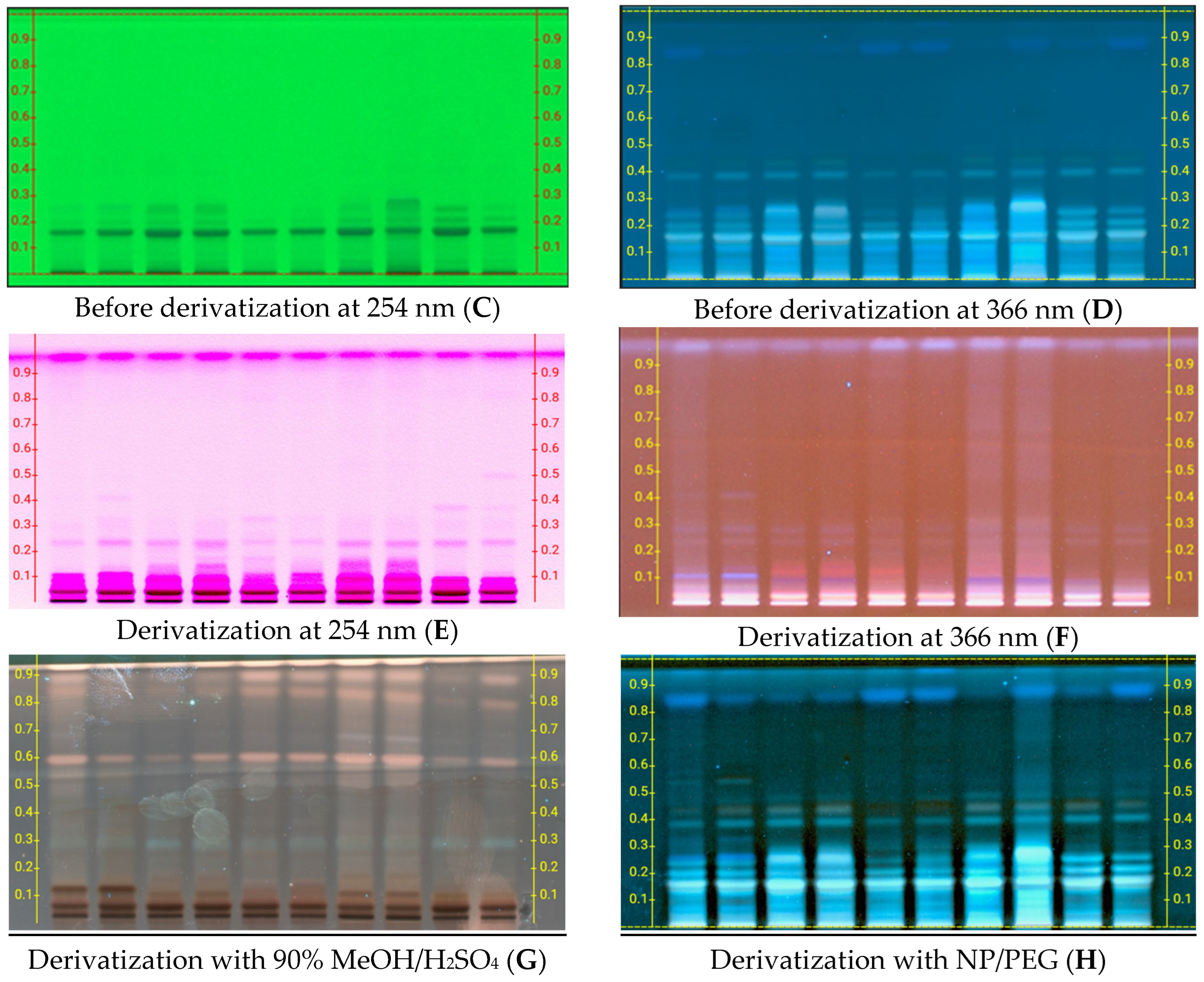
The five Hypoxis extracts were subjected to HPTLC to visualized the different phytochemical components. After developing the plates and applying all visualization methods, different compounds were detected with varying Rf values.
According to Figure 1, all the Hypoxis extracts exhibited different bands under visible light (plate A/B), UV 254 nm (plate C/E), and UV 366 nm (plate D/F), derivatization with 90% MeOH/H2SO4 (plate G), and with NP/PEG (plate H). The variation alluded to the various phytochemical classes present in the Hypoxis extracts. Bands appearing under 254 nm might represent phytochemicals with conjugated or aromatic compounds, while the ones appearing under 366 nm might indicate flavonoids, phenolic compounds, or other compounds that fluorescent naturally. The results also showed a broader range of colored band post-derivatization throughout the Hypoxis methanol extracts. This may be an indication of phytochemical compounds that may not be UV-active in their native state but become detectable after chemical modification, such as sugars, saponins, and certain terpenoids.

2.3. Quantitative Antioxidant Potential by DPPH Assay of the Five Hypoxis Species

2.3.1. The 2,2-Diphenyl-1-picrylhydrazyl (DPPH) Assay

The principle of 2,2-diphenyl-1-picrylhydrazyl (DPPH) free radical scavenging is based on the reduction of the stable DPPH radical, which is purple in color that changes to a yellow-colored diphenyl picrylhydrazine in the presence of an antioxidant. The degree of discoloration indicates the scavenging potential of the sample. Hence, the higher the DPPH percentage inhibition, the stronger the antioxidant activity of the extract.
As shown in Figure 2, all samples follow the same trend as the standards, whereby the antioxidant activity (% inhibition) across all the Hypoxis species increases as the concentration of the test samples increases. Among all samples, H. obtusa demonstrated the best activity with a percentage inhibition of 96.79% at a concentration of 1 mg/mL. The high activity may be attributed to the flavonoids they possess, which are known contributors to antioxidant mechanisms. On the other hand, H. colchicifolia showed the lowest activity with a percentage inhibition of 81.43% at the highest concentration 1 mg/mL. The standards, gallic acid, and BHT exhibited average percentage inhibition of 94.57 and 94.33, respectively, which is slightly lower than that of H. obtusa, H. galpinii, and H. hemerocallidea, suggesting that some Hypoxis species may possess superior natural antioxidant capacities compared to commonly used synthetic compounds and reference antioxidants.

2.3.2. Hydrogen Hydroxide (H2O2) Free Radical Scavenging Assay

Hydrogen peroxide is a reactive oxygen species that has the ability to cross cell membranes and produce hydroxyl radicals (-OH) in the presence of transition metals. The assay is therefore based on the ability of the antioxidants to scavenge and neutralize H2O2 and thus reduce the number of reactive species it generates. The percentage inhibition of H2O2 by extracts are displayed in Figure 3.
The percentage inhibition of H2O2 by extracts exhibited a concentration-dependent curve, in which the percentage of H2O2 inhibition increased as the concentration of the extract used increased. The same concentration-dependent curve was also observed with the reference standards gallic acid and BHT. Among all the samples evaluated, H. obtusa demonstrated the highest average percentage inhibition of 96.93% across all tested concentrations, reflecting its exceptional H2O2 scavenging capacity and strong antioxidant potential. In contrast, H. colchicifolia exhibited the lowest average inhibition (81.25%), indicating relatively weak antioxidant activity compared to the other species.
Gallic acid, a well-known phenolic antioxidant, showed considerable H2O2 scavenging activity but was outperformed by H. obtusa. The strong antioxidant activity of H. obtusa was further supported by its remarkably low IC50 value of 0.0423 mg/mL, the lowest among all plant samples tested, highlighting its potency in neutralizing H2O2 radicals. On the other end of the spectrum, H. colchicifolia presented the highest IC50 value (0.0516 mg/mL), corroborating its weaker antioxidant performance.
As expected, the synthetic antioxidant BHT exhibited the strongest scavenging ability, with the lowest IC50 value of 0.0356 mg/mL, followed closely by gallic acid (IC50 = 0.036 mg/mL). The other species—H. hemerocallidea, H. galpinii, and H. rigidula var. rigidula—demonstrated moderate antioxidant potential, with IC50 values ranging from 0.042 to 0.043 mg/mL, suggesting comparable H2O2 neutralizing capacities.
These findings confirm the significant antioxidant potential of Hypoxis species, particularly H. obtusa, and highlight its promising role in mitigating oxidative stress through effective hydrogen peroxide scavenging.

2.3.3. Ferric Reducing Antioxidant Assay

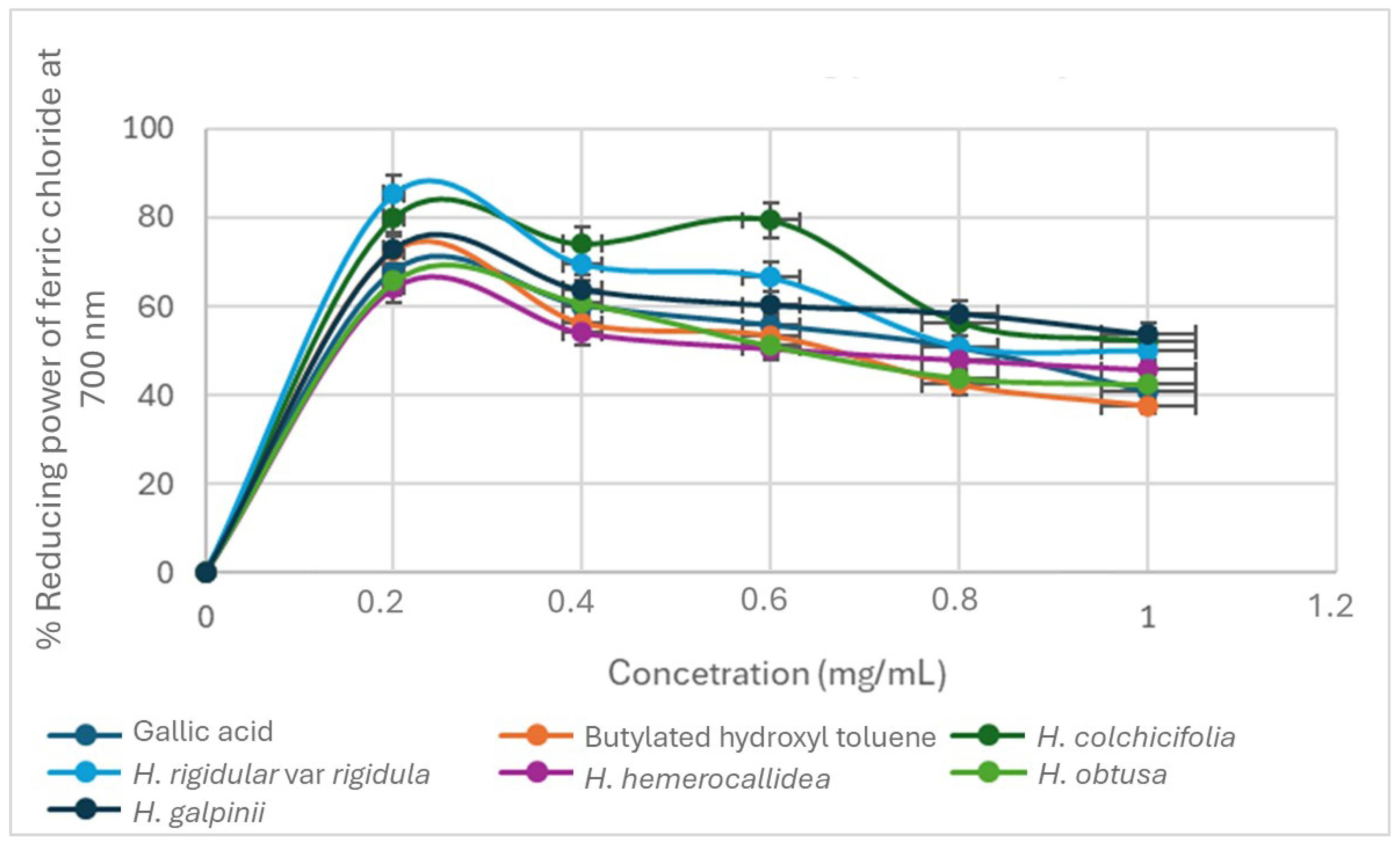
The detailed and very informative Figure 4 depicts samples H. obtusa and H. hemerocollidea as having the lowest average percentage oxidation of 52.32% and 62.24%, respectively. Therefore, we can deduce that both species had the highest antioxidant activity compared to the other three samples, H. galpinii, H. colchicifolia and H. rigidular var. rigidular with 67.3, 84.0 and 88.2%, respectively. Compared to the standards, this assay proved that Gallic and BHT have a similar antioxidant profile of 70%. In addition, IC50 values and antioxidant activity from the ferric reducing power assay are inverse proportionality inherent in H. obtusa and H. hemerocollidea, with the lowest IC50 values that translate to the highest antioxidant activity. As anticipated, H. galpinii, H. colchicifolia, and H. rigidula var. rigidula with comparatively higher IC50 values displayed the lowest antioxidant potentials in the FRAP assay. However, regarding all the test samples and the two standards, BHT exhibited the lowest IC50 value of 0.25 mg/mL (Table 2) and was nominated as having the best antioxidant activity.

2.4. Total Phenolic Content (TPC) Determination of the Five Hypoxis Species

The concentration of total phenolic content in the samples was determined by using the regression line equation y = 3.6883x + 0.278 with R2 = 0.9506 obtained from the Standard gallic calibration curve. The total phenolic content was expressed as Gallic acid equivalents per gram of dry extract of the Hypoxis sample (mg GAE/g of sample), converted to the concentration of the standard unit. The absorbance was measured in triplicate to ascertain the accuracy, consistency, and reliability of the results. The mean TPC in each sample and the standard deviations were calculated to check the variability of the obtained TPC results, summarized in Table 3.
In principle, the sample with the highest absorbance value was expected to exhibit a corresponding higher total phenolic content and vice versa. Hence, H. obtusa, with a highest instrument (y-value) of 0.52, which is tantamount to the highest percentage absorbance, exhibited a mean TPC of 335.61 ± 1.820 mg GAE/g, indicating a greater phenolic concentration. In contrast, H. colchicifolia showed a lower absorbance value of 0.287 nm, which correlated to a lower TPC of 12.20 ± 0.13 mg GAE/g, and an expected lowest phenolic content. This study demonstrated the correlation between total phenolic content and antioxidant activity of the tested samples. Notably, H. obtusa exhibited the highest total phenolic content among the five Hypoxis species, and by extension, the strongest antioxidant activity in the DPPH assay, H2O2, and the ferric reducing power assay (Table 1). These findings support the notion that phenolic compounds contribute to the antioxidant properties of plants.

2.5. Determination of the Antidiabetic Potentials of the Five Hypoxis Species

Diabetes, and its type-2, is a global public health concern that is affecting the world health care system and economy, with regard to how it impacts the per capita income negatively. Weighing in on the numerous complications from using the first-line antidiabetic drugs has ushered in positive frontiers in the use of alternative antidiabetics, including medicinal plants. The synergistic roles of phenolic content of natural products, dietary antioxidants with antidiabetic effects, may improve diabetic conditions by regulating glucose metabolism, improving insulin secretion, and decreasing insulin resistance. H. hemerocallidea has been indicated traditionally for the management of type 2 diabetes. It significantly reduced hyperglycaemia and hyperglycaemic-induced oxidative stress in the liver and kidney tissues of streptozotocin-induced diabetic male Wistar rats [22]. Whereas the Hypoxis species is listed in the South African National Biodiversity Institute (SANBI) endangered or red list of medicinal plants, we report possible species that can be used as it substitute for the management of diabetes. The diabetic mitigating penitential of four other Hypoxis species was compared with that of H. hereocallidea. The promising results obtained are depicted in Figure 5 and Figure 6 as well as summarized in Table 4.

3. Discussion

Alkaloids are among the most common bioactive compounds due to their preventative activity on a wide range of diseases and act as antidotes, such as antimicrobial, anticancer, anti-inflammatory, and wound healing capacity. In the current study, negative outcomes revealed the absence of alkaloids in all five Hypoxis species investigated. This observation, the lack of alkaloids in the Hypoxis species investigated, mirrored that previously reported [23]. Hypoxis species contains more tannins and zero terpenoids, and literature underscores tannins as having biological properties that aid in the prevention and treatment of a variety of oxidative stresses and are effective in lowering blood pressure, total cholesterol, and stimulating the immune system. The study conducted by Kumari M. and Jain S. [24] also indicated that tannins have a positive effect on managing diabetes, where they are believed to facilitate the uptake of glucose through mediators of the insulin signaling pathway. Furthermore, tannins are also considered to be cardio-protective, anti-inflammatory, anticarcinogenic, and antimutagenic [25]. On the other hand, terpenoids are reported to have antibacterial, antifungal, antiviral, cytotoxic, analgesic, and anticancer properties [26].
Generally, some natural products, regardless of their source are useful in treating burns, psoriasis, preventing scar formation following surgery, and recovery from an episiotomy following vaginal delivery of a newborn [27]. Our analysis revealed considerable variations in the extract’s composition of different Hypoxis species. Phytochemicals such as alkaloids, flavonoids, phenolics, and terpenoids were present in the extracts with significant differences in their concentrations. These variations in phytochemical profiles may be attributed to genetic factors, environmental conditions, and geographical locations in which the plants were cultivated or collected.
H. colchicifolia has four bands with Rf values of 0.2, 0.4, 0.8, and 0.9, and six bands with Rf values of 0.1, 0.2, 0.3, 0.4, 0.8, and 0.9. H. galpinii indicated four bands with Rf values of 0.2, 0.3, 0.8, and 0.9, and another three bands with Rf values of 0.2, 0.8, and 0.9. Furthermore, H. hemerocallidea displayed five bands with Rf values of 0.2, 0.5, 0.7, 0.8, and 0.9, as well as four other bands with Rf values of 0.2, 0.4, 0.8, and 0.9. The remaining two species, H. obtusa and H. ridigula var. rigidula from the different locations, equally speak to the occurrence of intra- and inter-species variations in their phytoconstituents. However, certain bands with Rf values of 0.2, 0.8, and 0.9 cut across all the Hypoxis species, while there were no compound bands with Rf values of 0.5 and 0.6 for all the species investigated.
The bands on the TLC plates indicate the different phytochemicals found in the plant extract at various Rf values. These phytochemicals were mostly antioxidants, as indicated by the cream color after dipping the TLC plate in the DPPH solution. Significant variations in antioxidant potential among different Hypoxis species were observed. Some species displayed notably higher antioxidant activity compared to others. The antioxidant potential was assessed using various assays, such as DPPH, to visualize the varying degrees of radical scavenging abilities. The findings of this study suggest that the five Hypoxis species investigated hold promise as potential sources of natural antioxidants, which could have implications for health and pharmaceutical applications.
Similar trends in antioxidant activities were observed for the DPPH and H2O2 assays; the ferric chloride reducing power assay was different. Unlike DPPH and hydrogen peroxide assays, where higher inhibition means stronger antioxidant potential, the ferric chloride results show an inverse relationship—lower inhibition percentages reflect greater antioxidant power. Among the species evaluated, H. obtusa emerged as the star, boasting the highest antioxidant activity with an average inhibition of 32.7% across all concentrations. This placed it at the top, while H. colchicifolia fell behind with the lowest antioxidant activity, averaging a 66.94% inhibition rate. Surprisingly, H. obtusa even outperformed gallic acid, a widely used antioxidant standard, showing that its antioxidant potential is worth noting.
Further investigation of IC50 values confirmed H. obtusa’s lead, with the lowest IC50 value of 0.25, underscoring its strong antioxidant potential. H. colchicifolia lagged, with a much higher IC50 value of 0.80. However, when comparing free radical reduction abilities, H. obtusa did not surpass every standard; it ranked behind BHT (IC50 = 0.96) and gallic acid. These findings illuminate H. obtusa’s exceptional antioxidant profile, warranting a deeper study to unlock its full potential. Its mixed performance in various assays also highlights how multifaceted antioxidant activity can be, underscoring the need for comprehensive testing across different assays to grasp the compound’s full antioxidant profile.
This study revealed that Hypoxis obtusa exhibited the highest total phenolic content (TPC) at 335.61 mg GAE/g, followed by H. hemerocallidea with 251.33 mg GAE/g. These results suggest that both Hypoxis species are rich in phenolic compounds known for their antioxidant properties [26]. The findings of this study align with previous studies that have emphasized the antioxidant potential of H. hemerocallidea [28,29], but notably, H. obtusa showed even greater phenolic content, indicating its superior potential as a natural antioxidant source. This high phenolic content directly correlates with the results from the DPPH assay, where H. obtusa displayed the lowest IC50 value, thus highlighting that H. obtuse has the highest antioxidant activity, further confirming that the antioxidant capacity of these species is linked to their phenolic content. Similarly, in the hydrogen peroxide assay, H. obtusa again demonstrated the strongest free radical scavenging ability, supporting the fact that phenolic compounds play a significant role in its antioxidant function. The lower phenolic content and antioxidant activity observed in H. colchicifolia (12.20 mg GAE/g) underscore the variability in bioactivity among Hypoxis species, reinforcing the conclusion that phenolic compounds are key contributors to the antioxidant properties of these plants. The higher phenolic content in H. obtusa compared to H. hemerocallidea could be attributed to factors such as environmental conditions, genetic variation, or other intrinsic factors that determine phenolic biosynthesis. While H. hemerocallidea continues to show strong phenolic content, the significantly higher values for H. obtusa highlight the need for more focused research on this species over the more highly researched and commercialized H. hemerocallidea.
Studies have demonstrated that α-amylase and β-glucosidase can break down carbohydrates and simultaneously increase the post-meal amount of glucose in diabetic patients [30]. In particular, β-glucosidase is implicated in the hydrolysis of complex carbohydrates, especially oligosaccharides or glycoconjugates. Therefore, it can be used in an antidiabetic assay because inhibitors of this enzyme can help manage blood sugar levels in individuals with diabetes [31]. We therefore explored the use of β-glucosidase in this study. Previous studies have targeted inhibitors of both α-amylase and β-glucosidase enzymes having potential in the management of type 2 diabetes [32]. Therefore, agents, including medicinal plant decoctions that can deter the activity of these two enzymes, can control postprandial hyperglycaemia and reduce the risk of developing diabetes. Aqueous extracts and metallic nanoparticles of H. hemerocallidea exhibited potency against α-amylase and α-glucosidase with an IC50 value < 10 mg/mL in a previous study [33]. This study further gives credence to the inhibitory effects of H. hemerocallidea and four other related Hypoxis species extracts on α-amylase and β-glucosidase. However, H. hemorecallidea and H. obtusa equally inhibited the two enzymes, with an IC50 of 0.21, 0.24, just like the standard acarbose with an IC50 of 0.20 mg/mL. The other three Hypoxis extracts exhibited comparative inhibitory effects with an IC50 of 0.34–0.55 mg/mL. These results unravel the potential of using the related species as alternatives to H. hemerocallidea as interventions to manage diabetes in humans, provided their cytotoxicity and chemistry are equilibrated. However, stakeholders interested in using related species, especially H. obtusa, should expect potential loopholes to the practical application of H. obtusa, including limited ethnobotanical documentation, an undefined safety profile, and challenges associated with its large-scale cultivation. In as much as different antioxidant assays (e.g., ORAC, FRAP, DPPH, and H2O2) have numerous advantages, they come with many disadvantages as well [34]. These disadvantages include, but are not limited to, challenges of solubility of antioxidant agents, their interferences with colorants and related reducing phytochemicals, reaction kinetics, and physiological radicals used in these assays. In addition, the antioxidant assay methods do not afford equivalent antioxidant potential values, such that DPPH, H2O2, and FRAP assays often have distinct specific values. This discrepancy negatively impacts the interpolation of in vitro outcomes to expected in vivo models. Consequently, assays that bridge in vitro to in vivo translational gaps [35] are pivotal for future research design.

4. Materials and Methods

4.1. Chemicals

All chemicals used in this study were of analytical reagent (AR) grade. Methanol, chloroform, dichloromethane (DCM), 2,2 diphenyl-1-picrylhydrazyl (DPPH), sulfuric acid, polyethylene glycol (PEG), hydrochloric acid, Dragnendorffs reagent, hydrogen peroxide, ferric chloride, olive oil, and glacial acetic acid were all purchased from either Rochelle Chemicals or Sigma-Aldrich, Johannesburg, South Africa.

4.2. Plant Collection, Comminution, and Extraction

Corms of the five species were purchased from Mountain Herbs Nursery (25°43′27.6″ S 27°57′54.8″ E) in the Northern part of Pretoria, Gauntrng, South Africa. The specimens of five Hypoxis species were available from previous research, and voucher specimens (HH1, HO1, HC1, HRVR1, and HG1) were subsequently deposited at the Department of Pharmaceutical Sciences, Sefako Makgatho Health Sciences University, Pretoria, South Africa. The tap roots were removed from the fresh corms, which were separated from the plant. After washing, the corms were chopped into small pieces and oven dried at 30 °C for 36 h prior to extraction. The resulting dried plant material was then pulverized using a Retsch1MM 400 ball milling machine (Monitoring and Control Pty Ltd., Haans, Germany) at a frequency of 30.0 Hz for 120 s to yield fine brown powders. These powders were sieved using a 500 mm mesh size (Endcotts Filters Ltd., London, UK) to ensure consistency of particle size.
All the solvents used in this study were purchased from Rochelle Chemicals and Lab Equipment Cc, Johannesburg, South Africa. The ground sample (0.5 kg) of the plant was extracted using a NUVE shaking water bath (ZT10.ST 30, KK05F01, Akyurt, Turkey) at 100 RPM with 2.5 L of dichloromethane and methanol (1:1 v/v) using ultrasonication. The extracts, which resulted from the process, were then filtered and concentrated using a Stuart rotary evaporator (Re400, Cole-Parmer Ltd., Stone, Staffordshire, UK) and were evaporated to dryness. The total mass of each of the extracts obtained was weighed, and the percentage yield was calculated.

4.3. Phytochemical Analysis

The phytochemicals present in the extracts were analyzed using standard colorimetric tests as recorded by [36], with slight modifications. The extracts were dissolved in methanol, a solvent that was used to extract them prior to the standard colorimetric tests as follows:

4.3.1. Alkaloids

The Dragnendorffs reagent (potassium–bismuth–iodide solution) test was used to determine the presence of alkaloids in the plant extracts. An amount of 0.05 g of plant extract was dissolved in 0.5% aqueous hydrochloric acid. Followed by filtration and treatment of 1.0 mL of filtrate with five drops of Dragnendorffs reagent. The formation of brownish/red precipitate indicated a positive test for the presence of alkaloid [37].

4.3.2. Tannins

To test for the presence of tannins in the extracts, 0.05 g of dry plant material was boiled in 20 mL of distilled water and filtered. A few drops of 0.1 g w/v ferric chloride was added to the filtrate. The presence of blue-black precipitate was a positive test for the presence of tannins [38].

4.3.3. Phlobatannins

The crude extract (0.05 g) was dissolved in 10 mL of aqueous solution and boiled, then 1.0% hydrochloric acid was added to the boiling tube. The deposition of red precipitate indicated the presence of phlobatannins [39].

4.3.4. Terpenoids

To determine the presence of terpenoids in the extracts, 5.0 mL of each plant extract was mixed with 2.0 mL of chloroform, followed by the dropwise addition of 3.0 mL of concentrated sulfuric acid. The formation of reddish/brownish coloration at the interphase of the mixture indicated the presence of terpenoids [40].

4.3.5. Deoxy Sugar of Cardenolides

Each crude extract (5.0 mL) was mixed with 2.0 mL of glacial acetic acid containing five drops of ferric chloride solution, followed by the addition of 1.0 mL of concentrated sulfuric acid. The formation of a brown ring at the interphase of the solution and a violet ring below the brown ring indicated the presence of deoxy sugar of cardenolides [41].

4.3.6. Saponins

A frothing test was used to determine the presence of saponins in the extracts. To 20 mL of distilled water was added 2.0 g of plant material. A total of 0.5 mL filtrate was diluted to 5 mL with distilled water and shaken vigorously for 2 min. Formation of stable foam indicates the presence of saponins [38].

4.4. High-Performance Thin-Layer Chromatography (HPTLC) Analysis

An amount of 5.0 mg of each plant extract was dissolved in 10 mL of methanol. The resulting solution was then filtered with syringe filters and spotted on HPTLC silica gel 60 F254, 20 × 10 cm glass plates. The plates were then developed in the CAMAAG (Muttenz, Switzerland), automatic developing chamber using chloroform–methanol–distilled water (70:30:4 v/v/v) as the mobile phase. After developing the HPTLC plate, the visualization was performed by CAMAAG HPTLC visualizer before capturing was conducted first at naked eye (white light), under 254 nm wavelength, then 366 nm wavelength. To further visualize the plates, they were derivatized with a solution of methanol–sulfuric acid (9:1 v/v) or polyethylene glycol–dichloromethane solution (90:10 v/v). Plates derivatized with methanol–H2SO4 (9:1 v/v) were heated on a CAMMAG heater at 100 °C for 5 min for clearer bands.

4.5. Determination of Antioxidant Potential of the Five Hypoxis Species

4.5.1. DPPH Radical Scavenging Activity

The DPPH radical scavenging assay was performed according to the method described by Mapfumari and co-workers [42] as follows: A sample stock solution was prepared by dissolving 20 mg of the extract in 20 mL of methanol to yield a concentration of 1.0 mg/mL. Dilutions of this stock solution were made to obtain concentrations ranging from 0.2 mg/mL to 1.0 mg/mL. Equal volumes of 1 mL of each sample and DPPH solution were mixed in a 1:1 (v/v) ratio. The resulting mixtures were incubated at room temperature for 30 min for the reaction to occur. After incubation, the absorbances were measured at 517 nm using a spectrophotometer (SprectraMax®, Molecular Devices, San Jose, CA, USA). The same concentration of Gallic acid and butylated hydroxyl toluene (BHT) reference standards were also measured at the same wavelength, and the percentage radical scavenging activity of the extracts and standards was calculated using Equation (1) below.
Equation (1):
%   D P P H   r a d i c a l   s c a v e n g i n g   a c t i v i t y = A 0 A s A 0 × 100
where A0 = absorbance of the negative control and As = the absorbance of the sample.

4.5.2. Hydrogen Peroxide Free Radical Scavenging Activity

The hydrogen peroxide (H2O2) assay was conducted according to the method described by [41], but with slight modifications. A stock solution was prepared by dissolving 20 mg of the plant extract in 20 mL of methanol to yield a concentration of 1.0 mg/mL. A range of concentrations (0.2–1 mg/mL) was then prepared from this stock solution. A 20 mM working solution of H2O2 was subsequently prepared in phosphate-buffered saline (PBS) with a pH of 7.4. 2.0 mL of the working solution was added to each sample, and then vortexed. The samples were then incubated for 10 min, and absorbances were measured at 560 nm using a spectrophotometer. The same procedure was followed with the standards, and the percentage hydrogen peroxide inhibition was calculated using Equation (1).

4.5.3. Ferric Chloride Reducing Power Method

The procedure adapted from [41], was used to assess the ferric chloride reducing power of the extracts. Different concentrations of the extracts were prepared, and each 1.0 mL of the prepared extracts were combined with 2.5 mL of potassium ferricyanide (K3Fe (CN) 6) at 1% (w/v) and 2.5 mL of 0.2 M phosphate buffer (pH 6.6). After mixing the components, they were incubated for 20 min at 50 °C. The mixture was then centrifuged for 10 min at 3000 revolutions per minute (rpm). This was followed by the addition of 2.5 mL of 10% (w/v) trichloroacetic acid to the mixture. A total of 2.5 mL of the resultant solution’s upper layer was combined with 0.5 mL of ferric chloride (0.1% w/v) and 2.5 mL of distilled water. The same procedure was followed with the standards. Post incubation, the absorbance was measured using a spectrophotometer at 700 nm, and the percentage reducing power activity was calculated using Equation (1).

4.6. The Determination of Total Phenolic Content

A Folin–Ciocalteu reagent method explained by Aryal and co-workers (2019) [43] was employed with fewer modifications to determine the total phenolic content of the extracts. A standard curve was generated to quantify phenols by using gallic acid solutions with concentrations of 0.2–1.0 (mg/mL). To prepare the standard solutions, 9.0 mL of distilled water was added to a 25 mL flat-bottomed flask, followed by the addition of 1.0 mL of the respective gallic acid solutions. Next, 1.0 mL of Folin–Ciocalteu reagent was added to each flask, and the mixtures were shaken and incubated at room temperature for 5 min. After incubation, 10 mL of 7% sodium carbonate was added, and the volume was adjusted to 25 mL with distilled water. The samples were then incubated for 90 min at room temperature, and absorbance was measured using a spectrophotometer at 550 nm. Hypoxis samples were prepared in the same way as the gallic acid standard. Samples of varying concentrations were measured in triplicate, and their absorbances were recorded. The phenolic content of the samples was expressed as milligrams of gallic acid equivalents per gram of extract (mg GAE/g extract).

4.7. Determination of the Antidiabetic Activity

4.7.1. α-Amylase Inhibition Assay

The α-amylase inhibitory activity of Hypoxis sample extracts was accomplished according to the standard technique described in a previous study [31], with minor adjustments. The dry extracts were redissolved in the solvents used to extract them, and a concentration of 1.0 mg/mL was prepared for each extract. An amount of 100 μL of potassium phosphate buffer (pH 7) was added to all the wells of a 96-well plate. A 100 μL of each extract was added to the test well and serially diluted until the last row of the wells. A 100 μL of acarbose was added to the control well and serially diluted. Thereafter, 20 μL of the α-amylase was added to all wells and incubated for 5 min at 37 °C. After the first incubation, 50 μL of substrate was added to all wells and further incubated for 20 min. Absorbance was then read at 405 nm using a 96-well UV spectrophotometer Section. The ability of the extracts to inhibit alpha amylase was calculated using Equation (2).
%   α a m y l a s e   i n h i b i t i o n = A 0 A S A 0 × 100
where A0 = absorbance of the control and As = absorbance of a sample and standards.

4.7.2. β-Glucosidase Inhibition Assay

To determine the ability of the five Hypoxis extracts to inhibit enzyme β-glucosidase that is implicated in the progression of diabetes, a method described by [31], was followed with minor modifications. In all wells of a 96-well plate, 50 μL of potassium phosphate buffer (pH 7) was added. An aliquot of 50 μL of the prepared five Hypoxis extracts was added to all the test wells and serially diluted to the last row of the plate. This was followed by the addition of 50 μL of sucrose and then 100 μL of the commercial glucose kit. An amount of 30 μL of intestinal acetone rat powder was added to all wells, followed by incubation of the plate at 37 °C for 30 min. A positive control of acarbose was prepared in the same manner, and the absorbances were read at 505 nm using a UV spectrophotometer. The ability of the extracts to inhibit β-glucosidase enzyme was calculated using Equation (3).
Equation (3):
%   i n h i b i t i o n   o f   b e t a g l u c o s i d a s e = A 0 A s A 0 × 100
where A0 = absorbance of positive control and AS = absorbance of sample and standards.

5. Conclusions

This study indicated variation in phytochemicals amongst the five selected South African Hypoxis species. However, the study’s milestone is unraveling the equivalent amount of phenolic content and significant potential antioxidant properties of H. obtusa and H. hemerocallidea in addition to those of the other three species. The findings of this study strongly suggest further investigations on the five Hypoxis species of the South African ecotype for the potential management of oxidative stress diabetes and related neurological diseases like Parkinson’s disease, Alzheimer’s disease (AD), amyotrophic lateral sclerosis (ALS), multiple sclerosis, depression, and memory loss due to the strong free radicals scavenging properties of the plant. Additionally, H. obtusa could serve as a potential substitute for H. hemerocallidea for conservation and sustainability.

Author Contributions

Conceptualization, K.B.; methodology, K.B.; formal analysis, B.M. and K.B.; resources, M.P., P.D. and K.B.; writing—original draft preparation, B.M. and K.B.; writing—review and editing, M.P. and K.B.; supervision, K.B.; project administration, M.P., P.D. and K.B. All authors have read and agreed to the published version of the manuscript.

Funding

This research received no external funding, and the APC was funded by the Department of Pharmaceutical Sciences, Sefako Makgatho Health Sciences University.

Data Availability Statement

The data generated in this study are part of this manuscript.

Acknowledgments

The author is indebtedly appreciative of the Sefako Makgatho Health Sciences University 2023 (Dlamini U., Mbatha S., Racheku J. and Zulu N.P.) and 2024 (Dlamini B.N., Hassim S., Hlongwane P. and Moraba I.) Bachelor of Pharmacy fourth year group Matlou M.K., Masinga V.S., Moabelo K., Makhaza N.T., Mathe B.C., Thabela K.A. for the laboratory investigation work performed.

Conflicts of Interest

The authors declare no conflicts of interest.

References

  1. Lobo, V.; Patil, A.; Phatak, A.; Chandra, N. Free radicals, antioxidants, and functional foods: Impact on human health. Pharmacogn. Rev. 2010, 4, 118–126. [Google Scholar] [CrossRef]
  2. Engwa, G.A.; Nweke, F.N.; Nkeh-Chungag, B.N. Free radicals, oxidative stress-related diseases and antioxidant supplementation. Altern. Ther. Health Med. 2022, 28, 114. [Google Scholar] [PubMed]
  3. Pizzino, G.; Irrera, N.; Cucinotta, M.; Pallio, G.; Mannino, F.; Arcoraci, V.; Squadrito, F.; Altavilla, D.; Bitto, A. Oxidative stress: Harms and benefits for human health. Oxidative Med. Cell. Longev. 2017, 2017, 8416763. [Google Scholar] [CrossRef]
  4. Forni, C.; Facchiano, F.; Bartoli, M.; Pieretti, S.; Facchiano, A.; D’Arcangelo, D.; Norelli, S.; Valle, G.; Nisini, R.; Beninati, S.; et al. Beneficial role of phytochemicals on oxidative stress and age-related diseases. BioMed Res. Int. 2019, 2019, 8748253. [Google Scholar] [CrossRef]
  5. Preiser, J.C. Oxidative stress. J. Parenter. Enter. Nutr. 2012, 36, 147–154. [Google Scholar] [CrossRef]
  6. Sharma, V.; Mehdi, M.M. Oxidative stress, inflammation and hormesis: The role of dietary and lifestyle modifications on aging. Neurochem. Int. 2023, 164, 105490. [Google Scholar] [CrossRef]
  7. Alfadda, A.A.; Sallam, R.M. Reactive oxygen species in health and disease. BioMed Res. Int. 2012, 2012, 936486. [Google Scholar] [CrossRef] [PubMed]
  8. Liguori, I.; Russo, G.; Curcio, F.; Bulli, G.; Aran, L.; Della-Morte, D.; Gargiulo, G.; Testa, G.; Cacciatore, F.; Bonaduce, D.; et al. Oxidative stress, aging, and diseases. Clin. Interv. Aging 2018, 13, 757–772. [Google Scholar] [CrossRef]
  9. Hajhashemi, V.; Vaseghi, G.; Pourfarzam, M.; Abdollahi, A. Are antioxidants helpful for disease prevention? Res. Pharm. Sci. 2010, 5, 1. [Google Scholar] [PubMed]
  10. Viana da Silva, M.; Santos, M.R.C.; Alves Silva, I.R.; Macedo Viana, E.B.; Dos Anjos, D.A.; Santos, I.A.; Barbosa de Lima, N.G.; Wobeto, C.; Jorge, N.; Lannes, S.C.D.S. Synthetic and natural antioxidants used in the oxidative stability of edible oils: An overview. Food Rev. Int. 2022, 38, 349–372. [Google Scholar] [CrossRef]
  11. Ayza, M.A.; Zewdie, K.A.; Yigzaw, E.F.; Ayele, S.G.; Tesfaye, B.A.; Tafere, G.G.; Abrha, M.G. Potential protective effects of antioxidants against cyclophosphamide-induced nephrotoxicity. Int. J. Nephrol. 2022, 2022, e5096825. [Google Scholar] [CrossRef] [PubMed]
  12. Caleja, C.; Barros, L.; Antonio, A.L.; Carocho, M.; Oliveira, M.B.P.; Ferreira, I.C. Fortification of yogurts with different antioxidant preservatives: A comparative study between natural and synthetic additives. Food Chem. 2016, 210, 262–268. [Google Scholar] [CrossRef] [PubMed]
  13. Lourenço, S.C.; Moldão-Martins, M.; Alves, V.D. Antioxidants of natural plant origins: From sources to food industry applications. Molecules 2019, 24, 4132. [Google Scholar] [CrossRef] [PubMed]
  14. Xu, X.; Liu, A.; Hu, S.; Ares, I.; Martínez-Larrañaga, M.R.; Wang, X.; Martínez, M.; Anadón, A.; Martínez, M.A. Synthetic phenolic antioxidants: Metabolism, hazards and mechanism of action. Food Chem. 2021, 353, 129488. [Google Scholar] [CrossRef] [PubMed]
  15. Sharma, N.; Kumar, P.; Shukla, K.S.; Maheshwari, S. AGE RAGE Pathways: Cardiovascular disease and oxidative stress. Drug Res. 2023, 73, 408–411. [Google Scholar] [CrossRef]
  16. Uzombah, T.A. The implications of replacing synthetic antioxidants with natural ones in the food systems. In Natural Food Additives; IntechOpen: London, UK, 2022. [Google Scholar]
  17. Thakur, M.; Singh, K.; Khedkar, R. Phytochemicals: Extraction process, safety assessment, toxicological evaluations, and regulatory issues. In Functional and Preservative Properties of Phytochemicals; Academic Press: Cambridge, MA, USA, 2020; pp. 341–361. [Google Scholar]
  18. Singh, Y. Systematics of Hypoxis (Hypoxidaceae) in Southern Africa. Ph.D. Thesis, University of Pretoria, Pretoria, South Africa, 2006. [Google Scholar]
  19. Bassey, K.; Viljoen, A.; Combrinck, S.; Choi, Y.H. New phytochemicals from the corms of medicinally important South African Hypoxis species. Phytochem. Lett. 2014, 10, lxix–lxxv. [Google Scholar] [CrossRef]
  20. Oguntibeju, O.O.; Meyer, S.; Aboua, Y.G.; Goboza, M. Hypoxis hemerocallidea Significantly Reduced Hyperglycaemia and Hyperglycaemic-Induced Oxidative Stress in the Liver and Kidney Tissues of Streptozotocin-Induced Diabetic Male Wistar Rats. Evid. Based Complement. Altern. Med. 2016, 2016, 8934362. [Google Scholar] [CrossRef] [PubMed] [PubMed Central]
  21. Pereus, D.; Otieno, J.; Ghorbani, A.; Kocyan, A.; Hilonga, S.; de Boer, H. Diversity of Hypoxis species used in ethnomedicine in Tanzania. S. Afr. J. Bot. 2019, 122, 336–341. [Google Scholar] [CrossRef]
  22. Muscolo, A.; Mariateresa, O.; Giulio, T.; Mariateresa, R. Oxidative stress: The role of antioxidant phytochemicals in the prevention and treatment of diseases. Int. J. Mol. Sci. 2024, 25, 3264. [Google Scholar] [CrossRef]
  23. Zimudzi, C. African Potato (Hypoxis Spp.): Diversity and Comparison of the Phytochemical Profiles and Cytotoxicity Evaluation of four Zimbabwean Species. J. Appl. Pharm. Sci. 2014, 4, 79–83. [Google Scholar] [CrossRef]
  24. Kumari, M.; Jain, S. Tannins: An Antinutrient with Positive Effect to Manage Diabetes. Res. J. Recent Sci. 2012, 2277, 2502. [Google Scholar]
  25. Masyita, A.; Sari, R.M.; Astuti, A.D.; Yasir, B.; Rumata, N.R.; Emran, T.B.; Nainu, F.; Simal-Gandara, J. Terpenes and terpenoids as main bioactive compounds of essential oils, their roles in human health and potential application as natural food preservatives. Food Chem. X 2022, 13, 100217. [Google Scholar] [CrossRef] [PubMed]
  26. McLoone, P.; Oladejo, T.O.; Kassym, L.; McDougall, G.J. Honey Phytochemicals: Bioactive Agents with Therapeutic Potential for Dermatological Disorders. Phytother. Res. 2024, 38, 5741–5764. [Google Scholar] [CrossRef] [PubMed]
  27. Zeb, A. Concept, mechanism, and applications of phenolic antioxidants in foods. J. Food Biochem. 2020, 44, e13394. [Google Scholar] [CrossRef] [PubMed]
  28. Nair, V.D.; Dairam, A.; Agbonon, A.; Arnason, J.T.; Foster, B.C.; Kanfer, I. Investigation of the antioxidant activity of African potato (Hypoxis hemerocallidea). J. Agri. Food Chem. 2007, 55, 1707–1711. [Google Scholar] [CrossRef]
  29. Kumar, V.; Okem, A.; Moyo, M.; Gruz, J.; Doležal, K.; Van Staden, J. Effect of zinc on the production of phenolic acids and hypoxoside in micropropagated Hypoxis hemerocallidea. Plant Growth Regul. 2019, 89, 19–24. [Google Scholar] [CrossRef]
  30. Poovitha, S.; Parani, M. In vitro and in vivo α-amylase and α-glucosidase inhibiting activities of the protein extracts from two varieties of bitter gourd (Momordica charantia L.). BMC Complement. Altern. Med. 2016, 16, 185. [Google Scholar] [CrossRef]
  31. Chokki, M.; Cudălbeanu, M.; Zongo, C.; Dah-Nouvlessounon, D.; Ghinea, I.O.; Furdui, B.; Raclea, R.; Savadogo, A.; Baba-Moussa, L.; Avamescu, S.M.; et al. Exploring Antioxidant and Enzymes (A-Amylase and B-Glucosidase) Inhibitory Activity of Morinda lucida and Momordica charantia Leaves from Benin. Foods 2020, 9, 434. [Google Scholar] [CrossRef] [PubMed] [PubMed Central]
  32. Verma, N.; Behera, B.C.; Sharma, B.O. Glucosidase inhibitory radical scavenging properties of lichen metabolites salazinic acid sekikaic acid usnic acid Hacettepe. J. Biol. Chem. 2012, 40, 7–21. [Google Scholar]
  33. Mahlo, S.J.; More, G.K.; Oladipo, A.O.; Lebelo, S.L. In vitro α-amylase/α-glucosidase, cytotoxicity and radical scavenging potential of Hypoxis hemerocallidea synthesized magnesium oxide nanoparticles. Discov. Appl. Sci. 2024, 6, 62. [Google Scholar] [CrossRef]
  34. Kotha, R.R.; Tareq, F.S.; Yildiz, E.; Luthria, D.L. Oxidative stress and antioxidants—A critical review on in vitro antioxidant assays. Antioxidants 2022, 11, 2388. [Google Scholar] [CrossRef] [PubMed]
  35. de Torre, M.P.; Cavero, R.Y.; Calvo, M.I.; Vizmanos, J.L. A simple and a reliable method to quantify antioxidant activity in vivo. Antioxidants 2019, 8, 142. [Google Scholar] [CrossRef]
  36. Mujeeb, F.; Bajpai, P.; Pathak, N. Phytochemical evaluation, antimicrobial activity, and determination of bioactive components from leaves of Aegle marmelos. Biomed. Res. Int. 2014, 2014, 497606. [Google Scholar] [CrossRef] [PubMed] [PubMed Central]
  37. Harborne, J.B. Phenolic compounds. In Phytochemical Methods; Springer: Dordrecht, The Netherlands, 1973; pp. 33–88. [Google Scholar]
  38. Evans, W.C. Trease and Evans Pharmacognosy, 15th ed.; Sanders Co., Ltd.: Singapore, 2002. [Google Scholar]
  39. Edeoga, H.O.; Okwu, D.E.; Mbaebie, B.O. Phytochemical constituents of some Nigerian medicinal plants. Afr. J. Biotechnol. 2005, 4, 685–688. [Google Scholar] [CrossRef]
  40. Salkowski, E. Ueber cholesterin. Z. Phys. Chem. 1871, 1, 1–8. [Google Scholar]
  41. DagerAlbalawi, M.A. Chemistry spectroscopic characteristics biological activity of natural occurring cardiac glycosides. IOSR J. Biotechnol. Biochem. 2016, 2, 20–35. [Google Scholar]
  42. Mapfumari, S.; Matseke, B.; Bassey, K. Isolation of a Marker Olean-12-en-28-butanol Derivative from Viscum continuum E. Mey. Ex Sprague and the Evaluation of Its Antioxidant and Antimicrobial Potentials. Plants 2024, 13, 1382. [Google Scholar] [CrossRef] [PubMed]
  43. Aryal, S.; Baniya, M.K.; Danekhu, K.; Kunwar, P.; Gurung, R.; Koirala, N. Total phenolic content, flavonoid content and antioxidant potential of wild vegetables from Western Nepal. Plants 2019, 8, 96. [Google Scholar] [CrossRef] [PubMed]
Figure 1. Analysis of Hypoxis extracts: HPTLC chromatograms indicating the phytochemical variations at different UV wavelengths and derivatizing conditions. For all the images, band 1 and 2: H. colchifolia, band 3 and 4: H. galpinii, band 5 and 6: H. hemerocallidea, band 7 and 8: H. obtusa, and band 9 and 10: H. rigidula var. rigidula.
Stresses 05 00053 g001aStresses 05 00053 g001b
Figure 2. DPPH free radical scavenging activity of the five Hypoxis species extracts. Each value is expressed as mean ± standard deviation of (n = 3).
Stresses 05 00053 g002
Figure 3. H2O2 free radical scavenging activity of the five Hypoxis species extracts. Each value is expressed as mean ± standard deviation of (n = 3).
Stresses 05 00053 g003
Figure 4. Ferric chloride reducing power activity of the five Hypoxis species extracts. Each value is expressed as mean ± standard deviation of (n = 3).
Stresses 05 00053 g004
Figure 5. α-Amylase inhibitory assay of the five Hypoxis extracts. Where % absorbance indicated the level of enzyme activity remaining after the addition of a potential inhibitor or plant extract.
Stresses 05 00053 g005
Figure 6. β-Glucosidase inhibitory assay of the five Hypoxis extracts. Where % enzymatic inhibition indicated the level of enzyme activity remaining after the addition of a potential inhibitor or plant extract.
Stresses 05 00053 g006
Table 1. Preliminary phytochemical screening results for the five Hypoxis species.
Phytochemical
Tests
Observation for Different Extracts
H. colchifoliaH. galpiniiH. rigidular var. rigidulaH.
hemerocallidea
H. obtusa
Alkaloids
Flavonoids+++++
Tannins
Phlobatannins
Saponins+++++
Terpenoids+++++
Deoxy sugar+++++
Key: + present; − absent.
Table 2. IC50 values and the regression used to calculate them for the three assays.
IC50 (mg/mL)
Hypoxis SpeciesRegression EquationDPPHH2O2FRAP
H. rigidular var. rigidulay = 70.344x + 41.294
y = 63.502x + 47.305
y = 20.652x + 43.39
0.1240.04240.320
H. obtusay = 68.866x + 46.353
y = 60.489x + 46.881
y = 19.647x + 34.164
0.0530.0520.806
H. colchicifoliay = 66.23x + 34.71
y = 65.496x + 47.228
y = 2797x + 43.003
0.2300.04230.2500
H. hemerocallideay = 68.838x + 45.862
y = 57.806x + 47.49
y = 25.214x + 31.167
0.0600.0430.745
H. galpiniiy = 63.686x + 39.92
y = 64.085x + 47.271
y = 31.671x + 35.72
0.1580.0430.451
Gallic acidy = 66.23x + 34.715
y = 70.26x + 47.358
y = 21.091x + 35.31
0.0870.0370.695
BHTy = 74.866x + 40.384
y = 70.271x + 47.498
y = 13.598x + 36.859
0.1280.0350.966
Table 3. Total phenolic content (mgGAE/g dry extract) of the five extracts of Hypoxis species.
H. SpeciesH. rigidula var. reigidulaH. obtusaH.
colchicifolia
H. hemerocallideaH. galpiniiGallic AcidBHT
TPC (mg (GAE/g)188.52 ± 2.6335.61 ± 1.812.201 ± 0.13251.34 ± 0.2727.43 ± 0.14--
%DPPH89.6 ± 3.2797.17 ± 0.2174.06 ± 4.5596.34 ± 0.4283.06 ± 1.7997.78 ± 3.9587.85 ± 4.55
H2O297.08 ± 2.3698.42 ± 3.1296.58 ± 1.7897.95 ± 4.2597.85 ± 2.1667.92 ± 0.0372.41 ± 0.02
FRAP85.28 ± 0.0165.64 ± 0.0179.90 ± 0.0364.23 ± 0.0873.01 ± 0.0267.92 ± 0.0172.41 ± 0.02
Antioxidant IC500.083 ± 0.0570.052 ± 0.0010.1366 ± 0.1330.065 ± 0.0310.100 ± 0.0810.097 ± 0.0850.082 ± 0.066
Table 4. The IC50 (mg/mL) values of the test extracts versus the acarbose standard.
H. SpeciesH. rigidular var. rigidulaH. obtusaH.
colchifolia
H. hemerocallideaH. galpiniiAcarbose
α-amylase IC50 (mg/mL)0.3460.3950.3750.4040.3700.209
β-glucosidase IC50 (mg/mL)0.5530.2100.3840.2410.4250.209
Disclaimer/Publisher’s Note: The statements, opinions and data contained in all publications are solely those of the individual author(s) and contributor(s) and not of MDPI and/or the editor(s). MDPI and/or the editor(s) disclaim responsibility for any injury to people or property resulting from any ideas, methods, instructions or products referred to in the content.