Review

Bacterial Biofilm Development and Its Relationship with Catheter-Associated Urinary Tract Infection

Botany and Microbiology Department, Faculty of Science, Cairo University, Giza 12613, Egypt
*
Author to whom correspondence should be addressed.
Stresses 2025, 5(3), 58; https://doi.org/10.3390/stresses5030058
Submission received: 23 May 2025 / Revised: 23 August 2025 / Accepted: 27 August 2025 / Published: 4 September 2025
(This article belongs to the Collection Feature Papers in Human and Animal Stresses)

Abstract

Biofilms are structured communities of microorganisms embedded in a self-produced extracellular polymeric substance (EPS) matrix; they form by sticking to a surface, growing in number, spreading out, developing fully, and breaking apart. Biofilm represents a risk of infections linked to healthcare environments. It can be one of the leading causes of nosocomial infections, which can colonize the surface of medical equipment, including respirators, urinary and central venous catheters, prosthetic heart valves, and orthopaedic devices. Biofilm formation in urinary catheters is the most common and plays a role in multidrug resistance, especially in patients with catheter-associated urinary tract infections. The supply of antibiotics for the treatment of biofilm bacteria is still inadequate due to continued antibiotic resistance, and the search for a cure for biofilm bacteria in urinary catheters is still under development. Most research currently focuses on preventing biofilm bacteria from adhering to the urinary catheter. This review discusses biofilm bacteria that form with catheter-associated urinary tract infection mechanisms and pathogenesis. In addition, the factors affecting the biofilm development by catheter-associated urinary tract infections were explained.

1. Introduction

Catheter-associated urinary tract infections (CAUTI) have negative effects on patients and the availability of healthcare resources [1]. The term “CAUTI” refers to a urinary tract infection (UTI) caused by an indwelling urinary catheter that is present or that manifests 48 h after it has been removed. Upon insertion, tubular silicone or latex urinary catheters are prone to colonization by biofilms on both their luminal and external surfaces [2].
These devices are commonly contaminated, and the Gram-negative bacteria Escherichia coli, Proteus mirabilis, and Pseudomonas aeruginosa, and the Gram-positive bacteria Staphylococcus epidermidis and Enterococcus faecalis, among others, create biofilms. The longer the urine catheter remains in the urinary tract, the more likely these microorganisms are to form biofilms that could cause urinary tract infections [3,4]. These strains contain a variety of adhesins in their walls, and when they come into contact with a surface, they release exopolysaccharides to facilitate attachment. Then, as colonies of these bacteria grow and spread across the surface, they are embedded in a matrix of polysaccharides that resembles a gel [5].
Groups of populations known as biofilms are encased in dense extracellular polymeric substances (EPS), which are made up of extracellular proteins, lipids, DNA, and polysaccharides and serve as a matrix to hold the cells together [6,7]. Like real multicellular organisms, biofilms control their internal activities, such as quorum sensing by external signals [8,9,10].
A heterogeneous biofilm’s high cell density promotes the exchange of genetic information, including the transmission of resistance genes between strains and species. Besides protecting biofilm-forming microorganisms from environmental challenges like antibiotics, immune responses, and physical stress, the extracellular matrix may also limit the colonization of competing species, but this is not its primary function [10].
In general, the matrix of EPS has essential roles in biofilm formation. It can hold onto water, defending the embedded microorganisms from desiccation. Also, it can capture and store nutrients from the environment, enabling survival in oligotrophic environments. In addition, it can protect the bacteria from both antibacterial and harmful metals [10], Therefore, biofilm has a significant role in developing antibiotic resistance [11,12]; and bacteria in a biofilm can be up to 1000-fold more resistant to antimicrobial drugs [13].
About 80% of infections classified as CAUTI are thought to be caused by urinary equipment, most notably urinary catheterization [3,10].
The urinary catheter is connected to 95% of urinary tract infections, mechanical ventilation is linked to 80% of pneumonia, and intravascular devices are connected to 87% of bloodstream infections. Staphylococci and Enterococci cause common cases of hospital-acquired infections. These two bacterial genera live in the human skin, upper respiratory system, lower gastrointestinal tract, and urogenital tract as commensal residents. They are therefore among the organisms most prone to colonize medical equipment that has been implanted [14].
Bacteria can enter the bladder during catheter insertion, through the catheter lumen, or from the area around the catheter’s exterior. As is widely recognized, the external surface is heavily favoured by biofilm development during brief catheterization. However, prolonged catheterization results in biofilm growth on the catheter’s inner lumen. On the other hand, data on the spread of resistance among urinary pathogens and considerable changes in the antibiogram of CAUTI bacterial isolates have made antimicrobial resistance among them, which has been a growing problem in recent decades; this resistance makes the bacteria a significant challenge for patient recovery [15].
Due to their reduced susceptibility to antimicrobial treatment, biofilms are of significant medical importance. Horizontal gene transfer is significantly increased in biofilms. The proximity of cells within a biofilm facilitates the exchange of plasmids, which promotes the spread of antibiotic resistance genes to other microorganisms. Additionally, many bacteria within biofilms turn into metabolically inactive forms. Therefore, they are unaffected by antimicrobial treatments because they only impact bacteria with an active metabolism [12]. Bacterial biofilm research and management can be critical in developing innovative approaches to treating infections.
Scientists have been focused on the modifications of urinary catheter walls for the prevention or reduction in biofilm adherence, as well as to reduce encrustation on the catheter wall while maintaining its durability [16,17]. Moreover, some researchers indicate that antibiotic prophylaxis significantly reduced the rate of catheter-associated UTIs. However, guidelines do not recommend the routine use of antibiotics or antimicrobial-coated catheters to prevent catheter-associated UTIs [16].
The present review summarized biofilm development and CAUTI issues, the formation of bacterial biofilms and their characteristics, and the diseases linked to biofilm formation.

2. Urinary Catheter

Urinary catheters have been used in human medicine for over 3500 years. Ancient civilizations such as the Greeks, Egyptians, and Chinese are known to have practiced catheterization as early as the third century B.C. Early catheters were made from a wide variety of materials, including copper, tin, bronze, gold, lead, papyrus, onion stems, dried reeds, and palm leaves [18]. The term “catheter” originates from the ancient Greek word kathiénai, meaning “to thrust into” or “to send down,” referring to a medical device used to drain fluids from bodily cavities [19]. Over time, catheter materials evolved, and more flexible and biocompatible options became available. Recent decades have seen the widespread use of materials such as gum-elastic, plastic (polyvinyl chloride, PVC), polyurethanes, latex rubber, and silicone [17,18,20].
A catheter is a hollow, flexible tube made of latex, polyvinyl chloride, or silicone that drains urine or infuses fluids into the bladder. There are two main types of catheters: indwelling catheters (IDCs) and intermittent catheters (ICs) [21].
Advances in material science have led to the development of biofilm-resistant catheter materials, such as silicone and hybrid composites. The hypoallergenic and non-toxic properties of silicone make it suitable for long-term use within the human body, making it the “gold standard” [22]. Its smooth and non-adhesive surface minimizes microbial attachment and delays the early stages of biofilm formation, thereby improving its resistance to infection [23].

3. Formation of Biofilm

In biotic (e.g., host tissues) and abiotic (e.g., medical device surfaces) environments, biofilm formation is an integral survival strategy that bacteria utilize. This process allows microorganisms to withstand various environmental and host-derived stressors such as desiccation, immune responses, and exposure to disinfectants [24]. Forming biofilms on urinary catheters is of particular concern in the context of healthcare-associated infections. The process begins with initial bacterial attachment to the catheter surface, followed by microcolony formation, extracellular polymeric substance (EPS) production, and biofilm maturation, which enhances bacterial persistence and antibiotic resistance [25].

3.1. Steps of Biofilm Formation

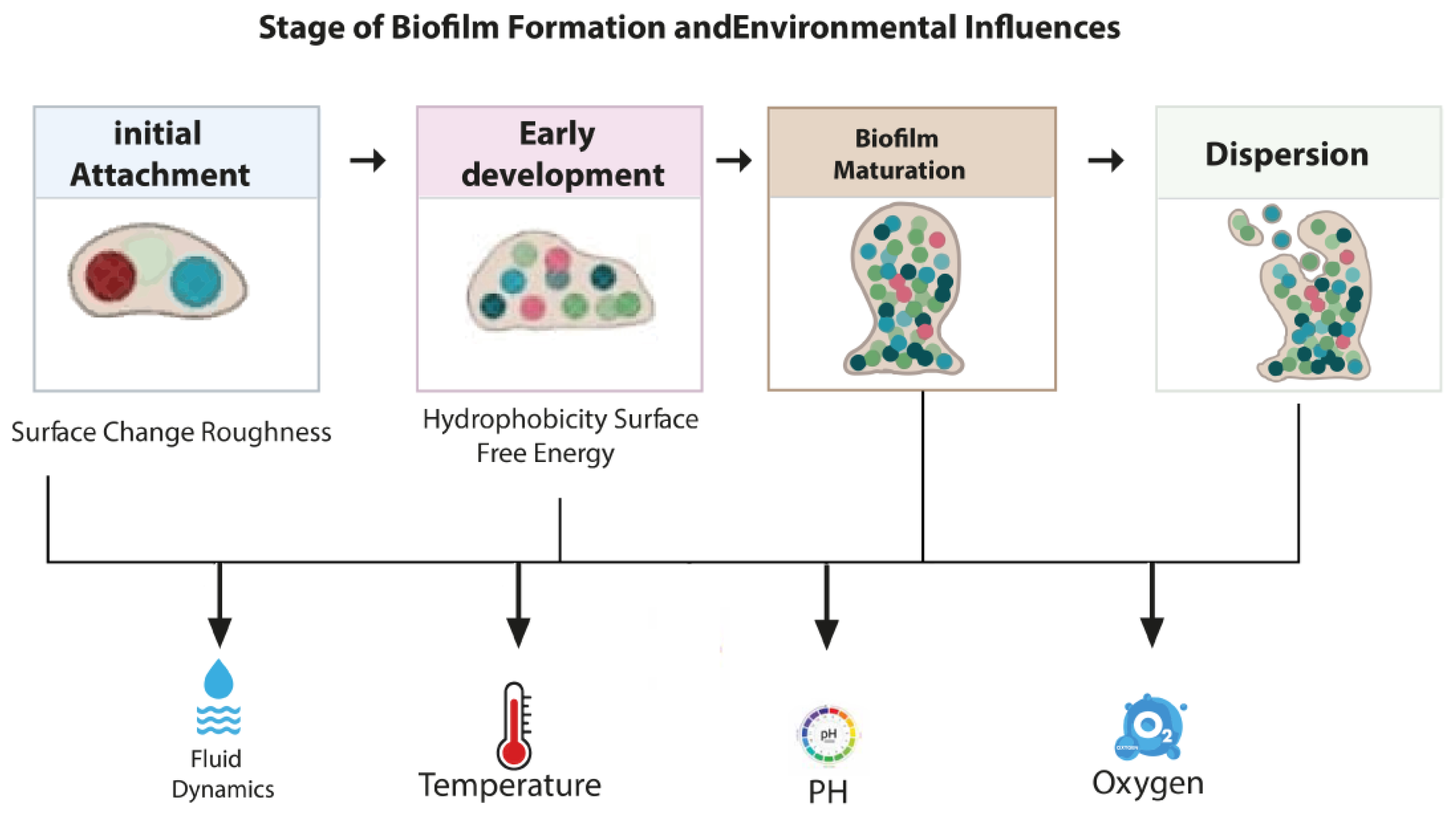
Generally, bacterial pathogens typically go through five primary stages while forming biofilms on any substratum or layer [24,26,27] (Figure 1). (A) Attachment: Initially, weak interactions like acid-base, hydrophobic, and electrostatic forces allow free-swimming planktonic cells to adhere to biotic or abiotic surfaces reversibly, (B) Colonization: Stronger contacts between bacteria and the surface, such as those involving flagella, pili, lipopolysaccharides, and adhesive proteins that bind to collagen, allow bacteria to form permanent attachments, (C) Proliferation: The production of vast quantities of EPS occurs along with the substantial accumulation of multilayered bacterial cells, (D) Maturation: The multilayered bacterial cells that were adhered developed into a mature biofilm with a typical 3D biofilm structure, and (E) Dispersion: After biofilm has fully developed, it is broken down or dispersed via mechanical and active procedures.
A biofilm forms on the surface of urinary catheters in a series of sequential steps. In some cases, the urinary tract can be infected by urease-producing bacterial species. A key example is Proteus mirabilis, which produces urease. These bacteria facilitate the colonization process by adhering to an organic conditioning film that forms on the catheter surface due to the deposition of urinary components, including ions, proteins, and other biomolecules. Once adhered, the bacteria begin to produce an extracellular polysaccharide matrix, leading to the establishment of a mature biofilm community [28].
As the bacterial population within the biofilm expands, the concentration of secreted urease increases. This enzyme hydrolyzes urea into ammonia and carbon dioxide, resulting in a rise in local pH levels in both the biofilm microenvironment and the surrounding urine. The elevated pH promotes the attraction of divalent cations such as calcium and magnesium to the negatively charged gel matrix of the biofilm. These ions subsequently combine with phosphate to form crystalline deposits, primarily struvite (magnesium ammonium phosphate) and apatite (calcium phosphate), which integrate into the biofilm structure and adhere strongly to the catheter surface. This crystallization process contributes significantly to catheter encrustation and blockage [28].
Figure 1. Steps of Biofilm Formation and Environmental Influences [29].
Stresses 05 00058 g001

3.2. Environmental Factors Affecting Biofilm Formation

The initial stages of biofilm formation are strongly influenced by bacterial properties such as surface charge, hydrophobicity, and surface free energy, all of which promote bacterial adhesion. In contrast, long-term biofilm development and stability are primarily affected by environmental factors, including fluid dynamics, temperature, pH, nutrient availability, and oxygen levels. While physicochemical traits play a dominant role during the early attachment phase, environmental conditions become increasingly significant in shaping biofilm structure and behavior over time [30,31].

3.3. Biofilm Architecture (Structure)

In the early days, the formation of biofilms was considered to be moderately homogeneous structures of microcolonies evenly embedded with a gel matrix of EPS. The biofilms, on the other hand, are comparatively flat and smear-like complex morphological formations. The three-dimensional (3D) structure of biofilm images can be seen thanks to the development of sophisticated imaging techniques such as confocal Raman spectroscopy, probe microscopy, epifluorescence microscopy, and confocal laser scanning microscopy (CLSM) [32].
In addition, biofilms have been seen to be more complex and compact structures, from patchy clumps with detailed morphological shapes such as pillars or mushrooms-like structures, and with water channels in between for the exchange of materials with the surroundings and within the biofilms [32].
Based on experimental observations, it can be shown that mature biofilms can exhibit a variety of surface patterns, such as concentric rings, radial ridges, branching, labyrinthine networks, and their combinations with different surface topographies. Furthermore, the biofilm’s physiological state and environmental signals influence the genetic characteristics and the choice of a specific pattern. Within individual microcolonies in this biofilm architecture, mixtures of living and non-living particles are prevalent [33].

3.4. Components in the Biofilm Matrix

A class of microorganisms known as biofilms produces extracellular polymeric substances (EPS), including proteins (<1–2%), polysaccharides (<2%), DNA (<1%), and RNA (<1%). A crucial component of biofilm, water (up to 97%), is also present. It is distributed non-homogeneously and is primarily responsible for the transport of nutrients within the biofilm matrix [27,34,35].
The capability to build and conserve an organized biofilm community mainly depends on EPS matrix components [27]. The EPS in the biofilm matrix commands a charter for the biofilms. The biofilm inhabitants are always shielded from the atmosphere (competitive microbes, temperature, host cells, antimicrobials, and desiccation) while having access to nutrients and the capacity to react to environmental changes. EPS can help the bacteria to adhere to many different surfaces and hosts, protecting the environment and reservoirs for nutrient acquisition [27].

3.5. Role of Quorum Sensing in Biofilm Formation

Quorum sensing (QS) occurs when bacteria communicate with each other and is mediated by diffusible signaling molecules called autoinducers (AIs) [36,37]. Autoinducers, for example, acyl-homoserine lactones (AHLs), auto-inducing oligopeptides (AIPs), are present in Gram-negative and Gram-positive bacteria, respectively. Autoinducer-2 (AI-2) QS is present in both Gram-negative and Gram-positive bacteria [36].
Bacteria use QS to regulate diverse functions, including virulence factor secretion, antibiotic production, and biofilm formation. QS also enables the bacteria to survive in a continuously changing environment and controls the changes required to allow nutrients to enter the cells. These changes usually include the formation of pores, channels, and pillar-like structures. QS also influences biofilm development, as it has a vital role in bacterial accumulation on solid surfaces [36].

3.6. Diseases Related to Biofilm Formation

Biofilms are a central factor in many human infections, which are challenging to treat [14,24]; as such, infections related to the presence of biofilms are considered a significant problem because they counteract the host’s immune defenses [38].
Because biofilms have the potential to be fatal colonizers of biomedical devices, they are to blame for the majority of hospital-acquired illnesses. The colonization of bacteria on the surface of implanted medical equipment, such as respirators, catheters (central venous, urinary), prosthetic heart valves, and orthopedic devices, is, in fact, a major contributor to nosocomial infections [14,39].
Various factors influence biofilm development when microbes contaminate an indwelling medical device. First, the microbes must cling to the device’s exposed surfaces long enough to attach permanently. After these cells form an irreversible bond and create extracellular polysaccharides to form a biofilm, the pace of growth is influenced by flow rate, medium nutrient content, antimicrobial drug concentration, and ambient temperature. Aside from temperature, earlier research has established the effects of nutrient mix and incubation duration [40].
In this context, Awoke et al. [15] reported that 43 (79.7%) bacterial isolates from patients receiving urinary catheters were biofilm formers overall in biofilm generation. 75% and 81% of the isolated Gram-positive and Gram-negative bacteria were biofilm formers.

4. Mechanisms of Catheter-Associated Urinary Tract Infection

UTIs are typically caused by urethral contamination with microbiota, followed by microbial migration to the bladder, adhesion, and colonization. Pili and adhesins then facilitate bladder invasion, and neutrophil infiltration begins. Bacteria grow and build biofilms, while bacterial proteases and toxins cause epithelial injury. These basic phases of infection are the same whether a urinary catheter is present. Urinary catheters directly channel the outside world to the urinary bladder. While this conduit is essential for urine outflow in some people, it also provides a pathway for rectal and periurethral microbes to enter the bladder and establish a foothold for infection [41].
Catheters bypass the urethral sphincters, reduce the turbulence associated with spontaneous voiding, and serve as a nidus for infection, thus increasing the risk for UTI. In addition, catheters may also irritate and traumatize the uroepithelium, thereby disrupting the physiologic mucopolysaccharide coating and rendering it susceptible to bacterial adhesion and entry [41]. The strong immune response to catheterization leads to fibrinogen accumulation on the catheter, thus providing an optimal environment for adherence by uropathogens that express fibrinogen-binding proteins [41].
For example, Enterococcus faecalis can grow in urine or bind to catheter material in vitro. Still, they can grow in fibrinogen-supplemented urine and adhere to a fibrinogen-coated catheter. Adherence is a key initial step in urinary tract infection. In uncomplicated UTIs, bacteria may adhere directly to the uroepithelium of the bladder, allowing them to gain a foothold for infection. However, in a urinary catheter, whether a urethral catheter or suprapubic tube, UTIs may be initiated upon bacterial adherence to the catheter, with subsequent biofilm formation [41].

5. Biofilm Formation in Urinary Catheters and Its Pathogenesis

The urinary catheters are tubular latex or silicone devices, which, when inserted, may readily acquire biofilms on the inner or outer surfaces [2]. It is believed that urinary instrumentation, primarily urinary catheterization, is responsible for nearly 80% of the infections, defined as catheter-associated urinary tract infections (CAUTI) [3,42].
The adhesion of microorganisms to catheter materials mainly depends on the hydrophobicity of the organisms and the catheter surface. Moreover, the attachment and bacterial colonization to the surface of catheters is enhanced by its increased ionic strength due to the presence of divalent cations such as calcium and magnesium in urine, and its relatively acidic pH [2].
One major issue in developing catheter-associated UTI (CA-UTI) is biofilm formation. The urinary catheter microorganisms colonize in the form of a biofilm, which is made up of single- or multi-layered cells encased in an extracellular polymeric material matrix [42].
These microorganisms get into the bladder through the catheter’s mucosal coating. The catheter’s mechanical urethral and bladder mucosa irritation is believed to increase these tissues’ sensitivity to microbial invasion. In patients with indwelling urinary catheters, bacteria can enter the bladder through three different routes: during catheter insertion, migration of bacteria present in the urethra around the catheter, and ascent of bacteria through the catheter’s lumen from a contaminated drainage system [43].
Numerous investigations documented the separation of biofilm bacteria from urinary catheters and urine from patients suffering from UTIs linked with catheter use. Table 1 displays the available research that succeeded in isolating bacterial biofilm. Methods for detecting biofilm-forming bacteria include (A) the Congo Red Agar method (CRAM): a simple qualitative test method, which has the benefit that colonies stay viable on the medium and are quick, sensitive, and repeatable [44], (B) the Tube adherence method (TM): a qualitative assay for the detection of biofilm-producing microorganisms [45], (C) the tissue culture plate method (TCP): regarded as the gold standard for biofilm identification [46], and (D) Fluorescence microscopic examination: Fluorescence-based microscopy offers several advantages over conventional optical and electronic microscopies, owing to sample fixing not being required and the possibility of observing bacterial vitality in live samples [47]. So, regarding the previously mentioned biofilm bacteria laboratory diagnosis, the benefits of these diagnostic methods for infections associated with biofilms are facilitating diagnosis and making patient treatment faster and more effective.

6. Catheter Insertion Techniques and Their Role in Reducing Biofilm Formation

Proper catheter insertion techniques are crucial to reducing the risk of infection and biofilm formation, which can result in catheter-associated bloodstream infections (CA-BSIs). Essential strategies consist of:

6.1. Aseptic Technique

A key component of infection prevention is aseptic insertion. Contamination hazards are significantly decreased by strict attention to procedures such as using sterile gloves, drapes, and equipment, and practicing good hand hygiene. According to studies, following aseptic procedures can lower CA-BSI rates by as much as 60% [52].

6.2. Catheter Design

Silver, chlorhexidine, or minocycline-rifampin are examples of anti-microbial or anti-thrombogenic coatings on catheters that have shown promise in lowering bacterial colonization and biofilm development [53].
We can conclude that lowering biofilm-associated infections requires evidence-based catheter insertion and maintenance procedures. Biofilm-targeted treatments and catheter material advancements have the potential to significantly lower infection risks and enhance patient outcomes.

7. Bacterial Biofilm Mechanism of Antibiotic Resistance

7.1. Reduced Antibiotic Penetration

Biofilms form a protective extracellular matrix that acts as a physical shield, limiting the penetration of antibiotics and other antimicrobial agents. This barrier prevents the drugs from effectively reaching and killing the bacteria embedded within the biofilm, often resulting in chronic and recurring infections despite ongoing antibiotic therapy. Infrared spectroscopy studies have shown that the antibiotic ciprofloxacin diffuses more slowly across biofilm-covered surfaces than sterile ones [54].

7.2. Layered Metabolic Activity in Biofilms

Due to limited nutrient diffusion, biofilms develop gradients in nutrient availability, where the outer layers have better access to nutrients than the inner layers. This results in metabolic stratification, with cells in the inner regions exhibiting reduced metabolic activity or entering a dormant state; these nutrient-deprived, slow-growing cells are less affected by antibiotics that target actively dividing bacteria. Additionally, differences in nutrient concentrations, waste accumulation, and signaling molecules contribute to biofilm heterogeneity [31,55].

7.3. Quorum Sensing and Population Density

Bacteria within biofilms communicate using quorum sensing (QS), which allows them to sense population density and regulate gene expression collectively, including genes related to antibiotic resistance. QS mechanisms trigger the initiation, maturation, and dispersal of biofilms. Bacterial cell density can be inferred by measuring the concentration of signaling molecules known as autoinducers (AIs) [31,56].

7.4. Enhanced Horizontal Gene Transfer

The dense structure of biofilms facilitates horizontal gene transfer (via conjugation, transformation, and transduction), allowing for the efficient exchange of genetic material, including antibiotic resistance genes. This transfer is often mediated through plasmid conjugation and is significantly more effective in biofilms than in free-floating (planktonic) bacterial populations. Additionally, some bacteria can take up extracellular DNA from the biofilm matrix, which supports transformation. The hydrated environment of the matrix creates favorable conditions for genetic exchange. Resistance gene sequences are found to be over 100 times more prevalent in biofilms than in planktonic cells [57].

7.5. Persister Cells

Persister cells are a dormant subpopulation within biofilms that are genetically like active cells but highly tolerant to antibiotics due to their low metabolic activity. Reduced ATP levels in these cells limit the effectiveness of antibiotics targeting active cellular processes [58,59].

7.6. Adaptive Gene Expression

Biofilm-associated stress can alter gene expression, enhancing resistance to antibiotics and biocides. In E. coli, the marRAB operon regulates multidrug resistance by controlling membrane permeability and reducing antibiotic entry. Regulatory genes like oxyR and soxR also trigger stress responses. Mutations occur more frequently in biofilms, and environmental acidification from extracellular DNA leads to membrane changes that block aminoglycoside entry and protect against antimicrobial peptides [31].

7.7. Efflux Pumps

Efflux pumps are transport proteins that expel antibiotics and toxins from bacterial cells, reducing intracellular drug concentrations. Genes encoding pumps are found in both chromosomal and plasmid DNA; they also regulate membrane permeability by controlling porin production and limiting the uptake of both hydrophilic and lipophilic substances [56,60,61].

8. Factors Affecting the Biofilm Development of Catheter-Associated Urinary Tract Infections

8.1. Gender and Age

The development of biofilm is a precursor to a catheter-associated urinary tract infection and is associated with many risk factors, including gender and age [49]. A study carried out in Egypt by Ramadan et al. [50] reported that the extremes of age in CAUTI patients were significantly associated with biofilm formation; the same results were reported in Indonesia [62].
A study conducted in Indonesia characterized risk factors for catheter-associated biofilm formation. The researchers found that female gender was a significant predictor for both the frequency and severity of biofilms, evidenced by a higher rate of positive biomass (p ≤ 0.001). Conversely, age did not show a significant association with biofilm formation (p = 0.151), although a numerically higher rate was observed in the elderly population [49].

8.2. Catheter Types

All types of catheters are prone to CAUTIs and biofilm development. The material of the urinary catheter plays a crucial part in the development of biofilms and the severity of infection [50].
Many studies have reported a relationship between the type of catheter and biofilm production in CAUTIs. In a survey about bacterial biofilm-dependent catheter-associated urinary tract infections: Characterization, antibiotic resistance pattern, and risk factors conducted by Ramadan et al. [50] reported in their study that the latex catheter showed a higher rate of biofilm production than silicon catheters.

8.3. Duration of Catheterization

Long-term catheterization has an important role and is a primary risk factor for developing CAUTI and biofilm formation. This might be explained by a daily risk of bacterial colonization that is higher, and a catheterization period that is longer [49,63].
Oumer et al. [3] reported in their study that the long duration of the catheter has been considered a risk factor for catheter-associated urinary tract infections. Another study concluded that the duration of catheterization has been a significant risk factor for biofilm formation [49].
Regarding catheter exchange timing, the duration of catheterization plays an essential role in CAUTI and biofilm formation; therefore, perhaps the first solution to reduce biofilm formation is to reduce the duration of the catheter and exchange it more frequently [40].
The catheter should be removed if no longer needed, or exchanged, before initiating antibiotic therapy (a urine culture must also be obtained before the initiation of antibiotics, as discussed in the diagnostic section), particularly if it has been in place for longer than two weeks [40]

9. Treatments and Challenges in the Future

Biofilms can become resistant to host defenses, including macrophage phagocytosis, and can develop antibiotic tolerance due to phenotypic changes or mutations, reducing their treatability.
In addition, for enhancing sterile practices, current commercialized CAUTI prevention options include coating with antibacterial substances like antibiotics, silver, or antiadhesive substances like hydrogels and polytetrafluoroethylene (PTFE) [64]. Numerous investigations have revealed that nanoparticles have a broad range of antibacterial activity against Gram-positive and Gram-negative bacteria [13].
Due to the antibacterial properties of metals or surface-tailored nanocarriers, which enable targeted medication delivery to desired locations of action and inhibit the development of cytotoxic processes in healthy cells, blocking the formation of biofilms and overcoming multidrug resistance, nanotechnology has brought about tremendous advancements in the treatment of urinary tract infections (UTIs) [65]. Therefore, most of the research focuses on coating urinary catheter surfaces with nanoparticles, which is thought to be an excellent way to lower CAUTIs.
Silver nanoparticles can destroy bacteria on their own and release silver ions. After adhering to the cell surface, silver nanoparticles can gather in the pits that develop on the cell wall. Denaturation of the cell membrane might result from the accumulated silver nanoparticles. Because of their nanoscale size, silver nanoparticles can also pass through bacterial cell walls and alter the structure of the cell membrane. The cytoplasmic membrane’s denaturation can potentially tear organelles and cause cell lysis. Furthermore, the transmission of bacterial signals may be facilitated by silver nanoparticles. Bacterial signal transduction is affected by the phosphorylation of protein substrates, and nanoparticles can dephosphorylate tyrosine residues on the peptide substrates. Disruption of the signal transduction can lead to cell apoptosis and termination of cell multiplication [66,67].

10. Conclusions

CAUTIs are the most critical healthcare-associated infection, and the prevalence of antibiotic resistance is concerningly high, especially since the bacteria associated with it can form biofilm. Many factors affect the biofilm development by CAUTIs and play an essential role in pathogenesis, such as gender, age, type, and duration of the catheter. Because there is currently not enough of an antibiotic pipeline to keep up with the rise in resistance resulting from biofilm, many studies have used nanoparticles to attempt treatment and eliminate biofilm bacteria. In the future, scientists need to investigate novel strategies that mainly stop bacteria from adhering to the catheter’s surface, since the physicochemistry or surface topography of the catheter might be changed to do this, or try to coat the catheter with materials that have no effect on human health and prevent the biofilm bacteria adhesion mechanism.

Author Contributions

Conceptualization, T.A.A.M. and N.L.J.; resources, R.M.H. and N.L.J.; writing—original draft preparation, R.M.H. and N.L.J.; writing—review and editing, T.A.A.M. and M.S.K. All authors have read and agreed to the published version of the manuscript.

Funding

This research received no external funding.

Conflicts of Interest

The authors declare no conflicts of interest.

References

  1. Wilks, S.A.; Koerfer, V.V.; Prieto, J.A.; Fader, M.; Keevil, C.W. Biofilm Development on Urinary Catheters Promotes the Appearance of Viable but Nonculturable Bacteria. MBio 2021, 12, 10–1128. [Google Scholar] [CrossRef]
  2. Goda, R.M.; El-Baz, A.M.; Khalaf, E.M.; Alharbi, N.K.; Elkhooly, T.A.; Shohayeb, M.M. Combating Bacterial Biofilm Formation in Urinary Catheter by Green Silver Nanoparticle. Antibiotics 2022, 11, 495. [Google Scholar] [CrossRef]
  3. Oumer, Y.; Regasa Dadi, B.; Seid, M.; Biresaw, G.; Manilal, A. Catheter-Associated Urinary Tract Infection: Incidence, Associated Factors and Drug Resistance Patterns of Bacterial Isolates in Southern Ethiopia. Infect. Drug Resist. 2021, 14, 2883–2894. [Google Scholar] [CrossRef] [PubMed]
  4. Vivehananthan, K.; Thevashayinath, S.; Abeygunawardena, I. Extraction and Screening of Biofilm Producing Bacterial Isolated in Short and Long-Term Catheters. Adv. Technol. 2021, 1, 329–345. [Google Scholar] [CrossRef]
  5. Verma, A.; Bhani, D.; Tomar, V.; Bachhiwal, R.; Yadav, S. Differences in Bacterial Colonization and Biofilm Formation Property of Uropathogens between the Two Most Commonly Used Indwelling Urinary Catheters. J. Clin. Diagn. Res. JCDR 2016, 10, PC01. [Google Scholar] [CrossRef] [PubMed]
  6. Vestby, L.K.; Grønseth, T.; Simm, R.; Nesse, L.L. Bacterial Biofilm and Its Role in the Pathogenesis of Disease. Antibiotics 2020, 9, 59. [Google Scholar] [CrossRef]
  7. Al-Wrafy, F.A.; Al-Gheethi, A.A.; Ponnusamy, S.K.; Noman, E.A.; Fattah, S.A. Nanoparticles Approach to Eradicate Bacterial Biofilm-Related Infections: A Critical Review. Chemosphere 2022, 288, 132603. [Google Scholar] [CrossRef]
  8. Flemming, H.C. EPS—Then and Now. Microorganisms 2016, 4, 41. [Google Scholar] [CrossRef]
  9. Penesyan, A.; Paulsen, I.T.; Kjelleberg, S.; Gillings, M.R. Three Faces of Biofilms: A Microbial Lifestyle, a Nascent Multicellular Organism, and an Incubator for Diversity. npj Biofilms Microbiomes 2021, 7, 80. [Google Scholar] [CrossRef]
  10. Ramírez-Larrota, J.S.; Eckhard, U. An Introduction to Bacterial Biofilms and Their Proteases, and Their Roles in Host Infection and Immune Evasion. Biomolecules 2022, 12, 306. [Google Scholar] [CrossRef]
  11. Werneburg, G.T.; Nguyen, A.; Henderson, N.S.; Rackley, R.R.; Shoskes, D.A.; Le Sueur, A.L.; Corcoran, A.T.; Katz, A.E.; Kim, J.; Rohan, A.J. The Natural History and Composition of Urinary Catheter Biofilms: Early Uropathogen Colonization with Intraluminal and Distal Predominance. J. Urol. 2020, 203, 357–364. [Google Scholar] [CrossRef]
  12. El-sayed, H.; Fahmy, Y. Correlation between Biofilm Formation and Multidrug Resistance in Clinical Isolates of Pseudomonas Aeruginosa. Microbes Infect. Dis. 2021, 2, 541–549. [Google Scholar] [CrossRef]
  13. Filipović, N.; Ušjak, D.; Milenković, M.T.; Zheng, K.; Liverani, L.; Boccaccini, A.R.; Stevanović, M.M. Comparative Study of the Antimicrobial Activity of Selenium Nanoparticles with Different Surface Chemistry and Structure. Front. Bioeng. Biotechnol. 2021, 8, 624621. [Google Scholar] [CrossRef]
  14. Schulze, A.; Mitterer, F.; Pombo, J.P.; Schild, S. Biofilms by Bacterial Human Pathogens: Clinical Relevance-Development, Composition and Regulation-Therapeutical Strategies. Microb. Cell 2021, 8, 28. [Google Scholar] [CrossRef] [PubMed]
  15. Awoke, N.; Kassa, T.; Teshager, L. Magnitude of Biofilm Formation and Antimicrobial Resistance Pattern of Bacteria Isolated from Urinary Catheterized Inpatients of Jimma University Medical Center, Southwest Ethiopia. Int. J. Microbiol. 2019, 2019, 5729568. [Google Scholar] [CrossRef] [PubMed]
  16. Nouri, S.; Sharif, M.R.; Hosseinpour, M.; Farokhi, S.; Sharif, M.H. A Comparison between Foley and Nelatone Urinary Catheters in Causing Urinary Tract Infection in Animal Models. Nurs. Midwifery Stud. 2015, 4, e24363. [Google Scholar] [CrossRef] [PubMed]
  17. Gayani, B.; Dilhari, A.; Kottegoda, N.; Ratnaweera, D.R.; Weerasekera, M.M. Reduced Crystalline Biofilm Formation on Superhydrophobic Silicone Urinary Catheter Materials. ACS Omega 2021, 6, 11488–11496. [Google Scholar] [CrossRef]
  18. Singha, P.; Locklin, J.; Handa, H. A Review of the Recent Advances in Antimicrobial Coatings for Urinary Catheters. Acta Biomater. 2017, 50, 20–40. [Google Scholar] [CrossRef]
  19. Pelling, H.; Nzakizwanayo, J.; Milo, S.; Denham, E.L.; MacFarlane, W.M.; Bock, L.J.; Sutton, J.M.; Jones, B.V. Bacterial Biofilm Formation on Indwelling Urethral Catheters. Lett. Appl. Microbiol. 2019, 68, 277–293. [Google Scholar] [CrossRef]
  20. Dias, L.D.; Duarte, L.S.; Naves, P.L.F.; Napolitano, H.B.; Bagnato, V.S. Self-Disinfecting Urethral Catheter to Overcome Urinary Infections: From Antimicrobial Photodynamic Action to Antibacterial Biochemical Entities. Microorganisms 2022, 10, 2484. [Google Scholar] [CrossRef]
  21. Murphy, C. Innovating Urinary Catheter Design: An Introduction to the Engineering Challenge. Proc. Inst. Mech. Eng. Part H. J. Eng. Med. 2019, 233, 48–57. [Google Scholar] [CrossRef] [PubMed]
  22. Zhang, L.; Cao, Z.; Bai, T.; Carr, L.; Ella-Menye, J.-R.; Irvin, C.; Ratner, B.D.; Jiang, S. Zwitterionic Hydrogels Implanted in Mice Resist the Foreign-Body Reaction. Nat. Biotechnol. 2013, 31, 553–556. [Google Scholar] [CrossRef]
  23. Bryers, J.D. Medical Biofilms. Biotechnol. Bioeng. 2008, 100, 1–18. [Google Scholar] [CrossRef] [PubMed]
  24. Berhe, N.; Tefera, Y.; Tintagu, T. Review on Biofilm Formation and Its Control Options. Int. J. Adv. Res. Biol. Sci. 2017, 8, 122–133. [Google Scholar]
  25. Mirghani, R.; Saba, T.; Khaliq, H.; Mitchell, J.; Do, L.; Chambi, L.; Diaz, K.; Kennedy, T.; Alkassab, K.; Huynh, T. Biofilms: Formation, Drug Resistance and Alternatives to Conventional Approaches. AIMS Microbiol. 2022, 8, 239. [Google Scholar] [CrossRef]
  26. Dewasthale, S.; Mani, I.; Vasdev, K. Microbial Biofilm: Current Challenges in Health Care Industry. J. Appl. Biotechnol. Bioeng. 2018, 5, 160–164. [Google Scholar] [CrossRef]
  27. Srinivasan, R.; Santhakumari, S.; Poonguzhali, P.; Geetha, M.; Dyavaiah, M.; Xiangmin, L. Bacterial Biofilm Inhibition: A Focused Review on Recent Therapeutic Strategies for Combating the Biofilm Mediated Infections. Front. Microbiol. 2021, 12, 676458. [Google Scholar] [CrossRef]
  28. Cortese, Y.J.; Wagner, V.E.; Tierney, M.; Devine, D.; Fogarty, A. Review of Catheter-associated Urinary Tract Infections and in Vitro Urinary Tract Models. J. Healthc. Eng. 2018, 2018, 2986742. [Google Scholar] [CrossRef]
  29. Karaguler, T.; Kahraman, H.; Tuter, M. Analyzing Effects of ELF Electromagnetic Fields on Removing Bacterial Biofilm. Biocybern. Biomed. Eng. 2017, 37, 336–340. [Google Scholar] [CrossRef]
  30. Xu, X.; Wang, S.; Gao, F.; Li, J.; Zheng, L.; Sun, C.; He, C.; Wang, Z.; Qu, L. Marine Microplastic-Associated Bacterial Community Succession in Response to Geography, Exposure Time, and Plastic Type in China’s Coastal Seawaters. Mar. Pollut. Bull. 2019, 145, 278–286. [Google Scholar] [CrossRef]
  31. Azeem, K.; Fatima, S.; Ali, A.; Ubaid, A.; Husain, F.M.; Abid, M. Biochemistry of Bacterial Biofilm: Insights into Antibiotic Resistance Mechanisms and Therapeutic Intervention. Life 2025, 15, 49. [Google Scholar] [CrossRef]
  32. Sharma, S.; Mohler, J.; Mahajan, S.D.; Schwartz, S.A.; Bruggemann, L.; Aalinkeel, R. Microbial Biofilm: A Review on Formation, Infection, Antibiotic Resistance, Control Measures, and Innovative Treatment. Microorganisms 2023, 11, 1614, Erratum in Microorganisms 2024, 12, 1961. [Google Scholar] [CrossRef]
  33. Silva, V.; Pereira, J.E.; Maltez, L.; Poeta, P.; Igrejas, G. Influence of Environmental Factors on Biofilm Formation of Staphylococci Isolated from Wastewater and Surface Water. Pathogens 2022, 11, 1069. [Google Scholar] [CrossRef] [PubMed]
  34. Jamal, M.; Ahmad, W.; Andleeb, S.; Jalil, F.; Imran, M.; Nawaz, M.A.; Hussain, T.; Ali, M.; Rafiq, M.; Kamil, M.A. Bacterial Biofilm and Associated Infections. J. Chin. Med. Assoc. 2018, 81, 7–11. [Google Scholar] [CrossRef] [PubMed]
  35. Amankwah, S.; Abdella, K.; Kassa, T. Bacterial Biofilm Destruction: A Focused Review on the Recent Use of Phage-Based Strategies with Other Antibiofilm Agents. Nanotechnol. Sci. Appl. 2021, 14, 161–177. [Google Scholar] [CrossRef] [PubMed]
  36. Zhou, L.; Zhang, Y.; Ge, Y.; Zhu, X.; Pan, J. Regulatory Mechanisms and Promising Applications of Quorum Sensing-Inhibiting Agents in Control of Bacterial Biofilm Formation. Front. Microbiol. 2020, 11, 589640. [Google Scholar] [CrossRef]
  37. Sahreen, S.; Mukhtar, H.; Imre, K.; Morar, A.; Herman, V.; Sharif, S. Exploring the Function of Quorum Sensing Regulated Biofilms in Biological Wastewater Treatment: A Review. Int. J. Mol. Sci. 2022, 23, 9751. [Google Scholar] [CrossRef]
  38. Jabir, D.M. Evaluation of the Ability of Some Bacterial Species Isolated from UTI to Form Biofilm. Al-Qadisiyah J. Pure Sci. 2022, 27, 8–14. [Google Scholar] [CrossRef]
  39. Murugan, K.; Selvanayaki, K.; Al-Sohaibani, S. Urinary Catheter Indwelling Clinical Pathogen Biofilm Formation, Exopolysaccharide Characterization and Their Growth Influencing Parameters. Saudi J. Biol. Sci. 2016, 23, 150–159. [Google Scholar] [CrossRef]
  40. Patel, F.M.; Goswami, P.N.; Khara, R. Detection of Biofilm Formation in Device Associated Clinical Bacterial Isolates in Cancer Patients. Sri Lankan J. Infect. Dis. 2016, 6, 43–50. [Google Scholar] [CrossRef]
  41. Werneburg, G.T. Catheter-Associated Urinary Tract Infections: Current Challenges and Future Prospects. Res. Rep. Urol. 2022, 14, 109–133. [Google Scholar] [CrossRef] [PubMed]
  42. Mota, É.C.; Oliveira, A.C. Catheter-Associated Urinary Tract Infection: Why Do Not We Control This Adverse Event? Rev. Da Esc. Enferm. Da USP 2019, 53, e03452. [Google Scholar] [CrossRef] [PubMed]
  43. Tiwari, A.A.; Ghnawate, N. Detection of Biofilm Forming Bacterial Communities from Urinary Catheter of Patients with Change in Its Antibiotic Susceptibility Pattern and Triclosan Effect from Different Hospitals of Amravati City Maharashtra, India. Open J. Med. Microbiol. 2017, 7, 51–66. [Google Scholar] [CrossRef]
  44. Freeman, D.J.; Falkiner, F.R.; Keane, C.T. New Method for Detecting Slime Production by Coagulase Negative Staphylococci. J. Clin. Pathol. 1989, 42, 872–874. [Google Scholar] [CrossRef]
  45. Sultan, A.M.; Nabiel, Y. Tube Method and Congo Red Agar versus Tissue Culture Plate Method for Detection of Biofilm Production by Uropathogens Isolated from Midstream Urine: Which One Could Be Better? Afr. J. Clin. Exp. Microbiol. 2019, 20, 60–66. [Google Scholar] [CrossRef]
  46. Basnet, A.; Tamang, B.; Shrestha, M.R.; Shrestha, L.B.; Rai, J.R.; Maharjan, R.; Dahal, S.; Shrestha, P.; Rai, S.K. Assessment of Four in Vitro Phenotypic Biofilm Detection Methods in Relation to Antimicrobial Resistance in Aerobic Clinical Bacterial Isolates. PLoS ONE 2023, 18, e0294646. [Google Scholar] [CrossRef]
  47. Mori, S.; Yamada, A.; Kawai, K. Evaluation of the Biofilm Detection Capacity of the Congo Red Agar Method for Bovine Mastitis-Causing Bacteria. Jpn. J. Vet. Res. 2024, 71, 109–116. [Google Scholar]
  48. Navarro, S.; Sherman, E.; Colmer-Hamood, J.A.; Nelius, T.; Myntti, M.; Hamood, A.N. Urinary Catheters Coated with a Novel Biofilm Preventative Agent Inhibit Biofilm Development by Diverse Bacterial Uropathogens. Antibiotics 2022, 11, 1514. [Google Scholar] [CrossRef]
  49. Gunardi, W.D.; Karuniawati, A.; Umbas, R.; Bardosono, S.; Lydia, A.; Soebandrio, A.; Safari, D. Biofilm-producing Bacteria and Risk Factors (Gender and Duration of Catheterization) Characterized as Catheter-associated Biofilm Formation. Int. J. Microbiol. 2021, 2021, 8869275. [Google Scholar] [CrossRef]
  50. Ramadan, R.; Omar, N.; Dawaba, M.; Moemen, D. Bacterial Biofilm Dependent Catheter Associated Urinary Tract Infections: Characterization, Antibiotic Resistance Pattern and Risk Factors. Egypt. J. Basic. Appl. Sci. 2021, 8, 64–74. [Google Scholar] [CrossRef]
  51. Zarmouh, H.M.; Karayem, K.J.; Swieb, S.M.; Alhadad, O.S.; Rhouma, N.R. Biofilm Formation of Pathogenic Bacterial Species Isolated From Urinary Catheters. J. Med. Sci. 2021, 16, 16–19. [Google Scholar]
  52. Marschall, J.; Mermel, L.A.; Fakih, M.; Hadaway, L.; Kallen, A.; O’Grady, N.P.; Pettis, A.M.; Rupp, M.E.; Sandora, T.; Maragakis, L.L. Strategies to Prevent Central Line-Associated Bloodstream Infections in Acute Care Hospitals: 2014 Update. Infect. Control Hosp. Epidemiol. 2014, 35, S89–S107. [Google Scholar] [CrossRef]
  53. Haque, M.; Sartelli, M.; McKimm, J.; Bakar, M.A. Health Care-Associated Infections–an Overview. Infect. Drug Resist. 2018, 11, 2321–2333. [Google Scholar] [CrossRef]
  54. Uruén, C.; Chopo-Escuin, G.; Tommassen, J.; Mainar-Jaime, R.C.; Arenas, J. Biofilms as Promoters of Bacterial Antibiotic Resistance and Tolerance. Antibiotics 2020, 10, 3. [Google Scholar] [CrossRef] [PubMed]
  55. Jo, J.; Price-Whelan, A.; Dietrich, L.E.P. Gradients and Consequences of Heterogeneity in Biofilms. Nat. Rev. Microbiol. 2022, 20, 593–607. [Google Scholar] [CrossRef] [PubMed]
  56. AlQurashi, D.M.; AlQurashi, T.F.; Alam, R.I.; Shaikh, S.; Tarkistani, M.A.M. Advanced Nanoparticles in Combating Antibiotic Resistance: Current Innovations and Future Directions. J. Nanotheranostics 2025, 6, 9. [Google Scholar] [CrossRef]
  57. Michaelis, C.; Grohmann, E. Horizontal Gene Transfer of Antibiotic Resistance Genes in Biofilms. Antibiotics 2023, 12, 328. [Google Scholar] [CrossRef]
  58. Shan, Y.; Brown Gandt, A.; Rowe, S.E.; Deisinger, J.P.; Conlon, B.P.; Lewis, K. ATP-Dependent Persister Formation in Escherichia coli. MBio 2017, 8, 10–1128. [Google Scholar] [CrossRef]
  59. Niu, H.; Gu, J.; Zhang, Y. Bacterial Persisters: Molecular Mechanisms and Therapeutic Development. Signal Transduct. Target. Ther. 2024, 9, 174. [Google Scholar] [CrossRef]
  60. Hajiagha, M.N.; Kafil, H.S. Efflux Pumps and Microbial Biofilm Formation. Infect. Genet. Evol. 2023, 112, 105459. [Google Scholar] [CrossRef]
  61. Gaurav, A.; Bakht, P.; Saini, M.; Pandey, S.; Pathania, R. Role of Bacterial Efflux Pumps in Antibiotic Resistance, Virulence, and Strategies to Discover Novel Efflux Pump Inhibitors. Microbiology 2023, 169, 1333. [Google Scholar] [CrossRef]
  62. Anggi, A.; Wijaya, D.W.; Ramayani, O.R. Risk Factors for Catheter-Associated Urinary Tract Infection and Uropathogen Bacterial Profile in the Intensive Care Unit in Hospitals in Medan, Indonesia. Open Access Maced. J. Med. Sci. 2019, 7, 3488. [Google Scholar] [CrossRef]
  63. Weiner, L.M.; Webb, A.K.; Limbago, B.; Dudeck, M.A.; Patel, J.; Kallen, A.J.; Edwards, J.R.; Sievert, D.M. Antimicrobial-Resistant Pathogens Associated with Healthcare-Associated Infections: Summary of Data Reported to the National Healthcare Safety Network at the Centers for Disease Control and Prevention, 2011–2014. Infect. Control Hosp. Epidemiol. 2016, 37, 1288–1301. [Google Scholar] [CrossRef]
  64. Zhang, S.; Wang, L.; Liang, X.; Vorstius, J.; Keatch, R.; Corner, G.; Nabi, G.; Davidson, F.; Gadd, G.M.; Zhao, Q. Enhanced Antibacterial and Antiadhesive Activities of Silver-PTFE Nanocomposite Coating for Urinary Catheters. ACS Biomater. Sci. Eng. 2019, 5, 2804–2814. [Google Scholar] [CrossRef]
  65. Qindeel, M.; Barani, M.; Rahdar, A.; Arshad, R.; Cucchiarini, M. Nanomaterials for the Diagnosis and Treatment of Urinary Tract Infections. Nanomaterials 2021, 11, 546. [Google Scholar] [CrossRef] [PubMed]
  66. Yin, I.X.; Zhang, J.; Zhao, I.S.; Mei, M.L.; Li, Q.; Chu, C.H. The Antibacterial Mechanism of Silver Nanoparticles and Its Application in Dentistry. Int. J. Nanomed. 2020, 15, 2555–2562. [Google Scholar] [CrossRef] [PubMed]
  67. Liao, C.; Li, Y.; Tjong, S.C. Bactericidal and Cytotoxic Properties of Silver Nanoparticles. Int. J. Mol. Sci. 2019, 20, 449. [Google Scholar] [CrossRef]
Table 1. Some examples of bacteria forming biofilm from urinary catheters and urine of patients with CAUTI.
Biofilm BacteriaSource of SampleReferences
Escherichia coli, Enterobacter cloacae, Pseudomonas aeruginosa, Citrobacter Proteus, Staphylococcus aureusurine samples[3,39,48]
E. coli, E. faecalis, Klebsiella pneumoniae and P. aeruginosaCatheter and Urine samples[49]
K. pneumoniae, E. coli, S. aureus, S. epidermidis, E. faecalis, Streptococcus spp., S. epidermidis, P. aeruginosa, and AcinetobacterCatheter[43,50,51]
Disclaimer/Publisher’s Note: The statements, opinions and data contained in all publications are solely those of the individual author(s) and contributor(s) and not of MDPI and/or the editor(s). MDPI and/or the editor(s) disclaim responsibility for any injury to people or property resulting from any ideas, methods, instructions or products referred to in the content.