Review

Exploring Stressors: Impact on Cellular Organelles and Implications for Cellular Functions

by
Zoofa Zayani
1,
Arash Matinahmadi
2,
Alireza Tavakolpournegari
3 and
Seyed Hesamoddin Bidooki
4,*
1
Centre for Modern Interdisciplinary Technologies, Nicolaus Copernicus University, 87-100 Torun, Poland
2
Department of Cellular and Molecular Biology, Nicolaus Copernicus University, 87-100 Torun, Poland
3
Department of Toxicology and Cancer Biology, University of Kentucky College of Medicine, Lexington, KY 40536, USA
4
Department of Biochemistry and Molecular and Cellular Biology, Faculty of Veterinary Medicine, Health Research Institute of Aragon, University of Zaragoza, 50013 Zaragoza, Spain
*
Author to whom correspondence should be addressed.
Stresses 2025, 5(2), 26; https://doi.org/10.3390/stresses5020026
Submission received: 25 February 2025 / Revised: 28 March 2025 / Accepted: 2 April 2025 / Published: 4 April 2025
(This article belongs to the Collection Feature Papers in Human and Animal Stresses)

Abstract

:
Cellular stressors have been demonstrated to exert a substantial influence on the functionality of organelles, thereby impacting cellular homeostasis and contributing to the development of disease pathogenesis. This review aims to examine the impact of diverse stressors, including environmental, chemical, biological, and physical factors, on critical organelles such as the cell membrane, mitochondria, endoplasmic reticulum, Golgi apparatus, lysosomes, and membrane-less organelles. The intricate molecular mechanisms underlying cellular stress responses, encompassing oxidative stress, protein misfolding, and metabolic reprogramming, have the capacity to elicit adaptive responses or culminate in pathological conditions. The interplay between these stressors and organelle dysfunction has been implicated in a myriad of diseases, including neurodegenerative disorders, cancer, metabolic disorders, and immune-related pathologies. A comprehensive understanding of the mechanisms by which organelles respond to stress can offer valuable insights into the development of therapeutic strategies aimed at mitigating cellular damage.

1. Introduction

The cellular stress refers to a condition wherein cells experience adverse effects due to various internal and external factors, disrupting their normal functioning and potentially leading to cell damage or death [1]. However, cells have evolved sophisticated mechanisms to adapt to such disturbances. One fundamental process is hormesis, where exposure to low levels of stressors induces beneficial responses that enhance cellular resilience and function. In contrast, high levels of stress can overwhelm these adaptive mechanisms, leading to cellular dysfunction and disease [2]. The impact of cellular stress extends beyond individual cells, influencing the overall function of tissues and organs [1,3]. Cellular stressors can be broadly categorized into four main types: environmental, chemical, biological, and physical stressors [4]. These stressors encompass a diverse range of challenges to cellular function. For instance, environmental stressors include temperature extremes, such as heat waves that denature proteins or cold snaps that damage cell membranes, as well as ultraviolet (UV) radiation from sunlight that causes DNA damage and heavy metals like lead or mercury that induce oxidative stress [5,6,7]. Chemical stressors involve exposure to substances such as pesticides (e.g., organophosphates like chlorpyrifos, which disrupt neural signaling) and industrial pollutants (e.g., benzene, which impairs metabolic pathways), alongside nutritional imbalances like excessive sugar intake leading to metabolic stress [8,9,10]. Biological stressors include pathogens, such as the SARS-CoV-2 virus that hijack cellular machinery or Escherichia coli bacteria that trigger immune responses, and conditions like glucose starvation that disrupt energy metabolism. Physical stressors, meanwhile, comprise mechanical forces, such as shear stress from blood flow affecting vascular endothelial cells, and osmotic pressure changes, such as those experienced by kidney cells in varying solute concentrations [11,12,13,14]. Cellular stress triggers a variety of responses in different organelles, which play critical roles in maintaining cellular homeostasis and managing the consequences of stress [15]. Key organelles involved in cellular stress responses include the cell membrane, mitochondria, endoplasmic reticulum (ER), Golgi apparatus, lysosomes, and membrane-less organelles [15,16]. Under normal conditions, these organelles perform essential roles in cellular homeostasis. The cell membrane acts as a selective barrier, regulating the transport of substances and facilitating cell signaling [17]. Mitochondria serve as the cell’s powerhouses, generating ATP through energy metabolism [18]. The ER is responsible for protein synthesis, folding, and lipid production, while the Golgi apparatus modifies, sorts, and packages proteins and lipids for transport [19]. Lysosomes function as recycling centers, degrading waste materials with hydrolytic enzymes [20]. Membrane-less organelles, such as stress granules, dynamically regulate RNA metabolism and stress adaptation [21]. This baseline physiology underpins their responses to stressors, which can disrupt these functions and trigger adaptive or pathological outcomes.
Prolonged exposure to various stressors can lead to detrimental effects on cellular function and metabolism, contributing to the onset of chronic diseases and neurodegenerative conditions [1,3,22]. Stress-induced protein modifications can disrupt normal cellular functions and contribute to the pathogenesis of conditions like amyotrophic lateral sclerosis and multiple sclerosis [1]. Moreover, oxidative stress can impair synaptic transmission and lead to neuroinflammation, further exacerbating cognitive decline and mood disorders [23,24]. Chronic stress has been implicated in the development of a variety of chronic diseases, such as cancer, where oxidative DNA damage plays a critical role in carcinogenesis [23]. Cancer cells experience stress from both intrinsic and extrinsic sources. Intrinsic factors include oncogenic stress, insufficient nutrient and oxygen supply, DNA damage, and ER stress caused by the accumulation of mutant proteins [25]. Extrinsic factors often involve therapeutic interventions, such as chemotherapy, which exacerbate cellular stress within the tumor environment [26]. These stress responses can also influence the tumor microenvironment, affecting immune responses and inflammation, making them a focal point for novel anti-cancer strategies [25]. The ability of chronic stress to induce apoptosis resistance in cancer cells further complicates therapeutic interventions, necessitating a deeper understanding of the underlying mechanisms to improve treatment outcomes [27]. This review summarizes the effects of cellular stressors and their associated molecular pathways across multiple cellular organelles, and we conclude by emphasizing potential areas for future research.

2. Cellular Stressors

2.1. Environmental Stressors

Environmental stressors are among the most prevalent causes of cellular stress. For instance, heat stress may lead to protein denaturation and disrupt cellular processes, while extreme cold can damage cell membranes and impair metabolic functions [1,3]. Moreover, exposure to ultraviolet (UV) radiation can cause DNA damage, potentially resulting in mutations and cell death [1]. Additionally, the pH of the surrounding environment is a critical factor that impacts cellular metabolism. Most mammalian cells thrive at a neutral pH (around 7), and deviations from this optimal range can negatively affect growth rates and product synthesis [28]. Environmental pollutants, including heavy metals and particulate matter, can induce oxidative stress and inflammation within cells, leading to various adverse health outcomes. For instance, heavy metals such as lead and mercury are known to disrupt normal cellular functions by altering enzymatic activity and damaging DNA, proteins, and lipids [29,30]. Over and above that, microplastics and nanoplastics interact with human cells through various mechanisms, leading to potential cellular stress and damage. These interactions can occur through passive transportation and active endocytosis, resulting in disruptions to the plasma membrane and other cellular structures [31,32,33,34]. Upon internalization, these particles can be entrapped in early endosomes and transported to lysosomes, where they may interact with hydrolytic enzymes and affect cellular metabolism [32]. These stressors trigger various cellular responses to repair damage and maintain cellular integrity [33,34,35].

2.2. Chemical Stressors

Chemical stressors primarily involve exposure to harmful substances that disrupt cellular processes [1,36]. Agricultural chemicals, such as pesticides and herbicides, pose a risk to cellular health as they often contain toxic compounds that can affect metabolic pathways. Exposure to these chemicals can activate stress response mechanisms, leading to alterations in gene expression and activation of detoxification processes [29,37]. Industrial chemicals, including solvents and plasticizers can leach into the environment and subsequently enter biological systems, where they may cause cellular dysfunction. The mechanisms of action often involve the activation of stress proteins that signal damage, resulting in impaired cellular processes and increased vulnerability to other stressors [38,39]. Nutritional chemical stressors can also act as chemical stressors. For example, high levels of saturated fats and sugars can lead to metabolic disorders by inducing cellular stress in adipose tissues and pancreatic β-cells, resulting in conditions such as insulin resistance and type 2 diabetes [37,39].

2.3. Biological Stressors

Biological stressors factors can trigger immune responses that can further stress the host cell [1]. Pathogens such as viruses and bacteria represent significant biological stressors that can hijack cellular machinery, leading to cellular dysfunction. The interaction between pathogens and host cells often results in complex responses that can compromise cellular integrity and function [40,41]. Furthermore, nutrient deprivation occurs when cells lack essential resources necessary for growth, maintenance, and metabolism. This form of biological stress can arise from an inadequate supply of nutrients such as glucose, amino acids, or vitamins. The absence of these vital components disrupts normal cellular processes, potentially leading to metabolic imbalances and impaired cellular function [37,39]. Over and above that, chronic inflammation caused by persistent infections or autoimmune diseases can exacerbate cellular stress, leading to further cellular damage and dysfunction [28]. Oxidative stress, characterized by an imbalance between reactive oxygen species (ROS) production and the antioxidant defenses of the cell, is a pervasive form of biological stress. This can occur as a result of exposure to different stressors, leading to cellular damage that manifests in various diseases, including cancer and neurodegenerative disorders [29,37]. The production of ROS can be triggered by both endogenous and exogenous sources, making it a critical factor in understanding the impacts of different stressors [37].

2.4. Physical Stressors

Cells often experience mechanical forces that can induce physical stress responses. For example, cells in the cardiovascular system are constantly exposed to the mechanical shear stress of blood flow. Similarly, bone cells respond to mechanical load by adapting their function and structure to maintain bone density and strength [42]. Changes in osmotic pressure, affecting the balance of water and solutes within and outside the cell, can also lead to cellular stress as cells may shrink or swell in response to these changes [1,3].

3. Cell Membrane

The cell membrane, also known as the plasma membrane, is a critical component of cellular architecture, functioning as a selective barrier that regulates the movement of substances into and out of the cell. It facilitates cell recognition, adhesion, and signal transduction, making it essential for various cellular processes, including tissue formation and immune responses [43,44]. Its unique structure primarily comprises a phospholipid bilayer, which forms the foundational matrix necessary for various cellular processes [45]. Integral and peripheral proteins play vital roles in the cell membrane function. These proteins play significant roles in signal transduction, acting as intermediaries that convey external signals to initiate intracellular responses [46]. Cholesterol is another important component of the cell membrane, interspersed among phospholipids to enhance structural integrity and functional versatility [47]. By preventing the membrane from becoming overly permeable or rigid, cholesterol helps maintain the balance of fluidity and firmness necessary for proper cellular function [48,49]. Finally, glycoproteins and glycolipids, which are formed by the attachment of carbohydrates to proteins and lipids, respectively, play key roles in cell recognition and communication [44].

3.1. Mechanisms of Cell Membrane in Stress

The cell membrane plays a critical role in how cells respond to various stressors, both internal and external. During stress, the activity and localization of integral proteins can be altered due to post-translational modifications. For example, stress-induced modifications can affect the functionality of transport proteins, ensuring that the cell can efficiently respond to changing environmental conditions (Figure 1) [50,51]. Moreover, heat shock proteins serve to maintain protein structure and function within the membrane by preventing misfolding and degradation [52,53]. The membrane’s fluidity is influenced by factors such as temperature and lipid composition, which are vital during stress responses. For instance, high temperatures trigger membrane fluidity, enabling better movement of integral and peripheral proteins, thus facilitating signaling pathways essential for stress adaptation [54,55]. Also, phosphorylation of specific proteins in response to stress can lead to a reduction in global protein synthesis, conserving resources and enabling the cell to focus on recovery and repair processes [56]. MAPK pathways are a well-conserved signaling module that transduces extracellular and intracellular stress signals through a series of phosphorylation events [57]. The signaling pathways activated by cellular stress in cell membranes, particularly the unfolded protein response (UPR) and MAPK pathways, are interconnected with immune signaling networks. For instance, the activation of toll-like receptors (TLRs) can influence UPR signaling, resulting in the production of inflammatory cytokines, thereby integrating stress responses with immune activation (Figure 1) [58].

3.2. Effects of Various Stressors on Cell Membrane

3.2.1. Oxidative Stress

Oxidative stress modifies membrane proteins, including ion channels, receptors (e.g., EGFR, toll-like receptors), and transporters (e.g., glucose transporters), often through the oxidation of cysteine residues, altering their function [59]. Additionally, lipid peroxidation of membrane phospholipids affects membrane fluidity and integrity [60]. Genes involved in antioxidant responses, such as Nrf2 (nuclear factor erythroid 2–related factor 2) and HO-1 (heme oxygenase-1), are upregulated to counteract oxidative damage [61]. Signaling pathways like MAPK/ERK, PI3K/AKT, and NF-κB are activated in response to oxidative stress, influencing cell survival, apoptosis, and pro-inflammatory gene expression (Figure 1) [62]. Cell types particularly sensitive to oxidative stress include endothelial cells, neurons, and epithelial cells. For instance, in endothelial cells, oxidative stress disrupts the barrier function of the cell membrane, contributing to vascular diseases [63].

3.2.2. Chemical Stress

Specific stressors, including bacterial endotoxins (e.g., lipopolysaccharides), environmental toxins (e.g., bisphenol A), chemotherapeutic agents (e.g., doxorubicin), antibiotics (e.g., gentamicin), heavy metals (e.g., arsenic, mercury, cadmium), and environmental pollutants such as pesticides (e.g., glyphosate) and industrial chemicals (e.g., polychlorinated biphenyls), can disrupt cell membrane integrity and signaling [63,64,65,66]. In response, membrane transporters like P-glycoprotein and multidrug resistance proteins (MRPs) are often upregulated to efflux toxic substances, while detoxification genes such as CYP450 enzymes are induced to metabolize harmful chemicals (Figure 1) [67]. Membrane receptors like GPCRs (G-protein-coupled receptors) can also be directly targeted by chemical stressors, altering downstream signaling. The Nrf2/ARE pathway is activated to enhance antioxidant and detoxification responses, while severe chemical stress often triggers apoptotic pathways, including caspase activation (Figure 1) [68]. Numerous studies have demonstrated that certain chemicals can induce cell membrane stress in bacteria, ultimately leading to their destruction. For example, antibiotics such as aminoglycosides and fluoroquinolones compromise membrane integrity, triggering cytoplasmic condensation and the leakage of intracellular contents, which contributes to bacterial cell death [69]. Similarly, dimethyl phthalate has been shown to disrupt the cell membrane of Escherichia coli K12, further highlighting the role of chemical stressors in bacterial eradication [70]. On the other side, osmotic stress, caused by changes in extracellular solute concentration, such as hypertonic or hypotonic conditions, disrupts cell volume and membrane tension, leading to cellular adaptations to maintain homeostasis [71,72]. These molecular and cellular responses aim to mitigate the damaging effects of chemical stressors and maintain cellular homeostasis.

3.2.3. Thermal Stress

Heat shock (e.g., >42 °C), causes protein denaturation and changes in membrane fluidity, while cold shock (e.g., <10 °C) induces membrane rigidification and alters ion channel activity [73]. Rapid temperature fluctuations can further disrupt membrane integrity and signaling. In response, heat shock proteins (HSPs) such as HSP70 and HSP90 are upregulated to protect proteins from denaturation and assist in refolding, while cold shock proteins like CIRP (cold-inducible RNA-binding protein) are induced during cold stress (Figure 1) [73]. Changes in membrane fluidity due to alterations in lipid composition also affect the function of embedded proteins. Signaling pathways like the heat shock factor (HSF) pathway are activated, leading to the transcription of HSP genes, and the UPR pathway is engaged to manage protein misfolding (Figure 1) [74]. Cell types particularly responsive to thermal stress include epithelial cells, such as those in the skin, and immune cells, which adapt to temperature changes to maintain function [75,76]. Neurons are also highly sensitive to thermal stress, as temperature fluctuations can affect membrane potential and synaptic transmission [77]. These molecular and cellular adaptations ensure survival and functionality under varying thermal conditions.

3.3. Consequences of Cell Membrane Stress

Cellular stress caused by factors such as oxidative stress, toxins, pathogens, or metabolic imbalances can disrupt the structure and function of the cell membrane, leading to a variety of diseases [77]. Neurodegenerative diseases, such as Alzheimer’s and Parkinson’s, are closely linked to cellular stress, particularly oxidative stress, which severely impacts neuronal cell membranes. In Alzheimer’s disease, oxidative damage to the lipid bilayer leads to the accumulation of amyloid-beta plaques and tau protein tangles, disrupting membrane fluidity and signaling pathways, and ultimately contributing to neuronal death [77]. Similarly, in Parkinson’s disease, mitochondrial dysfunction and oxidative stress compromise the cell membrane’s ability to maintain ion gradients, resulting in calcium dysregulation and progressive neuronal degeneration [78]. In atherosclerosis, oxidative stress damages endothelial cell membranes lining blood vessels, triggering inflammation and promoting the formation of atherosclerotic plaques [79]. Ischemia–reperfusion injury occurs when oxygen deprivation (ischemia) followed by the restoration of blood flow (reperfusion) causes increased membrane permeability, leading to cell swelling, rupture, and extensive tissue damage [80,81].
Furthermore, in diabetes, chronic hyperglycemia induces oxidative stress, damaging the membranes of pancreatic beta cells, impairing insulin secretion, and reducing membrane fluidity in muscle and fat cells, contributing to insulin resistance [82]. On the other side, cancer is associated with significant alterations in membrane properties and stress-induced adaptations that impact cellular behavior and treatment response. Membrane lipid composition in cancer cells often shifts toward increased levels of cholesterol and saturated fatty acids, enhancing membrane rigidity and resistance to apoptosis, thereby promoting cancer cell survival [82]. Additionally, stress-induced membrane changes can drive the overexpression of drug efflux pumps, such as P-glycoprotein, which actively expel chemotherapeutic agents from the cell, contributing to drug resistance and reduced treatment efficacy [82].

4. Mitochondria

Mitochondria act as the powerhouses of cells, using oxygen, sugars, and ketones to generate the energy required for various cellular activities, especially in high-energy-demand organs such as the heart, muscles, and brain [83,84]. The primary function of mitochondria involves the catabolism of redox equivalents derived from nutrient uptake, ultimately leading to ATP synthesis [85]. The oxidative phosphorylation system (OXPHOS) [85], located in the inner mitochondrial membrane, comprises five multi-oligomeric complexes that are part of the electron transport chain (ETC). As electrons flow through complexes, I to IV, they reduce molecular oxygen to water, coupling this reaction with proton pumping from the mitochondrial matrix to the intermembrane space. The subsequent return of protons through ATP synthase generates ATP, providing the energy required for cellular functions [86]. Mitochondrial function can be severely affected by cellular stress. This stress often leads to mitochondrial dysfunction, which is characterized by decreased ATP production and increased production of ROS, resulting in oxidative stress that can exacerbate cellular damage and contribute to various diseases [87,88,89].

4.1. Mechanisms of Mitochondria in Stress

Mitochondria are not merely passive responders to stress but active participants in cellular homeostasis [90,91]. They regulate redox balance by acting as sinks for free radicals, mitigating oxidative stress and influencing signaling pathways critical for metabolism and cellular adaptation [91,92,93]. Additionally, mitochondria sense environmental shifts, such as changes in oxygen or carbon dioxide levels, through a process termed mitohormesis, where low-level stress enhances mitochondrial function and cellular resilience [94,95]. This adaptive capacity is exemplified by exercise, a hormetic stressor that induces mitochondrial biogenesis, improves metabolic flexibility, and enhances resistance to oxidative and other stressors [96,97]. For instance, physical activity upregulates pathways like AMPK/PGC-1α, boosting mitochondrial efficiency and capacity, a mechanism critical for maintaining health across species [98]. In response to cellular stress, mitochondria can undergo biogenesis, a process regulated by factors such as peroxisome proliferator-activated receptor gamma coactivator 1-alpha (PGC1α). PGC1α is known as a master regulator of mitochondrial biogenesis, enhancing the activity of transcription factors that control the expression of genes related to OXPHOS [99,100]. This adaptive response aims to increase mitochondrial mass and function, which is crucial for tissues that depend heavily on ATP production for their physiological activities [100]. Changes in mitochondrial dynamics, such as mitochondrial fission and fusion, are essential for maintaining mitochondrial function. Dysregulation of these processes can significantly impact mitochondrial bioenergetics, especially under conditions of oxidative stress [101]. For instance, research has shown that lipid peroxidation increases the mitochondrial fission phenotype in isolated brain capillaries, leading to decreased bioenergetic efficiency [101,102]. The dynamin-like GTPase OPA1 is vital for mitochondrial fusion and shaping. Proper regulation of mitochondrial fission and fusion processes is critical for combating mitochondrial dysfunction. Increased expression of long isoforms of OPA1 has been shown to enhance respiration efficiency and improve cellular resilience against stressors such as ischemia and OXPHOS deficiencies [85]. Consequently, OPA1 serves as a key player in maintaining mitochondrial integrity and function during periods of cellular stress. Following changes in mitochondrial dynamics, another critical mechanism by which cells manage mitochondrial stress is mitophagy, the selective autophagic degradation of damaged or dysfunctional mitochondria. When mitochondria experience severe stress—such as oxidative damage or mtDNA mutations—they are targeted for removal to prevent the release of pro-apoptotic factors and excessive ROS [103]. A well-characterized pathway involves PINK1 and Parkin: PINK1 accumulates on the outer membrane of damaged mitochondria, recruiting the E3 ubiquitin ligase Parkin, which ubiquitinates mitochondrial proteins, signaling autophagosome formation. This process is essential for maintaining mitochondrial quality and cellular homeostasis, particularly in high-energy-demand tissues like the brain and muscles [104]. On the other side, in response to stressors such as calcium overload or oxidative pressure, mitochondria can release calcium ions into the cytoplasm, activating pathways that modulate gene transcription and promote mitochondrial biogenesis [102]. Additionally, the interaction between mitochondrial and nuclear signaling pathways, such as the mTOR/AMPK pathway, helps maintain homeostasis and adapt energy metabolism in response to cellular demands [100].

4.2. Effects of Various Stressors on Mitochondria

4.2.1. Oxidative Stress

Mitochondria counteract oxidative stress by acting as net sinks for free radicals, a process that supports their role in redox control and metabolic flexibility, allowing cells to adapt to fluctuating oxidative loads [105,106]. Oxidative stress, caused by an imbalance between ROS production and antioxidant defenses, is a major mitochondrial stressor that disrupts homeostasis and leads to mitochondrial dysfunction [103,107]. It induces changes in mitochondrial proteins such as superoxide dismutase 2 (SOD2), which is upregulated to neutralize ROS but can be inactivated under chronic stress, exacerbating damage [108], and uncoupling proteins (UCPs), which help mitigate ROS damage [109]. Oxidative stress also alters the expression of proteins involved in mitochondrial dynamics, including fission protein DRP1 and fusion proteins OPA1 and MFN2, thereby impairing mitochondrial dynamics [110]. Additionally, oxidative stress can directly damage mitochondrial DNA (mtDNA), which lacks the protective histones and robust repair mechanisms of nuclear DNA, making it highly susceptible to ROS-induced mutations and deletions. These mtDNA alterations compromise the integrity of the electron transport chain, further exacerbating mitochondrial dysfunction, though cells employ mitophagy to remove such damaged mitochondria and mitigate these effects [111]. Additionally, it activates the Nrf2/ARE signaling pathway, upregulating antioxidant defense genes like heme oxygenase-1 (HO-1) and glutathione peroxidase (GPx) (Figure 2) [112].

4.2.2. Nutrient Stress

Nutrient stress, including caloric restriction, high-fat diets, or nutrient deprivation, significantly impacts mitochondrial function [113]. Studies in the last five years have revealed that nutrient stress alters the expression of genes and proteins involved in mitochondrial biogenesis, energy metabolism, and stress resistance [114]. For instance, caloric restriction upregulates sirtuins (e.g., SIRT1 and SIRT3), which deacetylate and activate mitochondrial proteins, enhancing energy metabolism and stress resistance while promoting longevity and metabolic health [115]. Additionally, PGC-1α, a master regulator of mitochondrial biogenesis, is upregulated in response to nutrient stress in skeletal muscle and adipose tissue, enhancing mitochondrial capacity and oxidative metabolism (Figure 2) [116]. Tissue-specific effects include impaired mitochondrial fatty acid oxidation in liver cells under high-fat diets, leading to lipid accumulation and insulin resistance [117], while in muscle cells, exercise-induced nutrient stress enhances mitochondrial efficiency through AMPK (AMP-activated protein kinase) activation (Figure 2) [118]. Under glucose limitation, cells rely on mitochondrial oxidative metabolism, with PERK and eIF2α enhancing mitochondrial function by increasing cristae density, supercomplex (SC) assembly, and respiration. PERK and ATF4 promote SCAF1 expression, boosting mitochondrial respiration and ATP production. Targeting PERK and SC assembly may offer therapeutic potential for mitochondrial dysfunction by stabilizing SCs and improving OXPHOS function [119]. Nutrients like ω3 fatty acids, vitamin C, B12, folic acid, zinc, and magnesium protect mitochondrial function by reducing oxidative stress, supporting lipid raft formation, lowering toxic homocysteine levels, and enhancing mitochondrial enzyme activity (Figure 2). These nutrients may help prevent or mitigate stress-related mitochondrial dysfunction, suggesting their potential as therapeutic or preventive interventions for stress-induced psychiatric conditions [120].

4.2.3. Hypoxia

Recent studies have demonstrated that hypoxia stabilizes hypoxia-inducible factor 1-alpha (HIF-1α), a key regulator of mitochondrial metabolism, under low oxygen conditions [121]. HIF-1α downregulates mitochondrial ETC components and shifts cellular energy production from oxidative phosphorylation to glycolysis [122]. This metabolic adaptation is mediated by HIF-1α target genes, such as pyruvate dehydrogenase kinase 1 (PDK1), which inhibits pyruvate entry into the mitochondria, further reducing oxidative phosphorylation [123]. Tissue-specific effects of hypoxia include promoting survival and proliferation in cancer cells by enhancing glycolysis and suppressing mitochondrial respiration [124], while in cardiac tissue, chronic hypoxia downregulates mitochondrial ETC complexes, leading to reduced ATP production and contributing to heart failure [125]. These findings highlight the dual role of hypoxia in both promoting adaptive survival mechanisms in cancer cells and driving pathological outcomes in cardiac tissue.

4.2.4. Environmental Stress

Exposure to environmental toxins, such as heavy metals (e.g., cadmium, arsenic) and pollutants (e.g., pesticides like rotenone), has been shown to disrupt mitochondrial function across various tissues and cell types [126,127,128]. In renal cells, cadmium exposure leads to mitochondrial membrane potential loss and increased production of ROS, ultimately triggering apoptosis [128]. Similarly, in neuronal cells, rotenone inhibits mitochondrial complex I, resulting in Parkinson’s-like symptoms, highlighting the role of mitochondrial dysfunction in toxin-induced neurodegenerative pathologies (Figure 2) [129]. Additionally, toxins like arsenic upregulate genes involved in the mitochondrial unfolded protein response (UPRmt), such as HSP60 and HSP10, to counteract protein misfolding and maintain cellular homeostasis (Figure 2) [130]. Tissue-specific effects are also evident; for instance, in liver cells, toxin exposure impairs mitochondrial β-oxidation, contributing to the development of steatosis [131]. Studies highlight the detrimental effects of PET-based micro-nanoplastics (PETNPLs) on mitochondrial function in alveolar macrophages (MH-S cells). Exposure to PETNPLs caused disruption of mitochondrial activity, increased intracellular ROS levels, and alterations in macrophage polarization, shifting the balance between M1 and M2 phenotypes. These changes suggest significant mitochondrial stress induced by micro-nanoplastics [34,107]. Paraquat (PQ) has been reported to produce oxidative stress in the rat midbrain and various cell lines, such as human lung carcinoma cells, hepatocytes, and retinal pigmented epithelial cells. PQ can cause apoptosis through a ROS-mediated mitochondrion-dependent pathway [132]. Moreover, Chlorpyrifos is an organophosphate pesticide that can induce mitochondrial dysfunction by inhibiting complex I of the mitochondrial ETC, leading to ROS production [133]. These findings collectively underscore the diverse mechanisms through which environmental toxins disrupt mitochondrial function, leading to a range of pathologies.

4.2.5. Chemical Stress

LPS, a component of the outer membrane of Gram-negative bacteria, can activate the immune system via toll-like receptors (TLRs), specifically TLR4. LPS exposure leads to mitochondrial dysfunction and ROS production (Figure 2) [134]. Cisplatin is a chemotherapy agent that causes DNA crosslinking and disrupts DNA repair mechanisms. It also induces mitochondrial ROS production, leading to mitochondrial dysfunction and oxidative stress [135]. Cyanide directly inhibits mitochondrial respiration by binding to cytochrome c oxidase (complex IV) in the electron transport chain, disrupting oxidative phosphorylation [136]. Aflatoxin B1, a mycotoxin produced by Aspergillus species, induces mitochondrial dysfunction by generating ROS and altering mitochondrial membrane integrity [137]. Doxorubicin (DOX) can be considered a stressor, particularly in the context of mitochondrial stress and cardiac toxicity. The generation of damaging ROS and the interaction with cardiolipin, a lipid component of the inner mitochondrial membrane, is a key pathway through which DOX induces stress in mitochondria, leading to cellular damage and inflammation [138]. Mitochondria also regulate inflammation as an adaptive response to repair damage, sensing stress via redox changes and releasing signals like mitochondrial DNA to modulate immune responses, though chronic stress can shift this to pathological inflammation [139,140]. Chronic inflammation is a key driver of mitochondrial dysfunction across various tissues, mediated by pro-inflammatory cytokines such as TNF-α and IL-6. These cytokines disrupt mitochondrial ETC activity and increase ROS production, particularly in endothelial cells, contributing to the development of cardiovascular diseases [141]. In adipose tissue, inflammation-induced mitochondrial dysfunction exacerbates insulin resistance and metabolic syndrome, highlighting tissue-specific effects [142]. Additionally, mitochondrial dysfunction is closely linked to systemic inflammation through the release of mtDNA, which activates the NLRP3 inflammasome. NLRP3 was also activated upon mitochondrial damage and release of ROS (Figure 2) [143]. This mechanism further underscores the bidirectional relationship between mitochondrial stress and inflammatory responses, emphasizing the central role of mitochondria in chemical-related pathologies.

4.2.6. Physical Stress

Physical stressors, such as exercise or mechanical injury, have contrasting effects on mitochondrial function depending on the intensity and context. Moderate exercise exemplifies a beneficial stressor, enhancing mitochondrial health by increasing metabolic reserve and reducing sensitivity to other stressors [144]. Conversely, the removal of exercise, as seen in sedentary lifestyles, diminishes this reserve, accelerating aging phenotypes and heightening susceptibility to infections, as observed during the COVID-19 pandemic when reduced physical activity correlated with increased viral sensitivity [145,146]. Moderate exercise upregulates mitochondrial biogenesis and improves oxidative capacity in skeletal muscle through the activation of the AMPK/PGC-1α pathway, enhancing the expression of proteins like PGC-1α and TFAM (Figure 2) [147,148]. This promotes mitochondrial health and energy metabolism, demonstrating the beneficial effects of controlled physical stress (Figure 2). Conversely, excessive mechanical stress, such as in traumatic brain injury, leads to mitochondrial fragmentation and impaired energy production in neuronal cells, contributing to cell death and long-term neurological deficits [149]. These findings highlight the dual role of physical stress in either enhancing or disrupting mitochondrial function, depending on the nature and severity of the stressor.

4.3. Consequences of Mitochondrial Stress

Mitochondrial stress is linked to a broad spectrum of diseases, ranging from rare genetic disorders to common chronic conditions (62). These diseases can affect virtually any organ system, particularly those with high energy demands, such as the brain, heart, and muscles [150,151]. Disruption of mitochondrial redox control and metabolic flexibility, often exacerbated by sedentary lifestyles, reduces cellular resilience, increasing vulnerability to stressors like viruses or metabolic overload [152]. Conversely, exercise-induced mitochondrial adaptations enhance resistance to such pathologies, underscoring its role in maintaining health [153]. Neurodegenerative diseases, including Alzheimer’s disease, Parkinson’s disease and amyotrophic lateral sclerosis (ALS), are characterized by mitochondrial dysfunction, which plays a critical role in their pathogenesis. In Alzheimer’s disease, mitochondrial dysfunction contributes to neuronal energy deficits, oxidative stress, and the accumulation of amyloid-beta plaques [154,155]. Similarly, in Parkinson’s disease, impaired mitochondrial function and increased ROS production are associated with the degeneration of dopaminergic neurons [154]. In ALS, mitochondrial stress is implicated in motor neuron degeneration and disease progression [156]. These findings highlight the central role of mitochondrial dysfunction across various neurodegenerative conditions. Additionally, chronic stress alters mitochondrial dynamics and function in regions such as the hippocampus and prefrontal cortex, affecting proteins like DRP1 and OPA1, which regulate mitochondrial fission and fusion [157]. In brain tissue, these changes disrupt synaptic plasticity and contribute to mood disorders like depression and anxiety [158].
Furthermore, type 2 diabetes and obesity are both metabolic disorders linked to mitochondrial dysfunction, which plays a significant role in their development and progression. In type 2 diabetes, mitochondrial dysfunction in pancreatic beta cells and insulin-sensitive tissues contributes to impaired glucose metabolism [159]. Similarly, in obesity, excessive nutrient intake can overwhelm mitochondrial capacity, leading to oxidative stress and inflammation [159,160]. These interconnected mechanisms highlight the critical role of mitochondrial health in metabolic regulation and disease pathology. Cardiovascular diseases, including heart failure and ischemia–reperfusion injury, are closely associated with mitochondrial dysfunction, which significantly impacts their pathophysiology. In heart failure, mitochondrial dysfunction in cardiomyocytes reduces ATP production and impairs contractility, contributing to the deterioration of cardiac function [161,162]. Similarly, during ischemia–reperfusion injury, such as in heart attacks, mitochondrial stress exacerbates tissue damage due to increased ROS production and calcium overload [163]. Finally, mitochondrial stress can drive tumorigenesis by altering cellular metabolism and promoting resistance to apoptosis, thereby enabling cancer cells to survive and proliferate [164,165]. Conversely, certain cancer therapies exploit mitochondrial function as a therapeutic target, aiming to induce cancer cell death by disrupting mitochondrial integrity and function [166]. This dual role highlights the complex relationship between mitochondrial biology and cancer progression, as well as its potential as a target for innovative treatments.

5. Endoplasmic Reticulum

The ER is a crucial organelle found in eukaryotic cells, characterized by an extensive network of membranous tubules and sacs known as cisternae. This organelle extends from the nuclear envelope throughout the cytoplasm, playing a vital role in several cellular processes, including protein and lipid synthesis, detoxification, and calcium storage [167,168]. ER stress occurs when there is an accumulation of misfolded or unprocessed proteins within the ER, disrupting its normal function [169,170]. This condition can arise from various factors that compromise the ER’s capacity to manage protein folding and quality control [171]. Under normal conditions, the ER ensures that only properly folded proteins are exported to the Golgi apparatus for further processing. When proteins fail to fold correctly, they are retained in the ER for re-processing. However, if the folding process is unsuccessful, these proteins are ultimately sent to the cytosol for degradation via ER-associated degradation (ERAD) pathways [172].
Several intrinsic and extrinsic factors can contribute to protein misfolding within the ER, such as mutations in genes encoding for proteins that can lead to misfolded variants, which not only fail to function properly but may also interfere with the processing of normal proteins (Figure 3). Such mutations can result in dominant-negative effects where mutant proteins disrupt the function of wild-type counterparts [173]. Additionally, Abnormal chemical modifications of proteins, which may occur due to metabolic disturbances or oxidative stress, can impede proper folding. Oxidative stress, in particular, can lead to the formation of protein aggregates that are difficult for the ER to handle (Figure 3) [173]. Moreover, situations that significantly increase the need for protein synthesis, such as cellular stress or inflammation, can exceed the ER’s ability to efficiently fold proteins. The balance between the protein load and the folding capabilities of the ER becomes disrupted, leading to stress [171,174]. Pathogen-associated molecular patterns (PAMPs) and danger-associated molecular patterns (DAMPs) can stimulate inflammatory responses that exacerbate ER stress. For example, during infections, the immune response can drive the production of cytokines that increase the burden on the ER, thereby contributing to stress conditions [175].

5.1. Mechanisms of ER in Stress

In response to stressors, the ER initiates a cellular stress response known as the UPR [176]. The UPR is designed to restore homeostasis by enhancing the capacity of the ER to fold proteins, upregulating chaperone proteins, and ultimately facilitating the degradation of irreparably misfolded proteins. However, if the stress is too severe or prolonged, the UPR can trigger apoptosis to prevent damaged cells from proliferating [177,178,179]. The UPR is initiated by three primary signaling pathways: IRE1α, PERK, and ATF6 (Figure 4) [180].
IRE1α is a type I transmembrane protein that acts as a sensor for misfolded proteins in the ER. Upon activation, IRE1α undergoes dimerization and trans autophosphorylation, which leads to its endoribonuclease activity. This activity promotes the unconventional splicing of X-box binding protein 1 (XBP1) mRNA, resulting in the production of a potent transcription factor, XBP1s, that upregulates genes involved in protein folding and degradation, thus enhancing the ER’s capacity to cope with stress [180,181,182]. Additionally, IRE1α activation is linked to the regulation of cell survival and apoptosis (Figure 4) [181]. The PERK (protein kinase RNA-like endoplasmic reticulum kinase) pathway is another crucial component of the UPR. When activated, PERK phosphorylates the eukaryotic translation initiation factor 2α (eIF2α), leading to a global reduction in protein synthesis. This decrease in new protein production reduces the burden on the ER, allowing it to focus on processing existing proteins more effectively. However, the activation of PERK also promotes the expression of specific stress-response genes that aid in cell survival during stress conditions (Figure 4) [180,181]. The third pathway involves ATF6 (activating transcription factor 6), which, upon activation, translocate to the Golgi apparatus where it is cleaved to release an active transcription factor that enters the nucleus. This factor regulates the expression of genes involved in protein folding, quality control, and ERAD pathways. ATF6 signaling has also been shown to mitigate the effects of amyloidogenic proteins, suggesting a protective role against certain types of cellular stress (Figure 4) [181,183].

5.2. Effects of Various Stressors on ER

5.2.1. Oxidative Stress

Oxidative stress plays a pivotal role in various cellular processes, including the misfolding of ER-resident proteins. ROS inducers, such as hydrogen peroxide (H2O2) and superoxide radicals (O2), along with lipid peroxidation products like 4-hydroxynonenal (4-HNE), have been implicated in the disruption of protein homeostasis. Recent studies have shown that these oxidative agents contribute to the misfolding of critical ER-resident proteins such as BiP/GRP78 and protein disulfide isomerase (PDI), which are essential for protein folding and quality control [184]. In response to these stressors, cells upregulate antioxidant genes, notably heme oxygenase-1 (HO-1), which plays a crucial role in mitigating oxidative damage [185]. The activation of cellular signaling pathways is a key adaptive response to oxidative stress. One of the major pathways involved is the PERK-eIF2α-ATF4 pathway, which helps in managing the accumulation of misfolded proteins and modulating the cell’s response to stress. Studies investigating these processes are often conducted in various cell types, including neuronal cells, which are relevant in the context of neurodegenerative diseases such as Alzheimer’s disease, pancreatic β-cells associated with diabetes, and cardiovascular tissues, where oxidative stress contributes to diseases like atherosclerosis [186].

5.2.2. Nutrient Stress

Nutrient deprivation, including glucose deprivation, amino acid starvation (such as leucine or glutamine deprivation), and lipid scarcity, has significant effects on ER metabolism and its protein homeostasis. Recent studies have demonstrated that these forms of nutrient stress disrupt protein glycosylation and folding processes, leading to the accumulation of misfolded proteins in the ER. In response, cells activate compensatory mechanisms such as the upregulation of ER chaperones, particularly GRP94, which assists in protein folding and stabilizes the ER environment [187]. A critical pathway activated during nutrient deprivation is the IRE1-XBP1 signaling pathway, which enhances protein folding, degradation of misfolded proteins, and lipid biosynthesis, thereby maintaining cellular homeostasis under this stress conditions [188,189]. Lipotoxicity, resulting from the accumulation of excess lipids, disrupts ER homeostasis, particularly in the context of obesity [190]. Recent studies have shown that high-fat diets can elevate markers of ER stress. For instance, obese mice on mentioned diets exhibit increased phosphorylation of PERK. This heightened phosphorylation indicates an active ER stress response, which is associated with metabolic disturbances and insulin resistance [191]. This pathway is essential for managing the increased load of misfolded proteins while simultaneously promoting cellular adaptation to nutrient shortages [191]. Collectively, these findings underscore the critical role of nutrient balance in maintaining ER function and highlight the potential consequences of nutrient-induced ER stress on metabolic health.

5.2.3. Viral Infections

Recent studies have shown that various viruses, including SARS-CoV-2, influenza virus, hepatitis C virus (HCV), and herpes simplex virus (HSV), significantly impact host cellular processes, particularly those involved in protein homeostasis [192]. These viruses overwhelm the protein folding capacity of the ER, leading to cellular stress. In response, cells activate several compensatory mechanisms, including the upregulation of ERAD components such as EDEM1 and SEL1L. These components aid in the clearance of misfolded proteins, attempting to restore cellular balance [193]. In addition to the host’s response, these viruses have evolved strategies to hijack the UPR pathways for their benefit. One of the critical pathways activated during viral infections is the IRE1-XBP1 signaling pathway, which not only aids in protein folding and quality control but also promotes viral replication by creating an environment conducive to viral survival and proliferation [194]. Studies investigating these processes are commonly conducted in immune cells, such as macrophages, as well as epithelial cells in respiratory or gastrointestinal tracts, and hepatocytes, where viral infections like HCV and SARS-CoV-2 cause significant cellular disruption [195].

5.2.4. Chemical Stress

Recent studies have shown that agents such as thapsigargin (a SERCA pump inhibitor), ionomycin (a calcium ionophore), and excessive glutamate exposure lead to significant cellular stress, primarily through disruption of calcium homeostasis [196]. These stressors have been implicated in the dysfunction of calcium-dependent chaperones, including calnexin and calreticulin, which play crucial roles in protein folding and quality control within the ER. The loss of function of these chaperones compromises protein homeostasis and contributes to ER stress [196]. Additionally, these conditions can activate pro-apoptotic genes such as CHOP, which is associated with the induction of cell death in response to prolonged ER stress [197]. A key feature of the cellular response to these stressors is the activation of all three branches of the UPR. Studies examining these processes are commonly conducted in various cell types, including cardiac myocytes in the context of heart failure, neuronal cells in models of stroke, and skeletal muscle cells, where calcium dysregulation and glutamate toxicity contribute to cellular injury [198].
Recent studies have shown that pharmacological inhibitors such as tunicamycin (an N-linked glycosylation inhibitor), Brefeldin A (which disrupts ER-Golgi transport), and bortezomib (a proteasome inhibitor) have profound effects on ER protein homeostasis, primarily by inducing ER stress [199]. In response, cells activate protective mechanisms, including the upregulation of ER chaperones like BiP/GRP78, which assist in protein refolding and reduce the burden on the ER [200]. However, prolonged or excessive ER stress can lead to maladaptive responses, including the activation of CHOP [200]. The activation of the UPR is a critical component of the cellular response to these stressors. Specifically, the PERK and ATF6 pathways are activated, initiating adaptive responses that help cells cope with the stress of protein misfolding [200]. In another study, the investigation of TXNDC5 deficiency leads to altered mRNA and protein expression of key ER stress markers, with reduced IRE1 and PERK protein levels and increased BiP levels. The absence of TXNDC5 increases ROS production under tunicamycin-induced ER stress. Different stress inducers, such as tunicamycin and palmitic acid, selectively affect ER stress cascades, with TXNDC5 playing a pivotal role in modulating these pathways [169].

5.2.5. Protein Overloading

Recent studies have shown that the overexpression of misfolded proteins, such as amyloid-beta (Aβ) in Alzheimer’s disease, mutant insulin (e.g., Akita insulin) in diabetes, and mutant fibrinogen in liver disease, plays a significant role in the pathogenesis of these disorders [201]. These aberrant proteins accumulate in the ER, overwhelming the organelle’s protein-folding capacity and triggering cellular stress. As a result, the cell activates protective mechanisms, including the upregulation of ERAD components. Additionally, the UPR is activated to alleviate the burden of misfolded proteins and maintain cellular function [200]. The activation of specific UPR pathways, including PERK and IRE1, plays a crucial role in the cellular response to this stress. While these pathways initially attempt to restore balance by enhancing protein folding and degradation, prolonged or unresolved stress can lead to the activation of pro-apoptotic pathways, ultimately resulting in cell death [202]. In renal physiology, particularly within proximal tubular cells (PTCs), protein overload has been shown to induce ER stress. Studies have demonstrated that exposure of PTCs to excessive albumin leads to ER stress and subsequent apoptosis. This suggests that proteinuria, a condition characterized by elevated protein levels in the urine, can contribute to kidney injury through mechanisms involving ER stress [203].

5.3. Consequences of ER Stress

Recent studies indicate that ER stress plays a pivotal role in the development and progression of various neurodegenerative diseases, including Alzheimer’s and Parkinson’s disease. The accumulation of misfolded proteins triggers the UPR, which, if unresolved, can lead to neuronal apoptosis and neuroinflammation [172,201,204]. ER stress is also implicated in cancer biology [205]. Tumor cells often experience elevated levels of ER stress due to rapid proliferation and metabolic demands, leading to adaptive responses that promote survival. Proteins such as GRP78 are upregulated in various tumors, aiding in their resistance to therapies [181]. Inhibition of the UPR has been proposed as a therapeutic strategy, as the downregulation of stress-related proteins can enhance the sensitivity of cancer cells to treatments [181,206]. In cardiovascular pathology, ER stress is linked to the development of atherosclerosis and endothelial dysfunction. It has been shown that factors like hyper-homocysteinemia can induce ER stress in endothelial cells, contributing to inflammation and cellular dysfunction [175,207]. This relationship underscores the importance of ER stress in vascular diseases and suggests potential pathways for intervention to alleviate cardiovascular complications.
On the other side, many therapeutic targets identified are small molecule inhibitors designed to alleviate ER stress. Most of these candidates remain in preclinical stages due to concerns regarding their toxicity, safety, and metabolic processes in vivo [208]. However, some inhibitors have demonstrated efficacy; for instance, GSK2656157 has shown potential in enhancing the sensitivity of colorectal cancer cells to 5-fluorouracil (5-FU) treatment [208]. Furthermore, the PERK inhibitor 42,215 has been noted for its antitumor effects, inducing apoptosis and G2/M cell cycle arrest in HT-29 human colon adenocarcinoma cells [208]. These findings suggest that modulating ER stress pathways could be a viable strategy for cancer therapy.

6. Golgi Apparatus

The Golgi apparatus, also referred to as the Golgi complex or Golgi body, is a crucial organelle in eukaryotic cells, characterized by its unique structural organization and function. It consists of a series of flattened, membrane-bound sacs known as cisternae, which are typically arranged in stacks [209]. The arrangement of these cisternae is crucial for the effective processing of proteins and lipids, allowing for compartmentalized enzymatic activities necessary for various modifications, such as glycosylation [210]. The Golgi apparatus is composed of distinct regions, including the cis-Golgi, medial-Golgi, and trans-Golgi, each playing specific roles in processing and transporting cellular products [211,212]. Surrounding the Golgi apparatus is a network of vesicles and tubules that facilitate the transport of materials. Transitional vesicles, which emerge from the ER, fuse with the cis face of the Golgi, contributing to the formation of new cisternae [213]. Once proteins and lipids are processed within the Golgi, they are packaged into secretory vesicles that bud from the trans face, directing these materials to their final destinations within or outside the cell [213].

6.1. Mechanisms of Golgi Apparatus in Stress

The Golgi apparatus plays a pivotal role in cellular stress responses, particularly in the context of its structural integrity and functional capacity. Under various forms of stress, the Golgi can undergo significant morphological changes that trigger specific signaling pathways to mitigate stresses [214,215]. When the Golgi encounters elevated levels of unmodified proteins, it activates a response known as the Golgi Apparatus Stress (GAS) response, which mirrors the ER stress response [216]. This response is characterized by three primary signaling pathways: TFE3, HSP47, and CREB3-ARF4 (Figure 5). The TFE3 pathway is particularly crucial, as it involves the translocation of TFE3 to the nucleus, where it initiates the transcription of genes that encode enzymes necessary for post-translational modifications, including glycosyltransferases [217,218]. Emerging evidence suggests that fragments of Golgi proteins, such as golgin-160, produced through caspase-2 cleavage during Golgi stress can translocate to the nucleus and may play roles in gene expression modulation (Figure 5) [219]. These nuclear fragments potentially act as transcriptional enhancers or repressors, contributing to the cellular response to stress [219]. Notably, cells expressing caspase-resistant variants of these proteins exhibit reduced sensitivity to apoptosis under certain stress conditions, indicating a complex interplay between Golgi integrity and cell survival pathways [219]. Also, prolonged stress can lead to Golgi disassembly, resulting in the degradation of key structural proteins, such as GM130. This process is mediated by the association of the 26S proteasome with the cytosolic surface of Golgi membranes, facilitating Golgi Apparatus-Related Degradation (GARD) [220,221,222].

6.2. Effects of Various Stressors on Golgi Apparatus

6.2.1. Oxidative Stress

Oxidative stress disrupts Golgi structure and function by inducing fragmentation of the Golgi apparatus and altering the expression of Golgi-resident proteins such as GM130 and GRASP65, which are essential for Golgi stacking and maintenance [223]. This stress activates signaling pathways like ERK/MAPK and PI3K/AKT, leading to Golgi remodeling, and triggers the UPR, which affects Golgi function by altering protein trafficking and secretion (Figure 5) [224,225]. These effects have been observed in neuronal cells, where oxidative stress-induced Golgi fragmentation [226], as well as in cardiomyocytes, where it contributes to Golgi dysfunction [225]. Another specific stressor is the inhibition of O-glycosylation, which triggers the upregulation of HSP47, a chaperone that protects cells from Golgi fragmentation (Figure 5) [227]. Inhibition of this post-translational modification leads to an accumulation of collagen within the Golgi, resulting in structural perturbations and apoptosis in the absence of protective chaperone activity [228].

6.2.2. Nutrient Stress

Lipid stress induces the expression of GOLPH3, which promotes Golgi expansion and vesicular trafficking [229], while nutrient deprivation impairs protein glycosylation and secretion through the Golgi (Figure 5). Key signaling pathways, such as the mTOR pathway and SREBP signaling, regulate Golgi function under metabolic stress by influencing lipid and protein trafficking (Figure 5) [230]. These effects are observed in various cell types, including adipocytes, where metabolic stress-induced Golgi dysfunction contributes to obesity-related metabolic disorders [231], and hepatocytes, where lipid overload disrupts Golgi function, leading to fatty liver disease [231,232]. Together, these findings highlight the critical role of nutrient stress in altering Golgi structure and function, with significant implications for metabolic health and disease.

6.2.3. Hypoxia

Hypoxia downregulates the expression of GOLPH3, a protein critical for maintaining Golgi structure and function, leading to Golgi fragmentation and impaired protein trafficking (Figure 5) [233,234]. Additionally, hypoxia alters glycosylation processes in the Golgi, affecting the maturation of secreted and membrane-bound proteins [235]. Hypoxia also activates the AMPK pathway, which modulates Golgi dynamics and energy-dependent processes. These hypoxia-induced changes are particularly relevant in cancer cells, where Golgi alterations promote tumor progression and metastasis by enhancing the secretion of pro-invasive factors [236]. Similarly, in endothelial cells, hypoxia disrupts Golgi function, contributing to vascular dysfunction and angiogenesis defects [237]. These findings highlight the significant impact of hypoxia on Golgi structure and function across different cell types and its implications in disease progression.

6.2.4. Viral Infections

Infectious stress caused by viral and bacterial pathogens can hijack the Golgi apparatus for replication, immune evasion, or toxin delivery, leading to significant disruptions in its structure and function. For example, SARS-CoV-2 infection disrupts Golgi structure and alters the expression of Golgi-resident proteins such as TGN46 and GOLGA2 [238]. Similarly, bacterial toxins like Shiga toxin target the Golgi, impairing its function [239]. Influenza A virus (IAV) infections further exemplify this disruption, as multiple IAV subtypes induce morphological changes in the Golgi, including fragmentation and swelling within 36 h post infection [239]. This is associated with the activation of the TFE3 pathway, highlighting how viral infections can elicit a robust Golgi stress response, potentially impacting viral replication and host cell integrity (Figure 5) [227]. Pathogens often manipulate host cell signaling pathways, including the NF-κB and interferon pathways, to alter Golgi function and evade immune responses (Figure 5) [240]. These effects are studied in various cell types, including immune cells, where Golgi dysfunction impairs cytokine secretion and immune responses [240,241], and epithelial cells, where pathogen-induced Golgi changes contribute to tissue damage and inflammation [228]. Together, these findings demonstrate how infectious stress disrupts Golgi integrity and function, with significant implications for host–pathogen interactions, immune evasion, and disease progression.

6.2.5. Chemical Stress

Exposure to chemicals like Brefeldin A and Monensin inhibits protein trafficking through the Golgi, leading to Golgi fragmentation and impaired secretion [219,242]. Ionophores like Monensin and Nigericin neutralize Golgi luminal pH and block intra-Golgi trafficking [243,244]. Lithocholylglycine inhibits α-2,3-sialyltransferase, affecting glycosylation [245]. Targeting the ADP ribosylation factor (ARF) proteins with compounds, such as Brefeldin A and Golgicide A, induces Golgi stress by increasing redistribution of the Golgi in the ER [246,247]. Exo2 blocks ER-to-Golgi transport, disrupting Golgi function while sparing the trans-Golgi network (TGN) [248]. Camptothecin and doxorubicin cause Golgi fragmentation by triggering DNA-PK-mediated phosphorylation of GOLPH3, leading to impaired vesicular transport [249]. These chemicals also alter the expression of Golgi-associated genes involved in vesicle formation and fusion. Mechanistically, chemical stressors activate the UPR and autophagy pathways, which can lead to Golgi degradation or remodeling (Figure 5) [235]. Additionally, the JNK pathway is implicated in Golgi stress responses. These effects are observed in various cell types, including hepatocytes, where chemical stress-induced Golgi dysfunction contributes to liver injury and impaired detoxification [250].

6.2.6. Physical Stress

Physical stress such as shear stress or stretching, physically deforms cells and organelles, including the Golgi apparatus, leading to its reorganization. This reorganization alters the expression and localization of Golgi proteins, such as Giantin and GM130, and disrupts Golgi-mediated secretion and cell polarity [251]. Mechanistically, physical stress activates the YAP/TAZ pathway, part of the Hippo signaling network, which influences Golgi structure and function (Figure 5) [252]. Additionally, the RhoA/ROCK pathway plays a role in Golgi remodeling under physical stress (Figure 5). These effects are particularly prominent in epithelial cells and fibroblasts, where physical stress-induced Golgi changes impact cell migration and wound healing [253]. In cardiomyocytes, physical stress contributes to Golgi dysfunction, which is implicated in heart disease [254]. These findings demonstrate how physical stress disrupts Golgi structure and function, with significant implications for cellular processes and disease progression across different cell types.

6.3. Consequences of Golgi Apparatus Stress

When the Golgi apparatus experiences stress—due to factors such as oxidative stress, protein misfolding, or disruptions in membrane trafficking—it can lead to cellular dysfunction and contribute to the pathogenesis of various diseases [222]. Golgi stress is closely associated with neurodegenerative disorders, where impaired protein trafficking and processing contribute to neuronal dysfunction and cell death. In Alzheimer’s disease, the accumulation of misfolded proteins, such as amyloid-beta (Aβ) and tau, disrupts Golgi function, leading to Golgi fragmentation and impaired vesicular trafficking. These defects result in defective protein sorting and secretion, exacerbating neuronal damage [255]. Similarly, in Parkinson’s disease, mutations in genes such as LRRK2 and PARK2 are linked to Golgi dysfunction, causing disrupted Golgi structure and impaired vesicular transport. This contributes to the accumulation of toxic proteins like alpha-synuclein, a hallmark of Parkinson’s disease [256]. In ALS, mutations in genes like SOD1 and C9ORF72 disrupt Golgi integrity and function, leading to Golgi fragmentation and impaired protein trafficking in motor neurons, which further drives disease progression [257].
Moreover, Golgi stress contributes to tumorigenesis and cancer progression through several mechanisms. Dysregulated protein trafficking is a major factor, as alterations in Golgi function can lead to the mislocalization of oncogenic proteins and growth factors, promoting uncontrolled cell growth and metastasis [258]. Furthermore, the Golgi is responsible for post-translational modifications, including glycosylation, and glycosylation defects in cancer cells can lead to aberrant glycosylation of proteins and lipids. This alters cell signaling, immune evasion, and metastasis, further driving cancer progression [258]. Together, these mechanisms highlight the pivotal role of Golgi stress in cancer development and progression, emphasizing its impact on cellular processes critical to tumor biology.
Additionally, in type 2 Diabetes, the Golgi apparatus plays a critical role in insulin secretion and processing. Golgi stress can impair insulin trafficking and secretion in pancreatic beta cells, contributing to insulin resistance and the development of diabetes [259]. Similarly, in Non-Alcoholic Fatty Liver Disease (NAFLD), Golgi dysfunction disrupts lipid metabolism and secretion, leading to the accumulation of lipids in hepatocytes. This lipid buildup can drive the progression of NAFLD to more severe conditions, such as steatohepatitis and cirrhosis [260]. Also, viral infections, such as those caused by SARS-CoV-2, influenza, and hepatitis C virus (HCV), hijack the Golgi apparatus for viral assembly and secretion. This exploitation induces Golgi stress, disrupts cellular function, and contributes to disease pathology [238,239,261]. Similarly, bacterial infections involve pathogens like Chlamydia and Legionella, which manipulate Golgi function to create a niche for replication. This manipulation leads to Golgi stress and cellular damage, further exacerbating the infection [262]. Together, these examples highlight how pathogens exploit the Golgi apparatus, inducing stress and dysfunction that contribute to the progression and severity of metabolic and infectious diseases.

7. Lysosomes

Lysosomes are specialized, membrane-bound organelles predominantly found in animal cells [263,264] and contain a dense array of hydrolytic enzymes housed within their lumen. The lysosomal membrane is unique in its composition, containing highly glycosylated lysosomal-associated membrane proteins and lysosomal integral membrane proteins. These proteins form a protective coat on the inner surface of the membrane, shielding it from degradation by the hydrolytic enzymes contained within the lysosome [263,265]. Lysosomes contain approximately 50 different hydrolytic enzymes, including proteases, lipases, amylases, and nucleases, each specialized for the degradation of various biomolecules such as proteins, lipids, carbohydrates, and nucleic acids [263,264]. This diverse enzymatic arsenal allows lysosomes to perform complex roles in cellular maintenance, contributing significantly to overall cellular homeostasis [265]. Lysosomes are often referred to as the “digestive compartments” of the cell due to their critical role in the degradation and recycling of cellular components [266]. Lysosomes facilitate intracellular digestion by fusing with food vacuoles after the cell has engulfed food particles. Upon fusion, hydrolytic enzymes are released into the vacuole, where they break down complex biomolecules into simpler, usable forms that can permeate the vacuole membrane and provide nutrients for cellular energy production and growth [265,267].

7.1. Mechanisms of Lysosomes in Stress

Lysosomes play a crucial role in managing cellular stress through various mechanisms that involve the degradation and recycling of cellular components. One of the primary functions of lysosomes during cellular stress is autophagy, a physiological process that helps in the removal of damaged organelles and misfolded proteins [268,269]. This process is essential for cellular maintenance, especially during stress situations such as nutrient deprivation or oxidative stress. During autophagy, defective organelles are enveloped by double-membrane vesicles known as autophagosomes, which then fuse with lysosomes to degrade their contents [268,269]. The lysosomal enzymes break down these materials into basic building blocks that can be recycled to support cellular function and repair [270]. Lysosomes are also involved in heterophagy, where they digest exogenous materials taken up by the cell via phagocytosis or pinocytosis. This is particularly relevant in immune responses, where lysosomes degrade pathogens and debris, ensuring that cellular integrity is preserved during infection or injury [270,271].
Beyond degradation, lysosomes are integral to various signaling pathways that respond to cellular stress. They can sense nutrient availability and activate signaling cascades, such as PI3K/AKT, to regulate cell growth and survival [272]. When lysosomes are stressed, adaptive responses are triggered to cope with the dysfunction (Figure 6). This includes the translocation of transcription factors such as Transcription Factor EB (TFEB), TFE3, and MITF to the nucleus, which then upregulate genes involved in lysosomal biogenesis and autophagy (Figure 6) [273,274]. However, these compensatory mechanisms may not fully restore lysosomal function, particularly in chronic conditions, leading to a cycle of dysfunction and stress [274]. Additionally, the role of calcium and mTORC1 (mechanistic Target of Rapamycin Complex 1) modulation in these adaptive changes highlights the complexity of lysosomal stress responses and their impact on overall cellular health [275].

7.2. Effects of Various Stressors on Lysosomes

7.2.1. Oxidative Stress

Oxidative stress upregulates lysosomal membrane proteins such as LAMP1 and LAMP2, which protect lysosomes from damage [276,277]. In neuronal cells, oxidative stress activates the transcription factor TFEB, a master regulator of lysosomal biogenesis and autophagy [278]. TFEB activation promotes the expression of lysosomal genes and proteins, including cathepsins and V-ATPase subunits, to enhance lysosomal function and mitigate stress [279]. Neurons are particularly vulnerable to oxidative stress, which often leads to lysosomal dysfunction and TFEB activation, as observed in neurodegenerative diseases [280]. In tissues such as the liver and kidney, oxidative stress induces lysosomal membrane permeabilization (LMP), leading to the release of lysosomal proteases, including cathepsins B and D, into the cytosol and trigger cell death pathways [281]. Moreover, the p53 signaling pathway plays a critical role in regulating lysosomal stability under oxidative stress in these tissues by mediating LMP in response to oxidative damage (Figure 6) [282].

7.2.2. Nutrient Stress

Nutrient deprivation, such as amino acid or glucose starvation, triggers cellular stress responses to maintain energy homeostasis by inducing lysosomal adaptation. Under nutrient stress, the mTORC1 pathway is inhibited, leading to the activation of transcription factors TFEB and TFE3, which promote the expression of lysosomal and autophagic genes, enhancing the cell’s ability to recycle nutrients and sustain energy balance [283,284,285]. This process involves the upregulation of lysosomal components such as V-ATPase, a proton pump critical for maintaining lysosomal acidity, and enzymes like cathepsin D, which facilitate the breakdown of cellular components (Figure 6) [286]. Additionally, research indicates that glucose modulation induces lysosome formation and increases endocytosis. Variations in glucose levels can stimulate the formation of early endosomes and lysosomes, suggesting that glucose-induced stress directly influences lysosomal biogenesis and function [287]. Lipid overload, as seen in conditions like obesity, can impair lysosomal function and autophagy, leading to cellular dysfunction or death [19]. In cancer cells, nutrient deprivation acts as a survival mechanism, upregulating lysosomal biogenesis and autophagy; for example, glucose deprivation in breast cancer cells increases the expression of lysosomal enzymes like cathepsin D and LAMP2 to support tumor growth [288,289]. Similarly, in muscle tissue, nutrient stress activates the AMPK pathway, which coordinates lysosomal and autophagic activity to preserve muscle function during starvation [290]. Thus, nutrient deprivation impacts diverse cell and tissue types, including cancer cells and muscle tissue, through the interplay of signaling pathways like mTORC1 and AMPK, as well as the upregulation of lysosomal proteins and enzymes to ensure cellular survival and function under stress conditions (Figure 6).

7.2.3. Viral Infections

In immune cells like macrophages, bacterial infections such as mycobacterium tuberculosis impair lysosomal acidification by downregulating V-ATPase subunits and inhibiting the PI3K/Akt/mTOR pathway, which normally promotes lysosomal function, allowing the pathogen to survive (Figure 6) [291,292]. Salmonella actively manipulates host vesicular trafficking to avoid lysosomal targeting. It resides in a vacuole altered from the default lysosomal trafficking, thereby evading lysosomal degradation [292]. Additionally, pathogens often target lysosomal membrane proteins like LAMP1 and LAMP2 to evade degradation [291]. In contrast, viral infections, such as SARS-CoV-2, induce lysosomal membrane permeabilization and the release of cathepsins, triggering inflammatory responses and contributing to tissue damage. In epithelial cells, SARS-CoV-2 infection alters lysosomal pH, leading to the accumulation of undigested material and cellular dysfunction [293,294]. Thus, pathogens disrupt lysosomal function in various cell types, including macrophages and epithelial cells, through mechanisms such as impaired acidification, altered signaling pathways, and the release of lysosomal enzymes, ultimately contributing to immune evasion, inflammation, and tissue damage.

7.2.4. Environmental and Chemical Stress

Exposure to environmental toxins, such as heavy metals and nanoparticles, disrupts lysosomal function in various cell types. In hepatocytes, cadmium exposure induces LMP and the release of cathepsins into the cytosol, leading to cell death by activating the NLRP3 inflammasome pathway [295,296]. Similarly, in lung epithelial cells, silica nanoparticles accumulate in lysosomes, causing lysosomal dysfunction and the release of inflammatory cytokines [297]. In neuronal cells, exposure to amyloid-beta peptides lead to lysosomal alkalinization and impaired degradation of toxic aggregates. This process is associated with the downregulation of lysosomal genes and proteins, including cathepsins and LAMP1, as well as the activation of the JNK (c-Jun N-terminal kinase) signaling pathway, which contributes to neuronal cell death (Figure 6) [298,299]. TFEB activation in response to toxin-induced stress has been observed as a protective mechanism to enhance lysosomal clearance and mitigate cellular damage [300]. All cells with lysosomal storage disorders exhibit a heightened sensitivity to apoptosis induced by Brefeldin-A, indicating the presence of pre-existing ER stress [301]. Additionally, it has been revealed that chemically disrupting the lysosomal balance in normal cells leads to ER stress, implying an interaction between the lysosomes and the ER [301]. Overall, the disruption of lysosomal stability and function by toxic compounds underscores the critical role lysosomes play in maintaining cellular homeostasis and their vulnerability to environmental and chemical stressors.

7.3. Consequences of Lysosomes Stress

Lysosomal dysfunction or stress can lead to the accumulation of undigested substrates including lipids and proteins, disruption of cellular processes, and ultimately contribute to the development of various diseases [270,302]. Lysosomal storage disorders are a group of approximately 50 rare inherited metabolic diseases caused by deficiencies in lysosomal enzymes, membrane transporters, or other proteins essential for lysosomal function. This results in the accumulation of specific substrates within lysosomes, leading to cellular toxicity and tissue damage [303,304]. Lysosomal dysfunction is increasingly recognized as a contributing factor in several neurodegenerative diseases. In Alzheimer’s disease, lysosomal dysfunction contributes to the accumulation of amyloid-beta plaques and tau tangles, which are characteristic of Alzheimer’s pathology. Also, impaired autophagy and lysosomal degradation exacerbate neuronal damage [270,305]. Furthermore, lysosomal dysfunction impairs the clearance of alpha-synuclein aggregates, leading to neuronal cell death [306], and finally, lysosomal and autophagic dysfunction contribute to the accumulation of mutant huntingtin protein, which is toxic to neurons [307,308]. On the other side, dysfunctional lysosomes can contribute to inflammatory and autoimmune diseases. For instance, in systemic lupus erythematosus, impaired lysosomal degradation of cellular debris may lead to the accumulation of autoantigens, triggering autoimmune responses [309]. Also, lysosomal dysfunction can contribute to cancer progression by promoting cell survival, resistance to cell death, and metastasis. Cancer cells often upregulate lysosomal activity to support their high metabolic demands and survival under stress conditions [310]. Not only that, but resistance to chemotherapy in lysosomes can sequester and neutralize chemotherapeutic drugs, reducing their efficacy [311,312].

8. Membrane-Less Organelles

Membrane-less organelles (MLOs), such as stress granules (SGs), processing bodies (PBs), and Cajal bodies (CBs), have gained significant attention in recent years due to their dynamic roles in cellular responses to various stressors (Figure 7) [313,314]. These organelles are formed through liquid–liquid phase separation and lack membrane boundaries, allowing them to rapidly assemble and disassemble in response to cellular needs [313,314]. In addition to SGs, PBs, and CBs, various other types of MLOs have been identified. These include P-granules in germ cells and nucleoli within the nucleus, which also rely on liquid–liquid phase separation for their formation [314]. Each of these organelles plays specific roles in cellular processes, such as gene expression regulation, protein synthesis, and the response to environmental stimuli [314]. In summary, the mechanisms of formation of MLOs during cellular stress highlight a complex interplay between RNA and proteins, underpinned by dynamic structural changes and environmental factors that dictate their assembly and disassembly [313,314].

8.1. Stress Granules

SGs are prominent examples of MLOs that assemble in response to cellular stress in the cytoplasm. SGs are assembled by accumulating several proteins like PolyA binding protein (PABP), Eucaryote initiation translation factor (eIF), and GTPase-activating protein-binding protein (G3BP) (Figure 7) [315]. They serve as sites for the storage of translationally silent mRNA, where mRNA can be either redirected to PBs for degradation or returned to polysomes for translation [316]. The dynamics of SGs are influenced by ATP-dependent remodeling, enabling them to respond rapidly to changes in cellular conditions [317,318]. The assembly of SGs is primarily driven by weak electrostatic, hydrophobic, and protein–protein interactions among RNA-binding proteins (RBPs) that contain intrinsically disordered regions. RNA molecules also contribute significantly to the formation of SGs, indicating that RNA–RNA interactions play an essential role in their dynamics [317,318]. MLOs, such as SGs, play a crucial role in cellular responses to environmental stressors. When cells encounter stressful conditions, they must adapt their protein synthesis and resource allocation to ensure survival. SGs act as an emergency response mechanism, allowing cells to pause non-essential protein production and conserve critical resources, such as mRNAs and ribosomal subunits until the stress has subsided [319,320], then they can either send the non-translating mRNAs to re-translation by chaperon proteins or send non-translating mRNAs to PBs for decapping and degradation [321].
The formation of SGs is triggered by various environmental stressors, including heat shock, oxidative stress, and ER stress. These stressors activate specific cellular signaling pathways, leading to the assembly of proteins and RNA into SGs in the cytoplasm. The mRNA molecules found in SGs are typically stalled translation pre-initiation complexes, associated with essential translation initiation factors and RNA-binding proteins [319]. Once the stressful condition begins to resolve, cells initiate the dismantling of SGs to restore normal protein production. Specialized cellular machinery facilitates the extraction of mRNAs and the disassembly of SGs, allowing various protein synthesis projects to resume. This process is reminiscent of a city rebuilding after a disaster, highlighting the resilience of cellular mechanisms in the face of adversity [320].
In SGs, the RNA-binding proteins G3BP1/2, TIA1, and TTP are central to SGs assembly. Recent studies have shown that the heat shock and toxins cause phosphorylation of G3BP1 by protein kinase R (PKR) enhancing its interaction with RNA and promoting SG formation (Figure 7) [322]. In addition, other studies emphasized the SGs assembly during the heat shock stress [323]. The expression of HSPs is primarily controlled by HSFs, a group of transcription factors that bind to the heat-shock promoter element (HSE). In vertebrates, the HSF family includes four members (HSF1-HSF4) (Figure 8) [323]. Among these, HSF1 is activated in response to elevated temperatures. During cellular stress, HSF1 localizes to SGs, with its phosphorylation and high transcriptional activity being linked to SGs presence. Once the stress subsides, HSF1 detaches from SGs and disperses throughout the cell [323]. Nutrient deprivation triggers the activation of the integrated stress response (ISR). A lack of essential nutrients, such as amino acids or glucose, has been shown to trigger SGs assembly [324,325]. SGs also act as platforms for mTORC1 signaling during nutrient stress because of nutrient deprivation (Figure 8) [326]. Chemical substances such as sodium arsenite or thapsigargin disrupt cellular homeostasis, leading to SGs formation. These chemicals induce stress pathways that converge on translational control mechanisms, promoting SGs assembly [327].

8.2. Processing Bodies

PBs, or P-bodies, are another type of MLOs involved in mRNA metabolism, which is also known as a kind of cytoplasmic foci. PBs are assembled by accumulating several proteins like decapping proteins (DCP), enhancer of decapping (EDC), heptameric complex LSM1-7, and 5′ to 3′ exonucleolytic enzyme (XRN) (Figure 7) [328]. They serve as sites for the decapping and degradation of mRNA, facilitating the turnover of messenger RNA that is no longer needed by the cell [314,318]. PBs share similar biophysical properties with SGs, forming through liquid–liquid phase separation and containing RBPs and RNA [317,318]. While SGs are distinct from PBs, both structures interact with mRNA and share some protein components. PBs have traditionally been viewed as sites of mRNA decapping and degradation [328], whereas SGs are involved in mRNA storage and translational repression [319]. It has been proposed that mRNAs destined for degradation may be transferred from SGs to PBs, suggesting a coordinated relationship between these organelles during stress recovery [319,329]. The interplay between SGs and PBs highlights the cellular strategy for managing mRNA during times of stress or cell development, which is crucial for maintaining cellular homeostasis [318].
Stress conditions, such as hypoxia or viral infection, increase the abundance of PBs components such as DDX and DCP1/2, leading to enhancing PBs assembly [330]. Hypoxia-inducible factor 1α (HIF-1α) and mRNA decay pathways are key regulators of PBs function under stress (Figure 8) [315]. Stress-induced microRNAs, such as miR-122 and miR-16, have been implicated in PBs-associated mRNA degradation [331]. Studies showed that chemical substances such as cycloheximide, which traps mRNA in polysomes, cause loss of PBs [328]. Some viruses, predominantly RNA viruses, affect PBs number and stability and can recruit PBs components to viral replication centers [332,333,334].

8.3. Cajal Bodies

CBs are dynamic, spherical type of MLOs found in the nucleus, primarily involved in RNA metabolism and ribonucleoprotein (RNP) biogenesis [335]. They serve as assembly and maturation hubs for small nuclear RNPs (snRNPs) required for pre-mRNA splicing, as well as small nucleolar RNPs (involved in ribosomal RNA modification [336,337]. Additionally, CBs play a role in telomerase assembly, histone mRNA processing, and RNA modification [338]. Their structure is organized by coilin, a scaffold protein essential for CBs integrity, and they form through liquid–liquid phase separation (Figure 7) [339]. The size and number of CBs fluctuate depending on cell type, transcriptional activity, and the cell cycle [340].
In CBs, coilin, the scaffold protein of CBs, undergoes post-translational modifications (e.g., phosphorylation, methylation) in response to DNA damage or oxidative stress [341]. SMN (survival motor neuron) protein, another key component, is redistributed under stress conditions [342]. DNA damage response (DDR) pathways, such as ATM and ATR signaling, influence CBs dynamics [343]. Stress-induced changes in CBs have been observed in neuronal cells, fibroblasts, and cancer cells, particularly in the context of pathological aggregates in SGs and nucleoli [344]. SUMO-1 transiently localizes into neuronal CBs in response to osmotic stress or methyltransferase inhibition (Figure 8). These SUMO-1-positive CBs contain coilin, SMN, and snRNPs, indicating their functional role in pre-mRNA processing. The presence of SUMO-1 modification motifs in coilin and SMN suggests that SUMOylation may play a key role in reorganizing CBs during stress responses [345]. SMN and p53 colocalize in CBs upon proteasome inhibition and DNA damage, suggesting a potential link between CBs and stress sensing [346,347]. Additionally, CBs undergo structural changes during adenovirus infection or protein synthesis inhibition, redistributing into microfoci [348,349]. The presence of stress-related proteins like peroxiredoxin V in CBs, which regulates redox balance and counteracts ROS, further supports their role in stress responses. These findings suggest that CBs may act as stress-responsive domains, potentially influencing pathways involving p53 and redox regulation (Figure 8) [350,351].

8.4. Consequences of MLOs Stress

Evidence suggests that SGs can contribute to drug resistance in cancer cells, as the formation of SGs has been linked to the cellular response to chemotherapy [352,353]. For instance, cancer treatments such as bortezomib can induce SGs assembly, which may help protect cancer cells from apoptosis, complicating therapeutic outcomes [317,318]. Additionally, specific compounds that modulate SGs dynamics have shown the potential to enhance cancer treatment efficacy. Small molecules that target SGs components, such as G3BP, have been shown to influence T-cell proliferation in cancer models, indicating that manipulating SGs formation could be a viable strategy for cancer therapies [354]. Also, recent research has implicated SGs in the pathology of neurodegenerative diseases, particularly ALS and frontotemporal dementia (FTD) [355,356]. There is mounting evidence suggesting that SGs may act as precursors to pathological aggregates found in these conditions. The accumulation of RNA-binding proteins (RBPs) such as TDP-43 and FUS within SGs has been observed in various neurodegenerative disorders, indicating that dysregulation of SGs dynamics might contribute to the formation of toxic aggregates [319]. Mutations in genes encoding these proteins, such as TIA-1, hnRNPA1, and FUS/TLS, have been documented in ALS/FTD patients, further supporting the link between SGs dysfunction and disease pathology [317]. The formation of pathological SGs may disrupt normal cellular functions by sequestering essential RBPs, leading to impaired RNA metabolism and other critical cellular processes [318,357]. Furthermore, chronic cellular stress and aging can alter the composition of SGs, favoring the aggregation of proteins into rigid complexes that develop into insoluble inclusions. This transition from dynamic SGs to irreversible aggregates is a key factor in the progression of neurodegeneration, emphasizing the importance of understanding SGs dynamics in the context of ALS and FTD [357].
Recent studies have highlighted the role of PBs in tumor development and their potential clinical applications. Specifically, PBs mediators are being explored for their contributions to cancer progression. Additionally, PBs show promise as innovative drug-delivery systems, particularly as carriers similar to exosomes, which could be leveraged for targeted anti-cancer therapies in the future [358,359]. Mutations in DDX6, a protein essential for PBs assembly, disrupt PBs formation and are associated with intellectual developmental disorder with impaired language and dysmorphic facies (IDDILF). Additionally, autoantibodies targeting PBs components have been identified in autoimmune diseases, potentially contributing to the autoimmune phenotype. These findings underscore the role of PBs in developmental disorders and autoimmune conditions [360,361].

9. Conclusions

Cellular stressors, which may be environmental, chemical, biological, or physical in nature, are an inevitable challenge that affect various organelles, including cell membranes, mitochondria, ER, Golgi apparatus, lysosomes, and MLOs. This disruption of homeostasis can lead to functional impairments. In response to these stressors, each organelle employs specific adaptive mechanisms, such as activating signaling pathways, upregulating protective proteins, and initiating degradation or repair processes. The cell membrane is critical to how cells respond to internal and external stressors. Stress can alter the activity and localization of integral proteins in the membrane via post-translational modifications. Mitochondrial function is often affected, leading to decreased ATP production and increased ROS production, which contributes to oxidative stress and disease. Mitochondria play a central role due to their involvement in redox balance, inflammation, metabolism, and stress sensing via mitohormesis. This adaptability, often enhanced by stressors like exercise, underscores metabolic flexibility as a key determinant of cellular resilience. Viewed through an adaptive thermodynamic lens, these responses reflect energy reallocations to prioritize maintenance and repair, offering therapeutic potential—such as targeting mitochondrial function—to mitigate stress-related pathologies. The ER plays vital roles in protein/lipid synthesis, detoxification, and calcium storage. ER stress occurs when misfolded/unprocessed proteins build up, disrupting ER function. This condition stems from various issues that hinder the ER’s ability to fold and quality-test proteins. The Golgi apparatus is vital for cellular stress responses, especially regarding its structural integrity and functional capacity. Stress triggers changes in the Golgi, activating specific signaling pathways to alleviate the stress. Furthermore, lysosomes play a crucial role in managing cellular stress through various mechanisms, including the degradation and recycling of components. Autophagy is a physiological process in which lysosomes remove damaged organelles and misfolded proteins. Finally, the mechanisms of formation of MLOs during cellular stress highlight a complex interplay between RNA and proteins, underpinned by dynamic structural changes and environmental factors that dictate their assembly and disassembly. However, it is important to note that prolonged or excessive stress can overwhelm these defense systems in different cellular organs, contributing to cellular dysfunction and the onset of various diseases, including neurodegenerative disorders, cancer, and metabolic syndromes.

Author Contributions

Conceptualization, Z.Z., A.M., A.T. and S.H.B.; writing—original draft preparation, Z.Z., A.M., A.T. and S.H.B.; writing—review and editing, Z.Z., A.M., A.T. and S.H.B.; visualization, A.T.; supervision, A.M., A.T. and S.H.B. All authors have read and agreed to the published version of the manuscript.

Funding

This research received no external funding.

Data Availability Statement

No new data were created or analyzed in this study.

Conflicts of Interest

The authors declare no conflicts of interest.

References

  1. Hetz, C.; Zhang, K.; Kaufman, R.J. Mechanisms, Regulation and Functions of the Unfolded Protein Response. Nat. Rev. Mol. Cell Biol. 2020, 21, 421–438. [Google Scholar] [CrossRef] [PubMed]
  2. Calabrese, E.J. Hormesis: Path and Progression to Significance. Int. J. Mol. Sci. 2018, 19, 2871. [Google Scholar] [CrossRef] [PubMed]
  3. Evans, T.G.; Kültz, D. The Cellular Stress Response in Fish Exposed to Salinity Fluctuations. J. Exp. Zool. A Ecol. Integr. Physiol. 2020, 333, 421–435. [Google Scholar] [PubMed]
  4. Simmons, S.O.; Fan, C.-Y.; Ramabhadran, R. Cellular Stress Response Pathway System as a Sentinel Ensemble in Toxicological Screening. Toxicol. Sci. 2009, 111, 202–225. [Google Scholar]
  5. McKechnie, A.E.; Wolf, B.O. The Physiology of Heat Tolerance in Small Endotherms. Physiology 2019, 34, 302–313. [Google Scholar]
  6. Rastogi, R.P.; Richa; Kumar, A.; Tyagi, M.B.; Sinha, R.P. Molecular Mechanisms of Ultraviolet Radiation-induced DNA Damage and Repair. J. Nucleic Acids 2010, 2010, 592980. [Google Scholar]
  7. Balali-Mood, M.; Naseri, K.; Tahergorabi, Z.; Khazdair, M.R.; Sadeghi, M. Toxic Mechanisms of Five Heavy Metals: Mercury, Lead, Chromium, Cadmium, and Arsenic. Front. Pharmacol. 2021, 12, 643972. [Google Scholar] [CrossRef]
  8. Slotkin, T.A. Guidelines for Developmental Neurotoxicity and Their Impact on Organophosphate Pesticides: A Personal View from an Academic Perspective. Neurotoxicology 2004, 25, 631–640. [Google Scholar]
  9. Wang, L.; He, X.; Bi, Y.; Ma, Q. Stem Cell and Benzene-Induced Malignancy and Hematotoxicity. Chem. Res. Toxicol. 2012, 25, 1303–1315. [Google Scholar] [CrossRef]
  10. Stanhope, K.L. Sugar Consumption, Metabolic Disease and Obesity: The State of the Controversy. Crit. Rev. Clin. Lab. Sci. 2016, 53, 52–67. [Google Scholar]
  11. Wieczfinska, J.; Kleniewska, P.; Pawliczak, R. Oxidative Stress-related Mechanisms in SARS-CoV-2 Infections. Oxid. Med. Cell Longev. 2022, 2022, 5589089. [Google Scholar] [CrossRef] [PubMed]
  12. Verbrugghe, E.; Boyen, F.; Gaastra, W.; Bekhuis, L.; Leyman, B.; Van Parys, A.; Haesebrouck, F.; Pasmans, F. The Complex Interplay between Stress and Bacterial Infections in Animals. Vet. Microbiol. 2012, 155, 115–127. [Google Scholar] [CrossRef] [PubMed]
  13. Li, Y.-S.J.; Haga, J.H.; Chien, S. Molecular Basis of the Effects of Shear Stress on Vascular Endothelial Cells. J. Biomech. 2005, 38, 1949–1971. [Google Scholar] [CrossRef] [PubMed]
  14. Burg, M.B.; Ferraris, J.D.; Dmitrieva, N.I. Cellular Response to Hyperosmotic Stresses. Physiol. Rev. 2007, 87, 1441–1474. [Google Scholar] [CrossRef]
  15. Sasaki, K.; Yoshida, H. Organelle Autoregulation—Stress Responses in the ER, Golgi, Mitochondria and Lysosome. J. Biochem. 2015, 157, 185–195. [Google Scholar] [CrossRef]
  16. Lindström, M.S.; Latonen, L. The Nucleolus as a Stress Response Organelle. In Proteins of the Nucleolus: Regulation, Translocation, & Biomedical Functions; Springer: Dordrecht, The Netherlands, 2013; pp. 251–273. [Google Scholar]
  17. Mehta, D.; Malik, A.B. Signaling Mechanisms Regulating Endothelial Permeability. Physiol. Rev. 2006, 86, 279–367. [Google Scholar] [CrossRef]
  18. Schirrmacher, V. Mitochondria at Work: New Insights into Regulation and Dysregulation of Cellular Energy Supply and Metabolism. Biomedicines 2020, 8, 526. [Google Scholar] [CrossRef]
  19. Mierke, C.T.; Mierke, C.T. Mechanical View on the Endoplasmatic Reticulum and Golgi. In Cellular Mechanics and Biophysics: Structure and Function of Basic Cellular Components Regulating Cell Mechanics; Springer: Cham, Switzerland, 2020; pp. 191–262. [Google Scholar]
  20. Jaishy, B.; Abel, E.D. Lipids, Lysosomes, and Autophagy. J. Lipid Res. 2016, 57, 1619–1635. [Google Scholar] [CrossRef]
  21. Rabouille, C.; Alberti, S. Cell Adaptation upon Stress: The Emerging Role of Membrane-Less Compartments. Curr. Opin. Cell Biol. 2017, 47, 34–42. [Google Scholar] [CrossRef]
  22. Sharifi-Rad, M.; Anil Kumar, N.V.; Zucca, P.; Varoni, E.M.; Dini, L.; Panzarini, E.; Rajkovic, J.; Tsouh Fokou, P.V.; Azzini, E.; Peluso, I. Lifestyle, Oxidative Stress, and Antioxidants: Back and Forth in the Pathophysiology of Chronic Diseases. Front. Physiol. 2020, 11, 694. [Google Scholar] [CrossRef]
  23. Halliwell, B.; Adhikary, A.; Dingfelder, M.; Dizdaroglu, M. Hydroxyl Radical Is a Significant Player in Oxidative DNA Damage in Vivo. Chem. Soc. Rev. 2021, 50, 8355–8360. [Google Scholar] [CrossRef] [PubMed]
  24. van Leeuwen, W.; Rabouille, C. Cellular Stress Leads to the Formation of Membraneless Stress Assemblies in Eukaryotic Cells. Traffic 2019, 20, 623–638. [Google Scholar] [CrossRef] [PubMed]
  25. Flaherty, R.L.; Falcinelli, M.; Flint, M.S. Stress and Drug Resistance in Cancer. Cancer Drug Resist. 2019, 2, 773. [Google Scholar] [CrossRef] [PubMed]
  26. Labrie, M.; Brugge, J.S.; Mills, G.B.; Zervantonakis, I.K. Therapy Resistance: Opportunities Created by Adaptive Responses to Targeted Therapies in Cancer. Nat. Rev. Cancer 2022, 22, 323–339. [Google Scholar] [CrossRef]
  27. Eckerling, A.; Ricon-Becker, I.; Sorski, L.; Sandbank, E.; Ben-Eliyahu, S. Stress and Cancer: Mechanisms, Significance and Future Directions. Nat. Rev. Cancer 2021, 21, 767–785. [Google Scholar] [CrossRef]
  28. Teixeira, A.P.; Dias, J.M.L.; Carinhas, N.; Sousa, M.; Clemente, J.J.; Cunha, A.E.; von Stosch, M.; Alves, P.M.; Carrondo, M.J.T.; Oliveira, R. Cell Functional Enviromics: Unravelling the Function of Environmental Factors. BMC Syst. Biol. 2011, 5, 92. [Google Scholar] [CrossRef]
  29. Farhad, H.A. An Overview of Stress in Cellular and Molecular Levels and the Importance of Studying Responses to Stresses in Biology. Res. J. Biotechnol. Vol. 2021, 16, 4. [Google Scholar]
  30. McConkey, D.J. The Integrated Stress Response and Proteotoxicity in Cancer Therapy. Biochem. Biophys. Res. Commun. 2017, 482, 450–453. [Google Scholar] [CrossRef]
  31. Yan, Z.; Xu, L.; Zhang, W.; Yang, G.; Zhao, Z.; Wang, Y.; Li, X. Comparative Toxic Effects of Microplastics and Nanoplastics on Chlamydomonas Reinhardtii: Growth Inhibition, Oxidative Stress, and Cell Morphology. J. Water Process Eng. 2021, 43, 102291. [Google Scholar] [CrossRef]
  32. Banerjee, A.; Shelver, W.L. Micro-and Nanoplastic Induced Cellular Toxicity in Mammals: A Review. Sci. Total Environ. 2021, 755, 142518. [Google Scholar] [CrossRef]
  33. Tavakolpournegari, A.; Annangi, B.; Villacorta, A.; Banaei, G.; Martin, J.; Pastor, S.; Marcos, R.; Hernández, A. Hazard Assessment of Different-Sized Polystyrene Nanoplastics in Hematopoietic Human Cell Lines. Chemosphere 2023, 325, 138360. [Google Scholar] [PubMed]
  34. Annangi, B.; Villacorta, A.; Vela, L.; Tavakolpournegari, A.; Marcos, R.; Hernández, A. Effects of True-to-Life PET Nanoplastics Using Primary Human Nasal Epithelial Cells. Environ. Toxicol. Pharmacol. 2023, 100, 104140. [Google Scholar] [PubMed]
  35. Banaei, G.; García-Rodríguez, A.; Tavakolpournegari, A.; Martín-Pérez, J.; Villacorta, A.; Marcos, R.; Hernández, A. The Release of Polylactic Acid Nanoplastics (PLA-NPLs) from Commercial Teabags. Obtention, Characterization, and Hazard Effects of True-to-Life PLA-NPLs. J. Hazard. Mater. 2023, 458, 131899. [Google Scholar] [CrossRef] [PubMed]
  36. Sies, H.; Berndt, C.; Jones, D.P. Oxidative Stress. Annu. Rev. Biochem. 2017, 86, 715–748. [Google Scholar]
  37. Sulmon, C.; Van Baaren, J.; Cabello-Hurtado, F.; Gouesbet, G.; Hennion, F.; Mony, C.; Renault, D.; Bormans, M.; El Amrani, A.; Wiegand, C. Abiotic Stressors and Stress Responses: What Commonalities Appear between Species across Biological Organization Levels? Environ. Pollut. 2015, 202, 66–77. [Google Scholar]
  38. Rider, C.V.; Boekelheide, K.; Catlin, N.; Gordon, C.J.; Morata, T.; Selgrade, M.K.; Sexton, K.; Simmons, J.E. Cumulative Risk: Toxicity and Interactions of Physical and Chemical Stressors. Toxicol. Sci. 2014, 137, 3–11. [Google Scholar]
  39. Holmstrup, M.; Bindesbøl, A.-M.; Oostingh, G.J.; Duschl, A.; Scheil, V.; Köhler, H.-R.; Loureiro, S.; Soares, A.M.V.M.; Ferreira, A.L.G.; Kienle, C. Interactions between Effects of Environmental Chemicals and Natural Stressors: A Review. Sci. Total Environ. 2010, 408, 3746–3762. [Google Scholar]
  40. Kruk, J.; Aboul-Enein, H.Y.; Kładna, A.; Bowser, J.E. Oxidative Stress in Biological Systems and Its Relation with Pathophysiological Functions: The Effect of Physical Activity on Cellular Redox Homeostasis. Free Radic. Res. 2019, 53, 497–521. [Google Scholar]
  41. Münzel, T.; Daiber, A. Environmental Stressors and Their Impact on Health and Disease with Focus on Oxidative Stress. Antioxid. Redox Signal 2018, 28, 735–740. [Google Scholar]
  42. Poljšak, B.; Milisav, I. Clinical Implications of Cellular Stress Responses. Bosn. J. Basic. Med. Sci. 2012, 12, 122. [Google Scholar]
  43. Harrison, R. Biological Membranes: Their Structure and Function; Springer Science & Business Media: Berlin/Heidelberg, Germany, 2013; ISBN 9401168571. [Google Scholar]
  44. Hughes, R.C. Membrane Glycoproteins: A Review of Structure and Function; Elsevier: Amsterdam, The Netherlands, 2014; ISBN 1483163962. [Google Scholar]
  45. Mohandas, N.; Gallagher, P.G. Red Cell Membrane: Past, Present, and Future. Blood J. Am. Soc. Hematol. 2008, 112, 3939–3948. [Google Scholar]
  46. Lee, M.J.; Yaffe, M.B. Protein Regulation in Signal Transduction. Cold Spring Harb. Perspect. Biol. 2016, 8, a005918. [Google Scholar]
  47. Sezgin, E.; Levental, I.; Mayor, S.; Eggeling, C. The Mystery of Membrane Organization: Composition, Regulation and Roles of Lipid Rafts. Nat. Rev. Mol. Cell Biol. 2017, 18, 361–374. [Google Scholar] [PubMed]
  48. Ouweneel, A.B.; Thomas, M.J.; Sorci-Thomas, M.G. The Ins and Outs of Lipid Rafts: Functions in Intracellular Cholesterol Homeostasis, Microparticles, and Cell Membranes: Thematic Review Series: Biology of Lipid Rafts. J. Lipid Res. 2020, 61, 676–686. [Google Scholar]
  49. Mencarelli, C.; Martinez–Martinez, P. Ceramide Function in the Brain: When a Slight Tilt Is Enough. Cell. Mol. Life Sci. 2013, 70, 181–203. [Google Scholar]
  50. Hashiguchi, A.; Komatsu, S. Impact of Post-Translational Modifications of Crop Proteins under Abiotic Stress. Proteomes 2016, 4, 42. [Google Scholar] [CrossRef]
  51. Snead, W.T.; Gladfelter, A.S. The Control Centers of Biomolecular Phase Separation: How Membrane Surfaces, PTMs, and Active Processes Regulate Condensation. Mol. Cell 2019, 76, 295–305. [Google Scholar]
  52. Penke, B.; Bogár, F.; Crul, T.; Sántha, M.; Tóth, M.E.; Vígh, L. Heat Shock Proteins and Autophagy Pathways in Neuroprotection: From Molecular Bases to Pharmacological Interventions. Int. J. Mol. Sci. 2018, 19, 325. [Google Scholar] [CrossRef]
  53. Kampinga, H.H.; Bergink, S. Heat Shock Proteins as Potential Targets for Protective Strategies in Neurodegeneration. Lancet Neurol. 2016, 15, 748–759. [Google Scholar] [CrossRef]
  54. Ernst, R.; Ejsing, C.S.; Antonny, B. Homeoviscous Adaptation and the Regulation of Membrane Lipids. J. Mol. Biol. 2016, 428, 4776–4791. [Google Scholar] [CrossRef]
  55. Murata, N.; Los, D.A. Membrane Fluidity and Temperature Perception. Plant Physiol. 1997, 115, 875. [Google Scholar] [CrossRef] [PubMed]
  56. McNeil, P.L.; Steinhardt, R.A. Plasma Membrane Disruption: Repair, Prevention, Adaptation. Annu. Rev. Cell Dev. Biol. 2003, 19, 697–731. [Google Scholar] [CrossRef] [PubMed]
  57. Cargnello, M.; Roux, P.P. Activation and Function of the MAPKs and Their Substrates, the MAPK-Activated Protein Kinases. Microbiol. Mol. Biol. Rev. 2011, 75, 50–83. [Google Scholar] [CrossRef] [PubMed]
  58. Muralidharan, S.; Mandrekar, P. Cellular Stress Response and Innate Immune Signaling: Integrating Pathways in Host Defense and Inflammation. J. Leukoc. Biol. 2013, 94, 1167–1184. [Google Scholar] [CrossRef]
  59. Sies, H.; Jones, D.P. Reactive Oxygen Species (ROS) as Pleiotropic Physiological Signalling Agents. Nat. Rev. Mol. Cell Biol. 2020, 21, 363–383. [Google Scholar] [CrossRef]
  60. Catalá, A.; Díaz, M. Impact of Lipid Peroxidation on the Physiology and Pathophysiology of Cell Membranes. Front. Physiol. 2016, 7, 423. [Google Scholar] [CrossRef]
  61. Zhang, M.; Liu, J.; Yu, Y.; Liu, X.; Shang, X.; Du, Z.; Xu, M.L.; Zhang, T. Recent Advances in the Inhibition of Membrane Lipid Peroxidation by Food-Borne Plant Polyphenols via the Nrf2/GPx4 Pathway. J. Agric. Food Chem. 2024, 72, 12340–12355. [Google Scholar] [CrossRef]
  62. Sadiq, I.Z. Free Radicals and Oxidative Stress: Signaling Mechanisms, Redox Basis for Human Diseases, and Cell Cycle Regulation. Curr. Mol. Med. 2023, 23, 13–35. [Google Scholar] [CrossRef]
  63. Song, K.; Li, Y.; Zhang, H.; An, N.; Wei, Y.; Wang, L.; Tian, C.; Yuan, M.; Sun, Y.; Xing, Y. Oxidative Stress-Mediated Blood-Brain Barrier (BBB) Disruption in Neurological Diseases. Oxid. Med. Cell Longev. 2020, 2020, 4356386. [Google Scholar] [CrossRef]
  64. Lemma, T.; Ruiz, G.C.M.; Oliveira, O.N., Jr.; Constantino, C.J.L. Disruption of Giant Unilamellar Vesicles Mimicking Cell Membranes Induced by the Pesticides Glyphosate and Picloram. Biophys. Chem. 2019, 250, 106176. [Google Scholar] [CrossRef]
  65. Renu, K.; Chakraborty, R.; Myakala, H.; Koti, R.; Famurewa, A.C.; Madhyastha, H.; Vellingiri, B.; George, A.; Gopalakrishnan, A.V. Molecular Mechanism of Heavy Metals (Lead, Chromium, Arsenic, Mercury, Nickel and Cadmium)-Induced Hepatotoxicity–A Review. Chemosphere 2021, 271, 129735. [Google Scholar] [PubMed]
  66. Russo, G.; Capuozzo, A.; Barbato, F.; Irace, C.; Santamaria, R.; Grumetto, L. Cytotoxicity of Seven Bisphenol Analogues Compared to Bisphenol A and Relationships with Membrane Affinity Data. Chemosphere 2018, 201, 432–440. [Google Scholar] [CrossRef] [PubMed]
  67. Gnauck, A.; Lentle, R.G.; Kruger, M.C. The Characteristics and Function of Bacterial Lipopolysaccharides and Their Endotoxic Potential in Humans. Int. Rev. Immunol. 2016, 35, 189–218. [Google Scholar] [CrossRef] [PubMed]
  68. Joyce, H.; McCann, A.; Clynes, M.; Larkin, A. Influence of Multidrug Resistance and Drug Transport Proteins on Chemotherapy Drug Metabolism. Expert. Opin. Drug Metab. Toxicol. 2015, 11, 795–809. [Google Scholar]
  69. Liu, X.; Yang, H.; Liu, Z. Signaling Pathways Involved in Paraquat-Induced Pulmonary Toxicity: Molecular Mechanisms and Potential Therapeutic Drugs. Int. Immunopharmacol. 2022, 113, 109301. [Google Scholar]
  70. Wong, F.; Stokes, J.M.; Cervantes, B.; Penkov, S.; Friedrichs, J.; Renner, L.D.; Collins, J.J. Cytoplasmic Condensation Induced by Membrane Damage Is Associated with Antibiotic Lethality. Nat. Commun. 2021, 12, 2321. [Google Scholar] [CrossRef]
  71. Wang, Z.; Zhu, X.; Su, Y.; Xu, W.; Liu, H.; Liu, Z.; Chen, W.; Wang, J. Dimethyl Phthalate Damaged the Cell Membrane of Escherichia Coli K12. Ecotoxicol. Environ. Saf. 2019, 180, 208–214. [Google Scholar]
  72. Delpire, E.; Gagnon, K.B. Water Homeostasis and Cell Volume Maintenance and Regulation. Curr. Top. Membr. 2018, 81, 3–52. [Google Scholar]
  73. Török, Z.; Crul, T.; Maresca, B.; Schütz, G.J.; Viana, F.; Dindia, L.; Piotto, S.; Brameshuber, M.; Balogh, G.; Péter, M. Plasma Membranes as Heat Stress Sensors: From Lipid-Controlled Molecular Switches to Therapeutic Applications. Biochim. et Biophys. Acta (BBA)-Biomembr. 2014, 1838, 1594–1618. [Google Scholar] [CrossRef]
  74. Padmini, E. Physiological Adaptations of Stressed Fish to Polluted Environments: Role of Heat Shock Proteins. In Reviews of Environmental Contamination and Toxicology; Springer: New York, NY, USA, 2010; Volume 206, pp. 1–27. [Google Scholar]
  75. Kalmar, B.; Greensmith, L. Cellular Chaperones as Therapeutic Targets in ALS to Restore Protein Homeostasis and Improve Cellular Function. Front. Mol. Neurosci. 2017, 10, 251. [Google Scholar]
  76. Pockley, A.G.; Henderson, B. Extracellular Cell Stress (Heat Shock) Proteins—Immune Responses and Disease: An Overview. Philos. Trans. R. Soc. B Biol. Sci. 2018, 373, 20160522. [Google Scholar]
  77. Kelty, J.D.; Noseworthy, P.A.; Feder, M.E.; Robertson, R.M.; Ramirez, J.-M. Thermal Preconditioning and Heat-Shock Protein 72 Preserve Synaptic Transmission during Thermal Stress. J. Neurosci. 2002, 22, RC193. [Google Scholar] [PubMed]
  78. Penke, B.; Paragi, G.; Gera, J.; Berkecz, R.; Kovács, Z.; Crul, T.; Vígh, L. The Role of Lipids and Membranes in the Pathogenesis of Alzheimer’s Disease: A Comprehensive View. Curr. Alzheimer Res. 2018, 15, 1191–1212. [Google Scholar] [PubMed]
  79. Calì, T.; Ottolini, D.; Brini, M. Calcium Signaling in Parkinson’s Disease. Cell Tissue Res. 2014, 357, 439–454. [Google Scholar]
  80. Poznyak, A.V.; Grechko, A.V.; Orekhova, V.A.; Chegodaev, Y.S.; Wu, W.-K.; Orekhov, A.N. Oxidative Stress and Antioxidants in Atherosclerosis Development and Treatment. Biology 2020, 9, 60. [Google Scholar] [CrossRef]
  81. Soares, R.O.S.; Losada, D.M.; Jordani, M.C.; Évora, P.; Castro-e-Silva, O. Ischemia/Reperfusion Injury Revisited: An Overview of the Latest Pharmacological Strategies. Int. J. Mol. Sci. 2019, 20, 5034. [Google Scholar] [CrossRef]
  82. Römer, A.; Linn, T.; Petry, S.F. Lipotoxic Impairment of Mitochondrial Function in β-Cells: A Review. Antioxidants 2021, 10, 293. [Google Scholar] [CrossRef]
  83. Ray, S.; Das, S.; Suar, M. Molecular Mechanism of Drug Resistance. In Drug Resistance in Bacteria, Fungi, Malaria, and Cancer; Springer: Berlin/Heidelberg, Germany, 2017; pp. 47–110. [Google Scholar]
  84. Clemente-Suárez, V.J.; Redondo-Flórez, L.; Beltrán-Velasco, A.I.; Ramos-Campo, D.J.; Belinchón-deMiguel, P.; Martinez-Guardado, I.; Dalamitros, A.A.; Yáñez-Sepúlveda, R.; Martín-Rodríguez, A.; Tornero-Aguilera, J.F. Mitochondria and Brain Disease: A Comprehensive Review of Pathological Mechanisms and Therapeutic Opportunities. Biomedicines 2023, 11, 2488. [Google Scholar] [CrossRef]
  85. Dai, D.-F.; Chiao, Y.A.; Marcinek, D.J.; Szeto, H.H.; Rabinovitch, P.S. Mitochondrial Oxidative Stress in Aging and Healthspan. Longev. Heal. 2014, 3, 6. [Google Scholar]
  86. Fernandez-Vizarra, E.; Zeviani, M. Mitochondrial Disorders of the OXPHOS System. FEBS Lett. 2021, 595, 1062–1106. [Google Scholar]
  87. Ackerman, S.H.; Tzagoloff, A. Function, Structure, and Biogenesis of Mitochondrial ATP Synthase. Prog. Nucleic Acid. Res. Mol. Biol. 2005, 80, 95–133. [Google Scholar] [PubMed]
  88. Martín-Pérez, J.; Villacorta, A.; Banaei, G.; Morataya-Reyes, M.; Tavakolpournegari, A.; Marcos, R.; Hernández, A.; García-Rodriguez, A. Hazard Assessment of Nanoplastics Is Driven by Their Surface-Functionalization. Effects in Human-Derived Primary Endothelial Cells. Sci. Total Environ. 2024, 934, 173236. [Google Scholar] [CrossRef] [PubMed]
  89. Pacheu-Grau, D.; Rucktäschel, R.; Deckers, M. Mitochondrial Dysfunction and Its Role in Tissue-Specific Cellular Stress. Cell Stress 2018, 2, 184. [Google Scholar] [CrossRef] [PubMed]
  90. Bidooki, S.H.; Quero, J.; Sánchez-Marco, J.; Herrero-Continente, T.; Marmol, I.; Lasheras, R.; Sebastian, V.; Arruebo, M.; Osada, J.; Rodriguez-Yoldi, M.J. Squalene in Nanoparticles Improves Antiproliferative Effect on Human Colon Carcinoma Cells Through Apoptosis by Disturbances in Redox Balance. Int. J. Mol. Sci. 2024, 25, 13048. [Google Scholar] [CrossRef]
  91. Spinelli, J.B.; Haigis, M.C. The Multifaceted Contributions of Mitochondria to Cellular Metabolism. Nat. Cell Biol. 2018, 20, 745–754. [Google Scholar] [CrossRef]
  92. Murphy, M.P. How Mitochondria Produce Reactive Oxygen Species. Biochem. J. 2009, 417, 1–13. [Google Scholar] [CrossRef]
  93. Shadel, G.S.; Horvath, T.L. Mitochondrial ROS Signaling in Organismal Homeostasis. Cell 2015, 163, 560–569. [Google Scholar] [CrossRef]
  94. Ristow, M.; Schmeisser, K. Mitohormesis: Promoting Health and Lifespan by Increased Levels of Reactive Oxygen Species (ROS). Dose-Response 2014, 12, 288–341. [Google Scholar] [CrossRef]
  95. Yun, J.; Finkel, T. Mitohormesis. Cell Metab. 2014, 19, 757–766. [Google Scholar] [CrossRef]
  96. Hood, D.A.; Memme, J.M.; Oliveira, A.N.; Triolo, M. Maintenance of Skeletal Muscle Mitochondria in Health, Exercise, and Aging. Annu. Rev. Physiol. 2019, 81, 19–41. [Google Scholar] [CrossRef]
  97. Radak, Z.; Zhao, Z.; Koltai, E.; Ohno, H.; Atalay, M. Oxygen Consumption and Usage during Physical Exercise: The Balance between Oxidative Stress and ROS-Dependent Adaptive Signaling. Antioxid. Redox Signal 2013, 18, 1208–1246. [Google Scholar] [CrossRef] [PubMed]
  98. Palabiyik, A.A.; Palabiyik, E. Pharmacological Approaches to Enhance Mitochondrial Biogenesis: Focus on PGC-1A, AMPK, and SIRT1 in Cellular Health. Mol. Biol. Rep. 2025, 52, 270. [Google Scholar] [CrossRef] [PubMed]
  99. Jamwal, S.; Blackburn, J.K.; Elsworth, J.D. PPARγ/PGC1α Signaling as a Potential Therapeutic Target for Mitochondrial Biogenesis in Neurodegenerative Disorders. Pharmacol. Ther. 2021, 219, 107705. [Google Scholar] [CrossRef]
  100. Assaf, L.; Eid, A.A.; Nassif, J. Role of AMPK/MTOR, Mitochondria, and ROS in the Pathogenesis of Endometriosis. Life Sci. 2022, 306, 120805. [Google Scholar] [CrossRef]
  101. Xia, M.; Zhang, Y.; Jin, K.; Lu, Z.; Zeng, Z.; Xiong, W. Communication between Mitochondria and Other Organelles: A Brand-New Perspective on Mitochondria in Cancer. Cell Biosci. 2019, 9, 27. [Google Scholar] [CrossRef] [PubMed]
  102. Velmurugan, G.V.; Vekaria, H.J.; Hartz, A.M.S.; Bauer, B.; Hubbard, W.B. Oxidative Stress Alters Mitochondrial Homeostasis in Isolated Brain Capillaries. Fluids Barriers CNS 2024, 21, 81. [Google Scholar] [CrossRef]
  103. Zorov, D.B.; Juhaszova, M.; Sollott, S.J. Mitochondrial Reactive Oxygen Species (ROS) and ROS-Induced ROS Release. Physiol. Rev. 2014, 94, 909–950. [Google Scholar] [CrossRef]
  104. Poole, A.C.; Thomas, R.E.; Andrews, L.A.; McBride, H.M.; Whitworth, A.J.; Pallanck, L.J. The PINK1/Parkin Pathway Regulates Mitochondrial Morphology. Proc. Natl. Acad. Sci. USA 2008, 105, 1638–1643. [Google Scholar] [CrossRef]
  105. Andreyev, A.Y.; Kushnareva, Y.E.; Starkov, A.A. Mitochondrial Metabolism of Reactive Oxygen Species. Biochemistry 2005, 70, 200–214. [Google Scholar] [CrossRef]
  106. Balaban, R.S.; Nemoto, S.; Finkel, T. Mitochondria, Oxidants, and Aging. Cell 2005, 120, 483–495. [Google Scholar] [CrossRef]
  107. Tavakolpournegari, A.; Villacorta, A.; Morataya-Reyes, M.; Arranz, J.A.; Banaei, G.; Pastor, S.; Velázquez, A.; Marcos, R.; Hernández, A.; Annangi, B. Harmful Effects of True-to-Life Nanoplastics Derived from PET Water Bottles in Human Alveolar Macrophages. Environ. Pollut. 2024, 348, 123823. [Google Scholar] [CrossRef] [PubMed]
  108. Flynn, J.M.; Melov, S. SOD2 in Mitochondrial Dysfunction and Neurodegeneration. Free Radic. Biol. Med. 2013, 62, 4–12. [Google Scholar] [PubMed]
  109. Zhao, R.; Jiang, S.; Zhang, L.; Yu, Z. Mitochondrial Electron Transport Chain, ROS Generation and Uncoupling. Int. J. Mol. Med. 2019, 44, 3–15. [Google Scholar] [PubMed]
  110. Rodrigues, T.; Ferraz, L.S. Therapeutic Potential of Targeting Mitochondrial Dynamics in Cancer. Biochem. Pharmacol. 2020, 182, 114282. [Google Scholar]
  111. Kowalczyk, P.; Sulejczak, D.; Kleczkowska, P.; Bukowska-Ośko, I.; Kucia, M.; Popiel, M.; Wietrak, E.; Kramkowski, K.; Wrzosek, K.; Kaczyńska, K. Mitochondrial Oxidative Stress—A Causative Factor and Therapeutic Target in Many Diseases. Int. J. Mol. Sci. 2021, 22, 13384. [Google Scholar] [CrossRef]
  112. Hu, Q.; Ren, J.; Li, G.; Wu, J.; Wu, X.; Wang, G.; Gu, G.; Ren, H.; Hong, Z.; Li, J. The Mitochondrially Targeted Antioxidant MitoQ Protects the Intestinal Barrier by Ameliorating Mitochondrial DNA Damage via the Nrf2/ARE Signaling Pathway. Cell Death Dis. 2018, 9, 403. [Google Scholar] [CrossRef]
  113. Vargas-Mendoza, N.; Angeles-Valencia, M.; Morales-González, Á.; Madrigal-Santillán, E.O.; Morales-Martínez, M.; Madrigal-Bujaidar, E.; Álvarez-González, I.; Gutiérrez-Salinas, J.; Esquivel-Chirino, C.; Chamorro-Cevallos, G. Oxidative Stress, Mitochondrial Function and Adaptation to Exercise: New Perspectives in Nutrition. Life 2021, 11, 1269. [Google Scholar] [CrossRef]
  114. Kuppuswami, J.; Senthilkumar, G.P. Nutri-Stress, Mitochondrial Dysfunction, and Insulin Resistance—Role of Heat Shock Proteins. Cell Stress Chaperones 2023, 28, 35–48. [Google Scholar] [CrossRef]
  115. Valdecantos, M.P.; Pérez-Matute, P.; González-Muniesa, P.; Prieto-Hontoria, P.L.; Moreno-Aliaga, M.J.; Martínez, J.A. Lipoic Acid Improves Mitochondrial Function in Nonalcoholic Steatosis through the Stimulation of Sirtuin 1 and Sirtuin 3. Obesity 2012, 20, 1974–1983. [Google Scholar]
  116. Halling, J.F.; Pilegaard, H. PGC-1α-Mediated Regulation of Mitochondrial Function and Physiological Implications. Appl. Physiol. Nutr. Metab. 2020, 45, 927–936. [Google Scholar]
  117. Miotto, P.M.; LeBlanc, P.J.; Holloway, G.P. High-Fat Diet Causes Mitochondrial Dysfunction as a Result of Impaired ADP Sensitivity. Diabetes 2018, 67, 2199–2205. [Google Scholar]
  118. Herzig, S.; Shaw, R.J. AMPK: Guardian of Metabolism and Mitochondrial Homeostasis. Nat. Rev. Mol. Cell Biol. 2018, 19, 121–135. [Google Scholar] [PubMed]
  119. Balsa, E.; Soustek, M.S.; Thomas, A.; Cogliati, S.; García-Poyatos, C.; Martín-García, E.; Jedrychowski, M.; Gygi, S.P.; Enriquez, J.A.; Puigserver, P. ER and Nutrient Stress Promote Assembly of Respiratory Chain Supercomplexes through the PERK-EIF2α Axis. Mol. Cell 2019, 74, 877–890. [Google Scholar] [CrossRef] [PubMed]
  120. Du, J.; Zhu, M.; Bao, H.; Li, B.; Dong, Y.; Xiao, C.; Zhang, G.Y.; Henter, I.; Rudorfer, M.; Vitiello, B. The Role of Nutrients in Protecting Mitochondrial Function and Neurotransmitter Signaling: Implications for the Treatment of Depression, PTSD, and Suicidal Behaviors. Crit. Rev. Food Sci. Nutr. 2016, 56, 2560–2578. [Google Scholar] [PubMed]
  121. Kierans, S.J.; Taylor, C.T. Regulation of Glycolysis by the Hypoxia-inducible Factor (HIF): Implications for Cellular Physiology. J. Physiol. 2021, 599, 23–37. [Google Scholar] [CrossRef]
  122. Emelyanova, L.; Ashary, Z.; Cosic, M.; Negmadjanov, U.; Ross, G.; Rizvi, F.; Olet, S.; Kress, D.; Sra, J.; Tajik, A.J. Selective Downregulation of Mitochondrial Electron Transport Chain Activity and Increased Oxidative Stress in Human Atrial Fibrillation. Am. J. Physiol.-Heart Circ. Physiol. 2016, 311, H54–H63. [Google Scholar]
  123. Deng, X.; Wang, Q.; Cheng, M.; Chen, Y.; Yan, X.; Guo, R.; Sun, L.; Li, Y.; Liu, Y. Pyruvate Dehydrogenase Kinase 1 Interferes with Glucose Metabolism Reprogramming and Mitochondrial Quality Control to Aggravate Stress Damage in Cancer. J. Cancer 2020, 11, 962. [Google Scholar]
  124. Orang, A.V.; Petersen, J.; McKinnon, R.A.; Michael, M.Z. Micromanaging Aerobic Respiration and Glycolysis in Cancer Cells. Mol. Metab. 2019, 23, 98–126. [Google Scholar]
  125. Zhou, B.; Tian, R. Mitochondrial Dysfunction in Pathophysiology of Heart Failure. J. Clin. Investig. 2018, 128, 3716–3726. [Google Scholar]
  126. Jimi, S.; Uchiyama, M.; Takaki, A.Y.A.; Suzumiya, J.; Hara, S. Mechanisms of Cell Death Induced by Cadmium and Arsenic. In Mitochondrial Pathogenesis: From Genes and Apoptosis to Aging and Disease; Springer: Berlin/Heidelberg, Germany, 2004; pp. 325–331. [Google Scholar]
  127. Lee, H.K.; Pak, Y.K. Persistent Organic Pollutants, Mitochondrial Dysfunction, and Metabolic Syndrome. In Mitochondrial Dysfunction Caused by Drugs and Environmental Toxicants; Wiley Online Library: Hoboken, NJ, USA, 2018; pp. 691–707. [Google Scholar]
  128. Meyer, J.N.; Leung, M.C.K.; Rooney, J.P.; Sendoel, A.; Hengartner, M.O.; Kisby, G.E.; Bess, A.S. Mitochondria as a Target of Environmental Toxicants. Toxicol. Sci. 2013, 134, 1–17. [Google Scholar]
  129. Martinez, T.N.; Greenamyre, J.T. Toxin Models of Mitochondrial Dysfunction in Parkinson’s Disease. Antioxid. Redox Signal 2012, 16, 920–934. [Google Scholar] [PubMed]
  130. Inigo, J.R.; Chandra, D. The Mitochondrial Unfolded Protein Response (UPRmt): Shielding against Toxicity to Mitochondria in Cancer. J. Hematol. Oncol. 2022, 15, 98. [Google Scholar] [PubMed]
  131. Adeva-Andany, M.M.; Carneiro-Freire, N.; Seco-Filgueira, M.; Fernández-Fernández, C.; Mouriño-Bayolo, D. Mitochondrial β-Oxidation of Saturated Fatty Acids in Humans. Mitochondrion 2019, 46, 73–90. [Google Scholar] [PubMed]
  132. Jang, Y.J.; Won, J.H.; Back, M.J.; Fu, Z.; Jang, J.M.; Ha, H.C.; Hong, S.; Chang, M.; Kim, D.K. Paraquat Induces Apoptosis through a Mitochondria-Dependent Pathway in RAW264. 7 Cells. Biomol. Ther. 2015, 23, 407. [Google Scholar]
  133. Montanarí, C.; Franco-Campos, F.; Taroncher, M.; Rodríguez-Carrasco, Y.; Zingales, V.; Ruiz, M.J. Chlorpyrifos Induces Cytotoxicity via Oxidative Stress and Mitochondrial Dysfunction in HepG2 Cells. Food Chem. Toxicol. 2024, 192, 114933. [Google Scholar]
  134. Nair, S.; Sobotka, K.S.; Joshi, P.; Gressens, P.; Fleiss, B.; Thornton, C.; Mallard, C.; Hagberg, H. Lipopolysaccharide-induced Alteration of Mitochondrial Morphology Induces a Metabolic Shift in Microglia Modulating the Inflammatory Response in Vitro and in Vivo. Glia 2019, 67, 1047–1061. [Google Scholar]
  135. Lomeli, N.; Di, K.; Czerniawski, J.; Guzowski, J.F.; Bota, D.A. Cisplatin-Induced Mitochondrial Dysfunction Is Associated with Impaired Cognitive Function in Rats. Free Radic. Biol. Med. 2017, 102, 274–286. [Google Scholar]
  136. Zuhra, K.; Szabo, C. The Two Faces of Cyanide: An Environmental Toxin and a Potential Novel Mammalian Gasotransmitter. FEBS J. 2022, 289, 2481–2515. [Google Scholar]
  137. Moloi, T.P.; Ziqubu, K.; Mazibuko-Mbeje, S.E.; Mabaso, N.H.; Ndlovu, Z. Aflatoxin B1-Induced Hepatotoxicity through Mitochondrial Dysfunction, Oxidative Stress, and Inflammation as Central Pathological Mechanisms: A Review of Experimental Evidence. Toxicology 2024, 509, 153983. [Google Scholar]
  138. Songbo, M.; Lang, H.; Xinyong, C.; Bin, X.; Ping, Z.; Liang, S. Oxidative Stress Injury in Doxorubicin-Induced Cardiotoxicity. Toxicol. Lett. 2019, 307, 41–48. [Google Scholar]
  139. Marchi, S.; Guilbaud, E.; Tait, S.W.G.; Yamazaki, T.; Galluzzi, L. Mitochondrial Control of Inflammation. Nat. Rev. Immunol. 2023, 23, 159–173. [Google Scholar] [CrossRef] [PubMed]
  140. West, A.P.; Shadel, G.S. Mitochondrial DNA in Innate Immune Responses and Inflammatory Pathology. Nat. Rev. Immunol. 2017, 17, 363–375. [Google Scholar] [CrossRef] [PubMed]
  141. Hoffmann, R.F.; Jonker, M.R.; Brandenburg, S.M.; De Bruin, H.G.; Ten Hacken, N.H.T.; Van Oosterhout, A.J.M.; Heijink, I.H. Mitochondrial Dysfunction Increases Pro-Inflammatory Cytokine Production and Impairs Repair and Corticosteroid Responsiveness in Lung Epithelium. Sci. Rep. 2019, 9, 15047. [Google Scholar] [CrossRef] [PubMed]
  142. Ziolkowska, S.; Binienda, A.; Jabłkowski, M.; Szemraj, J.; Czarny, P. The Interplay between Insulin Resistance, Inflammation, Oxidative Stress, Base Excision Repair and Metabolic Syndrome in Nonalcoholic Fatty Liver Disease. Int. J. Mol. Sci. 2021, 22, 11128. [Google Scholar] [CrossRef]
  143. Liu, Q.; Zhang, D.; Hu, D.; Zhou, X.; Zhou, Y. The Role of Mitochondria in NLRP3 Inflammasome Activation. Mol. Immunol. 2018, 103, 115–124. [Google Scholar] [CrossRef]
  144. Neufer, P.D.; Bamman, M.M.; Muoio, D.M.; Bouchard, C.; Cooper, D.M.; Goodpaster, B.H.; Booth, F.W.; Kohrt, W.M.; Gerszten, R.E.; Mattson, M.P. Understanding the Cellular and Molecular Mechanisms of Physical Activity-Induced Health Benefits. Cell Metab. 2015, 22, 4–11. [Google Scholar] [CrossRef]
  145. Simpson, R.J.; Campbell, J.P.; Gleeson, M.; Krüger, K.; Nieman, D.C.; Pyne, D.B.; Turner, J.E.; Walsh, N.P. Can Exercise Affect Immune Function to Increase Susceptibility to Infection? Exerc. Immunol. Rev. 2020, 26, 8–22. [Google Scholar]
  146. Chastin, S.F.M.; Abaraogu, U.; Bourgois, J.G.; Dall, P.M.; Darnborough, J.; Duncan, E.; Dumortier, J.; Pavón, D.J.; McParland, J.; Roberts, N.J. Effects of Regular Physical Activity on the Immune System, Vaccination and Risk of Community-Acquired Infectious Disease in the General Population: Systematic Review and Meta-Analysis. Sports Med. 2021, 51, 1673–1686. [Google Scholar] [CrossRef]
  147. Abu Shelbayeh, O.; Arroum, T.; Morris, S.; Busch, K.B. PGC-1α Is a Master Regulator of Mitochondrial Lifecycle and ROS Stress Response. Antioxidants 2023, 12, 1075. [Google Scholar] [CrossRef]
  148. Wu, J.; Li, J.; Feng, B.; Bi, Z.; Zhu, G.; Zhang, Y.; Li, X. Activation of AMPK-PGC-1α Pathway Ameliorates Peritoneal Dialysis Related Peritoneal Fibrosis in Mice by Enhancing Mitochondrial Biogenesis. Ren. Fail. 2022, 44, 1546–1558. [Google Scholar] [CrossRef]
  149. Dawson, T.M.; Dawson, V.L. Mitochondrial Mechanisms of Neuronal Cell Death: Potential Therapeutics. Annu. Rev. Pharmacol. Toxicol. 2017, 57, 437–454. [Google Scholar] [CrossRef] [PubMed]
  150. Boengler, K.; Kosiol, M.; Mayr, M.; Schulz, R.; Rohrbach, S. Mitochondria and Ageing: Role in Heart, Skeletal Muscle and Adipose Tissue. J. Cachexia Sarcopenia Muscle 2017, 8, 349–369. [Google Scholar] [CrossRef] [PubMed]
  151. Panov, A.; Mayorov, V.I.; Dikalov, S. Metabolic Syndrome and β-Oxidation of Long-Chain Fatty Acids in the Brain, Heart, and Kidney Mitochondria. Int. J. Mol. Sci. 2022, 23, 4047. [Google Scholar] [CrossRef] [PubMed]
  152. Picard, M.; McEwen, B.S.; Epel, E.S.; Sandi, C. An Energetic View of Stress: Focus on Mitochondria. Front. Neuroendocr. 2018, 49, 72–85. [Google Scholar]
  153. Memme, J.M.; Erlich, A.T.; Phukan, G.; Hood, D.A. Exercise and Mitochondrial Health. J. Physiol. 2021, 599, 803–817. [Google Scholar] [CrossRef]
  154. Monzio Compagnoni, G.; Di Fonzo, A.; Corti, S.; Comi, G.P.; Bresolin, N.; Masliah, E. The Role of Mitochondria in Neurodegenerative Diseases: The Lesson from Alzheimer’s Disease and Parkinson’s Disease. Mol. Neurobiol. 2020, 57, 2959–2980. [Google Scholar]
  155. Carmo, C.; Naia, L.; Lopes, C.; Rego, A.C. Mitochondrial Dysfunction in Huntington’s Disease. In Polyglutamine Disorders; Springer: Berlin/Heidelberg, Germany, 2018; pp. 59–83. [Google Scholar]
  156. Smith, E.F.; Shaw, P.J.; De Vos, K.J. The Role of Mitochondria in Amyotrophic Lateral Sclerosis. Neurosci. Lett. 2019, 710, 132933. [Google Scholar]
  157. Hu, C.; Huang, Y.; Li, L. Drp1-Dependent Mitochondrial Fission Plays Critical Roles in Physiological and Pathological Progresses in Mammals. Int. J. Mol. Sci. 2017, 18, 144. [Google Scholar] [CrossRef]
  158. Kim, Y.; Vadodaria, K.C.; Lenkei, Z.; Kato, T.; Gage, F.H.; Marchetto, M.C.; Santos, R. Mitochondria, Metabolism, and Redox Mechanisms in Psychiatric Disorders. Antioxid. Redox Signal 2019, 31, 275–317. [Google Scholar] [CrossRef]
  159. Rovira-Llopis, S.; Bañuls, C.; Diaz-Morales, N.; Hernandez-Mijares, A.; Rocha, M.; Victor, V.M. Mitochondrial Dynamics in Type 2 Diabetes: Pathophysiological Implications. Redox Biol. 2017, 11, 637–645. [Google Scholar] [CrossRef]
  160. Burgos-Morón, E.; Abad-Jiménez, Z.; Martinez de Maranon, A.; Iannantuoni, F.; Escribano-López, I.; López-Domènech, S.; Salom, C.; Jover, A.; Mora, V.; Roldan, I. Relationship between Oxidative Stress, ER Stress, and Inflammation in Type 2 Diabetes: The Battle Continues. J. Clin. Med. 2019, 8, 1385. [Google Scholar] [CrossRef] [PubMed]
  161. Wu, S.; Zou, M.-H. AMPK, Mitochondrial Function, and Cardiovascular Disease. Int. J. Mol. Sci. 2020, 21, 4987. [Google Scholar] [CrossRef] [PubMed]
  162. Forte, M.; Schirone, L.; Ameri, P.; Basso, C.; Catalucci, D.; Modica, J.; Chimenti, C.; Crotti, L.; Frati, G.; Rubattu, S. The Role of Mitochondrial Dynamics in Cardiovascular Diseases. Br. J. Pharmacol. 2021, 178, 2060–2076. [Google Scholar] [CrossRef] [PubMed]
  163. Bertero, E.; Maack, C. Calcium Signaling and Reactive Oxygen Species in Mitochondria. Circ. Res. 2018, 122, 1460–1478. [Google Scholar] [CrossRef]
  164. Kopinski, P.K.; Singh, L.N.; Zhang, S.; Lott, M.T.; Wallace, D.C. Mitochondrial DNA Variation and Cancer. Nat. Rev. Cancer 2021, 21, 431–445. [Google Scholar] [CrossRef]
  165. Minarrieta, L.; Annis, M.G.; Audet-Delage, Y.; Kuasne, H.; Pacis, A.; St-Louis, C.; Nowakowski, A.; Biondini, M.; Khacho, M.; Park, M. Mitochondrial Elongation Impairs Breast Cancer Metastasis. Sci. Adv. 2024, 10, eadm8212. [Google Scholar] [CrossRef]
  166. Zhang, L.; Zhang, W.; Li, Z.; Lin, S.; Zheng, T.; Hao, B.; Hou, Y.; Zhang, Y.; Wang, K.; Qin, C. Mitochondria Dysfunction in CD8+ T Cells as an Important Contributing Factor for Cancer Development and a Potential Target for Cancer Treatment: A Review. J. Exp. Clin. Cancer Res. 2022, 41, 227. [Google Scholar] [CrossRef]
  167. Ajoolabady, A.; Kaplowitz, N.; Lebeaupin, C.; Kroemer, G.; Kaufman, R.J.; Malhi, H.; Ren, J. Endoplasmic Reticulum Stress in Liver Diseases. Hepatology 2023, 77, 619–639. [Google Scholar] [CrossRef]
  168. Krshnan, L.; van de Weijer, M.L.; Carvalho, P. Endoplasmic Reticulum–Associated Protein Degradation. Cold Spring Harb. Perspect. Biol. 2022, 14, a041247. [Google Scholar] [CrossRef]
  169. Bidooki, S.H.; Barranquero, C.; Sánchez-Marco, J.; Martínez-Beamonte, R.; Rodríguez-Yoldi, M.J.; Navarro, M.A.; Fernandes, S.C.M.; Osada, J. TXNDC5 Plays a Crucial Role in Regulating Endoplasmic Reticulum Activity through Different ER Stress Signaling Pathways in Hepatic Cells. Int. J. Mol. Sci. 2024, 25, 7128. [Google Scholar] [CrossRef]
  170. Bidooki, S.H.; Sánchez-Marco, J.; Martínez-Beamonte, R.; Herrero-Continente, T.; Navarro, M.A.; Rodríguez-Yoldi, M.J.; Osada, J. Endoplasmic Reticulum Protein TXNDC5 Interacts with PRDX6 and HSPA9 to Regulate Glutathione Metabolism and Lipid Peroxidation in the Hepatic AML12 Cell Line. Int. J. Mol. Sci. 2023, 24, 17131. [Google Scholar] [CrossRef] [PubMed]
  171. Ekundayo, B.E.; Obafemi, T.O.; Adewale, O.B.; Obafemi, B.A.; Oyinloye, B.E.; Ekundayo, S.K. Oxidative Stress, Endoplasmic Reticulum Stress and Apoptosis in the Pathology of Alzheimer’s Disease. Cell Biochem. Biophys. 2024, 82, 457–477. [Google Scholar] [PubMed]
  172. Chen, X.; Shi, C.; He, M.; Xiong, S.; Xia, X. Endoplasmic Reticulum Stress: Molecular Mechanism and Therapeutic Targets. Signal Transduct. Target. Ther. 2023, 8, 352. [Google Scholar] [CrossRef] [PubMed]
  173. Gariballa, N.; Mohamed, F.; Badawi, S.; Ali, B.R. The Double Whammy of ER-Retention and Dominant-Negative Effects in Numerous Autosomal Dominant Diseases: Significance in Disease Mechanisms and Therapy. J. Biomed. Sci. 2024, 31, 64. [Google Scholar] [CrossRef]
  174. Karna, K.K.; Choi, N.Y.; Kim, C.Y.; Kim, H.K.; Shin, Y.S.; Park, J.K. Gui-a-Gra Attenuates Testicular Dysfunction in Varicocele-Induced Rats via Oxidative Stress, Er Stress and Mitochondrial Apoptosis Pathway. Int. J. Mol. Sci. 2020, 21, 9231. [Google Scholar] [CrossRef]
  175. Awasthi, D.; Chopra, S.; Cho, B.A.; Emmanuelli, A.; Sandoval, T.A.; Hwang, S.-M.; Chae, C.-S.; Salvagno, C.; Tan, C.; Vasquez-Urbina, L. Inflammatory ER Stress Responses Dictate the Immunopathogenic Progression of Systemic Candidiasis. J. Clin. Investig. 2023, 133, e167359. [Google Scholar]
  176. Bidooki, S.H.; Navarro, M.A.; Fernandes, S.C.M.; Osada, J. Thioredoxin Domain Containing 5 (TXNDC5): Friend or Foe? Curr. Issues Mol. Biol. 2024, 46, 3134–3163. [Google Scholar] [CrossRef]
  177. Jin, J.-K.; Blackwood, E.A.; Azizi, K.; Thuerauf, D.J.; Fahem, A.G.; Hofmann, C.; Kaufman, R.J.; Doroudgar, S.; Glembotski, C.C. ATF6 Decreases Myocardial Ischemia/Reperfusion Damage and Links ER Stress and Oxidative Stress Signaling Pathways in the Heart. Circ. Res. 2017, 120, 862–875. [Google Scholar]
  178. Estébanez, B.; De Paz, J.A.; Cuevas, M.J.; González-Gallego, J. Endoplasmic Reticulum Unfolded Protein Response, Aging and Exercise: An Update. Front. Physiol. 2018, 9, 1744. [Google Scholar] [CrossRef]
  179. Sprenkle, N.T.; Sims, S.G.; Sánchez, C.L.; Meares, G.P. Endoplasmic Reticulum Stress and Inflammation in the Central Nervous System. Mol. Neurodegener. 2017, 12, 42. [Google Scholar] [CrossRef]
  180. Yang, Y.; Lu, D.; Wang, M.; Liu, G.; Feng, Y.; Ren, Y.; Sun, X.; Chen, Z.; Wang, Z. Endoplasmic Reticulum Stress and the Unfolded Protein Response: Emerging Regulators in Progression of Traumatic Brain Injury. Cell Death Dis. 2024, 15, 156. [Google Scholar] [CrossRef] [PubMed]
  181. Read, A.; Schröder, M. The Unfolded Protein Response: An Overview. Biology 2021, 10, 384. [Google Scholar] [CrossRef]
  182. Corazzari, M.; Gagliardi, M.; Fimia, G.M.; Piacentini, M. Endoplasmic Reticulum Stress, Unfolded Protein Response, and Cancer Cell Fate. Front. Oncol. 2017, 7, 78. [Google Scholar] [CrossRef] [PubMed]
  183. Lindholm, D.; Korhonen, L.; Eriksson, O.; Kõks, S. Recent Insights into the Role of Unfolded Protein Response in ER Stress in Health and Disease. Front. Cell Dev. Biol. 2017, 5, 48. [Google Scholar] [CrossRef] [PubMed]
  184. Resende, R.; Fernandes, T.; Pereira, A.C.; Marques, A.P.; Pereira, C.F. Endoplasmic Reticulum-Mitochondria Contacts Modulate Reactive Oxygen Species-Mediated Signaling and Oxidative Stress in Brain Disorders: The Key Role of Sigma-1 Receptor. Antioxid. Redox Signal 2022, 37, 758–780. [Google Scholar]
  185. Ong, G.; Logue, S.E. Unfolding the Interactions between Endoplasmic Reticulum Stress and Oxidative Stress. Antioxidants 2023, 12, 981. [Google Scholar] [CrossRef]
  186. Ren, J.; Bi, Y.; Sowers, J.R.; Hetz, C.; Zhang, Y. Endoplasmic Reticulum Stress and Unfolded Protein Response in Cardiovascular Diseases. Nat. Rev. Cardiol. 2021, 18, 499–521. [Google Scholar]
  187. Oakes, S.A. Endoplasmic Reticulum Stress Signaling in Cancer Cells. Am. J. Pathol. 2020, 190, 934–946. [Google Scholar]
  188. Roy, A.; Kumar, A. ER Stress and Unfolded Protein Response in Cancer Cachexia. Cancers 2019, 11, 1929. [Google Scholar] [CrossRef]
  189. Lucas, D.; Sarkar, T.; Niemeyer, C.Y.; Harnoss, J.C.; Schneider, M.; Strowitzki, M.J.; Harnoss, J.M. IRE1 Is a Promising Therapeutic Target in Pancreatic Cancer. Am. J. Physiol.-Cell Physiol. 2025, 328, 806–824. [Google Scholar] [CrossRef]
  190. Brito, M.L.; Coutinho-Wolino, K.S.; Almeida, P.P.; de Castro Trigueira, P.; de Paula Alves, A.P.; Magliano, D.C.; Stockler-Pinto, M.B. Unstressing the Reticulum: Nutritional Strategies for Modulating Endoplasmic Reticulum Stress in Obesity. Mol. Nutr. Food Res. 2024, 68, 2400361. [Google Scholar]
  191. Zhou, X.; Fouda, S.; Li, D.; Zhang, K.; Ye, J.-M. Involvement of the Autophagy-ER Stress Axis in High Fat/Carbohydrate Diet-Induced Nonalcoholic Fatty Liver Disease. Nutrients 2020, 12, 2626. [Google Scholar] [CrossRef] [PubMed]
  192. Bartolini, D.; Stabile, A.M.; Vacca, C.; Pistilli, A.; Rende, M.; Gioiello, A.; Cruciani, G.; Galli, F. Endoplasmic Reticulum Stress and NF-kB Activation in SARS-CoV-2 Infected Cells and Their Response to Antiviral Therapy. IUBMB Life 2022, 74, 93–100. [Google Scholar]
  193. Cirone, M. ER Stress, UPR Activation and the Inflammatory Response to Viral Infection. Viruses 2021, 13, 798. [Google Scholar] [CrossRef] [PubMed]
  194. Peng, J.; Qin, C.; Ramatchandirin, B.; Pearah, A.; Guo, S.; Hussain, M.; Yu, L.; Wondisford, F.E.; He, L. Activation of the Canonical ER Stress IRE1–XBP1 Pathway by Insulin Regulates Glucose and Lipid Metabolism. J. Biol. Chem. 2022, 298, 102283. [Google Scholar]
  195. Choi, J.-A.; Song, C.-H. Insights into the Role of Endoplasmic Reticulum Stress in Infectious Diseases. Front. Immunol. 2020, 10, 3147. [Google Scholar] [CrossRef]
  196. Lim, D.; Tapella, L.; Dematteis, G.; Genazzani, A.A.; Corazzari, M.; Verkhratsky, A. The Endoplasmic Reticulum Stress and Unfolded Protein Response in Alzheimer’s Disease: A Calcium Dyshomeostasis Perspective. Ageing Res. Rev. 2023, 87, 101914. [Google Scholar] [CrossRef]
  197. Gao, P.-C.; Wang, A.-Q.; Chen, X.-W.; Cui, H.; Li, Y.; Fan, R.-F. Selenium Alleviates ER Calcium Depletion-Induced Endoplasmic Reticulum Stress Dependent Apoptosis via PERK/ATF4/CHOP Pathway in Chicken Myocardium after Mercuric Chloride Exposure. Environ. Sci. Pollut. Res. 2023, 30, 51531–51541. [Google Scholar]
  198. Afroze, D.; Kumar, A. ER Stress in Skeletal Muscle Remodeling and Myopathies. FEBS J. 2019, 286, 379–398. [Google Scholar]
  199. Sun, Z.; Brodsky, J.L. Protein Quality Control in the Secretory Pathway. J. Cell Biol. 2019, 218, 3171–3187. [Google Scholar]
  200. Liu, X.; Green, R.M. Endoplasmic Reticulum Stress and Liver Diseases. Liver Res. 2019, 3, 55–64. [Google Scholar] [CrossRef] [PubMed]
  201. Ghemrawi, R.; Khair, M. Endoplasmic Reticulum Stress and Unfolded Protein Response in Neurodegenerative Diseases. Int. J. Mol. Sci. 2020, 21, 6127. [Google Scholar] [CrossRef] [PubMed]
  202. Guo, D.; Liu, Z.; Zhou, J.; Ke, C.; Li, D. Significance of Programmed Cell Death Pathways in Neurodegenerative Diseases. Int. J. Mol. Sci. 2024, 25, 9947. [Google Scholar] [CrossRef] [PubMed]
  203. Eleftheriadis, T.; Pissas, G.; Golfinopoulos, S.; Efthymiadi, M.; Poulianiti, C.; Polyzou Konsta, M.A.; Liakopoulos, V.; Stefanidis, I. Routes of Albumin Overload Toxicity in Renal Tubular Epithelial Cells. Int. J. Mol. Sci. 2023, 24, 9640. [Google Scholar] [CrossRef]
  204. Rowland, M.B.; Moore, P.E.; Correll, R.N. Regulation of Cardiac Fibroblast Cell Death by Unfolded Protein Response Signaling. Front. Physiol. 2024, 14, 1304669. [Google Scholar]
  205. Hashemi, M.; Hosseini, A.S.; Monjezi, S.; Hasany, S.; Binaei, S.; Nejat, M.; Melyani, H.; Bashandeh, N.; Matinahmadi, A.; Zayani, Z. Prostate Cancer, Apoptosis, Autophagy and Ferroptosis: Cell Death Mechanisms and Their Cross-Talk. In Prostate Cancer: Molecular Events and Therapeutic Modalities; Springer: Berlin/Heidelberg, Germany, 2024; pp. 71–107. [Google Scholar]
  206. Liu, Q.; Körner, H.; Wu, H.; Wei, W. Endoplasmic Reticulum Stress in Autoimmune Diseases. Immunobiology 2020, 225, 151881. [Google Scholar] [CrossRef]
  207. Ni, L.; Yang, L.; Lin, Y. Recent Progress of Endoplasmic Reticulum Stress in the Mechanism of Atherosclerosis. Front. Cardiovasc. Med. 2024, 11, 1413441. [Google Scholar]
  208. Yuan, S.; She, D.; Jiang, S.; Deng, N.; Peng, J.; Ma, L. Endoplasmic Reticulum Stress and Therapeutic Strategies in Metabolic, Neurodegenerative Diseases and Cancer. Mol. Med. 2024, 30, 40. [Google Scholar]
  209. Whaley, W.G. The Golgi Apparatus; Springer Science & Business Media: Berlin/Heidelberg, Germany, 2012; ISBN 3709176700. [Google Scholar]
  210. Zhang, X.; Wang, Y. Glycosylation Quality Control by the Golgi Structure. J. Mol. Biol. 2016, 428, 3183–3193. [Google Scholar]
  211. Luini, A.; Parashuraman, S. Golgi and TGN. Encycl. Cell Biol. 2016, 2, 183–191. [Google Scholar]
  212. Pavelka, M. Functional Morphology of the Golgi Apparatus; Springer Science & Business Media: Berlin/Heidelberg, Germany, 2013; ISBN 364272826X. [Google Scholar]
  213. Eşrefoğlu, M. The Golgi Apparatus: Morphology and Function with Recent Facts. Bezmialem Sci. 2019, 7, 331–338. [Google Scholar] [CrossRef]
  214. Machamer, C.E. The Golgi Complex in Stress and Death. Front. Neurosci. 2015, 9, 421. [Google Scholar] [CrossRef] [PubMed]
  215. Hicks, S.W.; Machamer, C.E. Golgi Structure in Stress Sensing and Apoptosis. Biochim. Biophys. Acta (BBA)-Mol. Cell Res. 2005, 1744, 406–414. [Google Scholar] [CrossRef] [PubMed]
  216. Oku, M.; Tanakura, S.; Uemura, A.; Sohda, M.; Misumi, Y.; Taniguchi, M.; Wakabayashi, S.; Yoshida, H. Novel Cis-Acting Element GASE Regulates Transcriptional Induction by the Golgi Stress Response. Cell Struct. Funct. 2011, 36, 1–12. [Google Scholar] [CrossRef]
  217. Yin, Y.; Kan, X.; Miao, X.; Sun, Y.; Chen, S.; Qin, T.; Ding, C.; Peng, D.; Liu, X. H5 Subtype Avian Influenza Virus Induces Golgi Apparatus Stress Response via TFE3 Pathway to Promote Virus Replication. PLoS Pathog. 2024, 20, e1012748. [Google Scholar] [CrossRef]
  218. Gao, J.; Gao, A.; Liu, W.; Chen, L. Golgi Stress Response: A Regulatory Mechanism of Golgi Function. Biofactors 2021, 47, 964–974. [Google Scholar] [CrossRef]
  219. Li, J.; Ahat, E.; Wang, Y. Golgi Structure and Function in Health, Stress, and Diseases. In The Golgi Apparatus and Centriole: Functions, Interactions and Role in Disease; Springer: Berlin/Heidelberg, Germany, 2019; pp. 441–485. [Google Scholar]
  220. Eisenberg-Lerner, A.; Benyair, R.; Hizkiahou, N.; Nudel, N.; Maor, R.; Kramer, M.P.; Shmueli, M.D.; Zigdon, I.; Cherniavsky Lev, M.; Ulman, A. Golgi Organization Is Regulated by Proteasomal Degradation. Nat. Commun. 2020, 11, 409. [Google Scholar] [CrossRef]
  221. Chang, H.; Yang, W.Y. Golgi Quality Control and Autophagy. IUBMB Life 2022, 74, 361–370. [Google Scholar] [CrossRef]
  222. Kim, W.K.; Choi, W.; Deshar, B.; Kang, S.; Kim, J. Golgi Stress Response: New Insights into the Pathogenesis and Therapeutic Targets of Human Diseases. Mol. Cells 2023, 46, 191–199. [Google Scholar] [CrossRef]
  223. He, Q.; Liu, H.; Deng, S.; Chen, X.; Li, D.; Jiang, X.; Zeng, W.; Lu, W. The Golgi Apparatus May Be a Potential Therapeutic Target for Apoptosis-Related Neurological Diseases. Front. Cell Dev. Biol. 2020, 8, 830. [Google Scholar] [CrossRef]
  224. Kumari, N.; Reabroi, S.; North, B.J. Unraveling the Molecular Nexus between GPCRs, ERS, and EMT. Mediat. Inflamm. 2021, 2021, 6655417. [Google Scholar] [CrossRef] [PubMed]
  225. Raja, R.; Fonseka, O.; Ganenthiran, H.; Liu, W. The Multifaceted Roles of ER and Golgi in Metabolic Cardiomyopathy. Front. Cardiovasc. Med. 2022, 9, 999044. [Google Scholar] [CrossRef] [PubMed]
  226. Liu, M.; Duan, Y.; Dong, J.; Zhang, K.; Jin, X.; Gao, M.; Jia, H.; Chen, J.; Liu, M.; Wei, M. Early Signs of Neurodegenerative Diseases: Possible Mechanisms and Targets for Golgi Stress. Biomed. Pharmacother. 2024, 175, 116646. [Google Scholar] [CrossRef] [PubMed]
  227. Taniguchi, M.; Yoshida, H. TFE3, HSP47, and CREB3 Pathways of the Mammalian Golgi Stress Response. Cell Struct. Funct. 2017, 42, 27–36. [Google Scholar] [CrossRef]
  228. Harapas, C.R.; Idiiatullina, E.; Al-Azab, M.; Hrovat-Schaale, K.; Reygaerts, T.; Steiner, A.; Laohamonthonkul, P.; Davidson, S.; Yu, C.-H.; Booty, L. Organellar Homeostasis and Innate Immune Sensing. Nat. Rev. Immunol. 2022, 22, 535–549. [Google Scholar] [CrossRef]
  229. Xu, Y.; Jin, Y.; Huang, Y.; Wen, Y.; Gu, Z.; Zhu, Y. Targeted Drug Delivery System for Golgi Apparatus’s Diseases. Eng. Regen. 2025, 6, 17–33. [Google Scholar] [CrossRef]
  230. Liu, Y.-N.; Chen, Y.-L.; Zhang, Z.-J.; Wu, F.-Y.; Wang, H.-J.; Wang, X.-L.; Liu, G.-Q. Phosphatidic Acid Directly Activates MTOR and Then Regulates SREBP to Promote Ganoderic Acid Biosynthesis under Heat Stress in Ganoderma Lingzhi. Commun. Biol. 2024, 7, 1503. [Google Scholar] [CrossRef]
  231. Fernandes-da-Silva, A.; Miranda, C.S.; Santana-Oliveira, D.A.; Oliveira-Cordeiro, B.; Rangel-Azevedo, C.; Silva-Veiga, F.M.; Martins, F.F.; Souza-Mello, V. Endoplasmic Reticulum Stress as the Basis of Obesity and Metabolic Diseases: Focus on Adipose Tissue, Liver, and Pancreas. Eur. J. Nutr. 2021, 60, 2949–2960. [Google Scholar] [CrossRef]
  232. Liu, J.; Huang, Y.; Li, T.; Jiang, Z.; Zeng, L.; Hu, Z. The Role of the Golgi Apparatus in Disease. Int. J. Mol. Med. 2021, 47, 38. [Google Scholar] [CrossRef]
  233. Khoder-Agha, F.; Kietzmann, T. The Glyco-Redox Interplay: Principles and Consequences on the Role of Reactive Oxygen Species during Protein Glycosylation. Redox Biol. 2021, 42, 101888. [Google Scholar] [CrossRef]
  234. Lee, P.; Chandel, N.S.; Simon, M.C. Cellular Adaptation to Hypoxia through Hypoxia Inducible Factors and Beyond. Nat. Rev. Mol. Cell Biol. 2020, 21, 268–283. [Google Scholar] [PubMed]
  235. Mennerich, D.; Kellokumpu, S.; Kietzmann, T. Hypoxia and Reactive Oxygen Species as Modulators of Endoplasmic Reticulum and Golgi Homeostasis. Antioxid. Redox Signal 2019, 30, 113–137. [Google Scholar] [CrossRef] [PubMed]
  236. Li, X.; Yu, J.; Gong, L.; Zhang, Y.; Dong, S.; Shi, J.; Li, C.; Li, Y.; Zhang, Y.; Li, H. Heme Oxygenase-1 (HO-1) Regulates Golgi Stress and Attenuates Endotoxin-Induced Acute Lung Injury through Hypoxia Inducible Factor-1α (HIF-1α)/HO-1 Signaling Pathway. Free Radic. Biol. Med. 2021, 165, 243–253. [Google Scholar] [PubMed]
  237. Huang, X.; Akgün, E.E.; Mehmood, K.; Zhang, H.; Tang, Z.; Li, Y. Mechanism of Hypoxia-Mediated Smooth Muscle Cell Proliferation Leading to Vascular Remodeling. Biomed. Res. Int. 2022, 2022, 3959845. [Google Scholar] [CrossRef]
  238. Gonzalez-Garcia, P.; Fiorillo Moreno, O.; Zarate Peñata, E.; Calderon-Villalba, A.; Pacheco Lugo, L.; Acosta Hoyos, A.; Villarreal Camacho, J.L.; Navarro Quiroz, R.; Pacheco Londoño, L.; Aroca Martinez, G. From Cell to Symptoms: The Role of SARS-CoV-2 Cytopathic Effects in the Pathogenesis of COVID-19 and Long COVID. Int. J. Mol. Sci. 2023, 24, 8290. [Google Scholar] [CrossRef]
  239. Pandey, K.P.; Zhou, Y. Influenza a Virus Infection Activates NLRP3 Inflammasome through Trans-Golgi Network Dispersion. Viruses 2022, 14, 88. [Google Scholar] [CrossRef]
  240. Mărunţelu, I.; Constantinescu, A.-E.; Covache-Busuioc, R.-A.; Constantinescu, I. The Golgi Apparatus: A Key Player in Innate Immunity. Int. J. Mol. Sci. 2024, 25, 4120. [Google Scholar] [CrossRef]
  241. Kaminska, P.; Tempes, A.; Scholz, E.; Malik, A.R. Cytokines on the Way to Secretion. Cytokine Growth Factor. Rev. 2024, 79, 52–65. [Google Scholar]
  242. Daussy, C.F.; Wodrich, H. “Repair Me If You Can”: Membrane Damage, Response, and Control from the Viral Perspective. Cells 2020, 9, 2042. [Google Scholar] [CrossRef]
  243. Zhang, Y.; Wang, Y.; Read, E.; Fu, M.; Pei, Y.; Wu, L.; Wang, R.; Yang, G. Golgi Stress Response, Hydrogen Sulfide Metabolism, and Intracellular Calcium Homeostasis. Antioxid. Redox Signal 2020, 32, 583–601. [Google Scholar]
  244. Suga, K.; Saito, A.; Mishima, T.; Akagawa, K. Data for the Effects of ER and Golgi Stresses on the ER–Golgi SNARE Syntaxin5 Expression and on the ΒAPP Processing in Cultured Hippocampal Neurons. Data Brief. 2015, 5, 114–123. [Google Scholar] [CrossRef] [PubMed]
  245. Chang, K.-H.; Lee, L.; Chen, J.; Li, W.-S. Lithocholic Acid Analogues, New and Potent α-2, 3-Sialyltransferase Inhibitors. Chem. Commun. 2006, 629–631. [Google Scholar] [CrossRef] [PubMed]
  246. Saenz, J.B.; Sun, W.J.; Chang, J.W.; Li, J.; Bursulaya, B.; Gray, N.S.; Haslam, D.B. Golgicide A Reveals Essential Roles for GBF1 in Golgi Assembly and Function. Nat. Chem. Biol. 2009, 5, 157–165. [Google Scholar] [CrossRef] [PubMed]
  247. Robineau, S.; Chabre, M.; Antonny, B. Binding Site of Brefeldin A at the Interface between the Small G Protein ADP-Ribosylation Factor 1 (ARF1) and the Nucleotide-Exchange Factor Sec7 Domain. Proc. Natl. Acad. Sci. USA 2000, 97, 9913–9918. [Google Scholar]
  248. Feng, Y.; Jadhav, A.P.; Rodighiero, C.; Fujinaga, Y.; Kirchhausen, T.; Lencer, W.I. Retrograde Transport of Cholera Toxin from the Plasma Membrane to the Endoplasmic Reticulum Requires the Trans-Golgi Network but Not the Golgi Apparatus in Exo2-treated Cells. EMBO Rep. 2004, 5, 596–601. [Google Scholar] [CrossRef]
  249. Farber-Katz, S.E.; Dippold, H.C.; Buschman, M.D.; Peterman, M.C.; Xing, M.; Noakes, C.J.; Tat, J.; Ng, M.M.; Rahajeng, J.; Cowan, D.M. DNA Damage Triggers Golgi Dispersal via DNA-PK and GOLPH3. Cell 2014, 156, 413–427. [Google Scholar]
  250. Tak, J.; Kim, S.G. Effects of Toxicants on Endoplasmic Reticulum Stress and Hepatic Cell Fate Determination. Toxicol. Res. 2023, 39, 533–547. [Google Scholar]
  251. Mohan, A.G.; Calenic, B.; Ghiurau, N.A.; Duncea-Borca, R.-M.; Constantinescu, A.-E.; Constantinescu, I. The Golgi Apparatus: A Voyage through Time, Structure, Function and Implication in Neurodegenerative Disorders. Cells 2023, 12, 1972. [Google Scholar] [CrossRef]
  252. Heng, B.C.; Zhang, X.; Aubel, D.; Bai, Y.; Li, X.; Wei, Y.; Fussenegger, M.; Deng, X. Role of YAP/TAZ in Cell Lineage Fate Determination and Related Signaling Pathways. Front. Cell Dev. Biol. 2020, 8, 735. [Google Scholar]
  253. Tortorella, I.; Argentati, C.; Emiliani, C.; Morena, F.; Martino, S. Biochemical Pathways of Cellular Mechanosensing/Mechanotransduction and Their Role in Neurodegenerative Diseases Pathogenesis. Cells 2022, 11, 3093. [Google Scholar] [CrossRef]
  254. Meng, S.; Liu, J.; Wang, Z.; Fan, Y.; Pei, S.; Wang, E.; Song, Y.; Cui, Y.; Xie, K. Inhibition of Golgi Stress Alleviates Sepsis-Induced Cardiomyopathy by Reducing Inflammation and Apoptosis. Int. Immunopharmacol. 2024, 133, 112103. [Google Scholar] [CrossRef] [PubMed]
  255. Mohamed Asik, R.; Suganthy, N.; Aarifa, M.A.; Kumar, A.; Szigeti, K.; Mathe, D.; Gulyás, B.; Archunan, G.; Padmanabhan, P. Alzheimer’s Disease: A Molecular View of β-Amyloid Induced Morbific Events. Biomedicines 2021, 9, 1126. [Google Scholar] [CrossRef] [PubMed]
  256. Singh, P.K.; Muqit, M.M.K. Parkinson’s: A Disease of Aberrant Vesicle Trafficking. Annu. Rev. Cell Dev. Biol. 2020, 36, 237–264. [Google Scholar] [CrossRef] [PubMed]
  257. Le Gall, L.; Anakor, E.; Connolly, O.; Vijayakumar, U.G.; Duddy, W.J.; Duguez, S. Molecular and Cellular Mechanisms Affected in ALS. J. Pers. Med. 2020, 10, 101. [Google Scholar] [CrossRef]
  258. Vlad, D.-B.; Dumitrascu, D.-I.; Dumitrascu, A.-L. Golgi’s Role in the Development of Possible New Therapies in Cancer. Cells 2023, 12, 1499. [Google Scholar] [CrossRef]
  259. Bone, R.N.; Oyebamiji, O.; Talware, S.; Selvaraj, S.; Krishnan, P.; Syed, F.; Wu, H.; Evans-Molina, C. A Computational Approach for Defining a Signature of β-Cell Golgi Stress in Diabetes. Diabetes 2020, 69, 2364–2376. [Google Scholar] [CrossRef]
  260. Khan, M.S.; Lee, C.; Kim, S.G. Non-Alcoholic Fatty Liver Disease and Liver Secretome. Arch. Pharm. Res. 2022, 45, 938–963. [Google Scholar] [CrossRef]
  261. Daussy, C.F.; Monard, S.C.; Guy, C.; Muñoz-González, S.; Chazal, M.; Anthonsen, M.W.; Jouvenet, N.; Henry, T.; Dreux, M.; Meurs, E.F. The Inflammasome Components NLRP3 and ASC Act in Concert with IRGM to Rearrange the Golgi Apparatus during Hepatitis c Virus Infection. J. Virol. 2021, 95, 10–1128. [Google Scholar] [CrossRef]
  262. Pais, S.V.; Key, C.E.; Borges, V.; Pereira, I.S.; Gomes, J.P.; Fisher, D.J.; Mota, L.J. CteG Is a Chlamydia Trachomatis Effector Protein That Associates with the Golgi Complex of Infected Host Cells. Sci. Rep. 2019, 9, 6133. [Google Scholar] [CrossRef]
  263. Bonam, S.R.; Wang, F.; Muller, S. Lysosomes as a Therapeutic Target. Nat. Rev. Drug Discov. 2019, 18, 923–948. [Google Scholar] [CrossRef]
  264. Xu, H.; Ren, D. Lysosomal Physiology. Annu. Rev. Physiol. 2015, 77, 57–80. [Google Scholar] [PubMed]
  265. Gu, J.; Geng, M.; Qi, M.; Wang, L.; Zhang, Y.; Gao, J. The Role of Lysosomal Membrane Proteins in Glucose and Lipid Metabolism. FASEB J. 2021, 35, e21848. [Google Scholar] [CrossRef] [PubMed]
  266. Kononenko, N.L. Lysosomes Convene to Keep the Synapse Clean. J. Cell Biol. 2017, 216, 2251. [Google Scholar]
  267. Braulke, T.; Carette, J.E.; Palm, W. Lysosomal Enzyme Trafficking: From Molecular Mechanisms to Human Diseases. Trends Cell Biol. 2024, 34, 198–210. [Google Scholar]
  268. Yim, W.W.-Y.; Mizushima, N. Lysosome Biology in Autophagy. Cell Discov. 2020, 6, 6. [Google Scholar]
  269. Mahapatra, K.K.; Mishra, S.R.; Behera, B.P.; Patil, S.; Gewirtz, D.A.; Bhutia, S.K. The Lysosome as an Imperative Regulator of Autophagy and Cell Death. Cell. Mol. Life Sci. 2021, 78, 7435–7449. [Google Scholar] [CrossRef]
  270. Nixon, R.A.; Rubinsztein, D.C. Mechanisms of Autophagy–Lysosome Dysfunction in Neurodegenerative Diseases. Nat. Rev. Mol. Cell Biol. 2024, 25, 926–946. [Google Scholar]
  271. Rudinskiy, M.; Morone, D.; Molinari, M. Fluorescent Reporters, Imaging, and Artificial Intelligence Toolkits to Monitor and Quantify Autophagy, Heterophagy, and Lysosomal Trafficking Fluxes. Traffic 2024, 25, e12957. [Google Scholar]
  272. Platonova, N.; Manzo, T.; Mirandola, L.; Colombo, M.; Calzavara, E.; Vigolo, E.; Cermisoni, G.C.; De Simone, D.; Garavelli, S.; Cecchinato, V. PI3K/AKT Signaling Inhibits NOTCH1 Lysosome-mediated Degradation. Genes. Chromosomes Cancer 2015, 54, 516–526. [Google Scholar]
  273. Ploper, D.; De Robertis, E.M. The MITF Family of Transcription Factors: Role in Endolysosomal Biogenesis, Wnt Signaling, and Oncogenesis. Pharmacol. Res. 2015, 99, 36–43. [Google Scholar] [CrossRef]
  274. Raben, N.; Puertollano, R. TFEB and TFE3: Linking Lysosomes to Cellular Adaptation to Stress. Annu. Rev. Cell Dev. Biol. 2016, 32, 255–278. [Google Scholar] [CrossRef] [PubMed]
  275. Medina, D.L. Lysosomal Calcium and Autophagy. Int. Rev. Cell Mol. Biol. 2021, 362, 141–170. [Google Scholar] [PubMed]
  276. Eriksson, I.; Wäster, P.; Öllinger, K. Restoration of Lysosomal Function after Damage Is Accompanied by Recycling of Lysosomal Membrane Proteins. Cell Death Dis. 2020, 11, 370. [Google Scholar] [CrossRef] [PubMed]
  277. Pascua-Maestro, R.; Diez-Hermano, S.; Lillo, C.; Ganfornina, M.D.; Sanchez, D. Protecting Cells by Protecting Their Vulnerable Lysosomes: Identification of a New Mechanism for Preserving Lysosomal Functional Integrity upon Oxidative Stress. PLoS Genet. 2017, 13, e1006603. [Google Scholar] [CrossRef]
  278. Wang, H.; Wang, N.; Xu, D.; Ma, Q.; Chen, Y.; Xu, S.; Xia, Q.; Zhang, Y.; Prehn, J.H.M.; Wang, G. Oxidation of Multiple MiT/TFE Transcription Factors Links Oxidative Stress to Transcriptional Control of Autophagy and Lysosome Biogenesis. Autophagy 2020, 16, 1683–1696. [Google Scholar] [CrossRef]
  279. Cao, M.; Luo, X.; Wu, K.; He, X. Targeting Lysosomes in Human Disease: From Basic Research to Clinical Applications. Signal Transduct. Target. Ther. 2021, 6, 379. [Google Scholar] [CrossRef]
  280. Yang, J.; Zhang, W.; Zhang, S.; Iyaswamy, A.; Sun, J.; Wang, J.; Yang, C. Novel Insight into Functions of Transcription Factor EB (TFEB) in Alzheimer’s Disease and Parkinson’s Disease. Aging Dis. 2023, 14, 652. [Google Scholar] [CrossRef]
  281. Chen, X.; Li, Z.; Yang, C.; Tang, J.; Lan, H.; Liu, H. Lysosome Depletion-Triggered Autophagy Impairment in Progressive Kidney Injury. Kidney Dis. 2021, 7, 254–267. [Google Scholar] [CrossRef]
  282. Yamashita, G.; Takano, N.; Kazama, H.; Tsukahara, K.; Miyazawa, K. P53 Regulates Lysosomal Membrane Permeabilization as Well as Cytoprotective Autophagy in Response to DNA-Damaging Drugs. Cell Death Discov. 2022, 8, 502. [Google Scholar] [CrossRef]
  283. De Santis, M.C.; Gozzelino, L.; Margaria, J.P.; Costamagna, A.; Ratto, E.; Gulluni, F.; Di Gregorio, E.; Mina, E.; Lorito, N.; Bacci, M. Lysosomal Lipid Switch Sensitises to Nutrient Deprivation and MTOR Targeting in Pancreatic Cancer. Gut 2023, 72, 360–371. [Google Scholar] [CrossRef]
  284. Rabanal-Ruiz, Y.; Korolchuk, V.I. MTORC1 and Nutrient Homeostasis: The Central Role of the Lysosome. Int. J. Mol. Sci. 2018, 19, 818. [Google Scholar] [CrossRef] [PubMed]
  285. Leprivier, G.; Rotblat, B. How Does MTOR Sense Glucose Starvation? AMPK Is the Usual Suspect. Cell Death Discov. 2020, 6, 27. [Google Scholar] [CrossRef] [PubMed]
  286. Tripathi, A.; Misra, S. Vacuolar ATPase (V-ATPase) Proton Pump and Its Significance in Human Health. In Ion Transporters-From Basic Properties to Medical Treatment; IntechOpen: London, UK, 2022; ISBN 1803555505. [Google Scholar]
  287. Seebacher, N.A.; Lane, D.J.R.; Jansson, P.J.; Richardson, D.R. Glucose Modulation Induces Lysosome Formation and Increases Lysosomotropic Drug Sequestration via the P-Glycoprotein Drug Transporter. J. Biol. Chem. 2016, 291, 3796–3820. [Google Scholar] [CrossRef] [PubMed]
  288. Mijanovic, O.; Petushkova, A.I.; Brankovic, A.; Turk, B.; Solovieva, A.B.; Nikitkina, A.I.; Bolevich, S.; Timashev, P.S.; Parodi, A.; Zamyatnin, A.A., Jr. Cathepsin D—Managing the Delicate Balance. Pharmaceutics 2021, 13, 837. [Google Scholar] [CrossRef]
  289. Yang, Y.; Wang, Q.; Song, D.; Zen, R.; Zhang, L.; Wang, Y.; Yang, H.; Zhang, D.; Jia, J.; Zhang, J. Lysosomal Dysfunction and Autophagy Blockade Contribute to Autophagy-Related Cancer Suppressing Peptide-Induced Cytotoxic Death of Cervical Cancer Cells through the AMPK/MTOR Pathway. J. Exp. Clin. Cancer Res. 2020, 39, 197. [Google Scholar] [CrossRef]
  290. Saikia, R.; Joseph, J. AMPK: A Key Regulator of Energy Stress and Calcium-Induced Autophagy. J. Mol. Med. 2021, 99, 1539–1551. [Google Scholar] [CrossRef]
  291. Teixeira, S.C.; Teixeira, T.L.; Tavares, P.C.B.; Alves, R.N.; da Silva, A.A.; Borges, B.C.; Martins, F.A.; Dos Santos, M.A.; de Castilhos, P.; Notário, A.F.O. Subversion Strategies of Lysosomal Killing by Intracellular Pathogens. Microbiol. Res. 2023, 277, 127503. [Google Scholar] [CrossRef]
  292. Sachdeva, K.; Sundaramurthy, V. The Interplay of Host Lysosomes and Intracellular Pathogens. Front. Cell Infect. Microbiol. 2020, 10, 595502. [Google Scholar] [CrossRef]
  293. Blaess, M.; Kaiser, L.; Sauer, M.; Csuk, R.; Deigner, H.-P. COVID-19/SARS-CoV-2 Infection: Lysosomes and Lysosomotropism Implicate New Treatment Strategies and Personal Risks. Int. J. Mol. Sci. 2020, 21, 4953. [Google Scholar] [CrossRef]
  294. He, W.; Gao, Y.; Zhou, J.; Shi, Y.; Xia, D.; Shen, H.-M. Friend or Foe? Implication of the Autophagy-Lysosome Pathway in SARS-CoV-2 Infection and COVID-19. Int. J. Biol. Sci. 2022, 18, 4690. [Google Scholar] [CrossRef]
  295. Nie, B.; Liu, X.; Lei, C.; Liang, X.; Zhang, D.; Zhang, J. The Role of Lysosomes in Airborne Particulate Matter-Induced Pulmonary Toxicity. Sci. Total Environ. 2024, 919, 170893. [Google Scholar] [CrossRef] [PubMed]
  296. Lima, H., Jr.; Jacobson, L.; Goldberg, M.; Chandran, K.; Diaz-Griffero, F.; Lisanti, M.P.; Brojatsch, J. Role of Lysosome Rupture in Controlling Nlrp3 Signaling and Necrotic Cell Death. Cell Cycle 2013, 12, 1868–1878. [Google Scholar] [CrossRef] [PubMed]
  297. Wang, M.; Li, J.; Dong, S.; Cai, X.; Simaiti, A.; Yang, X.; Zhu, X.; Luo, J.; Jiang, L.-H.; Du, B. Silica Nanoparticles Induce Lung Inflammation in Mice via ROS/PARP/TRPM2 Signaling-Mediated Lysosome Impairment and Autophagy Dysfunction. Part. Fibre Toxicol. 2020, 17, 23. [Google Scholar] [CrossRef]
  298. Dhakal, S.; Macreadie, I. Protein Homeostasis Networks and the Use of Yeast to Guide Interventions in Alzheimer’s Disease. Int. J. Mol. Sci. 2020, 21, 8014. [Google Scholar] [CrossRef] [PubMed]
  299. Anfinogenova, N.D.; Quinn, M.T.; Schepetkin, I.A.; Atochin, D.N. Alarmins and C-Jun N-Terminal Kinase (JNK) Signaling in Neuroinflammation. Cells 2020, 9, 2350. [Google Scholar] [CrossRef]
  300. Pei, X.; Liu, D.; Li, J.; Li, L.; Ding, X.; Zhang, W.; Li, Z.; Xu, G.; Li, C.; Li, D. TFEB Coordinates Autophagy and Pyroptosis as Hepatotoxicity Responses to ZnO Nanoparticles. Sci. Total Environ. 2023, 865, 161242. [Google Scholar] [CrossRef]
  301. Wei, H.; Kim, S.-J.; Zhang, Z.; Tsai, P.-C.; Wisniewski, K.E.; Mukherjee, A.B. ER and Oxidative Stresses Are Common Mediators of Apoptosis in Both Neurodegenerative and Non-Neurodegenerative Lysosomal Storage Disorders and Are Alleviated by Chemical Chaperones. Hum. Mol. Genet. 2008, 17, 469–477. [Google Scholar] [CrossRef]
  302. Udayar, V.; Chen, Y.; Sidransky, E.; Jagasia, R. Lysosomal Dysfunction in Neurodegeneration: Emerging Concepts and Methods. Trends Neurosci. 2022, 45, 184–199. [Google Scholar] [CrossRef]
  303. Huang, W.J.; Zhang, X.; Chen, W.W. Gaucher Disease: A Lysosomal Neurodegenerative Disorder. Eur. Rev. Med. Pharmacol. Sci. 2015, 19, 1219–1226. [Google Scholar]
  304. Ferreira, C.R.; Gahl, W.A. Lysosomal Storage Diseases. Transl. Sci. Rare Dis. 2017, 2, 1–71. [Google Scholar] [CrossRef]
  305. Lie, P.P.Y.; Nixon, R.A. Lysosome Trafficking and Signaling in Health and Neurodegenerative Diseases. Neurobiol. Dis. 2019, 122, 94–105. [Google Scholar] [PubMed]
  306. Pitcairn, C.; Wani, W.Y.; Mazzulli, J.R. Dysregulation of the Autophagic-Lysosomal Pathway in Gaucher and Parkinson’s Disease. Neurobiol. Dis. 2019, 122, 72–82. [Google Scholar] [CrossRef] [PubMed]
  307. Darios, F.; Stevanin, G. Impairment of Lysosome Function and Autophagy in Rare Neurodegenerative Diseases. J. Mol. Biol. 2020, 432, 2714–2734. [Google Scholar] [PubMed]
  308. Valionyte, E.; Yang, Y.; Roberts, S.L.; Kelly, J.; Lu, B.; Luo, S. Lowering Mutant Huntingtin Levels and Toxicity: Autophagy-Endolysosome Pathways in Huntington’s Disease. J. Mol. Biol. 2020, 432, 2673–2691. [Google Scholar]
  309. Monteith, A.J.; Kang, S.; Scott, E.; Hillman, K.; Rajfur, Z.; Jacobson, K.; Costello, M.J.; Vilen, B.J. Defects in Lysosomal Maturation Facilitate the Activation of Innate Sensors in Systemic Lupus Erythematosus. Proc. Natl. Acad. Sci. USA 2016, 113, E2142–E2151. [Google Scholar]
  310. Machado, E.R.; Annunziata, I.; van de Vlekkert, D.; Grosveld, G.C.; d’Azzo, A. Lysosomes and Cancer Progression: A Malignant Liaison. Front. Cell Dev. Biol. 2021, 9, 642494. [Google Scholar]
  311. Trybus, W.; Trybus, E.; Król, T. Lysosomes as a Target of Anticancer Therapy. Int. J. Mol. Sci. 2023, 24, 2176. [Google Scholar] [CrossRef]
  312. Zhitomirsky, B.; Assaraf, Y.G. Lysosomes as Mediators of Drug Resistance in Cancer. Drug Resist. Updates 2016, 24, 23–33. [Google Scholar]
  313. Scoca, V.; Di Nunzio, F. Membraneless Organelles Restructured and Built by Pandemic Viruses: HIV-1 and SARS-CoV-2. J. Mol. Cell Biol. 2021, 13, 259–268. [Google Scholar] [CrossRef]
  314. FERIC, M.; BRA, C. The Shape-Shifting Blobs That Rule Biology. Nature 2022, 611, 24–27. [Google Scholar]
  315. Anderson, P.; Kedersha, N.; Ivanov, P. Stress Granules, P-Bodies and Cancer. Biochim. Biophys. Acta (BBA)-Gene Regul. Mech. 2015, 1849, 861–870. [Google Scholar] [CrossRef] [PubMed]
  316. Riggs, C.L.; Kedersha, N.; Ivanov, P.; Anderson, P. Mammalian Stress Granules and P Bodies at a Glance. J. Cell Sci. 2020, 133, jcs242487. [Google Scholar] [CrossRef]
  317. Campos-Melo, D.; Hawley, Z.C.E.; Droppelmann, C.A.; Strong, M.J. The Integral Role of RNA in Stress Granule Formation and Function. Front. Cell Dev. Biol. 2021, 9, 621779. [Google Scholar] [CrossRef] [PubMed]
  318. Hubstenberger, A.; Courel, M.; Bénard, M.; Souquere, S.; Ernoult-Lange, M.; Chouaib, R.; Yi, Z.; Morlot, J.-B.; Munier, A.; Fradet, M. P-Body Purification Reveals the Condensation of Repressed MRNA Regulons. Mol. Cell 2017, 68, 144–157. [Google Scholar] [PubMed]
  319. Millar, S.R.; Huang, J.Q.; Schreiber, K.J.; Tsai, Y.-C.; Won, J.; Zhang, J.; Moses, A.M.; Youn, J.-Y. A New Phase of Networking: The Molecular Composition and Regulatory Dynamics of Mammalian Stress Granules. Chem. Rev. 2023, 123, 9036–9064. [Google Scholar] [CrossRef]
  320. Nawaz, M.S.; Vik, E.S.; Berges, N.; Fladeby, C.; Bjørås, M.; Dalhus, B.; Alseth, I. Regulation of Human Endonuclease V Activity and Relocalization to Cytoplasmic Stress Granules. J. Biol. Chem. 2016, 291, 21786–21801. [Google Scholar] [CrossRef]
  321. Mateju, D.; Franzmann, T.M.; Patel, A.; Kopach, A.; Boczek, E.E.; Maharana, S.; Lee, H.O.; Carra, S.; Hyman, A.A.; Alberti, S. An Aberrant Phase Transition of Stress Granules Triggered by Misfolded Protein and Prevented by Chaperone Function. EMBO J. 2017, 36, 1669–1687. [Google Scholar] [CrossRef]
  322. Yang, P.; Mathieu, C.; Kolaitis, R.-M.; Zhang, P.; Messing, J.; Yurtsever, U.; Yang, Z.; Wu, J.; Li, Y.; Pan, Q. G3BP1 Is a Tunable Switch That Triggers Phase Separation to Assemble Stress Granules. Cell 2020, 181, 325–345. [Google Scholar]
  323. Verma, A.; Sumi, S.; Seervi, M. Heat Shock Proteins-Driven Stress Granule Dynamics: Yet Another Avenue for Cell Survival. Apoptosis 2021, 26, 371–384. [Google Scholar] [CrossRef]
  324. Moon, S.L.; Parker, R. Analysis of EIF2B Bodies and Their Relationships with Stress Granules and P-Bodies. Sci. Rep. 2018, 8, 12264. [Google Scholar]
  325. Costa-Mattioli, M.; Walter, P. The Integrated Stress Response: From Mechanism to Disease. Science 2020, 368, eaat5314. [Google Scholar] [PubMed]
  326. Cadena Sandoval, M.; Heberle, A.M.; Rehbein, U.; Barile, C.; Ramos Pittol, J.M.; Thedieck, K. MTORC1 Crosstalk with Stress Granules in Aging and Age-Related Diseases. Front. Aging 2021, 2, 761333. [Google Scholar]
  327. Hodgson, R.E.; Varanda, B.A.; Ashe, M.P.; Allen, K.E.; Campbell, S.G. Cellular EIF2B Subunit Localization: Implications for the Integrated Stress Response and Its Control by Small Molecule Drugs. Mol. Biol. Cell 2019, 30, 942–958. [Google Scholar] [PubMed]
  328. Luo, Y.; Na, Z.; Slavoff, S.A. P-Bodies: Composition, Properties, and Functions. Biochemistry 2018, 57, 2424–2431. [Google Scholar] [CrossRef]
  329. Guzikowski, A.R.; Chen, Y.S.; Zid, B.M. Stress-induced MRNP Granules: Form and Function of Processing Bodies and Stress Granules. Wiley Interdiscip. Rev. RNA 2019, 10, e1524. [Google Scholar]
  330. Chu, C.; Geng, Y.; Zhou, Y.; Sicinski, P. Cyclin E in Normal Physiology and Disease States. Trends Cell Biol. 2021, 31, 732–746. [Google Scholar]
  331. Ansari Basir, S.; Adeli, K. MicroRNAs: Critical Regulators of MRNA Traffic and Translational Control with Promising Biotech and Therapeutic Applications. Iran. J. Biotechnol. 2013, 11, 147–155. [Google Scholar]
  332. Liu, L.; Weiss, E.; Panas, M.D.; Götte, B.; Sellberg, S.; Thaa, B.; McInerney, G.M. RNA Processing Bodies Are Disassembled during Old World Alphavirus Infection. J. Gen. Virol. 2019, 100, 1375–1389. [Google Scholar]
  333. Emara, M.M.; Brinton, M.A. Interaction of TIA-1/TIAR with West Nile and Dengue Virus Products in Infected Cells Interferes with Stress Granule Formation and Processing Body Assembly. Proc. Natl. Acad. Sci. USA 2007, 104, 9041–9046. [Google Scholar]
  334. Chahar, H.S.; Chen, S.; Manjunath, N. P-Body Components LSM1, GW182, DDX3, DDX6 and XRN1 Are Recruited to WNV Replication Sites and Positively Regulate Viral Replication. Virology 2013, 436, 1–7. [Google Scholar]
  335. Gall, J.G. Cajal Bodies: The First 100 Years. Annu. Rev. Cell Dev. Biol. 2000, 16, 273–300. [Google Scholar] [PubMed]
  336. Rudzka, M.; Wróblewska-Ankiewicz, P.; Majewska, K.; Hyjek-Składanowska, M.; Gołębiewski, M.; Sikora, M.; Smoliński, D.J.; Kołowerzo-Lubnau, A. Functional Nuclear Retention of Pre-MRNA Involving Cajal Bodies during Meiotic Prophase in European Larch (Larix decidua). Plant Cell 2022, 34, 2404–2423. [Google Scholar] [PubMed]
  337. Quinodoz, S.A.; Guttman, M. Essential Roles for RNA in Shaping Nuclear Organization. Cold Spring Harb. Perspect. Biol. 2022, 14, a039719. [Google Scholar]
  338. Morris, G.E. The Cajal Body. Biochim. Biophys. Acta (BBA)-Mol. Cell Res. 2008, 1783, 2108–2115. [Google Scholar] [CrossRef]
  339. Banani, S.F.; Lee, H.O.; Hyman, A.A.; Rosen, M.K. Biomolecular Condensates: Organizers of Cellular Biochemistry. Nat. Rev. Mol. Cell Biol. 2017, 18, 285–298. [Google Scholar]
  340. Neugebauer, K.M. Special Focus on the Cajal Body. RNA Biol. 2017, 14, 669–670. [Google Scholar]
  341. Tichy, E.D.; Ma, N.; Sidibe, D.; Loro, E.; Kocan, J.; Chen, D.Z.; Khurana, T.S.; Hasty, P.; Mourkioti, F. Persistent NF-ΚB Activation in Muscle Stem Cells Induces Proliferation-Independent Telomere Shortening. Cell Rep. 2021, 35, 109098. [Google Scholar]
  342. Dominguez, C.E.; Cunningham, D.; Chandler, D.S. SMN Regulation in SMA and in Response to Stress: New Paradigms and Therapeutic Possibilities. Hum. Genet. 2017, 136, 1173–1191. [Google Scholar]
  343. Coucoravas, C.; Dhanjal, S.; Henriksson, S.; Böhm, S.; Farnebo, M. Phosphorylation of the Cajal Body Protein WRAP53β by ATM Promotes Its Involvement in the DNA Damage Response. RNA Biol. 2017, 14, 804–813. [Google Scholar]
  344. Yu, J.; Zhu, H.; Lape, R.; Greiner, T.; Du, J.; Lü, W.; Sivilotti, L.; Gouaux, E. Mechanism of Gating and Partial Agonist Action in the Glycine Receptor. Cell 2021, 184, 957–968. [Google Scholar] [CrossRef]
  345. Navascues, J.; Bengoechea, R.; Tapia, O.; Casafont, I.; Berciano, M.T.; Lafarga, M. SUMO-1 Transiently Localizes to Cajal Bodies in Mammalian Neurons. J. Struct. Biol. 2008, 163, 137–146. [Google Scholar] [CrossRef] [PubMed]
  346. Young, P.J.; Day, P.M.; Zhou, J.; Androphy, E.J.; Morris, G.E.; Lorson, C.L. A Direct Interaction between the Survival Motor Neuron Protein and P53 and Its Relationship to Spinal Muscular Atrophy. J. Biol. Chem. 2002, 277, 2852–2859. [Google Scholar] [CrossRef] [PubMed]
  347. Jin, S.; Levine, A.J. The P53 Functional Circuit. J. Cell Sci. 2001, 114, 4139–4140. [Google Scholar] [CrossRef] [PubMed]
  348. Rebelo, L.; Almeida, F.; Ramos, C.; Bohmann, K.; Lamond, A.I.; Carmo-Fonseca, M. The Dynamics of Coiled Bodies in the Nucleus of Adenovirus-Infected Cells. Mol. Biol. Cell 1996, 7, 1137–1151. [Google Scholar] [CrossRef]
  349. Rodrigues, S.H.; Silva, N.P.; Delício, L.R.; Granato, C.; Andrade, L.E.C. The Behavior of the Coiled Body in Cells Infected with Adenovirus in Vitro. Mol. Biol. Rep. 1996, 23, 183–189. [Google Scholar] [CrossRef]
  350. Zhou, Y.; Kok, K.H.; Chun, A.C.S.; Wong, C.-M.; Wu, H.W.; Lin, M.C.M.; Fung, P.C.W.; Kung, H.; Jin, D.-Y. Mouse Peroxiredoxin V Is a Thioredoxin Peroxidase That Inhibits P53-Induced Apoptosis. Biochem. Biophys. Res. Commun. 2000, 268, 921–927. [Google Scholar] [CrossRef]
  351. Kropotov, A.V.; Grudinkin, P.S.; Pleskach, N.M.; Gavrilov, B.A.; Tomilin, N.V.; Zhivotovsky, B. Downregulation of Peroxiredoxin V Stimulates Formation of Etoposide-Induced Double-Strand DNA Breaks. FEBS Lett. 2004, 572, 75–79. [Google Scholar] [CrossRef]
  352. Gao, X.; Jiang, L.; Gong, Y.; Chen, X.; Ying, M.; Zhu, H.; He, Q.; Yang, B.; Cao, J. Stress Granule: A Promising Target for Cancer Treatment. Br. J. Pharmacol. 2019, 176, 4421–4433. [Google Scholar] [CrossRef]
  353. Song, M.-S.; Grabocka, E. Stress Granules in Cancer. In Organelles in Disease; Springer: Berlin/Heidelberg, Germany, 2020; pp. 25–52. [Google Scholar]
  354. Curdy, N.; Lanvin, O.; Cerapio, J.-P.; Pont, F.; Tosolini, M.; Sarot, E.; Valle, C.; Saint-Laurent, N.; Lhuillier, E.; Laurent, C. The Proteome and Transcriptome of Stress Granules and P Bodies during Human T Lymphocyte Activation. Cell Rep. 2023, 42, 112211. [Google Scholar] [CrossRef]
  355. Chen, L.; Liu, B. Relationships between Stress Granules, Oxidative Stress, and Neurodegenerative Diseases. Oxid. Med. Cell Longev. 2017, 2017, 1809592. [Google Scholar] [CrossRef]
  356. Wolozin, B.; Ivanov, P. Stress Granules and Neurodegeneration. Nat. Rev. Neurosci. 2019, 20, 649–666. [Google Scholar] [CrossRef] [PubMed]
  357. Ryan, V.H.; Fawzi, N.L. Physiological, Pathological, and Targetable Membraneless Organelles in Neurons. Trends Neurosci. 2019, 42, 693–708. [Google Scholar] [CrossRef] [PubMed]
  358. Nsengimana, B.; Khan, F.A.; Ngowi, E.E.; Zhou, X.; Jin, Y.; Jia, Y.; Wei, W.; Ji, S. Processing Body (P-Body) and Its Mediators in Cancer. Mol. Cell Biochem. 2022, 477, 1217–1238. [Google Scholar] [CrossRef] [PubMed]
  359. Kodali, S.; Proietti, L.; Valcarcel, G.; López-Rubio, A.V.; Pessina, P.; Eder, T.; Shi, J.; Jen, A.; Lupión-Garcia, N.; Starner, A.C. RNA Sequestration in P-Bodies Sustains Myeloid Leukaemia. Nat. Cell Biol. 2024, 26, 1745–1758. [Google Scholar] [CrossRef]
  360. Johnson, M.E.; Grassetti, A.V.; Taroni, J.N.; Lyons, S.M.; Schweppe, D.; Gordon, J.K.; Spiera, R.F.; Lafyatis, R.; Anderson, P.J.; Gerber, S.A. Stress Granules and RNA Processing Bodies Are Novel Autoantibody Targets in Systemic Sclerosis. Arthritis Res. Ther. 2016, 18, 27. [Google Scholar] [CrossRef]
  361. Balak, C.; Benard, M.; Schaefer, E.; Iqbal, S.; Ramsey, K.; Ernoult-Lange, M.; Mattioli, F.; Llaci, L.; Geoffroy, V.; Courel, M. Rare de Novo Missense Variants in RNA Helicase DDX6 Cause Intellectual Disability and Dysmorphic Features and Lead to P-Body Defects and RNA Dysregulation. Am. J. Hum. Genet. 2019, 105, 509–525. [Google Scholar] [CrossRef]
Figure 1. Molecular mechanisms of cell membrane in different stress conditions. Created by www.BioRender.com, accessed on 23 February 2025.
Stresses 05 00026 g001
Figure 2. Effects of various stressors on mitochondria. Created by www.BioRender.com, accessed on 23 February 2025.
Stresses 05 00026 g002
Figure 3. Intrinsic and extrinsic factors inducing ER stress. Created by www.BioRender.com, accessed on 1 February 2025.
Stresses 05 00026 g003
Figure 4. Molecular mechanisms of ER in stress. Created by www.BioRender.com, accessed on 1 February 2025.
Stresses 05 00026 g004
Figure 5. Molecular mechanisms of Golgi in stress. Created by www.BioRender.com, accessed on 19 February 2025.
Stresses 05 00026 g005
Figure 6. Molecular mechanisms of lysosomes in different stress circumstances. Created by www.BioRender.com, accessed on 14 February 2025.
Stresses 05 00026 g006
Figure 7. Membrane-less organelles and their protein markers. Created by www.BioRender.com, accessed on 5 February 2025.
Stresses 05 00026 g007
Figure 8. Effects of various stressors on membrane-less organelles. Created by www.BioRender.com, accessed on 7 February 2025.
Stresses 05 00026 g008
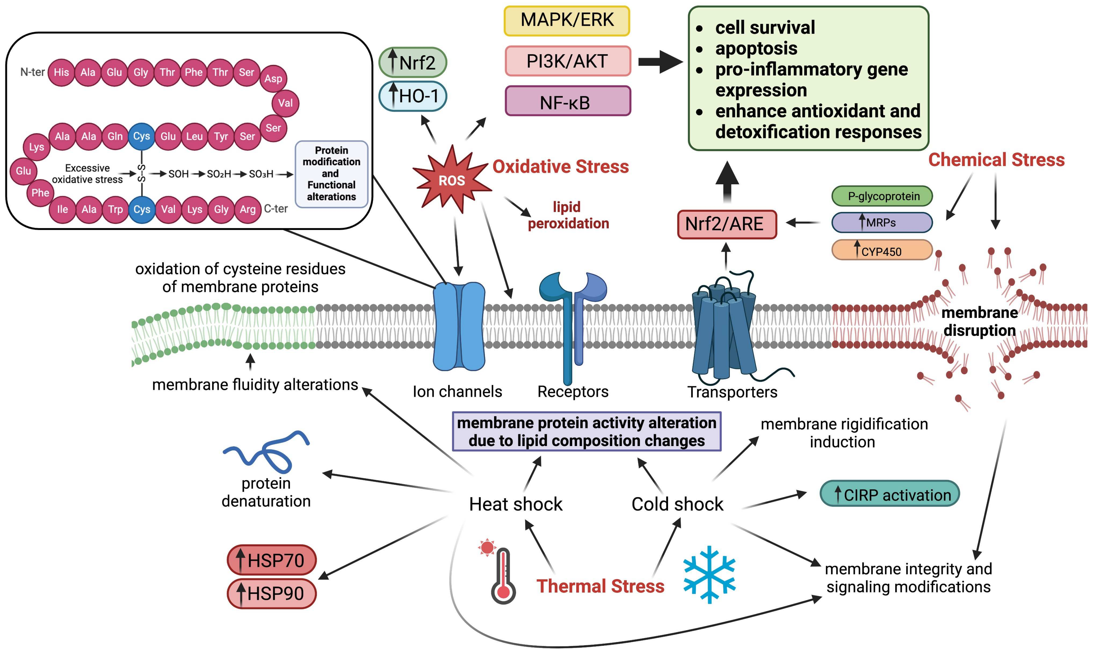
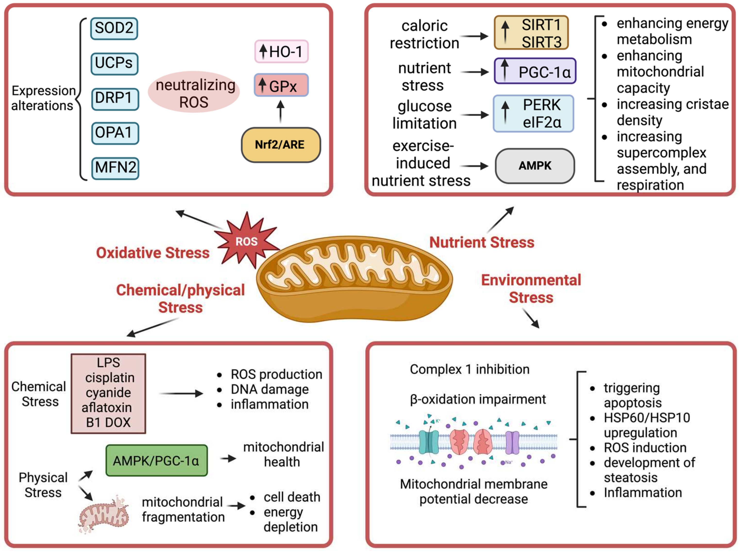
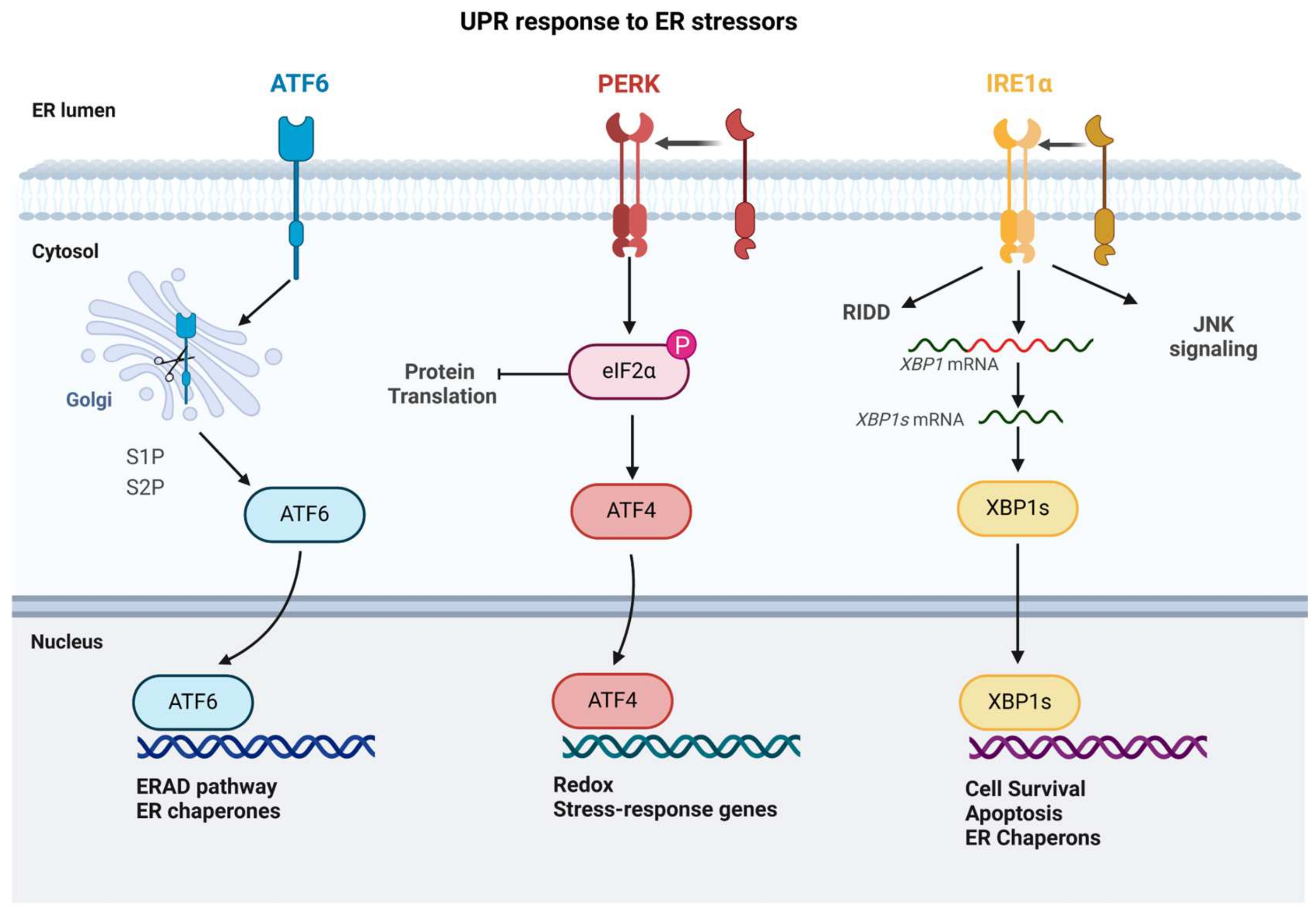
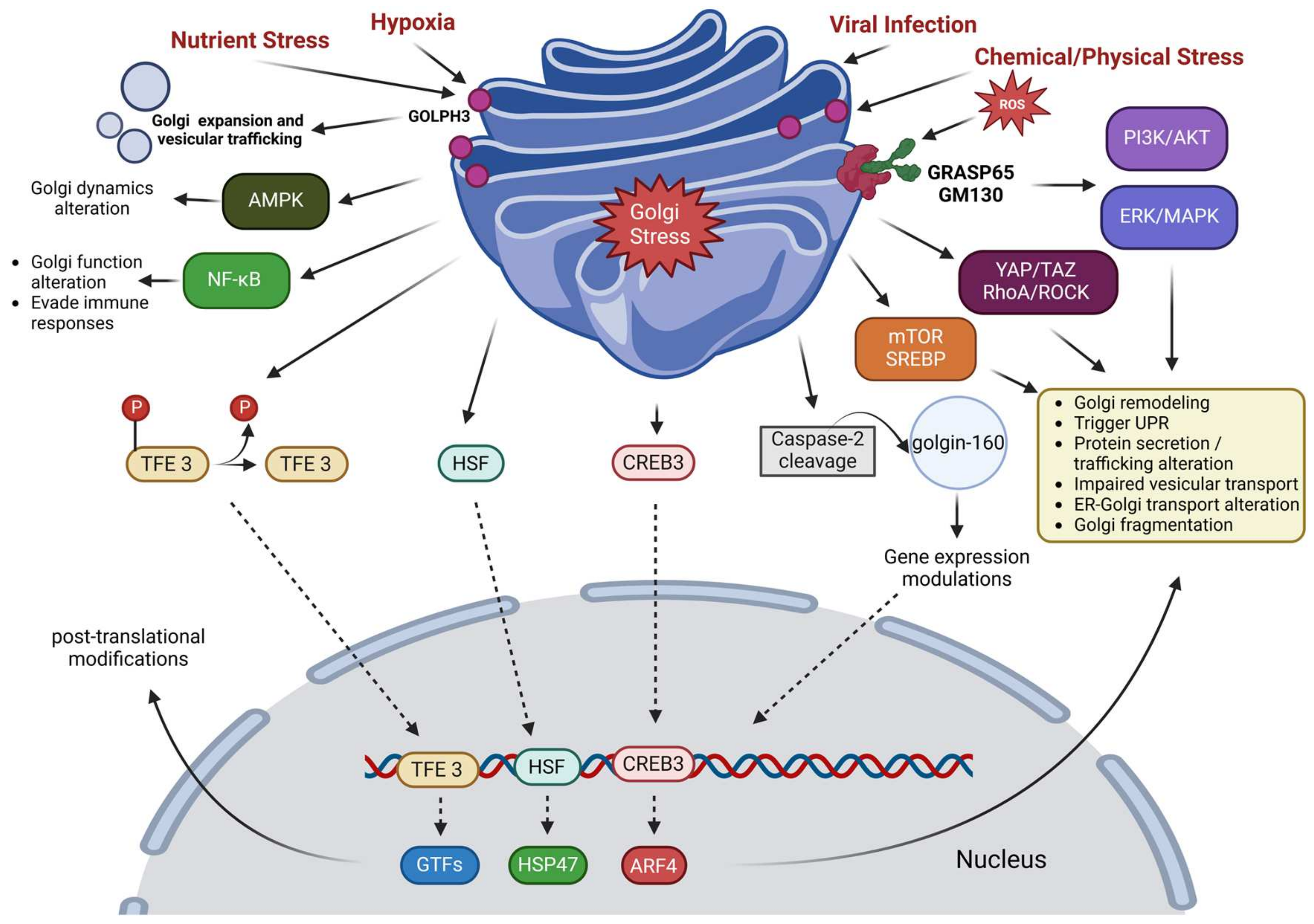
Disclaimer/Publisher’s Note: The statements, opinions and data contained in all publications are solely those of the individual author(s) and contributor(s) and not of MDPI and/or the editor(s). MDPI and/or the editor(s) disclaim responsibility for any injury to people or property resulting from any ideas, methods, instructions or products referred to in the content.

