Article

Rosmarinic Acid Attenuates Testicular Damage via Modulating Oxidative Stress and Apoptosis in Streptozotocin-Induced Diabetic Albino Mice

by
Omar Al-khawaldeh
1,
Zina M. Al-Alami
2,*,
Osama Y. Althunibat
3,
Tamer M. M. Abuamara
4,5,
Afnan Mihdawi
1 and
Mohammad H. Abukhalil
3,6
1
Department of Medical Laboratory Sciences, Faculty of Allied Medical Sciences, Al-Ahliyya Amman University, Amman P.O. Box 19328, Jordan
2
Department of Basic Medical Sciences, Faculty of Allied Medical Sciences, Al-Ahliyya Amman University, Amman P.O. Box 19328, Jordan
3
Department of Medical Analysis, Princess Aisha Bint Al-Hussein College of Nursing and Health Sciences, Al-Hussein Bin Talal University, Ma’an 71111, Jordan
4
Department of Basic Medical Science, Faculty of Dentistry, Al-Ahliyya Amman University, Amman P.O. Box 19328, Jordan
5
Department of Histology, Faculty of Medicine, Al-Azhar University, Cairo 11651, Egypt
6
Department of Biology, College of Science, Al-Hussein Bin Talal University, Ma’an 71111, Jordan
*
Author to whom correspondence should be addressed.
Stresses 2024, 4(3), 505-517; https://doi.org/10.3390/stresses4030032
Submission received: 11 July 2024 / Revised: 29 July 2024 / Accepted: 31 July 2024 / Published: 5 August 2024
(This article belongs to the Section Animal and Human Stresses)

Abstract

:
Diabetes mellitus (DM) induces the production of reactive oxygen species, which may lead to cell injury and death. This study aimed to assess the effects of rosmarinic acid (RA) on testicular damage, oxidative stress, and apoptosis in streptozotocin (STZ)-induced diabetic albino mice. DM in four- to six-week-old BALB/c male albino mice was induced via 50 mg/kg STZ, IP for 5 days. Twelve mice were randomly assigned into each of following groups: a control group, a diabetic (DM) group, RA5 mg/kg and RA15 mg/kg groups, and DM + RA5 mg/kg and DM + RA15 mg/kg groups. RA doses were intraperitoneally injected six times a week for seven weeks. Diabetes increased blood sugar and HbA1c levels and decreased all assessed sperm parameters. Testicular tissues of the diabetic mice showed increased lipid peroxidation, decreased reduced glutathione levels and catalase and superoxide dismutase activities, and increased apoptosis associated with histological abnormalities. Both RA doses had no effects on final body weight, blood sugar, and HbA1c in the diabetic mice. It is concluded that the administration of the potent antioxidant RA to diabetic mice improved the redox status in testicular tissues, protected them from diabetes-induced oxidative damage, and improved the quality of spermatozoa, mostly in a dose-dependent manner, which suggests a potential application value of RA in treating DM-related testicular injury and perhaps other complications.

1. Introduction

Diabetes mellitus (DM) is a disorder in which the level of blood glucose rises due to failing endogenous insulin synthesis or activity [1]. An uncontrolled glycemic level can significantly increase the risk of coronary artery disease, congestive heart failure, neuropathy, myocardial infarction, retinopathy, nephropathy, peripheral artery disease, and infertility [2]. Several published studies have reported that DM induces an enhanced production of reactive oxygen species (ROS) via disrupting different metabolic pathways and a reduction in the capacity of key antioxidant enzymes [3]. Indeed, ROS could directly damage cellular proteins, lipids, and DNA, leading to cell injury and death [4]. Overproduction of ROS leads to the peroxidation of polyunsaturated fatty acids, which are essential for spermatozoa’s fluidity during fertilization, and are present in high concentrations in plasma membranes; this produces high biological activity, which interferes with cellular functions and increases ROS [5]. In turn, excessive ROS and oxidative stress can affect spermatogenesis and even lead to male infertility [6]. Therefore, a potential compound that can protect the testes from such damage is in urgent demand.
Indeed, accumulating evidence has indicated that natural products represent a rich source of potential therapeutics, offering a variety of different benefits for testis health [7,8]. Rosmarinic acid (RA) is a bioactive phytochemical which exhibits several pharmacological activities; it is found naturally in a variety of plants such as rosemary, mint, sage, lemon balm, thyme, oregano, basil, and others [9,10]. It is an ester of caffeic acid and 3,4-dihydroxyphenyllactic acid, which is known to exhibit many biological activities and have beneficial health effects through its antibacterial, antiviral, antioxidant, and anti-inflammatory properties [11]. One previous study evaluated the effects of RA on infertility induced by metronidazole (MTZ), which is one of the most effective antibiotics and antiprotozoals that can also induce reversible male infertility by causing an abnormal production of immature sperm cells and decreasing the sperm count. The researchers used two doses of RA in sexually mature rats without and with MTZ, and they found that the RA reversed the effects of MTZ on male infertility and sperm count, morphology, and motility [12]. Another study found that RA can effectively attenuate ischemia/reperfusion-induced testicular damage, alleviate oxidative stress, and improve sperm parameters [13].
Despite its several pharmacological actions, including the previously reported positive effects it has on male infertility, the potential protective effect of RA on DM-induced testicular injury has not yet been fully investigated. Therefore, this study aimed to examine the protective effect of RA on testicular injury induced by streptozotocin (STZ) in terms of sperm parameters, oxidative stress, apoptosis, and histological changes, using a mouse model of STZ-induced DM.

2. Results

2.1. Effects of RA on Body Weight, FBG Levels, and HbA1c in Diabetic Mice

At the end of the experiment, the weights recorded in all diabetic groups (DM, DM + RA5 mg/kg, and DM + RA15 mg/kg) were considerably lower when compared to those of the healthy groups (the control, RA5 mg/kg, and RA15 mg/kg groups). However, RA had no significant effect on ultimate body weight in either the diabetic or non-diabetic groups (p > 0.05) (Figure 1a). FBS findings demonstrated normal glucose homeostasis in all groups at the beginning of the study. Administration of STZ (50 mg/kg) for five consecutive days resulted in the development of DM (p < 0.05). Nevertheless, both RA doses showed no influence on FBS levels throughout the course of the experiment (Figure 1b). In addition, diabetic animals in the three diabetic groups showed significantly higher levels of HbA1c than the control group. However, both RA doses showed no significant effect on HbA1c levels in either diabetic or non-diabetic mice (Figure 1c).

2.2. Effects of RA on Sperm Parameters in Diabetic Mice

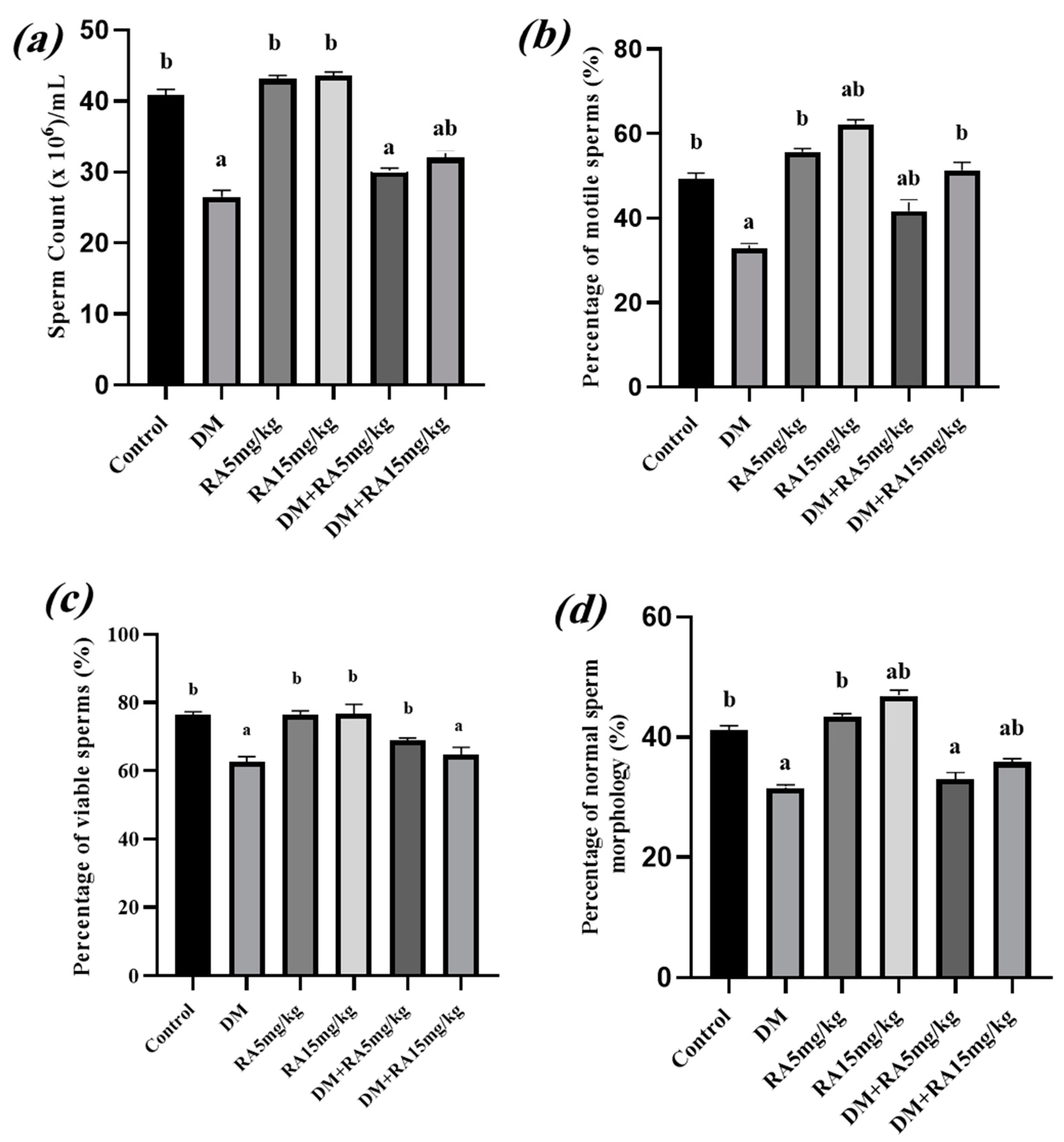
DM groups had significantly lower sperm counts than the control group (Figure 2a). The high RA dose (15 mg/kg) significantly increased the sperm count (p < 0.05) compared to the diabetic animals (STZ group). Nevertheless, both doses of RA did not affect the sperm count in non-diabetic animals.
The induction of DM significantly reduced sperm motility (Figure 2b). RA treatment of the diabetic mice (DM + RA5 mg/kg and DM + RA15 mg/kg groups) significantly (p < 0.05) enhanced sperm motility when compared to the non-treated diabetic group.
As shown in Figure 2c, the sperm viability of non-treated diabetic animals (DM group) declined significantly compared to the control group. On the other hand, the percentage of sperm viability in diabetic mice treated with RA5 mg/kg was considerably ameliorated (p < 0.05) compared the non-treated diabetic group and was normalized to the level of healthy animals (control group). Controversially, treatment of the diabetic mice with 15 mg/kg had no significant effect on sperm viability when compared to the positive control group. Moreover, none of the RA doses had a significant effect on sperm viability in the non-diabetic treated groups as compared to the control group.
The percentage of sperms with normal morphology was found to be significantly lower in all diabetic mice (DM, DM + RA5 mg/kg, and DM + RA15 mg/kg groups) compared to the control group. However, treatment of the diabetic animals with both doses of RA significantly (p < 0.05) enhanced their percentage of morphologically normal sperms as compared to non-treated diabetic animals (DM group). Also, the administration of 15 mg/kg RA considerably improved the proportion of sperms with normal morphology in non-diabetic mice compared to the control group (p < 0.05) (Figure 2d).

2.3. RA Mitigates Oxidative Stress in the Testes of Diabetic Mice

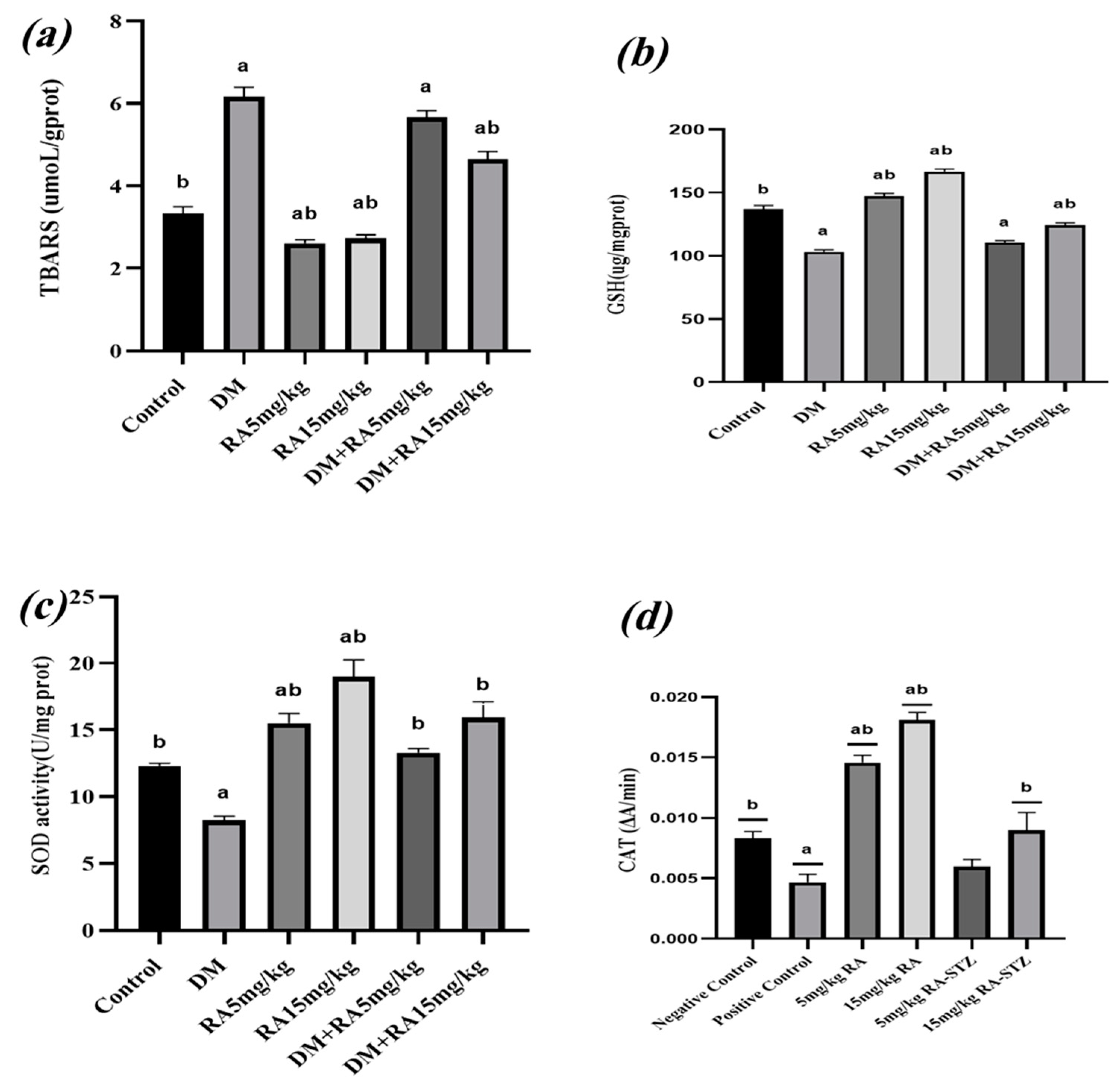
A TBARS assay was used to measure the levels of MDA, which is the end product of lipid peroxidation, in the mice’s testicular tissues. The results revealed that the diabetic groups had higher levels of MDA than the healthy groups (p < 0.05). Interestingly, RA15 mg/kg reduced testicular MDA levels in diabetic mice (DM + RA15 mg/kg) compared to the non-treated diabetes group (p < 0.05) (Figure 3a). Furthermore, both doses of RA remarkably lowered MDA levels in the tissue of healthy animals as compared to the control group (p < 0.05).
Reduced glutathione (GSH) levels in the testicular tissues of the DM animals were significantly lower than those in the control group (p < 0.05) (Figure 3b). However, RA treatment of the diabetic mice resulted in a dose-dependent increase in their GSH levels, as diabetic animals receiving the high-dose treatment (DM + RA15 mg/kg) showed a significant improvement in their testicular GSH levels compared to the non-treated diabetic group (p < 0.05). Yet, healthy animals given both RA doses had noticeably higher GSH levels than the control group animals (p > 0.05).
The DM group exhibited a substantial decline in superoxide dismutase (SOD) activity when compared to the control group (p < 0.05). Remarkably, treating diabetic mice with both RA doses significantly restored their SOD activity to the level seen in the control group. Likewise, both doses of RA treatment significantly increased the SOD activity in non- diabetic mice compared to the control group (p < 0.05) (Figure 3c).
In addition, both RA doses significantly increased the catalase (CAT) enzyme activity in non-diabetic mice (p < 0.05), yet only 15 mg/kg RA could significantly improve CAT enzyme activity in the diabetic mice (p < 0.05) (Figure 3d).

2.4. Histological Studies

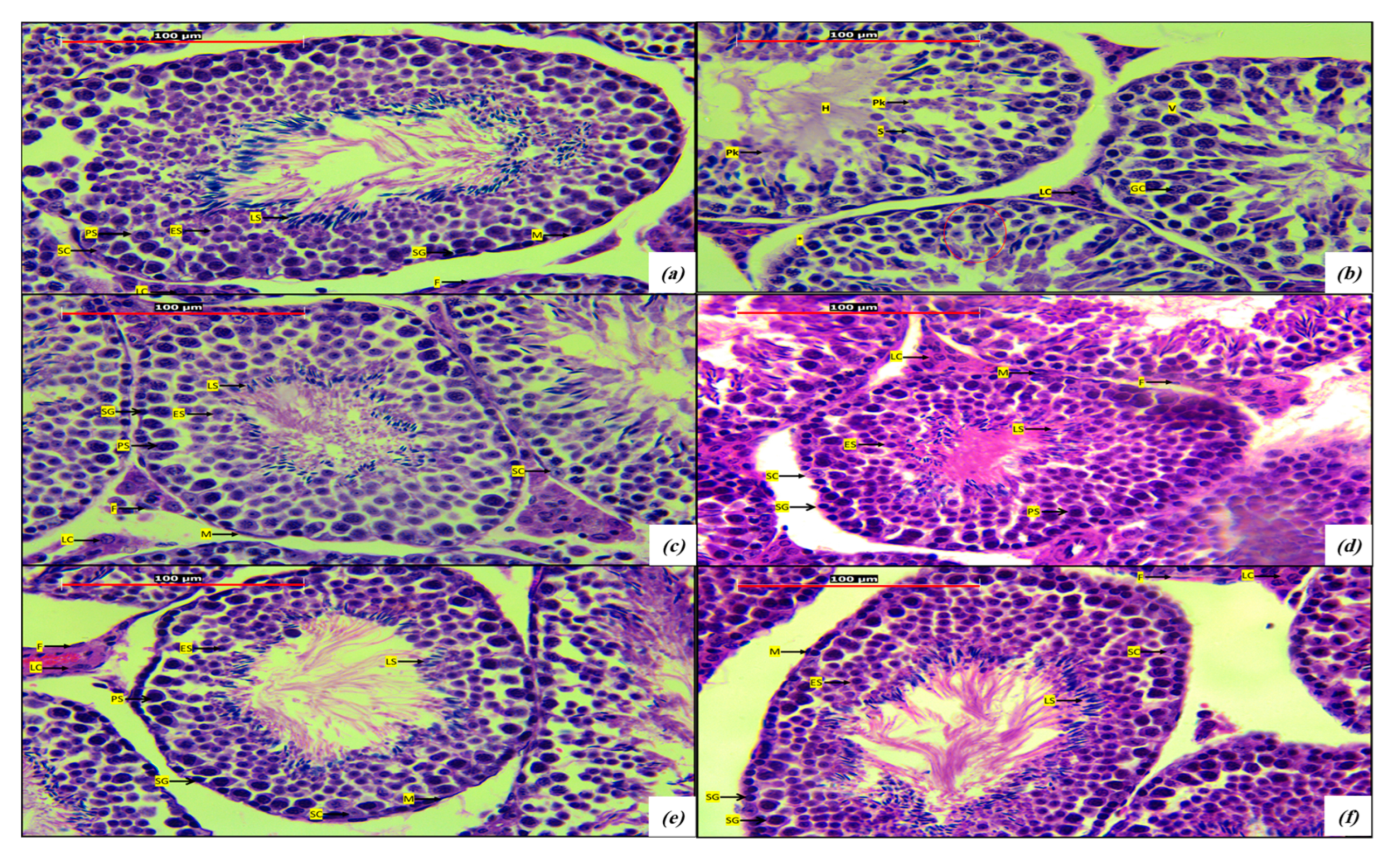
Microscopic sections stained with H&E showed that testis samples from the RA5 mg/kg (Figure 4c) and RA15 mg/kg (Figure 4d) groups contained cell types similar to those detected in the control group samples (Figure 4a). As for the spermatogenesis series, spermatogonia (SG), primary spermatocytes (PS), early spermatids (ES), late spermatids (LS), and, finally, highly specialized sperm cells were preserved well. Also, the Sertoli cells (SC) seemed normal. Moreover, the seminiferous tubules’ interstitial cells of Leydig (LC) seemed well preserved. The surrounding myoid cells (M) and fibroblasts (F) were also preserved. Testis samples from the control group (Figure 4a) showing cross-sections of normal seminiferous tubules of the testes. The seminiferous tubule wall consisted of a unique germinal epithelium composed of columnar Sertoli cells (SC) and dividing spermatogenic stem cells. Near the basement membrane there were many prominent spermatogonia (SG). The primary spermatocytes (PS) are the largest spermatogenic cells and are usually abundant at all levels between the basement membrane and the lumen. Newly formed round, early spermatids (ES) differentiate and lose volume in becoming late spermatids (LS) and finally highly specialized sperm cells. The seminiferous tubules (ST) were surrounded by stroma containing many interstitial cells of Leydig (LC) having vesicular nucleus with prominent nucleolus. Around the seminiferous tubules, we observed myoid cells (M) with elongated nuclei and fibroblasts (F).
When compared to the testis samples from the control group (Figure 4a), testis samples from the DM group (Figure 4b) showed severe degenerative changes: atrophy of the seminiferous tubules, the absence of spermatogenesis (red circle in the figure), pyknotic and necrotic spermatogenic cells (Pk) with immature spermatids (S), several spermatid giant cells (GC) in the tubules with noticeable degenerative changes like vacuoles (V), hyalinization in the lumen of some seminiferous tubules (H), sloughing germinal epithelia, and irregular basement membranes (*). Also, these samples showed widening of the intertubular space with distortion and pyknosis in the interstitial cells of Leydig (LC).
Interestingly, diabetic mice that were treated with 15 mg/Kg RA (Figure 4f) appeared to show a marked improvement in their testicular arrangement, seminiferous epithelium with spermatogonia (SG), primary spermatocytes (PS), early spermatids (ES), late spermatids (LS), and Sertoli cells (SC); also, a marked improvement in their interstitial cells of Leydig (ES) were observed in comparison to the DM group (Figure 4b). Nevertheless, these parameters showed moderate improvements when the diabetic mice were treated with 5 mg/Kg RA (Figure 4e). Photomicrographs were taken using a Leica, Leica Microsystems, Wetziar, Germany and Leica Geosystem v 11.7.0.524 software.

2.5. RA Attenuates Apoptosis in the Testes of Diabetic Mice

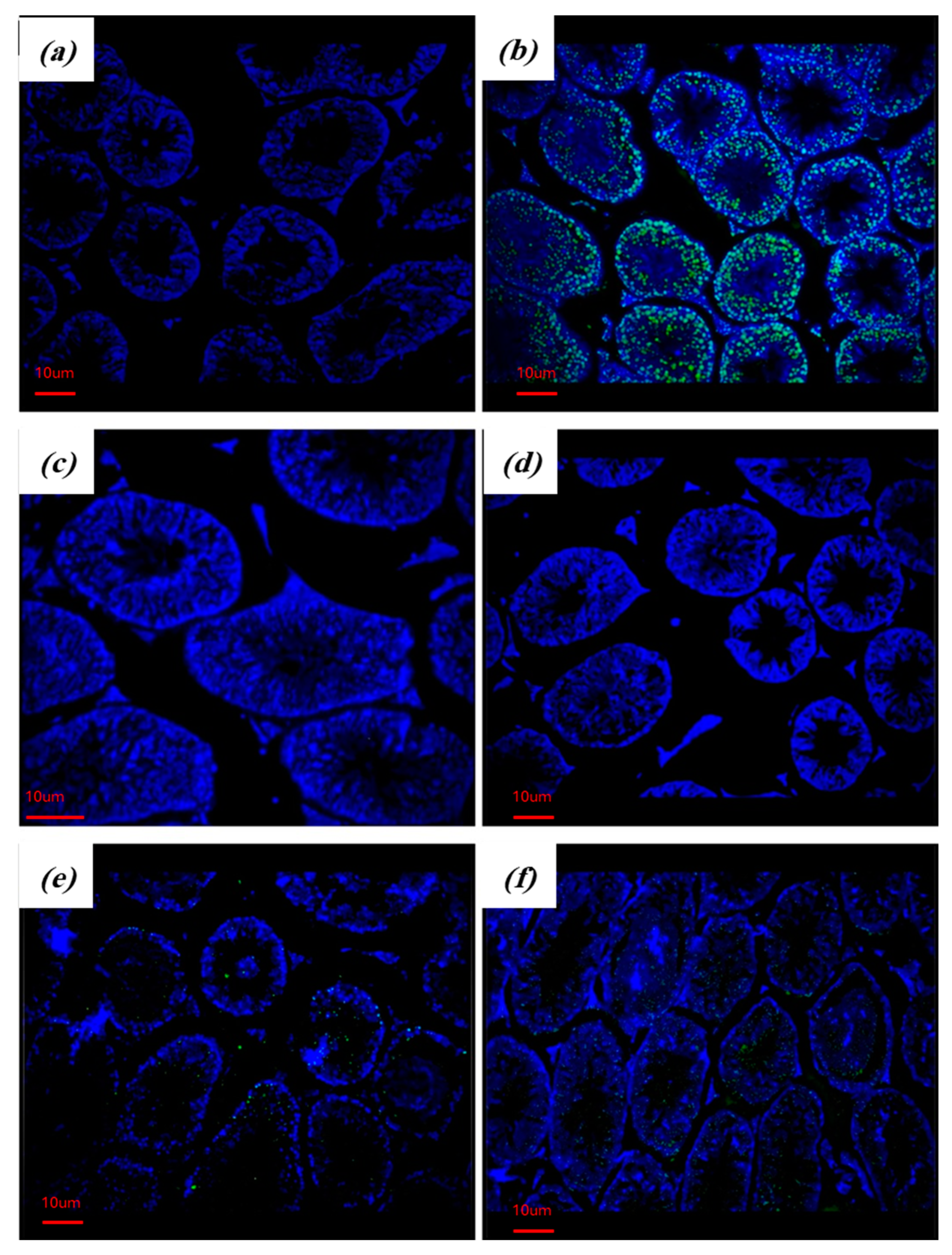
The TUNEL assay method was used to detect the apoptotic cells, particularly at the late stage of apoptosis when DNA fragmentation had occurred. Cells positive for DNA fragmentation displayed a bright green color under the fluorescent microscope, whilst negative results revealed a blue color of the background stain, which was a DAPI stain. As shown in Figure 5, only diabetic groups showed positive results for DNA fragmentation as a marker of late apoptosis. However, the frequency of DNA-fragmented cells was remarkably reduced in the testicular tissues of diabetic mice that received both RA doses compared to the untreated diabetic group. Images were taken using Zeiss axio imager 2, Oberkochen, Germany.

3. Discussion

DM is a chronic metabolic disorder characterized by hyperglycemia resulting from insufficient insulin production or ineffective utilization of insulin by the body cells. While DM is widely recognized for its impact on cardiovascular health, kidney function, and nerve damage, its association with reproductive health, particularly infertility, is an emerging concern [14]. Diabetes-induced infertility results from disrupted hormonal regulation, impaired ovarian function, and altered sperm quality [15]. Moreover, oxidative stress plays a crucial role in the pathogenesis of DM-related complications, including infertility. The prolonged exposure to high glucose levels leads to an overproduction of ROS, causing cellular damage and inflammation [16]. Accordingly, it is very important to support comprehensive treatment strategies that target not only blood glucose control but also oxidative stress reduction in order to reduce the risk of infertility and other chronic complications of DM. Thus, the present study aimed to investigate the effects of a natural antioxidant, RA, on testicular oxidative damage and the quality of sperms in STZ-induced diabetic mice.
STZ injection into experimental animals causes the destruction of pancreatic beta cells, which in turn mimics the insulin deficiency and hyperglycemia characteristics of DM [17]. STZ enters beta cells, resulting in DNA alkylation and damage, which triggers poly ADP-ribosylation, which reduces cellular NAD+ and ATP. Increased ATP dephosphorylation after STZ treatment provides a substrate for xanthine oxidase, producing superoxide radicals, hydrogen peroxide, and hydroxyl radicals. Furthermore, STZ releases toxic levels of nitric oxide, which in turn lead to DNA damage, eventually destroying beta cells through necrosis [18].
In this context, the current study successfully induced diabetes in mice, which was proven by the development of hyperglycemia in the STZ-injected groups. In addition, weight loss is a common manifestation of DM, which is attributed to multiple metabolic alterations associated with DM. Insulin deficiency reduces cellular uptake and utilization of glucose, resulting in a shift towards breaking down stored fats and proteins, contributing to muscle wasting and the loss of adipose tissue [19]
The results presented in the current study show a continuous reduction in the body weight of the diabetic mice, while there was an increase in the body weight of healthy mice throughout the course of the experiment. However, RA treatment did not improve glucose homeostasis or body weight in either the diabetic mice or the healthy mice. This may indicate that the antioxidant activity of RA is not involved in insulin production or its action. Diabetes-induced testicular dysfunction is characterized by several detrimental impacts on seminal fluid quality, including an increase in the frequency of abnormal-sperm morphology and decreases in the total sperm count, sperm viability, and sperm motility [20]. In the current investigation, the administration of RA therapy to diabetic mice considerably ameliorated the low quality of sperms reported in the untreated diabetic group, mostly in a dose-dependent manner. In agreement with our findings, it was previously reported that RA positively affected sperm motility and morphology in metronidazole-induced testicular dysfunction [12]. Additionally, in another study, a combined treatment of RA with ellagic acid reversed doxorubicin-induced testicular damage and improved sperm count and motility [21]. The protective effect of RA on testicular health against various pathological and toxicological stressors is mainly attributed to its potential antioxidant activity. In diabetes, hyperglycemia can trigger ROS accumulation in amounts beyond what can be neutralized through the ability of the antioxidant defense system, resulting in the development of oxidative stress conditions. The current study reports the biochemical markers in the testicular tissues of diabetic mice. In previous research, the reduced antioxidant enzyme levels in DM testes were associated with poor spermatogenesis and a substantial drop in sperm count and motility [22]. On the other hand, administrating RA to healthy and diabetic mice improved their antioxidant defense mechanisms, marked by enhanced SOD and CAT activities, elevated GSH levels, and reduced lipid peroxidation levels.
The radical-scavenging potential of RA is the main mechanism through which it restores cellular antioxidants, inhibiting oxidative tissue damage. Several studies have demonstrated the protective effect of RA against oxidative stress conditions. It was revealed that the nephroprotective effect of RA against diabetic nephropathy was associated with its antioxidant potential [23]. In another study, the treatment of diabetic rats with RA attenuated diabetes-induced vascular dysfunction and its associated oxidative stress [24]. Furthermore, the radical-scavenging activity of RA was linked to its actions in attenuating drug-induced organ and tissue damage, such as cisplatin-induced kidney injury [25], gentamicin sulfate-induced renal oxidative damage [26], β-amyloid-induced neurotoxicity [27], and carbon tetrachloride-intoxicated liver condition [28]. In testicular failure, DM is associated with spermatogenetic cell loss and seminiferous tubule shrinkage, which are morphological markers of spermatogenesis abnormalities [29,30]. In one study that evaluated curcumin’s impact on diabetes-induced testicular injury, the structures of seminiferous tubules were degraded and disrupted in diabetic rats, and curcumin treatment improved the tissue damage to the testicles [31]. In this study, it was found that DM mice had decreased spermatogenic cell series, and their seminiferous tubule structure was disrupted. In the DM + RA15 mg/kg mice, there was an increase in spermatogenic cells, and the seminiferous tubule structure was improved compared to the DM mice.
Likewise, DM has been shown to induce apoptotic signaling pathways in testicular tissues. RA treatment protected the diabetic mice from DNA fragmentation during the late apoptosis step, as demonstrated by the TUNEL assay results. This can be attributed to RA’s potential to inhibit DNA oxidative damage as well as accelerate DNA repair mechanisms. In accordance with the current findings, RA has been shown to protect DNA against the mutagenic and damaging effects of UV and H2O2 [32]. Moreover, RA can attenuate chromium-induced hepatic and renal oxidative damage and DNA damage in rats [33].

4. Materials and Methods

4.1. Experimental Animals

Male albino BALB/c mice (aged 4–6 weeks) were purchased from the animal house of Al-Yarmouk University, Irbid, Jordan. The animals were maintained under a controlled light–dark cycle at 18–25 °C at Al-Ahliyya Amman University, Amman, Jordan. All experiments were performed under the approval of the Al-Ahliyya Amman University’s ethics committee (approval number AUP: AAU/2/4/2022-2023).

4.2. Chemicals

The following chemicals were used in the experiments: Streptozotocin (Santa Cruz Biotechnology, Dallas, TX, USA). Rosmarinic acid (GenoChem World, Valencia, Spain). Trypan blue (Euroclone, Milan, Italy). Citric acid, used to prepare the citrate buffer (citric acid anhydrous, Pharmpur®, Königsbrunn, Germany). Ether (alpha chemika-diethyl ether, Andheri, Maharashtra, Indea). Phosphate-buffered saline (PBS) (Capricorn scientific, Dulbecco’s PBS (1x) Powder, Cat. No.: PBS-1A-P10, Ebsdorfergrund, Germany). Formalin (10%), prepared from formaldehyde solution (BP, Gainland chemical company, Clwyd, UK; Cat No.: 06380). Hematoxylin (Sigma-Aldrich Corporation, Saint Louis, MO, USA). Eosin (Riedel-de Haën, Seelze, Germany).

4.3. Induction of DM

DM was induced in the male BALB/C mice at 4–6 weeks old, when the mice weighed 23 to 25 g, via intraperitoneal injection of STZ (Santa Cruz Biotechnology, Dallas, TX, USA) at the dose of 50 mg/kg dissolved in 100 mM citrate buffer, pH 4.5 (pH meter, EUTECH instruments, Singapore, Singapore), for 5 consecutive days [34]. After 1 week (7 days after the first STZ dose), fasting blood glucose levels were measured using the OKmeter Blood Glucose meter (brand name: OKmeter; place of origin: Taiwan; model number: OK-1; certifications: approved by FDA 510K, CE0123, ISO13485, GMP) by tail-vein puncture blood sampling with 8–12 h fasting [35]. Mice that had blood sugar values of more than 200 mg/dL were used for the study.

4.4. Experimental Groups

The mice were randomly divided into the following groups: The control group: the non-diabetic group (n = 12)—the animals in this group did not receive any treatment. The DM group: the diabetic group (n = 12)—the animals in this group were treated with 50 mg/kg STZ dissolved in 0.1 mol/L citrate buffer, pH 4.5 (pH meter, EUTECH instruments, Singapore, Singapore ), for 5 days [34]. The RA5 mg/kg group: the low-dose RA non-diabetic group (n = 12)—the animals in this group were treated with a dose of 5 mg/kg RA, six times a week for seven weeks [12]. The RA15 mg/kg group: the high-dose RA non-diabetic group (n = 12)—the animals in this group were treated with a dose of 15 mg/kg of RA, six times a week for seven weeks [12]. The DM + RA5 mg/kg group: the low-dose RA diabetic group (n = 12)—the diabetic animals in this group were treated with a dose of 5 mg/kg of RA, six times a week for seven weeks. The DM + RA15 mg/kg group: the high-dose RA diabetic group (n = 12)—the diabetic animals in this group were treated with a dose of 15 mg/kg of RA, six times a week for seven weeks. RA weight was measured using a four-digit analytical balance manufactured by Boeckel & Co. GmbH & Co. KG, Hamburg, Germany, and the RA was dissolved in distilled water [12].

4.5. Animal Treatment and Sacrifice

RA (GenoChem World, Valencia, Spain) doses were injected intraperitoneally and freshly prepared each day according to the animal’s weight, which was documented weekly along with their fasting blood sugar (FBS) levels. The weights of the animals were recorded weekly; the first reading was obtained at day 0, before the administration of any drug, and the second reading was taken following the administration of the five STZ doses. At the end of experiment (day 50), the final body weight was recorded using a laboratory balance (Sartorius, Gottingen, Germany); then, the mice were anesthetized with ether and sacrificed by opening the pleural cavity. The left testes were homogenized in phosphate-buffered saline (PBS) and stored at −20 °C, while the right testes were cut and immersed in formalin and stored at room temperature to make paraffin blocks for the histological studies and TUNEL assay. For the histological studies, sections from all mice in all groups were prepared and stained with hematoxylin and eosin; for the TUNEL assay, three blocks were randomly chosen from each group and sectioned.

4.6. Sperm Parameters

Sperm samples were collected from the cauda epididymis using the method of Ikhlas and his group, with some modifications [36]. The left cauda epididymis was transferred to a Petri dish and immersed in 0.5 mL of warm PBS (pH 7.4) (pH meter, EUTECH instruments, Singapore, Singapore), then opened with a sharp blade, and the sperms were allowed to diffuse into the buffer by incubating the sample at 37 °C for 1–2 min (Binder incubator, Tuttlingen, Germany). Measurement of the sperm count and the percentage of motile sperms was performed from the prepared sperm suspension using a hemocytometer (Cat. No.: 1103, China) and a microscope (Nikon-ECLIPSE E100 microscope, Tokyo, Japan). A volume of 10 μL of sample was mixed with 1 μL of 4% trypan blue and stained for 5 min to test the sperm viability. The percentages of morphologically normal sperms were assessed using a modified version of Al-Alami’s method [12].

4.7. Glycated HemoglobinA1c (HbA1c)

The blood was collected from mice from the Retro-orbital plexus into a plain tube (AFCOVAC) and centrifugated at 1000× g for 20 min (eppendrof, Hamburg, Germany) to separate the serum. This assay was performed according to the manufacturer instructions provided in the kit HbA1c ELISA Kit (ELK Biotechnology CO, Cat. No.: ELK5108, Wuhan, China).

4.8. Testicular Oxidative Stress Markers and Antioxidants

During high levels of ROS, malondialdehyde (MDA) reacts with 2-thiobarbituric acid (TBA) and attacks polyunsaturated fatty acids, which forms lipid peroxidation and produces MDA, which is a biomarker of lipid peroxidation. The protocol used to assess this was taken from the manufacturer instructions provided with the thiobarbituric acid-reactive substance (TBARS) colorimetric assay kit we used (Elabscience CO, Cat. No.: E-BC-K298-M, Wuhan, China).
The contents of reduced glutathione (GSH) in the testicular tissues were estimated using a kit provided by GenoChem World S.L. (Cat. No.: GW0477, Valencia, Spain), according to the manufacturer’s instructions. The activity of catalase (CAT) was estimated in the testis samples using a kit obtained from GenoChem World S.L. (GenoChem World S.L. (Cat. No.: GW0579-100T-96S, Valencia, Spain)), following the manufacturer’s instructions. Last but not least, superoxide dismutase (SOD) testing was performed according to the manufacturer instructions provided with the relevant kit (Geochem World S.L., Cat. No.: GW0583-100T-48S, Valencia, Spain).

4.9. TUNEL Assay

TUNEL staining was used to analyze the DNA fragmentation of apoptotic cells using a kit provided by Elabscience CO (Cat. No.: ACK-A320, Wuhan, China) based on the manufacturer’s instructions. The results were observed using microscopy.

4.10. Statistical Analysis

The body weight and FBS results were analyzed using a two-way ANOVA, and all other results were analyzed using a one-way ANOVA followed by Tukey’s post hoc test using GraphPad Prism version 8. A p-value less than 0.05 was considered significant. All results are shown as the mean ± SE.

5. Conclusions

The current study concludes that DM stimulates oxidative damage in testicular tissues, induces cell apoptosis, and reduces the quality of spermatozoa. The administration of the potent antioxidant RA to mice improves the redox status in testicular tissues, protecting them from diabetes-induced oxidative damage and improving the quality of their spermatozoa, mostly in a dose-dependent manner. Further studies are recommended to clarify the precise underling mechanisms behind its testicular-protective effect and to identify the most effective RA dose.

Author Contributions

Conceptualization, Z.M.A.-A. and O.Y.A.; methodology, Z.M.A.-A., O.Y.A. and A.M.; validation, Z.M.A.-A., O.Y.A. and M.H.A.; formal analysis, O.A.-k., Z.M.A.-A. and O.Y.A.; investigation, O.A.-k., T.M.M.A. and A.M.; resources, O.A.-k., Z.M.A.-A., O.Y.A. and A.M.; data curation, Z.M.A.-A., O.Y.A., T.M.M.A. and M.H.A.; writing—original draft preparation, O.A.-k., Z.M.A.-A. and M.H.A.; writing—review and editing, Z.M.A.-A., O.Y.A., T.M.M.A., A.M. and M.H.A.; visualization, O.A.-k., Z.M.A.-A., O.Y.A. and T.M.M.A.; supervision, Z.M.A.-A. and O.Y.A.; project administration, Z.M.A.-A. and O.Y.A.; funding acquisition, Z.M.A.-A. All authors have read and agreed to the published version of the manuscript.

Funding

This research received funding from Al-Ahliyya Amman University upon the decision of the Appointment, Promotion and Transfer Committee (decision number (4/22/2022-2023)).

Data Availability Statement

The data used to generate the results presented in the paper are available and can be shared upon reasonable request to the corresponding author.

Conflicts of Interest

The authors declare no conflicts of interest.

References

  1. Asmat, U.; Abad, K.; Ismail, K. Diabetes mellitus and oxidative stress-A concise review. Saudi Pharm. J. 2016, 24, 547–553. [Google Scholar] [CrossRef] [PubMed]
  2. Lotfy, M.; Adeghate, J.; Kalasz, H.; Singh, J.; Adeghate, E. Chronic Complications of Diabetes Mellitus: A Mini Review. Curr. Diabetes Rev. 2017, 13, 3–10. [Google Scholar] [CrossRef]
  3. Hurrle, S.; Hsu, W.H. The etiology of oxidative stress in insulin resistance. Biomed. J. 2017, 40, 257–262. [Google Scholar] [CrossRef]
  4. Redza-Dutordoir, M.; Averill-Bates, D.A. Activation of apoptosis signalling pathways by reactive oxygen species. Biochim. Biophys. Acta 2016, 1863, 2977–2992. [Google Scholar] [CrossRef]
  5. Lam, P.L.; Wong, R.S.; Lam, K.H.; Hung, L.K.; Wong, M.M.; Yung, L.H.; Ho, Y.W.; Wong, W.Y.; Hau, D.K.; Gambari, R.; et al. The role of reactive oxygen species in the biological activity of antimicrobial agents: An updated mini review. Chem. Biol. Interact. 2020, 320, 109023. [Google Scholar] [CrossRef]
  6. Kaltsas, A. Oxidative Stress and Male Infertility: The Protective Role of Antioxidants. Medicina 2023, 59, 1769. [Google Scholar] [CrossRef]
  7. D‘Cruz, S.C.; Vaithinathan, S.; Jubendradass, R.; Mathur, P.P. Effects of plants and plant products on the testis. Asian J. Androl. 2010, 12, 468–479. [Google Scholar] [CrossRef] [PubMed]
  8. Nasim, N.; Sandeep, I.S.; Mohanty, S. Plant-derived natural products for drug discovery: Current approaches and prospects. Nucleus 2022, 65, 399–411. [Google Scholar] [CrossRef] [PubMed]
  9. Ngo, Y.L.; Lau, C.H.; Chua, L.S. Review on rosmarinic acid extraction, fractionation and its anti-diabetic potential. Food Chem. Toxicol. 2018, 121, 687–700. [Google Scholar] [CrossRef]
  10. Petersen, M.; Abdullah, Y.; Benner, J.; Eberle, D.; Gehlen, K.; Hücherig, S.; Janiak, V.; Kim, K.H.; Sander, M.; Weitzel, C.; et al. Evolution of rosmarinic acid biosynthesis. Phytochemistry 2009, 70, 1663–1679. [Google Scholar] [CrossRef]
  11. Petersen, M.; Simmonds, M.S. Rosmarinic acid. Phytochemistry 2003, 62, 121–125. [Google Scholar] [CrossRef] [PubMed]
  12. Al-Alami, Z.M.; Shraideh, Z.A.; Taha, M.O. Rosmarinic acid reverses the effects of metronidazole-induced infertility in male albino rats. Reprod. Fertil. Dev. 2017, 29, 1910–1920. [Google Scholar] [CrossRef] [PubMed]
  13. Raisi, A.; Davoodi, F.; Mohammadi, R. Protective effects of rosmarinic acid on testicular torsion-detorsion in an animal model. Iran. J. Vet. Surg. 2022, 17, 80–86. [Google Scholar] [CrossRef]
  14. Facondo, P.; Di Lodovico, E.; Delbarba, A.; Anelli, V.; Pezzaioli, L.C.; Filippini, E.; Cappelli, C.; Corona, G.; Ferlin, A. The impact of diabetes mellitus type 1 on male fertility: Systematic review and meta-analysis. Andrology 2022, 10, 426–440. [Google Scholar] [CrossRef] [PubMed]
  15. Condorelli, R.A.; La Vignera, S.; Mongioì, L.M.; Alamo, A.; Calogero, A.E. Diabetes Mellitus and Infertility: Different Pathophysiological Effects in Type 1 and Type 2 on Sperm Function. Front. Endocrinol. 2018, 9, 268. [Google Scholar] [CrossRef] [PubMed]
  16. Papachristoforou, E.; Lambadiari, V.; Maratou, E.; Makrilakis, K. Association of Glycemic Indices (Hyperglycemia, Glucose Variability, and Hypoglycemia) with Oxidative Stress and Diabetic Complications. J. Diabetes Res. 2020, 2020, 7489795. [Google Scholar] [CrossRef] [PubMed]
  17. Furman, B.L. Streptozotocin-Induced Diabetic Models in Mice and Rats. Curr. Protoc. Pharmacol. 2015, 70, 5–47. [Google Scholar] [CrossRef] [PubMed]
  18. Szkudelski, T. The mechanism of alloxan and streptozotocin action in B cells of the rat pancreas. Physiol. Res. 2001, 50, 537–546. [Google Scholar] [PubMed]
  19. Cummings, D.E.; Overduin, J.; Foster-Schubert, K.E. Gastric bypass for obesity: Mechanisms of weight loss and diabetes resolution. J. Clin. Endocrinol. Metab. 2004, 89, 2608–2615. [Google Scholar] [CrossRef]
  20. He, L.; He, T.; Farrar, S.; Ji, L.; Liu, T.; Ma, X. Antioxidants Maintain Cellular Redox Homeostasis by Elimination of Reactive Oxygen Species. Cell Physiol. Biochem. 2017, 44, 532–553. [Google Scholar] [CrossRef]
  21. Georgy, G.S.; Maher, O.W. Ellagic acid and rosmarinic acid attenuate doxorubicin-induced testicular injury in rats. J. Biochem. Mol. Toxicol. 2017, 31, e21937. [Google Scholar] [CrossRef]
  22. Nna, V.U.; Bakar, A.B.A.; Ahmad, A.; Mohamed, M. Diabetes-induced testicular oxidative stress, inflammation, and caspase-dependent apoptosis: The protective role of metformin. Arch. Physiol. Biochem. 2020, 126, 377–388. [Google Scholar] [CrossRef] [PubMed]
  23. Jiang, W.L.; Xu, Y.; Zhang, S.P.; Hou, J.; Zhu, H.B. Effect of rosmarinic acid on experimental diabetic nephropathy. Basic. Clin. Pharmacol. Toxicol. 2012, 110, 390–395. [Google Scholar] [CrossRef]
  24. Sotnikova, R.; Okruhlicova, L.; Vlkovicova, J.; Navarova, J.; Gajdacova, B.; Pivackova, L.; Fialova, S.; Krenek, P. Rosmarinic acid administration attenuates diabetes-induced vascular dysfunction of the rat aorta. J. Pharm. Pharmacol. 2013, 65, 713–723. [Google Scholar] [CrossRef]
  25. Domitrović, R.; Potočnjak, I.; Crnčević-Orlić, Z.; Škoda, M. Nephroprotective activities of rosmarinic acid against cisplatin-induced kidney injury in mice. Food Chem. Toxicol. 2014, 66, 321–328. [Google Scholar] [CrossRef] [PubMed]
  26. Tavafi, M.; Ahmadvand, H. Effect of rosmarinic acid on inhibition of gentamicin induced nephrotoxicity in rats. Tissue Cell 2011, 43, 392–397. [Google Scholar] [CrossRef]
  27. Alkam, T.; Nitta, A.; Mizoguchi, H.; Itoh, A.; Nabeshima, T. A natural scavenger of peroxynitrites, rosmarinic acid, protects against impairment of memory induced by Abeta(25-35). Behav. Brain Res. 2007, 180, 139–145. [Google Scholar] [CrossRef]
  28. Domitrović, R.; Skoda, M.; Vasiljev Marchesi, V.; Cvijanović, O.; Pernjak Pugel, E.; Stefan, M.B. Rosmarinic acid ameliorates acute liver damage and fibrogenesis in carbon tetrachloride-intoxicated mice. Food Chem. Toxicol. 2013, 51, 370–378. [Google Scholar] [CrossRef] [PubMed]
  29. Cameron, D.F.; Murray, F.T.; Drylie, D.D. Interstitial compartment pathology and spermatogenic disruption in testes from impotent diabetic men. Anat. Rec. 1985, 213, 53–62. [Google Scholar] [CrossRef]
  30. Sisman, A.R.; Kiray, M.; Camsari, U.M.; Evren, M.; Ates, M.; Baykara, B.; Aksu, I.; Guvendi, G.; Uysal, N. Potential novel biomarkers for diabetic testicular damage in streptozotocin-induced diabetic rats: Nerve growth factor Beta and vascular endothelial growth factor. Dis. Markers 2014, 2014, 108106. [Google Scholar] [CrossRef]
  31. Zha, W.; Bai, Y.; Xu, L.; Liu, Y.; Yang, Z.; Gao, H.; Li, J. Curcumin Attenuates Testicular Injury in Rats with Streptozotocin-Induced Diabetes. Biomed. Res. Int. 2018, 2018, 7468019. [Google Scholar] [CrossRef] [PubMed]
  32. Sevgi, K.; Tepe, B.; Sarikurkcu, C. Antioxidant and DNA damage protection potentials of selected phenolic acids. Food Chem. Toxicol. 2015, 77, 12–21. [Google Scholar] [CrossRef] [PubMed]
  33. Khalaf, A.A.; Hassanen, E.I.; Ibrahim, M.A.; Tohamy, A.F.; Aboseada, M.A.; Hassan, H.M.; Zaki, A.R. Rosmarinic acid attenuates chromium-induced hepatic and renal oxidative damage and DNA damage in rats. J. Biochem. Mol. Toxicol. 2020, 34, e22579. [Google Scholar] [CrossRef]
  34. Rajesh, M.; Mukhopadhyay, P.; Bátkai, S.; Patel, V.; Saito, K.; Matsumoto, S.; Kashiwaya, Y.; Horváth, B.; Mukhopadhyay, B.; Becker, L.; et al. Cannabidiol attenuates cardiac dysfunction, oxidative stress, fibrosis, and inflammatory and cell death signaling pathways in diabetic cardiomyopathy. J. Am. Coll. Cardiol. 2010, 56, 2115–2125. [Google Scholar] [CrossRef]
  35. Althunibat, O.Y.; Al-Mustafa, A.H.; Tarawneh, K.; Khleifat, K.M.; Ridzwan, B.; Qaralleh, H.N. Protective role of Punica granatum L. peel extract against oxidative damage in experimental diabetic rats. Process Biochem. 2010, 45, 581–585. [Google Scholar] [CrossRef]
  36. Ikhlas, S.; Ahmad, M. Acute and sub-acute bisphenol-B exposures adversely affect sperm count and quality in adolescent male mice. Chemosphere 2020, 242, 125286. [Google Scholar] [CrossRef]
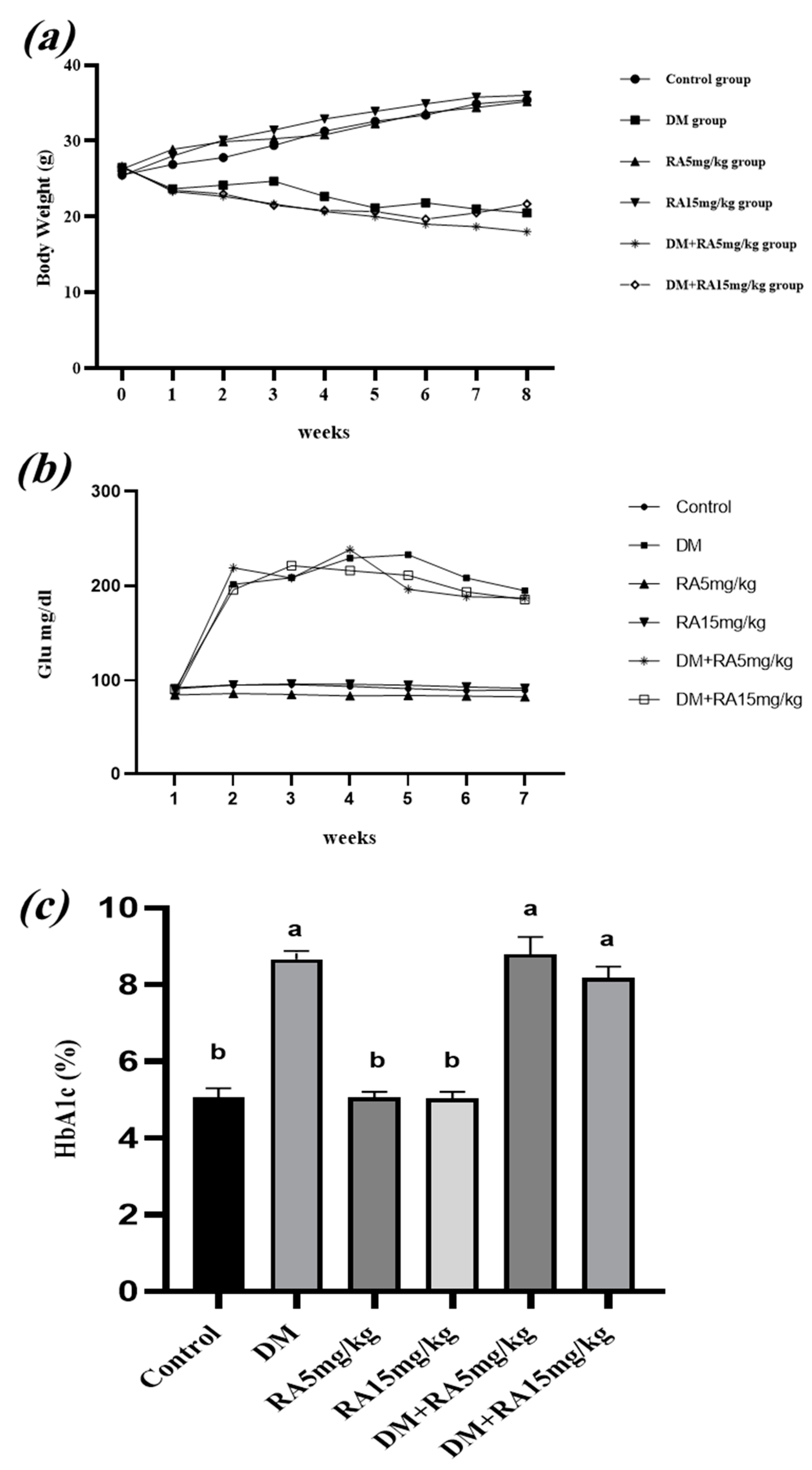
Figure 1. Effects of RA on body weight, FBG levels, and HbA1c in diabetic mice. (a) Average mean body weight during the experimental period. (b) Average mean values of fasting glucose level before and during treatment. (c) HbA1c levels. All values are expressed as the mean ± SEM. The letters on the bars mean the following: a: significant difference when compared to the control group at p < 0.05; b: significant difference when compared to the DM group at p < 0.05.
Stresses 04 00032 g001
Figure 2. Effects of RA on sperm parameters in diabetic mice: (a) Sperm count. (b) Percentage of motile sperms. (c) Percentage of viable sperm. (d) Percentage of sperm with normal morphology. All values are expressed as the mean ± SEM. The letters above the bars mean the following: a: significant difference when compared to the control group at p < 0.05; b: significant difference when compared to the DM group at p < 0.05; ab: significant difference when compared to the control and DM groups at p < 0.05.
Stresses 04 00032 g002
Figure 3. Effects of RA on oxidative stress in the testis of diabetic mice: (a) TBARS levels. (b) Concentration of reduced glutathione (GSH). (c) SOD activity. (d) Catalase (CAT) absorbance. All values are expressed as the mean ± SEM. The letters above the bars mean the following: a: significant difference when compared to the control group at p < 0.05; b: significant difference when compared to the DM group at p < 0.05; ab: significant difference when compared to control and DM group at p < 0.05.
Stresses 04 00032 g003
Figure 4. Photomicrographs obtained from the testes of mice from (a) the control group, (b) the DM group, (c) the RA5 mg/kg group, (d) the RA15 mg/kg group, (e) the DM + RA5 mg/kg group, and (f) the DM + RA15 mg/kg group (X400; H&E); scale bar: 100 uM. Early spermatids (ES), fibroblasts (F), hyalinization in the lumen of some seminiferous tubules (H), interstitial cells of Leydig (LC), late spermatids (LS), myoid cells (M), primary spermatocytes (PS), pyknotic and necrotic spermatogenic cells (Pk), seminiferous tubules (ST), Sertoli cells (SC), spermatid giant cells (GC), spermatids (S), spermatogonia (SG), vacuoles (V). Sloughing germinal epithelium and irregular basement membrane (*), absence of spermatogenesis (red circle).
Stresses 04 00032 g004
Figure 5. Representative images of TUNEL-stained sections in the testes of mice: (a) Control group. (b) DM group. (c) RA5 mg/kg group. (d) RA15 mg/kg group. (e) DM + RA5 mg/kg group. (f) DM + RA15 mg/kg group. In these fluorescent microscopic images, the bright green color represents DNA-fragmented cells, and the blue color represents the DAPI stain, meaning intact DNA. Scale bar: 10 μm.
Stresses 04 00032 g005
Disclaimer/Publisher’s Note: The statements, opinions and data contained in all publications are solely those of the individual author(s) and contributor(s) and not of MDPI and/or the editor(s). MDPI and/or the editor(s) disclaim responsibility for any injury to people or property resulting from any ideas, methods, instructions or products referred to in the content.