Article

Tools for In Vitro Propagation/Synchronization of the Liverwort Marchantia polymorpha and Application of a Validated HPLC-ESI-MS-MS Method for Glutathione and Phytochelatin Analysis

1
Dipartimento di Biologia, Università di Pisa, Via Luca Ghini 13, 56126 Pisa, Italy
2
Dipartimento di Patologia Chirurgica, Medica, Molecolare e dell’Area Critica, Università di Pisa, Via Roma 67, 56126 Pisa, Italy
*
Author to whom correspondence should be addressed.
Stresses 2022, 2(1), 136-145; https://doi.org/10.3390/stresses2010010
Submission received: 29 December 2021 / Revised: 18 February 2022 / Accepted: 22 February 2022 / Published: 24 February 2022
(This article belongs to the Special Issue Responses and Defense Mechanisms against Toxic Metals)

Abstract

:
Bryophytes, due to their poikilohydric nature and peculiar traits, are useful and versatile organisms for studies on metal accumulation and detoxification in plants. Among bryophytes, the liverwort Marchantia polymorpha is an excellent candidate as a model organism, having a key role in plant evolutionary history. In particular, M. polymorpha axenic cultivation of gametophytes offers several advantages, such as fast growth, easy propagation and high efficiency of crossing. Thus, the main purpose of this work was to promote and validate experimental procedures useful in the establishment of a standardized set-up of M. polymorpha gametophytes, as well as to study cadmium detoxification processes in terms of thiol-peptide production, detection and characterisation by HPLC-mass spectrometry. The results show how variations in the composition of the Murashige and Skoog medium impact the growth rate or development of this liverwort, and what levels of glutathione and phytochelatins are produced by gametophytes to counteract cadmium stress.

1. Introduction

Heavy metals are ubiquitous chemical elements present in all environmental matrices (soil, water, atmosphere), derived from both natural sources (e.g., pedogenetic processes) and anthropogenic emissions (e.g., as a byproduct of industrial activities, vehicle traffic, heating systems, etc.) [1,2]. Some studies have shown that, in nature, there is a correspondence between heavy metal and vegetation cover [3], which could offer an interesting reading key for future research in this field. The polluting capacity of heavy metals is due to their intrinsic toxicity combined with their long persistence, high solubility and ability to accumulate in organisms in higher concentrations than in the surrounding environment [2,4]. Historically, the term “heavy metals” indicates all transition elements with a density higher than 5 g cm−3 [2], although further methods of classification have recently been put forward, based on their affinity with functional groups such as thiols, carboxyls and amines [5].
The heavy metals which are toxic for plants, animals and the vast majority of organisms are cadmium (Cd), hexavalent chromium (CrVI), mercury (Hg), lead (Pb) and thallium (Tl), among others. However, certain heavy metals, such as iron (Fe), zinc (Zn), copper (Cu), molybdenum (Mo) and manganese (Mn), below a specific toxicity threshold, are indeed essential nutrients. For this reason, plants have evolved specific mechanisms for regulating essential metal uptake while avoiding deficiencies and toxicity effects [6].
In particular, bryophytes (including Marchantiophyta, Bryophyta and Anthocerotophyta) might prove to be useful model organisms for gaining a better understanding of metal accumulation and detoxification processes in plants [5,7]. Due to their poikilohydric nature, bryophytes do not possess strong hydrophobic barriers. Therefore, all nutrients and toxic elements can be absorbed by their entire body [7]. Moreover, because of their morpho-physiological characteristics, they can often be used as environmental biomonitors, particularly for monitoring air and precipitation quality [8,9].
Among bryophytes, the thalloid liverwort Marchantia polymorpha L. subsp. ruderalis Bischl. and Boisselier-Dubayle is currently recognized as an excellent model organism in studies of land plant evolution and phylogeny [10,11]. This liverwort is a dioecious species, having male and female gametophytes that can reproduce in a sexual or asexual manner. Clonal reproduction of M. polymorpha involves the formation of multicellular gemmae within gemmae “cups,” which are present on the dorsal part of the mature gametophyte, generally at each bifurcation [10]. Transplanted gemmae are able to develop, and, under axenic conditions, give rise to new clonal organisms [12,13]. Progress in transformation techniques [14,15,16,17] and the sequencing of the entire genome (about 280 Mb; [11]) have further promoted the use of M. polymorpha as a fundamental model organism in functional, molecular and evolutionary studies on land plants. Indeed, M. polymorpha gametophytes offer several advantages, such as a short life cycle, a relatively small genome size and a haploid number of chromosome sets. In addition, they can be easily cultivated and propagated axenically, avoiding genetic and metabolic variability from sexual processes or interaction with other organisms [16]. M. polymorpha could thus prove an excellent model organism for investigating the molecular and cellular process linked to heavy metal detoxification. Among these mechanisms, a key role is played by phytochelatins (PCn), small thiol-peptides whose general structure is (γ-Glu-Cys)n-Gly (n = 2–5) [18,19]. PCn mediate free metal chelation in cytosol and subsequent transport into the vacuole to avoid the free circulation of metal ions, thereby reducing their toxic effect. PCn synthesis is catalyzed by the phytochelatin synthase (PCS) enzyme, a gamma-glutamylcysteine-dipeptidyl-(trans)peptidase, belonging to the “Clan CA” of papain-like cysteine proteases [20,21,22,23]. It has been demonstrated that this enzyme’s activity is linked to the presence of heavy metals such as cadmium (Cd), mercury (Hg) and lead (Pb), as well as the metalloid arsenic (As) [19,24,25]. Recently, PCS activation has not only been detected in tracheophytes (chiefly in angiosperms), but also in bryophytes and charophytes, in response to metallic micronutrients such as Fe, Zn, Cu and the toxic metal Cd [26,27,28]. Moreover, as proposed by Maresca et al. (2020) [29], their role as metal detoxifier could make PCn indicators of metal stress, and biomarkers for monitoring such stress in plants. This demands the validation of PCn analytic methods which could be reliably employed with various plant systems.
Accordingly, the principal aims of this work are to clarify how M. polymorpha could be grown in axenic conditions, and to examine aspects of its ability to detoxify heavy metals using a validated mass spectrometry method.

2. Results and Discussion

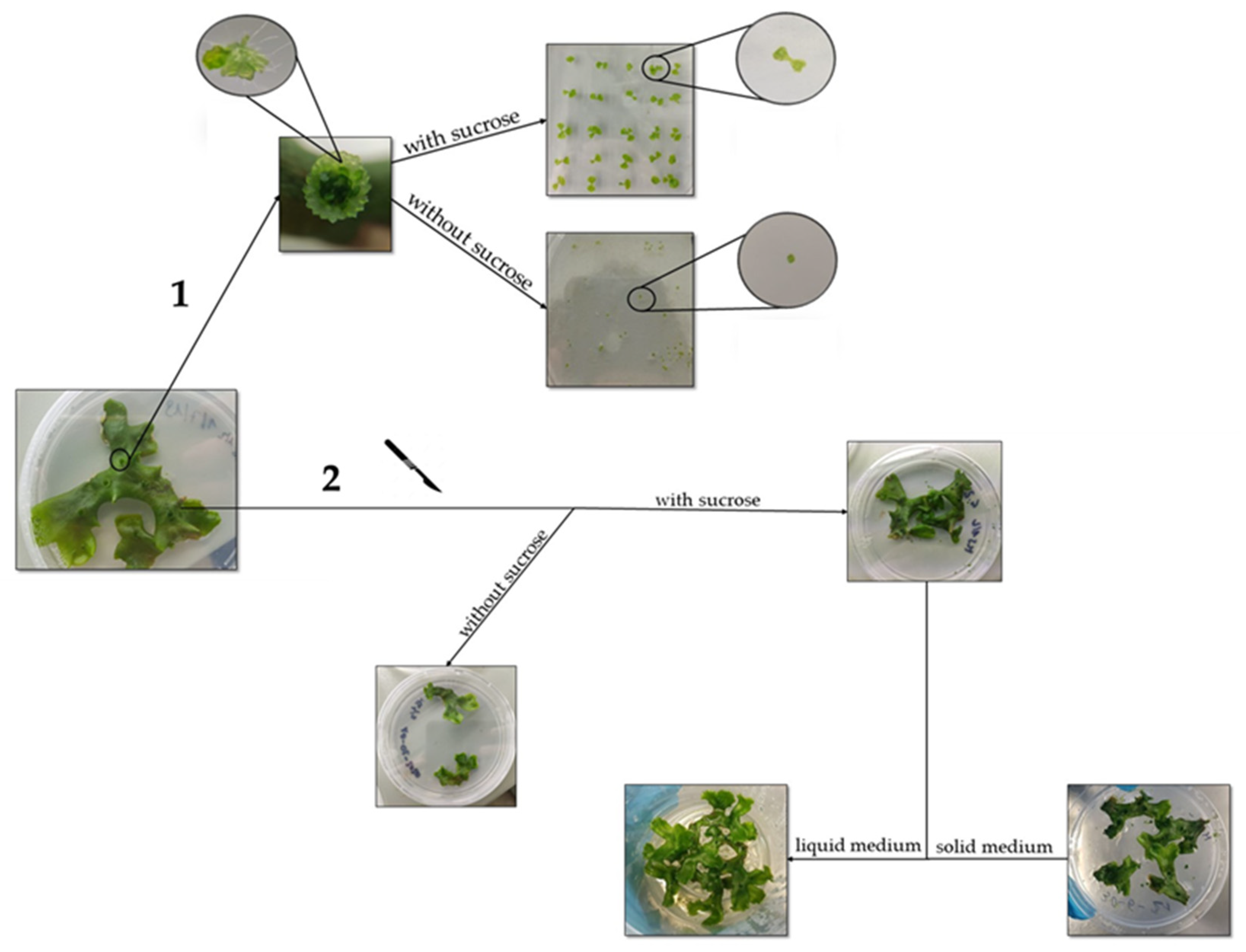
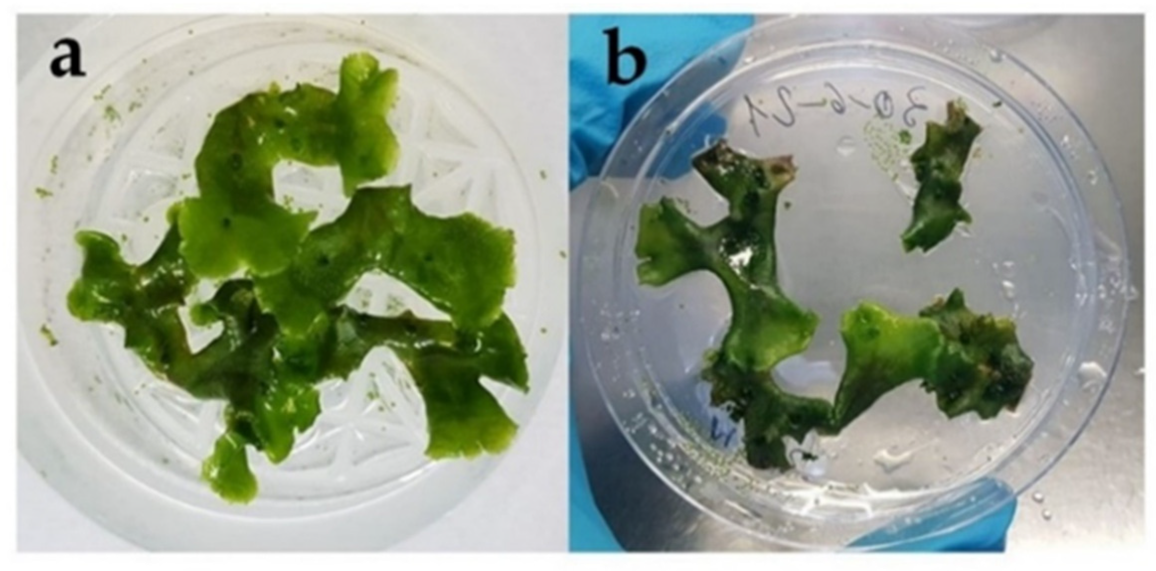
2.1. The In Vitro Propagation Protocol Employed Enables Rapid Synchronization of Gametophyte Growth

In this work, we tested two alternative methods of vegetative propagation of M. polymorpha gametophytes, both using a Murashige and Skoog half-strength (MS ½) medium, pH 5.7 (Figure 1).
In the first case, propagation commenced with gemmae taken from gemmae cups. The gemmae were produced in large quantities in cups, and were clones of the organism from which they were generated [12,13]. The gemmae, which were placed in a solid MS ½ medium and exposed to a light source, promptly developed and yielded a genetically identical clone. The results clearly showed that sucrose was fundamental for the gemmae to grow and develop into a new gametophyte (Figure 1). Indeed, as reported by Ishizaki et al. (2016) [30], sucrose is also essential for gemmae cup formation, which in turn depends on environmental factors and nutrients in the medium. Sucrose appears to contribute to cell cycle progression [31,32] and plays an important role in maintaining the gametophyte culture.
The second propagation method was based on cutting off the part of the gametophyte whose lobes contained the meristem [30]. One considerable advantage of M. polymorpha as a model organism is that a small part of the gametophyte suffices for regenerating the entire one, without the need for any growth regulator [16,33]. However, regenerated gametophytes grown in the absence of sucrose showed a significantly lower growth rate than those grown with sucrose (Figure 1). Once gametophytes of an appropriate size were obtained, particularly with sucrose addition, they could either be grown in a liquid medium using pots, or in a solid medium using Petri dishes, as shown in Figure 1.
After 2 weeks of cultivation, the differences between the growth rates of gametophytes grown in liquid and solid mediums were observed (Figure 2). In fact, the gametophytes grew faster in a liquid medium than in solid MS ½, becoming larger in size (Figure 2a).
This increased growth rate could likely be attributed to the greater facilitation of nutrient acquisition in the liquid medium than in the solid one. It is important to note that a liquid medium is suitable for metal treatment as it enables the homogeneous, enhanced absorption of the elements contained in the medium.

2.2. Gametophytes of M. polymorpha Lead to Rapid Medium Acidification

It has been widely demonstrated that the growth of plants may result in the acidification of the environment where they live [34]. In fact, acidification of the medium seems to increase the availability and acquisition of nutrients used for the organism’s growth. The pH value of the liquid MS ½ medium (initially set to a pH value equal to 5.7) was accordingly measured while growing our cultures, to understand the mechanisms behind the faster growth of M. polymorpha in the liquid medium. These data were monitored for 20 days in the liquid medium where M. polymorpha gametophytes were growing. The results are shown in Figure 3.
Interestingly, a rapid lowering of pH was already detected after the first 24 h of growth, reaching values of around 4.4. After additional few days, the pH progressively dropped to a value of 3.5–3.2, and remained constant around this value over the following days (Figure 3). The strong acidification observed in M. polymorpha cultures was also recorded by Rico-Resendiz and colleagues [35] under phosphate starvation conditions, highlighting that acidification can be a mechanism for increasing nutrient availability. Acidification of the medium could be due to the extrusion of metabolites such as organic acids by M. polymorpha gametophytes. It is well known that bryophytes produce large amounts of secondary metabolites [36]. The functions of many of these metabolites are not clearly understood, and many processes still need to be clarified. As a result, in the future, we aim to investigate which molecules extruded by M. polymorpha gametophytes might be responsible for the strong acidification of the medium.

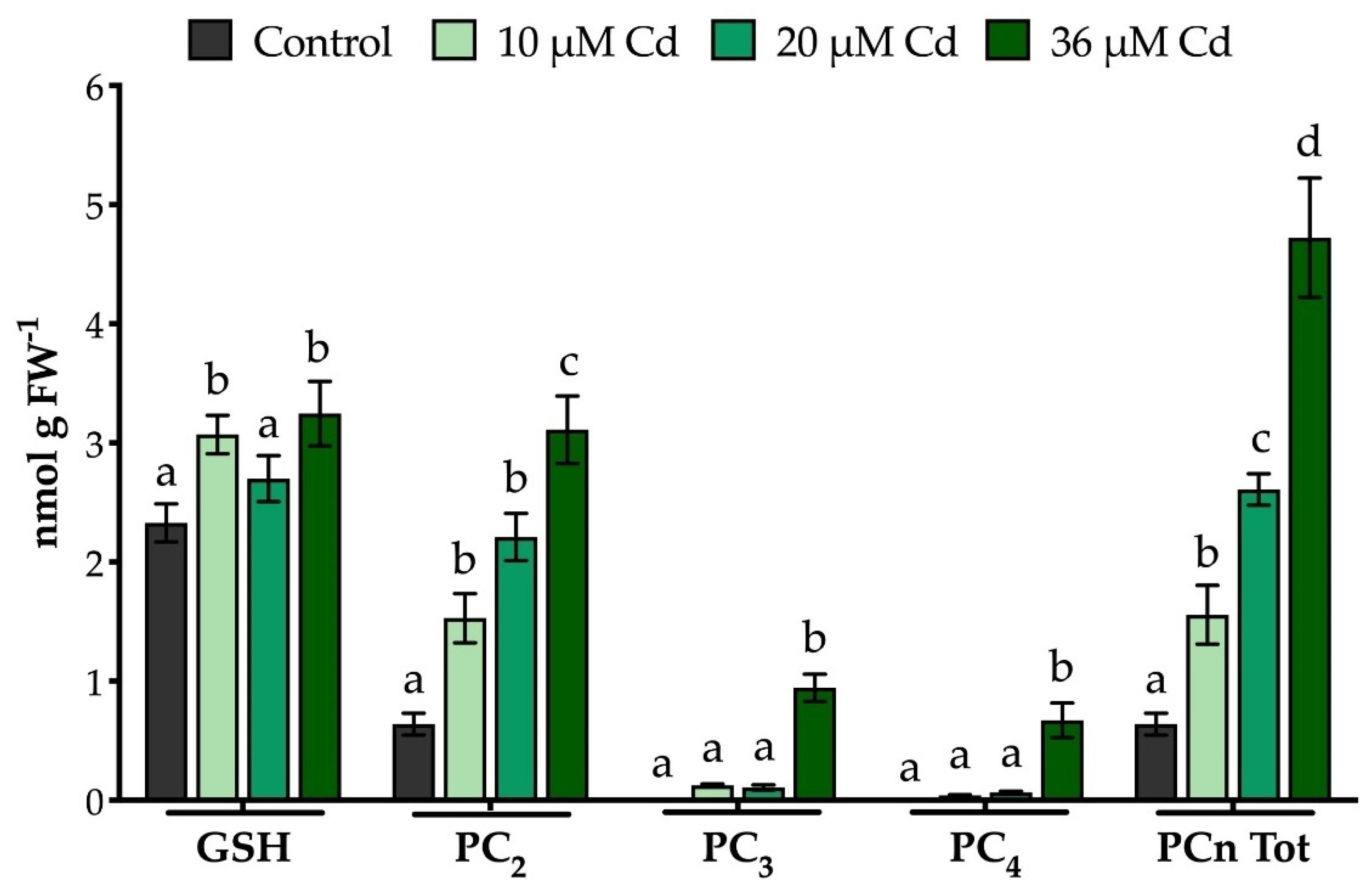
2.3. The Extraction Procedure and the Analytical Method Used Allow an Effective Quali-Quantitative Analysis of Thiol-Peptides in M. polymorpha Gametophytes

Liverworts are known for their ability to accumulate heavy metals in high concentrations through chelation and passive ion exchange mechanisms [5,7]. Their large surface/volume ratio allows them to efficiently absorb water and dissolved elements from the entire surface of the gametophyte [37]. As a result, liverworts such as M. polymorpha appear optimal for studying the mechanisms leading to metal detoxification. One such mechanism is the induction of the ubiquitous PCS enzyme, which is known to be constitutively expressed and fully activated in the presence of metal(loid)s, even in bryophytes and other early diverging streptophytes [26,27,28,38]. However, it should be noted that the extraction, characterization and quantification of analytes in plant extracts are often challenging due to the considerable variability in biological and instrumental terms. This is particularly the case of bryophytes, which produce large amounts of secondary metabolites [39,40]. The analysis of thiol-peptides is particularly critical due to their sensitivity to oxidation reactions, which frequently involve the formation of inter- and intra-molecular disulphide bonds during sample preparation or storage [41,42].
In this work, we tested the method used for thiol-peptide extraction and analysis in HPLC-ESI-MS-MS, as previously validated with Arabidopsis thaliana [42], in M. polymorpha gametophytes. Although slight alterations were made to the extraction procedure, such as adding a semi-purification step to reduce secondary metabolite interference, the method enabled the accurate characterization and quantification of GSH and PCn in M. polymorpha samples using HPLC-ESI-MS-MS analyses, as shown in Figure 4.
The results of the in vivo quantification of the thiol-peptides were consistent with those obtained by Bellini et al. (2020) (Figure 5) [43]. Indeed, the constitutive levels of GSH in the liverwort were somewhat lower than those normally found in higher plants, but wholly comparable with those quantified in other liverworts [26,27]. The sole exception is Pellia epiphylla, which is the only species with GSH levels 6–60 times higher than those of other liverworts [27], M. polymorpha included. In our experiments, the GSH levels increased under Cd treatments, possibly reflecting re-synthesis by the glutathione synthase enzyme and/or the reduction of oxidized glutathione (GSSG) by the NADPH-dependent glutathione reductase. Indeed, this thiol-tripeptide is a ubiquitous molecule in all organisms and is involved in a plethora of cellular responses, mainly functioning as an antioxidant and regulator of redox homeostatic processes [44,45]. With regard to PCn, the data confirmed the constitutive basal activity of M. polymorpha PCS [43], with control plants producing only PC2 (Figure 5). At all concentrations, the presence of Cd resulted in M. polymorpha PCS being promptly activated, with PC2, PC3 and PC4 synthesis (Figure 5). For total PCn, the observations seem to support an increasing trend in the production of PCn as the Cd concentrations grow. It is also important to note that the degree of polymerization of PCn is directly proportional to their chelating power, which depends on the number of thiol groups in the molecular structure [46]. Interestingly, as far as we know, PCn up to PC4 have only been detected in Conocephalum conicum and Lunularia cruciata liverworts [26,27], whereas in all the other species assayed so far, PCn production has been limited to only PC2 (and to PC2 and PC3 in just one case) [27]. This may be due to the actual non-production of PCn with high polymerization levels in other liverwort species, or to the fact that a non-optimized analytical method was adopted. As mentioned above, high levels of secondary metabolites in the extracts may actually generate a strong matrix effect, resulting in the masking of the analytes and causing a considerable reduction in the sensitivity of the method used [42].

3. Materials and Methods

3.1. Set-Up of the In Vitro Cultures, Growth Conditions and Vegetative Propagation of M. polymorpha Gametophytes

Axenic cultures of Marchantia polymorpha L. subsp. ruderalis Bischl. and Boisselier-Dubayle (Marchantiales, Marchantiophyta) female gametophytes (Cam2–Cambridge-2 wild type, University of Cambridge, UK) were grown both in a liquid medium using pots and in a solid medium using Petri dishes (Ø 100 mm). In both cases, sterile half-strength (½) Murashige & Skoog (MS) medium was used, pH 5.7, supplemented with 0.8 % (w/v) sucrose (Duchefa Biochemie). In the pots, 200 mL of liquid medium was employed, whereas in each Petri dish the volume used totalled 20 mL, supplemented with 0.7 % (w/v) Plant Agar (Duchefa Biochemie). Gametophytes in dishes and pots were placed in a growth chamber with a light/dark cycle of 16:8 h with a photosynthetic photon flux density of 60 μmol m−2 s−1, a temperature of 19 ± 1 °C and 60% relative humidity. The gametophytes were propagated from gemmae (collected from gemmae cups) in a solid MS ½ medium, or from pieces cut from the gametophyte and propagated using a solid or liquid medium. The axenic cultures of gametophytes were set up and propagated using sterile forceps and scalpel under a vertical laminar flow. Sterilization of the scalpel blade and forceps was performed using a glass bead sterilizer at 300 °C. The scalpel and forceps were then cooled using sterilized filter paper soaked with sterile deionized water. After that, the instruments were used immediately for handling the plant material.

3.2. Metal Treatments and pH Measurements

Axenic gametophytes started from gemmae were grown in a solid MS ½ medium for 6 weeks and transferred into fresh dishes every 2 weeks. The resulting gametophytes were moved into sterile pots filled with 200 mL of liquid MS ½ medium and left to grow for a further week to acclimatize to the new conditions. After 1 week in the liquid medium, 7-week-old gametophytes (about 0.8 g FW) were independently moved to Petri dishes (Ø 60 mm) with 5 mL of liquid MS ½ medium, pH 5.7, and placed in different treatment conditions. Control samples with homeostatic concentrations of micronutrients corresponding to the same concentrations in the MS ½ medium were set up. Samples treated with Cd had the following concentrations: 10, 20 and 36 μM Cd2+ (provided as 3 CdSO4·8 H2O) for 72 h. Five biological replicates were performed for both the control samples and treated ones. At the end of the exposure time, the gametophytes were collected, dried with filter paper, divided into samples with a fresh weight of 100 mg, frozen in liquid nitrogen and stored at −80 °C for further analyses. The pH values of the liquid MS ½ medium were measured to gauge whether they had been influenced by Cd or not. The pH of the liquid half-strength MS medium was measured in the dishes and in the pots during growth of gametophytes, and at the end of each treatment.

3.3. Extraction, Characterization and Quantification of Thiol-Peptides

Gametophyte samples of 100 mg FW, previously stored at −80 °C, were extracted mainly following Bellini et al. (2019) [42], with slight modifications. Briefly, each sample was ground into a powder using liquid nitrogen, a mixer mill (MM200, Retsch, Haan, Germany) and two agate grinding balls (Ø 5 mm) to facilitate mechanical cell breakage using a frequency of 30 Hz for 1 min. Then, 300 μL of a previously prepared and ice-cool extraction buffer (5% w/v of salicylsulfonic acid, 6.3 mM of diethylenetetraminopentaacetic acid (DTPA) and 2 mM of tris (2-carboxyethyl) phosphine acid (TCEP)) were added to each sample, together with 200 ng mL−1 of internal standards (GSH and PC2), which had glycine marked with stable isotopes (15N, 13C): 13C2,15N-GSH and 13C2,15N-PC2 (AnaSpec Inc., Fremont, CA, USA) The suspension obtained was vortexed for 30 s, kept in an ice bath for 15 min and vortexed every 5 min. The extract was then centrifuged at 20,000× g (Hermle, Z 300 K, Wehingen, Germany) for 20 min at 4 °C. Unlike the results published by Bellini et al. (2019) [42], in this work, each supernatant was semi-purified using Amicon Ultracentrifuge filters (10 kDa) as an alternative to Minisart RC4 0.45 μm filters (Sartorius, Goettingen, Germany) to reduce any matrix effect. The samples were centrifuged at 14,000× g (Hermle, Z 300 K, Wehingen, Germany) for 30 min at 4 °C using Amicon Ultracentrifuge filters (10 kDa). The flow-through was then saved. Samples were stored at −80 °C until analyzed in HPLC-ESI-MS-MS, which was conducted using the analytical method validated by Bellini et al. (2019) [42].

4. Conclusions

The in vitro growth and cultivation in axenic conditions of model plants such as the liverwort M. polymorpha can represent an important basis for the development of effective protocols useful for investigating the functional and molecular responses of early diverging streptophytes to toxic heavy metals, such as Cd. In this work, we developed a reliable in vitro cultivation protocol of M. polymorpha gametophytes. Moreover, we characterized this liverwort with respect to medium acidification, as well as its thiol-peptide production (GSH, PCn) following Cd supply for 72 h. The data collected may prove to be a reference point for any future study involving the model organism M. polymorpha. It may be of interest to evaluate the nature of molecules extruded by this liverwort that result in acidification of the growth medium, in order to deepen the causes of this process. In addition, M. polymorpha can be used to define the molecular and cellular pathways of heavy metal detoxification and the repair of damage caused by the induced stress. Therefore, it could be interesting to investigate the role of tonoplast and plasmalemma transporters involved in heavy metal movement across membranes, either as free ions or complexed to thiol-peptides. Overall, M. polymorpha represents an excellent model system for evolutionary, functional, molecular and biomonitoring studies of heavy metals present in the environment.

Author Contributions

Conceptualization, L.S.d.T. and E.B. (Erika Bellini); methodology, S.G. and E.B. (Elena Bandoni); validation, A.S. and L.S.d.T.; formal analysis, S.G., E.B. (Erika Bellini) and A.S.; investigation, L.S.d.T.; data curation, S.G., E.B. (Erika Bellini) and E.B. (Elena Bandoni); writing—original draft preparation, S.G., E.B. (Erika Bellini) and L.S.d.T.; writing—review and editing, S.G., E.B. (Erika Bellini), A.S. and L.S.d.T.; supervision, A.S. and L.S.d.T.; project administration, L.S.d.T. All authors have read and agreed to the published version of the manuscript.

Funding

This research received no external funding.

Conflicts of Interest

The authors declare no conflict of interest.

References

  1. Frausto da Silva, J.J.R.; Williams, R.J.P. The Biological Chemistry of the Elements: The Inorganic Chemistry of Life; OUP Oxford: Oxford, UK, 2001; ISBN 978-0-19-850848-9. [Google Scholar]
  2. Sanità di Toppi, L.; Gabbrielli, R. Response to Cadmium in Higher Plants. Environ. Exp. Bot. 1999, 41, 105–130. [Google Scholar] [CrossRef]
  3. Perrino, E.V.; Brunetti, G.; Farrag, K. Plant Communities in Multi-Metal Contaminated Soils: A Case Study in the National Park of Alta Murgia (Apulia Region - Southern Italy). Int. J. Phytoremed. 2014, 16, 871–888. [Google Scholar] [CrossRef]
  4. Tchounwou, P.B.; Yedjou, C.G.; Patlolla, A.K.; Sutton, D.J. Heavy Metal Toxicity and the Environment. In Molecular, Clinical and Environmental Toxicology: Volume 3: Environmental Toxicology; Luch, A., Ed.; Experientia Supplementum; Springer: Basel, Swizterland, 2012; pp. 133–164. ISBN 978-3-7643-8340-4. [Google Scholar]
  5. Stanković, J.D.; Sabovljević, A.D.; Sabovljević, M.S. Bryophytes and Heavy Metals: A Review. Acta Bot. Croat. 2018, 77, 109–118. [Google Scholar] [CrossRef]
  6. Tennstedt, P.; Peisker, D.; Böttcher, C.; Trampczynska, A.; Clemens, S. Phytochelatin Synthesis Is Essential for the Detoxification of Excess Zinc and Contributes Significantly to the Accumulation of Zinc. Plant Physiol. 2009, 149, 938–948. [Google Scholar] [CrossRef] [Green Version]
  7. Bellini, E.; Betti, C.; Sanità di Toppi, L. Responses to Cadmium in Early-Diverging Streptophytes (Charophytes and Bryophytes): Current Views and Potential Applications. Plants 2021, 10, 770. [Google Scholar] [CrossRef]
  8. Harmens, H.; Ilyin, I.; Mills, G.; Aboal, J.R.; Alber, R.; Blum, O.; Coşkun, M.; De Temmerman, L.; Fernández, J.Á.; Figueira, R.; et al. Country-Specific Correlations across Europe between Modelled Atmospheric Cadmium and Lead Deposition and Concentrations in Mosses. Environ. Pollut. 2012, 166, 1–9. [Google Scholar] [CrossRef] [Green Version]
  9. Shaw, J. Evolution of Heavy Metal Tolerance in Bryophytes Ii. an Ecological and Experimental Investigation of the “Copper Moss,” Scopelophila cataractae (Pottiaceae). Am. J. Bot. 1987, 74, 813–821. [Google Scholar] [CrossRef]
  10. Shimamura, M. Marchantia polymorpha: Taxonomy, Phylogeny and Morphology of a Model System. Plant Cell Physiol. 2016, 57, 230–256. [Google Scholar] [CrossRef] [Green Version]
  11. Bowman, J.L.; Kohchi, T.; Yamato, K.T.; Jenkins, J.; Shu, S.; Ishizaki, K.; Yamaoka, S.; Nishihama, R.; Nakamura, Y.; Berger, F.; et al. Insights into Land Plant Evolution Garnered from the Marchantia polymorpha Genome. Cell 2017, 171, 287–304.e15. [Google Scholar] [CrossRef] [Green Version]
  12. Berger, F.; Bowman, J.L.; Kohchi, T. Marchantia. Curr. Biol. 2016, 26, R186–R187. [CrossRef] [Green Version]
  13. Nakazato, T.; Kadota, A.; Wada, M. Photoinduction of Spore Germination in Marchantia polymorpha L. Is Mediated by Photosynthesis. Plant Cell Physiol. 1999, 40, 1014–1020. [Google Scholar] [CrossRef] [Green Version]
  14. Ishizaki, K.; Chiyoda, S.; Yamato, K.T.; Kohchi, T. Agrobacterium-Mediated Transformation of the Haploid Liverwort Marchantia polymorpha L., an Emerging Model for Plant Biology. Plant Cell Physiol. 2008, 49, 1084–1091. [Google Scholar] [CrossRef] [Green Version]
  15. Ishizaki, K.; Johzuka-Hisatomi, Y.; Ishida, S.; Iida, S.; Kohchi, T. Homologous Recombination-Mediated Gene Targeting in the Liverwort Marchantia polymorpha L. Sci. Rep. 2013, 3, 1532. [Google Scholar] [CrossRef]
  16. Kubota, A.; Ishizaki, K.; Hosaka, M.; Kohchi, T. Efficient Agrobacterium -Mediated Transformation of the Liverwort Marchantia polymorpha Using Regenerating Thalli. Biosci. Biotechnol. Biochem. 2013, 77, 167–172. [Google Scholar] [CrossRef] [Green Version]
  17. Sugano, S.S.; Shirakawa, M.; Takagi, J.; Matsuda, Y.; Shimada, T.; Hara-Nishimura, I.; Kohchi, T. CRISPR/Cas9-Mediated Targeted Mutagenesis in the Liverwort Marchantia polymorpha L. Plant Cell Physiol. 2014, 55, 475–481. [Google Scholar] [CrossRef]
  18. Bräutigam, A.; Schaumlöffel, D.; Krauss, G.-J.; Wesenberg, D. Analytical Approach for Characterization of Cadmium-Induced Thiol Peptides—a Case Study Using Chlamydomonas reinhardtii. Anal. Bioanal. Chem. 2009, 395, 1737–1747. [Google Scholar] [CrossRef]
  19. Grill, E.; Löffler, S.; Winnacker, E.-L.; Zenk, M.H. Phytochelatins, the Heavy-Metal-Binding Peptides of Plants, Are Synthesized from Glutathione by a Specific γ-Glutamylcysteine Dipeptidyl Transpeptidase (Phytochelatin Synthase). Proc. Natl. Acad. Sci. USA 1989, 86, 6838–6842. [Google Scholar] [CrossRef] [Green Version]
  20. Rea, P.A. Phytochelatin Synthase: Of a Protease a Peptide Polymerase Made. Physiol. Plant. 2012, 145, 154–164. [Google Scholar] [CrossRef]
  21. Romanyuk, N.D.; Rigden, D.J.; Vatamaniuk, O.K.; Lang, A.; Cahoon, R.E.; Jez, J.M.; Rea, P.A. Mutagenic Definition of a Papain-Like Catalytic Triad, Sufficiency of the N-Terminal Domain for Single-Site Core Catalytic Enzyme Acylation, and C-Terminal Domain for Augmentative Metal Activation of a Eukaryotic Phytochelatin Synthase. Plant Physiol. 2006, 141, 858–869. [Google Scholar] [CrossRef] [Green Version]
  22. Vivares, D.; Arnoux, P.; Pignol, D. A Papain-like Enzyme at Work: Native and Acyl-Enzyme Intermediate Structures in Phytochelatin Synthesis. Proc. Natl. Acad. Sci. USA 2005, 102, 18848–18853. [Google Scholar] [CrossRef] [Green Version]
  23. Rea, P.A.; Vatamaniuk, O.K.; Rigden, D.J. Weeds, Worms, and More. Papain’s Long-Lost Cousin, Phytochelatin Synthase. Plant Physiol. 2004, 136, 2463–2474. [Google Scholar] [CrossRef] [Green Version]
  24. Clemens, S. Toxic Metal Accumulation, Responses to Exposure and Mechanisms of Tolerance in Plants. Biochimie 2006, 88, 1707–1719. [Google Scholar] [CrossRef]
  25. Vatamaniuk, O.K.; Mari, S.; Lu, Y.-P.; Rea, P.A. Mechanism of Heavy Metal Ion Activation of Phytochelatin (PC) Synthase. J. Biol. Chem. 2000, 275, 31451–31459. [Google Scholar] [CrossRef] [Green Version]
  26. Degola, F.; De Benedictis, M.; Petraglia, A.; Massimi, A.; Fattorini, L.; Sorbo, S.; Basile, A.; Sanità di Toppi, L. A Cd/Fe/Zn-Responsive Phytochelatin Synthase Is Constitutively Present in the Ancient Liverwort Lunularia cruciata (L.) Dumort. Plant Cell Physiol. 2014, 55, 1884–1891. [Google Scholar] [CrossRef] [Green Version]
  27. Petraglia, A.; De Benedictis, M.; Degola, F.; Pastore, G.; Calcagno, M.; Ruotolo, R.; Mengoni, A.; Sanità di Toppi, L. The Capability to Synthesize Phytochelatins and the Presence of Constitutive and Functional Phytochelatin Synthases Are Ancestral (Plesiomorphic) Characters for Basal Land Plants. J. Exp. Bot. 2014, 65, 1153–1163. [Google Scholar] [CrossRef] [Green Version]
  28. Fontanini, D.; Andreucci, A.; Ruffini Castiglione, M.; Basile, A.; Sorbo, S.; Petraglia, A.; Degola, F.; Bellini, E.; Bruno, L.; Varotto, C.; et al. The Phytochelatin Synthase from Nitella mucronata (Charophyta) Plays a Role in the Homeostatic Control of Iron(II)/(III). Plant Physiol. Biochem. 2018, 127, 88–96. [Google Scholar] [CrossRef]
  29. Maresca, V.; Sorbo, S.; Loppi, S.; Funaro, F.; Del Prete, D.; Basile, A. Biological Effects from Environmental Pollution by Toxic Metals in the “Land of Fires” (Italy) Assessed Using the Biomonitor Species Lunularia cruciata L. (Dum). Environ. Pollut. 2020, 265, 115000. [Google Scholar] [CrossRef]
  30. Ishizaki, K.; Nishihama, R.; Yamato, K.T.; Kohchi, T. Molecular Genetic Tools and Techniques for Marchantia Polymorpha Research. Plant Cell Physiol. 2016, 57, 262–270. [Google Scholar] [CrossRef] [Green Version]
  31. Riou-Khamlichi, C.; Menges, M.; Healy, J.M.S.; Murray, J.A.H. Sugar Control of the Plant Cell Cycle: Differential Regulation of Arabidopsis D-Type Cyclin Gene Expression. Mol. Cell. Biol. 2000, 20, 4513–4521. [Google Scholar] [CrossRef] [Green Version]
  32. Xiong, Y.; McCormack, M.; Li, L.; Hall, Q.; Xiang, C.; Sheen, J. Glucose–TOR Signalling Reprograms the Transcriptome and Activates Meristems. Nature 2013, 496, 181–186. [Google Scholar] [CrossRef] [Green Version]
  33. Nishihama, R.; Ishizaki, K.; Hosaka, M.; Matsuda, Y.; Kubota, A.; Kohchi, T. Phytochrome-Mediated Regulation of Cell Division and Growth during Regeneration and Sporeling Development in the Liverwort Marchantia polymorpha. J. Plant Res. 2015, 128, 407–421. [Google Scholar] [CrossRef] [PubMed] [Green Version]
  34. Marschner, H. Marschner’s Mineral Nutrition of Higher Plants; Academic Press: Cambridge, MA, USA, 2011; ISBN 978-0-12-384906-9. [Google Scholar]
  35. Rico-Reséndiz, F.; Cervantes-Pérez, S.A.; Espinal-Centeno, A.; Dipp-Álvarez, M.; Oropeza-Aburto, A.; Hurtado-Bautista, E.; Cruz-Hernández, A.; Bowman, J.L.; Ishizaki, K.; Arteaga-Vázquez, M.A.; et al. Transcriptional and Morpho-Physiological Responses of Marchantia polymorpha upon Phosphate Starvation. Int. J. Mol. Sci. 2020, 21, 8354. [Google Scholar] [CrossRef] [PubMed]
  36. Asakawa, Y.; Ludwiczuk, A.; Nagashima, F. Chemical Constituents of Bryophyta. In Chemical Constituents of Bryophytes: Bio- and Chemical Diversity, Biological Activity, and Chemosystematics; Asakawa, Y., Ludwiczuk, A., Nagashima, F., Eds.; Progress in the Chemistry of Organic Natural Products; Springer: Vienna, Austria, 2013; pp. 563–605. ISBN 978-3-7091-1084-3. [Google Scholar]
  37. Tyler, G. Bryophytes and Heavy Metals: A Literature Review. Bot. J. Linn. Soc. 1990, 104, 231–253. [Google Scholar] [CrossRef]
  38. Bellini, E.; Maresca, V.; Betti, C.; Castiglione, M.R.; Fontanini, D.; Capocchi, A.; Sorce, C.; Borsò, M.; Bruno, L.; Sorbo, S.; et al. The Moss Leptodictyum riparium Counteracts Severe Cadmium Stress by Activation of Glutathione Transferase and Phytochelatin Synthase, but Slightly by Phytochelatins. Int. J. Mol. Sci. 2020, 21, 1583. [Google Scholar] [CrossRef] [Green Version]
  39. Xie, C.-F.; Lou, H.-X. Secondary Metabolites in Bryophytes: An Ecological Aspect. Chem. Biodivers. 2009, 6, 303–312. [Google Scholar] [CrossRef]
  40. Peters; Treutler; Döll; Kindt; Hankemeier; Neumann Chemical Diversity and Classification of Secondary Metabolites in Nine Bryophyte Species. Metabolites 2019, 9, 222. [CrossRef] [Green Version]
  41. El-Zohri, M.H.A.; Cabala, R.; Frank, H. Quantification of Phytochelatins in Plants by Reversed-Phase HPLC–ESI–MS–MS. Anal. Bioanal. Chem. 2005, 382, 1871–1876. [Google Scholar] [CrossRef]
  42. Bellini, E.; Borsò, M.; Betti, C.; Bruno, L.; Andreucci, A.; Ruffini Castiglione, M.; Saba, A.; Sanità di Toppi, L. Characterization and Quantification of Thiol-Peptides in Arabidopsis thaliana Using Combined Dilution and High Sensitivity HPLC-ESI-MS-MS. Phytochemistry 2019, 164, 215–222. [Google Scholar] [CrossRef]
  43. Bellini, E.; Varotto, C.; Borsò, M.; Rugnini, L.; Bruno, L.; Sanità di Toppi, L. Eukaryotic and Prokaryotic Phytochelatin Synthases Differ Less in Functional Terms Than Previously Thought: A Comparative Analysis of Marchantia polymorpha and Geitlerinema sp. PCC 7407. Plants 2020, 9, 914. [Google Scholar] [CrossRef]
  44. Schafer, F.Q.; Buettner, G.R. Redox Environment of the Cell as Viewed through the Redox State of the Glutathione Disulfide/Glutathione Couple. Free Radic. Biol. Med. 2001, 30, 1191–1212. [Google Scholar] [CrossRef]
  45. Foyer, C.H.; Noctor, G. Redox Regulation in Photosynthetic Organisms: Signaling, Acclimation, and Practical Implications. Antioxid. Redox Signal. 2008, 11, 861–905. [Google Scholar] [CrossRef] [PubMed]
  46. Kneer, R.; Zenk, M.H. The Formation of Cd-Phytochelatin Complexes in Plant Cell Cultures. Phytochemistry 1997, 44, 69–74. [Google Scholar] [CrossRef]
Figure 1. Two alternative vegetative propagation methods of M. polymorpha gametophytes: (1) from gemmae cultivated in solid MS ½ medium, with or without sucrose; (2) from portions of the gametophyte, propagated with and without sucrose in solid MS ½ medium, or supplied with sucrose in liquid MS ½ medium.
Stresses 02 00010 g001
Figure 2. M. polymorpha growth after 2 weeks in liquid (a) and solid (b) MS ½ medium, supplied with sucrose.
Stresses 02 00010 g002
Figure 3. pH values detected over 20 days in pots used for growing M. polymorpha in liquid MS ½ medium, containing sucrose. Values are mean ± SE.
Stresses 02 00010 g003
Figure 4. Representative mass spectrometry chromatograms of a partially purified extract from M. polymorpha in the time range of 0–14 min.
Stresses 02 00010 g004
Figure 5. Thiol-peptide content detected in M. polymorpha gametophytes (control and treated plants with 10, 20 and 36 μM Cd2+ for 72 h). Values are mean ± SE. Differences between levels of thiol-peptides were detected by one-way analysis of variance (ANOVA), followed by Tukey’s multiple comparison post hoc test. Within each group of thiol-peptides, bars not accompanied by the same letter are significantly different at p < 0.05.
Stresses 02 00010 g005
Publisher’s Note: MDPI stays neutral with regard to jurisdictional claims in published maps and institutional affiliations.

