1. Introduction
Manual material handling tasks such as lifting, carrying, lowering, and palletizing are associated with a high prevalence of musculoskeletal disorders, particularly affecting the lumbar spine (
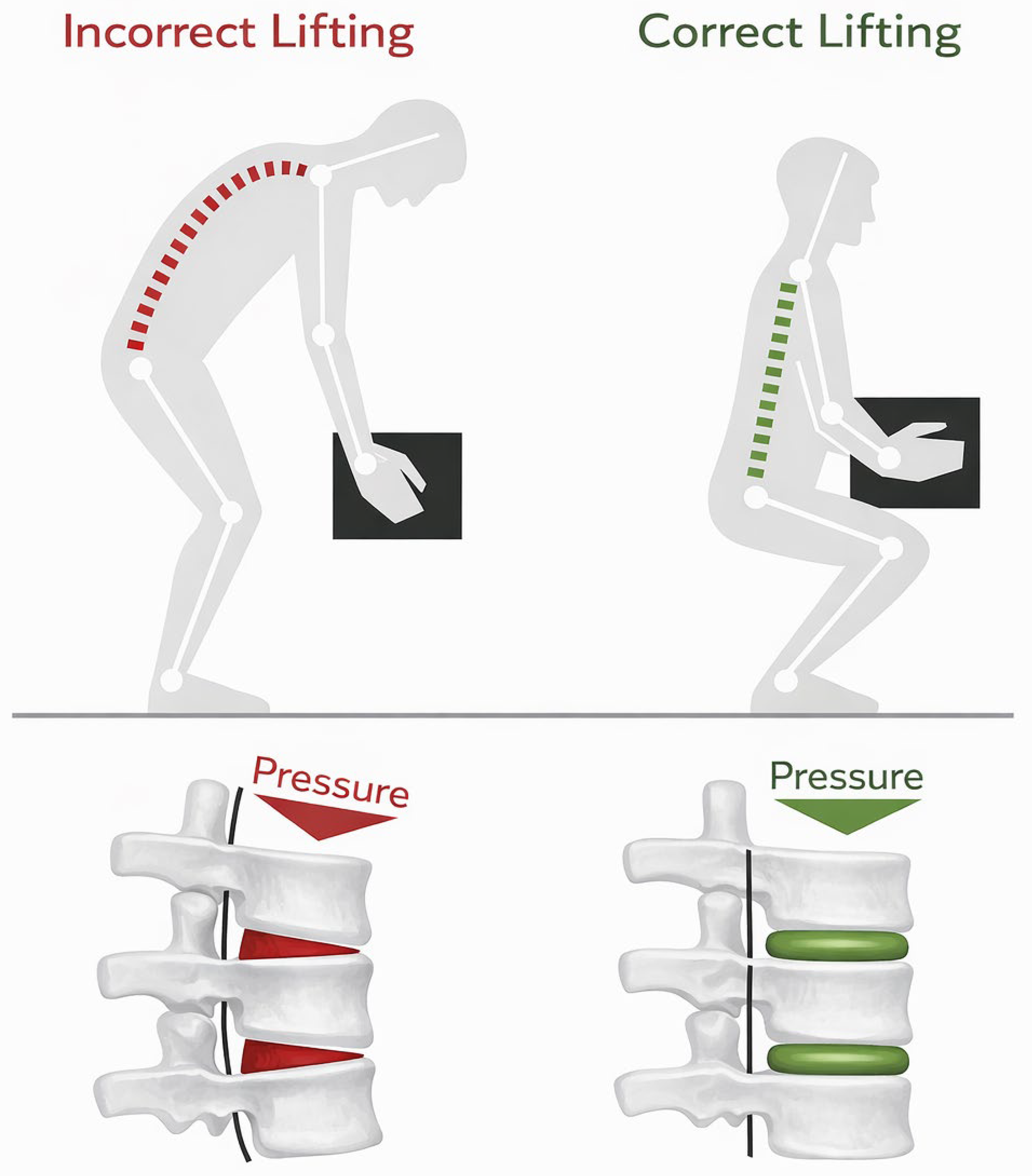
Figure 1) [
1,
2,
3,
4,
5]. Repetitive lifting combined with non-neutral spinal postures can increase biomechanical loading of the spine and may contribute to fatigue, discomfort, and long-term musculoskeletal complaints [
3,
4,
6,
7,
8]. Although ergonomic guidelines and assessment methods for manual handling are well established, their sustained implementation in industrial practice remains challenging [
3,
4,
9].
The human spine plays a central role in load transfer and postural stability during manual work. Its biomechanical behavior is strongly influenced by posture, load position, movement velocity, and repetition frequency [
6,
7,
8]. Forward bending while lifting may increase spinal compression and shear forces, whereas more neutral lifting postures can contribute to a more favorable distribution of mechanical loads [
6,
7]. Therefore, the quantitative assessment of spinal posture is an important component of ergonomic workplace evaluation.
While full automation can effectively reduce physical workload, high investment costs, limited flexibility, and variable task requirements often prevent its adoption, especially in small and medium-sized enterprises [
9,
12,
13]. In this context, wearable assistance systems such as industrial exoskeletons represent a complementary approach rather than a complete replacement of automation. They can be integrated into existing workplaces with comparatively low infrastructural changes and may support employees during physically demanding tasks while preserving human flexibility [
14,
15,
16,
17,
18].
Passive back-support exoskeletons are designed to reduce physical strain by storing mechanical energy during trunk flexion and releasing it during extension [
16,
17,
19]. Previous studies have shown reductions in back-muscle activity, spinal loading indicators, and perceived exertion during lifting or forward-bending tasks [
10,
11,
14,
15,
16,
17,
19,
20]. Many investigations focus on electromyographic measurements, subjective workload ratings, or laboratory-based lifting tasks [
10,
14,
15,
20,
21,
22]. Objective kinematic analyses of spinal posture during realistic work processes such as palletizing remain comparatively scarce [
13,
23,
24,
25].
This pilot study addresses this gap by quantitatively assessing spinal posture during a simulated palletizing task using optical motion capture. The primary aim of this pilot study was to examine the feasibility of a marker-based optical motion-capture procedure for quantifying posture-related angle changes during simulated palletizing. Measurements obtained under the activated, deactivated, and no-exoskeleton conditions were used to demonstrate the application and outputs of the proposed assessment procedure. The study was not designed or statistically powered to establish the biomechanical efficacy of the investigated exoskeleton or to identify factors explaining inter-individual differences.
The scope of the present feasibility study was restricted to the kinematic assessment of marker-defined dorsal segment angles. Complementary biomechanical variables such as muscle activation, external forces, and model-based estimates of spinal loading were not included in the measurement protocol. Consequently, the study was not intended to determine changes in muscular effort, spinal compression, shear forces, or injury risk.
2. Materials and Methods
2.1. Participants
Five healthy adults participated in the study. None of the participants were professionally engaged in logistics or material handling. Two participants had prior training in ergonomic lifting techniques. Anthropometric characteristics varied to capture inter-individual differences in body height and mass (
Table 1). All participants provided informed consent prior to participation.
2.2. Experimental Setup
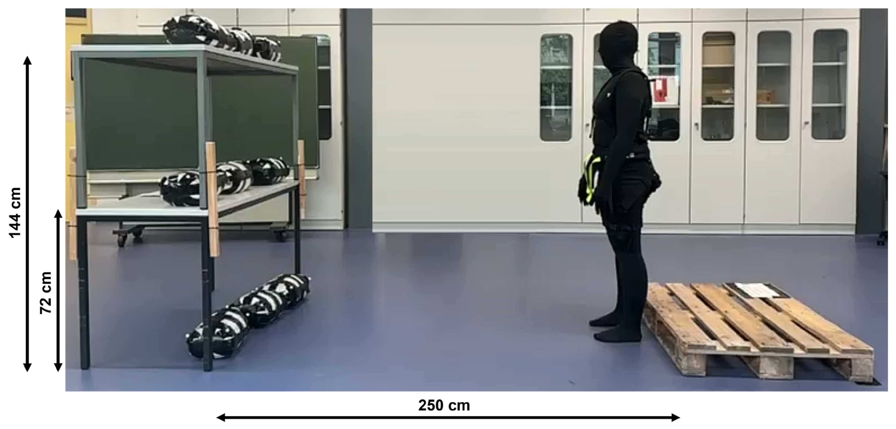
The experiment was conducted in a controlled indoor laboratory environment. A simulated palletizing workstation was constructed, consisting of three shelf levels (floor, hip, and chest height) and a standardized euro pallet placed two meters behind the shelves (
Figure 2).
Each participant performed nine identical palletizing cycles per condition, transferring nine packages from the shelves to the pallet. The packages are filled with 15 kg dust-free gravel, are sized 40 × 25 × 15 cm and are reinforced with duct tape. Three conditions were tested in following order:
All participants completed the three conditions in the same fixed order: activated exoskeleton, deactivated exoskeleton, and no exoskeleton. This sequence was selected as a pragmatic procedure for the initial feasibility study and allowed the level of assistance to be changed before the exoskeleton was removed, thereby limiting repeated removal, refitting, and potential changes in the experimental setup. The sequence was not selected on the basis of expected condition effects. No randomization or counterbalancing was applied; consequently, condition and order effects cannot be separated in the present study.
2.3. Exoskeleton
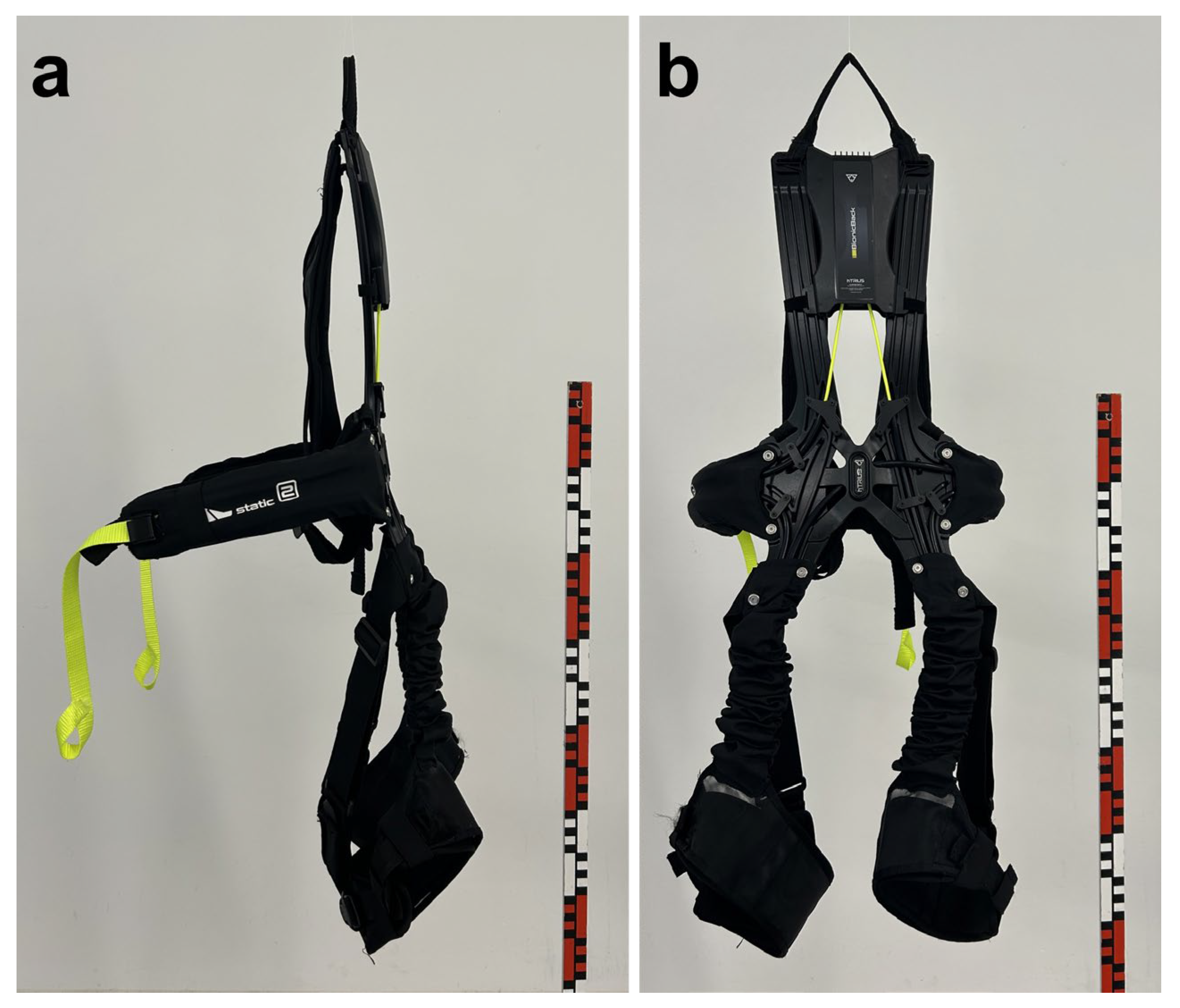
A commercially available passive back-support exoskeleton, the BionicBack (hTRIUS GmbH, Horb, Germany), was used (
Figure 3). The system employs elastic support elements running along the lower back and thigh regions. During trunk flexion, mechanical energy is stored in the elastic elements and released during extension to support the user when returning to an upright posture. This functional principle is consistent with the general mechanical approach of passive back-support exoskeletons described in previous studies [
16,
17,
19].
In the deactivated condition, the exoskeleton was worn without tension in the elastic elements. This condition was included to distinguish the effect of mechanical assistance from possible stabilization, restriction, or proprioceptive effects caused by wearing the device.
2.4. Motion Capture System
Spinal motion was recorded using an optical motion-capture system with twelve infrared cameras (PrimeX22) by Optitrack (NaturalPoint, Inc., Corvallis, OR, USA) operating at 120 Hz with a resolution of +/−0.15 mm. Optical motion-capture systems are widely used for quantitative movement analysis and allow marker-based reconstruction of body segment motion [
26,
27,
28,
29].
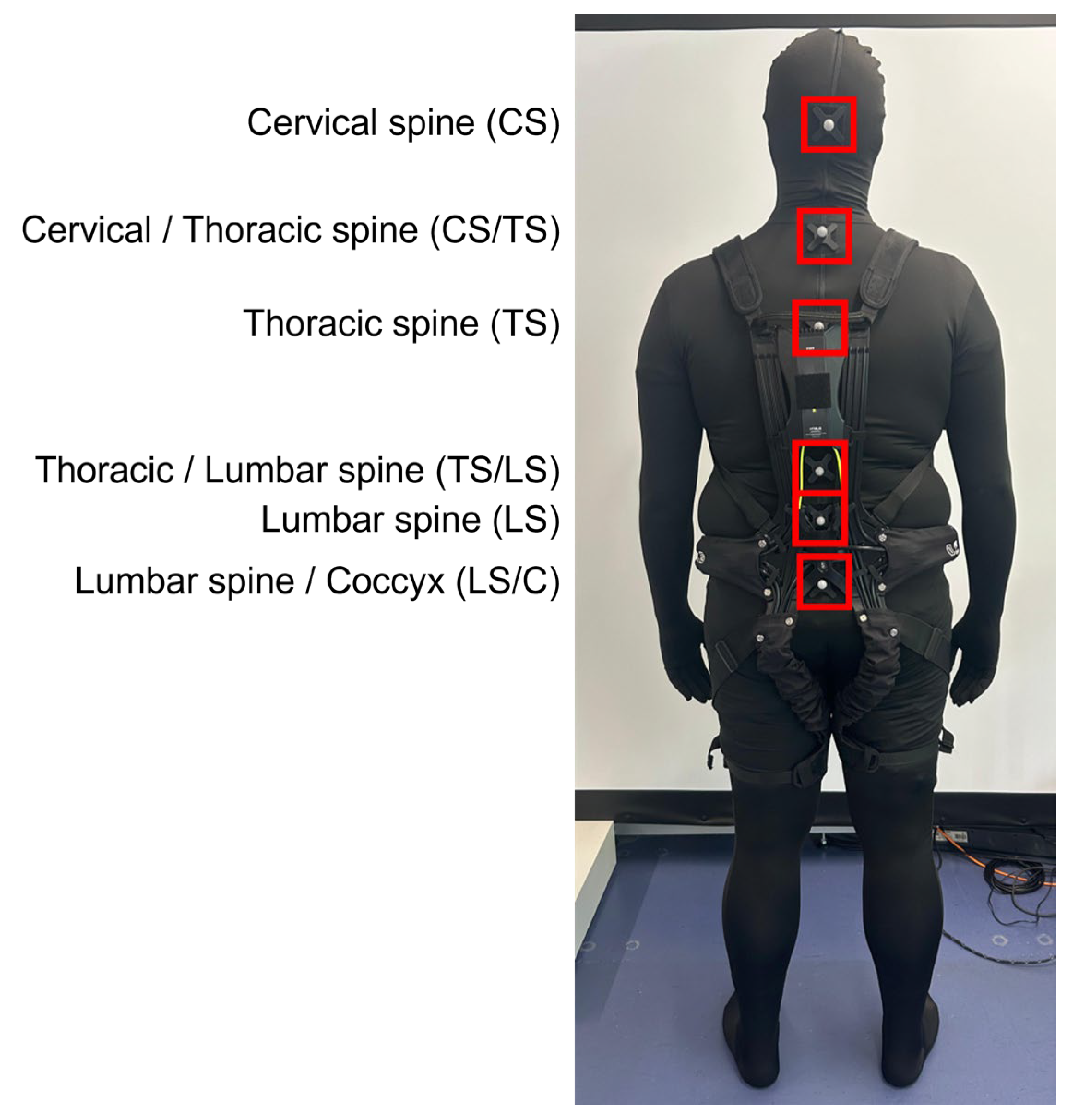
Reflective markers were positioned along the dorsal midline of the body at six predefined locations extending from the cervical to the sacral region (
Figure 4). The markers were attached to a tight-fitting elastic suit to reduce relative movement between the clothing and the markers. Within each participant’s measurement session, the same suit and marker configuration were retained for the upright reference measurement and all experimental conditions, avoiding repeated marker placement. In addition, all task-related angle values were normalized to the participant-specific upright reference obtained using the same marker configuration. This normalization reduces the influence of constant marker-placement offsets.
However, no independent quantitative assessment or correction of soft-tissue and clothing artefacts was performed. In particular, the marker trajectories were not validated against skin-mounted anatomical markers, a validated marker-cluster model, or an imaging-based reference. Moreover, normalization to an upright reference cannot eliminate dynamic movement between the skin, clothing, and external markers. The calculated angles should therefore be interpreted as marker-defined estimates of dorsal segment motion rather than direct measurements of anatomical intervertebral motion.
2.5. Motion Capture Data Processing
For each participant, an upright standing posture without an exoskeleton was recorded as an individual physiological reference. Marker-defined dorsal angles were calculated in Motive (Version 3.1.4, NaturalPoint, Inc., Corvallis, OR, USA) using three-marker vectors and were expressed relative to the participant-specific upright reference posture. Deviations were classified as flexion or extension.
The basic evaluation logic was as follows:
Positive and negative deviations were interpreted according to the respective segment definition. For each participant, condition, and spinal region, mean deviations, minimum values, maximum values, and ranges were calculated. The resulting values were used to compare the activated exoskeleton, deactivated exoskeleton, and no-exoskeleton conditions.
The measurement protocol focused exclusively on optical kinematic data. No synchronized electromyography, force-plate, pressure-insole, or other external-force measurements were collected. Furthermore, no inverse-dynamics or musculoskeletal model was applied to estimate spinal compression or shear forces. The calculated variables therefore describe marker-defined posture and movement only.
2.6. Data Analysis and Descriptive Statistics
This study was designed as an exploratory methodological feasibility study rather than as a confirmatory efficacy trial. Therefore, no a priori efficacy hypothesis or sample-size calculation was defined. The participant was considered the independent experimental unit, while the nine task cycles represented repeated observations within each participant. The individual cycles were not treated as independent experimental samples.
For each participant, condition, task phase, and marker-defined spinal region, the normalized angle deviations were summarized using means, standard deviations, minimum and maximum values, and ranges. These descriptive measures were used to illustrate the outputs of the proposed assessment procedure and the variability observed across repeated task executions.
The means and standard deviations calculated across the nine repeated cycles also provide a descriptive characterization of within-session cycle-to-cycle variability. However, this variability includes both natural differences in movement execution and potential measurement-related variation. These descriptive measures therefore cannot distinguish technical measurement error from biological or task-related variability and should not be interpreted as a formal assessment of the repeatability or reliability of the study-specific marker configuration and angle-calculation procedure. No intraclass correlation coefficients or other formal reliability statistics were calculated because the study was designed as an initial methodological feasibility investigation rather than as a reliability study.
No inferential between-condition hypothesis tests were performed because the study was neither designed nor statistically powered to establish population-level effects. Accordingly, all condition-related observations are interpreted as illustrative and exploratory rather than as statistically generalizable findings.
3. Results
The motion-capture-based evaluation enabled a quantitative comparison of spinal posture across the three test conditions. The upright standing trial without an exoskeleton served as the physiological reference posture for each participant. For every palletizing cycle, the measured spinal angles were compared with this individual reference posture.
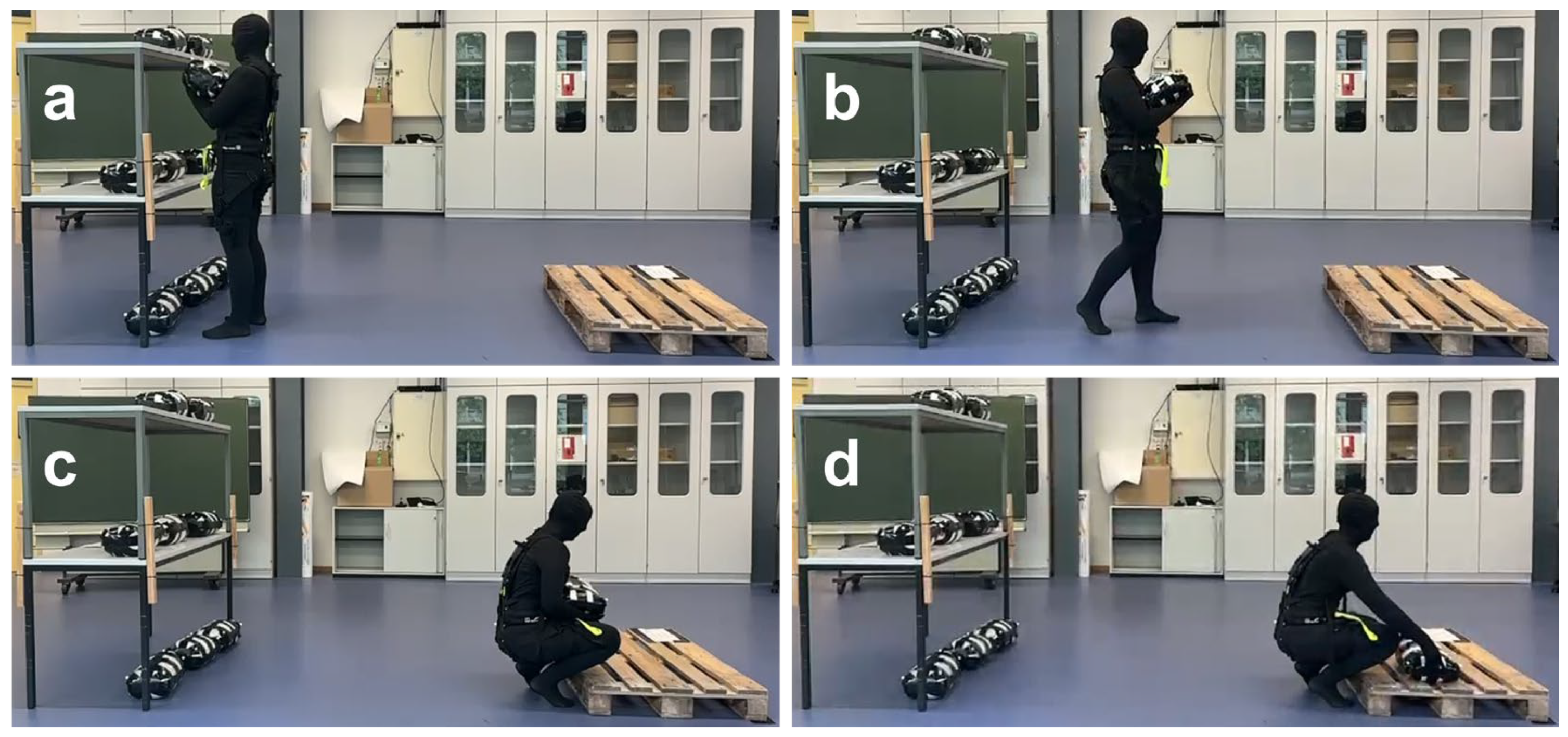
Movements are paused at the predefined locations (
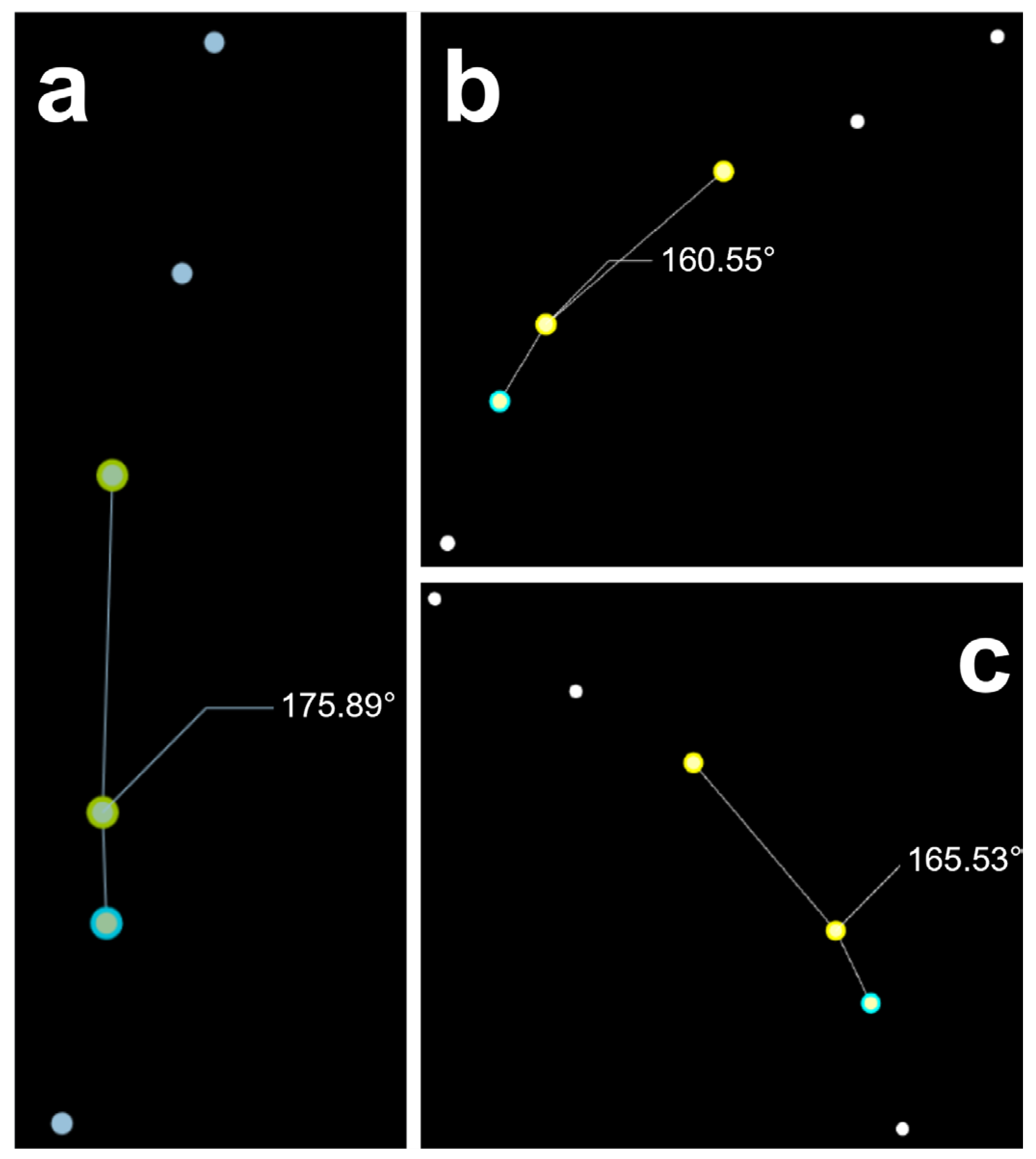
Figure 5) and evaluated using the angle-calculation tool of the software Motive (Version 3.1.4, NaturalPoint, Inc., Corvallis, OR, USA). For this purpose, three markers are selected; the smallest angle formed between the markers is computed by a scalar calculation and reported in degrees (
Figure 6).
To establish a baseline for spinal kinematics, the angular displacement of the cervical region was recorded over three consecutive cycles which simulate lifting sandbags from three different heights (chest, hip and floor). Each cycle comprised three repetitions of the same movement sequence, resulting in a total of nine repetitions per height. All trials were performed without the assistance of an exoskeleton and without a load being lifted, thereby representing the participants’ natural movement pattern.
The nine angle values obtained for a given shelf height were pooled. The mean and standard deviation were calculated (
Table 2).
The same protocol was applied to all remaining body segments and to trials performed with worn activated and deactivated exoskeleton. Across the three palletising cycles (nine repetitions per participant) also, the mean and standard deviation were computed of the angular data for every combination of body region, shelf height, and exoskeleton state (on/off). This systematic replication ensured that the kinematic effects of the exoskeleton could be quantified throughout the entire kinetic chain under identical loading and task-execution scenarios (
Table 3).
The loading phase was conducted by having participants lift sandbags from the three rack levels and place them onto a pallet, thereby introducing a known external load (15 kg per bag). For every lifted bag, the same sections described above were used, and the angles were extracted at the predefined pause points. Each measured angle α
task was then referenced against the participant-specific physiological baseline angle α
ref obtained in the unloaded, exoskeleton-off condition. The resulting deviation Δα quantifies the additional flexion/extension imposed by the load (
Table 4). If this result is a value less than zero, this angle is defined as flexion. If it is a value greater than zero, it is defined as extension.
For every body segment and each shelf height, the maximum and minimum deviation values across the nine repetitions were identified. Standardisation is carried out using the values for the physiologically straight spine (α
phy), the extreme values for flexion and extension (α
ext), and the individual angles of each segment (α):
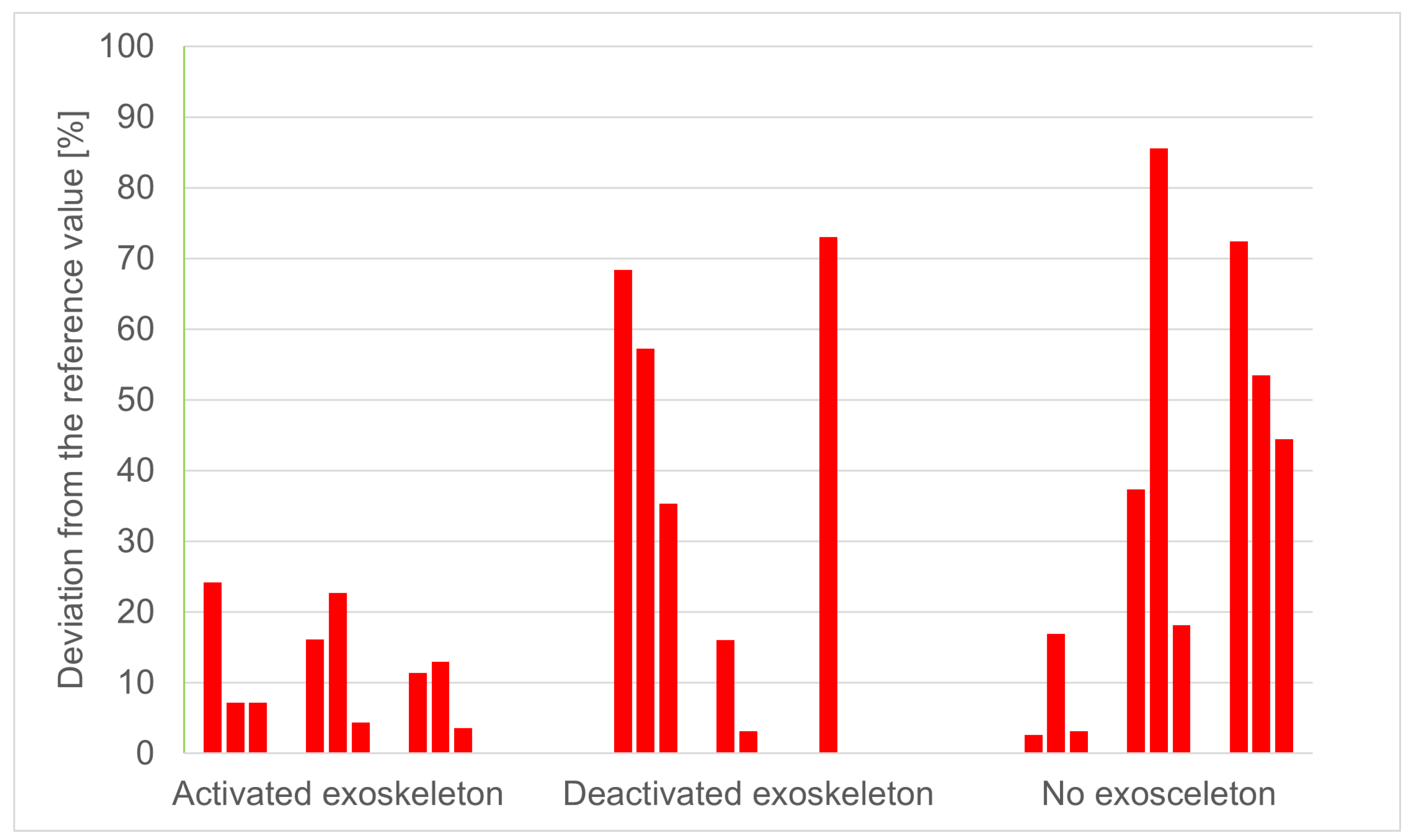
Once standardised, the angles associated with each task and person are plotted on bar charts (
Figure 7). These represent a single spinal segment for each work step. Each chart shows nine lifting processes, each with the exoskeleton activated, deactivated, or not in use. The data show that P1 has the least curvature of the thoracic/lumbar spine when lifting a load. When lifting from chest height, the average curvature is 14.9% lower than when using the deactivated exoskeleton or not using one at all. Furthermore, the variation in individual measurements when using the activated exoskeleton is 7.13%, which is at least 19.07% lower than in the other trials.
The descriptive application of the proposed assessment procedure produced condition-specific angle profiles for each participant and task phase. In several participants and marker-defined spinal regions, the activated exoskeleton condition showed smaller deviations from the individual upright reference posture. Such patterns were observed particularly in the thoracic and lumbar regions. However, the magnitude and consistency of these differences varied between participants and task phases.
In several observations, larger deviations from the upright reference occurred in the no-exoskeleton condition, particularly during lifting from the lower shelf level and during load placement on the pallet. These task phases involved pronounced forward bending, squatting, or combined trunk and lower-limb movements. However, the observed differences were not consistent across all participants and task phases.
The deactivated exoskeleton condition also produced varying angle patterns. For some participants and task phases, the recorded values differed from both the activated and no-exoskeleton conditions, whereas in other cases they were similar to the no-exoskeleton condition. Thus, no consistent condition-specific pattern was identified across the small participant sample.
Differences between participants were also visible in the recorded angle profiles. However, no statistical associations between these differences and body height, body mass, lifting technique, or previous ergonomic training were examined. Given the small sample size, the fixed condition order, and the absence of inferential hypothesis testing, the condition-related observations are presented solely to illustrate the outputs and applicability of the proposed assessment procedure. They should not be interpreted as population-level evidence of exoskeleton efficacy.
4. Discussion
The present pilot study examined the feasibility of a marker-based optical motion-capture procedure for describing posture-related angle changes during simulated palletizing. The proposed procedure generated participant-, task-, and condition-specific profiles of marker-defined dorsal segment angles. In several participants and task phases, smaller deviations from the individual upright reference posture were recorded in the activated exoskeleton condition, particularly in the thoracic and lumbar regions. These descriptive observations are compatible with the intended mechanical function of passive back-support exoskeletons, which are designed to support trunk extension during forward-bending and lifting activities [
10,
11,
14,
15,
16,
17,
19,
20]. However, because the study was not designed or statistically powered as an efficacy trial, the recorded differences cannot be interpreted as evidence of a general or causal effect of exoskeleton activation.
Previous studies have reported reductions in back-muscle activity, biomechanical loading indicators, or perceived exertion during the use of passive back-support exoskeletons [
10,
14,
15,
16,
17,
20]. The present study complements this research by focusing specifically on the feasibility of a marker-based procedure for quantitatively describing kinematic patterns during a simulated work process. Kinematic, electromyographic, and force-based measurements address different aspects of human–exoskeleton interaction [
30]. Motion capture provides information about posture and movement patterns, whereas electromyography quantifies muscle activation and force-measurement systems provide information about external loading. Estimates of spinal compression or shear forces additionally require external-force measurements, anthropometric data, and an appropriate inverse-dynamics or musculoskeletal model. Therefore, a smaller deviation from the upright reference posture should not automatically be interpreted as evidence of reduced muscular effort, lower spinal loading, or reduced injury risk.
The varying angle patterns observed in the deactivated exoskeleton condition demonstrate the relevance of including a condition in which the device is worn without mechanical assistance. Wearing the device may potentially influence movement through mechanical restriction, increased body awareness, proprioceptive feedback, or changes in movement strategy. However, these mechanisms were not measured in the present study and cannot be distinguished on the basis of the recorded kinematic data. Accordingly, the observations from the deactivated condition should be regarded as hypothesis-generating rather than as evidence of a specific stabilizing or posture-guiding effect. Future studies should continue to distinguish between the effects of mechanical assistance and those associated with wearing the device itself.
The recorded angle patterns also varied between participants. In the context of this methodological feasibility study, this variability demonstrates that the proposed procedure can represent participant-specific movement patterns. However, the small and heterogeneous sample does not permit conclusions regarding the factors responsible for these differences. Although body height, body mass, lifting technique, and previous ergonomic training may represent relevant variables, their associations with the recorded angle patterns were not statistically evaluated and should not be inferred from the present data. These variables should be investigated as potential moderating factors in future studies with an adequately powered sample.
Several limitations must be acknowledged. First, the sample size was small, and the study was not designed to provide statistically generalizable evidence regarding exoskeleton efficacy. The descriptive condition-related observations were used primarily to illustrate the outputs and applicability of the proposed assessment procedure. Second, the controlled laboratory setup and simulated palletizing task cannot fully reproduce the variability, duration, and environmental conditions of industrial workplaces.
Third, the condition sequence was fixed and identical for all participants, resulting in a complete confounding of condition and order effects. Familiarization or learning during repeated task execution may have altered the participants’ lifting strategies and improved movement consistency during the later conditions. Such an effect could have reduced the apparent advantage of the activated exoskeleton, which was always tested first. Conversely, cumulative fatigue may have increased spinal flexion or altered movement strategies during the later deactivated and no-exoskeleton conditions, thereby exaggerating the apparent advantage of the activated condition. The magnitude and direction of these potential sequence effects cannot be determined from the present data. Therefore, the observed condition-related differences cannot be attributed exclusively to exoskeleton activation. Future studies should use randomized or counterbalanced condition sequences and standardized rest periods.
Fourth, the reflective markers were attached to a tight-fitting suit rather than directly to the skin over palpated anatomical landmarks. Although the same marker configuration was retained throughout each participant’s measurement session and normalization to an individual upright reference reduced constant placement offsets, residual dynamic movement between the skin, clothing, and markers cannot be excluded. These artefacts were not independently quantified or corrected and may have affected the calculated angles. Consequently, the reported values represent marker-defined estimates of dorsal segment motion and should not be interpreted as direct measurements of vertebral or intervertebral motion. Future studies should validate the proposed marker configuration against an established anatomical reference method and quantify the magnitude of soft-tissue and clothing artefacts.
Furthermore, no formal intra-session repeatability analysis was performed for the study-specific multi-segment marker model. Although means and standard deviations across the nine repeated cycles were calculated to describe cycle-to-cycle variability, these values cannot distinguish technical measurement error from natural variation in movement execution. Consequently, the present study demonstrates the feasibility of the assessment procedure but does not establish its measurement reliability. Future studies should formally evaluate the intra-session and inter-session reliability of the proposed marker configuration and angle-calculation procedure.
Finally, the study was limited to kinematic measurements and did not include complementary biomechanical variables such as muscle activation, ground-reaction forces, external moments, or model-based estimates of spinal compression and shear forces. This limits the interpretation of the observed posture-related differences, as the present data cannot determine whether the recorded angle patterns were accompanied by changes in muscular effort or internal spinal loading. Future studies should therefore combine the proposed motion-capture procedure with synchronized surface electromyography, external-force measurements, and appropriate biomechanical modelling.
Despite these limitations, the study demonstrates the feasibility of motion-capture-based spinal angle analysis as an objective method for evaluating ergonomic effects of passive exoskeletons (
Figure 8). The approach allows the visualization and quantification of posture deviations and may therefore support future ergonomic workplace assessments.
5. Comparison with Other Motion Analysis Methods
Objective evaluation of industrial exoskeletons requires a reliable assessment of human kinematics, muscular activity, and biomechanical loading. Consequently, a variety of motion analysis techniques have been established, each addressing different aspects of human movement while exhibiting specific strengths and limitations (
Table 5). The most frequently applied methods include optical motion capture, inertial measurement units (IMUs), integrated exoskeleton sensors, electromyography (EMG), force measurement systems, and multimodal sensor fusion approaches [
31,
32,
33,
34].
Among these approaches, marker-based optical motion capture is generally regarded as the reference standard for three-dimensional kinematic analysis because of its high spatial accuracy, repeatability, and ability to reconstruct complete body movements [
24,
25,
26,
31]. For this reason, optical systems are commonly used to validate emerging wearable sensing technologies, including IMU-based motion capture [
31,
35].
However, the high accuracy of optical motion capture comes at the expense of considerable experimental effort. Multi-camera calibration, dedicated laboratory environments, and marker occlusions caused by body posture or exoskeleton components limit its applicability in industrial workplaces [
26,
27,
31]. Recent advances in computer vision have therefore promoted the development of markerless motion capture systems based on RGB or depth cameras and deep-learning algorithms. These systems considerably reduce preparation time and improve usability but currently exhibit lower accuracy and remain sensitive to occlusions, changing illumination conditions, and complex body–exoskeleton interactions [
36,
37].
As an alternative, IMU-based motion capture has become increasingly popular in ergonomics and wearable robotics. Because IMUs are directly attached to body segments or integrated into exoskeletons, they enable unrestricted measurements during realistic work tasks while providing continuous real-time kinematic data. Validation studies have demonstrated good agreement between modern IMU systems and laboratory-based optical motion capture, although measurement accuracy decreases during prolonged or highly dynamic movements due to sensor drift, magnetic disturbances, and biomechanical model differences [
35,
38].
While optical and inertial systems primarily quantify movement kinematics, surface electromyography (sEMG) provides complementary information on neuromuscular activation. Consequently, EMG has become one of the most frequently applied techniques for evaluating industrial exoskeletons, as reductions in muscle activity are commonly interpreted as indicators of effective physical assistance [
14,
15,
16,
17,
39]. In addition, EMG is increasingly employed for intention recognition and adaptive control of active exoskeletons [
39]. Nevertheless, EMG does not directly quantify posture or joint kinematics and is sensitive to electrode placement, sweat, and motion artefacts [
39].
Additional biomechanical information can be obtained from force plates, pressure insoles, or integrated joint sensors. Whereas force measurement systems quantify external loading conditions and gait characteristics, integrated encoders or Hall sensors accurately measure exoskeleton joint motion but cannot capture relative movement between the human body and the device [
32,
33]. Consequently, these methods are generally considered complementary rather than stand-alone solutions for comprehensive biomechanical assessment.
Current research therefore increasingly favours multimodal sensing approaches that combine optical motion capture, IMUs, EMG, and force measurements. Sensor fusion enables simultaneous assessment of body kinematics, muscular activity, and external loading while compensating for the limitations of individual sensing modalities [
31,
32,
33,
34,
39]. Such integrated approaches are considered one of the most promising directions for future industrial exoskeleton evaluation and human–robot interaction research [
32,
33,
34].
Against this background, optical motion capture was deliberately selected in the present study because the primary objective was the accurate quantification of spinal posture during palletizing. As the current work focuses on methodological validation of spinal kinematics, marker-based motion capture provides the required spatial accuracy and repeatability [
26,
27,
28,
29]. Future investigations should extend this methodology by integrating EMG, IMU-based motion capture, and force measurements to achieve a more comprehensive evaluation of human–exoskeleton interaction under realistic industrial conditions [
32,
33,
34].
6. Conclusions
This pilot study demonstrates the feasibility of a marker-based optical motion-capture procedure for quantifying posture-related angle changes during simulated palletizing. The procedure generated participant-, condition-, and task-specific profiles of marker-defined dorsal segment angles across repeated task executions.
In several participants and task phases, the activated exoskeleton condition showed smaller deviations from the individual upright reference posture than the deactivated and no-exoskeleton conditions. However, these descriptive patterns varied between participants and should be regarded solely as an illustrative application of the proposed assessment procedure. Given the small sample size, the fixed condition order, the absence of inferential hypothesis testing, and the potential influence of soft-tissue and clothing artefacts, the results do not provide population-level evidence of exoskeleton efficacy and do not permit a causal attribution to exoskeleton activation.
Furthermore, the study-specific marker configuration and angle-calculation procedure were not subjected to a formal intra-session or inter-session repeatability analysis or to an independent validity assessment. Although means and standard deviations across the repeated task cycles were calculated to describe cycle-to-cycle variability, these measures cannot distinguish technical measurement error from natural variation in movement execution. Consequently, the present study demonstrates the feasibility of the assessment procedure but does not establish its measurement reliability or validity.
The present study was limited to kinematic measurements. Therefore, the recorded angle patterns do not provide evidence of reduced muscle activation, external biomechanical loading, spinal compression, or injury risk. A smaller deviation from the upright reference posture should not be interpreted automatically as evidence of a reduced biomechanical workload.
Future studies should include larger and adequately powered participant samples, randomized or counterbalanced condition sequences, standardized rest periods, and longer task durations. They should also formally evaluate the intra-session and inter-session repeatability and validity of the proposed marker configuration and angle-calculation procedure. Combining optical motion capture with synchronized surface electromyography, external-force measurements, and biomechanical modelling would enable a more comprehensive evaluation of human–exoskeleton interaction.
The proposed procedure therefore represents a starting point for the further development and validation of quantitative kinematic assessment methods for wearable assistance systems in industrial work environments.
Author Contributions
Conceptualization, M.-G.W. and C.L.; methodology, T.K.; software, T.K.; validation, T.K.; formal analysis, T.K.; investigation, T.K.; resources, T.S. and T.V.; data curation, M.-G.W.; writing—original draft preparation, C.L.; writing—review and editing, M.-G.W.; visualization, C.L. and M.-G.W.; supervision, T.S.; project administration, T.S. and T.V.; funding acquisition, T.S. All authors have read and agreed to the published version of the manuscript.
Funding
This research received no external funding.
Institutional Review Board Statement
For this study, no ethical review or approval was required, as the participants had given their consent.
Informed Consent Statement
Informed consent was obtained from all subjects involved in the study.
Data Availability Statement
The data presented in this study are available from the corresponding author upon reasonable request. The data are not publicly available due to privacy and ethical considerations concerning the participant data.
Acknowledgments
We acknowledge support by the Open Access Publication Fund of the Westphalian University of Applied Sciences. The Motion Capture System was funded by an internal scheme. We also thank Michael M. Günther for his support in biomechanics of the human body and locomotion. During the preparation of this manuscript, the authors used ChatGPT (OpenAI, GPT-5.5) to assist with manuscript organization, language refinement, scientific discussion, and literature summarization. All generated content was critically evaluated, edited, and verified by the authors, who assume full responsibility for the final manuscript. The authors have reviewed and edited the output and take full responsibility for the content of this publication.
Conflicts of Interest
The authors declare no conflicts of interest.
Abbreviations
The following abbreviations are used in this manuscript:
| 3D | Three-dimensional |
| AV | Average |
| C | Coccyx |
| CS | Cervical spine |
| EMG | Electromyography |
| IMU | Inertial measurement unit |
| LS | Lumbar spine |
| P1–P5 | Patient |
| sEMG | Surface electromyography |
| TS | Thoracis spine |
| WC | Work cycle |
References
- World Health Organization. Low Back Pain. 2023. Available online: https://www.who.int/news-room/fact-sheets/detail/low-back-pain (accessed on 21 July 2026).
- World Health Organization. Musculoskeletal Health. 2022. Available online: https://www.who.int/news-room/fact-sheets/detail/musculoskeletal-conditions (accessed on 21 July 2026).
- Waters, T.R.; Putz-Anderson, V.; Garg, A. Applications Manual for the Revised NIOSH Lifting Equation; DHHS (NIOSH) Publication No. 94–110; revised September 2021; U.S. Department of Health and Human Services, Centers for Disease Control and Prevention, National Institute for Occupational Safety and Health: Cincinnati, OH, USA, 1994. [CrossRef] [Scilit]
- ISO 11228-1:2021; Ergonomics—Manual Handling—Part 1: Lifting, Lowering and Carrying. International Organization for Standardization: Geneva, Switzerland, 2021. Available online: https://www.iso.org/standard/76820.html (accessed on 21 July 2026).
- Serafin, P.; Schäfer, A.; Klußmann, A.; Lang, K.-H.; Liebers, F. Manuelles Heben, Halten und Tragen—Gefährdungsbeurteilung mit der Leitmerkmalmethode; Baua: Praxis; Bundesanstalt für Arbeitsschutz und Arbeitsmedizin: Dortmund, Germany, 2022. [Google Scholar] [CrossRef]
- Marras, W.S. Occupational Low Back Disorder Causation and Control. Ergonomics 2000, 43, 880–902. [Google Scholar] [CrossRef] [Scilit]
- McGill, S.M. Low Back Disorders: Evidence-Based Prevention and Rehabilitation, 3rd ed.; Human Kinetics: Champaign, IL, USA, 2016. [Google Scholar]
- Dreischarf, M.; Zander, T.; Shirazi-Adl, A.; Puttlitz, C.M.; Adam, C.J.; Chen, C.S.; Goel, V.K.; Kiapour, A.; Kim, Y.H.; Labus, K.M.; et al. Comparison of Eight Published Static Finite Element Models of the Intact Lumbar Spine: Predictive Power of Models Improves When Combined Together. J. Biomech. 2014, 47, 1757–1766. [Google Scholar] [CrossRef] [Scilit]
- Bundesanstalt für Arbeitsschutz und Arbeitsmedizin (BAuA). Gefährdungsbeurteilung bei Physischer Belastung—Die Neuen Leitmerkmalmethoden (LMM), 3rd ed.; Bundesanstalt für Arbeitsschutz und Arbeitsmedizin: Dortmund, Germany, 2019; Available online: https://shop.baua.de/baua-bericht/pub-baua-f2333-2 (accessed on 21 July 2026).
- Baltrusch, S.J.; van Dieën, J.H.; Koopman, A.S.; Näf, M.B.; Rodriguez-Guerrero, C.; Babič, J.; Houdijk, H. SPEXOR Passive Spinal Exoskeleton Decreases Metabolic Cost during Symmetric Repetitive Lifting. Eur. J. Appl. Physiol. 2020, 120, 401–412. [Google Scholar] [CrossRef] [Scilit]
- Iranzo, S.; Piedrabuena, A.; García-Torres, F.; Martinez-de-Juan, J.L.; Prats-Boluda, G.; Sanchis, M.; Belda-Lois, J.-M. Assessment of a Passive Lumbar Exoskeleton in Material Manual Handling Tasks under Laboratory Conditions. Sensors 2022, 22, 4060. [Google Scholar] [CrossRef] [Scilit]
- Lasota, A.M. Manual Palletizing Simulation: Time and Ergonomic Study. Int. J. Interact. Des. Manuf. (IJIDeM) 2026, 20, 1321–1334. [Google Scholar] [CrossRef] [Scilit]
- Gallagher, S.; Heberger, J.R. The Effects of Operator Position, Pallet Orientation, and Palletizing Condition on Low Back Loads in Manual Bag Palletizing Operations. Int. J. Ind. Ergon. 2015, 47, 84–92. [Google Scholar] [CrossRef] [Scilit]
- Kermavnar, T.; de Vries, A.W.; de Looze, M.P.; O’Sullivan, L.W. Effects of Industrial Back-Support Exoskeletons on Body Loading and User Experience: An Updated Systematic Review. Ergonomics 2021, 64, 685–711. [Google Scholar] [CrossRef] [Scilit]
- Ali, A.; Fontanari, V.; Schmoelz, W.; Agrawal, S.K. Systematic Review of Back-Support Exoskeletons and Soft Robotic Suits. Front. Bioeng. Biotechnol. 2021, 9, 765257. [Google Scholar] [CrossRef] [Scilit]
- Bosch, T.; van Eck, J.; Knitel, K.; de Looze, M. The Effects of a Passive Exoskeleton on Muscle Activity, Discomfort and Endurance Time in Forward Bending Work. Appl. Ergon. 2016, 54, 212–217. [Google Scholar] [CrossRef] [Scilit]
- Alemi, M.M.; Geissinger, J.; Simon, A.A.; Chang, S.E.; Asbeck, A.T. A Passive Exoskeleton Reduces Peak and Mean EMG during Symmetric and Asymmetric Lifting. J. Electromyogr. Kinesiol. 2019, 47, 25–34. [Google Scholar] [CrossRef] [Scilit]
- Terstegen, S.; Sandrock, S. Exoskelette—Physische Assistenzsysteme an Produktionsarbeitsplätzen. In Zahlen|Daten|Fakten; ifaa—Institut für Angewandte Arbeitswissenschaft: Düsseldorf, Germany, 2018; Available online: https://www.arbeitswissenschaft.net/fileadmin/Downloads/Angebote_und_Produkte/Zahlen_Daten_Fakten/ifaa_Zahlen_Daten_Fakten_Exoskelette.pdf (accessed on 21 July 2026).
- Näf, M.B.; Koopman, A.S.; Baltrusch, S.; Rodriguez-Guerrero, C.; Vanderborght, B.; Lefeber, D. Passive Back Support Exoskeleton Improves Range of Motion Using Flexible Beams. Front. Robot. AI 2018, 5, 72. [Google Scholar] [CrossRef] [Scilit]
- Baltrusch, S.J.; van Dieën, J.H.; van Bennekom, C.A.M.; Houdijk, H. The Effect of a Passive Trunk Exoskeleton on Functional Performance in Healthy Individuals. Appl. Ergon. 2018, 72, 94–106. [Google Scholar] [CrossRef] [Scilit]
- Hensel, R.; Keil, M. Subjektive Evaluation industrieller Exoskelette im Rahmen von Feldstudien an ausgewählten Arbeitsplätzen. Z. Arbeitswiss. 2018, 72, 252–263. [Google Scholar] [CrossRef] [Scilit]
- Riemer, J. Ergonomische Bewertung des Langzeiteinsatzes von Passiven Exoskeletten in der Arbeitswelt; Forschungsbericht F2466; Bundesanstalt für Arbeitsschutz und Arbeitsmedizin (BAuA): Dortmund, Germany, 2024. [Google Scholar] [CrossRef]
- van Sluijs, R.M.; Wehrli, M.; Brunner, A.; Lambercy, O. Evaluation of the Physiological Benefits of a Passive Back-Support Exoskeleton during Lifting and Working in Forward Leaning Postures. J. Biomech. 2023, 149, 111489. [Google Scholar] [CrossRef] [Scilit]
- Reimeir, B.; Calisti, M.; Mittermeier, R.; Ralfs, L.; Weidner, R. Effects of Back-Support Exoskeletons with Different Functional Mechanisms on Trunk Muscle Activity and Kinematics. Wearable Technol. 2023, 4, e12. [Google Scholar] [CrossRef] [Scilit]
- Kuber, P.M.; Rashedi, E. Investigating Spatiotemporal Effects of Back-Support Exoskeletons Using Unloaded Cyclic Trunk Flexion–Extension Task Paradigm. Appl. Sci. 2024, 14, 5564. [Google Scholar] [CrossRef] [Scilit]
- Gerdts, S.; Schepelmann, A. Characterization of Infrared Optical Motion Tracking System in NASA’s Simulated Lunar Operations (SLOPE) Laboratory; NASA Technical Memorandum NASA/TM-20220005304; National Aeronautics and Space Administration, Glenn Research Center: Cleveland, OH, USA, 2022. Available online: https://ntrs.nasa.gov/citations/20220005304 (accessed on 21 July 2026).
- Cappozzo, A.; Catani, F.; Della Croce, U.; Leardini, A. Position and Orientation in Space of Bones during Movement: Anatomical Frame Definition and Determination. Clin. Biomech. 1995, 10, 171–178. [Google Scholar] [CrossRef] [Scilit]
- Wu, G.; Siegler, S.; Allard, P.; Kirtley, C.; Leardini, A.; Rosenbaum, D.; Whittle, M.; D’Lima, D.D.; Cristofolini, L.; Witte, H.; et al. ISB Recommendation on Definitions of Joint Coordinate System of Various Joints for the Reporting of Human Joint Motion—Part I: Ankle, Hip, and Spine. J. Biomech. 2002, 35, 543–548. [Google Scholar] [CrossRef] [Scilit]
- OptiTrack. PrimeX Cameras: Product Details; dba OptiTracks; NaturalPoint, Inc.: Corvallis, OR, USA, 2025; Available online: https://d111srqycjesc9.cloudfront.net/primex-family-spec-sheet-v13.pdf (accessed on 21 July 2026).
- Grimmer, M.; Mohseni, O.; Seiler, J.; Sharbafi, M.A.; Findeisen, R.; Seyfarth, A.; Kupnik, M. Hip Exoskeleton Assistance during Sit-to-Stand Transitions. Biomechanics 2026, 6, 15. [Google Scholar] [CrossRef] [Scilit]
- Hubaut, R.; Guichard, R.; Greenfield, J.; Blandeau, M. Validation of an Embedded Motion-Capture and EMG Setup for the Analysis of Musculoskeletal Disorder Risks during Manhole Cover Handling. Sensors 2022, 22, 436. [Google Scholar] [CrossRef] [Scilit]
- Hasan, S.; Alam, N. Comprehensive Comparative Analysis of Lower Limb Exoskeleton Research: Control, Design, and Application. Actuators 2025, 14, 342. [Google Scholar] [CrossRef] [Scilit]
- Dollar, A.M.; Herr, H. Lower Extremity Exoskeletons and Active Orthoses: Challenges and State-of-the-Art. IEEE Trans. Robot. 2008, 24, 144–158. [Google Scholar] [CrossRef] [Scilit]
- Young, A.J.; Ferris, D.P. State of the Art and Future Directions for Lower Limb Robotic Exoskeletons. IEEE Trans. Neural Syst. Rehabil. Eng. 2017, 25, 171–182. [Google Scholar] [CrossRef] [Scilit]
- Robert-Lachaine, X.; Mecheri, H.; Larue, C.; Plamondon, A. Validation of Inertial Measurement Units with an Optoelectronic System for Whole-Body Motion Analysis. Med. Biol. Eng. Comput. 2017, 55, 609–619. [Google Scholar] [CrossRef] [Scilit]
- Desmarais, Y.; Mottet, D.; Slangen, P.; Montesinos, P. A Review of 3D Human Pose Estimation Algorithms for Markerless Motion Capture. Comput. Vis. Image Underst. 2021, 212, 103275. [Google Scholar] [CrossRef] [Scilit]
- Edwards, N.A.; Caccese, J.B.; Tracy, R.E.; Hagen, J.; Quatman-Yates, C.C.; Oñate, J. The Validity and Usability of Markerless Motion Capture and Inertial Measurement Units for Quantifying Dynamic Movements. Med. Sci. Sports Exerc. 2025, 57, 641–655. [Google Scholar] [CrossRef] [Scilit]
- Picerno, P. 25 Years of Lower Limb Joint Kinematics by Using Inertial and Magnetic Sensors: A Review of Methodological Approaches. Gait Posture 2017, 51, 239–246. [Google Scholar] [CrossRef] [Scilit]
- Sul, J.-H.; Piyathilaka, L.; Moratuwage, D.; Dunu Arachchige, S.; Jayawardena, A.; Kahandawa, G.; Preethichandra, D.M.G. Electromyography Signal Acquisition, Filtering, and Data Analysis for Exoskeleton Development. Sensors 2025, 25, 4004. [Google Scholar] [CrossRef] [Scilit]
Figure 1.
Healthy and unhealthy lifting of a load and their effects on the intervertebral discs. On the (
left), the person bends forward while lifting, causing the vertebral bodies to angle and the intervertebral discs to be compressed and wedge-shaped. This leads to local stress peaks and overload. On the (
right), the person lifts with a straight back, resulting in an even load distribution on the intervertebral discs and no local stress peaks [
10,
11].
Figure 1.
Healthy and unhealthy lifting of a load and their effects on the intervertebral discs. On the (
left), the person bends forward while lifting, causing the vertebral bodies to angle and the intervertebral discs to be compressed and wedge-shaped. This leads to local stress peaks and overload. On the (
right), the person lifts with a straight back, resulting in an even load distribution on the intervertebral discs and no local stress peaks [
10,
11].
Figure 2.
Starting position of a participant during an upright stance. The gaze is directed forward, while the abdomen and glutes are engaged. The legs are shoulder-width apart, and the arms hang relaxed. This upright stance is considered the physiological reference posture.
Figure 2.
Starting position of a participant during an upright stance. The gaze is directed forward, while the abdomen and glutes are engaged. The legs are shoulder-width apart, and the arms hang relaxed. This upright stance is considered the physiological reference posture.
Figure 3.
Passive exoskeleton “BionicBack” from hTRIUS GmbH: (a) medial view; (b) dorsal view. The yellow straps are pre-tensioned by the user and run bilaterally from the thigh level up to the lumbar spine, from there along the abdomen forward to the navel and back, rising up to the thoracic spine. The exoskeleton is activated when a person pre-tensions these straps and then squats. In this process, movement energy is stored in the tensioned straps and released as support when standing upright.
Figure 3.
Passive exoskeleton “BionicBack” from hTRIUS GmbH: (a) medial view; (b) dorsal view. The yellow straps are pre-tensioned by the user and run bilaterally from the thigh level up to the lumbar spine, from there along the abdomen forward to the navel and back, rising up to the thoracic spine. The exoskeleton is activated when a person pre-tensions these straps and then squats. In this process, movement energy is stored in the tensioned straps and released as support when standing upright.
Figure 4.
Dorsal view of a test subject wearing a full-body morphsuit and an attached exoskeleton. The red frames enclose the six markers that are directly applied to the suit including naming. The upper body is divided into four sections, which are marked by colours.
Figure 4.
Dorsal view of a test subject wearing a full-body morphsuit and an attached exoskeleton. The red frames enclose the six markers that are directly applied to the suit including naming. The upper body is divided into four sections, which are marked by colours.
Figure 5.
Experimental indoor setup: (a) lifting from different shelf heights; (b) carrying the load; (c) squatting to place the load; (d) load placement on the pallet.
Figure 5.
Experimental indoor setup: (a) lifting from different shelf heights; (b) carrying the load; (c) squatting to place the load; (d) load placement on the pallet.
Figure 6.
Exemplary illustrations of angle analysis in Motive 3.1.4 for different spinal postures. (a) the participant is standing upright; (b) she is bending forward to lift a load; and (c): she is bending forward to set down a load. The displayed number corresponds to the minimum angle between the three selected markers.
Figure 6.
Exemplary illustrations of angle analysis in Motive 3.1.4 for different spinal postures. (a) the participant is standing upright; (b) she is bending forward to lift a load; and (c): she is bending forward to set down a load. The displayed number corresponds to the minimum angle between the three selected markers.
Figure 7.
Relative deviation from the reference value for subject P1 whilst lifting a load from chest height. All diagrams show the spinal segment “TS|TS/LS|LS”. Means over all nine lifting processes: activated exoskeleton: 12.19 ± 7.56%; deactivated exoskeleton: 42.18 ± 28.70%; no exoskeleton: 37.09 ± 29.63%.
Figure 7.
Relative deviation from the reference value for subject P1 whilst lifting a load from chest height. All diagrams show the spinal segment “TS|TS/LS|LS”. Means over all nine lifting processes: activated exoskeleton: 12.19 ± 7.56%; deactivated exoskeleton: 42.18 ± 28.70%; no exoskeleton: 37.09 ± 29.63%.
Figure 8.
Flowchart of the measuring method.
Figure 8.
Flowchart of the measuring method.
Table 1.
Overview of participants, including biodata (m: male; f: female, x: applicable).
Table 1.
Overview of participants, including biodata (m: male; f: female, x: applicable).
| | P1 | P2 | P3 | P4 | P5 |
|---|
| sex | m | f | f | f | m |
| age [years] | 32 | 24 | 22 | 63 | 28 |
| bodyweight [kg] | 120 | 60 | 65 | 90 | 88 |
| body size [cm] | 186 | 167 | 163 | 178 | 185 |
got prior training in
ergonomic lifting | x | | | x | |
Table 2.
Initial position (P1) across three work cycles (WC), each with three executions without load and without exoskeleton in the upper-body region and calculated average (AV).
Table 2.
Initial position (P1) across three work cycles (WC), each with three executions without load and without exoskeleton in the upper-body region and calculated average (AV).
| CS|CS/TS|TS NE |
|---|
| | WC 1 [°] | WC 2 [°] | WC 3 [°] | AV [°] |
|---|
| Chest height | 163.79 | 163.80 | 163.75 | 165.54 ± 1.38 |
| 166.82 | 166.09 | 165.25 |
| 166.01 | 166.84 | 167.53 |
| Hip height | 174.40 | 167.58 | 166.69 | 168.55 ± 2.42 |
| 170.55 | 166.75 | 165.98 |
| 167.92 | 168.25 | 168.88 |
| Floor height | 169.31 | 167.86 | 170.63 | 168.76 ± 1.86 |
| 172.38 | 169.49 | 167.01 |
| 167.31 | 165.98 | 168.91 |
Table 3.
Initial position (P1) over all body regions, marked by colours.
Table 3.
Initial position (P1) over all body regions, marked by colours.
| | Starting Position [°] |
|---|
| | Activated Exoskeleton | Deactivated Exoskeleton | No Exoskeleton |
|---|
| Chest height | 166.91 ± 3.46 | 166.64 ± 1.27 | 165.54 ± 1.38 |
| Hip height | 166.10 ± 2.04 | 165.59 ± 1.42 | 168.55 ± 2.42 |
| Floor | 168.21 ± 3.39 | 166.60 ± 3.17 | 168.76 ± 1.86 |
| Chest height | 163.69 ± 1.66 | 165.28 ± 4.07 | 161.98 ± 1.55 |
| Hip height | 163.12 ± 1.25 | 162.31 ± 1.38 | 162.81 ± 1.04 |
| Floor | 163.40 ± 1.03 | 163.37 ± 0.94 | 162.13 ± 0.95 |
| Chest height | 175.01 ± 1.61 | 174.58 ± 2.05 | 176.92 ± 1.62 |
| Hip height | 172.37 ± 1.79 | 174.95 ± 2.82 | 176.42 ± 2.12 |
| Floor | 173.52 ± 2.46 | 173.06 ± 2.29 | 176.27 ± 1.91 |
| Chest height | 166.79 ± 3.93 | 164.15 ± 5.52 | 165.37 ± 4.56 |
| Hip height | 163.30 ± 5.73 | 166.07 ± 6.55 | 167.04 ± 4.74 |
| Floor | 164.00 ± 3.49 | 159.27 ± 4.44 | 163.04 ± 5.49 |
Table 4.
Difference between loaded minus starting position (P1) over all body regions while lifting.
Table 4.
Difference between loaded minus starting position (P1) over all body regions while lifting.
| | Difference Lifting-Starting Position [°] |
|---|
| | Activated Exoskeleton | Deactivated Exoskeleton | No Exoskeleton |
|---|
| Chest height | 10.82 ± 3.40 | 9.88 ± 2.58 | 7.35 ± 3.46 |
| Hip height | −3.13 ± 8.57 | −3.64 ± 6.62 | −0.92 ± 8.86 |
| Floor | 2.45 ± 4.02 | −0.66 ± 3.57 | 0.17 ± 2.81 |
| Chest height | 7.38 ± 2.33 | 8.84 ± 1.80 | 9.04 ± 2.02 |
| Hip height | 4.32 ± 1.51 | 7.39 ± 2.98 | 4.01 ± 1.65 |
| Floor | −2.74 ± 2.18 | −1.86 ± 1.81 | −2.26 ± 1.57 |
| Chest height | −1.13 ± 2.04 | 0.67 ± 2.15 | 0.64 ± 2.03 |
| Hip height | −3.16 ± 2.47 | −3.43 ± 2.70 | −5.19 ± 1.87 |
| Floor | −14.74 ± 3.05 | −15.41 ± 1.68 | −16.90 ± 1.77 |
| Chest height | 1.03 ± 5.73 | 4.48 ± 3.25 | 5.55 ± 4.23 |
| Hip height | 8.21 ± 4.88 | 9.69 ± 1.77 | 9.68 ± 1.45 |
| Floor | 3.96 ± 5.73 | 7.07 ± 1.75 | 5.70 ± 1.40 |
Table 5.
Comparison of commonly used motion analysis methods for industrial exoskeleton evaluation with respect to the measured variables and advantages.
Table 5.
Comparison of commonly used motion analysis methods for industrial exoskeleton evaluation with respect to the measured variables and advantages.
| Method | Measured Variables | Main Advantages |
|---|
Marker-based optical
motion capture | 3D body kinematics, joint angles, segment trajectories | Laboratory reference method for kinematic measurements; high spatial and temporal resolution; enables three-dimensional marker-trajectory reconstruction; full-body motion reconstruction |
| Markerless motion capture | 3D body kinematics, body posture | No markers required; rapid setup; natural movement; suitable for workplace observations |
| Inertial Measurement Units (IMUs) | Segment orientation, acceleration, angular velocity | Portable; wearable; real-time measurement; suitable for industrial environments |
| Integrated joint sensors (encoders/Hall sensors) | Exoskeleton joint angles | High temporal resolution; robust; directly available in active exoskeletons |
| Surface electromyography (sEMG) | Muscle activation | Quantifies muscular effort; evaluates assistance effectiveness; enables intention recognition |
| Disclaimer/Publisher’s Note: The statements, opinions and data contained in all publications are solely those of the individual author(s) and contributor(s) and not of MDPI and/or the editor(s). MDPI and/or the editor(s) disclaim responsibility for any injury to people or property resulting from any ideas, methods, instructions or products referred to in the content. |




 ,
,