Article

Morphology and Knee Joint Kinetics in National Football League Draft Prep Players: Implications for Osteoarthritis Development †

1
College of Osteopathic Medicine, Nova Southeastern University, 3200 S. University Drive, Fort Lauderdale, FL 33328, USA
2
College of Health Professions, Medical University of South Carolina, 171 Ashley Avenue, Charleston, SC 29425, USA
*
Author to whom correspondence should be addressed.
This paper represents an extension of the work previously presented at the International Society of Biomechanics in Sports (ISBS) 2024 under the title “Association between morphology and hip and knee joint reaction forces during running in American football players: Implications for osteoarthritis development”.
Biomechanics 2025, 5(4), 77; https://doi.org/10.3390/biomechanics5040077 (registering DOI)
Submission received: 20 August 2025 / Revised: 19 September 2025 / Accepted: 28 September 2025 / Published: 4 October 2025
(This article belongs to the Section Gait and Posture Biomechanics)

Abstract

Background/Objectives: National Football League (NFL) American football players are exposed to osteoarthritis risk factors of obesity and high joint loads. We sought to examine the association between total body mass (TBM), lean body mass (LBM), body fat percentage (BF%), and normalized compressive knee joint reaction forces (JRFcomp), peak knee adductor moments (KAM), and vertical ground reaction forces (vGRF) in NFL draft-eligible players during a high-speed run. Methods: A total of 125 participants ran a single trial at 5.5–6.5 m/s for 5 s on an instrumented treadmill. Bilateral vGRF and knee joint kinetics were calculated using inverse dynamics. Body composition was assessed using bioelectrical impedance. Results: LBM demonstrated significant moderate associations with vGRF (left, r(123) = −0.56, p < 0.001; right, r(123) = −0.60, p < 0.001) and low-to-negligible associations with KAM (left, r(123) = −0.20, p = 0.026; right, r(123) = −0.30, p < 0.001) and JRFcomp (left, r(123) = −0.39, p = 0.020; right, r(123) = −0.38, p = 0.015), respectively. TBM showed significant moderate negative associations with vGRF (left, r(123) = −0.56, p < 0.001; right, r(123) = −0.61, p < 0.001) and low-to-negligible associations with KAM (left, r(123) = −0.21, p = 0.021; right, r(123) = −0.28, p = 0.002) and JRFcomp (left, r(123) = −0.39, p < 0.001; right, r(123) = −0.37, p < 0.001), respectively. BF% showed significant low-to-negligible negative associations with JRFcomp (left, r(123) = −0.21, p < 0.001; right, r(123) = −0.22, p < 0.001) and vGRF (left, r(123) = −0.39, p < 0.001; right, r(123) = −0.41, p < 0.001), respectively, and no significant associations with KAM, p > 0.05. The heavier group exhibited significantly lower normalized JRFcomp, and vGRF, p < 0.05. Conclusions: Heavier, but not fatter, players attenuate knee loads. Dampening may be a short-term protective strategy for joints of heavier players.

1. Introduction

Knee osteoarthritis (OA) is a debilitating joint condition affecting over 15 million adults in the United States [1] and is increasingly seen in younger, active individuals, particularly those with a history of high joint loading. Half of all persons with symptomatic knee OA are younger than 65 years of age, with 2 million under the age of 45 years [1]. Risk factors for developing knee OA are multifactorial and include overweight, obesity, female sex, older age, and previous knee injury [2]. Silverwood and colleagues [2] reported that high levels of repetitive and habitual physical activity appear to increase the risk of knee OA compared to less intense exercise regimes. While biomechanical loading is necessary for cartilage homeostasis, occupations and sports involving heavy loading and high-intensity torsion of the knee have been associated with the onset and progression of knee OA [3]. The Osteoarthritis Initiative is a multi-center observational study of knee OA biomarker identification in men and women aged 45–79 years. Of 1166 eligible males, 31% had played American-style football at some point in their lifetime. Participation over a lifetime and in a vulnerable period between the ages of 12–18 years was associated with an increased prevalence of frequent knee pain, radiographic OA, and symptomatic OA even after adjustment for age, obesity, and previous injury [4]. Amongst retired National Football League (NFL) players, non-specific arthritis has been reported as three times more prevalent than in the general US population [5]. Further, retired players 50 years of age and older are ten times more likely to have had hip or knee arthroplasty [6].
Most American-style football players are large and heavy. Physical size is incentivized. University players are recruited for their size, strength, and talent, with further physical development fostered through diet and resistance training [7]. Total body mass (TBM), body mass index (BMI), and to a lesser extent, height, have progressively increased since the 1950s, with the greatest escalations in offensive and defensive linemen [7,8,9,10,11,12,13]. Over one-fifth of American-style football players are obese, meaning they possess ≥ 25% body fat [14]. When BMI is used as an indicator, as many as 97% are categorized as overweight (BMI of >25) and 56% as obese (BMI of >30) [15]. Given that obesity is one of the most potent risk factors for both the development and the progression of knee OA [16], examining morphology relevant to joint loads in American-style football players is paramount.
Excessive joint loading during gait has been linked to the onset, progression, severity, and even pain in knee OA [17,18,19,20,21]. The forces transmitted across the knee joint during normal walking are two to three times body weight, with the net effect of each additional kilogram of body weight being multiplied two to three times at the knee [22]. High BMI has been positively associated with knee joint loads during walking gait [18,21,23,24,25,26,27,28] and running at speeds of 4–5 m/s [28]. Chen and colleagues [29] stated that articular cartilage may be most sensitive to high-impact forces, such as those experienced during short high-speed running bouts, rather than long sustained loads. Further, high joint contact forces can result in articular cartilage degradation [30,31], although it is important to note that knee OA is not a localized disease of the cartilage alone. It is a chronic disease of the whole joint, including articular cartilage, menisci, ligaments, and surrounding muscle [32].
Tibiofemoral contact forces during gait have been directly measured in vivo via instrumented knee implants [33]. However, these methods are impractical for use in healthy, athletic populations. Thus, the external knee adduction moment (KAM) has been used as a surrogate measure and is well suited to predict medial-to-lateral joint load distribution in the knee joint during the stance phase of gait [33]. Peak KAM is elevated in patients with OA, with peak KAM being significantly higher in patients with more severe OA than in those with less severe OA [21,34]. In patients with medial compartment knee OA, KAM and knee flexion moments (KFMs) were associated with longitudinal changes in femoral and tibial medial-to-lateral thickness ratios [19]. The relationship between KFM and cartilage thickness distribution suggests that the overall loading environment and KFM have a different influence on joint load from KAM alone. However, the investigators cautioned that a significant increase in the magnitude of one moment may not necessarily affect the magnitude of the other. Increased KAM has also been associated with pain intensity in patients with medial compartment knee OA [20], furthering the role of KAM as a target variable in gait intervention. Relative to patient size, Miyazaki and colleagues [26] found that an increase of 1% body weight (BW) × height (ht) in knee adduction moment increased the risk of OA progression by 6.5 times. Thus, absolute knee loads and OA progression appear to be directly related to participant morphology (i.e., a heavier participant has larger loads). When knee loads are normalized to body mass, this relationship is less known and may provide insight into load mitigation strategies of active American football players.
Limiting disease progression is an important public health strategy, and understanding risk factors for progression is paramount. Despite evidence linking joint loading with OA risk, few studies have quantified how morphological traits influence knee kinetics in elite American football players. This study aimed to investigate whether body mass, lean mass, and adiposity are associated with normalized knee joint moments and forces during high-speed running in NFL draft prospects. Understanding these relationships may offer insight into long-term joint health in a high-risk athletic population. Specifically, we measured total body mass (TBM), lean body mass (LMM), BMI, body fat percentage (BF%), normalized knee adductor moments (KAMs), and normalized knee compression net joint reaction forces (JRFcomp) during a high-speed treadmill run [35].

2. Materials and Methods

One hundred twenty-five American-style football players (age, 22.9 ± 0.9 yrs; height, 1.86 ± 0.06 m; mass, 101.4 ± 19.3 kg) participated in this correlational study. Participants were sampled from players undergoing specialized training at a local performance center for the NFL drafts in 2022, 2023, and 2024. A priori sample size calculations were conducted using correlation coefficients derived from a pilot study of 54 players. Based on observed relationships between TBM and KAM (r = −0.322) and TBM and KJRF (r = −0.379), the required sample sizes to achieve 80% power at α = 0.05 were estimated to be 152 and 110 participants, respectively. Players represented a variety of field positions (e.g., wide receivers, tight ends, defensive linemen) and had competed for different university teams in the United States. All participants had just completed their collegiate football season, were active players training 5–6 times per week, and were cleared by licensed medical staff to participate in the physical activities of the study. We obtained written informed consent, and the study was approved by the university’s Institutional Review Board (#2018-684).
Participants reported to the Sports Performance and Gait Laboratory in groups of three at a designated time over two and a half days at the start of their training camp. During data collection, participants wore compression shorts and their own running shoes.

2.1. Body Morphology and Anthropometric Measurements

Body morphology measurements were obtained via bioelectrical impedance (BIA) using an InBody 270 multifrequency BIA device (InBody USA, Cerritos, CA, USA) prior to physical activity and per manufacturer specifications. In short, participants were instructed not to ingest or drink anything but water for at least 3 h and to use the restroom before their test. They stood on the scale unsupported, with bare hands and feet contacting the electrodes at the palms, thumbs, toes, and heels. Total body mass (kg) and the BIA indices were automatically calculated. The test took approximately 60 s. Total body mass (TBM; kg), lean body mass (LBM; kg), and percentage body fat (PBF, %) were automatically calculated.
Lower limb length, inter-anterior superior iliac spine (inter-ASIS), knee, and ankle breadths were measured using a standard fiberglass tape measure and anthropometers (Lafayette Instruments, Lafayette, IN, USA), respectively, according to the specifications of Vicon’s Plug-in Gait lower body model (Vicon, Centennial, CO, USA). These were used to estimate segment masses and moments of inertia necessary for inverse dynamics. To increase accuracy, the researchers used a felt-tip marker to identify the anterior superior iliac spines since these landmarks are used for inter-ASIS width, leg length, and marker placement. Height was self-reported. Limb laterality was not ascertained for this study.
Following anthropometrics, participants completed a standardized 25 min warm-up led by a single but different coach for each data collection year, consisting of dynamic stretching, muscle readiness, and reactivity exercises.

2.2. Running Mechanics

Running mechanics were captured during a single trial in a laboratory using a 10 infrared camera (120 Hz) Vicon motion analysis system (Vicon, Centennial, CO, USA). Sixteen 14 mm retroreflective markers were placed bilaterally on the participant’s pelvis and lower limbs according to the specifications of Vicon’s Plug-in Gait lower body model, and a local calibration was performed with the participant standing in anatomical position. Ground reaction forces at a running speed of 5.5–6.5 m/s were synchronously captured using an instrumented split-belt treadmill (Bertec Corporation, Columbus, OH, USA) at 1000 Hz with participants running on one side with zero incline. The treadmill speed was increased 1 m/s in one-second increments, with 3 s pauses at jogging (~3.5 m/s) and fast jogging (~5.0 m/s) speeds to facilitate comfort. When the participant reached their preferred maximum speed (the maximum speed allowed by the treadmill was 6.5 m/s), they ran for 1 to 2 s before a 5 s recording was taken. This capture time was selected to mimic most 36.6 m (40 yd) run durations at the NFL Combine. After the trial, participants transferred their weight to the non-moving belt, and the running treadmill belt was decelerated to a stop.

Data Processing

We examined normalized mean vGRF, KAM, and knee JRFcomp using inverse dynamics with rigid body assumptions. Marker coordinates were labeled and gap-filled, and joint angles were calculated using Vicon Nexus software (ver. 2.16). Since participants ran on one side of the split-belt treadmill, gait cycles were visually inspected to identify left and right ground contact and foot-off events in the software based on vertical ground reaction forces. This allowed for synchronization of the GRF with the motion capture data. Data processing was performed via Vicon Nexus software (ver. 2.16), where marker trajectories and ground reaction forces were filtered with a low-pass third-order Butterworth filter with a cutoff frequency of 10 Hz and 50 Hz, respectively. Running-induced knee loading was estimated using inverse dynamics to define the magnitude of peak KAM and JRFcomp. We acknowledge the limitation of examining net joint reaction forces since they are partial joint contact forces and do not reflect contributions from surrounding muscles. Figure 1 depicts the free-body diagram of the lower limb.
Joint moments (Mjoint) were computed in Vicon Nexus using the Newton–Euler approach of inverse dynamics. See Equations (1) and (2), where Equation (2) is specific to the knee.
M j o i n t = I c o m · α c o m
M k n e e = I c o m · α c o m R a n k l e _ y · L l e g cos θ + R a n k l e _ x · L l e g sin θ + m g · L p r o x cos θ M a n k l e
where Mknee and Mankle are the moments at the knee and ankle, respectively. Icom is the moment of inertia about the segment’s center of mass, αcom is the angular acceleration about the segment’s center of mass, acom is the linear acceleration of the segment’s center of mass, m and g represent the mass of the segment and gravity, respectively, and Rankle_x and Rankle_y are the x- and y-components of the ankle’s reaction force. θ is the joint angle, Lleg is the length of the lower leg segment, and Lprox is the length of the proximal portion of the segment. Data were then post-processed in a custom MATLAB® program (MathWorks, Natick, MA, USA) where ground contact was defined as the period of foot–ground contact with the treadmill when the vGRF exceeded 20 N.

2.3. Statistical Analysis

Statistical analyses of the data were performed using SPSS, ver. 28. Data were screened for normality of distribution and homogeneity of variance using a Shapiro–Wilk normality test and a Levene test, respectively. Pearson’s correlation coefficients were calculated to determine associations between all variables, p < 0.05. Correlation strength was determined according to Hinkle, Wiersma, and Jurs [36] and is as follows: 0.90 to 1.00 (−0.90 to −1.00) as very high, 0.70 to 0.90 (−0.70 to −0.90) as high, 0.50 to 0.70 (−0.50 to −0.70) as moderate, 0.30 to 0.50 (−0.30 to −0.50) as low, and 0.00 to 0.30 (0.00 to −0.30) as negligible. To control for multiple tests, we administered a Holm–Bonferroni sequential correction [37]. Additionally, participants were categorized into two groups, lighter (n1 = 64) and heavier (n2 = 61), based on a median mass of 94.5 kg. Group differences in normalized knee JRFcomp, KAM, and mean vGRF were determined using independent t-tests (p < 0.05).

3. Results

Table 1 and Table 2 present the means, standard deviations, and confidence intervals of the morphological and running kinetics variables, respectively, for all participants.

3.1. Associations

Table 3 presents the correlation coefficients between morphological and running kinetics for all participants. Figure 2, Figure 3 and Figure 4 show the associations between TBM, LBM, and BF% with JRFcomp, KAM, and mean vGRF, respectively. Visual inspection of the scatterplots in Figure 2, Figure 3 and Figure 4 confirmed linear relationships among all variable pairs.

3.2. Differences

Figure 5 depicts the differences in normalized JRFcomp, KAM, and mean vGRF for left and right limbs between heavy and light groups. Overall, the heavier group exhibited significantly lower bilateral normalized kinetics. Normalized knee JRFcomp was significantly lower in the heavy group for the left (t(123) = 3.52, p < 0.001) and right (t(123) = 2.84, p = 0.005) knees as was KAM [left (t(123) = 2.43, p < 0.016), right (t(123) = 2.44, p < 0.016)]. Similarly, normalized mean vGRF was significantly lower in the heavier group for the left (t(123) = 5.53, p < 0.001) and right (t(123) = 5.81, p < 0.001) limbs.

4. Discussion

We sought to determine the relationship between body composition variables and select knee kinetics during a high-speed run in elite American football players, aiming to offer insight into long-term joint health in a high-risk athletic population. An unexpected finding was that normalized mean vertical ground reaction forces (vGRF), knee adductor moments (KAMs), and compression net joint reaction forces (JRFcomp) were negatively associated with all body composition variables. Specifically, total body mass (TBM) and lean body mass (LBM) showed moderate-to-low negative correlations with vGRF (r = −0.56 to −0.61), JRFcomp (r = −0.37 to −0.39), and KAM (r = −0.20 to −0.30). Body fat percentage (BF%) was only negligibly associated with JRFcomp and KAM (r = −0.09 to −0.22), suggesting that absolute mass, rather than adiposity alone, may be more influential on joint kinetics during high-speed running.
Although heavier and fatter players exhibited lower normalized forces, this attenuation does not necessarily imply reduced internal joint stress. The knee flexion angles observed in our cohort of 33.8° ± 6.4° (right) and 34.9° ± 6.2° (left) were within typical limits for sprinters [38], indicating that force modulation was not achieved through exaggerated joint flexion. Instead, it is plausible that these athletes employ neuromuscular strategies, such as increased muscle co-contraction or altered segmental control, to dampen external forces. These adaptations may reflect a protective mechanism developed over years of high-impact sport participation, though they remain speculative without direct muscle force data.
Our findings align with those of Vincent et al. [39], who reported lower normalized vGRF in obese recreational runners compared to non-obese counterparts, despite differences in age and running speed (~2.5 m/s vs. 5.5–6.5 m/s in our study). The consistency across studies suggests that excess mass may prompt biomechanical adaptations that reduce peak external loading. However, the implications for joint health remain unclear. While reduced external forces may appear protective, they do not account for internal joint contact pressures, which may still be elevated due to muscle forces and altered joint mechanics.
The relationship between joint loading and osteoarthritis (OA) progression is well established, particularly in the medial compartment of the knee. Elevated KAM has been linked to cartilage thinning, pain severity, and OA progression [19,20,21]. In our cohort, normalized KAM values averaged 2.2 ± 0.5 Nm/kg bilaterally, and normalized knee joint reaction forces (JRFcomp) averaged 25.1 ± 2.2 N/kg (left) and 25.2 ± 2.3 N/kg (right). These values are modest relative to expected values for high-speed running, and notably lower than those reported in OA populations during walking gait, where KAM values often exceed 2.5–3.5 Nm/kg [21,26]. Miyazaki et al. [26] found that a 1% increase in body weight × height in KAM increased OA progression risk by 6.5 times, underscoring the importance of even small increases in joint moments over time. Although our participants demonstrated relatively low normalized KAM and JRFcomp values, the cumulative exposure to joint loading over years of high-impact sport may still pose a risk, particularly given their size and training history. A recent 1-year randomized controlled study by Uhlrich and colleagues [40] showed that individualized gait retraining of individuals with knee OA in the medial compartment reduced peak KAM and knee pain, and changes in cartilage microstructure were less than in the sham group. The gait retraining targeted modifying foot angle progression. The participants were not athletes, nor was the gait task running, but the results may have implications for joint load reduction strategies in other populations.
Despite the relatively low normalized KAM and JRFcomp values, the longitudinal effects of repeated joint loading in American football players remain to be studied. Our participants had played football for at least 8–10 years, and 41.6% weighed over 100 kg. While only 5.6% were classified as obese by BF%, 37.5% were obese by BMI, highlighting the limitations of BMI in athletic populations. In a systematic review focused on sports participation and knee OA, the authors concluded that the association between participation in high school American football was “unclear but possible” given the odds ratio was high at 9.17 but varied (95%CI: 1.00–83.77) [41]. The cumulative mechanical demands of training, competition, and body mass may contribute to joint degeneration, even in the presence of force attenuation strategies.
It is equally important to consider the implications for lighter athletes, who exhibited higher normalized vGRF, KAM, and JRFcomp. For example, participants with lower total body mass (<90 kg) showed normalized knee JRFcomp forces exceeding 26 N/kg and KAM values approaching 2.8 Nm/kg, which are relatively high for speed running and may reflect increased relative joint stress. These athletes may not rely on the same force attenuation strategies as their heavier counterparts, potentially due to differences in neuromuscular control, limb stiffness, or running technique. This highlights the need for individualized biomechanical assessments, where lighter players, often wide receivers and defensive backs, are monitored for movement efficiency and joint loading patterns. Disease mitigation strategies for these athletes could emphasize technique refinement, neuromuscular training, and early screening to ensure that high relative forces do not translate into cumulative joint damage over time.
Methodologically, the use of inverse dynamics provides valuable insight into external joint loading, but it does not capture internal muscle forces or joint contact pressures. Future studies should incorporate musculoskeletal modeling or electromyography to better understand the contributions of muscle activity to joint loading, particularly in high-mass athletes. Additionally, the use of treadmill-based running versus overground sprinting may influence the generalizability of our findings. Investigating joint kinetics under game-like conditions, including fatigue and directional changes, would provide a more comprehensive understanding of joint health risks in American-style football players.
This study is not without limitations. The first is the lack of muscle modeling or EMG data. This is a crucial piece for future studies. We also did not have access to collecting knee biomarker data to evaluate cartilage integrity and degeneration. Monitoring cartilage thickness and volume would provide valuable insight, as well as documenting meniscal damage. Limb laterality may have influenced the results and should be considered in future studies. Additionally, BIA has been scrutinized for its use in assessing obese individuals and may require adjusted algorithms for accuracy [42]. In the current study, 5.6% were classified as obese by BF%, while 37.5% were obese according to BMI. Future researchers may consider utilizing dual-energy X-ray absorptiometry or applying the BIA algorithms. Further, as previously mentioned, this study presents findings from a controlled treadmill run, where inferences about change in direction or deceleration activities are merely speculative.
Despite the limitations, the study’s findings offer several biomechanically grounded strategies for disease mitigation in this population. First, reinforcing force modulation techniques, such as optimal knee flexion, limb stiffness control, and neuromuscular coordination, may help athletes preserve joint integrity. By encouraging proper knee alignment during high-impact movements and training for optimal muscle activation around the joint, players can reduce forces transmitted through the knee and enhance shock absorption. Second, individualized mass management should be considered, emphasizing functional lean mass while avoiding excessive gains that elevate joint stress. A personalized approach to body composition, with a focus on building muscle rather than fat, can reduce the overall load on the knee joint during play. Third, longitudinal tracking of joint kinetics and morphology across seasons may help identify early signs of maladaptive loading. Integrating wearable technology and motion capture systems can provide real-time data on how knee mechanics evolve, allowing for early intervention if joint stress patterns shift or worsen. Fourth, expanding biomechanical testing to other sport-specific movements could provide information not captured in controlled lab settings. Field-based assessments, such as monitoring movements during tackles, sprints, or cutting, can offer a more accurate picture of how real-game forces affect joint health, informing position-specific training and recovery protocols. While this study represents one step of the OA risk problem in this group, it contributes valuable insight into how morphology and biomechanics interact in elite American football players and highlights opportunities for continued study, such as investigating position-specific demands, the effects of age, and longitudinal changes in knee mechanics to further refine training and injury prevention strategies.

5. Conclusions

This study resulted in sport-specific findings regarding body composition and speed-running kinetics. Lower mean vertical ground reactions, knee adductor moments, and knee compression joint reaction forces were associated with heavier and fatter participants. The associations were negligible for body fat percentage. Participants appear to dampen forces, perhaps as a method of tissue preservation. It is unknown if this strategy persists, but it does not seem likely to protect them from developing osteoarthritis. These data may serve in long-term tracking of American football players regarding knee health.

Author Contributions

Each author contributed in the following specific ways: Conceptualization, M.M., J.S., A.S., and S.M.; methodology, M.M.; software, M.M. and S.M.; validation, M.M., J.S., A.S., and S.M.; formal analysis, M.M., J.S., and A.S.; writing—original draft preparation, M.M., A.S., and J.S. writing—review and editing, J.S., A.S., and S.M. All authors have read and agreed to the published version of the manuscript.

Funding

This research received no external funding.

Institutional Review Board Statement

This study was conducted in accordance with the Declaration of Helsinki and approved by the Institutional Review Board of Nova Southeastern University (Protocol code 2018-684, 20 September 2021).

Informed Consent Statement

Informed consent was obtained from all participants involved in the study. Written informed consent has been obtained from the patients to publish this paper.

Data Availability Statement

The data presented in this study are available on request from the corresponding author. The data are not publicly available due to privacy protections of the participants.

Acknowledgments

The authors would like to thank high-performance coach Pete Bommarito of Bommarito Performance Systems for his support with participant recruitment and the numerous Exercise and Sport Science students at Nova Southeastern University for their assistance with data collection.

Conflicts of Interest

The authors declare no conflicts of interest.

Abbreviations

The following abbreviations are used in this manuscript:
BF%Body fat percentage
BIABioelectrical impedance analysis
BMIBody mass index
KAMKnee adductor moment
kgKilogram
JRFcompCompression joint reaction force
LBMLean body mass
N/kgNewton per kilogram
Nm/kgNewton-meter per kilogram
TBMTotal body mass

References

  1. Deshpande, B.R.; Katz, J.N.; Solomon, D.H.; Yelin, E.H.; Hunter, D.J.; Messier, S.P.; Suter, L.G.; Losina, E. Number of persons with symptomatic knee osteoarthritis in the US: Impact of race and ethnicity, age, sex, and obesity. Arthritis Care Res. 2016, 68, 1743–1750. [Google Scholar] [CrossRef]
  2. Silverwood, V.; Blagojevic-Bucknall, M.; Jinks, C.; Jordan, J.L.; Protheroe, J.; Jordan, K.P. Current evidence on risk factors for knee osteoarthritis in older adults: A systematic review and meta-analysis. Osteoarthr. Cartil. 2015, 23, 507–515. [Google Scholar] [CrossRef]
  3. Griffin, T.M.; Guilak, F. The role of mechanical loading in the onset and progression of osteoarthritis. Exerc. Sport Sci. Rev. 2005, 33, 195–200. [Google Scholar] [CrossRef]
  4. Lo, G.H.; McAlindon, T.E.; Kriska, A.M.; Price, L.L.; Rockette-Wagner, B.J.; Mandl, L.A.; Eaton, C.B.; Hochberg, M.C.; Jackson, R.D.; Kwoh, C.K.; et al. Football increases future risk of symptomatic radiographic knee osteoarthritis. Med. Sci. Sports Exerc. 2020, 52, 795–800. [Google Scholar] [CrossRef] [PubMed]
  5. Golightly, Y.M.; Marshall, S.W.; Callahan, L.F.; Guskiewicz, K. Early-onset arthritis in retired National Football League players. J. Phys. Act. Health 2009, 6, 638–643. [Google Scholar] [CrossRef]
  6. Tenforde, A.S.; Cortez, B.; Baker, J.; Borg-Stein, J.; Wasfy, M.; Baggish, A.L.; Zafonte, R. Prevalence of total hip and knee arthroplasty in former National Football League players: Comparison with the general US population and other populations of professional athletes. BMJ Open Sport Exerc. Med. 2020, 6, e000833. [Google Scholar] [CrossRef] [PubMed]
  7. Elliott, K.R.; Harmatz, J.S.; Zhao, Y.; Greenblatt, D.J. Body size changes among National Collegiate Athletic Association New England Division III football players, 1956–2014: Comparison with age-matched population controls. J. Athl. Train. 2016, 51, 373–381. [Google Scholar] [CrossRef]
  8. Anzell, A.R.; Potteiger, J.A.; Kraemer, W.J.; Otieno, S. Changes in height, body weight, and body composition in American style football players from 1942 to 2011. J. Strength Cond. Res. 2013, 27, 77–284. [Google Scholar] [CrossRef] [PubMed]
  9. Jacobson, B.H. Anthropometric cross-sectional comparisons of college football players and potential health implications. J. Strength Cond. Res. 2012, 26, 3358–3364. [Google Scholar] [CrossRef]
  10. Jacobson, B.H.; Dawes, J.; Smith, D.; Johnson, Q. Kinanthropometric characteristic comparisons of NCAA Division I offensive and defensive linemen spanning eight decades. J. Strength Cond. Res. 2022, 36, 3404–3408. [Google Scholar] [CrossRef]
  11. Robbins, D.W. Positional physical characteristics of players drafted into the National Football League. J. Strength Cond. Res. 2011, 25, 2661–2667. [Google Scholar] [CrossRef] [PubMed]
  12. Secora, C.A.; Latin, R.W.; Berg, K.E.; Noble, J.M. Comparison of physical and performance characteristics of NCAA Division I football players: 1987 and 2000. J. Strength Cond. Res. 2004, 18, 286–291. [Google Scholar] [CrossRef]
  13. Yamamoto, J.B.; Yamamoto, B.E.; Yamamoto, P.P.; Yamamoto, L.G. Epidemiology of college athlete sizes, 1950s to current. Res. Sports Med. 2008, 16, 111–127. [Google Scholar] [CrossRef]
  14. Provencher, M.T.; Chahla, J.; Sanchez, G.; Cinque, M.E.; Kennedy, N.I.; Whalen, J.; Price, M.D.; Moatshe, G.; LaPrade, R.F. Body mass index versus body fat percentage in prospective National Football League athletes: Overestimation of obesity rate in athletes at the National Football League scouting combine. J. Strength Cond. Res. 2018, 32, 1013–1019. [Google Scholar] [CrossRef]
  15. Harp, J.B.; Hecht, L. Obesity in the National Football League. JAMA 2005, 293, 1058–1062. [Google Scholar] [CrossRef]
  16. Murphy, L.; Helmick, C.G. The impact of osteoarthritis in the United States: A population-health perspective—A population-based review of the fourth most common cause of hospitalization in U.S. adults. Orthop. Nurs. 2012, 31, 85–91. [Google Scholar] [CrossRef] [PubMed]
  17. Bennell, K.L.; Bowles, K.A.; Wang, Y.; Cicuttini, F.; Davies-Tuck, M.; Hinman, R.S. Higher dynamic medial knee load predicts greater cartilage loss over 12 months in medial knee osteoarthritis. Ann. Rheum. Dis. 2011, 70, 1770–1774. [Google Scholar] [CrossRef]
  18. Chang, A.H.; Moisio, K.C.; Chmiel, J.S.; Eckstein, F.; Guermazi, A.; Prasad, P.V.; Zhang, Y.; Almagor, O.; Belisle, L.; Hayes, K.; et al. External knee adduction and flexion moments during gait and medial tibiofemoral disease progression in knee osteoarthritis. Osteoarthr. Cartil. 2015, 23, 1099–1106. [Google Scholar] [CrossRef]
  19. Chehab, E.F.; Favre, J.; Erhart-Hledik, J.C.; Andriacchi, T.P. Baseline knee adduction and flexion moments during walking are both associated with 5 year cartilage changes in patients with medial knee osteoarthritis. Osteoarthr. Cartil. 2014, 22, 1833–1839. [Google Scholar] [CrossRef]
  20. Huang, C.; Chan, P.K.; Chiu, K.Y.; Yan, C.H.; Yeung, S.S.; Fu, S.N. Exploring the relationship between pain intensity and knee moments in participants with medial knee osteoarthritis: A cross-sectional study. BMC Musculoskelet. Disord. 2021, 22, 685. [Google Scholar] [CrossRef] [PubMed]
  21. Sharma, L.; Hurwitz, D.E.; Thonar, E.J.; Sum, J.A.; Lenz, M.E.; Dunlop, D.D.; Andriacchi, T.P. Knee adduction moment, serum hyaluronan level, and disease severity in medial tibiofemoral osteoarthritis. Arthritis Rheum. 1998, 41, 1233–1240. [Google Scholar] [CrossRef]
  22. D’Lima, D.D.; Fregly, B.J.; Patil, S.; Steklov, N.; Colwell, C.W., Jr. Knee joint forces: Prediction, measurement, and significance. Proc. Inst. Mech. Eng. Part H 2012, 226, 95–102. [Google Scholar] [CrossRef] [PubMed]
  23. Guilak, F. Biomechanical factors in osteoarthritis. Best Pract. Res. Clin. Rheumatol. 2011, 25, 815–823. [Google Scholar] [CrossRef] [PubMed]
  24. Harding, G.T.; Dunbar, M.J.; Hubley-Kozey, C.L.; Stanish, W.D.; Astephen Wilson, J.L. Obesity is associated with higher absolute tibiofemoral contact and muscle forces during gait with and without knee osteoarthritis. Clin. Biomech. 2016, 31, 79–86. [Google Scholar] [CrossRef]
  25. Messier, S.P.; Pater, M.; Beavers, D.P.; Legault, C.; Loeser, R.F.; Hunter, D.J.; DeVita, P. Influences of alignment and obesity on knee joint loading in osteoarthritic gait. Osteoarthr. Cartil. 2014, 22, 912–917. [Google Scholar] [CrossRef]
  26. Miyazaki, T.; Wada, M.; Kawahara, H.; Sato, M.; Baba, H.; Shimada, S. Dynamic load at baseline can predict radiographic disease progression in medial compartment knee osteoarthritis. Ann. Rheum. Dis. 2002, 61, 617–622. [Google Scholar] [CrossRef] [PubMed]
  27. Reimann, I. Experimental osteoarthritis of the knee in rabbits induced by alteration of the load-bearing. Acta Orthop. Scand. 1973, 44, 496–504. [Google Scholar] [CrossRef] [PubMed]
  28. Saxby, D.J.; Modenese, L.; Bryant, A.L.; Gerus, P.; Killen, B.; Fortin, K.; Wrigley, T.V.; Bennell, K.L.; Cicuttini, F.M.; Lloyd, D.G. Tibiofemoral contact forces during walking, running and sidestepping. Gait Posture 2016, 49, 78–85. [Google Scholar] [CrossRef]
  29. Chen, C.T.; Burton-Wurster, N.; Lust, G.; Bank, R.A.; Tekoppele, J.M. Compositional and metabolic changes in damaged cartilage are peak-stress, stress-rate, and loading-duration dependent. J. Orthop. Res. 1999, 17, 870–879. [Google Scholar] [CrossRef]
  30. Kerin, A.J.; Coleman, A.; Wisnom, M.R.; Adams, M.A. Propagation of surface fissures in articular cartilage in response to cyclic loading in vitro. Clin. Biomech. 2003, 18, 960–968. [Google Scholar] [CrossRef]
  31. Souza, R.B.; Kumar, D.; Calixto, N.; Singh, J.; Schooler, J.; Subburaj, K.; Li, X.; Link, T.M.; Majumdar, S. Response of knee cartilage T1rho and T2 relaxation times to in vivo mechanical loading in individuals with and without knee osteoarthritis. Osteoarthr. Cartil. 2014, 22, 1367–1376. [Google Scholar] [CrossRef]
  32. Heidari, B. Knee osteoarthritis prevalence, risk factors, pathogenesis and features: Part I. Casp. J. Intern. Med. 2011, 2, 205–212. [Google Scholar]
  33. Kutzner, I.; Trepczynski, A.; Heller, M.O.; Bergmann, G. Knee adduction moment and medial contact force--facts about their correlation during gait. PLoS ONE 2013, 8, e81036. [Google Scholar] [CrossRef]
  34. Mündermann, A.; Dyrby, C.O.; Andriacchi, T.P. Secondary gait changes in patients with medial compartment knee osteoarthritis: Increased load at the ankle, knee, and hip during walking. Arthritis Rheum. 2005, 52, 2835–2844. [Google Scholar] [CrossRef]
  35. Mokha, M.; McBride, S.; Haberer, J.; Polletta, J.; Branum, N.; Bonsangue, M.; Schafer, A.; Stensland, J.; Hojeij, N.; Christoforides, E. Association between morphology and hip and knee joint reaction forces during running in American football players: Implications for osteoarthritis development. In Proceedings of the 42nd International Society of Biomechanics in Sports Conference, Salzburg, Austria, 15–19 July 2024. [Google Scholar]
  36. Hinkle, D.E.; Wiersma, W.; Jurs, S.G. Applied Statistics for the Behavioral Sciences, 5th ed.; Houghton Mifflin: Boston, MA, USA, 2003. [Google Scholar]
  37. Geatano, J. Holm–Bonferroni Sequential Correction: An EXCEL Calculator–Ver. 1.2; Microsoft Excel: Redmond, WA, USA, 2013. [Google Scholar]
  38. Ciacci, S.; Di Michele, R.; Fantozzi, S.; Merni, F. Assessment of kinematic asymmetry for reduction of hamstring injury risk. Int. J. Athl. Train. 2013, 18, 18–23. [Google Scholar] [CrossRef]
  39. Vincent, H.K.; Kilgore, J.E., III; Chen, C.; Bruner, M.; Horodyski, M.; Vincent, K.R. Impact of body mass index on biomechanics of recreational runners. Phys. Med. Rehabil. 2020, 12, 1106–1112. [Google Scholar] [CrossRef] [PubMed]
  40. Uhlrich, S.D.; Mazzoli, V.; Silder, A.; Finlay, A.K.; Kogan, F.; Gold, G.E.; Delp, S.L.; Beaupre, G.S.; Kolesar, J.A. Personalised gait retraining for medial compartment knee osteoarthritis: A randomised controlled trial. Lancet Rheumatol. 2025, 7, e708–e718. [Google Scholar] [CrossRef]
  41. Driban, J.B.; Hootman, J.M.; Sitler, M.R.; Harris, K.P.; Cattano, N.M. Is participation in certain sports associated with knee osteoarthritis? A systematic review. J. Athl. Train. 2017, 52, 497–506. [Google Scholar] [CrossRef]
  42. Becroft, L.; Ooi, G.; Forsyth, A.; King, S.; Tierney, A. Validity of multi-frequency bioelectric impedance methods to measure body composition in obese patients: A systematic review. Int. J. Obes. 2019, 43, 1497–1507. [Google Scholar] [CrossRef] [PubMed]
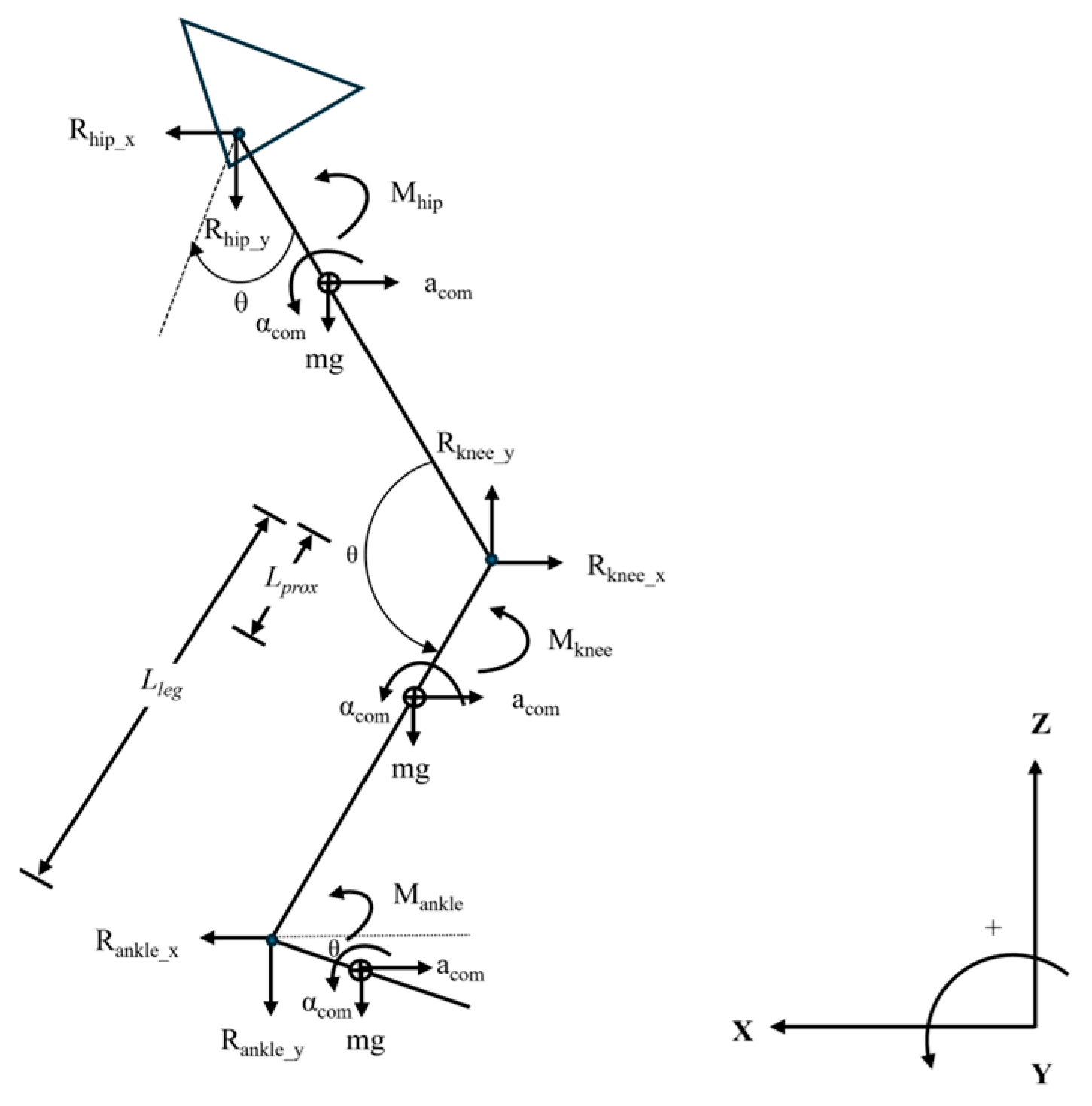
Figure 1. Free-body diagram of the lower limb.
Biomechanics 05 00077 g001
Figure 2. Relationship between left and right knee JRFcomp and (a) TBM, (b) LBM, and (c) BF%. Note: Blue and grey dashed lines represent regression lines for the left and right limb data.
Biomechanics 05 00077 g002
Figure 3. Relationship between left and right KAM and (a) TBM, (b) LBM, and (c) BF%. Note: Blue and grey dashed lines represent regression lines for the left and right limb data.
Biomechanics 05 00077 g003
Figure 4. Relationship between left and right mean vGRF and (a) TBM, (b) LBM, and (c) BF%. Note: Blue and grey dashed lines represent regression lines for the left and right limb data.
Biomechanics 05 00077 g004
Figure 5. Heavy vs. light players in normalized (a) JRFcomp, (b) KAM, and (c) mean vGRF. Note: ** denotes statistically significant difference, p ≤ 0.01; * denotes statistically significant difference, p ≤ 0.05.
Biomechanics 05 00077 g005
Table 1. Morphological characteristics (means ± SD; 95% CI).
GroupTotal Body Mass
(kg)
Lean Body Mass
(kg)
Body Fat
(%)
Total (N = 125)101.4 ± 19.387.2 ± 11.613.3 ± 6.5
(97.9, 104.8)(85.1, 89.2)(12.2, 14.5)
Lighter (n1 = 64)86.8 ± 6.778.3 ± 6.09.9 ± 3.6
(85.2, 88.5)(76.8, 79.8)(9.0, 10.8)
Heavier (n2 = 61)116.6 ± 16.196.6 ± 8.116.9 ± 7.0
(112.4, 120.7)(94.5, 98.6)(15.1, 18.5)
Table 2. Bilateral knee and normalized running kinetics (means ± SD; 95% CI), N = 125.
GroupLimbPeak Joint
Reaction Force
(N/kg)
Peak Knee
Adductor Moment
(Nm/kg)
Mean Vertical Ground
Reaction Force
(N/kg)
Total (N = 125)Left25.1 ± 2.22.2 ± 0.518.3 ± 1.4
(24.7, 25.5)(2.1, 2.3)(18.0, 18.5)
Right25.2 ± 2.32.2 ± 0.518.3 ± 1.4
(24.8, 25.6)(2.1, 2.3)(18.0, 18.5)
Lighter (n1 = 64)Left25.7 ± 2.12.3 ± 0.518.8 ± 1.2
(25.2, 26.3)(2.2, 2.5)(18.6, 19.2)
Right25.8 ± 2.12.3 ± 0.518.9 ± 1.1
(25.3, 26.3)(2.1, 2.4)(18.7, 19.2)
Heavier (n2 = 61)Left24.4 ± 2.02.1 ± 0.517.6 ± 1.3
(23.9, 24.9)(2.0, 2.3)(17.3, 18.0)
Right24.6 ± 2.42.1 ± 0.517.6 ± 1.5
(24.0, 25.2)(2.0, 2.2)(17.2, 18.0)
Table 3. Correlation coefficients for morphological and normalized running kinetics, N = 125.
VariableLimbTotal Body Mass
(kg)
Lean Body Mass
(kg)
Body Fat
(%)
Joint reaction
force
Left−0.39 **−0.39 *−0.21 **
Right−0.37 **−0.38 *−0.22 **
Knee adductor
moment
Left−0.21 *−0.20 *−0.14
Right−0.28 **−0.30 **−0.09
Mean vertical ground
reaction force
Left−0.56 **−0.56 **−0.39 **
Right−0.61 **−0.60 **−0.41 **
Note: ** denotes statistically significant association, p ≤ 0.01; * denotes statistically significant association, p ≤ 0.05.
Disclaimer/Publisher’s Note: The statements, opinions and data contained in all publications are solely those of the individual author(s) and contributor(s) and not of MDPI and/or the editor(s). MDPI and/or the editor(s) disclaim responsibility for any injury to people or property resulting from any ideas, methods, instructions or products referred to in the content.