1. Introduction
Body weight support (BWS) is a novel approach that provides an upward directed force to a person who is walking or running [
1]. There are several different mechanisms of providing BWS, one method that has become available in commercial fitness facilities as well as rehabilitation centers is a lower body positive pressure (LBPP) treadmill.
Part of the reason that BWS during locomotion has become more popular is that the user experiences less force during locomotion with BWS [
2,
3]. Less force may be useful during a rehabilitation program for those patients who may not be able to tolerate full weight bearing locomotion. Healthy runners may also benefit from reduced force with BWS since overuse running injuries have been hypothesized to be related to repetitively stressing the body [
4].
Muscle activity can also be influenced by BWS. For example, MacLean & Ferris [
3] examined overground walking across a wide range of simulated gravity levels and walking speeds. They reported that reductions in muscle activity with decreased load were not consistent across muscles; for example, gastrocnemius and quadriceps generally decreased with support, while tibialis anterior and biceps femoris increased at certain speeds. Their findings highlight the complex, non-linear effects of BWS and gait speed on neuromuscular control.
Similar unloading effects have also been described in aquatic treadmill and immersion studies, where buoyancy reduces effective body weight. In a systematic review, Silva et al. [
5] reported that muscle activity patterns during water-based gait were muscle-specific: distal muscles such as the tibialis anterior and gastrocnemius typically showed reduced activity, whereas proximal muscles such as the quadriceps and hamstrings were sometimes relatively more active. These findings support the idea that unloading does not uniformly reduce neuromuscular activity but instead induces complex, muscle activity adaptations. This perspective further underscores the importance of investigating muscle-specific responses when examining the interaction of BWS with other interventions.
Blood flow restriction (BFR) is a method that has become popular in strength and conditioning as a way to enhance a training stimulus at reduced loading [
6,
7,
8,
9,
10]. BFR was introduced in Japan in the 1960s under the term coined KAATSU [
7]. Although typically BFR is used in strength training [
7], it has also been used in low intensity aerobic exercise [
8,
9,
10]. There is also empirical evidence that walking with BFR provides a training stimulus [
11]. Clarkson and colleagues [
11] investigated how adding BFR to walking exercise could benefit older adults who might not tolerate heavy resistance training. Their study found that combining BFR with low-speed walking led to greater improvements in several indicators of functional fitness such as standing from a chair, walking endurance, mobility, and step tests as compared to walking alone [
11]. Notably, these gains were achieved with relatively low levels of effort as rated by participants, suggesting BFR walking could be a practical and tolerable option for enhancing physical function in older adults. These results highlight that BFR walking may help maintain or improve strength, mobility, and independence in populations at risk of muscle decline, especially when high-load training is not feasible.
The theoretical rationale for combining BFR and BWS is rooted in their apparent complementary physiological effects. BWS reduces musculoskeletal loading during locomotion, making walking and running feasible for individuals with pain, weakness, or impaired weight-bearing capacity [
1,
2,
3]. However, a limitation of BWS is the potential of reduction in muscle activation, particularly of the quadriceps and gastrocnemius muscles [
3,
12,
13], which may diminish the training stimulus. BFR, in contrast, has been shown to be beneficial to strength gains while strength training at lower intensities [
6,
7]. From a rehabilitation perspective, the combined use of BWS and BFR offers a unique opportunity: BWS reduces joint stress and impact forces, while BFR helps preserve or enhance muscle activation and adaptation despite the lower mechanical load. It may be that the pairing of BFR with BWS could allow the user to exercise at tolerable loading levels without compromising neuromuscular stimuli resulting in an adequate training stimulus as observed by Clarkson et al. [
11]. Therefore, the purpose of this study was to investigate whether lower extremity muscle activity was influenced by an interaction of BWS and BFR during walking at a self-selected pace. We also investigated stride frequency (SF) since this is a basic kinematic descriptor of gait that might provide insight into muscle activity results.
2. Materials and Methods
2.1. Participants
Participants (3 men and 4 women; age: 23.7 ± 3.0 years; height: 171.3 ± 6.9 cm; body mass: 64.4 ± 4.94 kg; body fat percentage: 18.8 ± 5.44 %) were free from any lower extremity injury that would interfere with walking and were not pregnant. None of the participants had prior experience using BFR. All participants gave written informed consent with the experimental procedure approved by the host institution Institutional Review Board.
2.2. Instrumentation
Testing took place at Lake Las Vegas Sports Club in the University of Nevada, Las Vegas satellite research lab. Body composition analysis was taken using bioimpedance (InBody 970, Biospace Co., Ltd., Eonju-ro, Gangnam-gu, Seoul Korea). BWS was done on a LBPP treadmill (Boost Treadmills, Redwood City, CA, USA) that had the capacity to provide up to 80% BWS. BFR was conducted utilizing using pneumatic pressure cuffs (B Strong Training Systems, Park City, UT, USA) applied to one thigh. Electromyography (EMG) was collected using wireless, surface EMG (Wave Plus Cometa system, Cometa Systems, Bareggio, Italy). Each EMG transmitting unit also measured acceleration in three orthogonal directions. Heart rate was monitored via telemetry chest strap (Wahoo Fitness, Atlanta, GA, USA). A Doppler system was utilized to listen for pulse (Edan SD3 Vascular Doppler System, Edan Instruments Inc., Shenzhen, China). The Rating of Perceived Exertion (RPE) scale was monitored using the Borg 6–20-point scale.
2.3. Procedures
Participants were asked to report to the lab for a 1-day test lasting approximately one hour and a half. Participants were screened using the Physical Activity Readiness Questionnaire (PAR-Q) as well as a thrombosis risk assessment survey [
14]. All anthropometrics were measured (height, mass, body composition) and then were then fit for the correct size of Boost shorts and size for the B-Strong pneumatic pressure cuff.
Prior to testing, participants first completed self-selected speed protocol. For this protocol, participants walked using the BWS treadmill with no BWS while wearing a BFR cuff with no pressure applied. Participants were blinded to the treadmill display screen to avoid seeing the treadmill speed. Participants were instructed to choose a comfortable speed that they would use for a long, leisurely walk. Once a speed was settled on, the participant would walk at this speed for one minute. This process was repeated 3 times with the average of the three speeds used as the test speed for all conditions. The group mean test speed was 1.14 ± 0.30 m·s−1.
While wearing the Boost shorts, the BFR cuff was placed as proximally on the participants’ right thigh as high as possible over the shorts. Participants were then told to sit down, while the researcher found the participants tibialis posterior pulse utilizing Doppler. Once the pulse was found, the researcher would then inflate the cuff until the pulse was absent. The researcher would note the pressure level of the cuff and then deflate the cuff. The test cuff pressure was set to 80% of the cuff pressure when the pulse went absent. The researcher would then reinflate to the new calculated cuff pressure and then have the participant walk around to get comfortable with walking with the cuff.
After the test speed and BFR pressure were determined, participants were then fit with a heart rate monitor and sensors to record EMG. Sensors were placed with the participants wearing the Boost treadmill shorts. Each location of sensors was prepared by cleaning the surface of the skin. Sensors were placed following published guidelines Surface Electromyography for the Non-Invasis Assessment of Muscles (SENAIM) [
15] of the Rectus Femoris (RF), Biceps Femoris (BF), medial head of Gastrocnemius (GA), and the Tibialis Anterior (TA).
The order of conditions was determined by first identifying all possible orders. Participants were then randomly assigned one of these orders with no order being repeated. Each participant completed each of the following conditions at the test speed:
Walking with no BWS, and no BFR
Walking with 50% BWS and no BFR
Walking with no BWS and BFR
Walking with 50% BWS and no BFR
For the BFR conditions, it was necessary to first inflate the BFR cuff to the target pressure, then have the participant secured in the LBPP treadmill. At that point, the treadmill speed was set to the test speed. There was approximately 30s between inflating the BFR cuff to the pressure and walking.
For each condition, the participant walked for approximately 2 min with EMG data collected in the first minute and RPE recorded at the end of the 2 min. In between each condition, BFR cuffs were deflated (when applicable) and the participant exited out of the Boost treadmill (shorts remained on). Participants were asked to sit down and not consume any food or liquids other than water. The participants began the next protocol 3 min after BFR cuffs were deflated.
2.4. Data Reduction
Stride frequency was determined using the resultant acceleration of the TA transmitting unit. Using distinct peaks that corresponded to foot strike, the time to complete 15 strides was measured. EMG data for each muscle were extracted over these 15 strides by removing any zero offset and then full-wave rectified. Average EMG was then calculated and used for analysis. EMG patterns were qualitatively analyzed by smoothing the data across the 15 strides using a 4th order, low pass Butterworth filter (fc = 4 Hz). Group ensemble plots were generated for each condition.
2.5. Statistical Analysis
The dependent variables Average EMG of each of the four muscles (RF, BF, GA, TA), SF, and RPE. Each dependent variable was analyzed using SPSS (IBM SPSS Statistics Data Editor, SPSS Version 29.01.00). The independent variables were BWS (0%, 50%) and BFR (with and without BFR). A two-way repeated measures analysis of variance was utilized to perform the analysis with significance set at an alpha level of 0.05. For descriptive purposes, ensemble plots for each muscle were created.
4. Discussion
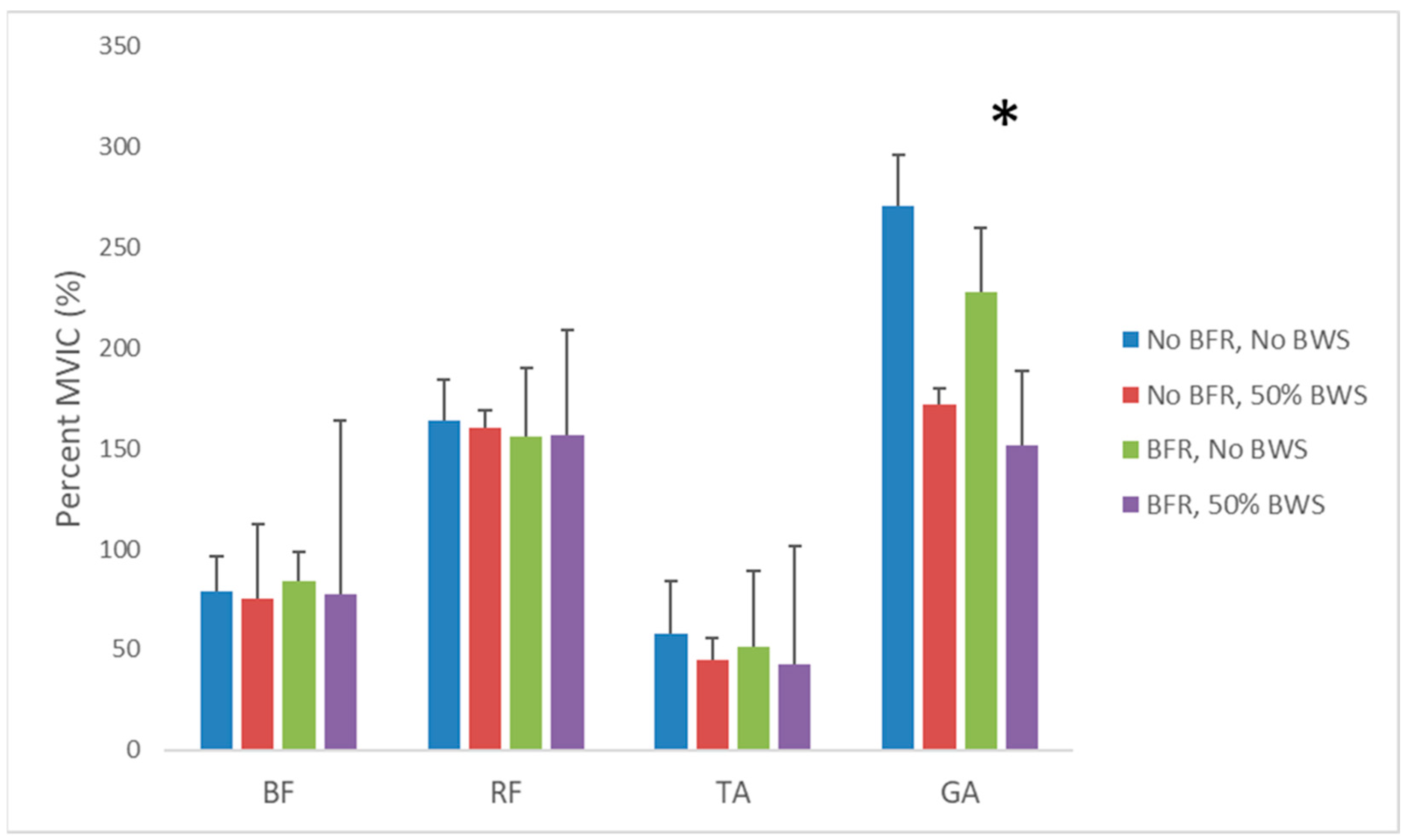
The main observation of this study was that muscle activity of the RF, BF, GA, and TA were not influenced by BFR regardless of BWS. Along with that, there was no change in SF across the different BWS and BFR conditions. The observations made are in the context of an acute (3 min) application of BFR during walking at a self-selected pace with and without 50% BWS.
We are not aware of any published data on muscle activity during combined BFR and BWS walking. However, there is research on the independent effects of BFR and BWS during walking. For example, Kristiansen et al. [
12] investigated lower extremity muscle activity while participants walked using 0%, 20%, 40%, and 80% BWS levels at 0.69 m·s
−1 and 1.0 m·s
−1 speeds. Similarly to the present study, Kristiansen et al. [
12] reported that the medial GA muscle activity decreased with increased BWS. Likewise, it was reported that the BF was not different across BWS nor was the TA at the faster walking speed, similar to what was observed in the present study. However, it was reported that the Vastus Lateralis (VL) decreased with BWS. In the present study, we were focused on the RF and observed no change in muscle activity between BWS conditions. It is not clear if the difference between studies for a knee extensor muscle is due to different knee extensor muscles (i.e., RF vs. VL) and/or that the RF also acts to cause hip flexion. Furthermore, it may also be that the results are influenced by walking speed since speeds used in the present study were faster (1.14 ± 0.30 m·s
−1) than that used by Kristiansen et al. [
12] (0.69 and 1.0 m·s
−1). Although Liebenberg et al. [
13] reported that muscle activity increased for each BWS during running and it is suspected this relationship would exist for walking as well, the effect of speed on muscle activity during walking is more nuanced [
12]. Kristiansen et al. [
12] reported that knee extensors were not influenced by speed when walking with BWS whereas the TA increased with speed while the GA decreased.
In a study investigating muscle activity during overground walking using a harness system to provide BWS, Fenuta & Hicks [
16] reported that BF did not change with BWS while GA decreased; both observations are similar to the present study. In contrast to the present study, both the RF and TA decreased with BWS [
16].
Also using a harness system during overground walking, MacLean and Ferris [
3], reported that RF, VL, and GA generally decreased with unloading, while TA and BF responses were more variable and sometimes increased at higher speeds. This pattern of muscle-specific and speed-dependent effects is consistent with our observation that only the gastrocnemius was significantly influenced by BWS. Together with previous research [
3,
12,
16], the results from BWS during walking research suggest that the neuromuscular adaptations to unloading are highly dependent on both the level of BWS and walking speed and that different muscles do not respond uniformly. It is important to note that it is not clear if data from overground-harness systems can be compared to treadmill walking with BWS provided via LBPP. Nevertheless, in a systematic review conducted by Apte et al. [
1] of research using BWS (in general), it was summarized that the medial GA demonstrated a decrease in muscle activity with increases in BWS while the RF, BF, and TA did not demonstrate consistent responses to BWS between studies for healthy subjects.
Another means to provide BWS during walking is walking in shallow water where the buoyancy force of water provides an upward directed force effectively reducing body weight. Our findings that the GA was the only muscle that was influenced by BWS only are somewhat consistent with evidence from aquatic locomotion studies. In a systematic review of water locomotion research, Silva et al. [
5] concluded that neuromuscular responses to immersion are not uniform across muscles. Of course, unloading via buoyancy force due to water comes with the complicating factor of drag force that provides resistance to movement in any direction.
Taken together, the EMG results we observed are reasonably in line with previous research using different mechanisms of BWS. Furthermore, given the nuanced differences between studies, it is speculated that there are multiple factors influencing muscle activity that could include type of BWS mechanism, level of BWS, treadmill vs. overground walking, and speed, for example. Any inconsistencies between studies on exact response of muscles to BWS seem to reflect a non-linear reorganization of neuromuscular control with unloading. Further research is needed to determine the influence of speed on muscle activity during walking with BWS.
The other main factor we were interested in understanding was the influence of BFR on muscle activity during walking. There is a paucity of research on muscle activity while walking with BFR. James et al. [
17] reported that muscle activity was greater when walking with BFR. However, there are some notable methodological differences between James et al. [
17] and the present study. For example, it appears that BFR was applied to both limbs in James et al. [
17] vs. unilaterally in the present study. The walk time was longer for James et al. [
16] (2 × 10 min bouts of walking) vs. the present study (3 min) and the speed was faster (set speed of 1.33 m·s
−1 vs. self-selected speed of 1.14 ± 0.30 m·s
−1). Finally, James et al. [
17] had participants complete one condition per day (i.e., two test days) whereas the present study was conducted on a single day. Both studies provide insight into muscle activity during walking with BFR. However, it is not clear if these methodological differences lead to differences in muscle activity responses. Along with this, the securing of the BFR cuff for the condition where there was no BFR needs to be done carefully. In pilot work for this project, we noted that if the participant was not appropriately set up with BFR in combination with the BOOST shorts, the cuff would slip off during the no BFR conditions.
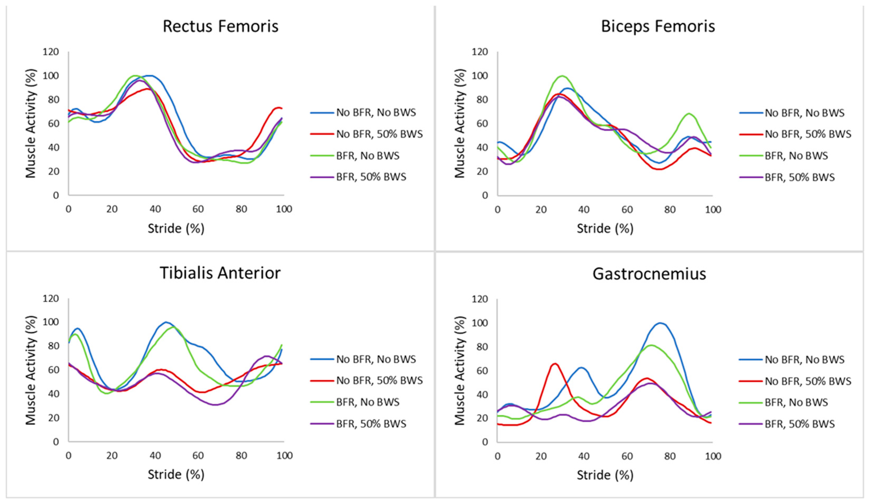
It is also important to note that James et al. [
17] extended their analysis to examine patterns of muscle activity and determined that BFR influenced the regularity of muscle contraction. We qualitatively inspected the patterns of muscle activity and noted that the patterns of the RF and BF were quite similar across conditions whereas the TA pattern seemed more disrupted by BWS while the GA pattern was quite variable between conditions. Inspection of muscle activity patterns are important observations since muscle activity patterns ultimately align with coordination and preferred movement patterns. In the present study, our analysis was focused on average muscle activity over 15 strides. In a different locomotor task of weighted sled push, Garcia et al. [
18] reported an increase in lower extremity muscle activity when using BFR. This seems to indicate that intensity of muscle contraction is likely another factor influencing muscle activity during locomotion. Clearly, there is a need for more research on the influence of BFR on muscle activity during walking.
Even with the limited research to date, the influence of BFR on muscle activity during walking remains equivocal. Presently, there are no specific standards when using BFR—especially during an activity such as walking. Additional research is needed to explore different parameters such as magnitude of occlusion, time of occlusion, walking speed, participant experience with BFR, BFR pressure, and applying BFR unilateral vs. bilaterally, for example, even cuff design should be explored since it has been reported that cuff width influences RPE and systolic blood pressure [
19].
Future studies could also explore different populations such as an older population or post stroke patients, for example. As the present study focuses on healthy subjects, it would be beneficial to see if there are possible gaps in understanding muscle activity responses in injured or unhealthy populations. In addition to exploring average muscle activity, future studies could examine other attributes of muscle activity such as muscle activity pattern as well as agonist-antagonist coordination. Furthermore, it seems important to also investigate different systemic biomarkers that are attributed to muscle repair and growth when using BFR [
7].
It is important to note that the mechanism of benefit of BFR use is not fully understood. In a literature review, Hwang et al. [
7] summarized several potential mechanisms behind BFR training that involve several physiological processes that work together to promote muscle adaptations. For example, the concept of BFR is limiting blood flow out of the muscle while maintaining some arterial inflow. Thus, BFR creates a low-oxygen environment that leads to a buildup of metabolic byproducts such as lactate. This metabolic stress can help trigger the release of anabolic hormones like growth hormone and insulin-like growth factor-1, which play roles in muscle repair and growth. Hwang et al. [
7] also stated that BFR may activate intracellular signaling pathways linked to protein synthesis and stimulate satellite cell activity, which supports muscle regeneration. Additionally, the increased metabolic demand under restricted blood flow may encourage greater recruitment of fast-twitch muscle fibers, even at lower loads. While our study did, overall, not find changes in muscle activity with BFR during walking with and without BWS, these underlying cellular and hormonal responses could still contribute to strength and hypertrophy gains over time, highlighting the complexity of the BFR effects.
The absence of an interactive effect between BFR and BWS in the present study should be interpreted in the context of their complementary rationale: BWS provides unloading to protect joints and reduce ground reaction forces [
1,
2,
3], whereas BFR is intended to offset the reduced neuromuscular demand associated with unloading [
17]. Although our acute application of BRF and BWS did not reveal additive changes in muscle activity, the theoretical basis for their combined use in rehabilitation remains compelling. Interestingly, we observed no changes in SF across the different conditions. This seems to suggest that the preferred SF selected was relatively robust to moderate unloading and circulatory occlusion.
A key limitation of the present study is the small sample size (
n = 7) and the study was not powered to detect small-to-moderate effects. For example, our observed increase in RPE with BFR corresponded to a large within-subject effect (Cohen’s dz ≈ 0.8–1.0), suggesting that relatively few participants would be required to replicate this finding. In contrast, muscle activity outcomes were not influenced by BWS (with the exception of GA) and/or BFR, may still be subject to Type II error if the true effects are small to moderate. Power analyses based on conventional benchmarks indicate that approximately 24–30 participants would be needed to detect small to moderate main effects. Thus, the null findings in the present study should be interpreted with caution, as subtle differences in EMG or SF may not have been detectable with the current sample size. Larger studies or studies that involve a higher intensity, for example, will be necessary to clarify whether the absence of combined effects of BFR and BWS represents a true physiological outcome or is attributable to insufficient statistical power. Nevertheless, the present study provides broad insight into an empirical approach to determine if the combination of BFR and BWS influences muscle activity. The similarity of muscle activity patterns (
Figure 2) between different combinations of BFR and BWS, seems to support the hypothesis that any potential changes in muscle activity will be nuanced—especially when intensity of workload is low, as was the case in the present study.


