Article

Critical Effect of Oxygen Concentration and Acidity on the Efficiency of Photodegradation of Levofloxacin with Solar UVB Light; Cytotoxicity on Mammalian Cells of the Photoproducts and Its Activity on Pathogenic Bacteria

by
Macarena Agostina Biondi
1,2,
Mariana Belén Spesia
1,3,
María Carola Sabini
4,
María Alicia Biasutti
1,3,
Hernán Alfredo Montejano
1,2,* and
Eugenia Reynoso
1,3,*
1
Departamento de Química, Facultad de Ciencias Exactas, Físico-Químicas y Naturales, Universidad Nacional de Río Cuarto (UNRC), Ruta Nacional 36 Km 601, Río Cuarto X5804BYA, Argentina
2
Instituto de Investigaciones en Tecnologías Energéticas y Materiales Avanzados (IITEMA), CONICET–UNRC, Ruta Nacional 36 Km 601, Río Cuarto X5804BYA, Argentina
3
Instituto para el Desarrollo Agroindustrial y de la Salud (IDAS), CONICET–UNRC, Ruta Nacional 36 Km 601, Río Cuarto X5804BYA, Argentina
4
Instituto de Investigaciones en Ciencias de la Salud (INICSA), CONICET–UNC, Ciudad Universitaria, Córdoba X5000, Argentina
*
Authors to whom correspondence should be addressed.
Compounds 2025, 5(2), 23; https://doi.org/10.3390/compounds5020023
Submission received: 14 May 2025 / Revised: 4 June 2025 / Accepted: 13 June 2025 / Published: 17 June 2025

Abstract

Levofloxacin is an antibiotic classified as an emerging contaminant. Its presence in aquatic environments represents potential risks to ecosystems and human health, making its removal during wastewater treatment of relevant importance. Here, we present a comprehensive kinetic analysis of levofloxacin photodegradation under UVB solar irradiation, with emphasis on the influence of pH and dissolved oxygen, two conditions that can vary widely in wastewater and impact treatment efficiency. We also investigated the formation and role of reactive oxygen species in the degradation mechanism, as well as the cytotoxicity and antibacterial activity of photoproducts. Our findings reveal that the efficiency of levofloxacin photodegradation is highly dependent on environmental conditions; it requires neutral or slightly alkaline pH and a high concentration of dissolved oxygen, a situation not always observed in contaminated waters. Several reactive oxygen species are generated, with singlet oxygen being the most reactive with the antibiotic. We report for the first time the singlet oxygen quantum yield from levofloxacin. Bioassays demonstrated that photoproducts neither exhibit antibacterial activity nor induce significant cytotoxicity. Our study suggests that UVB treatment of contaminated effluent containing levofloxacin could be an effective and environmentally safe strategy for the antibiotic degradation under certain conditions of pH and dissolved oxygen.

Graphical Abstract

1. Introduction

Emerging contaminants (ECs) are a group of synthetic chemicals that cannot be easily monitored in the environment and can have adverse effects on the environment and human health [1]. One of the main groups of ECs is pharmaceutical products [2]. These products often enter water bodies as biologically active compounds, mainly from household and hospital effluents [3,4]. Antibiotics are included in the group of potentially dangerous pollutants for their intensive use in human health, livestock farming, and agriculture. Final effluents from wastewater treatment plants (WWTPs) have been found to contain antibiotics; moreover, recent research has documented their presence in surface waters [5,6]. The environmental release of this kind of EC at sub-lethal concentrations contributes to the selection and dissemination of multidrug-resistant bacteria [6,7]. The clinical management of infections caused by these pathogens is increasingly challenging, often resulting in high morbidity and mortality rates, as well as a substantial burden on healthcare systems [6].
In recent years, many investigations have focused on the study of different processes to achieve the elimination of antibiotics in wastewater [6,7,8,9]. Among these, photodegradation involving sunlight has emerged as an effective approach for the degradation of a wide variety of pharmaceutical compounds [10,11,12,13]. When the substance to be degraded absorbs sunlight, excited electronic states are generated that can lead to bond breaking, isomerization, ionization, etc. [12,14,15]. On the other hand, when direct photolysis occurs in the presence of dissolved oxygen, reactive oxygen species (ROS) can be formed by self-sensitization mechanisms. These ROS can react with the contaminants themselves, generating additional degradation pathways [15,16].
Several and very complete works about direct photolysis of fluoroquinolone antibiotics have been carried out previously [17,18,19,20,21,22,23,24,25,26,27,28]; however, some aspects have not yet been fully clarified.
In the present study, we focused on investigating the influence of pH and dissolved oxygen concentration on the photodegradation rate of the antibiotic levofloxacin (Lev) in aqueous solution. Both conditions can have very different values in the contaminated water that reaches the wastewater cleaning plants.
Lev is a third-generation fluoroquinolone characterized by a broad spectrum of antibacterial effects [19]. Also, it has the highest excretion rate of unmetabolized molecules (85% in human urine), making its presence in wastewater very significant [9,19]. The concentration of Lev found in the environment can vary depending on the type of the effluent. Lev has been detected in sewage and wastewater in concentrations exceeding 7000 ng/L [9].
It is a fairly common practice in wastewater treatment processes to apply high doses of acids or alkalis to facilitate the effective action of various chemical purification methods. This is why the pH values found in wastewater can vary within a very wide range [29,30].
Moreover, biochemical oxygen demand (BOD) is commonly associated with aqueous systems containing elevated concentrations of biodegradable organic matter. High BOD levels indicate intense microbial activity, which can lead to significant depletion of dissolved oxygen, potentially reducing it to near-anoxic or undetectable levels [31].
For these reasons, we consider it is necessary to revisit the kinetic aspects of Lev photodegradation under different pH and dissolved oxygen concentration conditions.
Regarding the dependence of Lev photodegradation kinetic constants (kphot) on pH, there is some disagreement found in the literature. While Ahmad et al. [22] reports that kphot increases continuously from pH 2 to pH 10, other authors [23,27] have found, for some fluoroquinolone antibiotics structurally related to Lev, that kphot increases from acidic pH values, reaches a maximum value in the pH range of 7–8, and then decreases when the pH becomes more alkaline. With respect to the influence of dissolved oxygen concentration as a variable in the photodegradation of fluoroquinolone antibiotics, to the best of our knowledge, there is only one report [17] in which the authors determined the photodegradation quantum yields (Φphot) of four fluoroquinolones (excluding Lev) in aqueous solutions equilibrated with air and argon. These experiments showed a wide range of behaviors, with no general trend for all the molecules examined. While for norfloxacin, an 83% increase in Φphot is observed in an argon atmosphere, for ofloxacin, the Φphot decreases by 33%, in both cases, compared to solutions equilibrated with air.
In this context, our main objective is to expand and clarify the information regarding Lev degradation by UVB light performing a kinetic analysis of the photoprocesses varying pH and dissolved oxygen concentration conditions in the medium. Additionally, we investigated the generation and participation of ROS, the cytotoxicity of the photoproducts obtained in the presence and absence of oxygen on mammalian cells, and their residual activity against pathogenic bacteria, taking as a reference in each case the effects produced by the non-irradiated Lev.

2. Materials and Methods

2.1. Materials

Levofloxacin > 98% (Lev), superoxide dismutase (SOD) from bovine erythrocytes, catalase from bovine liver (CAT), D-mannitol, perinaphthenone 97% (PN), deuterium oxide 99.9% (D2O), and acetonitrile (CH3CN) HPLC quality 99.5%, were purchased from Sigma-Aldrich. Sodium azide 99% (NaN3) and isopropyl alcohol 99.7% (IPA) were purchased from Merck. All of these reagents were used as received. The pH of the final solutions for the Lev photolysis experiments was 4.0, 7.4, and 10.0. To adjust the pH, buffers were prepared according to a literature procedure [32], using citric acid, KH2PO4, NaCO3H, and NaOH, provided by Cicarelli pro-analysis.
In the microbiological test, the culture media used were purchased from Laboratorios Britania S.A., Buenos Aires, Argentina. These media were trypticase soy broth (TSB: casein peptone (pancreatic), 17 g; glucose, 2.5 g; soy peptone (papain digest.), 3 g; NaCl, 5 g; Na2HPO4, 2.5 g; and distilled water, 1000 mL) and trypticase soy agar (TSA: 12 g TSB and 6 g agar–agar; and distilled water, 400 mL). TSB and TSA were autoclaved at 121 °C for 20 min.
In the cytotoxicity analysis, Eagle’s minimum essential medium (EMEM), provided by GIBCO, and heat-inactivated fetal calf serum (FCS), provided by Natocor, were used. L-glutamine 99%, gentamicin 98%, neutral red 30% solution (NR), and 3-(4,5-dimethylthiazol-2-yl)-2,5-diphenyl tetrazolium bromide HPLC grade 97.5% (MTT) were provided by Sigma-Aldrich (Argentina). Ethanol 99.5% and glacial acetic acid 99.9% were provided by Cicarelli Pro-Analysis (Argentina), while dimethyl sulfoxide HPLC quality 99.9% was provided by Sintorgan (Argentina).
In all experiments, the water used was triply distilled.

2.2. Methods

2.2.1. Levofloxacin Photolysis Experiments

Stationary photolysis experiments were carried out via direct irradiation of 2 × 10⁻⁵ M Lev solutions, employing a Rayonet Ltd. (Branford, USA) circular photoreactor, provided with three UV RPR-3000A lamps, whose maximum emission is 300 nm ± 20 nm. The irradiance of each lamp is 1700 μW/cm2. The illumination of the samples was carried out at room temperature (25 ± 2 °C). The photoreactor has a ventilation system that prevents temperature fluctuation during the photolysis of the samples. The irradiation used corresponds to the UVB electromagnetic range of the solar spectrum (280–320 nm), in which Lev presents its main absorption band independently of the pH of the buffer used in the different studies (see Figure 1).
In order to monitor the photodegradation process, UV–Visible absorption spectra of the antibiotic solution were recorded on a Hewlett Packard 8453A diode array spectrophotometer using a quartz cuvette with 10 mm of optical path.
The variation in the absorbance of Lev with irradiation time at fixed wavelength was treated with a pseudo-first-order kinetic by writing the rate as −δA/δt = kphot A, where A is the Lev absorbance at different irradiation times, t; and kphot corresponds to the apparent photolysis rate constant experimentally obtained. Then, from plots of ln A/A0 vs. t (Equation (1)), where A0 refers to the Lev absorbance at zero time, the pseudo-first-order rate constant, kphot, could be obtained. This rate constant, under constant irradiation condition, was used as a comparative parameter for the photochemical behavior of the system.
ln A A 0 = k p h o t   t
To evaluate the participation of oxygen in the degradation mechanism of Lev induced by UVB light, kphot was determined under argon, air, and oxygen atmospheres. For this experiment, the solutions of Lev were independently irradiated in equilibrium with air, under continuous bubbling of oxygen and in a hermetically sealed cuvette in which the antibiotic solution was previously bubbled with argon for 20 min.
To prove the participation of different ROS in the photodegradation of Lev, kphot values were determined in the absence and in the presence of specific ROS scavengers. In this case, Lev solutions were irradiated in air atmosphere condition, adding 1 × 10⁻⁷ M of SOD, 1 × 10⁻⁷ M of CAT, 0.2 M of IPA, and 1 × 10⁻³ M of NaN3, all in separate experiments. The enzymes SOD and CAT were used for the detection of superoxide radical anion (O2∙−) and hydrogen peroxide (H2O2), respectively. Meanwhile, IPA and NaN3 were used for hydroxyl radical (HO) and singlet oxygen (O2(1Δg)) detection, respectively. The reactions involved between the ROS and each scavenger are described in Section 3.2.
In the particular case of O2(1Δg), the time-resolved phosphorescence detection method (TRPD) was also used, which has been previously described in depth [33]. For this, Lev was dissolved in D2O instead of water to lengthen the O2(1Δg) lifetime [34]. The excitation of the antibiotic was achieved by employing the Nd:YAG laser output at 355 nm. The radiation emitted by O2(1Δg) at 1270 nm was collected at a right angle with a germanium Judson J16/8Sp detector. The detected signal was acquired in a digital oscilloscope attached to a personal computer for further processing. Several laser shots were needed in order to obtain a good signal-to-noise ratio for decay time calculations [35].
The quantum yields of O2(1Δg) generation by Lev ( Φ Δ L e v ) were determined by the comparative method described elsewhere [36], using PN in CH3CN as a reference compound. Briefly, the zero-time extrapolated O2(1Δg) phosphorescence intensity (I0) was measured against the laser fluence (EL) for Lev and PN. The change in the laser fluency was achieved by employing filters of neutral density. With the slope comparison of the linear plots of I0 vs. EL for the reference and the sample, and by using Equation (2), the Φ Δ L e v was calculated.
ϕ Δ L e v = s l o p e L e v s l o p e P N   k C H 3 C N O 2 k D 2 O O 2   ϕ Δ P N 1 10 A P N 1 10 A L e v
where kO2 is the relative rate constants for O2(1Δg) radiative deactivation in D2O ( k D 2 O O 2 = 0.12) and in CH3CN ( k C H 3 C N O 2 = 0.30) [36]; Φ Δ P N is the quantum yields of O2(1Δg) generation by PN in CH3CN ( Φ Δ P N = 0.98) [36]; and A is the absorbance of PN in CH3CN and of Lev in D2O at the laser excitation wavelength (355 nm).

2.2.2. In Vitro Bioassays on Bacteria

To assess if Lev retains or loses its bactericidal activity after treatment with UVB light or if the photoproducts generated in the absence and in the presence of oxygen have effect on microbial growth, the determination of the number of viable microorganisms and the agar diffusion test were carried out.
The pathogenic bacteria employed was an Escherichia coli strain (EC7) recovered from clinical urogenital material. The strain was identified according to conventional procedures and using the automated system mini-API rapid ID32 STREP (BioMérieux) method. EC7 was resistant to ampicillin and sulfamethoxazole–trimethoprim [37].
For the microbiological assays, a solution of 2 × 10⁻⁵ M Lev in buffer of pH 7.4 was exposed to UVB light in the dispositive used in the stationary photolysis experiment already described. Buffer of pH 7.4 was selected for the mentioned experiments because it provides an optimal and stable environment that allows for bacterial growth conditions.
The Lev photolysis was carried out in an air-equilibrated and argon-saturated atmosphere. For the latter, argon gas was bubbled into the Lev solution for 20 min to ensure complete removal of oxygen before irradiation, and then the reaction cuvette was sealed.
The number of viable microorganisms, expressed as colony-forming units per milliliter (CFU mL−1), was determined as described below. E. coli strain was grown aerobically at 37 °C in TSB overnight. An aliquot (50 μL) of this culture was aseptically transferred to 50 mL of fresh medium (TSB). This microbial suspension was distributed in Eppendorf tubes to which was added the solution of Lev before irradiation, Lev irradiated in air atmosphere conditions, and Lev irradiated in argon atmosphere conditions. The initial Lev concentration in each tube was 2.0 × 10⁻⁷ M. The volume of Lev solution added to each tube was replaced with buffer of pH 7.4 for cell control. All tubes were incubated at 37 °C for 4 h. After that, cell suspensions were 10-fold serially diluted with phosphate-buffered saline (PBS, pH = 7.4), and each dilution was quantified by the spread-plate technique in triplicate on TSA. The plates were examined for viable E. coli cells after 24 h of incubation at 37 °C, and CFU mL−1 was determined.
For the agar diffusion method, filter paper discs (previously sterilized in an autoclave) were impregnated with unirradiated 2 × 10⁻⁵ M Lev solution and with Lev solution irradiated at different times in air atmosphere conditions. The paper discs were then placed on plates containing TSA previously seeded with an overnight culture grown of E. coli strain in TSB. After that, the plates were incubated at 37 °C for 24 h, and the diameter of the inhibition halo (clear zone around the discs) was measured. The results were expressed as the ratio between the diameter of the halo after (h) and before (h0) photolysis, h/h0, which represents the relative rates of decrease in the bactericidal effect of the antibiotic against the selected bacteria. All of the experiments described were performed in triplicate.

2.2.3. In Vitro Bioassays on Mammalian Cell

The potential cytotoxicity effects of Lev and its photoproducts on mammalian cells were evaluated using the Vero cells (Cercopithecus aethiops green monkey kidney epithelial cell line). This normal cell line was acquired from ATCC as Vero CCL-81TM. The Vero cells were grown in EMEM supplemented with 10% (v/v) FCS, 3% (v/v) L -glutamine, and 5% (v/v) gentamicin at pH 7.2–7.4. Cell cultures were maintained at 37 °C with 5% CO2 and humidity. For the assay, the cells were plated in 96-well culture plates, incubated until cell monolayer formation, and then washed with PBS and incubated with the samples at 37 °C for 48 h.
Cell monolayers were exposed to different concentrations of Lev (between 0 and 2 × 10⁻⁵ M or 7.2 μg mL−1) before and after irradiation, in the absence and in the presence of oxygen. For this, Lev solutions were 2-fold serially diluted with EMEM. Untreated cells were used as a control, in which the antibiotic sample was replaced by buffer at pH 7.4.
Cell viability was evaluated by the colorimetric test NR uptake and MTT metabolism assay, as described below. Cell monolayers treated with Lev and its photoproducts were washed with PBS and incubated with NR or MTT for 4 h at 37 °C. Subsequently, the supernatant was removed by inverting the plates, and the cells were washed three times with PBS.
In the NR test, the dye within viable cells was released via extraction with a solution of acetic acid, ethanol, and water (1:49:50). After agitation of the cultures for 20 min, the absorbance was measured at 540 nm, using a microplate ELISA reader (LabSystems Multiskan MS, Finland). In the MTT assay, formazan crystals (from the reduction reaction of MTT) were solubilized with dimethyl sulfoxide, and the absorbance was measured at 560 nm in the same absorbance reader. The relative viability was expressed as a percentage with respect to control cells as follows:
%   V i a b i l i t y = A t r e a t e d   c e l l A c o n t r o l   c e l l × 100
where A is the absorbance at 540 nm and 560 nm for NR and MTT, respectively. Assays were carried out in triplicate on independent plates. Variation between each experiment was calculated using one-way ANOVA, with a confidence level of 95% (p < 0.05) considered statistically significant. Data were represented as the mean standard deviation of each group.

3. Results and Discussion

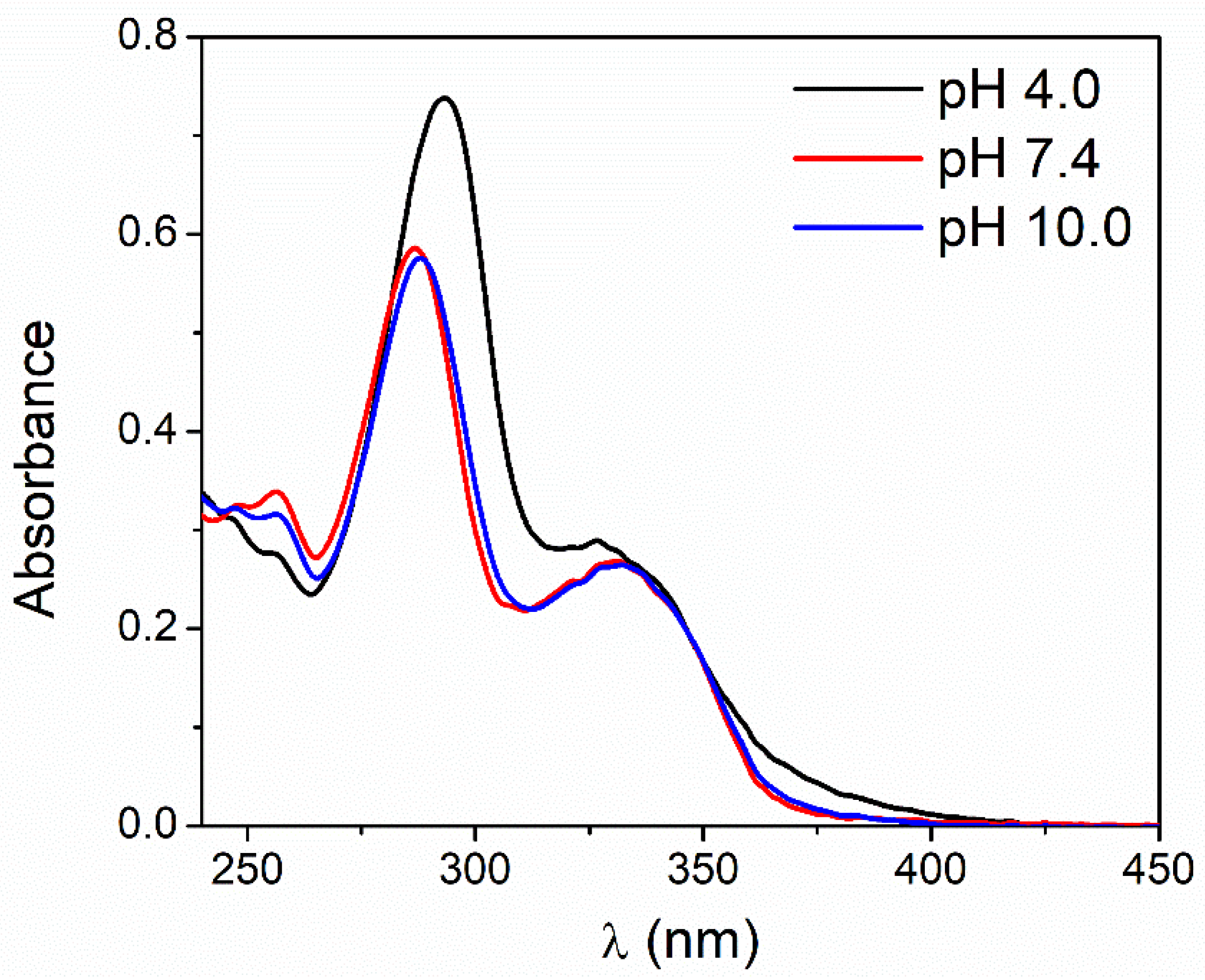
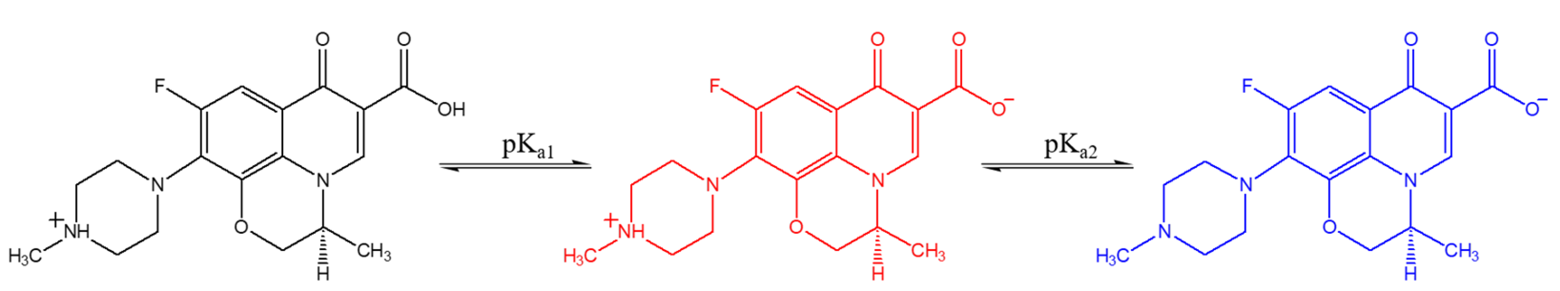
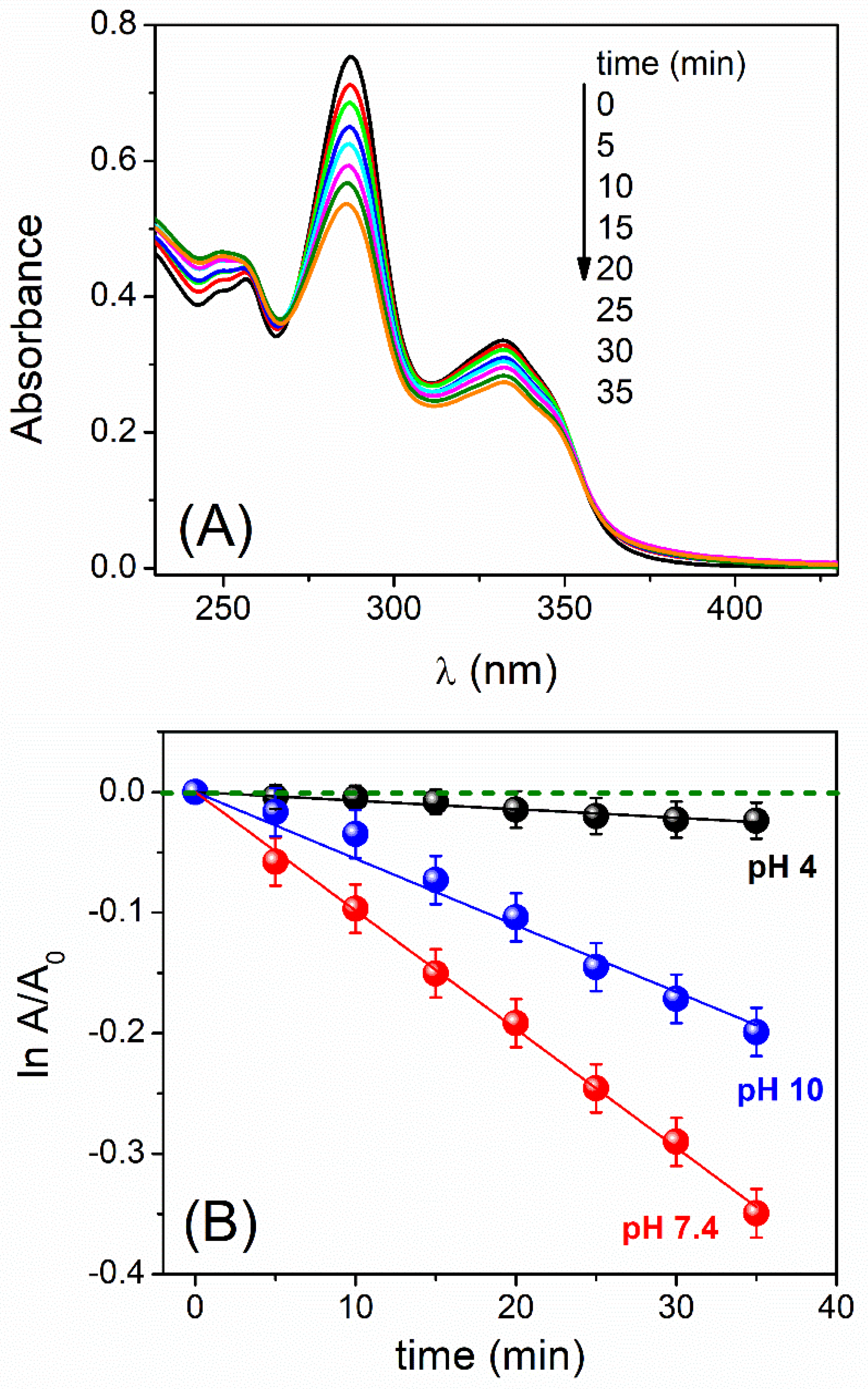
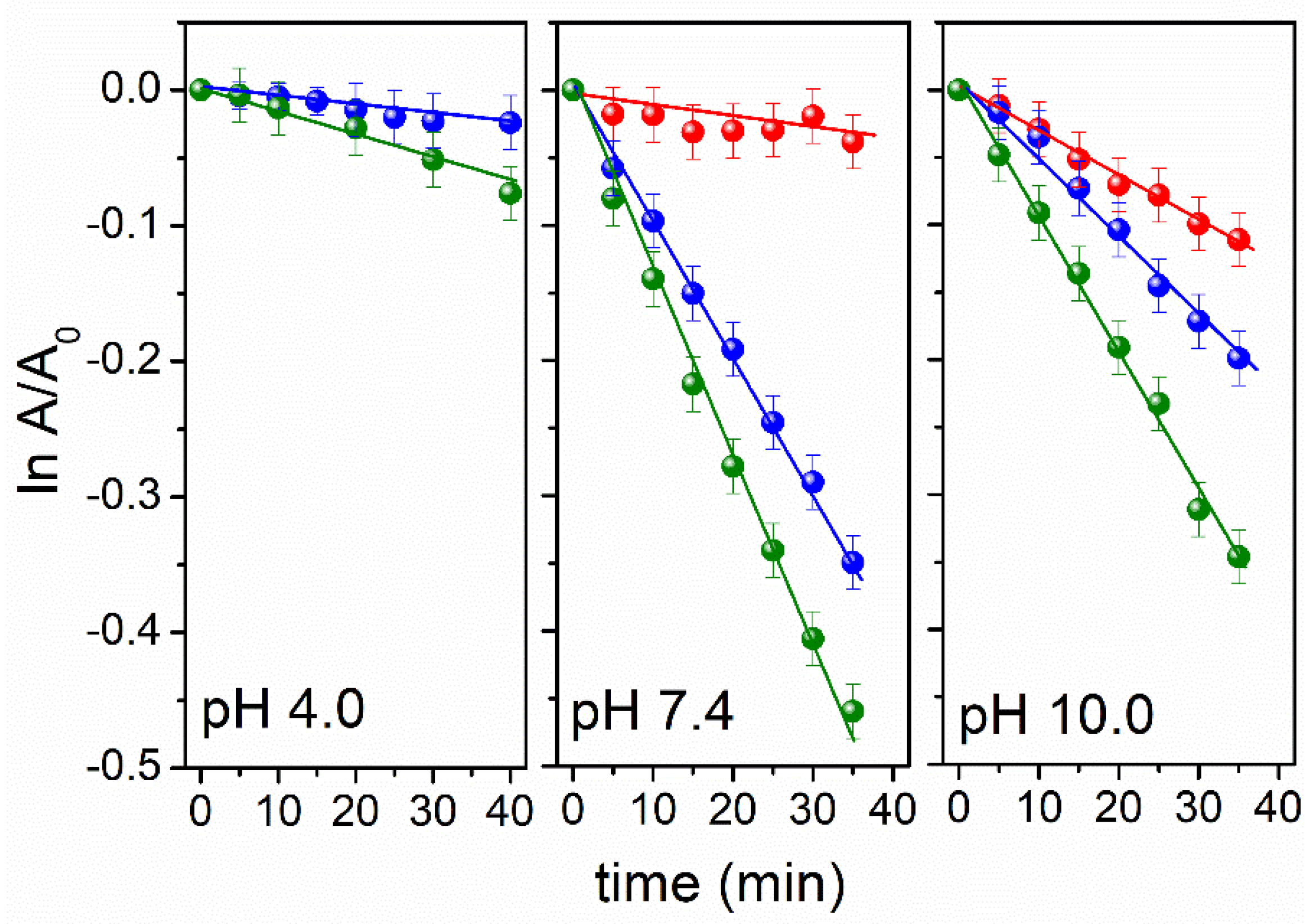
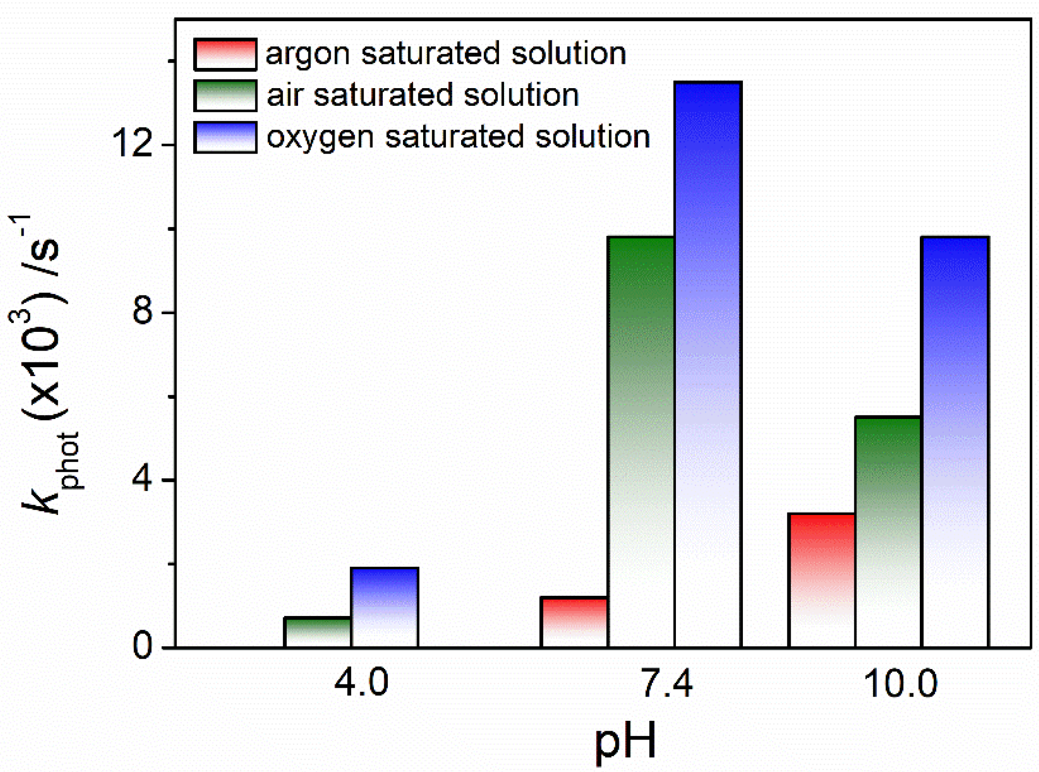
3.1. pH and Dissolved-Oxygen-Concentration Effect on Lev Photolysis

Lev has two reported pKa values, 6.0 and 8.1, corresponding to the deprotonation of the carboxylic group attached to the fluoroquinolone core (containing pyridine and benzene ring with a fluorine atom at C-6 position) and the amino group of the piperazine ring, respectively [38]. In this investigation, buffers of pH 4.0, 7.4 (physiological pH), and 10.0 were used as solvents of Lev. In acidic media, the antibiotic is found mainly as a cation; at pH 7.4, as a zwitterion; and in alkaline media anion form, it will be dominant [38,39]. The chemical structures of each ionic species are included in Scheme 1, while Figure 1 shows the absorption spectra of Lev obtained at each pH value studied.
Independently of pH value, the spectra show a main band at 260–320 nm assigned to the π → π* transitions of the delocalized electrons in the aromatic ring, and a weak band at 330–400 nm caused by an n → π* electronic transition [39]. Likewise, at acidic pH, the maximum of the main band shifts 6 nm towards longer wavelengths and has a higher molar absorption coefficient (ε294 nm = 36,800 ± 300 M−1 cm−1) compared with pH 7.4 and 10.0, in which the maxima are practically the same and the molar absorption coefficients are very similar, being ε288 nm = 28,900 ± 200 M−1 cm−1 and ε289 nm = 28,700 ± 200 M−1 cm−1 for pH 7.4 and 10.0, respectively. The molar absorption coefficients were determined in this work at the three pH conditions studied.
The photodegradation with UVB light was evaluated spectrophotometrically for Lev at pH 4.0, 7.4, and 10.0. Figure 2A shows the variation in the absorption spectrum of the antibiotic obtained at pH 7.4 in air-equilibrated solutions in which there is a clear decrease in the absorbance observed as there is an increase in the irradiation time exposure, indicating that Lev is sensitive to UVB light. On the other hand, Figure 2B shows the pseudo-first-order plot from which the apparent photodegradation rate constants (kphot) at each pH were determined. The values of kphot are summarized in Table 1.
The values of kphot expressed in Table 1 indicate that Lev can undergo UVB photodegradation in acidic, neutral, or alkaline media. Also, a clear dependence between the pH of the medium and the kphot was observed: at acidic pH, the value of kphot is markedly lower compared with that obtained at pH 7.4 and 10.0; meanwhile, pH 7.4 is the condition in which the kphot was the highest. This shows that Lev ionic species are not equally sensitive to UVB-light treatment, since the cationic species of Lev is much less susceptible to direct irradiation than the zwitterionic or anionic species.
In a previous work, Ahmad et al. studied the photodegradation of Lev under UV irradiation (λirradiation = 287 nm) in the pH range of 2.0–12.0 and found a very low sensitivity of the antibiotic to light in acidic medium [22]. On the other hand, they observed that the degradation rate increased as the pH was raised from 5.0 to values close to 10.0. These results differ from those we have found in our research, since the kphot value was greater at pH 7.4 than at pH 10.0. Nevertheless, our results are completely in agreement with those reported by Wammer et al. [23] and Ge et al. [27]. In the first case, the authors studied the degradation process of the fluoroquinolones enrofloxacin, norfloxacin, and ofloxacin with simulated sunlight at a pH between 4.0 and 10.0. For all three antibiotics, the direct photolysis rate for the zwitterionic form, which predominates at a pH between 6.0 and 8.0, was the fastest, followed by the anionic form and finally by the cationic form. Moreover, for the fluoroquinolones studied, the quantum yields experimentally obtained were always higher for the zwitterionic species than for the anionic species, and the quantum yield of the cationic species was very small, essentially indistinguishable from zero. Ge et al. [27] found a behavior similar when they studied the photolysis of the fluoroquinolones gatifloxacin and balofloxacin at a pH between 3.0 and 11.0, under simulated sunlight. They observed that the photolysis rates for the mentioned antibiotic are pH-dependent, following the order of zwitterionic form (neutral pH) > anionic form (alkaline pH) > cationic form (acid pH).
The strong dependence of the photodegradation of Lev on the pH of the medium found in our results could have a very important connotation with respect to the treatment of contaminated effluents. It is notable that if the effluent has high acidity, photodegradation is practically negligible. However, for neutral or moderately alkaline effluents, photodegradation of the antibiotic may be feasible.
Another relevant aspect evaluated in the present work was the influence of dissolved oxygen concentration on the degradation process of Lev induced by UVB light. For this, the kphot values under argon, air, and oxygen atmosphere at pH 4.0, 7.4, and 10.0 were determined. The pseudo-first-order plots are shown in Figure 3 for the three pH studies, and the values of kphot obtained for each atmosphere conditions at each pH are shown in Table 1.
It is necessary to clarify that in an argon atmosphere at pH 4.0, the kphot could not be determined since no changes in Lev absorbance were observed with the irradiation time. For this reason, no data are included in Figure 3 and Table 1. Under acidic medium and in complete absence of oxygen, it is interpreted that Lev is not photodegraded.
To facilitate the visualization of the kinetic data obtained, Figure 4 summarizes the results of UVB light photodegradations of Lev at different pH values and at different dissolved oxygen concentrations.
As can be seen in Figure 4, the efficiency of Lev degradation with UVB light not only depend on pH but also highly depend on the dissolved oxygen in the medium.
The kinetic dependence on oxygen concentration is clear, since in all cases, kphot increases noticeably in solutions in equilibrium with air (oxygen concentration: 0.27 mM at 25 °C) [40] and even more in solutions in equilibrium with pure oxygen (oxygen concentration: 1.27 mM at 25 °C) [40], compared with solutions equilibrated with pure argon (anoxic conditions).
These data could be highly relevant when evaluating a decontamination process, since highly contaminated effluents generally have a very high BOD, and under these conditions, the results achieved may be significantly lower than those expected if the analysis is based on results obtained in aqueous media with moderate or high oxygen concentrations.
In summary, if direct photolysis with sunlight is intended to be applied for the degradation of Lev, it is advisable to ensure that the conditions of the effluent to be treated correspond to a neutral or slightly alkaline pH and that the concentration of dissolved oxygen is as high as possible.

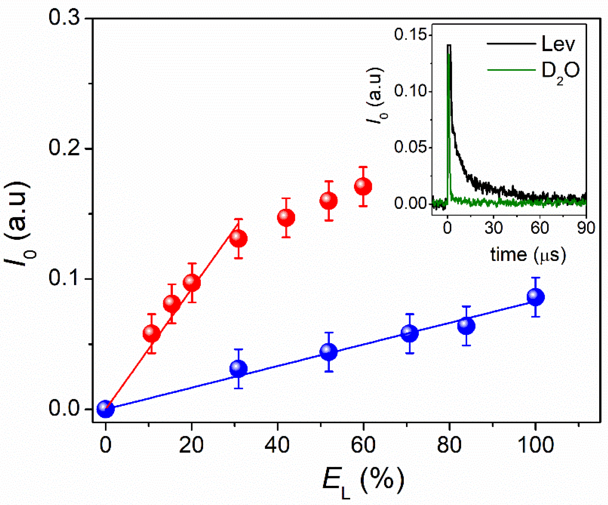
3.2. ROS Photogenerated and Singlet Oxygen Quantum Yield

Based on the kinetic results previously discussed, we could affirm that Lev is sensitive to direct irradiation with UVB light even in the absence of oxygen, thus indicating the presence of a unimolecular degradation pathway from the excited electronic states of Lev. The presence of oxygen significantly increases the kphot values at all pHs studied, thus demonstrating that a bimolecular degradation process is also involved. The oxygen-dependent pathway is associated with the photogeneration of ROS and the participation of these species in a self-sensitized degradation process.
Upon absorbing UVB light, Lev molecule is excited to a singlet state (1Lev*) which undergoes intersystem crossing to a triplet state (3Lev*). Formation of 3Lev* has been verified by laser flash photolysis by other authors [18,38]. The photodegradation occurs through 3Lev*, which can undergo typical reaction of fluoroquinolones photolysis, such decarboxylation, demethylation, and defluorination, or can react with the dissolved oxygen to generate ROS able to oxidize Lev [17,19,20,21,25,38,41]. The quenching rate constant of 3Lev* by oxygen is 1–2 × 10⁹ s−1 [18,38].
To confirm which ROS are formed and participate in the UVB photoinduced degradation of Lev, the kinetic experiments were carried out in the absence and in the presence of specific ROS scavengers. The studies were carried out at pH 7.4, considering that the kphot is much higher under this pH condition compared with the other pHs analyzed. Also, these studies were performed in air atmosphere since the process is only ~25% slower than in saturated oxygen atmosphere.
The enzymes superoxide dismutase (SOD) and catalase (CAT) were used as inhibitors of superoxide radical anion (O2•−) and hydrogen peroxide (H2O2), respectively, since they interact efficiently with the mentioned ROS according to (R1) and (R2) [42,43]. On the other hand, isopropyl alcohol (IPA) and sodium azide salt (NaN3) were used as hydroxyl radical (OH) and singlet oxygen (O2(1Δg)) scavengers, respectively ((R3) and (R4)) [34,42].
2 O2•− + 2 H+ + SOD → H2O2 + O2(3Σg)
2 H2O2 + CAT → 2 H2O + O2(3Σg)
(CH3)2CH(OH) + 2 OH → (CH3)2CO + 2 H2O
O2(1Δg) + NaN3 → O2(3Σg) + NaN3
The variation in Lev absorbance as a function of photolysis time was recorded, both in the absence and in the presence of ROS scavengers. Then, a pseudo-first-order kinetic analysis analogous to that already described was performed with the experimental data obtained. The corresponding kphot obtained in the presence of each inhibitor ( k p h o t i n h ) was relativized to the rate constant value in their absence ( k p h o t 0 ). The ratios obtained ( k p h o t i n h / k p h o t 0 ) are represented in the bar graph shown in Figure 5.
In Figure 5, a clear decrease in the kphot values can be observed when ROS inhibitors are present in the solution, thus allowing us to infer the generation of O2•−, H2O2, OH, and O2(1Δg) in the photodegradation mechanism of Lev, and that the antibiotic is susceptible to attack by these species. A more pronounced effect in the reduction of kphot was observed in the presence of NaN3, possibly suggesting that Lev is especially susceptible to O2(1Δg).
As additional evidence of the presence of O2(1Δg) in the photodegradation mechanism of Lev, the TRPD technique was used, which allows for the direct detection of the mentioned ROS. For this, a solution of Lev in D2O was used, and it was excited by employing a 355 nm laser. The signal obtained, shown in the insert of Figure 6, unambiguously confirms the generation of O2(1Δg) from the excitation of Lev. This signal corresponds to the phosphorescence emitted by O2(1Δg) at 1270 nm, which decays mono-exponentially with a lifetime of 15 μs approximately. The large lifetime of this ROS is a necessary factor to explain the reactivity observed with the antibiotic.
To quantify the capacity of photogeneration in the mentioned ROS, the determination of the quantum yield of O2(1Δg) generation by Lev ( Φ Δ L e v ) was carried out using the comparative method described in Section 2.2.1 and Equation (2), with PN as a reference compound. The results are shown in Figure 6, and the Φ Δ L e v value obtained was 0.30 ± 0.03. The value obtained is substantially higher than that found by other authors for the Lev enantiomer ofloxacin, but it is similar to that of other fluoroquinolone antibiotics, such as flumequine and rufloxacin [17].
Previous works by other authors have demonstrated the photogeneration of ROS by fluoroquinolone antibiotics, including Lev, and their participation in the photodegradative mechanism, employing both direct and indirect methods [17,18,19,25,44,45,46]. They report that Lev can produce O2•−, OH, and O2(1Δg) under UVB, UVA, and simulated sunlight, and also prove that these species could attack the antibiotic.
Likewise, our work provides additional information: although the ROS O2•−, OH, H2O2, and O2(1Δg) can be generated from Lev and react with it, the antibiotic appears to be more susceptible to O2(1Δg). Because this is the ROS that most oxidizes Lev, we quantify its generation by self-sensitization, using the direct TRPD method. Furthermore, to the best of our knowledge, the quantum yield of O2(1Δg) generation by Lev ( Φ Δ L e v ) has not been previously reported until now.

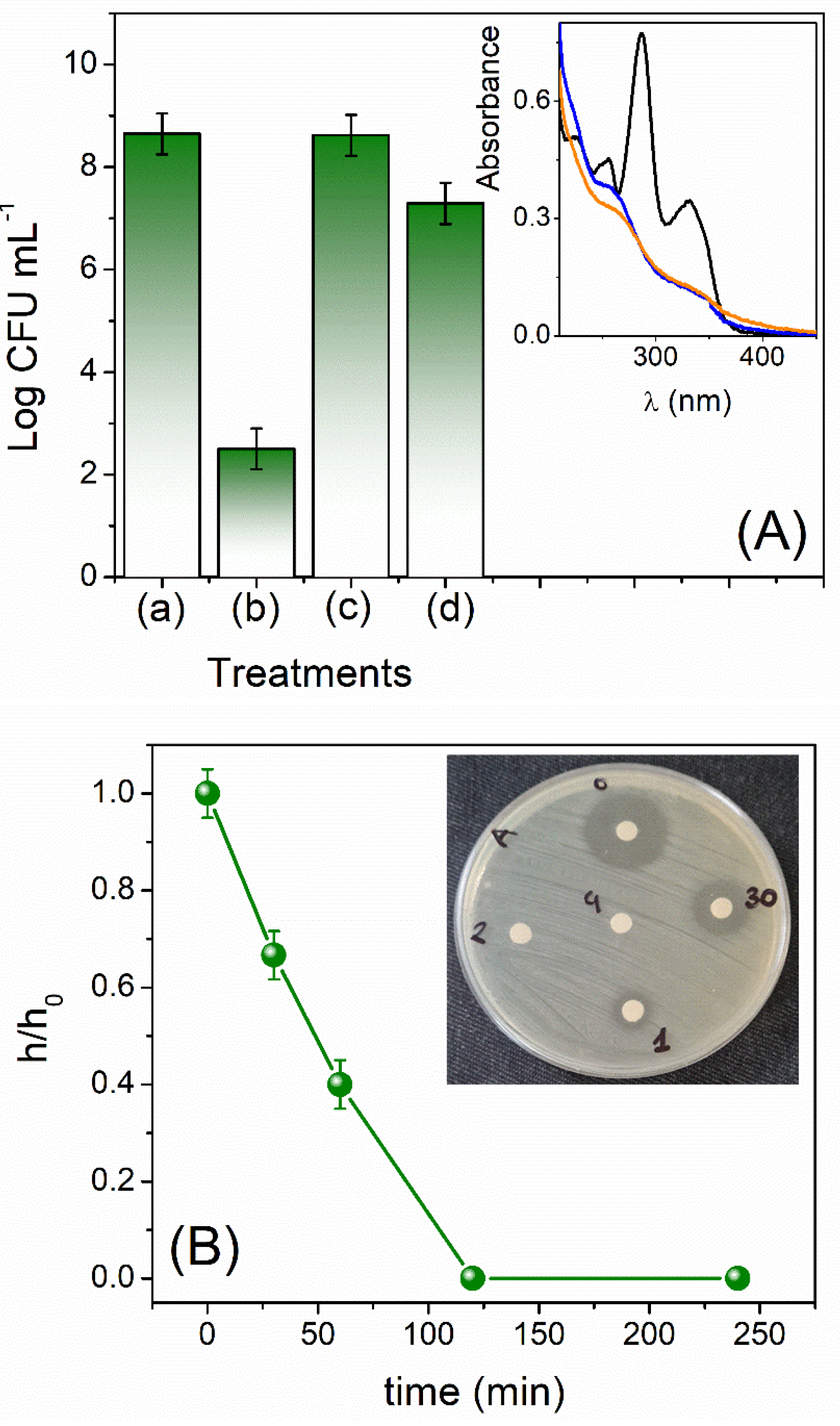
3.3. Activity of Levofloxacin and Its Photodegradation Products on Escherichia Coli

To evaluate if the photodegradation of Lev leads to alteration of its antimicrobial activity, the number of viable microorganisms, expressed as colony-forming units per milliliter (CFU mL−1), was determined using the pathogenic bacteria E. coli EC7 strain.
The 2 × 10⁻⁵ M Lev solutions at pH 7.4 were irradiated in an air and argon atmosphere long enough to achieve a high antibiotic degradation and the accumulation of photoproducts. The Lev consumption was monitored by absorption spectrophotometry.
The results obtained are shown as Log CFU mL−1 for each treatment in Figure 7A.
Additionally, the inset shows the absorption spectrum of Lev solutions employed in this experiment before and after UVB irradiation, and in an air and argon atmospheric condition. In both cases, it was observed that the main absorption band decreases substantially, reaching a diminution approximately 70% of the initial absorbance. This decrease in absorbance was achieved by irradiating a Lev solution equilibrated with air, with a light dose of 55.1 J/cm2, and a Lev solution equilibrated with argon, with a light dose of 266.2 J/cm2.
Figure 7A (main) shows a reduction of approximately 6 log in the number of microorganisms treated with non-irradiated Lev with respect to E. coli cell control, an expected result because of the bactericidal effect of the antibiotic.
On the other hand, after UVB irradiation, the antimicrobial activity of Lev was markedly affected both in argon and air atmospheres. When the solution was irradiated in an argon atmosphere, the number of bacteria resembled the cell control, while when irradiated in air, the number of microorganisms was reduced by only 1 log.
Complementarily, the effect of Lev irradiation with UVB light on its activity was also evaluated by the agar diffusion test. For this, the E. coli strain was exposed to the antibiotic (2 × 10⁻⁵ M) in buffer of pH 7.4 before and after 0.5, 1, 2, and 4 h of direct irradiation in air atmosphere. In this assay, we selected irradiation of air-equilibrated Lev solution because it allows for both degradation pathways: the unimolecular and the dissolved oxygen-dependent.
The results obtained were expressed as the relative index of bactericidal activity (h/h0) as a function of irradiation time (Figure 7B).
Figure 7B clearly shows a decrease in the bactericidal capacity of Lev with the increase in the irradiation time. Moreover, 2 h of irradiation is enough for the complete loss of the antimicrobial effect of the antibiotic, which is consistent with the results obtained in the viable microorganism-determination experiments described above.
Our results of both microbial assays indicate that Lev loses its bactericidal capacity against E. coli strain via exposure to direct UVB light. This can be attributed to the degradation of the antibiotic as a consequence of the photolysis process. It is clear that this light-induced degradation process affects essential parts of the molecule for it to be active, either by unimolecular or oxidative pathways. Likewise, this result suggests that the photoproducts generated under the experimental conditions employed do not have antimicrobial activity on the bacterial strain analyzed.
It should also be noted that the fact that some differences were found between the activity of irradiated Lev in air and argon atmospheres also leads us to suspect that the predominant products generated under each condition are different from each other.
Sturnini et al. evaluated the antibacterial activity of several fluoroquinolone antibiotics by determining the minimum inhibitory concentration (MIC) in different microorganisms, before and after exposing the compounds to natural sunlight [21]. In their research, they found that the antimicrobial action generally decreased after irradiation due to the degradation of the parent compound. Notably, several photoproducts retained significant functionality, as in the case of the antibiotics marbofloxacin, enrofloxacin, and danofloxacin. In agreement with our findings, they observed that the photoproducts obtained from the degradation of Lev are less effective against tested strains, including E. coli, compared to the non-irradiated antibiotic.
Ge et al. also found similar results in their study, using a solar simulator to irradiate fluoroquinolones antibiotics and evaluating antimicrobial activity before and after light exposure, using an agar diffusion test similar to the one performed in our work [25]. They observed that Lev and ciprofloxacin showed a decrease in their antibacterial potency after irradiation over the E. coli strain, while enrofloxacin and difloxacin retained significant antibacterial activity during initial photodegradation, indicating the presence of active primary degradation intermediates.
Contrarily, Geng et al. observed that irradiation with 254 nm UV light did not produce significant changes in Lev activity against E. coli over short exposure times (less than 30 min), attributing this phenomenon to the retention of photoproduct activity as a result of little changes in essential parts of the molecule [47]. However, these authors did not evaluate what occurred at longer UV irradiation times, where structural modifications may be enough to generate photoproducts with lower antimicrobial activity.
Based on the information available in the literature and the results obtained in our study, we conclude that direct photolysis generally does not lead to antibiotic mineralization but rather to the formation of photoproducts. The change in the antimicrobial activity is highly dependent on the structure of the antibiotic and the irradiation conditions, which determine the type of intermediates generated during the degradation process.

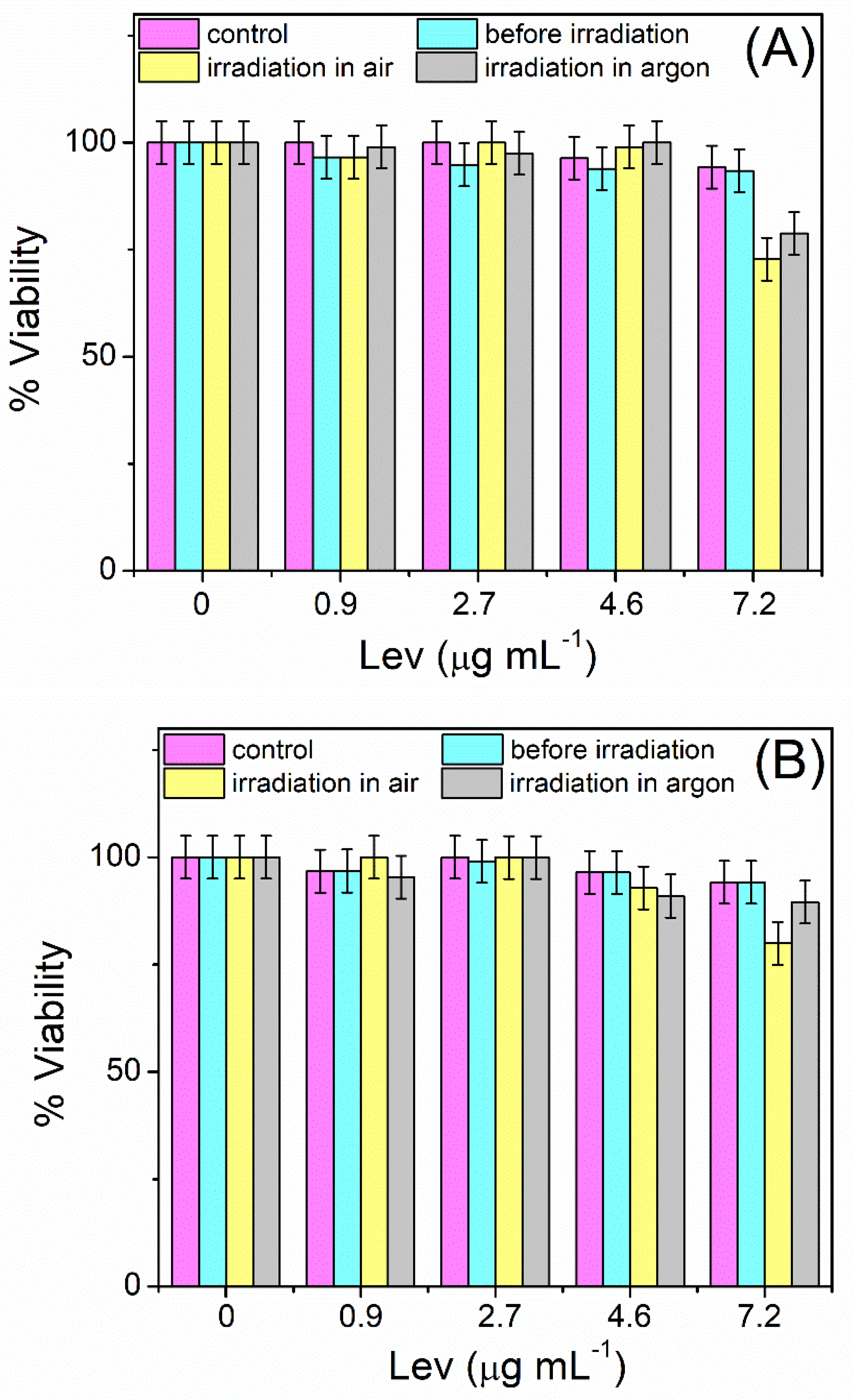
3.4. Toxicity of Levofloxacin and Its Photodegradation Products on Mammalian Cells

To detect the effect of Lev photodegradation on eukaryotic cells, cytotoxicity assays were performed using the mammalian cell line Vero. A monolayer of cells was exposed to different concentrations of the Lev in buffer at pH 7.4 before and after irradiation with UVB light, in air and argon atmosphere. The Lev solutions (2 × 10⁻⁵ M or 7.2 μg mL−1) irradiated and non-irradiated used in these experiments were prepared in the same way as in the microbiological assays describe in Section 3.3.
Cell viability was then assessed using neutral red uptake (NR) and MTT metabolic reduction assays. The results obtained are shown in Figure 8A and Figure 8B, respectively.
As can be seen in Figure 8A, the RN uptake assay indicates that cell viability was not significantly affected by either the buffer used or the non-irradiated antibiotic at any of the concentrations tested. In addition, no significant cytotoxic effect of the irradiated antibiotic was observed at concentrations less than or equal to 4.6 μg mL−1 and at both atmospheric conditions evaluated. However, at the maximum concentration tested (7.2 μg mL−1), a slight decrease in cell viability was observed (less than 20%). This decrease was more pronounced in Lev solution irradiated in an air atmosphere than in Lev solution irradiated in an argon atmosphere.
The results obtained suggest that, in general, UVB-irradiated solutions of Lev do not induce lysosomal damage in Vero cells; however, at higher concentrations, a slight cytotoxic effect may occur.
On the other hand, as shown in Figure 8B, similar profiles were obtained in the MTT assay. No alteration of mitochondrial respiration was observed as a consequence of the exposure of Vero cells to Lev or its photoproducts obtained after UVB exposure at concentrations less than or equal to 4.6 μg mL−1 and at both atmospheric conditions evaluated. However, at the highest concentration of Lev tested, a slight cytotoxic effect (less than 15%) was observed, which was more pronounced when the antibiotic was irradiated under air atmosphere conditions.
Furthermore, in both the RN and MTT bioassays, at high concentrations of Lev and its photoproducts, a difference in cell viability was observed depending on the dissolved oxygen concentration of the irradiated solutions. This indicates that the compounds formed in the absence and in the presence of oxygen are different and consequently have different effects on mammalian cells.
In the work carried out by Viola et al., the authors found that although Lev and other fluoroquinolone antibiotics have significant phototoxicity, the products generated under irradiation with UVA, UVB, and visible light are not involved in cellular damage processes [18]. The observed phototoxicity is a consequence of the ROS generated through self-sensitized processes. They verify this by evaluating the integrity of red blood cells and by using a fibroblast cell line.
On the other hand, Sturnini et al. evaluated the cytotoxicity of several fluoroquinolones using the bioluminescence test of the marine bacteria Vibrio fischeri and found that Lev, after being irradiated with a solar simulator for 15 min, generates photoproducts with a notable inhibition of light emission in Vibrio fischeri compared to non-irradiated Lev, indicating a toxic effect [24]. Similar results were found by de Oliveira et al. using the same bioassay on the fluoroquinolone lomefloxacin and its photoproducts obtained by irradiation with 254 nm UV light for 60 min [48].
We emphasize the importance of evaluating the toxicity of the products generated by each compound under a particular treatment and by more than one bioassay, without generalizing the behaviors, since they may be different as a consequence of the generation of different intermediaries.
In summary, the experimental conditions used in our research generated photoproducts that have no antimicrobial activity and do not present significant toxicity on mammalian cells, regardless of the concentration of dissolved oxygen in the medium. These demonstrate that UVB light could signify an adequate treatment for degrading Lev in a contaminated effluent without the compounds formed representing a potential risk to the environment and human health.

4. Conclusions

In the present investigation, we demonstrated that the antibiotic levofloxacin is sensitive to UVB solar irradiation; however, the rate of photodegradation depends considerably on the pH and oxygen concentration of the medium. The highest photodegradation rate was achieved at pH 7.4 and in the presence of oxygen. Moreover, at a higher dissolved oxygen concentration, a higher photodegradation rate of levofloxacin was reached. On the other hand, photodegradation of the antibiotic is negligible under acidic and anoxic conditions.
In the presence of oxygen, levofloxacin is degraded by a self-sensitizing mechanism involving the reactive oxygen species superoxide anion, hydroxyl radical, hydrogen peroxide, and singlet oxygen; levofloxacin is especially sensitive to the singlet oxygen. For this reason, the generation of this oxidizing species was quantified directly through its phosphorescent signal emitted at 1270 nm. The quantum yield of singlet oxygen by levofloxacin was determined and reported in this work for the first time.
The results of the microbiological tests on a pathogenic Escherichia coli strain reveal that photodegradation of levofloxacin leads to the loss of its antimicrobial activity. Furthermore, cytotoxicity tests on the mammalian cell line Vero indicate that the photoproducts do not exhibit significant cytotoxicity.
Finally, we can conclude that UVB sunlight treatment of contaminated effluents containing levofloxacin could be a very efficient and ecologically safe strategy to degrade the antibiotic provided that pH and oxygen conditions are adequate. However, we consider that additional experiments are necessary by using complex water matrices that offer more information to ensure the greatest efficiency of the treatment.

Author Contributions

M.A.B. (Macarena Agostina Biondi), investigation, methodology, and writing–original draft; M.B.S. and M.C.S., investigation and methodology; M.A.B. (María Alicia Biasutti), data curation, validation, and writing–review and editing; H.A.M. and E.R., conceptualization, supervision, data curation, validation, visualization, writing–review and editing, and project administration. All authors have read and agreed to the published version of the manuscript.

Funding

This research was funded by: Consejo Nacional de Investigaciones Científicas y Técnicas (CONICET), Agencia Nacional de Promoción de la Investigación, el Desarrollo Tecnológico y la Innovación (ANPCyT), Fundación Williams and Secretaría de Ciencia y Técnica de la Universidad Nacional de Río Cuarto (SECyT, UNRC).

Institutional Review Board Statement

Not applicable.

Informed Consent Statement

Not applicable.

Data Availability Statement

The data that support this study are contained within the article.

Conflicts of Interest

The authors declare no conflicts of interest.

References

  1. Patel, N.; Khan, M.D.; Shahane, S.; Rai, D.; Chauhan, D.; Kant, C.; Chaudhary, V.K. Emerging pollutants in aquatic environment: Source, effect, and challenges in biomonitoring and bioremediation—A review. Pollution 2020, 6, 99–113. [Google Scholar] [CrossRef]
  2. Rivera-Utrilla, J.; Sánchez-Polo, M.; Ferro-García, M.Á.; Prados-Joya, G.; Ocampo-Pérez, R. Pharmaceuticals as emerging contaminants and their removal from water. A review. Chemosphere 2013, 93, 1268–1287. [Google Scholar] [CrossRef] [PubMed]
  3. Bottoni, P.; Caroli, S.; Caracciolo, A.B. Pharmaceuticals as priority water contaminants. Toxicol. Environ. Chem. 2010, 92, 549–565. [Google Scholar] [CrossRef]
  4. González-González, R.B.; Sharma, P.; Singh, S.P.; Américo-Pinheiro, J.H.P.; Parra-Saldívar, R.; Bilal, M.; Iqbal, H.M. Persistence, environmental hazards, and mitigation of pharmaceutically active residual contaminants from water matrices. Sci. Total Environ. 2022, 821, 153329. [Google Scholar] [CrossRef]
  5. Hazra, M.; Watts, J.E.; Williams, J.B.; Joshi, H. An evaluation of conventional and nature-based technologies for controlling antibiotic-resistant bacteria and antibiotic-resistant genes in wastewater treatment plants. Sci. Total Environ. 2024, 917, 170433. [Google Scholar] [CrossRef]
  6. Mishra, P.; Tripathi, G.; Mishra, V.; Ilyas, T.; Firdaus, S.; Ahmad, S.; Farooqui, A.; Yadav, N.; Rustagi, S.; Shreaz, S.; et al. Antibiotic contamination in wastewater treatment plant effluents: Current research and future perspectives. Environ. Nanotechnol. Monit. Manag. 2025, 23, 101047. [Google Scholar] [CrossRef]
  7. Phoon, B.L.; Ong, C.C.; Saheed, M.S.M.; Show, P.L.; Chang, J.S.; Ling, T.C.; Lam, S.S.; Juan, J.C. Conventional and emerging technologies for removal of antibiotics from wastewater. J. Hazard. Mater. 2020, 400, 122961. [Google Scholar] [CrossRef]
  8. Yang, X.; Chen, Z.; Zhao, W.; Liu, C.; Qian, X.; Zhang, M.; Wei, G.; Khan, E.; Ng, Y.H.; Ok, Y.S. Recent advances in photodegradation of antibiotic residues in water. Chem. Eng. J. 2021, 405, 126806. [Google Scholar] [CrossRef]
  9. De Ilurdoz, M.S.; Sadhwani, J.J.; Reboso, J.V. Antibiotic removal processes from water & wastewater for the protection of the aquatic environment-a review. J. Water Process Eng. 2022, 45, 102474. [Google Scholar] [CrossRef]
  10. Vialaton, D.; Richard, C. Phototransformation of aromatic pollutants in solar light: Photolysis versus photosensitized reactions under natural water conditions. Aquat. Sci. 2002, 64, 207–215. [Google Scholar] [CrossRef]
  11. Andreozzi, R.; Raffaele, M.; Nicklas, P. Pharmaceuticals in STP effluents and their solar photodegradation in aquatic environment. Chemosphere 2003, 50, 1319–1330. [Google Scholar] [CrossRef] [PubMed]
  12. Lin, A.Y.C.; Reinhard, M. Photodegradation of common environmental pharmaceuticals and estrogens in river water. Environ. Toxicol. Chem. 2005, 24, 1303–1309. [Google Scholar] [CrossRef] [PubMed]
  13. Gomis, J.; Prevot, A.B.; Montoneri, E.; Gonzalez, M.C.; Amat, A.M.; Martire, D.O.; Arques, A.; Carlos, L. Waste sourced bio-based substances for solar-driven wastewater remediation: Photodegradation of emerging pollutants. Chem. Eng. J. 2014, 235, 236–243. [Google Scholar] [CrossRef]
  14. Parsons, S. Advanced Oxidation Processes for Water and Wastewater Treatment; IWA Publishing: London, UK, 2004. [Google Scholar]
  15. Biondi, M.A.; Cacciari, R.D.; Sabini, M.C.; Spesia, M.B.; Biasutti, M.A.; Reynoso, E.; Montejano, H.A. Natural degradation of ceftriaxone promoted by direct UVB light in aqueous media. Mechanistic analysis and cytotoxic effects on a eukaryotic cell line and on bacteria. New J. Chem. 2023, 47, 17799–17809. [Google Scholar] [CrossRef]
  16. Challis, J.K.; Hanson, M.L.; Friesen, K.J.; Wong, C.S. A critical assessment of the photodegradation of pharmaceuticals in aquatic environments: Defining our current understanding and identifying knowledge gaps. Environ. Sci. Process. Impacts 2014, 16, 672–696. [Google Scholar] [CrossRef]
  17. Albini, A.; Monti, S. Photophysics and photochemistry of fluoroquinolones. Chem. Soc. Rev. 2003, 32, 238–250. [Google Scholar] [CrossRef]
  18. Viola, G.; Facciolo, L.; Canton, M.; Vedaldi, D.; Dall’Acqua, F.; Aloisi, G.G.; Amelia, M.; Barbafina, A.; Elisei, F.; Latterini, L. Photophysical and phototoxic properties of the antibacterial fluoroquinolones levofloxacin and moxifloxacin. Chem. Biodivers. 2004, 1, 782–801. [Google Scholar] [CrossRef]
  19. Dwivedi, A.; Mujtaba, S.F.; Kushwaha, H.N.; Ali, D.; Yadav, N.; Singh, S.K.; Ray, R.S. Photosensitizing mechanism and identification of levofloxacin photoproducts at ambient UV radiation. Photochem. Photobiol. 2012, 88, 344–355. [Google Scholar] [CrossRef]
  20. Sturini, M.; Speltini, A.; Maraschi, F.; Profumo, A.; Pretali, L.; Irastorza, E.A.; Fasani, E.; Albini, A. Photolytic and photocatalytic degradation of fluoroquinolones in untreated river water under natural sunlight. Appl. Cat. B Environ. 2012, 119, 32–39. [Google Scholar] [CrossRef]
  21. Sturini, M.; Speltini, A.; Maraschi, F.; Pretali, L.; Profumo, A.; Fasani, E.; Albini, A.; Migliavacca, R.; Nucleo, E. Photodegradation of fluoroquinolones in surface water and antimicrobial activity of the photoproducts. Water Res. 2012, 46, 5575–5582. [Google Scholar] [CrossRef]
  22. Ahmad, I.; Bano, R.; Sheraz, M.A.; Ahmed, S.; Mirza, T.; Ansari, S.A. Photodegradation of levofloxacin in aqueous and organic solvents: A kinetic study. Acta Pharm. 2013, 63, 223–229. [Google Scholar] [CrossRef] [PubMed]
  23. Wammer, K.H.; Korte, A.R.; Lundeen, R.A.; Sundberg, J.E.; McNeill, K.; Arnold, W.A. Direct photochemistry of three fluoroquinolone antibacterials: Norfloxacin, ofloxacin, and enrofloxacin. Water Res. 2013, 47, 439–448. [Google Scholar] [CrossRef] [PubMed]
  24. Sturini, M.; Speltini, A.; Maraschi, F.; Pretali, L.; Ferri, E.N.; Profumo, A. Sunlight-induced degradation of fluoroquinolones in wastewater effluent: Photoproducts identification and toxicity. Chemosphere 2015, 134, 313–318. [Google Scholar] [CrossRef] [PubMed]
  25. Ge, L.; Na, G.; Zhang, S.; Li, K.; Zhang, P.; Ren, H.; Yao, Z. New insights into the aquatic photochemistry of fluoroquinolone antibiotics: Direct photodegradation, hydroxyl-radical oxidation, and antibacterial activity changes. Sci. Total Environ. 2015, 527, 12–17. [Google Scholar] [CrossRef]
  26. Frąckowiak, A.; Kamiński, B.; Urbaniak, B.; Dereziński, P.; Klupczyńska, A.; Darul-Duszkiewicz, M.; Kokot, Z.J. A study of ofloxacin and levofloxacin photostability in aqueous solutions. J. Med. Sci. 2016, 85, 238–244. [Google Scholar] [CrossRef]
  27. Ge, L.; Halsall, C.; Chen, C.E.; Zhang, P.; Dong, Q.; Yao, Z. Exploring the aquatic photodegradation of two ionisable fluoroquinolone antibiotics–gatifloxacin and balofloxacin: Degradation kinetics, photobyproducts and risk to the aquatic environment. Sci. Total Environ. 2018, 633, 1192–1197. [Google Scholar] [CrossRef] [PubMed]
  28. Czyrski, A.; Anusiak, K.; Teżyk, A. The degradation of levofloxacin in infusions exposed to daylight with an identification of a degradation product with HPLC-MS. Sci. Rep. 2019, 9, 3621. [Google Scholar] [CrossRef]
  29. Wei, H.; Tang, Y.; Li, A.; Yang, H. Insights into the effects of acidification on sewage sludge dewaterability through pH repeated adjustment. Chemosphere 2019, 227, 269–276. [Google Scholar] [CrossRef]
  30. Boczkaj, G.; Fernandes, A. Wastewater treatment by means of advanced oxidation processes at basic pH conditions: A review. Chem. Eng. J. 2017, 320, 608–633. [Google Scholar] [CrossRef]
  31. Penn, M.R.; Pauer, J.J.; Mihelcic, J.R. Biochemical oxygen demand. Environ. Ecol. Chem. 2009, 2, 278–297. [Google Scholar]
  32. Lide, D.R. (Ed.) CRC Handbook of Chemistry and Physics; CRC Press: Boca Raton, FL, USA, 2004; Volume 85. [Google Scholar]
  33. Cacciari, R.D.; Reynoso, E.; Spesia, M.B.; Criado, S.; Biasutti, M.A. Vancomycin-sensitized photooxidation in the presence of the natural pigment vitamin B2: Interaction with excited states and photogenerated ROS. Redox Rep. 2017, 22, 166–175. [Google Scholar] [CrossRef] [PubMed]
  34. Xie, Z.H.; He, C.S.; Pei, D.N.; Dong, Y.; Yang, S.R.; Xiong, Z.; Zhou, P.; Pan, Z.-C.; Yao, G.; Lai, B. Review of characteristics, generation pathways and detection methods of singlet oxygen generated in advanced oxidation processes (AOPs). Chem. Eng. J. 2023, 468, 143778. [Google Scholar] [CrossRef]
  35. Schweitzer, C.; Schmidt, R. Physical mechanisms of generation and deactivation of singlet oxygen. Chem. Rev. 2003, 103, 1685–1758. [Google Scholar] [CrossRef]
  36. Scurlock, R.D.; Nonell, S.; Braslavsky, S.E.; Ogilby, P.R. Effect of solvent on the radiative decay of singlet molecular oxygen (1Δg). J. Phys. Chem. 1995, 99, 3521–3526. [Google Scholar] [CrossRef]
  37. Spesia, M.B.; Rovera, M.; Durantini, E.N. Photodynamic inactivation of Escherichia coli and Streptococcus mitis by cationic zinc (II) phthalocyanines in media with blood derivatives. Eur. J. Med. Chem. 2010, 45, 2198–2205. [Google Scholar] [CrossRef]
  38. Zhao, J.; Liu, Y.; Jiang, X.; Guo, P.; Xu, Y.; Zhang, P.; Ji, T.; Lin, Z.; Wang, W. Effect of C-5 position on the photochemical properties and phototoxicity of antofloxacin and levofloxacin: A stable and transient study. J. Photochem. Photobiol. B Biol. 2016, 155, 122–129. [Google Scholar] [CrossRef]
  39. Polishchuk, A.; Emelina, T.; Karaseva, E.; Cramariuc, O.; Chukharev, V.; Karasev, V. Photochemical behavior and photolysis of protonated forms of levofloxacin. Photochem. Photobiol. 2014, 90, 79–84. [Google Scholar] [CrossRef]
  40. Murov, S.L.; Carmichael, I.; Hug, G.L. Handbook of Photochemistry; CRC Press: New York, NY, USA, 1993. [Google Scholar]
  41. Lu, J.; Ji, Y.; Chovelon, J.M.; Lu, J. Fluoroquinolone antibiotics sensitized photodegradation of isoproturon. Water Res. 2021, 198, 117136. [Google Scholar] [CrossRef]
  42. Cacciari, R.D.; Reynoso, E.; Montejano, H.A.; Biasutti, M.A. Photodegradation of prednisolone under UVB solar irradiation. Role of photogenerated ROS in the degradation mechanism. Photochem. Photobiol. Sci. 2017, 16, 1717–1726. [Google Scholar] [CrossRef]
  43. Winterbourn, C.C. Biological chemistry of superoxide radicals. ChemTexts 2020, 6, 7. [Google Scholar] [CrossRef]
  44. Lam, M.W.; Mabury, S.A. Photodegradation of the pharmaceuticals atorvastatin, carbamazepine, levofloxacin, and sulfamethoxazole in natural waters. Aquat. Sci. 2005, 67, 177–188. [Google Scholar] [CrossRef]
  45. Niu, X.Z.; Busetti, F.; Langsa, M.; Croué, J.P. Roles of singlet oxygen and dissolved organic matter in self-sensitized photo-oxidation of antibiotic norfloxacin under sunlight irradiation. Water Res. 2016, 106, 214–222. [Google Scholar] [CrossRef] [PubMed]
  46. Zhou, J.; Chen, Y.; Wang, C.; He, Y.; Lebedev, A.T.; Zhang, Y. Singlet oxygen presenting a higher detoxification potential on enrofloxacin than sulfate and hydroxyl radicals. J. Hazard. Mater. 2025, 487, 137146. [Google Scholar] [CrossRef] [PubMed]
  47. Geng, C.; Liang, Z.; Cui, F.; Zhao, Z.; Yuan, C.; Du, J.; Wang, C. Energy-saving photo-degradation of three fluoroquinolone antibiotics under VUV/UV irradiation: Kinetics, mechanism, and antibacterial activity reduction. Chem. Eng. J. 2020, 383, 123145. [Google Scholar] [CrossRef]
  48. Oliveira, A.M.D.; Maniero, M.G.; Guimaraes, J.R. Lomefloxacin degradation: Antimicrobial activity, toxicity and byproducts. J. Adv. Oxid. Technol. 2015, 18, 211–220. [Google Scholar] [CrossRef]
Figure 1. Absorption spectra of 2 × 10⁻⁵ M Lev in buffer of pH 4.0, pH 7.4, and pH 10.0.
Compounds 05 00023 g001
Scheme 1. Chemical structures of the predominant ionic species at each pH studied.
Compounds 05 00023 sch001
Figure 2. (A) Spectral evolution of 2 × 10⁻⁵ M Lev in buffer at pH 7.4 as a function of UVB light irradiation time in air-equilibrated atmosphere. (B) Graphic representation of Equation (1) for the determination of the photodegradation rate constants (kphot) for Lev in buffer of pH 4.0 (black circles), 7.4 (red circles), and 10.0 (blue circles). Error bars indicate one standard deviation.
Compounds 05 00023 g002
Figure 3. Graphic representation of Equation (1) for the determination of the photodegradation rate constants (kphot) of Lev in argon-saturated (red circles), air (blue circles), and oxygen-saturated (green circles) atmosphere conditions in buffer at pH 4.0, pH 7.4, and pH 10.0. Error bars indicate one standard deviation.
Compounds 05 00023 g003
Figure 4. Dependence of kphot on pH and dissolved oxygen concentration for UVB irradiated solutions of Lev.
Compounds 05 00023 g004
Figure 5. Ratios of the kinetic constant in the absence (Lev) and in the presence of the ROS inhibitors isopropyl alcohol (Lev + IPA), superoxide dismutase (Lev + SOD), catalase (Lev + CAT), and sodium azide salt (Lev + NaN3).
Compounds 05 00023 g005
Figure 6. Dependence of the amplitude of the O2(1Δg) phosphorescence emission extrapolated at time zero (I0) on the laser fluence (EL) for (red circle) PN in CH3CN and (blue circle) Lev in D2O. Inset: O2(1Δg) phosphorescence decay signals at 1270 nm for Lev in D2O; the signal of D2O was added for comparative purposes.
Compounds 05 00023 g006
Figure 7. (A) Effect of 2 × 10⁻⁷ M of Lev on E. coli EC7 strain: (a): cellular control without Lev, (b) Lev before irradiation with UVB light, (c) Lev irradiated with UVB light in argon atmospheric condition, and (d) Lev irradiated with UVB light in air atmospheric condition. Inset: Absorption spectrum of 2 × 10⁻⁵ M Lev before (black line) and after irradiation, in an argon atmosphere (orange line) and in an air atmosphere (blue line). (B) Relative normalized diameter of the bactericidal inhibitory activity of 2 × 10⁻⁵ M of Lev irradiated under air atmospheric conditions over E. coli EC7 strain. h0 and h represent the inhibitory halo diameter at t = 0 (h0) and at different UVB irradiation times (h), respectively. Inset: Variation in the inhibition halo as a function of Lev photolysis time. The numbers in the image indicate 0:0 min photolysis, 30:30 min photolysis, 1:60 min photolysis, 2:120 min photolysis, and 4:240 min photolysis.
Compounds 05 00023 g007
Figure 8. Percentage cell viability of Vero cells exposed to different concentrations of Lev before and after UVB irradiation under argon or air atmospheric conditions, evaluated by (A) NR uptake and (B) MTT metabolic assay.
Compounds 05 00023 g008
Table 1. Photodegradation rate constant (kphot) values of Lev under irradiation with UVB light in buffer of pH 4.0, 7.4, and 10.0 and in argon, air, and oxygen atmosphere conditions.
kphot (×103)/s−1
AtmosphereArgonAirOxygen
pH
4.0~00.71 ± 0.031.9 ± 0.3
7.41.2 ± 0.39.8 ± 0.113.5 ± 0.1
10.03.2 ± 0.25.5 ± 0.29.8 ± 0.1
Disclaimer/Publisher’s Note: The statements, opinions and data contained in all publications are solely those of the individual author(s) and contributor(s) and not of MDPI and/or the editor(s). MDPI and/or the editor(s) disclaim responsibility for any injury to people or property resulting from any ideas, methods, instructions or products referred to in the content.

