Article

Laccase from Melanocarpus albomyces: Molecular Docking Analysis with First-Generation Tetracyclines Through a Mechanistic Approach

by
José Muñoz-Espinoza
1,
Germán Barriga-González
2,
Gino Corsini
3,
Sebastián Lagos
3,
Andrés Barriga González
4,* and
Nadia Gavilán de Fátima
3,*
1
Laboratorio de Química Teórica, Departamento de Química, Facultad de Ciencias, Universidad de Chile, Santiago 7800003, Chile
2
Laboratorio MACEDONIA, Departamento de Química, Universidad Metropolitana de Ciencias de la Educación, Santiago 7760197, Chile
3
Laboratorio de Microbiología Molecular y Compuestos Bioactivos, Instituto de Ciencias Biomédicas, Facultad de Ciencias de la Salud, Universidad Autónoma de Chile, Santiago 8320000, Chile
4
Unidad de Espectrometría de Masas-CEPEDEQ, Facultad de Ciencias Químicas y Farmacéuticas, Universidad de Chile, Santiago 8320000, Chile
*
Authors to whom correspondence should be addressed.
Compounds 2025, 5(2), 17; https://doi.org/10.3390/compounds5020017
Submission received: 9 March 2025 / Revised: 14 April 2025 / Accepted: 22 April 2025 / Published: 1 May 2025

Abstract

:
Laccases are versatile enzymes capable of oxidizing a wide variety of antibiotics. In this study, the mechanism of catalytic oxidation of first-generation tetracyclines, namely, oxytetracycline, tetracycline, and chlortetracycline, by the Melanocarpus albomyces laccase enzyme was investigated using molecular docking and DFT calculations. Molecular docking studies revealed that all three substrates exhibit negative interaction energies, indicating stable enzyme–substrate complexes, with tetracycline and chlortetracycline showing the highest binding affinities. Global reactivity indices obtained by DFT confirmed the high electrophilicity of the enzyme active site, particularly the aminoacidic residues Glu235 and His508, favoring electron transfer from the substrates. In addition, NBO analysis allowed quantification of the energy of hydrogen bonds in enzyme–substrate interactions, evidencing their key role in the stabilization of the complex. Proton transfer analysis suggested two possible mechanisms: (1) a direct concerted transfer and (2) a process mediated by water molecules. The results provide insights into the thermodynamics, electronic structure, and nature of intermolecular interactions governing the oxidation of tetracyclines by the enzyme, highlighting their potential in bioremediation strategies for antibiotic degradation.

1. Introduction

Laccases (1.3.10.2) are multi-copper oxidase enzymes widely distributed in fungi [1] and bacteria [2]. This type of enzyme belongs to the oxidoreductase family [3,4], which catalyzes the oxidation of a wide variety of phenolic substrates, using molecular oxygen ( O 2 ) as an electron acceptor and hydrogen atoms, reducing them to water molecules. The oxidation-reduction (redox) reaction mechanism of laccase has been extensively studied [4,5], either experimentally and/or from a theoretical and computational approach [6]. Briefly, the redox mechanism consists of three simple steps: (1) oxidation of the substrate due to the abstraction of a proton and an electron by the site defined by Cu-T1; (2) transfer of electrons and protons from the Cu-T1 site to the trinuclear center (Cu-T2/T3); and (3) the reduction in molecular oxygen forming two water molecules [4,7].
Tetracyclines (TCs) are broad-spectrum antibiotics characterized by their wide range when treating bacterial [8] and even protozoan and parasitic diseases [9]. Due to its versatility, this type of drug has been utilized in both the livestock and aquaculture industries. In addition, first-generation TCs have low production costs, high purity, and are highly consumable. However, this has resulted in environmental challenges because these compounds are highly stable and degrade poorly in nature. Given their low metabolization by human and animal organisms, approximately 75% of these antibiotics are excreted in their intact form [10]. To address contamination by first-generation tetracycline antibiotics, numerous studies have investigated the degradation of these antibiotics using laccase enzymes from various sources. These sources include Trametes versicolor [11,12], Botrytis aclada [13], Aspergillus sp. [14,15], Cerrena unicolor [16], and Pycnoporus sp. [17], among others. The oxidation of TCs depends on how the substrate enters into the active site of the enzyme, which is determined by its aminoacidic residues and the structure of the substrate. Research shows that chlortetracycline (CTC) has the highest removal rate, followed by tetracycline (TC) and then oxytetracycline (OTC). Based on these observations, in the present study, a theoretical approach to the reaction mechanism of the catalytic oxidation of molecules belonging to the first-generation tetracyclines, namely OTC, TC, and CTC, in the active site of the laccase enzyme from the fungus Melanocarpus albomyces (MaL) by molecular docking is presented in order to compare with existing evidence in the literature (Figure 1) [18].

2. Materials and Methods

2.1. Molecular Docking

The conformational analysis of the substrates OTC, TC, and CTC in the active site of the laccase enzyme from the fungus Melanocarpus albomyces (PDB id: 3FU9) was performed by molecular docking using the Lamarckian Genetic Algorithm (LGA) in the software Autodock v4.2 [19]. The analysis was performed under the methodology established by Torres et al. [20], where the crystal structure of the enzyme was first refined by removing the water molecules present inside and outside its environment, then adding the polar hydrogen atoms, and finally calculating its total charge. The active site of the enzyme was delimited by the key aminoacidic residues Glu235 and His508, reported by Kallio et al. [18], where a grid of dimensions of  50 × 50 × 50   Å  with the corresponding coordinates, axes  x = 16.896   Å ,   y = 31.030   Å , and  z = 19.058   Å , were generated. The methodology used in the enzyme–substrate coupling was the semi-flexible type, where the enzyme presents a rigid structure, while the substrates present a flexible structure capable of adapting to the active site through bond rotations. To obtain the lowest energy conformer of the tetracyclines, molecular docking was performed to generate a series of 20 different conformers. This approach allows a comprehensive exploration of the conformational diversity of the substrates, considering the multiple possible orientations and affinities within the active site, obtaining variable conformations and binding energies. All the results obtained from molecular docking were visualized in the software Discovery Studio Visualizer v24.1.0.23298.

2.2. Electronic Properties and Proton Transfer Mechanism

Density Functional Theory (DFT) calculations and the global electronic properties, such as electrophilicity ( Δ E + / ω + ) [21], softness (S), hardness ( η ) [22], and electronic chemical potential ( μ ) [23], were calculated to evaluate the interaction energy between the substrate and the aminoacidic residues of the active site. All calculations were performed based on the energies of the frontier orbitals (HOMO and LUMO) derived from the software Gaussian v9.5 (RevD.01). To determine the electronic properties, the most probable conformers presenting a low interaction energy and a low Root Mean Square Deviation (RMSD), whose orientation and interactions are preferential with residues Glu235 and His508, were used. For this, the substrates with their interacting residues (named Sites) were isolated in the software Gaussview v5.0.9 [24], and then their global electronic properties were calculated with the energies of the HOMO and LUMO orbitals (Frontier Molecular Orbital approximation, FMO).
The literature [18] highlights the importance of residues Glu235 and His508, responsible for the abstraction of protons and electrons from substrates during the enzymatic redox process, due to their electrophilic nature. To quantify this strong electrophilicity, the electrophilicity model proposed by Contreras et al. [25], based on the transfer of one electron ( Δ N = 1 ), was used. Following the formalism of Parr and Yang [26] and considering the finite difference approximation for the electron chemical potential ( μ ) and chemical hardness ( η ) [27], in addition to the theorem of Perdew and Levy [28], one-electron transfer energy ( Δ E + ) can be expressed in terms of the HOMO ( ε H ) and LUMO ( ε L ) energies as
E + = 1 4 ε H + 3 4 ε L ,
where  ε H  and  ε L  represent the HOMO and LUMO energies, respectively. Given the trivial nature of these values, the strong electrophilicity described by Equation (1) is considered in terms of its absolute value ( | Δ E + | ), where species with high values of  | Δ E + |  are associated with strong electrophilicity.
To contrast the energy values obtained from molecular docking, a calculation of the interaction energy ( E i n t ) was performed using the DFT methodology, which considers the electron density and its superposition between the interacting species (Basis Set Superposition Error, BSSE), defined by
E i n t = E ( A B ) [ E ( A ) + E ( B ) ] + B S S E ,
where E(AB) corresponds to the energy of the complex, and E(A) and E(B) correspond to the energies of the isolated interacting species [29].
On the other hand, to quantify the energy associated with the hydrogen bonds generated between the aminoacidic residues of the active site and the tetracycline-type substrates, a second-order perturbative analysis (E2PERT,  E 2 ) was used, which, in a donor–acceptor context of Lewis-type orbitals, can quantify the electron delocalization between the hydrogen bond donor and acceptor segments [30]. The Second-Order Perturbative method was obtained from the following expression
E ϕ ϕ * 2 = 2 ϕ F ^ ϕ * ε ϕ * ε ϕ ,
where  F ^  is the diagonal element of the Fock matrix (Fock operator),  ϕ  and  ϕ *  are the bonding and anti-bonding NBO orbitals, and  ε ϕ ε ϕ *  are the energies of the donor and acceptor orbitals.
To model the proton and electron transfer reaction mechanisms, a potential energy surface (PES) analysis was performed by searching for transition and intermediate states using the QST2 (Synchronous Transit-Guided Quasi-Newton) methodology [31] implemented in the software Gaussian 09 v9.5 (RevD.01). The PES analysis was carried out in the gas phase ( ε = 1.00 ) and in the presence of an implicit solvent described by the dielectric constant ( ε = 78.40 ). The reaction models are analogous to those described by Li et al. [6], where proton transfer is generated in two possible ways: (1) directly between the substrate and the aminoacidic residues and (2) proton transfer mediated by water molecules. To model proton transfer, an active site was constructed comprising the aminoacidic residues glutamine (Glu) and histidine (His), where the carboxyl group of Glu is in its deprotonated form ( C O O ). This arrangement allows the system to facilitate the proton transfer through specific interactions, such as hydrogen bonds.

3. Results and Discussion

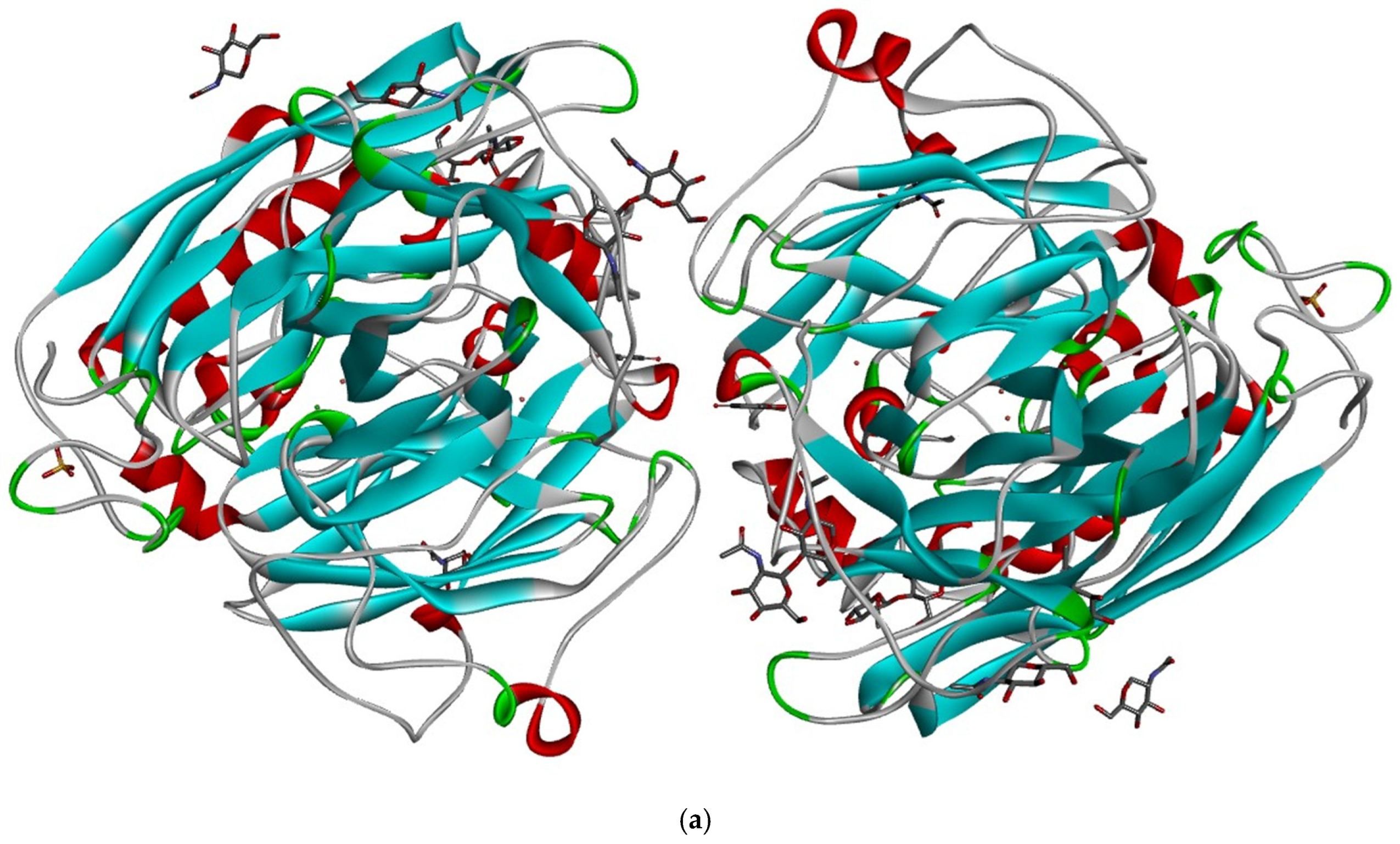
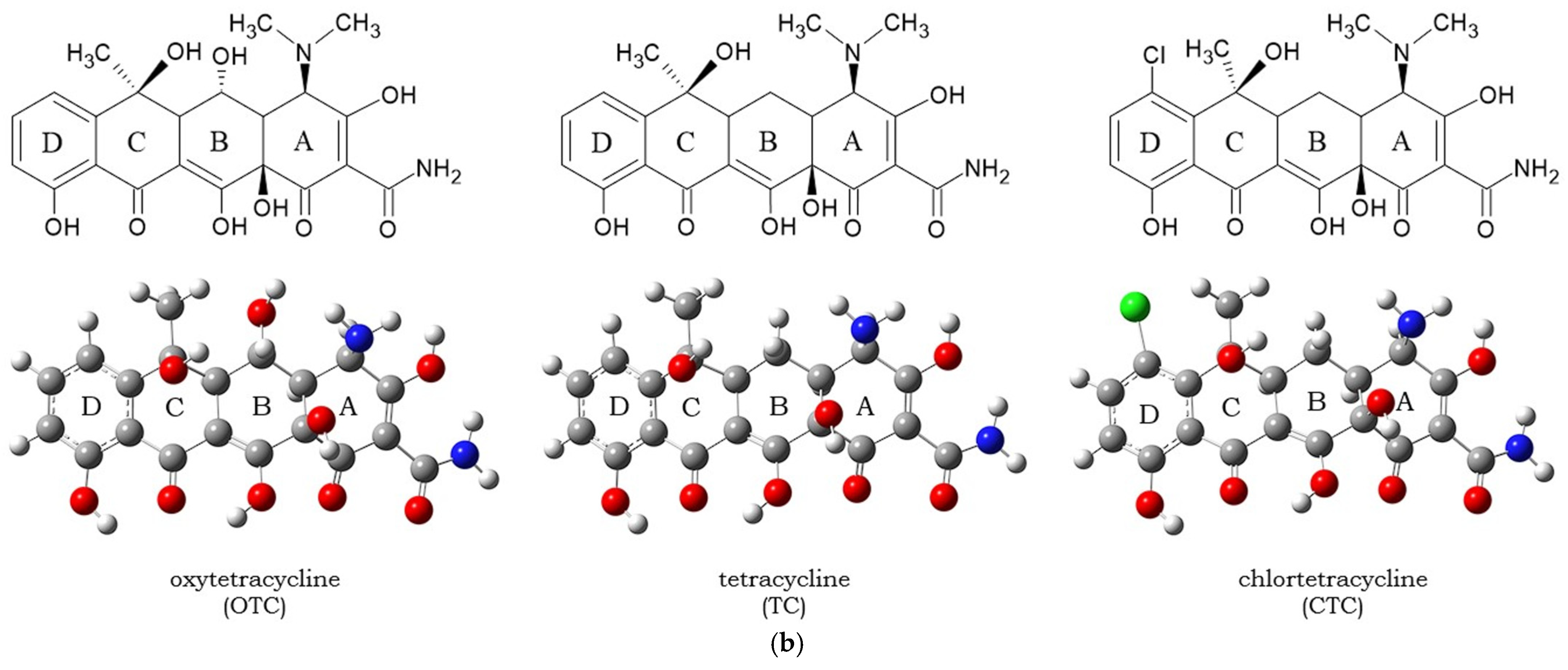
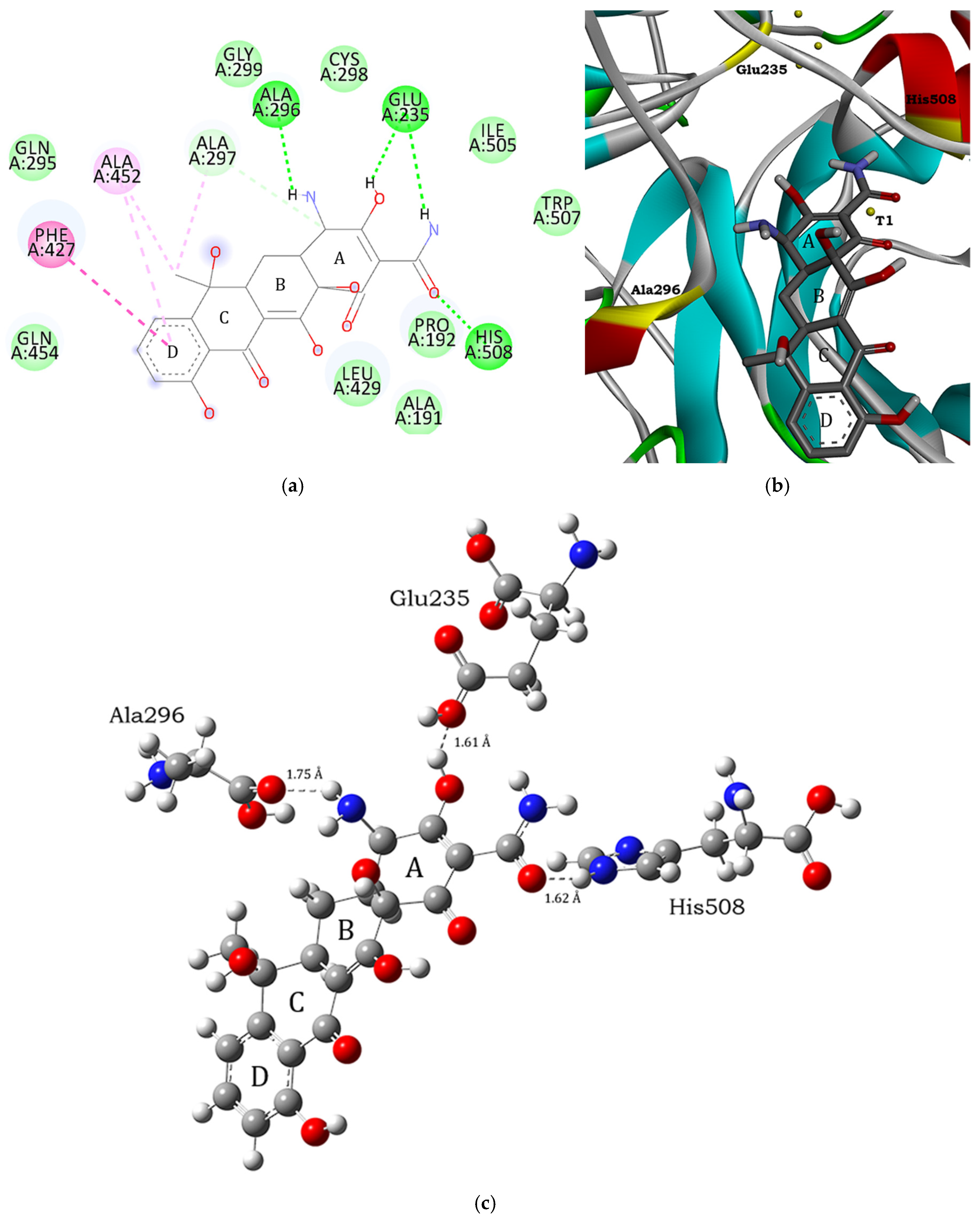
3.1. Molecular Docking and Intermolecular Interactions of Substrates–MaL Active Site

Molecular docking is an in silico technique that predicts conformational space between a substrate and an enzyme [32]. In general, docking is based on a model of non-covalent intermolecular interactions, mainly hydrogen bonding and van der Waals interactions [33]. To find the conformational space in the active site and optimal molecular conformation of the ligand within the enzyme, mixed search algorithms such as the LGA were used, which can perform a global conformational search followed by a local search [34,35]. Therefore, the result obtained is more optimal than a genetic algorithm (GA), since this method explores the surface of the protein or a larger interaction site.
The results for each substrate were analyzed using the methodology of Morris et al. [35], which focuses on interaction energies ( E i n t ) and RMSD values. According to this methodology, lower values of interaction energy and RMSD indicate better coupling and interaction between the enzyme and substrate. Table 1 presents the interaction energy and RMSD values for OTC, TC, and CTC at the active site of the MaL enzyme.
Table 1 shows that the systems formed by the substrates OTC, TC, and CTC interacting with the active site of the MaL enzyme have negative interaction energies. This indicates that these systems are both viable and stable due to the presence of non-covalent interactions, predominantly hydrogen bonds. The selected systems correspond to OTC conformer 20, TC conformer 2, and CTC conformer 1, whose interaction energies were  4.76 , 5.95 , and  5.85   k c a l / m o l , respectively. Thermodynamically, the systems conformed by the substrates OTC, TC, and CTC with the active site of the MaL enzyme are governed by a spontaneous complexation process given the negative Helmholtz free energy values ( A < 0 , where  A = U T S ) and are preferentially guided by the entropy of the process ( T S ), which is associated with the translational degrees of freedom of the substrate within the active site (Table 2).
To contrast the energetic values obtained from molecular docking (molecular mechanics, MM), an interaction energy calculation was performed using the DFT methodology, which considers the electronic properties of the interacting species (quantum mechanics, QM). For this purpose, the theoretical level ωB97XD/6-311+G(d,p), a functional that has been shown to be effective in describing long-range interactions, was employed [36]. As a model, conformers 20, 2, and 1 from OTC, TC, and CTC, respectively (Table 1), were considered, which presented a higher amount of hydrogen interactions with the residues Glu235 and His508. Table 3 shows the interaction energy values obtained by molecular docking and DFT, as well as the hydrogen bonds of each conformer with the aminoacidic residues of the active site, which were calculated through Second-Order Perturbative analysis (E2PERT,  E 2 ).
Table 3 shows that the interaction energies from molecular docking (MM) and DFT (QM) both exhibit a similar stability pattern, indicated by their negative values, which describe efficient and stable interactions between the substrates and the aminoacidic residues of the active site. TC and CTC presented a higher affinity for the active site, given the higher and negative interaction energy values and the higher energy values associated with hydrogen bonds. Suda et al. [11] and other investigators [12,14,15] also observed this behavior through the analysis of TCs degradation employing HPLC. It should be noted that the energy difference between the two methods may be due to the number of atoms considered in the calculation. In the MM method, several parameters were considered that contribute to the total interaction energy, mainly classical parameters, namely  E v d W  (short-range interaction energy),  E e l e c t  (electrostatic interaction energy),  E s o l v  (desolvation energy), and  E t o r  (internal rotations due to accommodation in the active site), which were considered in the conformational space of n atoms. Conversely, quantum mechanics (QM) methods primarily rely on the electronic characteristics of interacting species, including electrostatic interactions, dispersion, exchange repulsion, and charge transfer. These considerations are applied to segments with a number m of atoms, where m < n, as m only accounts for the aminoacidic segments formed by Glu235, Ala296, and His508, as well as the substrates, while n encompasses all the atoms within the grid that defines the conformational space.
According to Jeffrey’s classification, the hydrogen interactions listed in Table 3 range from moderate to strong [37]. This classification describes the nature of the hydrogen bonds through their distance and relative energy, being 1.5–2.7 Å and 15–40 k c a l / m o l  for strong hydrogen bonds, while moderate hydrogen bonds presented distances and relative energies of 2.7–3.2 Å and 10–15  k c a l / m o l . It should be noted that hydrogen bridges are a concerted phenomenon between the hydrogen donor and acceptor segments of substrates and aminoacidic residues, such that electron density transfer stabilizes both the substrate and the active site (TCs system–MaL). Figure 2, Figure 3 and Figure 4 show the conformers and intermolecular interactions resulting from molecular docking with the active site of the enzyme.
Figure 2 illustrates that OTC does not form a hydrogen bond with the aminoacidic residue Glu235. In comparison, TC and CTC establish interactions of this type with residues Glu235, His508, and Ala296. The variation in hydrogen bond formation between OTC, TC, and CTC may be due to the larger distances between the donor and acceptor groups of these bonds in OTC. This difference in hydrogen bond formation may be related to the nature of the semi-flexible molecular docking methodology, in which the rotation of bonds within the substrate functional groups determines the formation of non-covalent interactions of this type. Furthermore, Figure 2, Figure 3 and Figure 4 show that the hydroxyl groups of the A, B, and C rings of the substrates exhibit rotations with respect to their optimized equilibrium geometry shown in Figure 1. Additionally, all three substrates presented an  A D  interaction orientation, i.e., the A-ring is found interacting directly inside the active site, while the D-ring presents an external arrangement. The conformational arrangement of TC within the active site and its minimal interaction energy makes it an optimal candidate for modeling its proton and electron transfer oxidation reaction to be described in later sections (vide infra).

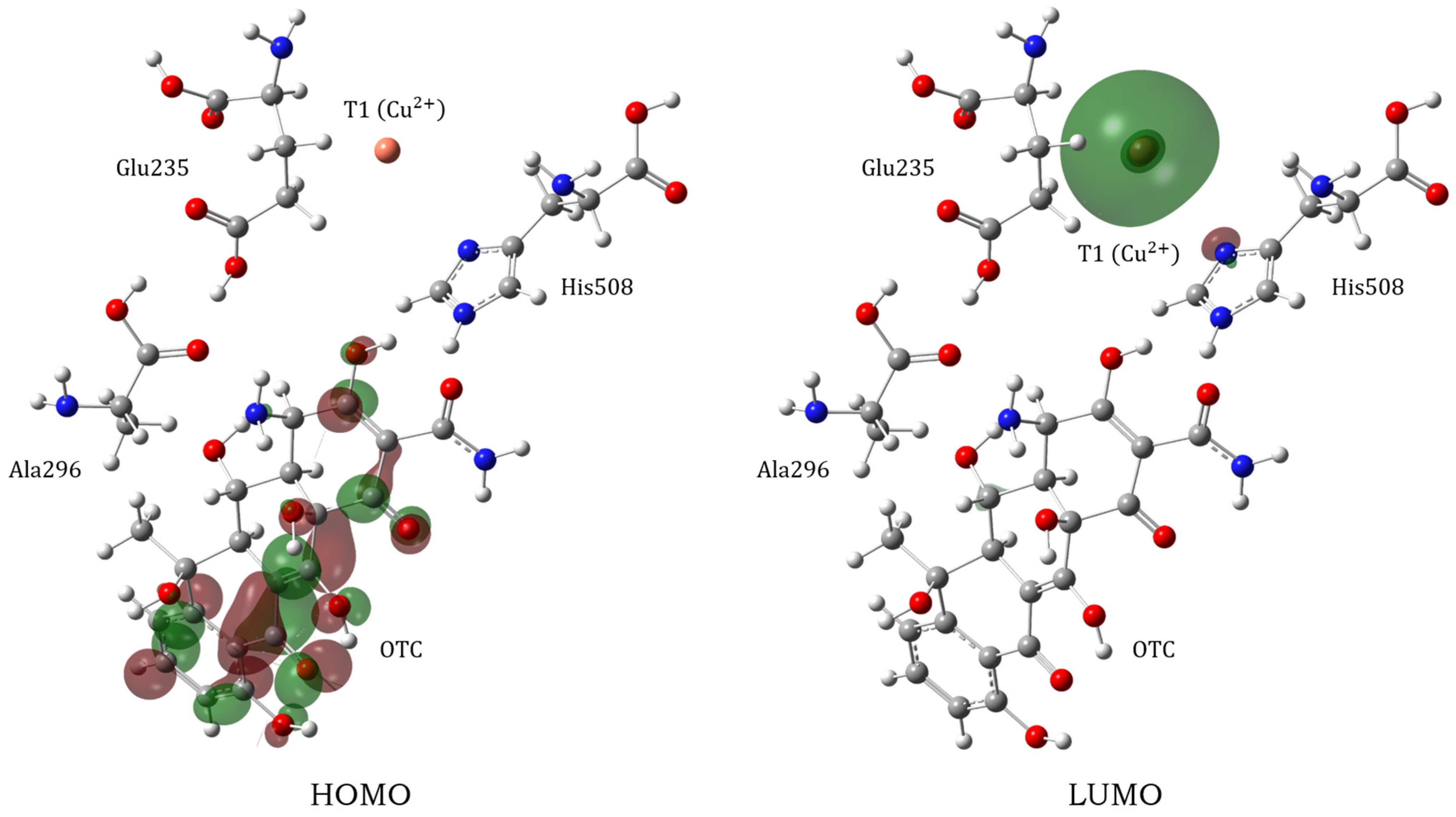
3.2. Electronic Properties and Proton Transfer Mechanism

Since molecular docking is based on classical mechanics and electrostatics, there is no quantum mechanical effect associated with the electron density and the properties derived from it. After detailing the thermodynamics of the complexation process and interpreting stability from intermolecular interactions, the subsequent step involved conducting a global reactivity analysis of the active site, particularly focusing on the main residues Glu235 and His508. Table 4 shows the global reactivity indices of the OTC, TC, and CTC substrates, as well as their corresponding aminoacidic residues at the active site: Ala296, Glu235, and His508. The active site, including Cu-T1, was considered for the calculations, as illustrated in Figure 2b, Figure 3b, and Figure 4b. Consequently, a pseudopotential approximation calculation was performed [38].
Table 4 shows properties related to the charge transfer from the substrate to the active site (AS), where AS1–MaL, AS2–MaL, and AS3–MaL correspond to the active sites interacting with the OTC, TC, and CTC substrates, respectively. These active sites are mainly characterized by the aminoacidic residues Ala296, Glu235, and His508, and Cu-T1. The electronic chemical potential values ( μ n T C s ) indicate a charge transfer from the substrate to the active site, as the latter has a lower value ( μ A S n M a L < μ n T C ). Due to the difference in chemical potential (inhomogeneity), electrons (or charge density) naturally transfer from the system with a higher chemical potential to the one with a lower chemical potential. In this case, the active site exhibits a lower chemical electronic potential than that of the substrates OTC, TC, and CTC. Therefore, this inhomogeneity causes the substrates to tend to donate electrons (or charge density) to the active site defined by the mentioned aminoacidic residues.
Moreover, considering the electrophilicity values ( ω + / E + ), it is evident that the active site is highly electrophilic. This observation aligns with the model and findings of Kallio et al. [18], which attribute this characteristic to the presence of Cu-T1. The positive charge of Cu-T1 generates a hole in the active site, making it prone to attract charge density and/or electrons. As demonstrated in Table 4, the electric charge density and electrons seem to originate from the OTC, TC, and CTC substrates. This can be attributed to their high overall softness and low overall hardness values. This suggests that they are reactive species with a tendency to transfer electric charge or electrons, aligned with their higher electronic chemical potential value compared to the active site. The electron transfer limit model ( | E + | ) illustrates a scenario comparable to global electrophilicity. In this context, AS1–MaL exhibits a higher tendency to attract electrons, while AS3-MaL displays a lower tendency. Thus, the proposed model serves as a reasonable approximation for electron transfer phenomena, such as those involving enzymes with redox capabilities.
The electrophilic character of the Cu-T1 center can be further rationalized by considering its electronic structure. The +2 oxidation state of copper ( C u 2 + ), together with the nature and geometry of its coordination environment, stabilizes the LUMO and localizes it primarily on the metal center. This configuration enhances the ability of Cu-T1 to accept electrons efficiently. The coordination field finely modulates the energy of the frontier orbitals, lowering the LUMO energy and increasing the electron affinity of the site. Consequently, the synergy between the oxidation state, LUMO localization, and coordination effects underlies the strong electrophilicity observed at the T1 site, in agreement with both theoretical predictions and experimental data [3]. Graphic evidence of the great electrophilic tendency of copper is the location of the LUMO, which can be observed in Figure 5.
Once the tendency of the active site and the aminoacidic residues to capture electrons to carry out the redox process that has been described, the proton transfer phenomenon between the substrate and the active site was modeled. Proton transfer in enzymes has been a widely studied phenomenon, one of the pioneers being Bender et al. [39]. Recent models of proton transfer include quantum-mechanical approaches in which the proton can tunnel through the space (potential barrier) presented between the donor (substrate) and hydrogen acceptor (aminoacidic residues of the active site) [40]. Simpler models propose concerted proton (and electron) transfers [41], or even mediation by water molecules [6,42]. The methodology established by Li et al. [6] was used to describe the proton transfer process, where the proton is transferred directly and mediated by water molecules in both the gas phase and implicit solvent (water), respectively. The discussion starts with the direct proton transfer between the TC substrate and the aminoacidic residue Glu235 (Figure 6).
Figure 6a shows the potential energy surface (PES) generated by direct proton transfer (See Supplementary Material for coordinates and imaginary frequencies of transition states). It indicates a concerted  R e a c t i v e T S P r o d u c t  process, where the gas phase transition state (TS) exhibits an energy of  12.26   k c a l / m o l . By considering the solvent, the stationary points for the transition state and product are energetically stabilized due to the solvation of the ionic residues by water. Figure 6b shows the proton transfer process (in the gas phase) occurring between a terminal hydroxyl group of the D-ring of TC and the carboxyl terminal of the Glu235 residue. The results now detail the proton transfer mediated by water molecules, as illustrated in Figure 7.
Proton transfer via water molecules is more complex than direct transfer, involving two transition states (TS1 and TS2) and an intermediate reaction. The PES analysis indicates that the proton transfer from the substrate to the water molecule is the determining reaction step due to its higher activation energy compared to the reference state of the reactants. As shown in Figure 6a, the PES depicted in Figure 7a exhibits a lower energy curve when considering the implicit solvent. This indicates that the systems formed by transition states TS1/TS2, intermediate, and product are stabilized by the generation of an implicit solvation sphere around them. From a mechanistic perspective (Figure 7b), the first transition state involves proton transfer from the substrate TC to a water molecule, forming an intermediate reaction where a  H 3 O +  cation bridges the substrate and the Glu235 residue. The presence of the  H 3 O +  cation is transient, as it subsequently donates the proton to the aminoacidic residue, thereby protonating it and regenerating the original water molecule.
Proton transfer between the substrate TC and the active site residue can occur via two potential mechanisms: direct transfer or water molecule-mediated transfer. Direct proton transfer is a concerted process involving the terminal hydroxyl group of the substrate and the carboxyl terminus of the aminoacidic residue Glu235. Conversely, the water molecule-mediated transfer is a non-concerted process that proceeds through two transition states and one intermediate reaction.
Analysis of the energy profiles reveals that the concerted mechanism presents an activation barrier of  12.36   k c a l / m o l , while water-mediated proton transfer exhibits significantly lower barriers, with values of  9.42   k c a l / m o l  and  8.73   k c a l / m o l  corresponding to the two transition states identified. Given the absence of theoretical and experimental evidence on the proton transfer mechanism between the TC substrate and the MaL enzyme, the results obtained suggest that the presence of water molecules facilitates proton transfer, reducing the energy required for reaction progression. This behavior is justified by the ability of water to act as an efficient mediator in the transfer of protons through the formation of hydrogen bonds, analogous to the model established by Grotthuss [43].
In order to adequately model the coupled proton–electron transfer (PCET) phenomenon, it is necessary to resort to multiscale theoretical approaches, such as the one developed by Li et al. [6], who propose that the catalytic oxidation of phenolic compounds by laccases occurs through a concerted mechanism of simultaneous charge and proton transfer. However, in the present work, the model proposed by Jones and Solomon [4], Solomon et al. [44], and Randall et al. [45] is taken as a basis, which establishes that this process does not necessarily occur in a coupled manner but rather follows a sequential scheme in which the electron is first transferred to the cupric center T1, followed by the dissociation of the proton towards the medium. This approach allows a clearer description of the redox mechanism from the electronic point of view. It should be noted that the detailed study of electron transfer, as well as the characterization of the transition states involved, requires the use of spin-polarized (open-shell) models, which implies a considerably more complex theoretical development than that addressed by Li et al. [6], especially when considering the temporal separation of the charge and proton events.

4. Conclusions

In this study, a theoretical analysis was carried out on the docking feasibility and electronic reactivity of first-generation tetracycline antibiotics (OTC, TC, and CTC) at the active site of the laccase enzyme from Melanocarpus albomyces (MaL). Molecular docking studies and DFT calculations have identified the main noncovalent interactions responsible for the formation and stabilization of enzyme–substrate complexes, as well as characterized the parameters associated with the redox and proton transfer processes.
The results indicated that the systems formed by TC and CTC exhibit a higher affinity for the active site due to their lower interaction energies and a greater number of hydrogen bonds with the key aminoacidic residues Glu235 and His508. Analysis of the global reactivity indices confirmed that the active site of the enzyme exhibits high electrophilicity, which is attributed to the location of the LUMO on the Cu2+ of the Cu-T1 center, a characteristic that favors the transfer of electrons from the substrates to the enzyme.
Regarding the proton transfer mechanism, two possible pathways were evaluated: a direct, concerted proton transfer between the TC substrate and the Glu235 residue and a water-mediated proton transfer pathway. The latter pathway proved to be thermodynamically more favorable due to the lower activation barriers observed. However, although some recent models, such as that of Li et al. [6], propose a coupled proton–electron transfer (PCET), our results, described by the classical model proposed by Solomon et al. [44], support a sequential mechanism. In this case, the electron is first transferred to the Cu-T1 center, followed by the release of the proton to the medium or to the acceptor residue, as evidenced by previous spectroscopic and theoretical studies.
Taken together, these findings contribute to the understanding of the biotransformation process of tetracycline antibiotics by fungal laccases and may be useful for the design of bioremediation strategies based on these enzymes. Furthermore, the proposed proton transfer model could be extended to other laccases, providing a useful theoretical framework for investigating similar redox mechanisms in enzymatic systems of environmental and biotechnological interest.

Supplementary Materials

The following supporting information can be downloaded at: https://www.mdpi.com/article/10.3390/compounds5020017/s1, Cartesian coordinates of all the intermediates and transition states.

Author Contributions

Conceptualization, G.B.-G., G.C. and N.G.d.F.; methodology, G.B.-G. and J.M.-E.; formal analysis, J.M.-E.; investigation, J.M.-E. and N.G.d.F.; resources, G.B.-G. and J.M.-E.; writing—original draft preparation, J.M.-E., G.C. and N.G.d.F.; writing—review and editing, G.B.-G., S.L. and A.B.G.; visualization, J.M.-E. and N.G.d.F.; supervision, N.G.d.F. and A.B.G.; project administration, A.B.G. and N.G.d.F.; funding acquisition, G.C. All authors have read and agreed to the published version of the manuscript.

Funding

This research was funded by the Universidad Autónoma de Chile, grant number DIUA 260-2023.

Institutional Review Board Statement

Not applicable.

Informed Consent Statement

Not applicable.

Data Availability Statement

The original contributions presented in this study are included in the article/Supplementary Material. Further inquiries can be directed to the corresponding authors.

Acknowledgments

The authors thank PIDi-UTEM for the high-performance computing system (SCC-PIDi UTEM CONICYT-FONDEQUIM-EQM180180) of the Universidad Tecnológica Metropolitana, Santiago, Chile, and for providing computing support.

Conflicts of Interest

The authors declare no conflicts of interest.

References

  1. Rivera-Hoyos, C.M.; Morales-Álvarez, E.D.; Poutou-Piñales, R.A.; Pedroza-Rodríguez, A.M.; Rodríguez-Vázquez, R.; Delgado-Boada, J.M. Fungal laccases. Fungal Biol. Rev. 2013, 27, 67–82. [Google Scholar] [CrossRef]
  2. Kudanga, T. Bacterial laccases: A general introduction. In Bacterial Laccases Engineering, Immobilization, Heterologous Production, and Industrial Applications; Academic Press: New York, NY, USA, 2024; Volume 1, pp. 1–9. [Google Scholar] [CrossRef]
  3. Tron, T. Laccases. In Encyclopedia of Metalloproteins; Kretsinger, R.H., Uversky, V.N., Permyakov, E.A., Eds.; Springer: New York, NY, USA, 2013. [Google Scholar]
  4. Jones, S.M.; Solomon, E.I. Electron transfer and reaction mechanism of laccases. Cell Mol. Life Sci. 2015, 72, 869–883. [Google Scholar] [CrossRef] [PubMed]
  5. Orlando, C.; Rizzo, I.C.; Arrigoni, F.; Zampolli, J.; Mangiagalli, M.; Di Gennaro, P.; Lotti, M.; De Gioia, L.; Marino, T.; Greco, C.; et al. Mechanism of non-phenolic substrate oxidation by the fungal laccase Type 1 copper site from Trametes versicolor: The case of benzo[a]pyrene and anthracene. Dalton Trans. 2024, 53, 12152–12161. [Google Scholar] [CrossRef] [PubMed]
  6. Li, X.; Xie, Q.; Luo, M.; Chen, X. Theoretical insights into the catalytic oxidation of phenols and arylamines by laccases via the proton-coupled electron transfer mechanism. J. Phys. Chem. B 2024, 128, 8915–8926. [Google Scholar] [CrossRef]
  7. Guan, Z.-B.; Luo, Q.; Wang, H.-R.; Chen, Y.; Liao, X.-R. Bacterial laccases: Promising biological green tools for industrial applications. Cell Mol. Life Sci. 2018, 75, 3569–3592. [Google Scholar] [CrossRef]
  8. Chopra, I.; Roberts, M. Tetracycline antibiotics: Mode of action, applications, molecular biology, and epidemiology of bacterial resistance. Microbiol. Mol. Biol. Rev. 2001, 65, 232–260. [Google Scholar] [CrossRef]
  9. Dahl, E.L.; Shock, J.L.; Shenai, B.R.; Gut, J.; DeRisi, J.L.; Rosenthal, P.J. Tetracyclines specifically target the apicoplast of the malaria parasite Plasmodium falciparum. Antimicrob. Agents Chemother. 2006, 50, 3124–3131. [Google Scholar] [CrossRef]
  10. Amangelsin, Y.; Semenova, Y.; Dadar, M.; Aljofan, M.; Bjørklund, G. The impact of tetracycline pollution on the aquatic environment and removal strategies. Antibiotics 2023, 12, 440. [Google Scholar] [CrossRef]
  11. Suda, T.; Hata, T.; Kawai, S.; Okamura, H.; Nishida, T. Treatment of tetracycline antibiotics by laccase in the presence of 1-hydroxybenzotriazole. Bioresour. Technol. 2012, 103, 498–501. [Google Scholar] [CrossRef]
  12. Ding, H.; Wu, Y.; Zou, B.; Lou, Q.; Zhang, W.; Zhong, J.; Lu, L.; Dai, G. Simultaneous removal and degradation characteristics of sulfonamide, tetracycline, and quinolone antibiotics by laccase-mediated oxidation coupled with soil adsorption. J. Hazard. Mater. 2016, 307, 350–358. [Google Scholar] [CrossRef]
  13. de Fátima, N.G.; Barriga, A.; Cáceres, J.C.; Pinto, E.; Cabrera, R. Oxidation of chlortetracycline and its isomers by Botrytis aclada laccase in the absence of mediators: pH dependence and identification of transformation products by LC–MS. Biodegradation 2024, 35, 155–171. [Google Scholar] [CrossRef] [PubMed]
  14. Wang, X.; Meng, F. Construction of a continuous packed bed laccase reactor for the elimination of tetracyclines in seawater. J. Environ. Chem. Eng. 2024, 12, 111939. [Google Scholar] [CrossRef]
  15. Wang, X.; Meng, F.; Zhang, B.; Xia, Y. Elimination of tetracyclines in seawater by laccase-mediator system. Chemosphere 2023, 333, 138916. [Google Scholar] [CrossRef]
  16. Yang, J.; Lin, Y.; Yang, X.; Ng, T.B.; Ye, X.; Lin, J. Degradation of tetracycline by immobilized laccase and the proposed transformation pathway. J. Hazard. Mat. 2017, 322, 525–531. [Google Scholar] [CrossRef]
  17. Tian, Q.; Dou, X.; Huang, L.; Wang, L.; Meng, D.; Zhai, L.; Shen, Y.; You, C.; Guan, Z.; Liao, X. Characterization of a robust cold-adapted and thermostable laccase from Pycnoporus sp. SYBC-L10 with a strong ability for the degradation of tetracycline and oxytetracycline by laccase-mediated oxidation. J. Hazard. Mater. 2020, 382, 121084. [Google Scholar] [CrossRef] [PubMed]
  18. Kallio, J.P.; Auer, S.; Jänis, J.; Andberg, M.; Kruus, K.; Rouvinen, J.; Koivula, A.; Hakulinen, N. Structure–function studies of a Melanocarpus albomyces laccase suggest a pathway for oxidation of phenolic compounds. J. Mol. Biol. 2009, 392, 895–909. [Google Scholar] [CrossRef] [PubMed]
  19. Morris, G.M.; Huey, R.; Lindstrom, W.; Sanner, M.F.; Belew, R.K.; Goodsell, D.S.; Olson, A.J. AutoDock4 and AutoDockTools4: Automated docking with selective receptor flexibility. J. Comput. Chem. 2009, 30, 2785–2791. [Google Scholar] [CrossRef]
  20. Torres, P.H.M.; Sodero, A.C.R.; Jofily, P.; Silva, F.P., Jr. Key topics in molecular docking for drug design. Int. J. Mol. Sci. 2019, 20, 4574. [Google Scholar] [CrossRef] [PubMed]
  21. Parr, R.G.; Szentpály, L.V.; Liu, S. Electrophilicity index. J. Am. Chem. Soc. 1999, 121, 1922–1924. [Google Scholar] [CrossRef]
  22. Gazquez, J.L. Hardness and softness in density functional theory. In Chemical Hardness; Sen, K.D., Ed.; Springer: Berlin, Germany, 1993; Volume 80, p. 27. [Google Scholar]
  23. Parr, R.G.; Donnelly, R.A.; Levy, M.; Palke, W.E. Electronegativity: The density functional viewpoint. J. Chem. Phy. 1978, 68, 3801–3807. [Google Scholar] [CrossRef]
  24. Frisch, M.J.; Trucks, G.W.; Schlegel, H.B.; Scuseria, G.E.; Robb, M.A.; Cheeseman, J.R.; Scalmani, G.; Barone, V.; Mennucci, B.; Petersson, G.A.; et al. Fox. Gaussian 09, Revision E. 01; Gaussian, Inc.: Wallingford, CT, USA, 2013. [Google Scholar]
  25. Contreras, R.; Muñoz-Espinoza, J.; Sánchez, B. Theoretical models of solubility for organic solvents. A conceptual density functional theory approach. J. Mol. Liq. 2024, 403, 124736. [Google Scholar] [CrossRef]
  26. Parr, R.G.; Yang, W. Density-Functional Theory of Atoms and Molecules; Oxford University Press: Oxford, UK, 1994; Chapters 4–5; pp. 70–104. [Google Scholar]
  27. Franco-Pérez, M.; Gázquez, J.L.; Ayers, P.W.; Vela, A. Revisiting the definition of the electronic chemical potential, chemical hardness, and softness at finite temperatures. J. Chem. Phys. 2015, 143, 154103. [Google Scholar] [CrossRef] [PubMed]
  28. Perdew, J.P.; Levy, M. Comment on “Significance of the highest occupied Kohn-Sham eigenvalue”. Phys. Rev. B Condens. Matter Mater. Phys. 1997, 56, 16021–16028. [Google Scholar] [CrossRef]
  29. Salvador, P.; Paizs, B.; Duran, M.; Suhai, S. On the effect of the BSSE on intermolecular potential energy surfaces. Comparison of a priori and a posteriori BSSE correction scheme. J. Comput. Chem. 2001, 22, 765–786. [Google Scholar] [CrossRef]
  30. Reed, A.E.; Curtiss, L.A.; Weinhold, F. Intermolecular interactions from a natural bond orbital, donor-acceptor viewpoint. Chem. Rev. 1988, 88, 899–926. [Google Scholar] [CrossRef]
  31. Peng, C.; Bernhard Schlegel, H. Combining synchronous transit and Quasi-Newton methods to find transition states. Isr. J. Chem. 1993, 33, 449–454. [Google Scholar] [CrossRef]
  32. Agu, P.C.; Afiukwa, C.A.; Orji, O.U.; Ezeh, E.M.; Ofoke, I.H.; Ogbu, C.O.; Ugwaju, E.I.; Aja, P.M. Molecular docking as a tool for the discovery of molecular targets of nutraceuticals in diseases management. Sci. Rep. 2023, 13, 13398. [Google Scholar] [CrossRef]
  33. Pantsar, T.; Poso, A. Binding affinity via docking: Fact and fiction. Molecules 2018, 23, 1899. [Google Scholar] [CrossRef]
  34. Chen, T.; Shu, X.; Zhou, H.; Beckford, F.A.; Misir, M. Algorithm selection for protein-ligand docking strategies and analysis on ACE. Sci. Rep. 2023, 13, 8219. [Google Scholar] [CrossRef]
  35. Morris, G.M.; Goodsell, D.S.; Halliday, R.S.; Huey, R.; Hart, W.E.; Belew, R.K.; Olson, A.J. Automated docking using a Lamarckian genetic algorithm and an empirical binding free energy function. J. Comput. Chem. 1998, 19, 1639–1662. [Google Scholar] [CrossRef]
  36. Chai, J.-D.; Head-Gordon, M. Long-range corrected hybrid density functionals with damped atom–atom dispersion corrections. Phys. Chem. Chem. Phys. 2008, 10, 6615. [Google Scholar] [CrossRef] [PubMed]
  37. Jeffrey, G.A. An Introduction to Hydrogen Bonding; Oxford University Press: New York, NY, USA, 1997. [Google Scholar]
  38. Pritchard, B.P.; Altarawy, D.; Didier, B.T.; Gibson, T.D.; Windus, T.L. A new basis set exchange: An open, up-to-date resource for the molecular sciences community. J. Chem. Inf. Model. 2019, 59, 4814–4820. [Google Scholar] [CrossRef] [PubMed]
  39. Bender, M.L.; Kézdy, F.J. Proton-Transfer in Enzymatic Catalysis. In Proton-Transfer Reactions; Caldin, E., Gold, V., Eds.; Springer: Boston, MA, USA, 1975. [Google Scholar]
  40. Pusuluk, O.; Farrow, T.; Deliduman, C.; Burnett, K.; Vedral, V. Proton tunnelling in hydrogen bonds and its implications in an induced-fit model of enzyme catalysis. Proc. R. Soc. A 2018, 474, 20180037. [Google Scholar] [CrossRef]
  41. Galli, C.; Madzak, C.; Vadalà, R.; Jolivalt, C.; Gentili, P. Concerted electron/proton transfer mechanism in the oxidation of phenols by laccase. Chembiochem 2013, 14, 2500–2505. [Google Scholar] [CrossRef]
  42. Siegbahn, P. How protons move in enzymes-The case of nitrogenase. J. Phys. Chem. B 2023, 127, 2156–2159. [Google Scholar] [CrossRef]
  43. Riccardi, D.; König, P.; Prat-Resina, X.; Yu, H.; Elstner, M.; Frauenheim, T.; Cui, Q. “Proton Holes” in Long-Range Proton Transfer Reactions in Solution and Enzymes: A Theoretical Analysis. J. Am. Chem. Soc. 2006, 128, 16302–16311. [Google Scholar] [CrossRef] [PubMed]
  44. Solomon, E.I.; Sundaram, U.M.; Machonkin, T.E. Multicopper Oxidases and Oxygenases. Chem. Rev. 1996, 96, 2563–2606. [Google Scholar] [CrossRef]
  45. Randall, D.W.; Gamelin, D.R.; LaCroix, L.B.; Solomon, E.I. Electronic Structure Contributions to Electron Transfer in Blue Cu and CuA. J. Biol. Inorg. Chem. 2000, 5, 16–29. [Google Scholar] [CrossRef]
Figure 1. (a) Dimeric structure of laccase enzyme from Melanocarpus albomyces (PDB id: 3FU9) [18]. (b) Molecular structure of first-generation tetracyclines. Two-dimensional (2D) representation and optimized 3D representation.
Compounds 05 00017 g001aCompounds 05 00017 g001b
Figure 2. (a) Intermolecular interactions between OTC and the aminoacidic residues of the active site of MaL (2D) visualized in the software Discovery Studio Visualizer. (b) OTC–MaL active site complex. (c) Hydrogen bond distance generated between OTC and the MaL active site.
Compounds 05 00017 g002
Figure 3. (a) Intermolecular interactions between TC and the aminoacidic residues of the active site of MaL (2D) visualized in the software Discovery Studio Visualizer. (b) TC–-MaL active site complex. (c) Hydrogen bond distance generated between TC and the MaL active site.
Compounds 05 00017 g003
Figure 4. (a) Intermolecular interactions between CTC and the aminoacidic residues of the active site of MaL (2D) visualized in the software Discovery Studio Visualizer. (b) CTC–-MaL active site complex. (c) Hydrogen bond distance generated between CTC and the MaL active site.
Compounds 05 00017 g004
Figure 5. Highest Occupied Molecular Orbital (HOMO) and Lowest Unoccupied Molecular Orbital (LUMO) of oxytetracycline (OTC) in the active site– C u 2 +  complex. It can be seen that the LUMO is completely located over the copper atom T1, while the HOMO is located over the OTC substrate.
Compounds 05 00017 g005
Figure 6. (a) Gibbs free energy profile for the direct proton transfer reaction between TC and Glu235. The red curve represents the gas phase ( ε = 1.0 ) while the blue curve corresponds to corrections for the solvent effect ( ε = 78.4 ) by SMD at the level of theory ωB97XD/6−31+G(d,p). (b) Proposed concerted reaction mechanism of proton transfer between substrate and Glu235 aminoacidic residue.
Compounds 05 00017 g006
Figure 7. (a) Gibbs free energy profile for the proton transfer reaction between TC and Glu235 residue mediated by water molecules. The red curve represents the gas phase ( ε = 1.0 ), while the blue curve corresponds to corrections for the solvent effect ( ε = 78.4 ) by SMD at the ωB97XD/6− 31+G(d,p) level of theory. (b) Proposed reaction mechanism for proton transfer between the substrate and water molecule-mediated Glu235 aminoacidic residue.
Compounds 05 00017 g007
Table 1. Conformers and interaction energies of OTC, TC and CTC substrates with the active site of MaL.
SubstrateConformers E i n t ( k c a l / m o l ) RMSDConformers E i n t ( k c a l / m o l ) RMSD
OTC1−5.800.0011−5.330.22
2−5.520.0012−5.320.16
3−5.500.1613−5.280.27
4−5.500.2114−5.290.00
5−5.480.0615−5.170.35
6−5.480.1116−5.160.36
7−5.470.0717−5.120.32
8−5.460.1518−4.850.00
9−5.380.1019−4.820.15
10−5.370.1520−4.760.10
TC1−6.200.0011−5.790.08
2−5.950.0012−5.730.23
3−5.920.6313−5.730.10
4−5.031.0414−5.730.23
5−4.891.7915−5.700.08
6−5.860.0016−5.660.22
7−5.860.1017−5.250.00
8−5.830.1118−5.220.06
9−5.820.1419−5.200.30
10−5.810.0720−4.710.00
CTC1−5.850.0011−4.430.42
2−4.681.9512−4.321.02
3−4.490.8913−4.291.07
4−4.440.8814−4.540.00
5−5.470.0015−4.490.13
6−5.220.7716−4.450.15
7−4.840.0017−4.380.32
8−4.680.2118−4.280.50
9−4.580.0019−4.210.00
10−4.570.2220−4.070.00
Table 2. Thermodynamic parameters of molecular coupling between the OTC, TC, and CTC substrates with the MaL enzyme under normal temperature and pressure conditions (298.15 K and 1 atm).
Complex A ( k c a l / m o l ) U ( k c a l / m o l ) S ( k c a l / m o l K )
OTC—MaL−1780.22−5.305.95
TC—MaL−1780.51−5.595.95
CTC—MaL−1779.53−4.615.95
Table 3. Interaction energy calculated by molecular docking and DFT. Hydrogen bond, distance, and energy obtained by the NBO-E2PERT method are shown. Energies and distances are in kcal/mol and  Å , respectively.
Complex E i n t D o c k E i n t D F T H BondDistanceDonor NBO 1Acceptor NBO 1 E 2
OTC (20)—MaL−4.76−1.92 O T C ( 20 ) H i s 508 1.69BD(2)C82-O86BD*(1)N38-H3914.90
H i s 508 O T C ( 20 ) BD(1)N38-H39BD*(2)C82-O860.46
TC (2)—MaL−5.95−2.12 G l u 235 T C 2 1.61LP(1)O10BD*(1)O77-H7816.45
T C 2 G l u 235 BD(1)O77-H78BD*(1)C8-0100.78
T C 2 H i s 508 1.62LP(2)O83BD*(1)N38-H3925.24
H i s 508 T C 2 BD(1)C37-H44BD*(2)C79-O830.16
A l a 296 T C 2 1.75LP(1)O21BD*(1)N74-H7510.24
T C 2 A l a 296 BD(1)N74-H75BD*(1)C20-O210.53
CTC (1)—MaL−5.85−2.21 C T C 1 G l u 235 1.64BD(1)O76-H77BD*(1)O96-H971.17
G l u 235 C T C 1 LP(1)O96BD*(1)O76-H770.67
C T C 1 H i s 508 1.71BD(2)C78-O82BD*(1)N37-H385.66
H i s 508 C T C 1 BD(1)N37-H38BD*(2)C78-O820.69
C T C 1 A l a 296 1.63BD(1)N73-H74BD*(1)C19-O200.81
A l a 296 C T C 1 BD(2)C19-O20BD*(1)N73-H741.33
1 Notation: LP, lone electron pairs; BD, bonding orbital; and BD*, antibonding orbital.
Table 4. HOMO and LUMO frontier orbital energies and global reactivity indices of the aminoacidic residues of the active site (AS) and the OTC, TC, and CTC substrates. All values are in eV units.
Isolated AS/Substrate ε H ε L μ η S ω + Δ E +
AS1–MaL a−0.8610.269−6.27511.9860.08341.6420.282
AS2–MaL b−0.8160.264−5.86011.4530.08731.4990.134
AS3–MaL c−0.8120.269−5.76511.4610.08721.4500.034
OTC−0.3020.005−3.1583.2550.30721.532
TC−0.2020.004−2.0952.1790.45901.008
CTC−0.3090.003−3.2483.3100.30101.593
a–c represent the aminoacidic residues of the isolated active site (AS).
Disclaimer/Publisher’s Note: The statements, opinions and data contained in all publications are solely those of the individual author(s) and contributor(s) and not of MDPI and/or the editor(s). MDPI and/or the editor(s) disclaim responsibility for any injury to people or property resulting from any ideas, methods, instructions or products referred to in the content.

