Abstract
Betel leaves are widely used as herbal medicine in Asia due to their antimicrobial properties. These properties have been attributed to the phenolic compound eugenol and its derivative, hydroxychavicol. Hydroxychavicol has also been shown to inhibit cancer cell proliferation. The main objective of this study was to investigate which structural components of hydroxychavicol are responsible for the antiproliferative property of this compound. Jurkat-E6 cells (JE6) were treated with increasing concentrations (5, 15, and 45 µM) of hydroxychavicol and structural variants of it for 48 h. The results of this study demonstrate that the catechol structure in hydroxychavicol is the structural component that exhibits the highest antiproliferative effect. More specifically, the data show that the six-carbon ring must be aromatic with the two hydroxyl groups attached in an ortho position. Furthermore, this study establishes that the oxygen in the hydroxyl groups has a vital role in the antiproliferative properties of catechol and hydroxychavicol.
Keywords:
hydroxychavicol; eugenol; catechol; cancer; chemotherapy; proliferation; antiproliferative 1. Introduction
The leaf of Piper betel has been used as a herbal remedy in Asian countries for generations due to its various benefits. Its usage encompasses cultural, spiritual, and medical matters. Research has shown that this herb has antioxidative, anti-inflammatory, antibacterial, and anticancer properties [1,2,3,4,5,6,7].
Studies have shown that these properties are linked to eugenol and hydroxychavicol [7,8,9,10]. Eugenol and its derivative, hydroxychavicol, are phenolic compounds commonly found in and extracted from essential oils from spices such as cloves and cinnamon [3,11,12,13]. A comparative study of both compounds showed that hydroxychavicol possesses more potent antimicrobial properties when tested against various species of oral and biofilm-forming bacteria [13,14,15].
Other studies have shown that eugenol and hydroxychavicol have anticarcinogenic properties as well. Both compounds appear to be exerting their antiproliferative effect through the PI3K/AKT/mTOR pathway in multiple cancer cell lines, including colon, breast, lung, and melanoma, by causing the downregulation of cell cycle-related transcription factors [16], the inhibition of oncogenes [17], and inducing apoptosis and autophagy [6,18,19,20,21,22,23,24,25,26].
As shown in Figure 1, the structures of eugenol and hydroxychavicol both consist of an aromatic benzene ring with an attached propenyl (allyl) side chain. Additional substituents on the benzene ring give rise to the difference in the structures of the two compounds. Eugenol possesses a hydroxyl and a methyl ether moiety, whereas hydroxychavicol has two free hydroxyl groups attached to the benzene ring (a catechol structure).

Figure 1.
Molecular structures of eugenol and hydroxychavicol.
Cell-based studies testing the antiproliferative effect of hydroxyflavones and trihydroxyflavones have shown that compounds with catechol-like structures have stronger anticancer properties. Almost all compounds with a catechol-like structure demonstrated this property. They have been shown to more effectively (requiring lower concentrations) suppress the proliferation of all the treated cancer cell lines [27,28].
The main objective of the present study was to investigate which structural elements—the aromatic ring, the hydroxyl groups and their positions, or the propenyl chain of the hydroxychavicol molecule—hold the key to the antiproliferative function of this compound. To help us achieve this goal, we used fourteen different compounds sharing various structural characteristics with hydroxychavicol. This study may lead to the discovery of more potent anticancer compounds.
2. Materials and Methods
2.1. Cell Culture
Jurkat, Clone E6 cells (JE6) were grown and subcultured regularly in complete growth media, RPMI 1640 with L-Glutamine and 25 mM HEPES (ThermoFisher Scientific, Grand Island, NY, USA) supplemented with 10% by volume of fetal bovine serum (ThermoFisher Scientific, Grand Island, NY, USA). All cells were grown under standard conditions (37 °C in a humidified 5% CO2 incubator).
2.2. Chemical Solution Preparation
A 10 mM stock solution in DMSO was prepared for each analyte. This solution was then sterilized by filtration through a 0.22 µm syringe disc filter. The only analyte used in an aqueous solution was mercaptophenol. Studies involving mercaptophenol were compared against water. For all other compounds, an appropriate concentration of DMSO was used as a control.
2.3. Treatment of Cells
Cell concentrations were determined using a Vi-Cell XR Automated Cell Viability Analyzer (Beckman Coulter, Miami, FL, USA). Individual wells of a 12-well plate were seeded with 120,000 cells in 1 mL of complete growth media. After a 24 h incubation period, the wells were treated with 5 μM, 15 μM, and 45 μM of analyte and DMSO (at 0.45%, which corresponds to the DMSO content in the treatment with the highest compound concentration) or water as solvent control. The plates were then incubated for 48 h under standard conditions. After the incubation period, the cells were counted.
2.4. Cell Counting and Viability Determination
JE6 cells were triturated to form a single-cell suspension. The cell viability and count in each well were determined using a Vi-Cell Automated Cell Viability Analyzer.
3. Results
To investigate which structural components of hydroxychavicol exhibit an antiproliferative effect, a select number of structural variants of the said compound were used. The antiproliferative effect of the analytes was compared to that of hydroxychavicol by treating Jurkat, Clone E6 (JE6) cells with increasing concentrations (5 µM, 15 µM, and 45 µM) of the analytes. Following the treatments, the total number of cells in each well was determined and used to calculate the fold increase in the number of cells by comparing it to the number of the initially plated cells. The fold increase observed in the control samples treated with solvent alone (DMSO or water) was set at one and used as a reference for the results obtained from the studied compounds.
The initial study compared the antiproliferative effect of hydroxychavicol and its closest derivatives, eugenol and methyl eugenol (Figure 2). The highest concentration-dependent suppression of cell growth was observed at a 45 µM concentration of hydroxychavicol (63%) compared to the DMSO control, followed by eugenol (53%) and methyl eugenol (39%). It appears that the addition of each hydroxyl group adds to the antiproliferative effect of the compound (t-test p value <0.01 between hydroxychavicol and methyl eugenol). The data clearly show that the reason for the stronger antiproliferative effect of hydroxychavicol compared to that of eugenol is that there are two adjacent hydroxyl groups in the molecule of hydroxychavicol.
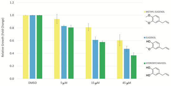
Figure 2.
Relative growth (fold change) of the JE6 cells after treatment with methyl eugenol, eugenol, and hydroxychavicol. The JE6 cells were treated with increasing concentrations (5 μM, 15 μM, 45 μM) of methyl eugenol, eugenol, and hydroxychavicol. The control group was treated only with the solvent, DMSO, at a concentration corresponding to the DMSO concentration in the wells treated with the highest compound concentration (45 µM). The error bars represent the standard error of the mean (SEM) from four independent repeats.
To understand if the propenyl chain of hydroxychavicol contributes to the antiproliferative effect of the compound, the JE6 cells were treated with catechol. Figure 3 shows that catechol exerts more potent antiproliferative properties than hydroxychavicol, which becomes apparent at a 45 µM concentration (t-test p-value = 0.059 at 45 µM). This observation demonstrates that the propenyl chain is the reason why hydroxychavicol has a lower antiproliferative effect than catechol.
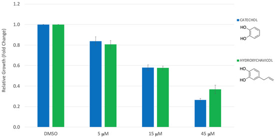
Figure 3.
Relative growth (fold change) of the JE6 cells after treatment with catechol and hydroxychavicol. The JE6 cells were treated with catechol, hydroxychavicol, and DMSO (control) as described previously. The error bars represent the standard error of the mean (SEM) from five independent repeats.
The next objective of this study was to investigate whether the benzene ring contributes to the antiproliferative effect of hydroxychavicol. To assess this, a sample containing a mixture of cis and trans isomers of cyclohexane-1,2-diol was compared against the activity of catechol. As shown in Figure 4, cyclohexane-1,2-diol had no antiproliferative effect on the JE6 cells. (p-value < 0.00001 at 45 µM), indicating that the two hydroxyl groups must be attached to a benzene ring if the compound is to have any antiproliferative effect.
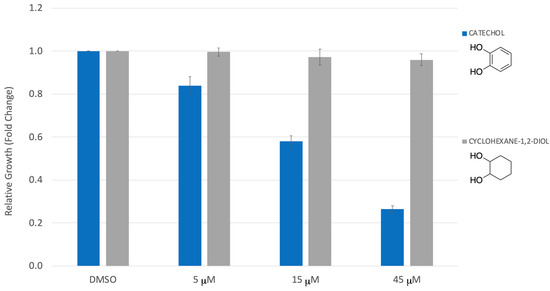
Figure 4.
Relative growth (fold change) of the JE6 cells after treatment with catechol and cyclohexane-1,2-diol. The JE6 cells were treated with catechol, cyclohexane-1,2-diol, and DMSO (control) as described previously. The error bars represent the standard error of the mean (SEM) from four independent repeats.
Next, the proximity, as well as the number of hydroxyl groups on the benzene ring, were investigated (Figure 5). To test the effect of the position of the hydroxyl groups, the JE6 cells were treated with catechol and resorcinol. Catechol’s hydroxyl groups are ortho to one another, whereas resorcinol’s hydroxyl groups are meta (Figure 5). Changing the orientation of the two hydroxyl groups was critical. Resorcinol had no observed effect on the proliferation of the cells (p-value < 0.05 at 45 µM compared to catechol). The number of hydroxyl groups attached to the benzene ring also proved to be important for hindering cell growth. Pyrogallol has an additional hydroxyl group, while phenol lacks one hydroxyl group compared to catechol (Figure 5). Adding a third or removing one of the hydroxyl groups from the catechol molecule also had a detrimental effect on the antiproliferative function of the compound (p-value < 0.01 at 45 µM compared to catechol). It should be noted that pyrogallol had a significantly lower inhibitory effect on cell proliferation than phenol.
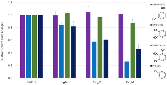
Figure 5.
Relative growth (fold change) of the JE6 cells after treatment with resorcinol, catechol, pyrogallol, and phenol. The JE6 cells were treated with resorcinol, catechol, pyrogallol, phenol, and DMSO (solvent control) as described previously. The error bars represent the standard error of the mean (SEM) from three independent repeats.
Having established that the catechol structure is vital for antiproliferative activity, the proximity of the hydroxyl groups to the benzene was subsequently investigated. Salicyl and phthalyl alcohol were chosen because their hydroxyl groups are spaced from the benzene ring by one or two methylene units (Figure 6). Even though phthalyl alcohol appears to have a slightly stronger antiproliferative effect than salicyl alcohol, both compounds had a much lower impact on the relative cell growth compared to that of catechol (p-values < 0.05 (phthalyl) and =0.06 (salicyl) at 45 µM compared to catechol). These observations confirm that the oxygens of the two hydroxyl groups must be adjacent and directly bound to the aromatic ring.
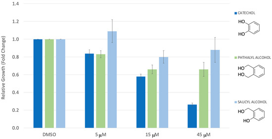
Figure 6.
Relative growth (fold change) of the JE6 cells after treatment with catechol, phthalyl alcohol, and salicyl alcohol. The JE6 cells were treated with catechol, phthalyl and salicyl alcohol, and DMSO (control) as described previously. The error bars represent the standard error of the mean (SEM) from three independent repeats.
The role of the heteroatoms in the analyte structure was also investigated by using chlorine and sulfur analogs of catechol (Figure 7). The effect on cell proliferation of 2-mercaptophenol, containing a thiol functional group instead of a hydroxy group, was almost nonexistent (p-value = 0.02 at 45 µM compared to catechol). This observation showed that the oxygens of the hydroxyl groups are essential for the antiproliferative effect of catechol and hydroxychavicol. Additional studies were conducted with compounds containing chlorine substitutions of one or both of the hydroxyl groups. When one of the hydroxyl groups was substituted (2-chlorophenol), no major difference in the activity was observed. However, when both hydroxyl groups were replaced with chlorine atoms (dichlorobenzene) or when the chlorines were attached to a non-aromatic cyclohexane ring (1,2-dichlorohexane), the antiproliferative functionality of the molecule was almost completely lost (p-value < 0.00001 at 45 µM for 1,2-dichlorohexane compared to catechol; the data from dichlorobenzene were not statistically significant). These observations further supported the essential role of the hydroxyl groups (and specifically the essential role of the oxygen atoms) in the catechol structure of hydroxychavicol.
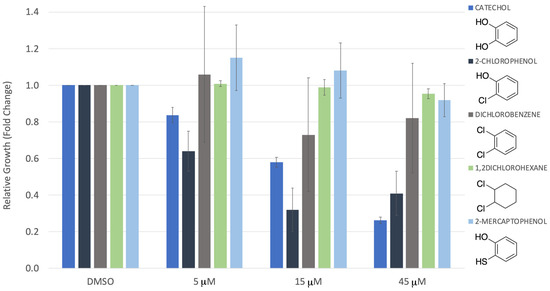
Figure 7.
Relative growth (fold change) of the JE6 cells after treatment with catechol, 2-chlorophenol, dichlorobenzene, 1,2-dichlorohexane, and 2-mercaptophenol. The JE6 cells were treated with catechol, 2-chlorophenol, dichlorobenzene, 1,2-dichlorohexane, 2-mercaptophenol, and DMSO (control) as described previously. The error bars represent the standard error of the mean (SEM) from three independent repeats.
Lastly, the antiproliferative effect of nordihydroguaiaretic acid (NDGA), a structural dimer of hydroxychavicol, was examined (Figure 8). As anticipated, NDGA performed similarly to hydroxychavicol at lower concentrations and significantly better than hydroxychavicol at higher concentrations (Figure 8). These results indicate a proportional influence of the catechol structure on an analyte’s antiproliferative activity (p-value = 0.02 at 45 µM NDGA compared to hydroxychavicol).
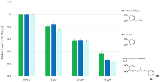
Figure 8.
Relative growth (fold change) of the JE6 cells after treatment with hydroxychavicol, catechol, and nordihydroguaiaretic acid (NDGA). The JE6 cells were treated with hydroxychavicol, catechol, nordihydroguaiaretic acid (NDGA), and DMSO (control) as described previously. The error bars represent the standard error of the mean (SEM) from four independent repeats.
4. Discussion
Through conducting the present study, we aimed to identify the structural components of the hydroxychavicol molecule that contribute to its antiproliferative effect. To achieve this, a series of structural variants of hydroxychavicol were investigated, and their antiproliferative properties were compared to those of hydroxychavicol itself. Our findings provide valuable insights into the key structural features responsible for the observed antiproliferative activity.
The structure of hydroxychavicol contains catechol, which is known to have antiproliferative properties [3,4,6,7,8,13,27,28]. Our study demonstrates that the presence of the catechol-like structure is key for the antiproliferative activity of hydroxychavicol and that the attachment of an aliphatic chain to the catechol structure dampens that activity (Figure 3). The presented data show conclusively that the aromatic ring is essential (Figure 4) and also demonstrate that the two hydroxyl groups must be directly attached to the ring and adjacent to one another (Figure 5 and Figure 6). Furthermore, this work establishes that the oxygen of the hydroxyl groups plays a critical role, which cannot be achieved by atoms with similar valence electron configuration, such as sulfur (Figure 7). It appears that the unique size and electronegativity of oxygen are critical for giving catechol, and analogous structures, antiproliferative capabilities.
Understanding the structural features of catechol and hydroxychavicol provides valuable information for the rational design of novel compounds with enhanced antiproliferative activity for potential therapeutic applications. Further studies are required to elucidate the underlying mechanisms of the observed properties and explore the potential clinical applications of these compounds.
Author Contributions
Conceptualization, J.J., R.G.C. and G.L.L.; methodology, D.H., G.M.R., J.J. and G.L.L.; validation, G.L.L. and J.J.; formal analysis, G.M.R., D.H., J.J. and G.L.L.; investigation, J.J., G.M.R. and D.H.; resources, G.L.L.; writing—original draft preparation, J.J., D.H., G.M.R. and G.L.L.; writing—review and editing, J.J., R.G.C. and G.L.L.; visualization, D.H. and G.L.L.; supervision, G.L.L.; project administration, G.L.L.; funding acquisition, G.L.L. All authors have read and agreed to the published version of the manuscript.
Funding
This research received no external funding.
Data Availability Statement
All data related to this study has been included in this report.
Acknowledgments
The resources for this work were provided by research funds dedicated to undergraduate research by Brigham Young University–Hawaii.
Conflicts of Interest
The authors declare no conflict of interest.
References
- Madhumita, M.; Guha, P.; Nag, A. Bio-actives of betel leaf (Piper betle L.): A comprehensive review on extraction, isolation, characterization, and biological activity. Phytother. Res. 2020, 34, 2609–2627. [Google Scholar] [CrossRef] [PubMed]
- Abrahim, N.N.; Kanthimathi, M.S.; Abdul-Aziz, A. Piper betle shows antioxidant activities, inhibits MCF-7 cell proliferation and increases activities of catalase and superoxide dismutase. Chin. J. Integr. Med. 2012, 12, 220. [Google Scholar] [CrossRef] [PubMed]
- Nayaka NM DM, W.; Sasadara MM, V.; Sanjaya, D.A.; Yuda PE, S.K.; Dewi NL KA, A.; Cahyaningsih, E.; Hartati, R. Piper betle (L): Recent Review of Antibacterial and Antifungal Properties, Safety Profiles, and Commercial Applications. Molecules 2021, 26, 2321. [Google Scholar] [CrossRef] [PubMed]
- Fazal, F.; Mane, P.P.; Rai, M.P.; Thilakchand, K.R.; Bhat, H.P.; Kamble, P.S.; Palatty, P.L.; Baliga, M.S. The phytochemistry, traditional uses and pharmacology of Piper Betel. linn (Betel Leaf): A pan-asiatic medicinal plant. Chin. J. Integr. Med. 2014. [Google Scholar] [CrossRef] [PubMed]
- Tran, V.T.; Nguyen, T.B.; Nguyen, H.C.; Do, N.H.N.; Le, P.K. Recent applications of natural bioactive compounds from Piper betle (L.) leaves in food preservation. Food Control 2023, 152, 110026. [Google Scholar] [CrossRef]
- Mohamad, N.A.; Rahman, A.A.; Kadir, S.H.S.A. Hydroxychavicol as a potential anticancer agent (Review). Oncol. Lett. 2023, 25, 34. [Google Scholar] [CrossRef] [PubMed]
- Vinusri, S.; Gnanam, R.; Caroline, R.; Santhanakrishnan, V.P.; Kandavelmani, A. Anticancer Potential of Hydroxychavicol Derived from Piper betle L: An in Silico and Cytotoxicity Study. Nutr. Cancer 2022, 74, 3701–3713. [Google Scholar] [CrossRef]
- Yadav, Y.; Owens, E.A.; Sharma, V.; Aneja, R.; Henary, M. Synthesis and evaluation of antiproliferative activity of a novel series of hydroxychavicol analogs. Eur. J. Med. Chem. 2014, 75, 1–10. [Google Scholar] [CrossRef]
- Gundala, S.R.; Aneja, R. Piper betel leaf: A reservoir of potential xenohormetic nutraceuticals with cancer-fighting properties. Cancer Prev. Res. 2014, 7, 477–486. [Google Scholar] [CrossRef]
- Gupta, R.K.; Guha, P.; Srivastav, P.P. Phytochemical and biological studies of betel leaf (Piper betle L.): Review on paradigm and its potential benefits in human health. Acta Ecol. Sin. 2023, 43, 721–732. [Google Scholar] [CrossRef]
- Vazhappilly, C.G.; Hodeify, R.; Siddiqui, S.S.; Laham, A.J.; Menon, V.; El-Awady, R.; Matar, R.; Merheb, M.; Marton, J.; Al Zouabi, H.A.K.; et al. Natural compound catechol induces DNA damage, apoptosis, and G1 cell cycle arrest in breast cancer cells. Phytother. Res. 2021, 35, 2185–2199. [Google Scholar] [CrossRef] [PubMed]
- Khalil, A.A.; Rahman, U.; Khan, M.R.; Sahar, A.; Mehmood, T.; Khan, M. Essential oil eugenol: Sources, extraction techniques and nutraceutical perspectives. RSC Adv. 2017, 7, 32669–32681. [Google Scholar] [CrossRef]
- Sharma, S.; Khan, I.A.; Ali, I.; Ali, F.; Kumar, M.; Kumar, A.; Johri, R.K.; Abdullah, S.T.; Bani, S.; Pandey, A.; et al. Evaluation of the antimicrobial, antioxidant, and anti-inflammatory activities of hydroxychavicol for its potential use as an oral care agent. Antimicrob. Agents Chemother. 2009, 53, 216–222. [Google Scholar] [CrossRef] [PubMed]
- Lopez, M.K.N.; Hadisurya, M.; Cornwall, R.G. Antimicrobial Investigation and Structure- Activity Analysis of Natural Eugenol Derivatives Against Several Oral Bacteria. J. Pharm. Microbiol. 2019, 5, 1. [Google Scholar]
- Syahidah, A.; Saad, C.R.; Hassan, M.D.; Rukayadi, Y.; Norazian, M.H.; Kamarudin, M.S. Phytochemical Analysis, Identification and Quantification of Antibacterial Active Compounds in Betel Leaves, Piper betle Methanolic Extract. Pak. J. Biol. Sci. 2017, 20, 70–81. [Google Scholar] [CrossRef][Green Version]
- Al-Sharif, I.; Remmal, A.; Aboussekhra, A. Eugenol triggers apoptosis in breast cancer cells through E2F1/survivin down-regulation. BMC Cancer 2013, 13, 600. [Google Scholar] [CrossRef]
- Abdullah, M.L.; Hafez, M.M.; Al-Hoshani, A.; Al-Shabanah, O. Anti-metastatic and anti-proliferative activity of eugenol against triple negative and HER2 positive breast cancer cells. BMC Complement. Altern. Med. 2018, 18, 321. [Google Scholar] [CrossRef]
- Manikandan, P.; Murugan, R.S.; Priyadarsini, R.V.; Vinothini, G.; Nagini, S. Eugenol induces apoptosis and inhibits invasion and angiogenesis in a rat model of gastric carcinogenesis induced by MNNG. Life Sci. 2010, 86, 936–941. [Google Scholar] [CrossRef]
- Jaganathan, S.K.; Supriyanto, E. Antiproliferative and Molecular Mechanism of Eugenol-Induced Apoptosis in Cancer Cells. Molecules 2012, 17, 6290–6304. [Google Scholar] [CrossRef]
- Petrocelli, G.; Farabegoli, F.; Valerii, M.C.; Giovannini, C.; Sardo, A.; Spisni, E. Molecules Present in Plant Essential Oils for Prevention and Treatment of Colorectal Cancer (CRC). Molecules 2021, 26, 885. [Google Scholar] [CrossRef]
- Fathy, M.; Fawzy, M.A.; Hintzsche, H.; Nikaido, T.; Dandekar, T.; Othman, E.M. Eugenol Exerts Apoptotic Effect and Modulates the Sensitivity of HeLa Cells to Cisplatin and Radiation. Molecules 2019, 24, 3979. [Google Scholar] [CrossRef] [PubMed]
- Abdullah, M.L.; Al-Shabanah, O.; Hassan, Z.K.; Hafez, M.M. Eugenol-Induced Autophagy and Apoptosis in Breast Cancer Cells via PI3K/AKT/FOXO3a Pathway Inhibition. Int. J. Mol. Sci. 2021, 22, 9243. [Google Scholar] [CrossRef] [PubMed]
- Rajedadram, A.; Pin, K.Y.; Ling, S.K.; Yan, S.W.; Looi, M.L. Hydroxychavicol, a polyphenol from Piper betle leaf extract, induces cell cycle arrest and apoptosis in TP53-resistant HT-29 colon cancer cells. J. Zhejiang Univ. Sci. B 2021, 22, 112–122. [Google Scholar] [CrossRef]
- Chakraborty, J.B.; Mahato, S.K.; Joshi, K.; Shinde, V.; Rakshit, S.; Biswas, N.; Choudhury, I.; Mandal, L.; Ganguly, D.; Chowdhury, A.A.; et al. Hydroxychavicol, a Piper betle leaf component, induces apoptosis of CML cells through mitochondrial reactive oxygen species-dependent JNK and endothelial nitric oxide synthase activation and overrides imatinib resistance. Cancer Sci. 2012, 103, 88–99. [Google Scholar] [CrossRef]
- Thamaraikani, I.; Kulandhaivel, M. Purification of Hydroxychavicol from Piper Betle Linn and evaluation of antimicrobial activity against some food poison causing bacteria. J. Pure Appl. Microbiol. 2017, 11, 1883–1889. [Google Scholar] [CrossRef]
- Lim, D.Y.; Shin, S.H.; Lee, M.-H.; Malakhova, M.; Kurinov, I.; Wu, Q.; Xu, J.; Jiang, Y.; Dong, Z.; Liu, K.; et al. A natural small molecule, catechol, induces c-Myc degradation by directly targeting ERK2 in lung cancer. Oncotarget 2016, 7, 35001–35014. [Google Scholar] [CrossRef] [PubMed]
- Grigalius, I.; Petrikaite, V. Relationship between Antioxidant and Anticancer Activity of Trihydroxyflavones. Molecules 2017, 22, 2169. [Google Scholar] [CrossRef]
- Park, Y.; Lim, Y.; Woo, Y.; Lee, S. Relationships between structure and anti-oxidative effects of hydroxyflavones. Bull. Korean Chem. Soc. 2009, 30, 1397–1400. [Google Scholar]
Disclaimer/Publisher’s Note: The statements, opinions and data contained in all publications are solely those of the individual author(s) and contributor(s) and not of MDPI and/or the editor(s). MDPI and/or the editor(s) disclaim responsibility for any injury to people or property resulting from any ideas, methods, instructions or products referred to in the content. |
© 2023 by the authors. Licensee MDPI, Basel, Switzerland. This article is an open access article distributed under the terms and conditions of the Creative Commons Attribution (CC BY) license (https://creativecommons.org/licenses/by/4.0/).