Article

Feline Parasitic Infections, Risk Factors, and Their Association with Parasitic Treatment in Mexico

by
Julio César Segura-Tinoco
1,
Rocío Estefanía Morales-Guerrero
2,
Juan José Pérez-Rivero
3,
Oscar Rico-Chávez
4,
Victor Hugo Del Río-Araiza
5 and
Yazmin Alcala-Canto
1,*
1
Doctorado en Ciencias de la Producción y de la Salud Animal, Universidad Nacional Autónoma de México, Ciudad de México 04510, Mexico
2
Maestría en Ciencias de la Producción y de la Salud Animal, Universidad Nacional Autónoma de México, Ciudad de México 04510, Mexico
3
Departamento de Producción Agrícola y Animal, Universidad Autónoma Metropolitana, Ciudad de México 04960, Mexico
4
Departamento de Etología, Fauna Silvestre y Animales de Laboratorio, Facultad de Medicina Veterinaria y Zootecnia, Universidad Nacional Autónoma de México, Ciudad de México 04510, Mexico
5
Departamento de Parasitología, Facultad de Medicina Veterinaria y Zootecnia, Universidad Nacional Autónoma de México, Ciudad de México 04510, Mexico
*
Author to whom correspondence should be addressed.
Parasitologia 2025, 5(3), 48; https://doi.org/10.3390/parasitologia5030048
Submission received: 6 August 2025 / Revised: 4 September 2025 / Accepted: 5 September 2025 / Published: 10 September 2025

Abstract

Due to their zoonotic potential and close interaction with humans, feline parasitic infections are an important public health concern. This study investigated 2758 domiciled and feral cats sampled across Mexico to assess the occurrence of parasites, coinfections, and associated risk factors. Twelve genera of parasites were identified, with Ancylostoma and Ctenocephalides being the most frequent. Coinfections were common, often involving both intestinal and ectoparasites. Multivariable logistic regression revealed that feral lifestyle, absence of recent antiparasitic treatment, female sex, and climatic conditions were significant predictors of infection. Cats with unrestricted outdoor access and direct contact with other cats, where hunting behavior and the ingestion of prey cannot be ruled out (ESCCAP risk group B), were more than five times as likely to be infected as those cats that live indoors (ESCCAP risk group A). Although antiparasitic use was reported in some cats, inappropriate drug choice and long treatment intervals reduced effectiveness, while nearly seven out of ten cats had never received treatment. These findings highlight major gaps between current practices in Mexico and international guidelines. Strengthening surveillance, promoting owner education, and implementing risk-based strategies are critical to reducing feline parasitism and associated zoonotic risks within a One Health framework.

1. Introduction

Globally, parasitic diseases remain an important concern despite the implementation of control and eradication campaigns [1]. In this context, the domestic cat (Felis catus)—one of the most common animals in urban and peri-urban areas—represents a challenge for epidemiological surveillance systems because of its role as a reservoir and transmitter of parasites that can also affect humans [2].
High human population densities may create favorable conditions for the persistence of pathogens in animals such as cats [3,4]. Nevertheless, studies addressing the impacts of cats on the emergence, diversity, transmission, and prevalence of parasites remain limited [5]. Some research has documented their role as hosts of relevant parasitic infections, including toxoplasmosis, dirofilariasis, toxocariasis, and ancylostomiasis [6].
The behaviors of cats strongly influence their exposure to parasites [2]. Their diverse lifestyles determine the degree of interaction with other animals and the environment [7]. For example, free-roaming individuals, whether feral or semi-owned, are more likely to encounter infection sources compared with those living indoors [3,5]. Predatory behaviors also contribute to transmission, as many parasites require intermediate hosts to complete their life cycles [6].
The European Scientific Counsel Companion Animal Parasites (ESCCAP) is an independent organization that provides evidence-based guidelines for the control of endo- and ectoparasites in companion animals. These guidelines classify cats and dogs into different risk groups based on factors such as lifestyle, environment, ingestion of raw meat and viscera, hunting activity, and exposure to other animals, which allows for the tailoring of antiparasitic protocols to the actual risk of infection. According to ESCCAP, cats are classified into two main risk groups based on lifestyle. Risk group A includes cats that live exclusively indoors or have access only to enclosed patios or gardens, with little likelihood of hunting or ingesting prey. These cats should be treated 1–2 times per year against intestinal nematodes or alternatively undergo fecal examinations with treatment as indicated. Risk group B comprises cats with unrestricted outdoor access and direct contact with other cats, where hunting behavior and the ingestion of prey cannot be ruled out. For these cats, treatment is recommended 4–12 times per year against intestinal nematodes and tapeworms or fecal examinations performed at the same frequency with treatment according to findings [8]. Although these recommendations were designed for Europe, they provide a valuable framework for assessing parasite risk in other regions, where official national guidelines are often lacking.
Understanding the geographic distribution of parasites in cat populations is essential to identifying risk factors, optimizing surveillance, and implementing preventive actions such as education, sanitation, and veterinary control measures [9,10]. However, several studies have highlighted that protozoa, helminths, and arthropod vectors remain under-reported globally and regionally [11]. This underestimation results from diagnostic limitations, a lack of active surveillance measures, and poor integration of veterinary data into broader monitoring systems [6]. The absence of accurate records hinders a realistic assessment of the epidemiological role of cats in urban and peri-urban environments [9].
Research on feline parasites in Mexico is relatively recent, and studies describing and statistically analyzing their frequency remain limited [12]. The scarcity of investigations is partly due to the challenges inherent to feline-related sampling, such as their habit of burying feces and the risks associated with handling feral individuals.
Between 2018 and 2023, a helminthological study was conducted on free-ranging cats (Felis silvestris catus, n = 36) in Mexico City. The following helminths were detected: the cestodes Hydatigera taeniaeformis, Mesocestoides sp., and Taenia rileyi; Dipylidium caninum; and the nematode Toxocara cati [13]. A survey of feline parasites in Mexico City and the metropolitan area identified the most prevalent ectoparasite to be Ctenocephalides felis (17.73%), followed by Notoedres cati (4.98%), Felicola subrostratus (4.78%), C. canis (4.18%), and Rhipicephalus sanguineus (1.59%). Among gastrointestinal parasites, the highest prevalence was recorded for D. caninum (16.14%) [12]. Another study reported the following parasitic arthropods in cats in Mexico: F. subrostratus, Heterodoxus spiniger, Eutrombicula alfreddugesi, and Cimex lectularius [14]. In central Mexico (Querétaro), 53% of free-roaming cats were infested with fleas, mainly C. felis (53%), followed by C. canis (18%) and Echidnophaga gallinacea (7%). Gastrointestinal parasites were found in 45% of cats, with D. caninum (36%) as the most prevalent, followed by Physaloptera praeputialis (7%) and H. taeniformis (4%) [15]. To date, no nationwide study on cat parasites has been conducted in Mexico, providing the rationale for undertaking the present study.
The purpose of this study was to evaluate the frequency and trends of parasitism in cats in central Mexico, as well as to analyze host- and environment-related factors that influence infection patterns. This approach provides a better understanding of the dynamics of parasite occurrence in cats and the risks they pose in different climatic regions in the country.

2. Materials and Methods

2.1. Study Area

Samples were collected in 2024 and 2025 from 320 localities across all 32 states of Mexico, providing broad and representative geographic coverage. This extensive sampling allowed for the assessment of parasitic prevalence under diverse climatic, ecological, and socioeconomic conditions. Regions were classified into the following three climate types (as shown in Figure 1): A (tropical and subtropical), B (dry), and C (temperate) [16].

2.2. Georeferencing of Sampling Sites

A geolocation map was generated using QGIS 3.32.3 (QGIS Development Team, Grüt, Switzerland), showing sampling sites for feral cats and zip codes for addresses for domiciled cats.

2.3. Sampling

The study used a convenience sample. Although no a priori sample size calculation was performed, a post hoc verification was carried out to ensure adequacy for prevalence estimation. The required sample size for estimating parasite prevalence in cats was calculated using the classical Cochran’s formula for proportions, applying a 95% confidence level (Z = 1.96), a conservative estimated prevalence of 50% (p = 0.5), and a desired precision of 5% (d = 0.05) [18,19].
n0 = (Z2 × p(1 − p))/d2
where Z = 1.96 for a 95% confidence level, p = 0.3005 (30.05% observed prevalence), and d = 0.03 (3% absolute precision).
To account for potential clustering and non-random sampling factors resulting from the use of a convenience sampling framework, a design effect was applied; design effects greater than 1 reflect increased sampling variance due to cluster effects [1,2].
Based on an estimated cat population in Mexico of approximately 7 million domiciled cats and 16 million feral cats [20,21], the minimum sample size required was 898 cats. A total of 2758 samples were collected, thus exceeding the calculated requirement. Given the practical challenges of random sampling under field conditions, a convenience sampling approach was employed which, while not probabilistic, allowed for the inclusion of a large and diverse number of animals from both domiciled and feral populations.
Stools from domiciled cats were brought in by their owners or collected from veterinary clinics, while samples from feral cats were obtained using video surveillance cameras (by collecting feces shortly after elimination) and through Trap–Neuter–Return (TNR) programs conducted within the study areas. Fecal samples (3–5 g) were gathered from veterinary clinics, directly from cat owners, or from the ground within four hours of stool deposition. Fecal samples for the diagnosis of endoparasites in feral cats were obtained from the environment in 553 cases and directly from the animal, during TNR procedures, in 228 cases. Regarding ectoparasites, 352 samples were obtained directly from feral cats captured using feral cat handlers (Tomahawk® traps) (Tetengo, Nuevo León, Mexico). All capture procedures were performed by veterinary students and veterinarians specifically trained in the ethical sampling and handling of both feral and domiciled cats. Procedures were conducted under institutional ethical approval and in strict accordance with the guidelines of the U.S. organization Alley Cat Allies for the care and management of feral cats [22]. These procedures aligned with recognized protocols from institutional policies and national codes of practice, ensuring that animal welfare remained a central consideration throughout the study. Seventeen ectoparasites from feral cats were identified through video surveillance images, in which flea infestations were inferred based on clinical signs (e.g., scratching) and the visible presence of fleas on animals residing in the monitored households. In domiciled cats, 183 ectoparasite samples were collected directly from the animals, and an additional 21 cases were identified from photographs presented to the veterinarians responsible for those cats. Sampling was carried out in 2024 and 2025 during field surveys conducted in different localities. Fecal samples were collected during the daytime or, when cats were observed defecating at night through a video surveillance system, samples were obtained at dawn the following day. In addition, cat owners were instructed to deliver fecal samples within four hours of collection. When delivery exceeded this period, the samples were stored under refrigeration at 4 °C until submission to the laboratory. All samples were sent to the Department of Parasitology at the Faculty of Veterinary Medicine and Animal Science, National Autonomous University of Mexico. Ectoparasites were collected directly from domiciled cats. Whenever possible, feral cats were carefully handled to collect ectoparasites.

2.4. Parasite Detection

2.4.1. Macroscopic Technique

Macroscopic examination was used to detect adult parasites, proglottids of cestodes, or entire helminths that could be observed with the naked eye in feces. For this procedure, feces were diluted in water. Samples were placed in dark trays and examined with the naked eye or under a stereoscopic magnifying glass. The fecal material was gently dispersed using a spatula or dissecting needle to facilitate visualization of helminths, proglottids, or other macroscopic parasitic structures. Any observed parasites were carefully separated with a dissecting needle or fine brush for subsequent identification. This process was repeated with successive portions of the sample until the entire specimen was examined [23].

2.4.2. Microscopic Technique

Microscopic examination (direct smear) enabled the identification of eggs, cysts, trophozoites, and oocysts in fresh stool preparations. For this test, the following two preparations were made on a clean glass slide: one with 0.9% saline solution and another with Lugol’s iodine. Using a wooden applicator, approximately 1–2 mg of feces (about the size of a rice grain) was placed on each drop and homogenized with the solution. Fibers and coarse debris were removed with the applicator before placing a coverslip. Both preparations were examined under a compound microscope at 10X and 40X objectives to detect protozoan cysts, trophozoites, oocysts, and helminth eggs [23].

2.4.3. Fecal Flotation Technique

Flotation techniques were applied to recover nematode and cestode eggs of low specific gravity, as well as protozoan oocysts such as Cystoisospora. For this procedure, approximately 3 to 5 g of feces were homogenized. One milliliter of saturated sucrose solution was added to the feces to form a paste, which was subsequently diluted with 60–100 mL of the same solution until a homogeneous suspension was obtained. The suspension was filtered through a fine sieve into a second beaker to remove coarse debris and then allowed to stand for 15–20 min. After this period, three drops were collected from the surface of the suspension using a sterilized inoculation loop, which had been flamed before use to avoid contamination with eggs or oocysts. Each drop was placed on a clean glass slide, covered with a coverslip, and examined under a compound microscope at 10X objective, focusing on the surface layer where parasitic structures (e.g., nematode eggs and protozoan oocysts) are concentrated [23].

2.4.4. Faust Technique

Faust centrifugal flotation with zinc sulfate was used to concentrate and recover protozoan cysts such as Giardia, as well as light nematode and cestode eggs. For this test, approximately 5 g of feces was placed in a beaker, diluted in 50 mL of distilled water, and homogenized until a uniform suspension was obtained. The suspension was strained into a second container and transferred into centrifuge tubes, filled up to approximately 1 cm from the rim. The tubes were centrifuged at 650 g for 2 min, after which the supernatant was discarded. The sediment was resuspended in distilled water and centrifuged again, discarding the supernatant each time. This washing process was repeated three to four times until the supernatant appeared clear. The cleaned sediment was then resuspended in saturated zinc sulfate solution (ZnSO4) and centrifuged again at 650 g for 2 min. After centrifugation, the tubes were placed on a rack, and the floating material was collected for microscopic observation. This was performed either by transferring drops from the surface of the liquid onto a glass slide using a previously flame-sterilized inoculating loop, placing a drop of the supernatant on a slide and mixing it with Lugol’s iodine before covering it with a 16 × 16 mm coverslip or by adding more saturated zinc sulfate solution until a meniscus was formed and then placing a coverslip directly onto the meniscus. After 10 min, the coverslip was carefully removed and placed on a slide containing a drop of Lugol’s iodine. All preparations were examined under the microscope, scanning in a zigzag pattern across the entire sample. Initial observations were made using the 10× objective, and when structures of interest were detected, higher magnification with the 40× objective was used for a more detailed evaluation [23].

2.4.5. Sedimentation Technique

This procedure was mainly intended for heavy eggs, such as those of trematodes, as well as some protozoan cysts, which would otherwise not float. For the sedimentation technique, 3 to 5 g of fecal material was homogenized. A beaker was labeled with the sample identification, and the feces were placed inside with enough tap water to dissolve them, mixing thoroughly until a homogeneous suspension was obtained. The mixture was then strained through a fine mesh or sieve into another container to remove large forage fragments. The strained material was topped with tap water up to the neck of the container and left to stand for 5 min. After this, the supernatant was decanted, and the sediment was resuspended in tap water. This washing process was repeated as many times as necessary until the supernatant appeared clear. The final sediment was then transferred to a Petri dish for microscopic examination. The Petri dish was observed under a compound microscope using the 10X objective, scanning the entire sediment in a zigzag pattern to ensure that no parasitic structures were overlooked [23].

2.4.6. Graham Technique

The Graham method (“Scotch-tape” test) was employed to recover perianal egg capsules of Dipylidium. For this test, a transparent adhesive tape was placed around one end of a wooden tongue depressor, with the sticky surface facing outward. Holding the depressor firmly between the index finger and thumb, the adhesive tape was pressed against the perianal region, applying pressure alternately to both sides to maximize the adherence of parasitic structures. Once this step was completed, the tape was carefully separated from the depressor and mounted directly onto a clean glass slide. The preparation was then examined under a compound microscope using the 10X objective to detect the presence of eggs or other diagnostic elements [23].

2.4.7. Kinyoun Technique

The Kinyoun (modified Ziehl–Neelsen) staining technique was used to visualize acid-fast oocysts of Cryptosporidium. For this procedure, approximately 25 μL of fecal suspension from suspected samples was placed at the center of a clean glass slide and evenly spread to form a thin smear. The preparation was allowed to dry at room temperature, then fixed with methanol for 3 min and left to dry completely. The fixed smear was subsequently covered with carbol fuchsin for 5 min, followed by rinsing with running water until the washout was clear. Decolorization was performed using acid–alcohol (3% hydrochloric acid in 95% ethanol), rinsing again with water until the background appeared clear. Alternatively, a milder solution of 1% sulfuric acid in 95% ethanol could be used as a decolorizing agent, especially when organisms were highly susceptible to acid treatment. The preparation was then counterstained with malachite green for 1 min, rinsed with tap water, and air-dried. Finally, the smear was examined under oil immersion (100X objective). When present, oocyst structures appeared as purplish-red elements contrasting against a blue or green background, depending on the counterstain applied [23].

2.4.8. Ectoparasite Collection

Mites that cause mange were collected using skin scrapings from lesions using a cotton swab, scalpel, or glass slide, with glycerin as a vehicle. In general, skin scrapings were placed on one or several slides, cotton swabs were smeared onto a slide or sent separately, and all the samples were placed in a plastic container or plastic bag, with a few drops of glycerin added. Lice were collected by carefully examining the hair, using a cotton ball soaked in ether–alcohol, which was rubbed over different anatomical regions. The cotton balls were then placed in jars containing 70% alcohol or in sealable plastic bags. Fleas were collected from the cats using a cotton ball soaked in ether–alcohol and subsequently placed in jars containing 70% alcohol. Another method for collecting lice and fleas from cats that were difficult to handle consisted of placing the animals over a sheet of white paper and brushing their hair. A limitation of this procedure was that the ectoparasites could be lost or escape; to avoid this, a few drops of glycerin were applied directly to the area being brushed. The ectoparasites were then placed in plastic or glass jars, or in vials containing 70–80% ethyl alcohol [24].

2.5. Statistical Analysis

The proportion of cats positive for each parasitic genus was calculated based on the total sampled population. If a cat was infected with more than one genus, it was counted once per genus.
An in-depth analysis was conducted on a database documenting parasitism cases in cats, considering host-related variables (sex, age, living condition, and prior antiparasitic treatment) and environmental factors (climate). The objective was to explore associations with host and environmental characteristics.
The collected data and identified parasites were recorded in a Microsoft Excel spreadsheet. A logistic regression analysis was performed to identify factors associated with the presence of parasites in cats. The dependent variable was parasitism status (parasitized = 1; not parasitized = 0). Independent variables included lifestyle (domiciled cats, living in households with regular human care, or feral cats, free-ranging with little or no human contact), sex (male, female, or not determined), and age group (kittens: 0–6 months; young: 6–12 months; adults: 1–7 years; and seniors: >7 years), as well as information regarding antiparasitic treatment, including whether cats had received any previous antiparasitic treatment (yes/no), the time elapsed since the last administration, classified into intervals (three months ago, six months ago, and one year ago), climate (temperate, tropical, or arid) [17], and risk group (A/B) according to the ESCCAP guidelines. Categorical variables with more than two categories (age group, sex, climate, and interval of previous antiparasitic treatment) were introduced into the model using indicator (dummy) variables. The final model was constructed using a backward stepwise selection approach, starting with all variables of interest and sequentially removing those without statistical significance. This approach was combined with epidemiological reasoning to ensure that biologically relevant variables were not excluded solely based on statistical criteria. The model was fitted using the maximum likelihood method, and the results are expressed as adjusted odds ratios (aORs) with 95% confidence intervals (95% CIs) and p-values, with p < 0.05 considered statistically significant. Model fit was assessed using the Hosmer–Lemeshow goodness-of-fit test, and the discriminatory performance was evaluated using the area under the ROC curve (AUC). All analyses were performed using EpiInfo™ version 7.2 (Centers for Disease Control and Prevention–CDC, 2022).
To evaluate associations between Dipylidium infection and the presence of ectoparasites (Ctenocephalides and Felicola), 2 × 2 contingency tables were constructed. Odds ratios (ORs) with 95% confidence intervals (CIs) were calculated, and statistical significance was assessed using Pearson’s χ2 test or Fisher’s exact test when expected frequencies were <5.

2.6. Ethics Statement

Permission to collect samples from cats was granted from the Subcomité Interno para el Cuidado y Uso de los Animales de Experimentación of the Programa de Maestría y Doctorado en Ciencias de la Producción y de la Salud Animal of the Universidad Nacional Autónoma de México code SICUAE.MC-2024/5-3, on 11 March 2024.

2.7. Data Availability Statement

The original contributions presented in this study are included in the Supplementary Materials (Database S1: Feline parasitic infections in Mexico database). Further inquiries can be directed to the corresponding author.

3. Results

3.1. Study Population Data

Population data for sampled cats are presented in Table 1. Total prevalence was estimated as 30.1% (95% CI: 28.4–31.8%).

3.2. Parasite Positivity Frequencies

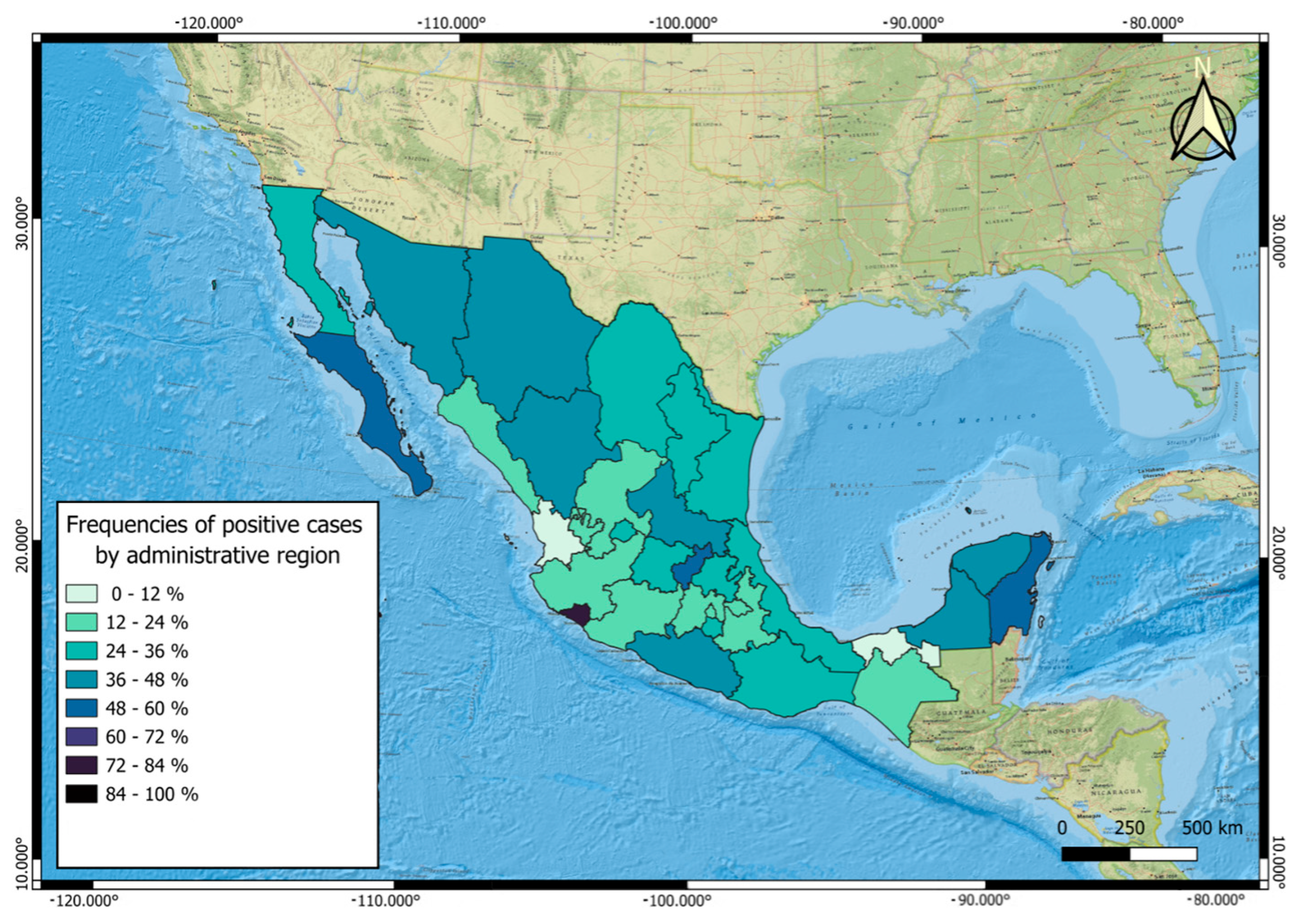
Positivity frequencies for each parasitic genus were calculated and represented on a map, expressed as the number of cats positive for each genus (n) and the percentage they represent of the total number of cats evaluated (n = 2758). The category “Negative” refers to animals in which no parasitic genus was detected, representing 1929 cases (69.9%). A total of 734 cats were sampled in climate type A (tropical and subtropical), of which 286 (39.0%) were parasitized, with a 95% confidence interval (CI) ranging from 35.5% to 42.5%. In climate type B (dry), 135 out of 683 cats were parasitized, corresponding to a prevalence of 19.8% (95% CI: 17.0–22.9%). In climate type C (temperate), 408 of 1341 cats were parasitized, yielding a prevalence of 30.4% (95% CI: 28.0–32.9%) (Figure 2).
Table 2 presents the distribution of the genera of all identified parasites, reflecting the proportion of each genus relative to the total number of parasites detected, regardless of whether they were from the same animal. Representative photographs of the identified parasites are provided in the Supplementary Materials (File S1: Photographs of parasites).
The most common genus was Ctenocephalides, followed by the nematode Ancylostoma and the cestode Dipylidium.

3.3. Coinfections

The distribution of coinfections was analyzed based on the number of distinct parasitic genera identified per cat, as presented in Table 3.
The most frequent parasitic genus combinations (coinfections) were Ancylostoma + Dipylidium (n = 57), Ancylostoma + Ctenocephalides + Dipylidium (n = 36), Ctenocephalides + Dipylidium (n = 33), Ancylostoma + Cryptosporidium + Ctenocephalides (n = 25), Ancylostoma + Cryptosporidium + Ctenocephalides + Dipylidium (n = 23), Ancylostoma + Ctenocephalides (n = 22), Ancylostoma + Cryptosporidium (n = 15), and Ancylostoma + Cryptosporidium + Dipylidium (n = 13). These associations reflect the most common coinfection patterns in the studied population. All other parasitic genus combinations occurred in fewer than eight cats each.
A significant association was identified between the presence of fleas (Ctenocephalides spp.) and infection with Dipylidium. Among the 495 cats positive for Ctenocephalides, 183 (37.0%) were also positive for Dipylidium, whereas only 126 (5.6%) of the 2263 cats without fleas were infected. Statistical analysis showed that cats infested with fleas were 9.96 times more likely to be infected with D. caninum compared with flea-free cats (OR = 9.96; 95% CI: 7.8–12.6; p < 0.001, Pearson’s χ2). A significant association could not be established between the presence of Felicola and infection with Dipylidium. Among the 22 cats positive for Felicola, all (100%) were also positive for Dipylidium, whereas all the 286 cats without Felicola (100%) were also infected. Statistical analysis showed no significant difference in the likelihood of infection between cats with and without Felicola (OR = 0.08; 95% CI: 0.001–4.05; p = 1.0, Fisher’s exact test).

3.4. Risk Factors

3.4.1. Overall Parasitism

Multivariable logistic regression identified several independent predictors of overall parasitism in cats. Feral lifestyle was the strongest risk factor, with feral cats being more than four times more likely to be parasitized compared with domiciled cats (aOR ≈ 4.4; 95% CI: 3.6–5.3; p < 0.001). Climate type also had a significant effect: cats from climate A (aOR ≈ 2.1; 95% CI: 1.7–2.6; p < 0.001) and climate C (aOR ≈ 1.9; 95% CI: 1.5–2.4; p < 0.001) were more likely to be parasitized than those from climate B. Sex was associated with infection risk: males had lower odds than females (aOR ≈ 0.66; 95% CI: 0.51–0.86; p = 0.002), while cats of undetermined sex had substantially higher odds (aOR ≈ 3.1; 95% CI: 2.1–4.7; p < 0.001). Age group was also a significant predictor. Kittens (0–6 months) and young cats (6–12 months) had lower odds of parasitism compared with adults (aOR ≈ 0.68 and 0.53, respectively; both p < 0.001), whereas senior cats (>7 years) did not differ significantly (p = 0.186). Previous deworming was independently associated with reduced odds of parasitism (aOR ≈ 0.77; 95% CI: 0.63–0.96; p = 0.018). Furthermore, when the time of antiparasitic treatment was considered, cats treated within the past six months had significantly lower odds of being parasitized compared with those never treated or treated more than six months prior (aOR ≈ 0.6; 95% CI: ~0.5–0.8; p < 0.05). This finding supports a time-dependent protective effect of recent deworming. Finally, classification based on the ESCCAP risk group was highly informative, with cats in group B having more than five times the odds of parasitism compared to those in group A (aOR = 5.414, 95% CI: 4.125–7.112; p < 0.001). The overall model demonstrated a moderate explanatory power (Nagelkerke R2 = 0.278) and was based on the evaluation of 2758 cats. In the multivariable analysis, variables such as lifestyle, ESCCAP risk group category and antiparasitic treatment remained consistently significant across most parasite genera, while sex, age, and climate also showed significant associations for several infections.

3.4.2. Endoparasites

Multiple logistic regression analysis identified several significant risk factors associated with endoparasite infection in cats. Age was strongly associated with infection, with kittens (aOR = 2.02, p < 0.001), young cats (aOR = 4.13, p < 0.001), and senior cats (aOR = 3.37, p < 0.001) presenting higher odds compared with adult cats. Sex was relevant only for feral cats in which sex remained undetermined due to handling conditions (aOR = 3.51, p < 0.001), whereas males did not differ significantly from females (p = 0.095). Lifestyle was also significant, with feral cats showing increased odds compared with domiciled cats (aOR = 1.39, p = 0.026). Belonging to ESCCAP risk group B emerged as the strongest predictor (aOR = 5.00, p < 0.001). A shorter interval since the last treatment against endoparasites (< 6 months) was a protective factor (aOR = 0.54, p < 0.001). Climatic conditions also played a role, with cats from climate type B showing significantly reduced odds of infection (aOR = 0.41, p < 0.001).

3.4.3. Ectoparasites

For ectoparasites, younger and older age groups were associated with higher risks, with kittens (aOR = 3.08, p < 0.001), young cats (aOR = 2.96, p < 0.001), and senior cats (aOR = 2.77, p < 0.001) showing increased odds compared with adults. Sex was relevant only for feral cats, in which sex remained undetermined due to handling conditions (aOR = 2.42, p < 0.001), while males were not significantly different from females (p = 0.473). Lifestyle was a strong predictor, with feral cats being at a markedly higher risk (aOR = 3.48, p < 0.001). A short interval since the last treatment against ectoparasites (<6 months) showed a borderline association with higher risk (aOR = 1.38, p = 0.054). Climatic conditions were protective, particularly in climate type B (aOR = 0.37, p < 0.001).

3.5. Antiparasitic Treatments

Of the 2758 records, 882 cats had been treated against endo- or ectoparasites, while 1876 had not been treated. A total of 705 dewormed cats tested negative for parasitic infections, suggesting effective antiparasitic control. However, 177 (20.1%) of the treated cats tested positive for parasites. Table 4 presents data on the previous antiparasitic treatment administered to the cats. Of the 177 treated cats that tested positive for parasites, 95 (53.7%) exhibited mismatches between the antiparasitic drug administered and the parasites detected, indicating inadequate spectrum coverage. Additionally, 101 cats (57.1%) had excessively long intervals between treatments, with deworming reported as having occurred one year prior to sampling.

4. Discussion

Despite their close interaction with human populations and increasing density in both urban and rural contexts of Latin America, domestic and feral cats have been underestimated as parasitic hosts [5]. In this context, the frequency of parasitism observed in this study (n = 829), along with the diversity of parasites identified (12 genera), reveals a considerable parasitic burden in both domiciled and feral cats.
It has been proposed that domiciled cats are less susceptible to endoparasitic infections as they have access to processed food, regular veterinary care, and reduced exposure to intermediate hosts such as wild prey [9]. Our findings support this statement, as a significant difference was observed between the following two populations: feral cats, despite constituting a smaller sampled population (n = 918), exhibited a considerably higher parasite positivity rate compared with domiciled cats. This finding was obtained despite the operational challenges of sampling feral animals or the fact that, at the time of collection during TNR surgeries, their intestines were often empty. In agreement with these findings, studies by Nagamori et al. [25,26] reported a higher prevalence of parasites in free-roaming cats (63.9%) in Oklahoma compared with domesticated cats, reinforcing the hypothesis that the environment and access to veterinary care influence parasitic susceptibility.
In this sense, the study by Wierzbowska et al. [27] suggests a higher frequency of parasites in cats inhabiting peri-urban areas compared with those in urban zones, likely due to the presence of feral individuals and synanthropic species. Nonetheless, these observations highlight the need for further research in diverse geographical contexts to better understand how environmental characteristics influence parasitic dynamics in feline populations.
Our findings revealed higher parasite prevalence in cats from tropical climate type A regions, followed by temperate (type C) and dry (type B) climates. This pattern is in agreement with previous reports indicating that warm and humid environments generally enhance the development and transmission of many parasitic species, whereas droughts and other extreme events may temporarily reduce parasite burdens but generate long-term ecological imbalances that sustain transmission risk [28]. However, these results must be taken with caution. First, the unequal distribution of samples across climate categories, with most cats originating from type C regions (n = 1341), may have introduced sampling bias. Second, microenvironmental conditions within each climate category (e.g., presence of water sources, vegetation cover, or soil type) were not directly measured but could have substantially influenced the survival and transmission of parasites. Third, host-related factors such as animal density, outdoor access, and owner management practices may have varied between locations and interacted with climatic conditions. Taken together, these considerations highlight that climate should be regarded as one of several interacting determinants of parasitism in cats and that further research with more balanced sampling and finer-scale environmental data are needed to clarify these associations.
Age and sex analysis revealed that adult cats accounted for the highest proportion of positive individuals, which may be attributed to greater exposure to contaminated environments [29]. Unlike studies such as that of Genchi et al. (2021), in which age was not a significant factor after multivariate adjustment, this analysis showed a lower risk of parasitism in adult cats, possibly reflecting behavioral differences and environmental access [9].
Regarding sex, females in the present study were significantly more prone to infections with parasites; in contrast, a previous study found that male cats had a higher prevalence of Toxocara cati compared with female cats [29]. The authors suggested that this may be associated with behavioral differences, such as roaming, fighting, or territorial marking, which increase environmental exposure to infective stages. However, a separate study did not identify sex as a consistent, statistically significant predictor across all parasite groups after controlling for other factors such as age, outdoor access, and frequency of antiparasitic treatment [30].
A high proportion of parasitized females was observed, many of which were feral. This might be linked to specific reproductive and epidemiological dynamics, such as behavioral differences (e.g., parental care and territoriality) and physiological factors, as female sex hormones can modulate immune responses, increasing susceptibility or promoting immunotolerance to certain parasites [31,32]. However, it is important to note that significantly more samples were collected from females than males; therefore, this result should be interpreted with caution.
Ancylostoma spp., which is responsible for cutaneous larva migrans, was the most frequently detected agent [33]. This parasitic genus has gained special attention in recent years, not only due to the emergence of multi-drug-resistant Ancylostoma spp. strains [34,35] but also because of the expansion of its geographic distribution, a phenomenon associated with climate change [36,37].
Among the zoonotically relevant parasites identified in this study were Cryptosporidium, Toxocara, Giardia, and Dipylidium. The persistence of oocysts, cysts, egg capsules, and eggs in the environment, along with the predatory and roaming behavior of unsupervised feral cats, significantly contributes to the perpetuation and spread of these agents in urban and peri-urban settings [10]. This study contrasts with previous reports on feline parasites in Mexico, where D. caninum was identified as the most prevalent species [12,13,15]. In the present study, however, Ancylostoma was the most frequently detected parasite. This discrepancy may be associated with the fact that not all cats could be handled to allow the Graham procedure; this may have led to underdiagnosis of D. caninum.
Five taxa of ectoparasites were identified (Ctenocephalides, Notoedres, Otodectes, Felicola, and Rhipicephalus). Ctenocephalides was the most prevalent ectoparasite, likely due to its cosmopolitan distribution, generalist habits, and affinity for domestic hosts [38]. Its importance in the context of public health lies in its role as a vector for Dipylidium, Bartonella, and Rickettsia, emphasizing the need for active epidemiological surveillance [39].
Regarding ectoparasites, a previous report [30] provided data on the specific prevalences of external parasites such as fleas, ticks, and mites in domestic cats in France, Germany, and the United Kingdom. The prevalence of Ctenocephalides felis was 29.6%, comparable to that reported in this study. In both studies, the methods used were strictly visual and entomological, and factors such as feral status and lack of deworming could explain the presence of ectoparasites [30]. The identification of Ctenocephalides as the most prevalent ectoparasite in this study is in accordance with previous reports in Mexico [12,15].
Another relevant finding was the high prevalence of parasitic coinfections in infected cats. The concurrent presence of two or more species, such as Ancylostoma, Cryptosporidium, Ctenocephalides, and Cystoisospora, not only worsens the clinical picture in hosts and complicates diagnosis and treatment [33] but also reflects ecological coexistence patterns that favor persistence and dissemination of pathogens in the same environment, increasing transmission risks [3,4]. Findings of coinfections are consistent with those reported previously in European cats [40], especially those with outdoor access. High positivity rates of ectoparasites such as Ctenocephalides and endoparasites such as Ancylostoma were observed, consistent with previous findings in Mexico and Europe [30,41] reporting high infestation rates by fleas and intestinal nematodes.
Many animals presented with mixed infections involving nematodes, cestodes, protozoa, and ectoparasites. These cases require combined or rotational treatment protocols that address all life stages of parasites and all taxonomic groups involved. Without collective deworming of all animals in the household, environmental control measures, and regular follow-up, even highly effective antiparasitic products may fail to keep the animal free of parasitic infections. The finding of a significant association between Ctenocephalides and Dipylidium is consistent with the biological cycle of this cestode, in which fleas act as intermediate hosts. The high magnitude of the association (OR ≈ 10) suggests that flea infestation represents a significant risk factor for the transmission of Dipylidium in cats. This result highlights the need to implement ectoparasite control programs as a key measure to reduce Dipylidium infection in feline populations and, consequently, to decrease the zoonotic risk to owners. Fleas are recognized as the principal intermediate hosts for Dipylidium, while lice such as Felicola are only infrequently involved and hold limited epidemiological significance [42]. The low prevalence of lice in our population (n = 22) may also have limited the statistical power to detect an association. Furthermore, as this was a cross-sectional study, causality cannot be established, and variables such as outdoor access or deworming frequency were not controlled for in this analysis.
Data on the deworming history of the 1840 domiciled cats reveal the insufficient implementation of public health preventive measures, as 58.64% of these animals had no prior deworming history. This situation underscores the urgent need to strengthen health education strategies targeting pet owners, as well as public and veterinary health professionals. Additionally, the guidelines proposed by Beugnet et al. [30] for assessing the efficacy of anthelmintic treatments in dogs and cats could be adapted to the Latin American epidemiological context, thereby optimizing regional control strategies. The lack of deworming was associated with a higher infection risk, in line with findings by Roussel et al. [43], who emphasized periodic deworming as a key factor in parasite control.
Among the dewormed cats, many individuals showed evidence of inappropriate drug selection based on the parasitic species detected. For instance, products such as Imidacloprid + Moxidectin are primarily effective against gastrointestinal nematodes and certain ectoparasites, but lack activity against protozoa such as Giardia or Cryptosporidium. In this group, despite recent treatment, infections with Giardia, Cryptosporidium, and Ctenocephalides were still detected. A similar situation was observed with other products such as Esafoxolaner and Lotilaner, which are mainly indicated for fleas and ticks but do not target intestinal parasites. This pharmacological gap helps to explain the high frequency of Ancylostoma, Dipylidium, and Cystoisospora infections in animals treated with these compounds.
Additionally, 57.1% of cats presented with an excessively long interval between deworming applications, with all treatments reportedly administered more than a year prior to sampling. A cat may be successfully dewormed and temporarily parasite-free, but can quickly become reinfected if exposed to contaminated sources such as soil, other animals, fleas, or feces. The persistent presence of Ctenocephalides, Dipylidium, and nematodes in the population suggests a strong reinfection pressure in the environment.
Of the dewormed cats that tested positive for parasites, most were specifically infected with intestinal helminths, such as Ancylostoma, Toxocara cati, and Dipylidium. These cases are of particular concern under the ESCCAP guidelines, which recommend at least four annual treatments for cats at moderate risk and monthly deworming for those with high-risk lifestyles to effectively control intestinal helminths. The presence of helminths in these 141 cats suggests a mismatch between current deworming practices and international guidelines-recommended frequencies and risk-based strategies [8]. Among the dewormed cats that tested negative for parasites, more than half had been dewormed three months prior to sampling. According to ESCCAP GL1 guidelines, cats at moderate risk should be dewormed at least every three months, while monthly treatment is recommended for those in high-risk situations. Thus, most parasite-free cats in this group had been dewormed within the minimum recommended timeframe, which likely contributed to the observed effectiveness of the antiparasitic treatments.
Beyond ESCCAP, other international guidelines also emphasize the need for more rigorous control strategies than those currently observed in our population. The Companion Animal Parasite Council (CAPC), a North American expert panel, recommends year-round broad-spectrum coverage against intestinal parasites, fleas, ticks, and heartworm, with fecal testing at least four times in the first year of life and twice annually in adults, or quarterly deworming when continuous prevention is not feasible [43]. The Tropical Council for Companion Animal Parasites (TroCCAP) further stresses the importance of early and frequent deworming in kittens, beginning at 2–3 weeks of age and continued fortnightly until at least 10 weeks; monthly treatment for outdoor cats, and simultaneous treatment of queens and their litters, while also highlighting environmental hygiene and avoidance of raw diets as critical preventive measures [44]. Likewise, ESCCAP extends its risk-based recommendations to adult cats, advising at least quarterly deworming and, in high-risk households (young children or immunocompromised individuals), monthly treatment or fecal examinations every four weeks [45].
Taken together, CAPC, TroCCAP, and ESCCAP guidelines converge in underscoring the importance of early, continuous, and risk-based parasite prevention in cats. The discrepancies among these international recommendations and our findings highlight a critical gap in feline parasite control in Mexico and reinforce the urgent need for education, owner compliance, and broader implementation of preventive strategies to safeguard both feline and public health.
Ectoparasites such as Ctenocephalides, Sarcoptes, Otodectes, Notoedres, Felicola, or Rhipicephalus were identified in 101 treated cats. The presence of ectoparasites in these treated cats suggests that either the selected antiparasitic agents lacked adequate ectoparasiticidal activity or that environmental sources of reinfestation were not sufficiently addressed. In addition to endoparasite control, international guidelines also highlight the need for strict ectoparasite prevention. CAPC recommends continuous, year-round control against fleas and ticks in cats, given their role as vectors of zoonotic pathogens and the difficulty of eliminating infestations once established [43]. TroCCAP also underscores that in tropical and subtropical environments, year-round ectoparasite pressure is high, making monthly application of ectoparasiticides essential, particularly in free-roaming cats. ESCCAP similarly advises continuous flea prevention, tick protection for cats with outdoor access, and combining pharmacological prophylaxis with environmental management such as prompt removal of fleas and ticks in the household [44]. Together, these guidelines emphasize that ectoparasite prevention must be comprehensive and sustained, which is particularly relevant in Mexico, where outdoor access and lack of owner compliance create a scenario of persistent risk.
A total of 705 dewormed cats tested negative for parasitic infections, indicating effective antiparasitic treatment. The success of these deworming treatments can be attributed to the appropriate selection of broad-spectrum drugs, such as Praziquantel + Pyrantel + Febantel, Milbemycin + Praziquantel, and Imidacloprid + Moxidectin, which cover a wide range of internal and external parasites. Furthermore, most treatments were administered within a relatively short timeframe (e.g., 3 to 6 months prior to sampling), minimizing the risk of reinfection and enhancing efficacy. However, caution is advised against more frequent treatments, repeated use of the same active substance, or underdosing. The risk of selecting drug-resistant parasites through frequent use of the same active compound or improper dosing warrants caution when recommending more intensive treatment regimens. Anthelmintic resistance is well recognized in Ancylostoma caninum, particularly within kennel environments in North America [35]. Although less common, there are documented reports of emerging resistance in Dipylidium caninum; notably, cases of resistance to praziquantel and epsiprantel have been described both in the United States and Europe, including challenging instances requiring high-dose mebendazole therapy to achieve parasite clearance [46]. Similarly, the protozoan Giardia has shown decreasing susceptibility to standard treatments such as nitroimidazoles. High drug pressure could be an explanation for this resistance [47]. Although our study did not directly assess the development of resistance, the growing body of evidence emphasizes the importance of judicious use of antiparasitic agents. Recommendations should prioritize correct dosing, consider rotation of drug classes, and tailor protocols based on individual risk factors, as outlined in ESCCAP guidelines. Additionally, we acknowledge this limitation in our study and propose that future research should monitor treatment failures as potential indicators of emerging resistance.
By determining the prevalence of gastrointestinal and ectoparasitic infections in the localities sampled in this study, our findings represent an advance in the epidemiological knowledge of the most common parasitoses affecting domiciled cats in Mexico. Based on these data, veterinarians can more accurately prescribe antiparasitic treatments that are aligned with the local parasite spectrum and tailored to specific risk factors. This approach not only improves treatment efficacy but also helps optimize preventive strategies within the Mexican epidemiological context.
Based on the data obtained in this study, Mexican practitioners are advised to always conduct deworming under the guidance of a veterinary professional. Regular coprological examination of fecal samples represents a valuable alternative to standard deworming protocols, as it enables targeted treatments based on the parasite species present. When the individual risk to an animal cannot be clearly assessed, the animal should be examined or dewormed at least four times per year.
Furthermore, given the limited awareness of zoonotic transmission risks in the general population, practitioners in Mexico can play a critical role in strengthening health education among cat owners. This includes explaining the importance of regular preventive treatments, tailoring drug choice to the local parasite species, and implementing integrated parasite control strategies (environmental hygiene, flea control, and avoidance of raw viscera or prey ingestion). By adapting these international guidelines to the Mexican epidemiological context, veterinarians can optimize preventive practices, reduce parasite circulation, and mitigate zoonotic risks.

Study Limitations

Our study has several limitations that should be acknowledged. First, although cats were classified according to ESCCAP risk groups and dietary habits, improper dosing of antiparasitics was not recorded and may have influenced infection outcomes. Second, potential cases of improper diagnosis, which may have affected prevalence estimates, could not be fully excluded. Third, the statistical power to detect associations with less prevalent ectoparasites was limited by the small number of infested animals. Fourth, the potential effect of oversampling in areas close to the authors’ institution may have introduced a geographical bias, limiting the representativeness of the results. Fifth, while our results revealed significant associations between parasite occurrence and certain variables, these findings should be interpreted with caution. The cross-sectional design of the study provides only a snapshot in time, allowing for the detection of correlations but not establishing temporality; therefore, causality cannot be inferred, as it is not possible to determine whether the exposure preceded the infection or whether other confounding factors were involved. Sixth, since cats were included based on accessibility rather than through random or stratified selection, the sampled population may not accurately reflect the characteristics of the broader feline population in Mexico. This non-probabilistic approach can introduce selection bias, particularly because many of the samples were obtained from locations near the authors’ institutions. Cats in these contexts may differ systematically from the general population in terms of health status, frequency of veterinary care, nutritional practices, and exposure to parasites. As a result, the prevalence estimates reported here should be interpreted with caution, as they may overestimate or underestimate the true national prevalence. Future studies should incorporate more representative and geographically balanced sampling strategies, ideally using probabilistic designs, to provide more robust and generalizable estimates of parasitic infections in domestic and feral cats across Mexico. Seventh, our study did not directly assess the emergence of drug resistance, which is increasingly recognized in Ancylostoma, Dipylidium, and Giardia, and should, therefore, be interpreted with caution. Eighth, a notable gender imbalance was present in the sampled population, with females being markedly overrepresented relative to males. This disproportion may have introduced bias and could partially account for certain observed statistical associations. Accordingly, the findings should be interpreted with due caution. Finally, it is important to highlight that the ESCCAP guidelines used for risk classification were developed under European epidemiological conditions; therefore, Mexico should develop its own deworming guidelines tailored to the local parasite ecology, animal management practices, and public health priorities.

5. Conclusions

The results of this study reinforce the need to address feline parasitism from a One Health perspective, considering the close relationships between animal, human, and environmental health. The high frequency of zoonotic parasites—especially in feral cats or those without sanitary control—represents a significant public health risk, particularly in urban and socially vulnerable settings. The distribution by age and sex showed that younger individuals, females, and feral cats are especially susceptible, underscoring the importance of considering age groups and environmental factors as key elements in transmission dynamics for public health programs. In urban, peri-urban, and rural contexts, it becomes a priority to strengthen epidemiological surveillance of zoonotic parasites and to implement integrated strategies for feline population control, community education, and access to veterinary services as essential pillars to reduce transmission risk and protect collective health. In conclusion, this study confirms and complements the existing literature, highlighting feral lifestyle and lack of deworming as the most critical factors affecting the risk of feline parasitism.

Supplementary Materials

The following supporting information can be downloaded at: https://www.mdpi.com/article/10.3390/parasitologia5030048/s1, File S1: Photographs: Cats, Ancylostoma, Toxocara, Giardia, Cryptosporidium, Cystoisospora, Dipylidium, Ctenocephalides, Sarcoptes, Otodectes, Rhipicephalus, Notoedres, Felicola; Database S1: Feline parasitic infections in Mexico database.

Author Contributions

Conceptualization, Y.A.-C., J.C.S.-T. and R.E.M.-G.; methodology, Y.A.-C., R.E.M.-G. and J.C.S.-T.; software, J.J.P.-R.; validation, Y.A.-C., O.R.-C., V.H.D.R.-A. and J.J.P.-R.; formal analysis, Y.A.-C., J.J.P.-R.; investigation, Y.A.-C., J.C.S.-T. and R.E.M.-G.; resources, Y.A.-C.; original draft preparation, Y.A.-C., J.C.S.-T. and R.E.M.-G.; writing, review and editing, Y.A.-C.; visualization, J.J.P.-R. and V.H.D.R.-A.; supervision, Y.A.-C.; project administration, Y.A.-C.; funding acquisition, Y.A.-C. All authors have read and agreed to the published version of the manuscript.

Funding

This research was funded by DIRECCION GENERAL DE ASUNTOS DEL PERSONAL ACADEMICO DE LA UNIVERSIDAD NACIONAL AUTONOMA DE MEXICO, Programa de Apoyo a Proyectos de Investigación e Innovación Tecnológica (PAPIIT) grant number IN213023: Desarrollo y análisis de una base de datos georreferenciada de los parásitos de los gatos en México.

Institutional Review Board Statement

Permission to take fecal samples from cats was obtained from the Ethics Committee (Subcomité Interno para el Cuidado y Uso de los Animales de Experimentación of the Programa de Maestría y Doctorado en Ciencias de la Producción y de la Salud Animal of the Universidad Nacional Autónoma de México) code SICUAE.MC-2024/5-3, on 11 March 2024.

Informed Consent Statement

Informed consent was obtained from the cat owners to collect fecal samples or ectoparasites from their cats, and they agreed either to bring the samples themselves or to allow the cats to be sampled. Informed consent was also obtained from the coordinators of the TNR programs to collect samples.

Data Availability Statement

The original contributions presented in this study are included in the Supplementary Materials. Further inquiries can be directed to the corresponding author.

Acknowledgments

To the Department of Parasitology at the Faculty of Veterinary Medicine and Animal Science of the National Autonomous University of Mexico for providing the laboratory facilities used for diagnosis. To the Trap-Neuter-Return (TNR) groups and feline pet guardians for providing the necessary samples.

Conflicts of Interest

Authors declare no conflicts of interest. The funders had no role in the design of the study; in the collection, analyses, or interpretation of data; in the writing of the manuscript; or in the decision to publish the results.

References

  1. Morchón, R.; Gabrielli, S.; Ciuca, L.; Napoli, E.; Carretón, E. Editorial: Advancements in understanding zoonotic parasitic diseases. Front. Vet. Sci. 2024, 11, 1539556. [Google Scholar] [CrossRef]
  2. Candela, M.G.; Fanelli, A.; Carvalho, J.; Serrano, E.; Domenech, G.; Alonso, F.; Martínez-Carrasco, C. Urban landscape and infection risk in free-roaming cats. Zoonoses Public Health 2022, 69, 295–311. [Google Scholar] [CrossRef]
  3. Bevins, S.N.; Carver, S.; Boydston, E.E.; Lyren, L.M.; Alldredge, M.; Logan, K.A.; Riley, S.P.D.; Fisher, R.N.; Vickers, T.W.; Boyce, W. Three pathogens in sympatric populations of pumas, bobcats, and domestic cats: Implications for infectious disease transmission. PLoS ONE 2012, 7, e31403. [Google Scholar] [CrossRef] [PubMed]
  4. Lindahl, J.; Magnusson, U. Zoonotic pathogens in urban animals: Enough research to protect the health of the urban population? Anim. Health Res. Rev. 2020, 21, 50–60. [Google Scholar] [CrossRef] [PubMed]
  5. Gonçalves, L.S.; de Souza Machado, D.; Caçador, M.E.; Ferreira, G.A.; Dickman, C.R.; Ceballos, M.C.; Prezoto, F.; Sant’Anna, A.C. The Wildcat That Lives in Me: A Review on Free-Roaming Cats (Felis catus) in Brazil, Focusing on Research Priorities, Management, and Their Impacts on Cat Welfare. Animals 2025, 15, 190. [Google Scholar] [CrossRef] [PubMed]
  6. Baneth, G.; Thamsborg, S.M.; Otranto, D.; Guillot, J.; Blaga, R.; Deplazes, P.; Solano-Gallego, L. Major Parasitic Zoonoses Associated with Dogs and Cats in Europe. J. Comp. Pathol. 2016, 155, S54–S74. [Google Scholar] [CrossRef]
  7. Sparkes, A.H.; Bessant, C.; Cope, K.; Ellis, S.L.H.; Finka, L.; Halls, V.; Hiestand, K.; Horsford, K.; Laurence, C.; MacFarlaine, I. ISFM guidelines on population management and welfare of unowned domestic cats (Felis catus). J. Feline Med. Surg. 2013, 15, 811–817. [Google Scholar] [CrossRef]
  8. ESCCAP. Worm control in dogs and cats. In Guideline 01; European Scientific Counsel Companion Animal Parasites: Malvern, UK, 2017. [Google Scholar]
  9. Genchi, M.; Vismarra, A.; Zanet, S.; Morelli, S.; Galuppi, R.; Cringoli, G.; Lia, R.; Diaferia, M.; Frangipane di Regalbono, A.; Venegoni, G.; et al. Prevalence and risk factors associated with cat parasites in Italy: A multicenter study. Parasites Vectors 2021, 14, 475. [Google Scholar] [CrossRef]
  10. Mendoza Roldan, J.A.; Otranto, D. Zoonotic parasites associated with predation by dogs and cats. Parasites Vectors 2023, 16, 55. [Google Scholar] [CrossRef]
  11. Adhikari, R.B.; Dhakal, M.A.; Ale, P.B.; Regmi, G.R.; Ghimire, T.R. Survey on the prevalence of intestinal parasites in domestic cats (Felis catus Linnaeus, 1758) in central Nepal. Vet. Med. Sci. 2023, 9, 559–571. [Google Scholar] [CrossRef]
  12. Morales-Guerrero, R.E. Desarrollo y Análisis de Una Base de Datos de Los Parásitos de Los Gatos en la Ciudad de México y Área Metropolitana. Bachelor’s Thesis, Universidad Nacional Autónoma de México, Mexico City, Mexico, 2023. [Google Scholar]
  13. Camacho-Giles, V.; Hortelano-Moncada, Y.; Torres-Carrera, G.; Gil-Alarcón, G.; Oceguera-Figueroa, A.; García-Prieto, L.; Osorio-Sarabia, D.; Cervantes, F.A.; Arenas, P. Helminths of free-ranging dogs and cats in an urban natural reserve in Mexico City and their potential risk as zoonotic agents. PLoS ONE 2024, 19, e0310302. [Google Scholar] [CrossRef]
  14. Baak-Baak, C.; Garcia-Rejon, J.; Tzuc-Dzul, J.; Nuñez-Corea, D.; Arana-Guardia, R.; Cetina-Trejo, R.; Machain-Williams, C.; Jimenez-Coello, M.; Acosta-Viana, K.; Torres-Chable, O.; et al. Four Species of Under-Reported Parasitic Arthropods in Mexico and Their Potential Role as Vectors of Pathogens. J. Parasitol. 2020, 106, 835–842. [Google Scholar] [CrossRef]
  15. Cantó, G.J.; Guerrero, R.I.; Olvera-Ramírez, A.M.; Milian, F.; Mosqueda, J.; Aguilar-Tipacamú, G. Prevalence of fleas and gastrointestinal parasites in free-roaming cats in central Mexico. PLoS ONE 2013, 8, e60744. [Google Scholar] [CrossRef]
  16. Méndez-Arriaga, F. The temperature and regional climate effects on communitarian COVID-19 contagion in Mexico throughout phase 1. Sci. Total Environ. 2020, 735, 139560. [Google Scholar] [CrossRef] [PubMed]
  17. Naing, L.; Winn, T.; Rusli, B.N. Practical issues in calculating the sample size for prevalence studies. Arch. Orofac. Sci. 2006, 1, 9–14. [Google Scholar]
  18. Ahmed, S.K. How to choose a sampling technique and determine sample size for research: A simplified guide for researchers. Oral Oncol. Rep. 2024, 12, 100662. [Google Scholar] [CrossRef]
  19. Peña-Corona, S.I.; Gomez-Vazquez, J.P.; López-Flores, E.A.; Vargas Estrada, D.; Arvizu-Tovar, L.O.; Pérez-Rivero, J.J.; Juárez Rodríguez, I.; Sierra Resendiz, A.; Soberanis-Ramos, O. Use of an extrapolation method to estimate the population of cats and dogs living at homes in Mexico in 2022. Vet. México OA 2022, 9. [Google Scholar] [CrossRef]
  20. INEGI. Instituto Nacional de Estadística y Geografía. Estadísticas Sobre Disponibilidad y Características de Animales de Compañía en los Hogares de México. 2023. Available online: https://www.inegi.org.mx/ (accessed on 5 September 2025).
  21. Allies, A.C. How to Help Community Cats: A Step-by-Step Guide to Trap-Neuter Return. 2019. Available online: https://www.alleycat.org/resources/how-to-help-community-cats-a-step-by-step-guide-to-trap-neuter-return/ (accessed on 5 September 2025).
  22. Figueroa, C.J.A.; Jasso, V.C.; Liébano, H.E.; Martínez, L.P.; Rodríguez, V.R.I.; Zárate, R.J.J. Examen Coproparasitoscópico. In Técnicas Para el Diagnóstico de Parásitos Con Importancia en Salud Pública y Veterinaria; Rodríguez Vivas, R.I.e., Ed.; AMPAVE-CONASA: Mexico City, Mexico, 2015; p. 517. [Google Scholar]
  23. Guerrero, M.M.C.; Cruz, V.C.; Cruz, M.I.; Escutia, S.I. Muestras biológicas y muestreos para estudios parasitológicos. In Técnicas Para el Diagnóstico de Parásitos Con Importancia en Salud Pública y Veterinaria. México; Rodríguez-Vivas, R.I., Ed.; AMPAVE-CONASA: Mexico City, Mexico, 2015. [Google Scholar]
  24. INEGI. Guía Para la Interpretación de Cartografía Climática. 2017. Available online: http://internet.contenidos.inegi.org.mx/contenidos/productos/prod_serv/contenidos/espanol/bvinegi/productos/geografia/publicaciones/guias-carto/clima/CLIMATIII.pdf#page=1&zoom=auto,-102,792 (accessed on 5 September 2025).
  25. Nagamori, Y.; Payton, M.E.; Duncan-Decocq, R.; Johnson, E.M. Fecal survey of parasites in free-roaming cats in northcentral Oklahoma, United States. Vet. Parasitol. Reg. Stud. Rep. 2018, 14, 50–53. [Google Scholar] [CrossRef]
  26. Nagamori, Y.; Payton, M.E.; Looper, E.; Apple, H.; Johnson, E.M. Retrospective survey of parasitism identified in feces of client-owned cats in North America from 2007 through 2018. Vet. Parasitol. 2020, 277, 109008. [Google Scholar] [CrossRef]
  27. Wierzbowska, I.A.; Kornaś, S.; Piontek, A.M.; Rola, K. The Prevalence of Endoparasites of Free Ranging Cats. Animals 2020, 10, 748. [Google Scholar] [CrossRef]
  28. Poglayen, G.; Gelati, A.; Scala, A.; Naitana, S.; Musella, V.; Nocerino, M.; Cringoli, G.; Frangipane di Regalbono, A.; Habluetzel, A. Do natural catastrophic events and exceptional climatic conditions also affect parasites? Parasitology 2023, 150, 1158–1166. [Google Scholar] [CrossRef]
  29. Iturbe Cossío, T.L.; Montes Luna, A.D.; Ruiz Mejia, M.; Flores Ortega, A.; Heredia Cárdenas, R.; Romero Núñez, C. Risk factors associated with cat parasites in a feline medical center. JFMS Open Rep. 2021, 7, 20551169211033183. [Google Scholar] [CrossRef]
  30. Beugnet, F.; Bourdeau, P.; Chalvet-Monfray, K.; Cozma, V.; Farkas, R.; Guillot, J.; Halos, L.; Joachim, A.; Losson, B.; Miró, G.; et al. Parasites of domestic owned cats in Europe: Co-infestations and risk factors. Parasites Vectors 2014, 7, 291. [Google Scholar] [CrossRef] [PubMed]
  31. Salcedo-Jiménez, J.; Alcala-Canto, Y.; Segura-Tinoco, J.; Valadez-Moctezuma, E.; Pérez-Rivero, J.J. Identifying Zoonotic Parasites in Domiciled and Non-Domiciled Dogs (Canis lupus familiaris) Within an Urban Zone of the Eastern State of Mexico. Vet. Med. Sci. 2024, 10, e70059. [Google Scholar] [CrossRef] [PubMed]
  32. Wesołowska, A. Sex—The most underappreciated variable in research: Insights from helminth-infected hosts. Vet. Res. 2022, 53, 94. [Google Scholar] [CrossRef]
  33. Beugnet, F.; Taweethavonsawat, P.; Traversa, D.; Fourie, J.; McCall, J.; Tielemans, E.; Geurden, T. World Association for the Advancement of Veterinary Parasitology (WAAVP): Second edition of guidelines for evaluating the efficacy of anthelmintics for dogs and cats. Vet. Parasitol. 2022, 312, 109815. [Google Scholar] [CrossRef]
  34. Currie, B.J.; Hoopes, J.; Cumming, B. Cutaneous Larva Migrans Refractory to Therapy with Ivermectin: Case Report and Review of Implicated Zoonotic Pathogens, Epidemiology, Anthelmintic Drug Resistance and Therapy. Trop. Med. Infect. Dis. 2025, 10, 163. [Google Scholar] [CrossRef]
  35. Jimenez Castro, P.D.; Howell, S.B.; Schaefer, J.J.; Avramenko, R.W.; Gilleard, J.S.; Kaplan, R.M. Multiple drug resistance in the canine hookworm Ancylostoma caninum: An emerging threat? Parasites Vectors 2019, 12, 576. [Google Scholar] [CrossRef]
  36. Aguilar-Rodríguez, D.; Seco-Hidalgo, V.; Lopez, A.; Romero-Sandoval, N.; Calvopiña, M.; Guevara, A.; Baldeón, L.; Rodríguez, A.; Mejia, R.; Nutman, T.B.; et al. Geographic Distribution of Human Infections with Zoonotic Ancylostoma ceylanicum and Anthropophilic Hookworms in Ecuador: A Retrospective Analysis of Archived Stool Samples. Am. J. Trop. Med. Hyg. 2024, 110, 460–469. [Google Scholar] [CrossRef]
  37. Cervantes-Candelas, L.A.; Aguilar-Castro, J.; Buendía-González, F.O.; Fernández-Rivera, O.; Nolasco-Pérez, T.d.J.; López-Padilla, M.S.; Chavira-Ramírez, D.R.; Legorreta-Herrera, M. 17β-Estradiol is involved in the sexual dimorphism of the immune response to malaria. Front. Endocrinol. 2021, 12, 643851. [Google Scholar] [CrossRef]
  38. Moore, C.O.; André, M.R.; Šlapeta, J.; Breitschwerdt, E.B. Vector biology of the cat flea Ctenocephalides felis. Trends Parasitol. 2024, 40, 324–337. [Google Scholar] [CrossRef]
  39. Abdullah, S.; Helps, C.; Tasker, S.; Newbury, H.; Wall, R. Pathogens in fleas collected from cats and dogs: Distribution and prevalence in the UK. Parasites Vectors 2019, 12, 71. [Google Scholar] [CrossRef] [PubMed]
  40. Bett, B.; Kiunga, P.; Gachohi, J.; Sindato, C.; Mbotha, D.; Robinson, T.; Lindahl, J.; Grace, D. Effects of climate change on the occurrence and distribution of livestock diseases. Prev. Vet. Med. 2017, 137, 119–129. [Google Scholar] [CrossRef] [PubMed]
  41. Beugnet, F.; Labuschagne, M.; Vos, C.; Crafford, D.; Fourie, J. Analysis of Dipylidium caninum tapeworms from dogs and cats, or their respective fleas—Part 2. Distinct canine and feline host association with two different Dipylidium caninum genotypes. Parasite 2018, 25, 31. [Google Scholar] [CrossRef] [PubMed]
  42. Roussel, C.; Drake, J.; Ariza, J.M. French national survey of dog and cat owners on the deworming behaviour and lifestyle of pets associated with the risk of endoparasites. Parasites Vectors 2019, 12, 480. [Google Scholar] [CrossRef]
  43. CAPC. Companion Animal Parasite Council. 2025. Available online: https://capcvet.org/ (accessed on 5 September 2025).
  44. Dantas-Torres, F.; Ketzis, J.; Mihalca, A.D.; Baneth, G.; Otranto, D.; Tort, G.P.; Watanabe, M.; Linh, B.K.; Inpankaew, T.; Jimenez Castro, P.D.; et al. TroCCAP recommendations for the diagnosis, prevention and treatment of parasitic infections in dogs and cats in the tropics. Vet. Parasitol. 2020, 283, 109167. [Google Scholar] [CrossRef]
  45. ESCCAP. European Scientific Counsel Companion Animal Parasites. Control of Ectoparasites in Dogs and Cats. 2025. Available online: https://www.esccap.org/guidelines/gl3/ (accessed on 5 September 2025).
  46. Beugnet, F.; Halos, L.; Guillot, J. Textbook of Clinical Parasitology in Dogs and Cats; Servet Editorial—Grupo Asís Biomedia, S.L.: Zaragoza, Spain, 2018. [Google Scholar]
  47. Ydsten, K.A.; Öhd, J.N.; Hellgren, U.; Asgeirsson, H. Prevalence of Nitroimidazole-Refractory Giardiasis Acquired in Different World Regions, Sweden, 2008–2020. Emerg. Infect. Dis. 2025, 31, 1235–1238. [Google Scholar] [CrossRef]
Figure 1. Climate regions of Mexico [17].
Parasitologia 05 00048 g001
Figure 2. Frequencies of cat parasites in Mexico.
Parasitologia 05 00048 g002
Table 1. Population data from sampled felines.
CategorySubcategoryn%
ParasitismPositive82930.1
Negative192969.9
SexFemale209175.8
Male48417.5
Not determined1836.6
AgeKittens (0–6 months)123644.8
Young (6–12 months)41815.2
Adults (1–7 years)86231.3
Senior (>7 years)2428.8
LifestyleDomesticated184066.7
Feral91833.3
ClimateTropical (A)73439.0
Arid (B)68319.8
Temperate (C)134130.4
Table 2. Relative frequency of each genus within the total set of parasites detected.
Parasiten%95% CI
Endoparasites
Ancylostoma44816.214.9–17.6
Dipylidium30911.210.0–12.4
Cryptosporidium2057.46.5–8.4
Cystoisospora1846.75.7–7.6
Toxocara943.42.7–4.1
Giardia722.62.0–3.2
Ectoparasites
Ctenocephalides49517.916.5–19.4
Notoedres531.91.4–2.4
Felicola501.81.3–2.3
Otodectes461.71.2–2.1
Rhipicephalus40.10.0–0.3
Table 3. Coinfections identified in cats.
Number of Generan (Cats)%95% CI
1 genus137249.847.9–51.7
2 genera69225.123.5–26.8
3 genera38614.012.8–15.4
4 genera1886.85.9–7.8
5 genera772.82.2–3.5
6 genera311.10.7–1.6
7 genera100.40.2–0.7
8 genera20.10.0–0.3
Table 4. Previous antiparasitic treatments (n = 882).
Categoryn%95% CI
Treated 3 months prior45416.515.1–17.9
Treated 6 months prior2649.68.5–10.7
Treated 1 year prior1645.95.1–6.9
Disclaimer/Publisher’s Note: The statements, opinions and data contained in all publications are solely those of the individual author(s) and contributor(s) and not of MDPI and/or the editor(s). MDPI and/or the editor(s) disclaim responsibility for any injury to people or property resulting from any ideas, methods, instructions or products referred to in the content.