Article

Differential Distribution of Trypanosoma vivax and Trypanosoma theileri in Cattle from Distinct Agroecological Regions of Central Argentina

by
Maria Celeste Facelli Fernández
1,
Johann Barolin
1,
Martin Allassia
2,
Javier Hernan Gonzalez
2,
Pablo Martin Beldomenico
1,2 and
Lucas Daniel Monje
1,3,*
1
Laboratorio de Ecología de Enfermedades, Instituto de Ciencias Veterinarias del Litoral (ICIVET-Litoral), Universidad Nacional del Litoral (UNL)/Consejo Nacional de Investigaciones Científicas y Técnicas (CONICET), Esperanza PC 3080, Santa Fe, Argentina
2
Facultad de Ciencias Veterinarias, Universidad Nacional del Litoral (UNL), Esperanza PC 3080, Santa Fe, Argentina
3
Facultad de Bioquímica y Ciencias Biológicas, Universidad Nacional del Litoral (UNL), Santa Fe PC 3000, Santa Fe, Argentina
*
Author to whom correspondence should be addressed.
Parasitologia 2025, 5(2), 27; https://doi.org/10.3390/parasitologia5020027
Submission received: 15 April 2025 / Revised: 13 May 2025 / Accepted: 3 June 2025 / Published: 5 June 2025

Abstract

:
Bovine trypanosomiasis, caused by Trypanosoma vivax, affects livestock productivity and is increasingly being reported in South America. This study aimed to detect and characterize Trypanosoma spp. infections, with a focus on T. vivax, in cattle from two distinct agroecological regions of central Argentina: a dairy-producing plain, located in the Espinal ecoregion, and a riparian zone, dedicated to beef production, located in the Delta and Islands of Paraná ecoregion. A total of 220 blood samples were collected from nine cattle farms and analyzed using real-time PCR, melting curve analysis, and the sequencing of 18S rRNA gene fragments. Trypanosoma vivax was detected at low prevalence (2.73%), exclusively in dairy cattle. In contrast, the prevalence of Trypanosoma theileri was much higher (10.91%), and it was found mainly in beef cattle from the riparian region. Phylogenetic analyses confirmed the species identity in all sequenced samples. No trypanosomes were observed by microscopy, and none of the animals showed clinical signs. The results indicate a differential distribution of T. vivax and T. theileri between regions and production systems. Although the study initially focused on T. vivax, the detection of T. theileri highlights the need to consider multiple Trypanosoma species in epidemiological surveys. This study contributes new data on the occurrence of bovine trypanosomes in central Argentina under extensive and semi-intensive management systems.

1. Introduction

Trypanosomes (Euglenozoa; Kinetoplastea) are a group of digenetic blood parasites with a global distribution, primarily transmitted by hematophagous insects. These flagellated protozoa are of significant public and veterinary health importance, as they are the causative agents of trypanosomiasis, a potentially fatal disease affecting both humans and animals worldwide [1]. Trypanosoma vivax infects both domestic and wild ungulates and is the primary causative agent of Nagana disease, which affects livestock and leads to substantial economic losses, particularly in sub-Saharan Africa [2,3] and South America [4,5,6]. In Africa, transmission occurs mainly through tsetse flies, whereas in the Americas, species of Tabanidae have been identified as the primary mechanical vectors [7]. However, any blood-feeding insect thriving in a farming environment could potentially serve as a mechanical vector for livestock trypanosomes, particularly for species like T. vivax and Trypanosoma evansi, which can reach high parasitemia levels [7]. In contrast, the mechanical transmission of Trypanosoma theileri is less likely due to its typically low parasitemia [7]. Additionally, although not recommended, the reuse of veterinary needles and syringes is a common practice even in developed countries such as the United States, where more than 80% of producers reportedly reuse the same needle on multiple animals [8]. Notably, T. vivax remains viable in injectable veterinary products, and when the same syringe and needle are reused after contact with an acutely infected animal, up to eight other cattle can become infected [9], contributing to the spread of T. vivax [10].
The first outbreak of T. vivax in Argentina was recorded in 2006 in a livestock farm in the northeastern region of Chaco. The infected animals exhibited fever, anemia, emaciation, neurological signs, and high mortality rates [11]. Since then, additional outbreaks have been documented in different regions of the country, affecting both dairy and beef production systems [12,13]. The economic losses from a single outbreak on a dairy farm were estimated at nearly USD 60,000.00, attributable to animal losses, deaths, abortions, and medical costs [12].
The Trypanosoma theileri group (hereafter referred to as T. theileri) consists of multiple species, including Trypanosoma theileri, Trypanosoma melophagium, Trypanosoma cervi, and Trypanosoma trinaperronei, which have been reported to infect cervids and bovids [14,15]. Trypanosoma theileri has been detected in a wide range of Diptera, including members of the family Tabanidae [16], keds [14], mosquitoes [17], sandflies [18], and tsetse flies [19], as well as in ticks [20,21]. Although T. theileri is generally considered to have low pathogenicity, recent studies have reported its presence in vital organs, such as the brain, liver, lungs, and lymph nodes [22,23], and evidence from dairy cattle suggests it may negatively affect productivity, with significant reductions in milk protein and solids-not-fat levels observed in infected multiparous cows [24].
In central Argentina, the province of Santa Fe has approximately 6 million hectares of grazing land, primarily consisting of natural grasslands and hosting an average of 1.5 million heads of cattle. It is also one of the most important dairy-producing regions in Latin America, accounting for 28% of Argentina’s total dairy production. Given the emerging economic impact of Trypanosoma on livestock in the region, this study aimed to detect and characterize Trypanosoma spp. infections, with a focus on T. vivax, in cattle from two distinct agroecological regions of central Argentina: a dairy-producing plain located in the Espinal ecoregion [25] and a riparian zone dedicated to beef production located in the Delta and Islands of Paraná ecoregion [25].
Our findings provide new insights into the distribution and ecology of T. vivax and T. theileri in central Argentina. We observed distinct patterns of trypanosome prevalence between dairy and beef cattle, indicating that environmental and management factors influence transmission dynamics. These results help to clarify the factors driving trypanosomiasis in the region and can guide future research and control efforts.

2. Materials and Methods

This study was conducted in livestock farms across two distinct regions within Santa Fe province in central Argentina (Figure 1), spanning approximately 80 km between the closest sites and up to 160 km between the farthest. One region corresponds to the dairy basin, a vast plain with favorable conditions for cattle breeding and traditionally dedicated to dairy production, located in the Espinal ecoregion [25]. Within the dairy basin, farms from the Castellanos department (CD) are located at its center, while those from the Las Colonias department (LCD) are at its eastern limit (Figure 1). The second region, located within the Delta and Islands of Paraná ecoregion [25], represented by farms from the Garay department (GD), is a riparian area that is characterized by extensive lowlands, riverbanks, and wetlands (Figure 1), where cattle farming is primarily focused on beef production.
Blood samples were collected from September 2021 to October 2022 from bovines (Bos taurus) in both dairy (Holando-Argentino breed) and beef (Braford breed) farms.
Sampling was performed on a convenience basis on farms to which we had access through the veterinarians responsible for the animal care and management of each farm. Blood was drawn from the coccygeal vein and stored in 1.5 mL tubes with EDTA. The samples were kept at −20 °C until further processing. Genomic DNA was extracted from 200 µL of blood from each bovine using the Genomic DNA Mini Kit (Geneaid Biotech, New Taipei, Taiwan) according to the manufacturer’s instructions. The concentration and purity of genomic DNA were evaluated using SPECTROstar Nano (BMG Labtech, Ortenberg, Germany) and the data analysis software MARS version 3.31 (BMG Labtech, Ortenberg, Germany). A 260/280 nm absorbance ratio above 1.6 was used as a quality cutoff; samples falling below this threshold were re-extracted. DNA concentrations were then standardized to 100 ng/µL for downstream analyses. Additionally, blood smears were prepared in situ for 37 beef cattle from a farm in GD, where animals were available for sampling during a routine veterinary control visit. Smears were air-dried, stained with the May–Grünwald Giemsa method, and thoroughly examined by microscopy to assess the presence of structures compatible with trypanosomatids.
Real-time PCR assays were performed on a StepOne system (Applied Biosystems, Waltham, MA, USA) using HOT FIREPol EvaGreen qPCR Mix Plus (Solis Biodyne, Tartu, Estonia). The integrity of DNA was first checked using primers that amplify a portion of the bovine 18S rRNA, as previously described [26,27]. For Trypanosome DNA detection, each PCR run included two positive controls (Trypanosoma evansi from an infected dog and T. vivax from an infected calf) and a negative control (ultrapure water). We used the TrypF AGCCTGAGAAATAGCTACCAC and TrypR CGAACCCTTTAACAGCAACA primers, which amplify a 246 bp region of the Trypanosoma spp. 18S rRNA gene [28], allowing species discrimination through melting curve analysis. Each PCR assay consisted of an initial denaturation step at 95 °C for 12 min, followed by 50 cycles of 5 s at 95 °C and 30 s at 60 °C. Immediately after PCR, a melting curve analysis was performed, increasing the temperature from 60 °C to 95 °C by 0.2 °C increments. The melting temperature (Tm) was determined using StepOne Software V2.3, and the melting profiles of the samples were compared with controls to identify the presence of Trypanosome DNA. A representative number of samples from different melting peak groups compatible with Trypanosome DNA were purified and sequenced. Phylogenetic relationships among the Trypanosome species were reconstructed with the maximum likelihood (ML) method by using MEGA 7.0 [29]. Best-fitting substitution models were determined with the Akaike Information Criterion using the ML model test. Support for the topologies was tested by bootstrapping over 1000 replications, and all positions containing gaps and missing data were excluded from the comparisons.
Associations between Trypanosoma positivity and the type of production (beef or dairy) and site (CD, LCD, and GD) were assessed using the Chi-squared test or Fisher’s exact test when the Chi-squared test’s assumptions were not met under the R Core Team version 4.2.2 (2022) software.

3. Results

Two hundred and twenty blood samples were collected over a period of two years (2021–2023) from nine different farms. Two of them were dedicated to beef cattle production in GD (n = 87), while the others were dairy farms, four located in LCD (n = 78) and three in CD (n = 55). None of the sampled animals showed clinical signs that were suggestive of trypanosomosis.
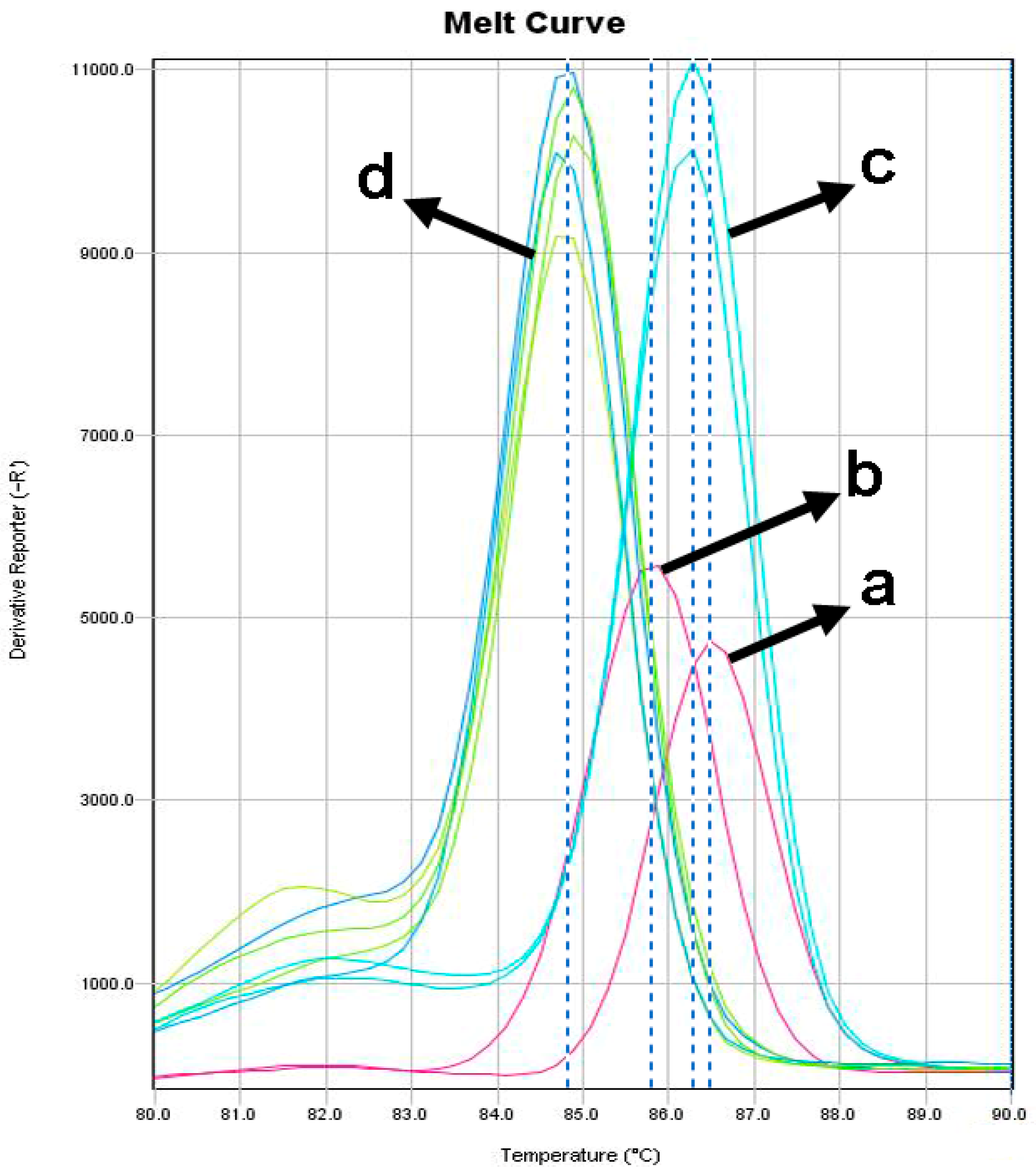
All DNA samples were positive for the bovine 18S rRNA gene, indicating DNA integrity and the absence of PCR inhibitors. Thirty samples tested positive for the presence of Trypanosoma DNA, with six samples showing melting temperatures matching those of T. vivax, while the remaining twenty-four formed a distinct group not matching the melting temperatures of either T. vivax or T. evansi. Among the T. vivax-matched samples, the difference in melting temperature compared to the T. vivax internal control in each PCR reaction was 0.12 °C ± 0.05 (SEM). The distinct group of twenty-four samples exhibited a melting temperature difference of 1.43 °C ± 0.09 compared to the T. vivax internal control (Figure 2).
Ten samples showing clear bands on agarose gels and representing both groups were purified and sequenced. The four samples that had melting temperatures matching T. vivax were 100% identical to each other and to the 18S rRNA sequence of T. vivax (KM391829). The remaining six sequenced samples, which did not match the melting temperatures of either the T. vivax or T. evansi controls, were 100% identical to each other and 99.36–100% identical to the corresponding sequences of several members of the T. theileri group (OM256612, PP945814).
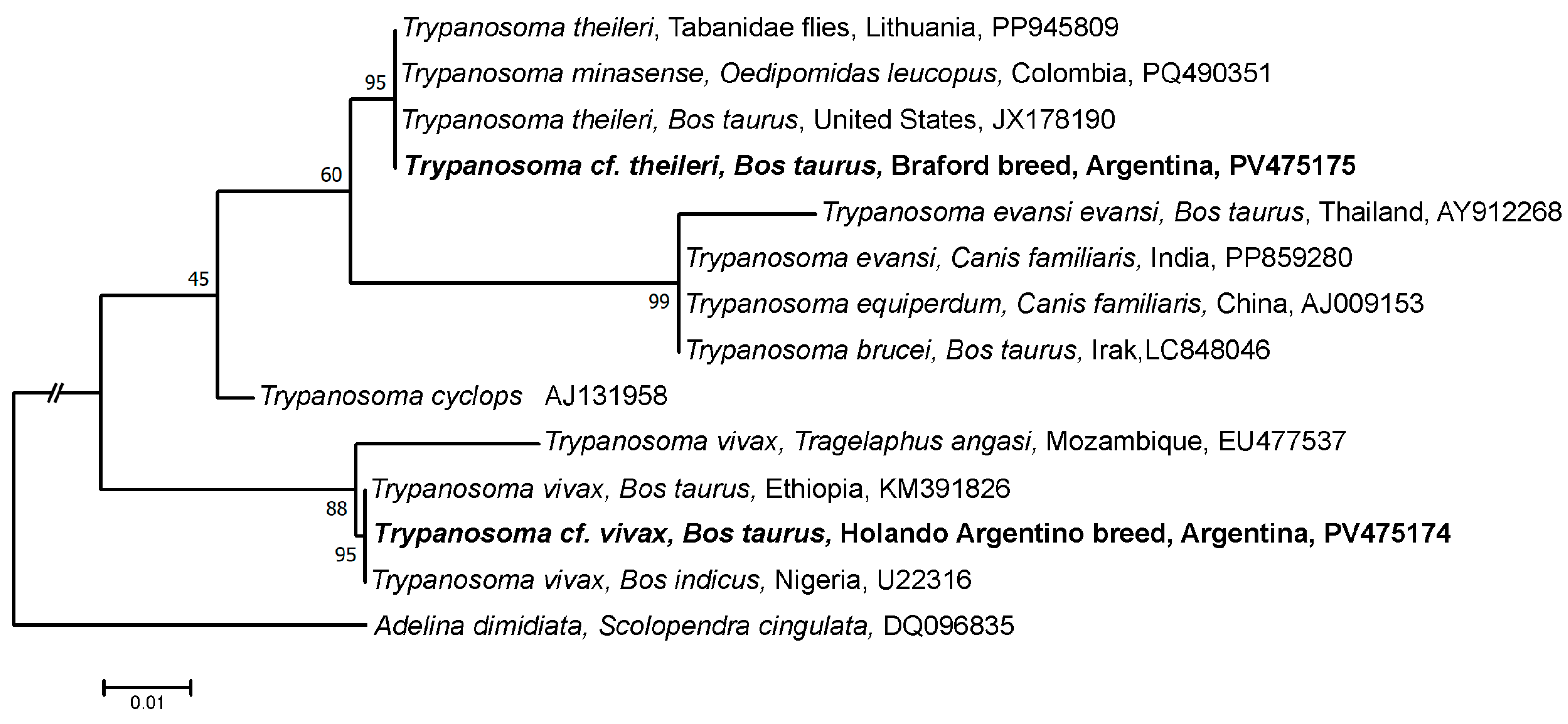
The phylogenetic analysis using 18S rRNA sequences placed the detected Trypanosoma into two distinct clades: the T. vivax sequence clustered with other T. vivax sequences, while the second sequence was grouped within the T. theileri clade (Figure 3), confirming their identity.
The overall prevalence of Trypanosoma in our study was 13.63% (30/220), with T. theileri being more prevalent than T. vivax (Table 1). Prevalence varied by location and production type, with T. theileri being significantly more common in beef cattle and in the GD region, while T. vivax was detected only in dairy herds (Table 1). A full breakdown of prevalence by site and cattle type is provided in Table 1, and the corresponding statistical analyses are summarized in Table 2.
Statistical comparisons revealed a significant variation in Trypanosoma spp. prevalence by region and cattle type, with T. theileri being more common in beef cattle and in the GD region, and T. vivax being detected only in dairy cattle, without significant regional differences (Table 2).
Thirty-seven blood smears, including one from a bovine PCR-positive for T. theileri, were examined in their entirety at 1000× magnification. The presence of trypomastigotes was not detected in any of the samples observed.

4. Discussion

This study provides new insights into the epidemiology of the Trypanosoma species in cattle raised in two ecologically distinct regions of Santa Fe province, Argentina. The detection of T. vivax and T. theileri highlights key differences in their distribution, likely shaped by environmental conditions, vector ecology, and host–parasite interactions.
Previous studies have reported T. vivax in both dairy and beef cattle from northern Argentina [13]. In contrast, our results confirm its presence only in dairy cattle from the dairy basin region but not in beef cattle from the riparian zone. This pattern supports earlier observations that T. vivax outbreaks are more commonly associated with dairy production systems [5,12,30]. Notably, the absence of T. vivax in beef cattle from the riparian area contrasts with the high prevalence of T. theileri, suggesting that ecological factors may differentially influence their distribution. However, potential confounding factors (e.g., management practices, herd size) could not be fully accounted for in the analysis and should be considered when interpreting these findings.
The riparian environment of GD, characterized by wetlands, non-flowing water, and high humidity, provides favorable conditions for the proliferation of hematophagous Diptera, such as Aedes mosquitoes, phlebotomine sandflies, and tabanids. These conditions differ markedly from the more temperate, drier pasturelands of CD and LCD, where cattle are raised on managed fields with lower vector densities. This ecological distinction is significant, as the development of T. theileri trypanosomes has been reported in tabanids, Aedes mosquitoes, and phlebotomine sandflies [17,31], which are competent vectors of T. theileri. Conversely, T. vivax transmission in South America is primarily attributed to mechanical vectors such as tabanids or through iatrogenic means [9]. In line with this ecological context, phlebotomine sandflies are notably more abundant in the Delta and Islands of Paraná ecoregion (where GD is located) than in the Espinal ecoregion (which includes CD and LCD) [32]. Additionally, studies from a neighboring region in Uruguay indicate that tabanids are significantly more prevalent in riparian lowland environments compared to open field habitats—a pattern that is consistent with the ecological conditions of GD [33].
In this context, the higher T. theileri prevalence observed in the riparian region, alongside the lower T. vivax seroprevalence reported in the same area [4], suggests that the abundance and composition of vector species could potentially influence transmission dynamics. Additionally, the possibility of immune responses induced by repetitive T. theileri infections offering cross-protection against T. vivax, as observed in other host–pathogen systems [34,35], cannot be excluded. This potential immunological interaction may contribute to the absence of T. vivax in beef cattle from the riparian zone, although further studies are required to confirm such effects under natural conditions.
The detection of T. vivax in this study aligns with the broader trend of geographic expansion described by Florentin et al. [13], who reported its southward spread from the Gran Chaco into the Pampas. In endemic areas, infections are typically cryptic and asymptomatic; however, in newly affected dairy regions, T. vivax has caused acute outbreaks with significant economic consequences, including decreased milk production, reproductive failures, and increased treatment costs [12].
Although T. theileri is traditionally considered non-pathogenic in cattle, evidence suggests it can act opportunistically under stress or during co-infection, potentially contributing to subclinical disease and impaired productivity [22,23,24,36,37,38]. In our study, no trypanosome-like structures were observed in the blood smears, including samples that tested positive for T. theileri by PCR. However, low Ct values in the Trypanosome-positive samples indicated the presence of T. theileri DNA circulating in the blood. This suggests that T. theileri infection intensity might have been too low for detection by microscopy or that the infective forms may have been localized in other tissues such as the bone marrow, testis, brain [22,23], or body fluids like peritoneal and cerebrospinal fluid [22,38] rather than in peripheral blood.
Overall, our findings emphasize the complex and regionally variable epidemiology of bovine trypanosomiasis under extensive and semi-intensive management systems. Differences in T. vivax and T. theileri prevalence between the two regions underscore the importance of vector ecology, environmental suitability, and possible immunological interactions. Moreover, the greater frequency of veterinary interventions in semi-intensive dairy systems (e.g., reproductive hormones are injected on a regular basis to synchronize estrus and maintain reproductive efficiency in lactating cows) compared to the limited handling practices in riparian beef cattle systems (i.e., cattle are gathered for veterinary procedures approximately four times per year), suggests that iatrogenic transmission is a plausible route of T. vivax infection that should be taken into consideration [8,10]. Future research should aim to clarify the interactions between T. theileri and T. vivax, assess the clinical impact of T. theileri on cattle health and productivity, and evaluate the role of local vectors in disease transmission. Continued molecular surveillance and preventive strategies will be essential to mitigate the expanding impact of T. vivax in Argentine livestock production.

Author Contributions

Conceptualization, P.M.B. and L.D.M.; methodology, M.C.F.F., J.B., M.A. and J.H.G.; formal analysis, M.C.F.F., P.M.B. and L.D.M.; investigation, M.C.F.F., J.B. and L.D.M.; resources, M.A., J.H.G., P.M.B. and L.D.M.; data curation, M.C.F.F., P.M.B. and L.D.M.; writing—original draft preparation, M.C.F.F. and L.D.M.; writing—review and editing, P.M.B. and L.D.M.; project administration, L.D.M.; funding acquisition, P.M.B. and L.D.M. All authors have read and agreed to the published version of the manuscript.

Funding

This research was funded by the Santa Fe Province Government, grant number IO-2019-00069.

Institutional Review Board Statement

This animal study protocol was approved by the Ethics Committee of Facultad de Ciencias Veterinarias, Universidad Nacional del Litoral (file FCV-1010135-19, protocol 536/19, 28 November 2019).

Informed Consent Statement

Not applicable.

Data Availability Statement

The sequence data presented in this study are deposited in GenBank with the accession numbers PV475174 and PV475175.

Acknowledgments

We thank Med. Vet. Jose Maciel for providing part of the samples used in this study.

Conflicts of Interest

The authors declare no conflicts of interest. The funders had no role in the design of the study, in the collection, analysis, or interpretation of data, in the writing of the manuscript, or in the decision to publish the results.

Abbreviations

The following abbreviations are used in this manuscript:
CDCastellanos department
LCDLas Colonias department
GDGaray department
PCRPolymerase chain reaction
DNADeoxyribonucleic acid
rRNARibosomal ribonucleic acid

References

  1. Magez, S.; Radwanska, M. Trypanosomes and Trypanosomiasis; Springer: Vienna, Austria, 2014; ISBN 978-3-7091-1555-8. [Google Scholar]
  2. Muhanguzi, D.; Mugenyi, A.; Bigirwa, G.; Kamusiime, M.; Kitibwa, A.; Akurut, G.G.; Ochwo, S.; Amanyire, W.; Okech, S.G.; Hattendorf, J.; et al. African animal Trypanosomiasis as a constraint to livestock health and production in Karamoja region: A detailed qualitative and quantitative assessment. BMC Vet. Res. 2017, 13, 355. [Google Scholar] [CrossRef] [PubMed]
  3. Hounyèmè, R.E.; Kaboré, J.; Gimonneau, G.; Somda, M.B.; Salou, E.; Missihoun, A.A.; Bengaly, Z.; Jamonneau, V.; Boulangé, A. Molecular epidemiology of animal African Trypanosomosis in southwest Burkina Faso. PLoS Negl. Trop. Dis. 2022, 16, e0010106. [Google Scholar] [CrossRef]
  4. Bontempi, I.A.; Arias, D.G.; Castro, G.V.; Peverengo, L.M.; Díaz, G.F.; Allassia, M.; Greif, G.; Marcipar, I. Improved serodiagnosis of Trypanosoma vivax infections in cattle reveals high infection rates in the livestock regions of Argentina. PLoS Negl. Trop. Dis. 2024, 18, e0012020. [Google Scholar] [CrossRef]
  5. Cadioli, F.A.; Barnabé, P.D.A.; Machado, R.Z.; Teixeira, M.C.A.; André, M.R.; Sampaio, P.H.; Fidélis Junior, O.L.; Teixeira, M.M.G.; Marques, L.C. First report of Trypanosoma vivax outbreak in dairy cattle in São Paulo state, Brazil. Rev. Bras. Parasitol. Vet. 2012, 21, 118–124. [Google Scholar] [CrossRef] [PubMed]
  6. Fritzen, A.; De Vitt, M.G.; Deolindo, G.L.; Signor, M.H.; Correa, N.G.; Ribeiro, B.G.; Marques, J.; Das Neves, G.B.; Miletti, L.C.; Da Silva, A.S. Outbreak of Trypanosoma vivax in dairy cows: Hematologic, immunological and antioxidant responses before and after treatment with isometamidium chloride. Pathogens 2025, 14, 143. [Google Scholar] [CrossRef]
  7. Desquesnes, M. Livestock Trypanosomoses and Their Vectors in Latin America; OIE: Paris, France, 2004; ISBN 978-92-9044-634-7. [Google Scholar]
  8. USDA. Dairy Heifer Raiser Health and Management Practices on U.S. Dairy Operations, 2014. Available online: https://www.aphis.usda.gov/sites/default/files/dairy14_dr_partiii.pdf (accessed on 27 March 2025).
  9. De Melo Junior, R.D.; Azeredo Bastos, T.S.; Heller, L.M.; Couto, L.F.M.; Zapa, D.M.B.; De Assis Cavalcante, A.S.; Cruvinel, L.B.; Nicaretta, J.E.; Iuasse, H.V.; Ferreira, L.L.; et al. How many cattle can be infected by Trypanosoma vivax by reusing the same needle and syringe, and what is the viability time of this protozoan in injectable veterinary products? Parasitology 2022, 149, 270–282. [Google Scholar] [CrossRef]
  10. Bastos, T.S.A.; Faria, A.M.; Madrid, D.M.D.C.; Bessa, L.C.D.; Linhares, G.F.C.; Fidelis Junior, O.L.; Sampaio, P.H.; Cruz, B.C.; Cruvinel, L.B.; Nicaretta, J.E.; et al. First outbreak and subsequent cases of Trypanosoma vivax in the state of Goiás, Brazil. Rev. Bras. Parasitol. Vet. 2017, 26, 366–371. [Google Scholar] [CrossRef] [PubMed]
  11. Monzón, C.M.; Mancebo, O.A.; Giménez, J.M.; Russo, A.M. Evolución de la Trypanosomosis bovina por Trypanosoma vivax en Formosa (Argentina). Años 2007-2012 y su potencial dispersión en el país. Rev. Ibero-Latinoam. de Parasitol. 2013, 72, 38–44. [Google Scholar]
  12. Abdala, A.A.; Larriestra, A.J.; Signorini, M. Estimación de pérdidas económicas causadas por Trypanosoma vivax en un rodeo lechero de Argentina. Rev. Vet. 2020, 31, 115. [Google Scholar] [CrossRef]
  13. Florentin, A.S.; Garcia Perez, H.A.; Rodrigues, C.M.F.; Dubois, E.F.; Monzón, C.M.; Teixeira, M.M.G. Molecular epidemiological insights into Trypanosoma vivax in Argentina: From the endemic Gran Chaco to outbreaks in the Pampas. Transbounding Emerg. Dis. 2022, 69, 1364–1374. [Google Scholar] [CrossRef]
  14. Garcia, H.A.; Blanco, P.A.; Rodrigues, A.C.; Rodrigues, C.M.F.; Takata, C.S.A.; Campaner, M.; Camargo, E.P.; Teixeira, M.M.G. Pan-American Trypanosoma (Megatrypanum) trinaperronei n. sp. in the white-tailed deer Odocoileus virginianus Zimmermann and its deer ked Lipoptena mazamae Rondani, 1878: Morphological, developmental and phylogeographical characterisation. Parasites Vectors 2020, 13, 308. [Google Scholar] [CrossRef]
  15. Rodrigues, A.C.; Campaner, M.; Takata, C.S.A.; Dell’ Porto, A.; Milder, R.V.; Takeda, G.F.; Teixeira, M.M.G. Brazilian isolates of Trypanosoma (Megatrypanum) theileri: Diagnosis and differentiation of isolates from cattle and water buffalo based on biological characteristics and randomly amplified DNA sequences. Vet. Parasitol. 2003, 116, 185–207. [Google Scholar] [CrossRef]
  16. Kostygov, A.Y.; Frolov, A.O.; Malysheva, M.N.; Ganyukova, A.I.; Drachko, D.; Yurchenko, V.; Agasoi, V.V. Development of two species of the Trypanosoma theileri complex in Tabanids. Parasites Vectors 2022, 15, 95. [Google Scholar] [CrossRef]
  17. Brotánková, A.; Fialová, M.; Čepička, I.; Brzoňová, J.; Svobodová, M. Trypanosomes of the Trypanosoma theileri group: Phylogeny and new potential vectors. Microorganisms 2022, 10, 294. [Google Scholar] [CrossRef]
  18. Calzolari, M.; Rugna, G.; Clementi, E.; Carra, E.; Pinna, M.; Bergamini, F.; Fabbi, M.; Dottori, M.; Sacchi, L.; Votýpka, J. Isolation of a trypanosome related to Trypanosoma theileri (Kinetoplastea: Trypanosomatidae) from Phlebotomus perfiliewi (Diptera: Psychodidae). BioMed Res. Int. 2018, 2018, 2597074. [Google Scholar] [CrossRef]
  19. Votýpka, J.; Rádrová, J.; Skalický, T.; Jirků, M.; Jirsová, D.; Mihalca, A.D.; D’Amico, G.; Petrželková, K.J.; Modrý, D.; Lukeš, J. A tsetse and Tabanid fly survey of African great apes habitats reveals the presence of a novel trypanosome lineage but the absence of Trypanosoma brucei. Int. J. Parasitol. 2015, 45, 741–748. [Google Scholar] [CrossRef]
  20. Latif, A.A.; Bakheit, M.A.; Mohamed, A.E.E.; Zweygarth, E. High infection rates of the tick Hyalomma anatolicum anatolicum with Trypanosoma theileri. Onderstepoort J. Vet. Res. 2004, 71, 251–256. [Google Scholar] [CrossRef]
  21. Martins, J.R.; Leite, R.C.; Doyle, R.L. Tripanosomatides like Trypanosoma theileri in the cattle tick Boophilus microplus. Rev. Bras. Parasitol. Vet. 2008, 17, 113–114. [Google Scholar] [CrossRef]
  22. Braun, U.; Rogg, E.; Walser, M.; Nehrbass, D.; Guscetti, F.; Mathis, A.; Deplazes, P. Trypanosoma theileri in the cerebrospinal fluid and brain of a heifer with suppurative meningoencephalitis. Vet. Rec. 2002, 150, 18–19. [Google Scholar] [CrossRef]
  23. Amato, B.; Mira, F.; Di Marco Lo Presti, V.; Guercio, A.; Russotto, L.; Gucciardi, F.; Vitale, M.; Lena, A.; Loria, G.R.; Puleio, R.; et al. A case of bovine trypanosomiasis caused by Trypanosoma theileri in Sicily, Italy. Parasitol. Res. 2019, 118, 2723–2727. [Google Scholar] [CrossRef]
  24. Suganuma, K.; Kayano, M.; Kida, K.; Gröhn, Y.T.; Miura, R.; Ohari, Y.; Mizushima, D.; Inoue, N. Genetic and seasonal variations of Trypanosoma theileri and the association of Trypanosoma theileri infection with dairy cattle productivity in northern Japan. Parasitol. Int. 2022, 86, 102476. [Google Scholar] [CrossRef]
  25. Morello, J.H.; Matteucci, S.D.; Rodriguez, A.F.; Silva, M.E. Ecorregiones y Complejos Ecosistémicos Argentinos, Primera edición; Facultad de Arquitectura, Diseño y Urbanismo, GEPAMA Grupo de Ecología del Paisaje y Medio Ambiente, Universidad de Buenos Aires: Buenos Aires, Argentina, 2012; ISBN 978-987-1922-00-0. [Google Scholar]
  26. Eberhardt, A.T.; Monje, L.D.; Zurvera, D.A.; Beldomenico, P.M. Detection of Trypanosoma evansi infection in wild capybaras from Argentina using smear mcroscopy and real-time PCR assays. Vet. Parasitol. 2014, 202, 226–233. [Google Scholar] [CrossRef]
  27. Monje, L.D.; Costa, F.B.; Colombo, V.C.; Labruna, M.B.; Antoniazzi, L.R.; Gamietea, I.; Nava, S.; Beldomenico, P.M. Dynamics of exposure to Rickettsia parkeri in cattle in the Paraná River Delta, Argentina. J. Med. Entomol. 2016, 53, 660–665. [Google Scholar] [CrossRef]
  28. Keatley, S.; Botero, A.; Fosu-Nyarko, J.; Pallant, L.; Northover, A.; Thompson, R.C.A. Species-level identification of trypanosomes infecting Australian wildlife by high-resolution melting—real time quantitative polymerase chain reaction (HRM-qPCR). Int. J. Parasitol. Parasites Wildl. 2020, 13, 261–268. [Google Scholar] [CrossRef]
  29. Kumar, S.; Stecher, G.; Tamura, K. MEGA7: Molecular evolutionary genetics analysis version 7.0 for bigger datasets. Mol. Biol. Evol. 2016, 33, 1870–1874. [Google Scholar] [CrossRef]
  30. De Souza Pimentel, D.; Do Nascimento Ramos, C.A.; Ramos, R.A.D.N.; De Araújo, F.R.; Borba, M.L.; Da Gloria Faustino, M.A.; Alves, L.C. First report and molecular characterization of Trypanosoma vivax in cattle from state of Pernambuco, Brazil. Vet. Parasitol. 2012, 185, 286–289. [Google Scholar] [CrossRef]
  31. Böse, R.; Friedhoff, K.T.; Olbrich, S. Transmission of Megatrypanum trypanosomes to Cervus dama by Tabanidae. J. Protozool. 1987, 34, 110–113. [Google Scholar] [CrossRef]
  32. Quintana, M.G.; Fernández, M.S.; Salomón, O.D. Distribution and abundance of Phlebotominae, vectors of Leishmaniasis, in Argentina: Spatial and temporal analysis at different scales. J. Trop. Med. 2012, 2012, 652803. [Google Scholar] [CrossRef]
  33. Lucas, M.; Krolow, T.K.; Riet-Correa, F.; Barros, A.T.M.; Krüger, R.F.; Saravia, A.; Miraballes, C. Diversity and seasonality of horse flies (Diptera: Tabanidae) in Uruguay. Sci. Rep. 2020, 10, 401. [Google Scholar] [CrossRef]
  34. Maraghi, S.; Molyneux, D.H. Studies on cross-immunity in Herpetosoma trypanosomes of Microtus, Clethrionomys and Apodemus. Parasitol. Res. 1989, 75, 175–177. [Google Scholar] [CrossRef]
  35. Welburn, S.; Picozzi, K.; Coleman, P.G.; Packer, C. Patterns in age-seroprevalence consistent with acquired immunity against Trypanosoma brucei in Serengeti lions. PLoS Negl. Trop. Dis. 2008, 2, e347. [Google Scholar] [CrossRef] [PubMed]
  36. Doherty, M.L.; Windle, H.; Voorheis, H.P.; Larkin, H.; Casey, M.; Clery, D.; Murray, M. Clinical disease associated with Trypanosoma theileri infection in a calf in Ireland. Vet. Rec. 1993, 132, 653–656. [Google Scholar] [CrossRef]
  37. Hajihassani, A.; Maroufi, S.; Esmaeilnejad, B.; Khorram, H.; Tavassoli, M.; Dalir-Naghadeh, B.; Samiei, A. Hemolytic anemia associated with Trypanosoma theileri in a cow from Kurdistan province, west of Iran. Vet. Res. Forum. 2020, 11, 191–193. [Google Scholar] [CrossRef] [PubMed]
  38. Sood, N.K.; Singla, L.D.; Singh, R.S.; Uppal, S.K. Association of Trypanosoma theileri with peritonitis in a pregnant cross-bred cow: A case report. Vet. Med. 2011, 56, 82–84. [Google Scholar] [CrossRef]
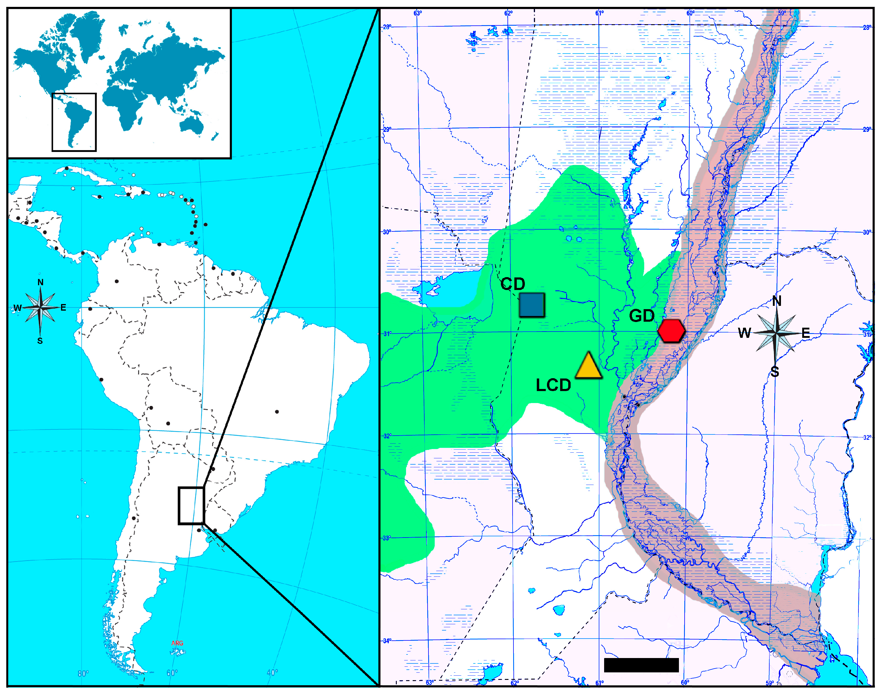
Figure 1. Geographic location of the study regions for the detection of Trypanosoma spp. in cattle. The main map shows the ecoregions, hydrography, and sampling areas within Santa Fe province, Argentina. Sites in the dairy basin (square and triangle) and in the riparian zone (hexagon). CD: Castellanos department; LCD: Las Colonias department; GD: Garay department. Green area: Espinal ecoregion; Brown area: Delta and Islands of Paraná ecoregion. Scale bar: 80 km. Maps were created using datasets and cartographic material from the Instituto Geográfico Nacional (https://www.ign.gob.ar/ (accessed on 27 March 2025)).
Parasitologia 05 00027 g001
Figure 2. Representative melting curve analysis of Trypanosoma PCR-positive bovines. (a) Trypanosoma vivax control, (b) Trypanosoma evansi control, (c) T. vivax-matching positive bovines (n = 6), and (d) Trypanosoma positive bovines (n = 24).
Parasitologia 05 00027 g002
Figure 3. Maximum likelihood tree constructed from 18S rRNA sequences of Trypanosoma species. Best-fitting substitution models using the maximum likelihood model test were determined with the Akaike Information Criterion. The Kimura 2 parameter with a discrete Gamma distribution was selected as the best model. The numbers represent the bootstrap support generated from 1000 replications. The GenBank accession numbers are shown. The sequences from this study are in boldface. The scale bars indicate nucleotide substitutions/sites.
Parasitologia 05 00027 g003
Table 1. Prevalence (mean ± 95% confidence interval) and positive cases (positive animals/total animals) of Trypanosoma spp. by region and production type.
Trypanosoma Prevalence by Breed Trypanosoma Prevalence by SiteTrypanosoma spp. Prevalence
Dairy Breed (Holando Argentino)Beef Breed (Braford)Delta and Islands of ParanáEspinal
Garay DepartmentCastellanos DepartmentLas Colonias Department
Trypanosoma spp.8.27 ± 4.68% (11/133)21.8 ± 8.68% (19/87)8.63 ± 3.71% (19/220)0.91 ± 1.25% (2/220)4.09 ± 2.62% (9/220)13.63 ± 4.53% (30/220)
Trypanosoma vivax4.51 ± 3.53% (6/133)NDND0.91 ± 1.25% (2/220)1.82 ± 1.77% (4/220)2.73 ± 2.15% (6/220)
Trypanosoma theileri3.75 ± 3.23% (5/133)21.8 ± 8.68% (19/87)8.63 ± 3.71% (19/220)ND2.27 ± 1.97% (5/220)10.91 ± 4.12% (24/220)
ND, none detected.
Table 2. Results of Chi-squared and Fisher’s exact tests comparing Trypanosoma prevalence across regions and cattle production types.
Prevalence ComparisonObserved Trendp-Value
Trypanosoma spp. prevalence by siteLCD = CD < GD0.0069 *,a
Trypanosoma theileri prevalence by siteLCD = CD < GD7.29 × 10−5 *,a
Trypanosoma vivax prevalence by siteLCD = CD = GD0.087 b
Trypanosoma spp. prevalence by cattle typeDairy < Beef0.0197 *,b
Trypanosoma theileri prevalence by cattle typeDairy < Beef0.0001 *,b
Trypanosoma vivax prevalence by cattle typeDairy > Beef0.027 *,b
* Statistically significant. a Chi-squared test. b Fisher’s exact test.
Disclaimer/Publisher’s Note: The statements, opinions and data contained in all publications are solely those of the individual author(s) and contributor(s) and not of MDPI and/or the editor(s). MDPI and/or the editor(s) disclaim responsibility for any injury to people or property resulting from any ideas, methods, instructions or products referred to in the content.