Article

Go West: Hirudins and Decorsin/Ornatin-like Platelet Aggregation Inhibitors in Two Representatives of American Hematophagous Leeches

by
Victoria Pfordt
1,†,
Pegah Kalatehjari
1,2,†,
Céline Tolksdorf
3,4,
Bernhard H. Rauch
4 and
Christian Müller
1,*
1
Animal Physiology, Zoological Institute and Museum, University of Greifswald, 17489 Greifswald, Germany
2
Natural Resources Engineering, Department of Fisheries Science, University of Agricultural Sciences and Natural Resources, Gorgan 4913815739, Iran
3
Department of General Pharmacology, Center of Drug Absorption and Transport (C_DAT), University Medicine Greifswald, 17489 Greifswald, Germany
4
Division of Pharmacology and Toxicology, School of Medicine and Health Sciences, Carl von Ossietzky University Oldenburg, 26129 Oldenburg, Germany
*
Author to whom correspondence should be addressed.
These authors contributed equally to this work.
Parasitologia 2022, 2(4), 313-325; https://doi.org/10.3390/parasitologia2040026
Submission received: 20 September 2022 / Revised: 24 October 2022 / Accepted: 27 October 2022 / Published: 1 November 2022

Abstract

:
Hematophagous leeches express a broad variety of secretory factors in their salivary glands; among them are hirudins, inhibitors of blood coagulation, and decorsins/ornatins, inhibitors of platelet aggregation. Here, we describe the identification and molecular and functional characterization of putative hirudins and decorsins/ornatins in two leech species of American origin, Limnobdella mexicana and Haementeria vizottoi. The leech species represent two orders of leeches, the proboscis-bearing Rhynchobdellida and the non-proboscis-bearing Arhynchobdellida. Members of the hirudin superfamily, such as hirudins or decorsins/ornatins, are described for the first time in the genus Haementeria. Both species expressed very potent inhibitors of platelet aggregation, but only the putative hirudins of L. mexicana displayed high thrombin-inhibitory potency, whereas the putative hirudin of H. vizottoi turned out to be a hirudin-like factor. The results of our study provide new insights into the evolutionary background of the blood-sucking lifestyle in leeches.

1. Introduction

“Blood is a juice of very special kind” (Goethe, Faust), and hematophagous animals have to cope with numerous challenges to ensure a successful blood meal [1]. Securing a continuous flow of blood through an open wound and preventing premature coagulation of the ingested blood within the sucking apparatus and/or the gut of the parasite are two sides of the same coin. The inhibition of both platelet aggregation (primary hemostasis) and the blood coagulation cascade (secondary hemostasis) are hence crucial tasks for hematophagic animals [2]. Primary hemostasis refers to platelet aggregation and plug formation, whereas secondary hemostasis refers to the formation and deposition of insoluble fibrin generated by a proteolytic coagulation cascade. Both processes are interlinked by certain factors acting in either mechanism [3].
Leeches are a taxonomic group of invertebrates that comprises both predatory (non-blood-feeding) and parasitic (blood- and hemolymph-feeding) representatives [4]. Hematophagy is very likely the ancestral state and was independently lost (and regained) in several lineages of leeches throughout evolution [5]. Blood-feeding leeches such as Hirudo medicinals, probably the most famous one, comprise a broad variety of bioactive compounds in their saliva, including inhibitors of both primary and secondary hemostasis [6]. The inhibitors of platelet aggregation in the saliva of H. medicinalis are calin and saratin [7,8]. Decorsins are platelet inhibitors in the saliva of Macrobdella decora [9,10,11], and ornatins fulfill this function in Placobdella ornata [12]. The molecular identity of calin remains obscure, but saratin, decorsin variant 1, and ornatin variant E have been successfully purified and functionally characterized [8,9,13]. Strikingly, both M. decora and P. ornata encode several variants, namely five decorsin variants (1–5) and six ornatin variants (A2, A3, and B–E), respectively.
Inhibitors of secondary hemostasis target different steps within the blood coagulation cascade, but leech-derived factors mainly inhibit factor Xa or thrombin (factor IIa). Yagin from H. medicinals [14] and antistasin from the Mexican leech Haementeria officinalis [15] are inhibitors of factor Xa, whereas hirudin from H. medicinalis represents the archetype of leech-derived thrombin-inhibitors [16,17]. Hirudin is composed of three functionally distinct domains: a short N-terminal stretch of five amino acid residues that blocks the active site of thrombin, a central globular domain that is stabilized by the formation of three disulfide bonds, and an elongated C-terminal tail that blocks the fibrinogen-binding site of thrombin (exosite I) [18]. Several putative hirudins have been described in different leech species, but only a few of them have actually been functionally characterized [11,19,20,21,22]. Only recently, a new class of leech-derived factors has been described: the hirudin-like factors (HLFs) [21,23]. HLFs share some characteristic features with hirudins, such as the numbers and positions of the six disulfide bond-forming cysteine residues and common gene structures composed of four exons and three introns, but may considerably differ in others, such as molecular mass and isoelectric points (pI values). Some HLFs exhibit thrombin-inhibitory potency that is equal to that of hirudins, whereas others do not or only very poorly inhibit thrombin [24]. Structure–function analyses have revealed that the interplay of all three domains of hirudins and HLFs is crucial for their thrombin-inhibitory potency [25,26].
Hirudin is a hallmark of hematophagy in leeches, but its presence is actually not restricted to blood feeders. The Asian non-hematophagous leech Whitmania pigra expresses several hirudins and HLFs, and some of them are potent thrombin inhibitors [22]. It may not come as a surprise that W. pigra is widely used as an anti-thrombotic drug in Traditional Chinese Medicine (TCM) [27]. By contrast, the presence of hirudins (or HLFs) has not yet been confirmed in some leech species that are blood-feeders, such as H. officinalis and its close relative Haementeria ghilianii, the giant Amazonas leech, although other inhibitors of secondary hemostasis have been found in close relatives: vizottin, a potent inhibitor of factor Xa [28], and cystatin-Hv, a serine protease inhibitor that acts on cathepsin-L [29], have been identified in Haementeria vizottoi, a leech that is native to southeastern Brazil. Hirudin or ornatin, however, could not be identified in a transcriptome library that was generated from the salivary gland tissue of H. vizottoi [30]. Members of the genus Haementeria belong to the proboscis-bearing leeches (Rhynchobdellida), and so far, the expression of hirudin and ornatins in Rhynchobdellida has only been confirmed in members of the genus Placobdella [5,12].
Limnobdella mexicana, by contrast, is a member of the family of Praobdellidae and hence belongs to the jawed or non-proboscis-bearing leeches (Arhynchobdellida) [31]. It parasitizes mucosal surfaces, such as the noses and oral cavities of mammals. Recently, Iwama et al. [32] described the generation and analysis of a salivary transcriptome of L. mexicana, including the identification of two putative hirudins. Unfortunately, the functional characterization of both factors is still missing. In addition, the authors did not find any evidence for the expression of decorsins or decorsin-like factors in L. mexicana. Decorsins have been found in the North American leech M. decora [9] but not yet in members of Arhynchobdellida outside America.
In the present study, we describe the re-analysis of the transcriptome datasets of H. vizottoi and L. mexicana and the identification of additional putative hirudins and putative decorsins in L. mexicana as well as the identification of an HLF and a putative ornatin in H. vizottoi. All factors were successfully expressed, purified, and functionally characterized using well-established coagulation and platelet aggregation assays. The results of our study shed new light on the evolutionary background of the hirudin superfamily and support the concept of a single origin of blood-feeding in leeches.

2. Results

2.1. Identification of Putative Hirudins/HLFs and Decorsins in L. mexicana

In a previous study, Iwama et al. [32] reported on the identification of two putative hirudins (DN24002_c0_g1_i2 and DN24002_c0_g1_i1) in the salivary gland transcriptome of L. mexicana. Both factors exhibited 87%/90% sequence identity/similarity to each other and about 32%/48% to hirudin variant HV1 of H. medicinalis, respectively. Functional characterization of both putative hirudins, however, is pending. Decorsins were not identified during the course of the study.
We thoroughly re-investigated the freely accessible raw transcriptome dataset of L. mexicana (SRX5688711) and confirmed the predictions of Iwama et al. [32]. For factor DN24002_c0_g1_i2, we identified two slightly different sequence variants (Lmex_HV1a and Lmex HV1b in our nomenclature), and for factor DN24002_c0_g1_i1, we identified the exact same sequence (Lmex_HV2 in our nomenclature). Furthermore, we were able to predict three additional putative hirudin variants, namely Lmex_HV3a, Lmex_HV3b, and Lmex_HV4. A multiple sequence alignment of all putative hirudin variants of L. mexicana is shown in Figure 1A. With the exception of Hmex_HV3a, all variants were about the same size as the hirudin variant HV1 of H. medicinalis. The same was applied for the isoelectric points: all pI values were in the range of about 3.9 to 4.7, typical values for hirudins (Table 1).
Our investigation also revealed the presence of coding sequences for two putative decorsin variants, namely Lmex_DV1 and Lmex_DV2. Both variants exhibited 80%/86% sequence identity/similarity to each other and contained the canonical RGD/KGD motif located between the cysteine residues C5 and C6 [33], but the pI values differed markedly (5.08 vs. 8.30, Table 1). The overall degrees of sequence identity/similarity of both putative decorsin variants to decorsin variant D1 of M. decora were quite low (about 20%/27%, respectively), and identical amino acid residues were virtually limited to the six cysteine residues and the RGD/KGD motif (Figure 1B).
The black background indicates conserved residues; the gray background indicates similar residues. The six conserved cysteine residues giving rise to the three-dimensional structure are marked in bold. The canonical RGD/KGD motif in decorsins and ornatins is marked in bold and red. Abbreviations are used according to the IUPAC code.

2.2. Identification of Putative Hirudins/HLFs and Ornatin in H. vizottoi

The same experimental approach outlined for L. mexicana was also applied to the freely accessible transcriptome dataset of H. vizottoi [30]. The authors of the study did not report on the identification of either hirudin- or ornatin-like sequences. However, our investigation revealed clear evidence that the coding sequences for both types of factors are expressed in the salivary complex of H. vizottoi: a putative HLF (Hviz_HV1) and a putative ornatin (Hviz_OV1). Hviz_HV1 exhibited characteristic features of hirudins (size and pI value, Table 1), but the N-terminus was short, contained only three amino acid residues in front of the first cysteine residue, and lacked the highly conserved Tyr3/Phe3 residue (Figure 1A). The overall degrees of sequence identity/similarity compared with hirudin variant HV1 of H. medicinalis were 27%/41%, respectively. For Hviz_OV1, the corresponding values were 42%/52% and 29%/37% compared with ornatin variants A2 and B of P. ornata. Like all decorsins and ornatins, Hviz_OV1 harbors an RGD motif located between cysteine residues C5 and C6 (Figure 1B).

2.3. Functional Characterization I: Thrombin Inhibition

As mentioned above, the putative hirudin variants of L. mexicana exhibited all characteristic structural and biochemical features of hirudins. By contrast, Hviz_HV1 of H. vizottoi comprises a substantially altered N-terminus that very likely is prohibitive for efficient interaction with the active site of thrombin. We thus constructed four variants of Hviz_HV1, namely Hviz_HV1b–e, that contained N-terminal sequences of H. medicinalis HV1 (Hviz_HV1c), a Placobdella costata hirudin (Hviz_HV1d), and Lmex_HV1 of L. mexicana (Hviz_HV1e). The modified N-terminal sequences are listed in Table 2.
All putative hirudin/HLF variants were successfully expressed, purified, and functionally tested in a thrombin time test that specifically addressed the activity of thrombin. To our disappointment and in sharp contrast to our expectations, none of the five Hviz-HV1 variants displayed any thrombin-inhibitory potency at the tested concentrations, despite the presence of “suitable” N-termini in Hviz_HV1c–e (data not shown).
However, five of the six hirudin variants of L. mexicana were highly potent thrombin inhibitors. In fact, the factors completely inhibited thrombin activity even at the lowest concentration of 0.32 µmol/L, comparable to the inhibitory potency of hirudin variants HV1 and HLF1V of H. medicinalis (Müller et al., 2020a) [24]. Lmex_HV4, by contrast, was only a very weak thrombin inhibitor, even at the high concentration of 3.2 µmol/L. As expected, the putative decorsin Lmex_DV1 did not display any thrombin inhibitory effect (Figure 2).

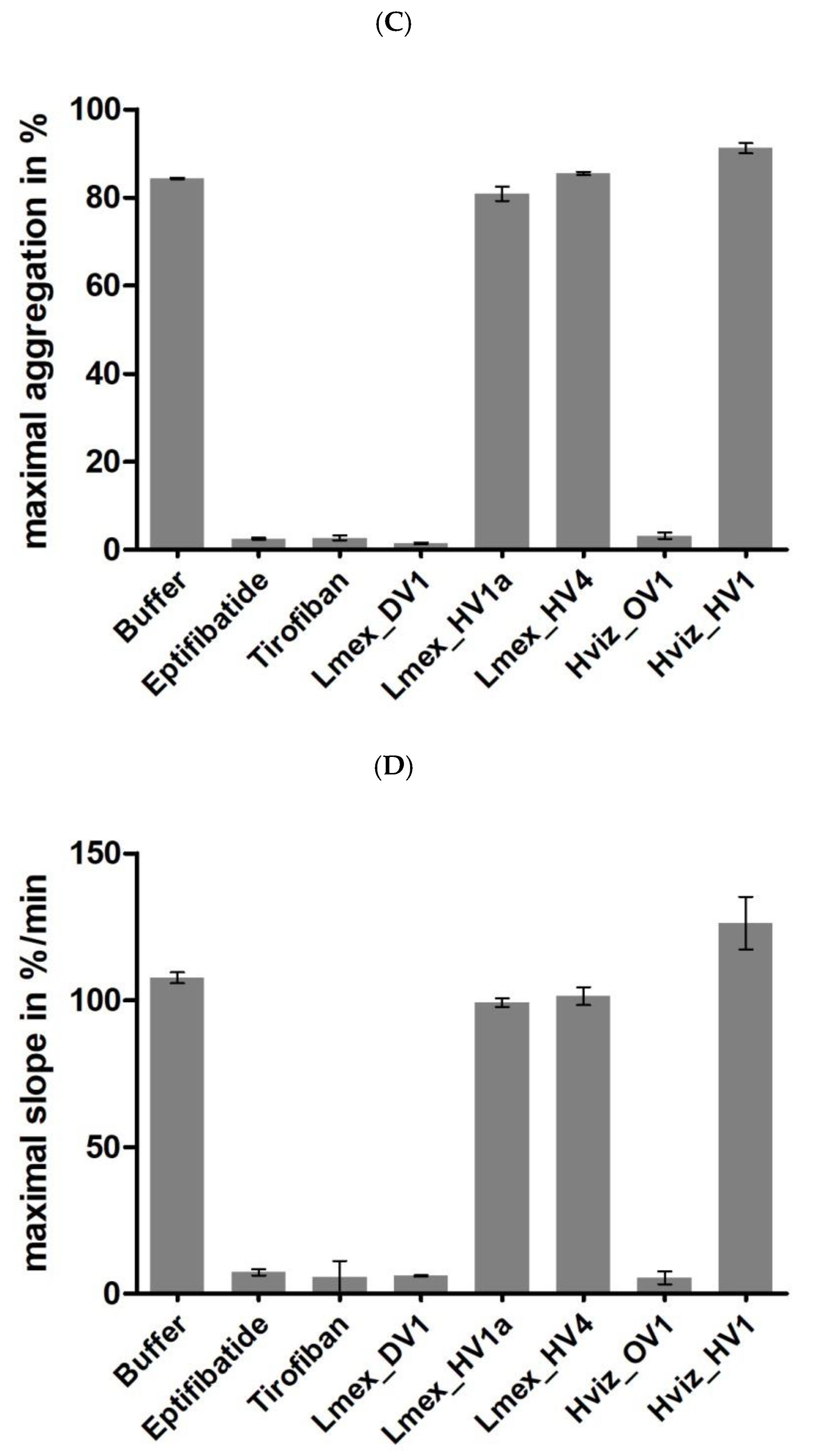
2.4. Functional Characterization II: Platelet Aggregation

Two putative decorsins of L. mexicana (Lmex_DV1 and DV2) and one putative ornatin of H. vizottoi (Hviz_OV1) were identified in the course of the present study. For practical reasons, only Lmex_DV1 and Hviz_OV1 were functionally tested in platelet aggregation assays. The strong inhibitors eptifibatide and tirofiban served as positive controls for the inhibition of platelet aggregation. Lmex_HV1a and Lmex_HV4 served as negative controls (i.e., no inhibition of platelet aggregation) for the measurement of Lmex_DV1 and Hviz_HV1 (see Table 2) served as the negative control for the measurement of Hviz_OV1. The aggregation curves were recorded, and two quantitative readout parameters were determined: maximal aggregation in percentage and maximal slope of the curve in percent per minute. The aggregation curves are shown in Figure 3A,B. Both Lmex_DV1 and Hviz_OV1 inhibited platelet aggregation to an extent that was comparable to eptifibatide and tirofiban in terms of maximal aggregation effect (Figure 3C) and maximal slope of the curve (Figure 3D). By contrast, neither Lmex_HV1a, Lmex_HV4, nor Hviz_HV1 inhibited platelet aggregation.

3. Discussion

The emergence of next- and third-generation sequencing technologies has substantially changed the field of genomic research [34,35]. A steadily increasing number of genome and transcriptome datasets has become available in public databases, providing almost infinite possibilities to conduct all kinds of sequence-based research projects. However, whereas the costs for sequence data generation are constantly falling [36], the costs for sequence data analysis are not; it is still a challenging, time-consuming, and tedious task, despite all the computational help and support.
The current manuscript describes the identification and subsequent functional characterization of several previously undiscovered putative anticoagulants in two American leech species, L. mexicana and H. vizottoi. For the first time, both a hirudin-like factor (Hviz_HV1) and an ornatin (Hviz_OV1) were described in a member of the genus Haementeria. Whereas antistasin and its related factors were identified in H. officinalis [15] and H. ghilianii [37] a long time ago, the presence of members of the hirudin superfamily remained obscure. Natural Hviz_HV1 is not an inhibitor of thrombin; the short N-terminal stretch and the lack of the canonical Tyr3/Phe3 residue can easily explain this phenomenon. However, the central globular domain and/or the C-terminal tail of Hviz_HV1 are obviously not optimal for interaction with thrombin since the replacement of the N-terminus of Hviz_HV1 with respective sequences of very potent thrombin inhibitors (see Table 2) did not convert Hviz_HV1 into a thrombin inhibitor. Previous studies in our lab have revealed that not only the N-terminus and the C-terminal tail but also all three domains, including the central globular domain, are essential for the thrombin-inhibitory potency of putative hirudins [25,26]. The central globular domain of Hviz_HV1 is quite different from those of the hirudin variant HV1 of H. medicinalis and the hirudins of L. mexicana, including an exceptionally long stretch between cysteine residues C1 and C2 (see Figure 1). We hence assume that the structure of the central globular domain of Hviz_HV1 is incompatible with the thrombin-inhibitory potency of the protein. Similar observations were previously made for both HLF3 and HLF4 of H. medicinalis/verbana and HLF6 and HLF7 of H. manillensis [25,26]. The biological target of Hviz_HV1 remains to be determined.
The putative hirudins of L. mexicana, by contrast, displayed substantial thrombin-inhibitory potency, equal to those of HV1 and HLF1V in H. medicinalis. The only exception was Lmex_HV4 (Figure 2). The main reason for the very low but still detectable inhibitory potency of Lmex_HV1 is probably the presence of two asparagine residues at the very end of the N-terminus (see Figure 1). To our knowledge, an Asn1Asn2 mutant of hirudin has never been constructed, but the replacement of Val1Val2 of hirudin variant HV1 of H. medicinalis with amino acid residues with hydrophilic side chains, such as serine, has the potential to dramatically increase the Ki values of respective mutants of hirudin HV1 [38,39]. By contrast, the presence of a positively charged aspartate residue at position 2 or 4 (as in Lmex_HV4, see Figure 1) can be well tolerated [24,37]. The very high thrombin-inhibitory potency of Lmex_HV1-3 is likely due to the presence of a phenylalanine residue at position 3 (Phe3) instead of the canonical tyrosine residue (Tyr3) in each of these factors (see Figure 1). The replacement of Tyr3 with Phe3 in hirudin variant HV1 of H. medicinalis increases its affinity to thrombin approximately sixfold [40].
Altogether, at least six different hirudin/HLF variants are expressed in L. mexicana. The presence of multiple copies and variants of anticoagulants in leeches has been reported several times [21,41,42,43,44] and indicates that the potential expression of multiple variants of effectors in salivary gland cells is a general feature in many leech species. However, whereas the text of the song seems to be the same, the melodies differ: in M. decora, only one hirudin was found, but five different decorsins could be identified [10]; in L. mexicana, the ratio was almost the opposite: six hirudins/HLFs versus two decorsins. A greater diversity of anticoagulants has been linked to greater diversity in putative host species [43]. On the basis of this hypothesis, the host spectrum of H. vizottoi must be narrow: only one ornatin and one HLF have been identified so far. However, it cannot be ruled out that additional variants of both factors are present in H. vizottoi but remained undetected in our investigations due to an inappropriate search strategy.
Both L. mexicana and H. vizottoi express highly competent inhibitors of platelet aggregation, namely Lmex_DV1 and Hviz_OV1 (see Figure 3A,B), and both factors very likely belong to the hirudin superfamily [11]. So far, the presence of decorsins/ornatins has only been confirmed in representatives of American leech species: M. decora [9], P. ornata [12], L. mexicana, and H. vizottoi (this study). The only exception is the European freshwater turtle leech Placobdella costata (Müller, unpublished data), but the genus Placobdella is of North American origin, and the presence of P. costata in the Palearctic is very likely due to a single dispersal event [45].
The absence of decorsins/ornatins in leech species outside America raises questions about the evolutionary background of this phenomenon. The genera Macrobdella and Hirudo belong to different families of the suborder Hirudiniformes within the order Arhynchobdellida [46,47], whereas Haementeria and Placobdella belong to the family Glossiphoniidae within the order Rhynchobdellida. The split between both orders of leeches chronologically precedes the split between the families [48]. Consequently, the presence of decorsins/ornatins as a trait must have been either lost (assuming a last common ancestor of both leech orders expressing this trait) or gained (assuming a last common ancestor without this trait) independently within the different lineages of leeches during evolution. The question of whether the first or the second option is correct cannot be definitively answered yet. It would certainly be helpful, in this context, to determine the gene sequences and evaluate the gene structures of both Lmex_DV1 and Hviz_OV1 to compare them with the corresponding gene structures of hirudins and the decorsins of M. decora [11].

4. Materials and Methods

4.1. Transcriptome Data

Transcriptome data of Limnobdella mexicana were obtained from GenBank sequence read archive SRX5688711, BioProject PRJNA532931 [32]. Transcriptome data of Haementeria vizottoi were obtained from GenBank sequence read archive SRX960561, BioProject PRJNA276902 [30].

4.2. Bioinformatics Tools

Basic Local Alignment Search Tool (BLAST) searches were performed using the respective NCBI web portal and modified settings for search algorithm parameters.
Multiple sequence alignments were generated using the CLC Sequence Viewer software package v8.0 (CLC bio) and default settings. Alignments were exported as msf files and further processed using GeneDoc v2.7 [49]. Signal peptide sequences were predicted using the Phobius web server [50] and SignalP6.0 [51].

4.3. Gene Synthesis

cDNA fragments of putative hirudin, decorsin/ornatin, and HLF variants were generated using the gene synthesis service of Synbio Technologies (Monmouth Junction, NJ, USA). Modifications were generated by PCR using appropriate primers to incorporate changes in the nucleotide sequences that led to the desired alterations in the amino acid sequences.

4.4. Amplification and Cloning of Hirudin, Decorsin/Ornatin, and HLF cDNAs

For the amplification of putative hirudin, decorsin/ornatin, and HLF cDNAs, primers were derived from the respective transcriptome database sequences. A list of all primers that were used in the study is provided in the Supplementary Material (Table S1). PCR reactions were performed using Q5 polymerase (New England Biolabs, Frankfurt a. M., Germany); fragments of relevant sizes were purified and cloned, and their sequences were determined.

4.5. Expression, Purification, Processing, and Quantification of Putative Hirudins and Decorsins

The entire protocol to clone cDNAs encoding putative hirudins/HLFs and decorsins/ornatins as well as to express, purify, and quantify the respective proteins has been previously described in great detail [21,24,25]. Briefly, we applied a system developed by Qiagen (Hilden, Germany). The pQE30Xa vector encodes a factor Xa protease recognition site between the His-tag coding region on the 5′ side and the multiple cloning site on the 3′ side. Factor Xa protease treatment cleaved off the His-tag and resulted in a recombinant protein that was free of any vector-derived amino acids at the N-terminus. The molar concentrations of the final protein solutions were calculated by dividing the absorbance at 280 nm by the molar absorption coefficient according to the equation ε = (nW × 5500) + (nY × 1490) + (nC × 125) [52,53].

4.6. Blood Coagulation Assays

To verify the thrombin-inhibitory potency of putative hirudins/HLFs, we performed the thrombin time test (TT; reference range 16.8–21.4 s) using a BFT II analyzer (Siemens Healthcare, Erlangen, Germany). All steps were carried out according to the instructions outlined by the manufacturer. For the coagulation tests, protein samples were diluted with buffer to reach final concentrations in the reaction assays of 3.2 µmol/L and 0.32 µmol/L. The desired amount of substrate was directly transferred into the cuvette immediately before the plasma was added. Dade® Ci-Trol® 1 (Siemens Healthcare, Erlangen, Germany) was used as standardized human plasma. The incubation of the reaction mixtures was carried out at 37.4 °C. Measurements that exceeded 300 s were stopped and considered complete inhibition of clot formation.

4.7. Platelet Aggregation Assays

All assays were performed with human blood samples that were obtained from a healthy volunteer (CM) after written informed consent and approval from the institutional ethics committee were obtained. The blood collection, blood preparation, and subsequent experiment followed the procedure as described in Lukas et al. (2019) [23] with few modifications. Briefly, 10 mL of venous blood was taken from the antecubital vein using an S-Monovette® (Sarstedt, Nürnbrecht, Germany) prefilled with citrate buffer. The first centrifugation step of the blood collection tube was performed at 200× g for 20 min. After centrifugation, the supernatant (platelet-rich plasma, PRP) was transferred, and the remaining blood was centrifuged again for 10 min at 2000× g. The supernatant was dedicated as platelet-poor plasma (PPP), transferred, and used as a reference value for maximal platelet aggregation. Measurements were performed using an APACT-4004 (LABiTec, Ahrensburg, Germany). The potent platelet aggregation inhibitors tirofiban and eptifibatide (Sigma-Aldrich, Taufkirchen, Germany) were used as controls. A volume of 170 μL PRP was transferred into cuvettes, and 10 μL of the respective test and control compounds (64 μmol/L) or buffer was added. The cuvettes were transferred into an APACT-4004 measuring cell, and the measurement was started. After 1 min, 20 μL of an adenosine-diphosphate (ADP) dilution (200 μmol/L; Hart Biologicals, Hartlepool, UK) was added as an agonist for a final ADP concentration of 5 µmol/L and a final sample concentration of 3.2 μmol/L. All experiments were performed at 37 °C over a time period of 400 s. Maximal aggregation in percentage and maximal slope of the curve in percentage per minute were calculated as quantitative output parameters [54].

5. Conclusions

Our work, for the first time, presents strong evidence for the presence and expression of members of the hirudin superfamily, both an HLF and an ornatin, within the genus Haementeria. In addition, we determined that the praobdellid leech L. mexicana expresses a broad variety of hirudins, HLFs, and decorsins. Both observations expand the current knowledge on the distribution of the members of the hirudin superfamily among different leech orders, families, and genera and may help to better understand the evolutionary processes that underlie the gains and losses of hematophagous lifestyles among members of this fascinating group of animals.

Supplementary Materials

The following supporting information can be downloaded at: https://www.mdpi.com/article/10.3390/parasitologia2040026/s1, Table S1: List of primers that were used in the study.

Author Contributions

C.M. conceived the ideas, designed the methodology, and performed the sequence-based investigations; V.P., P.K. and C.T. performed the experiments; C.M., V.P., P.K. and C.T. analyzed the data and drafted the manuscript; C.M. and B.H.R. supervised the experimental work. All authors have read and agreed to the published version of the manuscript.

Funding

P.K. received a stipend from Iran’s Ministry of Science, Research, and Technology to support a research stay in Germany.

Institutional Review Board Statement

The study was conducted in accordance with the Declaration of Helsinki, and approved by the Institutional Review Board (or Ethics Committee) of the University Medicine Greifswald (protocol code BB 20/12; 28 February 2012).

Informed Consent Statement

Written informed consent was obtained from all subjects involved in the study.

Data Availability Statement

Original data are deposited at BioStudies (EMBL-EBI) (https://www.ebi.ac.uk/biostudies/) under the accession number S-BSST927.

Acknowledgments

The authors are thankful to the colleagues that generated and provided the transcriptome datasets explored in the present study. In addition, we would like to thank all members of the Animal Physiology Working Group at the University of Greifswald for their help and support. A special thank goes to (em) Jan-Peter Hildebrandt and Undine Lauf for their critical reading of the manuscript.

Conflicts of Interest

The authors declare that they have no conflict of interest.

References

  1. Boulton, R.A. Hematophagy. In Encyclopedia of Animal Cognition and Behavior; Vonk, J., Shackelford, T.K., Eds.; Springer International Publishing: Cham, Switzerland, 2018; pp. 1–5. [Google Scholar] [CrossRef]
  2. Urata, J.; Shojo, H.; Kaneko, Y. Inhibition mechanisms of hematophagous invertebrate compounds acting on the host blood coagulation and platelet aggregation pathways. Biochimie 2003, 85, 493–500. [Google Scholar] [CrossRef]
  3. Gale, A.J. Continuing education course #2: Current understanding of hemostasis. Toxicol. Pathol. 2011, 39, 273–280. [Google Scholar] [CrossRef] [PubMed]
  4. Kvist, S.; Brugler, M.R.; Goh, T.G.; Giribet, G.; Siddall, M.E. Pyrosequencing the salivary transcriptome of Haemadipsa interrupta (Annelida: Clitellata: Haemadipsidae) Anticoagulant diversity and insight into the evolution of anticoagulation capabilities in leeches. Invertebr. Biol. 2014, 133, 74–98. [Google Scholar] [CrossRef]
  5. Siddall, M.E.; Brugler, M.R.; Kvist, S. Comparative transcriptomic analyses of three species of Placobdella (Rhynchobdellida: Glossiphoniidae) confirms a single origin of blood feeding in leeches. J. Parasitol. 2016, 102, 143–150. [Google Scholar] [CrossRef] [PubMed]
  6. Hildebrandt, J.-P.; Lemke, S. Small bite, large impact–saliva and salivary molecules in the medicinal leech. Hirudo medicinalis. Naturwissenschaften 2011, 98, 995–1008. [Google Scholar] [CrossRef]
  7. Munro, R.; Jones, C.P.; Sawyer, R.T. Calin-a platelet adhesion inhibitor from the saliva of the medicinal leech. Blood Coagul. Fibrinolysis 1991, 2, 179–184. [Google Scholar] [CrossRef]
  8. Barnes, C.S.; Krafft, B.; Frech, M.; Hofmann, U.R.; Papendieck, A.; Dahlems, U.; Gellissen, G.; Hoylaerts, M.F. Production and characterization of saratin, an inhibitor of von Willebrand factor-dependent platelet adhesion to collagen. Semin. Thromb. Hemost. 2001, 27, 337–348. [Google Scholar] [CrossRef]
  9. Seymour, J.L.; Henzel, W.J.; Nevins, B.; Stults, J.T.; Lazarus, R.A. Decorsin. A potent glycoprotein IIb-IIIa antagonist and platelet aggregation inhibitor from the leech Macrobdella decora. J. Biol. Chem. 1990, 265, 10143–10147. [Google Scholar] [CrossRef]
  10. Min, G.-S.; Sarkar, I.N.; Siddall, M.E. Salivary transcriptome of the North American medicinal leech, Macrobdella decora. J. Parasitol. 2010, 96, 1211–1221. [Google Scholar] [CrossRef]
  11. Müller, C.; Lukas, P.; Lemke, S.; Hildebrandt, J.-P. Hirudin and decorsins of the North American medicinal leech Macrobdella decora: Gene structure reveals homology to hirudins and hirudin-like factors of Eurasian medicinal leeches. J. Parasitol. 2019, 105, 423–431. [Google Scholar] [CrossRef]
  12. Mazur, P.; Henzel, W.J.; Seymour, J.L.; Lazarus, R.A. Ornatins: Potent glycoprotein IIb-IIIa antagonists and platelet aggregation inhibitors from the leech Placobdella ornata. Eur. J. Biochem. 1991, 202, 1073–1082. [Google Scholar] [CrossRef] [PubMed]
  13. Mazur, P.; Dennis, M.S.; Seymour, J.L.; Lazarus, R.A. Expression, purification, and characterization of recombinant ornatin E, a potent glycoprotein IIb-IIIa antagonist. Protein Expr. Purif. 1993, 4, 282–289. [Google Scholar] [CrossRef] [PubMed]
  14. Werber, M.M.; Zeelon, E.; Levanon, A.; Goldberg, M.; Greenstein, L.A.; Haim, B.; Ashkenazi, S.; Gelbhart, H.; Rigbi, M.; Ezov, N.; et al. Yagin, a leech derived FXa inhibitor, expressed in and recovered from E. coli: Anti-thrombotic potency in vitro and in vivo. Thromb. Haemost. 1995, 73, 1312. [Google Scholar]
  15. Tuszynski, G.P.; Gasic, T.B.; Gasic, G.J. Isolation and characterization of antistasin. An inhibitor of metastasis and coagulation. J. Biol. Chem. 1987, 262, 9718–9723. [Google Scholar] [CrossRef]
  16. Markwardt, F. Past, present and future of hirudin. Haemostasis 1991, 21 (Suppl. 1), 11–26. [Google Scholar] [CrossRef]
  17. Montinari, M.R.; Minelli, S. From ancient leech to direct thrombin inhibitors and beyond: New from old. Biomed. Pharmacother. 2022, 149, 112878. [Google Scholar] [CrossRef]
  18. Warkentin, T.E. Bivalent direct thrombin inhibitors: Hirudin and bivalirudin. Best Pract. Res. Clin. Haematol. 2004, 17, 105–125. [Google Scholar] [CrossRef]
  19. Electricwala, A.; Sawyer, R.T.; Jones, C.P.; Atkinson, T. Isolation of thrombin inhibitor from the leech Hirudinaria manillensis. Blood Coagul. Fibrinolysis 1991, 2, 83–89. [Google Scholar] [CrossRef]
  20. Steiner, V.; Knecht, R.; Börnsen, K.O.; Gassmann, E.; Stone, S.R.; Raschdorf, F.; Schlaeppi, J.M.; Maschler, R. Primary structure and function of novel O-glycosylated hirudins from the leech Hirudinaria manillensis. Biochemistry 1992, 31, 2294–2298. [Google Scholar] [CrossRef]
  21. Müller, C.; Mescke, K.; Liebig, S.; Mahfoud, H.; Lemke, S.; Hildebrandt, J.-P. More than just one: Multiplicity of hirudins and hirudin-like factors in the medicinal leech, Hirudo medicinalis. Mol. Genet. Genom. 2016, 291, 227–240. [Google Scholar] [CrossRef]
  22. Müller, C.; Wang, Z.; Hamann, M.; Sponholz, D.; Hildebrandt, J.-P. Life without blood: Molecular and functional analysis of hirudins and hirudin-like factors of the Asian non-hematophagous leech Whitmania pigra. J. Thromb. Haemost. 2022, 20, 1808–1817. [Google Scholar] [CrossRef] [PubMed]
  23. Lukas, P.; Wolf, R.; Rauch, B.H.; Hildebrandt, J.-P.; Müller, C. Hirudins of the Asian medicinal leech, Hirudinaria manillensis: Same same, but different. Parasitol. Res. 2019, 118, 2223–2233. [Google Scholar] [CrossRef] [PubMed]
  24. Müller, C.; Lukas, P.; Böhmert, M.; Hildebrandt, J.-P. Hirudin or hirudin-like factor-that is the question: Insights from the analyses of natural and synthetic HLF variants. FEBS Lett. 2020, 594, 841–850. [Google Scholar] [CrossRef] [Green Version]
  25. Müller, C.; Lukas, P.; Sponholz, D.; Hildebrandt, J.-P. The hirudin-like factors HLF3 and HLF4-hidden hirudins of European medicinal leeches. Parasitol. Res. 2020, 119, 1767–1775. [Google Scholar] [CrossRef]
  26. Müller, C.; Eickelmann, C.; Sponholz, D.; Hildebrandt, J.-P. Short tail stories: The hirudin-like factors HLF6 and HLF7 of the Asian medicinal leech, Hirudinaria manillensis. Parasitol. Res. 2021, 120, 3761–3769. [Google Scholar] [CrossRef]
  27. Ma, C.J.; Li, X.; Chen, H. Research progress in the use of leeches for medical purposes. Tradit. Med. Res. 2021, 6, 15. [Google Scholar] [CrossRef]
  28. Oliveira, D.G.L.; Alvarez-Flores, M.P.; Lopes, A.R.; Chudzinski-Tavassi, A.M. Functional characterisation of vizottin, the first factor Xa inhibitor purified from the leech Haementeria vizottoi. Thromb. Haemost. 2012, 108, 570–578. [Google Scholar] [CrossRef] [Green Version]
  29. Linhares, D.D.C.; Faria, F.; Kodama, R.T.; Amorim, A.M.X.P.; Portaro, F.C.V.; Trevisan-Silva, D.; Ferraz, K.F.; Chudzinski-Tavassi, A.M. Novel cysteine protease inhibitor derived from the Haementeria vizottoi leech: Recombinant expression, purification, and characterization. Toxins 2021, 13, 857. [Google Scholar] [CrossRef]
  30. Amorim, A.M.X.P.; de Oliveira, U.C.; Faria, F.; Pasqualoto, K.F.M.; Junqueira-de-Azevedo, I.D.L.M.; Chudzinski-Tavassi, A.M. Transcripts involved in hemostasis: Exploring salivary complexes from Haementeria vizottoi leeches through transcriptomics, phylogenetic studies and structural features. Toxicon 2015, 106, 20–29. [Google Scholar] [CrossRef]
  31. Phillips, A.J.; Arauco-Brown, R.; Oceguera-Figueroa, A.; Gomez, G.P.; Beltrán, M.; Lai, Y.-T.; Siddall, M.E. Tyrannobdella rex N. Gen. N. Sp. and the evolutionary origins of mucosal leech infestations. PLoS ONE 2010, 5, e10057. [Google Scholar] [CrossRef]
  32. Iwama, R.E.; Oceguera-Figueroa, A.; Giribet, G.; Kvist, S. The salivary transcriptome of Limnobdella mexicana (Annelida: Clitellata: Praobdellidae) and orthology determination of major leech anticoagulants. Parasitology 2019, 146, 1338–1346. [Google Scholar] [CrossRef] [PubMed]
  33. Krezel, A.M.; Wagner, G.; Seymour-Ulmer, J.; Lazarus, R.A. Structure of the RGD protein decorsin: Conserved motif and distinct function in leech proteins that affect blood clotting. Science 1994, 264, 1944–1947. [Google Scholar] [CrossRef]
  34. Koboldt, D.C.; Steinberg, K.M.; Larson, D.E.; Wilson, R.K.; Mardis, E.R. The next-generation sequencing revolution and its impact on genomics. Cell 2013, 155, 27–38. [Google Scholar] [CrossRef] [PubMed] [Green Version]
  35. Athanasopoulou, K.; Boti, M.A.; Adamopoulos, P.G.; Skourou, P.C.; Scorilas, A. Third-Generation Sequencing: The spearhead towards the radical transformation of modern genomics. Life 2022, 12, 30. [Google Scholar] [CrossRef] [PubMed]
  36. Wetterstrand, K.A. DNA Sequencing Costs: Data from the NHGRI Genome Sequencing Program (GSP). Available online: www.genome.gov/sequencingcostsdata (accessed on 12 August 2022).
  37. Blankenship, D.T.; Brankamp, R.G.; Manley, G.D.; Cardin, A.D. Amino acid sequence of ghilanten: Anticoagulant-antimetastatic principle of the South American leech, Haementeria ghilianii. Biochem. Biophys. Res. Commun. 1990, 166, 1384–1389. [Google Scholar] [CrossRef]
  38. Wallace, A.; Dennis, S.; Hofsteenge, J.; Stone, S.R. Contribution of the N-terminal region of hirudin to its interaction with thrombin. Biochemistry 1989, 28, 10079–10084. [Google Scholar] [CrossRef]
  39. Betz, A.; Hofsteenge, J.; Stone, S.R. Interaction of the N-terminal region of hirudin with the active-site cleft of thrombin. Biochemistry 1992, 31, 4557–4562. [Google Scholar] [CrossRef]
  40. Lazar, J.B.; Winant, R.C.; Johnson, P.H. Hirudin: Amino-terminal residues play a major role in the interaction with thrombin. J. Biol. Chem. 1991, 266, 685–688. [Google Scholar] [CrossRef]
  41. Kvist, S.; Manzano-Marín, A.; de Carle, D.; Trontelj, P.; Siddall, M.E. Draft genome of the European medicinal leech Hirudo medicinalis (Annelida, Clitellata, Hirudiniformes) with emphasis on anticoagulants. Sci. Rep. 2020, 10, 9885. [Google Scholar] [CrossRef]
  42. Iwama, R.E.; Tessler, M.; Kvist, S. Leech anticoagulants are ancestral and likely to be multifunctional. Zool. J. Linn. Soc. 2022, 196, 137–148. [Google Scholar] [CrossRef]
  43. Tong, L.; Dai, S.-X.; Kong, D.-J.; Yang, P.-P.; Tong, X.; Tong, X.-R.; Bi, X.-X.; Su, Y.; Zhao, Y.-Q.; Liu, Z.-C. The genome of medicinal leech (Whitmania pigra) and comparative genomic study for exploration of bioactive ingredients. BMC Genom. 2022, 23, 76. [Google Scholar] [CrossRef] [PubMed]
  44. Kvist, S.; Oceguera-Figueroa, A.; Tessler, M.; Jimenéz-Armenta, J.; Freeman, R.M., Jr.; Giribet, G.; Siddall, M.E. When predator becomes prey: Investigating the salivary transcriptome of the shark-feeding leech Pontobdella macrothela (Hirudinea: Piscicolidae). Zool. J. Linn. Soc. 2016, 179, 725–737. [Google Scholar] [CrossRef]
  45. de Carle, D.; Oceguera-Figueroa, A.; Tessler, M.; Siddall, M.E.; Kvist, S. Phylogenetic analysis of Placobdella (Hirudinea: Rhynchobdellida: Glossiphoniidae) with consideration of COI variation. Mol. Phylogenet. Evol. 2017, 114, 234–248. [Google Scholar] [CrossRef]
  46. Borda, E.; Siddal, M.E. Arhynchobdellida (Annelida: Oligochaeta: Hirudinida) Phylogenetic relationships and evolution. Mol. Phylogenet. Evol. 2004, 30, 213–225. [Google Scholar] [CrossRef]
  47. Phillips, A.J.; Siddall, M.E. Poly-paraphyly of Hirudinidae: Many lineages of medicinal leeches. BMC Evol. Biol. 2009, 9, 246. [Google Scholar] [CrossRef] [Green Version]
  48. Phillips, A.J.; Dornburg, A.; Zapfe, K.L.; Anderson, F.E.; James, S.W.; Erséus, C.; Lemmon, E.M.; Lemmon, A.R.; Williams, B.W. Phylogenomic analysis of a putative missing link sparks reinterpretation of leech evolution. Genome Biol. Evol. 2019, 11, 3082–3093. [Google Scholar] [CrossRef] [PubMed] [Green Version]
  49. Nicholas, K.B.; Nicholas, H.B., Jr. GeneDoc: A Tool for Editing and Annotating Multiple Sequence Alignments. Distributed by the Author 1997. Available online: https://nrbsc.org/gfx/genedoc (accessed on 15 September 2022).
  50. Käll, L.; Krogh, A.; Sonnhammer, E.L.L. Advantages of combined transmembrane topology and signal peptide prediction-the Phobius web server. Nucleic Acids Res. 2007, 35, W429–W432. [Google Scholar] [CrossRef] [Green Version]
  51. Teufel, F.; Armenteros, J.J.A.; Johansen, A.R.; Gíslason, M.H.; Pihl, S.I.; Tsirigos, K.D.; Winther, O.; Brunak, S.; von Heijne, G.; Nielsen, H. SignalP 6.0 predicts all five types of signal peptides using protein language models. Nat. Biotechnol. 2022, 40, 1023–1025. [Google Scholar] [CrossRef]
  52. Gill, S.C.; von Hippel, P.H. Calculation of protein extinction coefficients from amino acid sequence data. Anal. Biochem. 1989, 182, 319–326. [Google Scholar] [CrossRef]
  53. Pace, C.N.; Vajdos, F.; Fee, L.; Grimsley, G.; Gray, T. How to measure and predict the molar absorption coefficient of a protein. Protein Sci. 1995, 4, 2411–2423. [Google Scholar] [CrossRef]
  54. Zhou, L.; Schmaier, A.H. Platelet aggregation testing in platelet-richplasma: Description of procedures with the aim to develop standards in the field. Am. J. Clin. Pathol. 2005, 123, 172–183. [Google Scholar] [CrossRef] [PubMed]
Figure 1. Multiple sequence alignments. (A) Hirudin-variant 1 (HV1) of Hirudo medicinalis (Hmed_HV1) and putative hirudins and hirudin-like factors of Limnobdella mexicana (Lmed_HV1–4) and Haementeria vizottoi (Hviz_HV1). (B) Decorsin variant 1 of Macrobdella decora (Mdec_D1), ornatin variants A2 and B of Placobdella ornata (Plor_OA2 and OB), a putative decorsin of L. mexicana (Lmex_DV1 + 2), and a putative ornatin of H. vizottoi (Hviz_OV1).
Parasitologia 02 00026 g001
Figure 2. Standard blood coagulation assays of putative hirudin variants (Lmex_HV1–4) and a decorsin variant (Lmex_DV1) of L. mexicana using the thrombin time test (TT). Results are means of three independent measurements.
Parasitologia 02 00026 g002
Figure 3. Standard platelet aggregation assays of putative hirudin variants (Lmex_HV1a and HV4) and a decorsin variant (Lmex_DV1) of L. mexicana as well as an HLF (Hviz_HV1) and an ornatin variant (Hviz_OV1) of H. vizottoi. Eptifibatide and tirofiban were used as positive control substances for complete inhibition of aggregation; buffer was used as a negative control. Platelet aggregation was induced by addition of ADP to a final concentration of 5 µmol/L. Results are means of two independent measurements. (A) Platelet aggregation curves of L. mexicana-derived factors. (B) Platelet aggregation curves of H. vizottoi-derived factors. (C) Values of maximal aggregation. (D) Values of maximal slope.
Parasitologia 02 00026 g003aParasitologia 02 00026 g003b
Table 1. Molecular properties of hirudin-variant 1 (HV1) of H. medicinalis (Hmed_HV1), putative hirudins and hirudin-like factors of L. mexicana (Lmed_HV1–4) and H. vizottoi (Hviz_HV1), decorsin variant 1 of M. decora (Mdec_D1), ornatin variants A2 and B of P. ornata (Plor_OA2 and OB), putative decorsins of L. mexicana (Lmex_DV1 + 2), and a putative ornatin of H. vizottoi (Hviz_OV1). pI = isoelectric point; MW = molecular mass.
FactorpILength in aa MW in Da
Hmed_HV14.04657026.58
Lmex_HV1a4.64667573.23
Lmex_HV1b4.69667539.21
Lmex_HV24.64667502.19
Lmex_HV3a3.88778539.1
Lmex_HV3b4.07667391.04
Lmex_HV44.03636843.29
Hviz_HV14.22636983.51
Mdec_D14.46394383.86
Lmex_DV15.08505757.4
Lmex_DV28.3505621.46
Plor_OA28.32414455.03
Plor_OB7.75525872.61
Hviz_OV14.59505425.93
Table 2. N-terminal sequences of natural and synthetic variants of Hviz_HV1 of H. vizottoi. Abbreviations are used according to the IUPAC code.
FactorN-TerminusSource
Hviz_HV1aQPECnative
Hviz_HV1bGQPECnative
Hviz_HV1cVVYTDCHV1 of H. medicinalis
Hviz_HV1dVHFPPChirudin of Placobdella costata
Hviz_HV1eVHFMPCLmex_HV1 of L. mexicana
Publisher’s Note: MDPI stays neutral with regard to jurisdictional claims in published maps and institutional affiliations.

