Review

Snake Bite Management and Medico-Legal Considerations: An Italian Case and a Narrative Review with International Comparison


1
Department of Anatomical, Histological, Forensic and Orthopedic Sciences, Sapienza University of Rome, 00161 Rome, Italy
2
Department of Surgery, “Pietro Valdoni”, Sapienza University of Rome, 00161 Rome, Italy
3
Department of Medico-Surgical Sciences and Biotechnologies, Sapienza University of Rome, 04100 Latina, Italy
*
Author to whom correspondence should be addressed.
Forensic Sci. 2026, 6(1), 27; https://doi.org/10.3390/forensicsci6010027
Submission received: 5 January 2026 / Revised: 13 February 2026 / Accepted: 2 March 2026 / Published: 5 March 2026

Abstract

Background/Objectives: Viper envenomation in Italy is uncommon but carries significant clinical and forensic implications: an average of 257 bites per year are estimated, with only one fatality. This epidemiological context necessitates careful consideration of the standard of care and professional liability The article aims to outline the clinical and medico-legal dimensions of viper bites within both national and international contexts. Methods: We report the case of a 40-year-old woman bitten by a viper in an urban environment. At the initial emergency department visit the presentation was classified as Grade 0 (“dry bite”) according to Boels and, after 21 h, was discharged in good condition. Three days later, she returned with worsening symptoms and CT imaging revealed intrafascial and subcutaneous edema. The subsequent onset of complications prompted a criminal malpractice investigation. Therefore, we performed a PubMed search which yielded 125 records; after applying eligibility criteria, 33 articles were included, supplemented by manual reference checking for a total of 60 sources reviewed. Results: Comparison with the Australian model suggests the need for more standardized care pathways in Italy, while accounting for local toxicological and epidemiological specificities. Conclusions: This case and the accompanying literature analysis highlight that, even in low-incidence settings, structured patient communication, multidisciplinary management, collaboration with Poison Control Centers, and adherence to good clinical practice are crucial for patient safety and for mitigating medico-legal risk.

1. Introduction

Snakebites represent a serious public health problem in many parts of the world, with several species of venomous snakes posing a threat to humans and animals [1,2,3,4,5,6]. They can lead to a wide range of consequences, which vary depending on the snake species, the amount of venom injected, and the patient’s clinical condition. Snake envenomation may cause local effects such as severe pain, progressive edema, ecchymosis, and tissue necrosis, often associated with systemic manifestations including nausea, vomiting, hypotension, and coagulopathy with altered coagulation times and thrombocytopenia. Rarer complications include anaphylactic shock, acute renal failure, and neurotoxicity with clinical manifestations such as ptosis or paralysis; therefore, timely access to appropriate medical care following a bite can make the difference between life and death [4].
Each year, an estimated 5.4 million venomous snakebites occur worldwide, resulting in between 1.8 and 2.7 million cases of envenomation and approximately 100,000 deaths globally [1,5]. However, it is important to emphasize that many cases are not reported or recorded; consequently, these estimates may be underestimated.
Snakebite incidence varies considerably across continents. Asia is the region with the highest number of snakebites, with countries such as India, Bangladesh, and Sri Lanka reporting a high number of cases annually. Snakebites are also common in Africa, particularly in sub-Saharan Africa and North Africa. In the Americas, the burden is more significant in Latin America, especially in Brazil, Colombia, and Mexico [2]. In Oceania, snakebites are mainly distributed in Australia and Papua New Guinea and are largely attributable to species such as the taipan, brown snake, and tiger snake. Inland taipan in particular (Oxyuranus microlepidotus) is known to be the most venomous terrestrial snake in the world in terms of venom toxicity [3].
Snakebites are less common in Europe than in other parts of the world, but they remain relatively frequent in certain regions, such as the Balkans [2]. In Italy, snakebite envenomation is a relatively rare occurrence. Venomous snakebites are so rare in Italy that it is difficult to provide even an approximate estimate of their incidence. The majority of snakes found in Italy belong to non-venomous species. The family Viperidae includes the only venomous species present in the country. The asp viper, or common viper (Vipera aspis), is the most common and widely distributed species throughout the Italian territory. According to available data, an average of 257 viper bites occur annually in Italy [5]. On average, only one case per year is fatal, often due to individual characteristics or pre-existing conditions of the victim, such as low body mass in children or poor health status in adults (particularly in cases of cardiovascular disease).
The coexistence of numerous non-venomous species raises the issue of correct snake identification, which is of fundamental importance for establishing appropriate therapy in cases of envenomation.
From a medico-legal standpoint, careful and thorough clinical documentation is essential in the management of viper bites. The timeline of the event should be clearly recorded, including the time of the bite, emergency department admission, and any progression of symptoms. It is equally important to document the severity assessment, serial vital signs, laboratory results, and the clinical reasoning behind treatment decisions, including why antivenom was or was not administered. Clear informed consent, as well as detailed discharge and follow-up instructions, are also crucial. In low-incidence settings, where healthcare professionals may have limited direct experience with envenomation, structured documentation and early consultation with Poison Control Centers not only support good clinical practice but also help reduce medico-legal risk.
Following a brief overview of the problem, a clinical case was analyzed from a medico-legal perspective, relating to judicial proceedings brought against a healthcare professional following the management of a viper bite. The analysis focused on the correct clinical and medico-legal management of such events, with particular attention to the identification of the most appropriate diagnostic–therapeutic pathway. This aspect is especially relevant in contexts such as Italy, where viper bites represent a rare event of relatively limited severity compared with countries such as Australia [6], where these incidents constitute a more significant public health concern. A case report is presented, and the medico-legal implications emerging from the case are discussed.

2. Materials and Methods

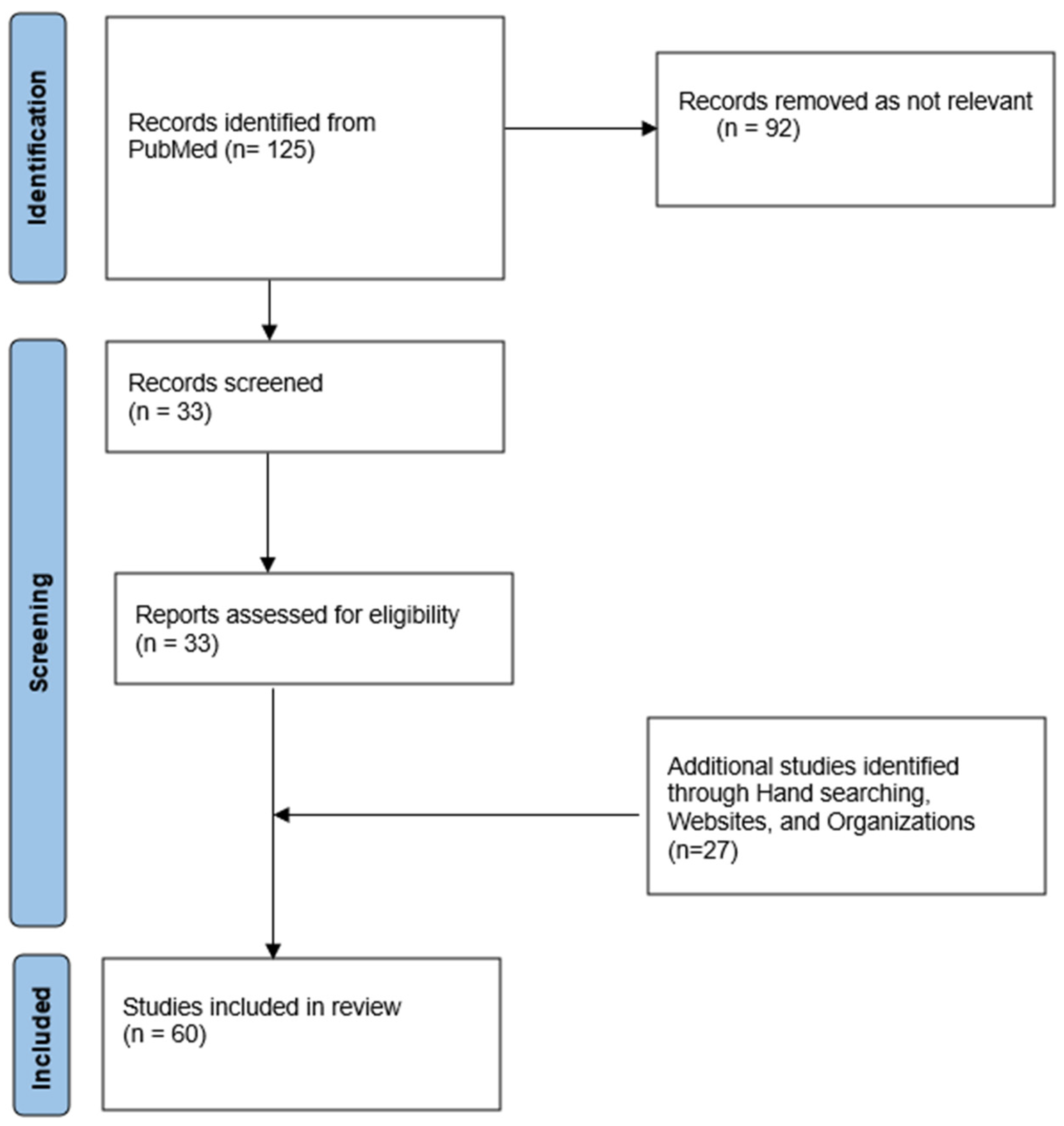
The study began with the analysis of a clinical case brought to our attention, followed by a review of scientific literature concerning snake envenomation, conducted with the aim of outlining the clinical and medico-legal aspects of viper bites in both national and international contexts. The bibliographic search was performed using PubMed, employing the following keywords: forensic OR medicolegal AND snakebite OR envenomation AND treatment protocol. We applied a time frame from 2015 to 2025, and the initial search yielded a total of 125 results. After removing duplicates and applying eligibility criteria (inclusion of articles only in English and relevant for medico-legal or clinical-toxicological content), 33 articles were included in the final review. The exclusion criteria applied were: in vitro studies, studies concerning viper species not present in Italy or Australia, as well as venomous organisms other than vipers; publications with outcomes not aligned with the objectives of the present study, and articles without full-text availability. A complementary manual search allowed the retrieval of additional relevant documents, bringing the total number of analyzed papers to 60, as shown in Figure 1.
Concurrently, a comparative analysis was conducted between the Italian and Australian models for the management of snake envenomation. Australia was selected as a reference point due to its high epidemiological burden associated with bites from highly venomous snakes and the well-established structure of its clinical and antivenom response protocols [6,7]. This analysis allowed for the identification of differences in clinical management and medico-legal implications, providing useful insights to improve diagnostic, therapeutic, and preventive strategies that could also be applied in contexts with lower incidence and relative severity, such as Italy.
The aim of this study is therefore to examine in depth the diagnostic–therapeutic management criteria and medico-legal implications of viper bites, with particular attention to their handling in contexts characterized by low incidence and relative severity, such as Italy. Furthermore, the research seeks to identify critical issues in the healthcare and medico-legal management of such events, outlining possible intervention strategies to improve treatment standards and reduce the risk of legal disputes.

2.1. The Case

2.1.1. Clinical Case

We report the case of a 40-year-old woman who presented to the Emergency Department after being bitten on the right foot by a snake, likely a viper. Upon arrival, she exhibited a small puncture wound on the middle phalanx of the third toe of the right foot and a tourniquet applied to the mid-third of the ipsilateral leg. The patient was alert and oriented, with stable vital signs and no evidence of systemic compromise. The bite was irrigated and treated with a lymphatic bandage. Blood tests were within normal limits. After consultation with the Poison Control Center (CAV), the healthcare team decided to monitor the patient without administering antivenom. Serial laboratory tests continued to show values within the normal range. The patient was discharged the following day with a prescription for antibiotics, corticosteroids, and local anti-inflammatory treatment, with instructions to return immediately in case of any changes in her condition. Three days later, the patient was readmitted to the Emergency Department due to significant worsening of symptoms, including diffuse hematoma and edema of the ankle and leg affected by the bite, as well as evident blood collections in the popliteal and medial thigh regions, marked tenderness on palpation, and decreased local sensation, associated with headache. The patient reported that symptoms had begun immediately after discharge. Peripheral pulses were present and palpable. Laboratory tests showed mild anemia (Hb 9.2 g/dL), coagulation abnormalities (prolonged PT and PTT), and signs of systemic inflammation. After repeated consultations with the CAV, treatment was initiated with enoxaparin 4000 IU, broad-spectrum antibiotics, and intravenous hydration. Contrast-enhanced CT of the lower limb revealed extensive intrafascial and subcutaneous edema. Following the onset of compartment syndrome, the patient underwent fasciotomy, followed by administration of two doses of antivenom. The postoperative course was uneventful, with progressive improvement of edema, limb function, and general symptoms. Blood tests, including coagulation parameters, normalized during hospitalization. The patient was subsequently discharged in good general condition after a period of monitoring, without further complications.

2.1.2. Medico-Legal History

Following the events described, a legal proceeding was initiated after the patient filed a complaint against the healthcare personnel involved, alleging alleged negligence and imprudence during observation and treatment. Specifically, the accusation concerned a delayed administration of antivenom and discharge after a period of observation deemed insufficient, allegedly non-compliant with recommendations for Grade 1–3 envenomation [8].
A detailed review of the medical records and applicable guidelines demonstrated that case management was appropriate and consistent with clinical protocols. At initial presentation, the patient had a small puncture wound on the third toe of the right foot, stable vital signs, and normal laboratory values, including coagulation and renal function. The case was classified as Grade 0 according to Boels’ classification [8], corresponding to a “dry bite,” for which immediate antivenom is not indicated unless symptoms progress rapidly.
During approximately 21 h of hospital observation, repeated clinical and laboratory monitoring showed no significant deterioration. According to the Stockholm criteria, antivenom is indicated only in the presence of hypotension, circulatory shock, severe prolonged gastrointestinal symptoms, mucosal edema with risk of airway obstruction, rapid extension of edema to an entire limb or trunk, neurological symptoms, or in extreme cases when laboratory markers show significant leukocytosis, elevated AST/ALT/CK, metabolic acidosis, hemolysis, ECG changes, or coagulopathy. As none of these criteria were met, conservative management was adopted, confirmed by consultation with the Poison Control Center. The patient was discharged in stable condition with clear instructions for home monitoring and immediate return in case of symptom progression. The subsequent worsening leading to compartment syndrome occurred in an extra-hospital setting, as the patient returned to the ED only three days after discharge, reporting a gradual progression rather than a sudden deterioration. At readmission, the patient received antivenom and appropriate treatment for the new clinical scenario. Follow-up examination one year later revealed good general condition, absence of edema, cyanosis, or skin lesions, and only residual scarring from the fasciotomy. Review of initial management confirmed adherence to national and international guidelines, appropriate observation, specialist consultations, and clear discharge instructions. Had the patient followed medical advice promptly, treatment could have been more effective and timely. Consequently, the healthcare personnel were not held responsible for the subsequent complications, and the legal proceedings concluded with dismissal of the accusations, affirming the appropriateness of the initial clinical management.

3. Results

3.1. Clinical Features and Classification of Viper Bites

Viper bites represent a potentially serious medical emergency, particularly in rural and mountainous areas where these snakes are more common. The characteristic impression of the two fangs can be easily distinguished [9,10], in contrast to bites from non-venomous snakes, which typically leave a semicircular mark [11] (Table 1).
Vipers, through their venomous fangs, can inject a variable amount of venom (5–40 mg) [12], causing local and systemic reactions that depend on the injected dose. Typically, venom injection induces within 30 min a firm edema and intense pain at the bite site. However, the absence of local manifestations within 2–3 h after the bite indicates a “dry bite,” meaning no venom was injected. In cases where venom is indeed injected, local signs may progress to severe systemic complications [13], including hemodynamic, digestive, coagulation, and renal disturbances [14]. Coagulation abnormalities can range from thrombocytopenia and fibrinolysis to disseminated intravascular coagulation (DIC) [15], occasionally accompanied by hemolysis [11].
For more effective clinical management, viper bites are classified into four severity grades (Boels Classification) [8]:
Grade 0 (dry bite): presence of bite marks without local or systemic signs.
Grade 1 (minimal envenomation): localized edema at the bite site without systemic symptoms.
Grade 2 (moderate envenomation): regional edema extending to part of the affected limb, moderate hypotension, vomiting and/or diarrhea.
Grade 3 (severe envenomation): extensive edema reaching the trunk, prolonged hypotension or shock, hemorrhages, and significant hemodynamic alterations [16].
Timely diagnosis and treatment, including potential antivenom administration, are crucial to prevent severe or fatal complications. Management of viper bites requires a multidisciplinary approach, encompassing careful monitoring and symptomatic or specific treatment according to the severity of the clinical presentation [17].

3.2. Snake Venom Toxins and Pathophysiological Mechanisms

Snake venom is a complex substance secreted by the venom glands located in the snake’s head, consisting of a mixture of proteins, peptides, organic molecules, and salts dissolved in an aqueous medium. Its composition varies considerably both among different species and within the same species. Toxic effects depend on the amount injected, which varies according to bite depth and local conditions, such as the presence of clothing [18,19].
The main classes of toxic proteins include phospholipase A2, serine proteases, metalloproteases, disintegrins, and lectin-like proteins [20,21]. Phospholipase A2 enzymes are responsible for the destruction of cellular membranes, causing hemolysis, inflammation, and local pain [22]. Serine proteases and metalloproteases act on coagulation factors, inducing hemorrhage or consumptive coagulopathy. Disintegrins interfere with cell adhesion, hindering coagulation and angiogenesis. Lectin-like proteins exert immunomodulatory and inflammatory effects, contributing to the severity of envenomation. Together, these components can produce local manifestations as well as systemic symptoms [23,24,25].
From a toxicokinetic perspective, venoms are primarily absorbed through subcutaneous or intramuscular tissues. Distribution occurs in an initial rapid phase, followed by a slower elimination phase. Bioavailability can vary widely, ranging from 4% to 81%, depending on the absorption route and snake species [26].
In Italy, viper venoms (Vipera aspis, Vipera ammodytes, Vipera dell’Orsini) are characterized mainly by hemotoxic and cytotoxic effects, with a moderate risk of neurotoxicity. Vipera aspis venom, for example, contains phospholipase A2, proteases, and hyaluronidase, causing local tissue damage and systemic alterations [27]. In Australia, by contrast, highly venomous species such as taipans (Oxyuranus spp.) [28], brown snakes (Pseudonaja spp.) [29], and tiger snakes (Notechis spp.) [30] produce potent neurotoxic and procoagulant venoms. These can induce rapid paralysis, consumptive coagulopathy, and systemic hemorrhage [31].
This diversity in venom toxicology reflects the evolutionary adaptation of snakes to their environments and prey, making personalized clinical management essential, based on the snake species involved, the severity of envenomation, and the availability of specific antivenoms.

3.3. Local and Systemic Complications Following Envenomation

Bites from vipers endemic to Italy, such as Vipera aspis, Vipera ammodytes, and Vipera dell’Orsini, primarily cause local and systemic effects, with severity varying according to the amount of venom injected, bite location, and patient condition. Local lesions include progressive edema, intense pain, ecchymosis, lymphangitis, tissue necrosis, and lymphadenopathy, while systemic manifestations may involve coagulopathies, hypotension, shock, renal damage (e.g., myoglobinuria or coagulopathy) [14], and, less frequently, neurotoxic effects such as peripheral axonal degeneration [24].
Bites from highly venomous Australian snakes, such as taipans (Oxyuranus spp.), brown snakes (Pseudonaja spp.), and tiger snakes (Notechis spp.), are characterized by higher lethality and a different spectrum of injuries. Their venoms are predominantly neurotoxic, causing rapid paralysis through neuromuscular transmission blockade, and procoagulant, leading to consumptive coagulopathy and systemic hemorrhage. Additional complications may include acute renal injury, shock, and multiorgan failure in severe cases.
While Italian viper bites are primarily hemotoxic and cytotoxic with limited neurotoxicity, Australian snake bites are dominated by neurotoxic and procoagulant effects, making timely and targeted treatment crucial, often requiring the administration of species-specific antivenoms.
It should therefore be recalled that in fatal viper bites, autopsy often represents the decisive tool for certification of the cause of death, especially when death occurs before any clinical assessment. The investigation must integrate scene examination, anamnestic reconstruction, macroscopic and microscopic autopsy findings, and toxicological confirmation.
At external examination, the presence of venom inoculation marks, namely characteristic fang impressions, together with local hemorrhage and cutaneous necrosis provides an initial diagnostic orientation; however, causal diagnosis is based on systemic lesions consistent with the action of the venom. As reported in the literature [32], these may include venom-induced consumption coagulopathy (VICC), diffuse hemorrhages, and acute renal failure with tubular necrosis; alternatively, acute respiratory failure due to neuromuscular paralysis [33] with hypoxic encephalic damage may be observed. Pulmonary edema, hepatic necrosis, and encephalopathy may also be associated [34].
A careful macroscopic examination must therefore be integrated with an accurate histological microscopic assessment, without neglecting toxicological analysis of the collected biological samples [35]. Enzyme-linked immunosorbent assay (ELISA) allows venom detection, whereas LC-MS/MS enables more specific venom characterization and exclusion of alternative toxic causes. As in many other not fully clear forensic cases, correlation between autopsy findings and toxicological results ensures medico-legal diagnostic certainty [36].

3.4. Management Strategies for Snake Bites: A Comparison Between Italy, France, and Australia

Australia represents an ideal context for comparing snakebite management strategies, given the high incidence and severity of these events compared to Italy. With its unique biodiversity, the country hosts some of the world’s most dangerous snake species, responsible for thousands of envenomation cases each year. The presence of highly neurotoxic and cytotoxic venoms, combined with the widespread distribution of snakes in rural and remote areas, poses significant challenges to the Australian healthcare system, necessitating a standardized and effective approach [37].
In Italy, as in most of Europe [38,39,40,41,42], viper bites are relatively rare and generally less severe, predominantly causing local toxicity with limited systemic effects. The management of viper bites is guided by recommendations from the Italian Society of Toxicology (SITOX) [43] and the Italian Society of Emergency and Urgent Care Medicine (SIMEU) [44], which include the Stockholm Criteria for antivenom use [45,46]. These guidelines emphasize the importance of prompt first aid and appropriate hospital-based management. The primary territorial reference for envenomation management in Italy is represented by the Poison Control Centers [47]. These specialized institutes provide medical consultation via telephone and, in some cases, online, for the management of poisoning or exposure to toxic substances. CAV staff include specialist physicians, pharmacists, and toxicologists who provide guidance on poisoning management, treatment, and preventive measures. Operators at CAVs provide precise instructions on the appropriate actions in case of any type of poisoning [36], addressing both the citizens and healthcare professionals. The telephone service offered by CAVs is continuously available to anyone who has experienced, or suspects exposure to, a toxic agent. CAVs manage cases of drug overdose, food poisoning, and chemical or biological contamination.
Following a suspected viper bite, it is essential for the patient to remain calm to reduce venom dissemination and immediately contact emergency services. Immobilization of the affected limb, with an elastic pressure bandage applied proximally to the bite, is a crucial measure to limit venom absorption, while the use of tourniquets or incision of the wound should be avoided.
In the hospital setting, according to SIMEU recommendations, the patient should be monitored for at least 24 h, with periodic clinical and laboratory evaluations every 6 h. Antivenom administration, based on the Stockholm Criteria [48,49], is indicated in cases of envenomation with severe systemic symptoms, rapid progression of edema, or neurotoxic, hemorrhagic, or shock signs. These criteria include persistent hypotension, neurological alterations such as paralysis, extensive edema, and coagulopathy. Antivenom administration must be carefully evaluated due to the risk of adverse reactions and is preferably performed in a hospital setting with intensive care support available for the management of potential complications [44].
In Australia, guidelines are defined by the Australian Resuscitation Council (ARC) and the Australian Snakebite Project (ASP) [50,51], which provide detailed recommendations for snakebite management. ARC Guideline 9.4.8 Envenomation Snakebite emphasizes the importance of the Pressure Immobilization Technique (PIT) [52] as first aid, which involves applying a compressive bandage to the entire affected limb to slow venom dissemination through the lymphatic system. Unlike in Italy, snake identification is facilitated using the Snake Venom Detection Kit (SVDK), which guides the selection of species-specific antivenom [6]. Australia is the only country worldwide with SVDKs. Antivenom administration is often urgent and can be initiated based on clinical presentation and suspected species, in accordance with ASP guidelines [30].
Patient monitoring includes frequent assessment of neurological, coagulation, and renal parameters, due to the high incidence of neurotoxicity and consumptive coagulopathy associated with Australian venoms. Australian guidelines also include protocols for managing complications such as respiratory failure, which may require intubation and mechanical ventilation.
Relying exclusively on Australian model could limit the generalizability of the proposed recommendations; therefore, we integrated comparative data from France, which has snakebite incidence and mortality rates more similar to those of Italy, in order to improve the practical applicability of the results.
In Italy and France, viper bites show many similarities both in terms of epidemiology and clinical management. In both countries, the main species involved are Vipera aspis and, in some areas, Vipera berus [53]. Snakebites are relatively uncommon and usually occur in rural, mountainous, or wooded areas, particularly during warmer months when outdoor activities increase. Fortunately, deaths are extremely rare, largely because both countries have well-developed healthcare systems and clear treatment protocols.
When a viper bite occurs, the approach to treatment is quite similar in Italy and France. The first step is careful medical evaluation and immobilization of the affected limb to limit the spread of venom. Patients are then closely monitored, both clinically and through laboratory tests such as blood counts, coagulation studies, and kidney function tests. Most cases are mild and can be managed with supportive care, including pain relief and observation to ensure that symptoms do not worsen [54].
In more serious cases, when swelling progresses rapidly or systemic symptoms such as low blood pressure or clotting abnormalities appear, intravenous antivenom may be administered in a hospital setting [55]. In both countries, antivenom is available in referral centers and is used according to well-defined clinical criteria, balancing its benefits with the small risk of adverse reactions. French Poison Control Centers (PCCs) also provide guidance to healthcare professionals, helping to standardize care. Overall, thanks to similar medical infrastructures and evidence-based guidelines, the management and outcomes of viper bites in Italy and France are closely aligned and generally favorable.
In France, eight Poison Control Centers (PCCs) operate 24 h a day, 7 days a week, responding to all calls from the public and healthcare professionals regarding any type of exposure to toxic substances. The data collected are stored anonymously in the French National Poisoning Database (FNDP). The FNDP is managed by the French Ministry of Health [56].
In the presence of moderate symptoms or rapid progression of edema, involvement of an entire limb, or systemic signs such as hypotension, persistent vomiting, diarrhea, neurological alterations, or coagulopathy, antivenom is indicated. France tends to use antivenom relatively early in these situations, as national studies have shown a reduction in symptom duration and local complications with timely administration. The antivenoms primarily used are Viperfav® or Vipertab, which are specific for the main European vipers (Vipera aspis, Vipera berus, Vipera ammodytes) [57,58].
From a medico-legal point of view, viper bites in Europe require careful documentation and adherence to standard clinical protocols. In both Italy and France, patients are usually observed in hospital, monitored for symptom progression and lab changes, and treated with antivenom when symptoms are moderate or severe. Keeping detailed records of the bite, the clinical course, treatments given, and any side effects is essential for both patient safety and legal protection. One important difference between the two countries is how compensation works. In France, a “no-fault compensation system” allows patients to receive compensation for injury without needing to prove medical negligence, making the process faster and more straightforward [59]. In Italy, compensation usually requires showing a causal link or, in some cases, medical liability, which can make the process more complex and dependent on proving fault. So, while the clinical management of viper bites is very similar in both countries, the legal and compensation frameworks differ, with France prioritizing patient protection through automatic support and Italy relying more on fault-based assessments.

4. Discussion

This study conducted an in-depth analysis of a clinical case of viper bite within a medico-legal context and provided a critical comparison of Italian and Australian management strategies, highlighting significant differences determined by epidemiological context, prevalent clinical severity, and the availability of specific resources.
In the presented clinical case, the initial management of the bite was appropriate and in accordance with SIMEU guidelines and the Stockholm Criteria, considering the bite classification as a “dry bite”. The decision to monitor the patient without immediate antivenom administration was consistent with international clinical recommendations, which reserve such treatment for cases of moderate to severe envenomation with systemic signs or rapidly evolving symptoms. The onset of delayed complications, such as compartment syndrome, was managed promptly and appropriately upon readmission, underscoring the importance of patient compliance with discharge instructions, a crucial factor in preventing serious complications.
Analysis of the two management contexts shows that in Italy, where viper bites are rare and generally less severe, a conservative approach prevails, based on prolonged monitoring and selective antivenom administration. In contrast, in Australia, the high incidence and lethality of bites from highly venomous snakes have led to the development of more aggressive therapeutic strategies, emphasizing rapid snake identification using Snake Venom Detection Kits and early administration of species-specific antivenoms.
This difference in management also reflects the toxicological profiles of the snakes present in the two countries. In Italy, the predominance of hemotoxic and cytotoxic effects, with limited neurotoxicity, justifies a more cautious approach focused on local complications such as tissue necrosis and compartment syndrome, which represent the main risks. In Australia, the predominance of neurotoxic and procoagulant effects, which can rapidly lead to paralysis, shock, and disseminated coagulopathy, necessitates a rapid and highly targeted response.
From a medico-legal perspective, the analysis of the two systems shows that professional responsibility is closely linked to the epidemiological context and the availability of resources. In Australia, the presence of strictly standardized protocols and dedicated diagnostic tools allows clinicians to demonstrate correct conduct objectively. In this scenario, failure to apply the Pressure Immobilization Technique or delay in initiating antivenom therapy could constitute negligence, given the widespread availability of detailed guidelines and diagnostic means.
In Italy, the rarity of snakebite cases makes adherence to SIMEU and SITOX recommendations and the Stockholm Criteria central, with particular attention to the timeliness of first aid, clinical and laboratory monitoring, and accurate documentation of therapeutic decisions, including the rationale for withholding antivenom if applicable. Proper completion of the medical record and obtaining informed consent represent essential medico-legal safeguards, especially considering the risk of adverse reactions associated with antivenom. While in Australia the presence of standardized protocols facilitates demonstration of compliant conduct, in Italy the infrequency of cases requires even greater attention to documenting therapeutic decisions and the involvement of Poison Control Centers as specialist support.
This case illustrates that, in the absence of specific national guidelines, medical practice grounded in full patient information, a multidisciplinary approach, and adherence to good clinical practices is crucial for reducing the risk of litigation or unfavorable judicial outcomes. In this perspective, the Australian experience, characterized by high incidence of envenomation and the presence of dedicated guidelines and protocols, offers valuable insights for the Italian context. In particular, through the definition of more standardized procedures and the adoption of validated diagnostic tools and decision algorithms, the margin of clinical error could be reduced. Indeed, despite the toxicological and epidemiological differences from the Australian paradigm, applying these principles could encourage the establishment of tailored management pathways, enhancing patient safety, traceability of clinical decisions, and the robustness of medico-legal defense in the event of judicial scrutiny.
In this context, artificial intelligence solutions, based on machine learning techniques and international, multidisciplinary data sharing, could be integrated into viper bite management pathways, with potential clinical and medico-legal benefits [8,60,61,62]. Predictive algorithms fed with clinical, laboratory, and toxicological data could facilitate severity classification and guide clinical management. Beyond decision support and early warning, AI tools could contribute to the analysis of epidemiological and environmental data to identify high-risk areas, supporting preventive strategies and more targeted healthcare planning. In this perspective, such systems could not only improve clinical safety but also strengthen the defensibility of medical conduct in legal settings, facilitating a multidisciplinary approach involving physicians, toxicologists, and Poison Control Centers.

5. Limitations

The study has several limitations that should be considered when interpreting the results. First, the data are based on a single clinical case, which limits the generalizability of the observations to other contexts or populations. Second, the literature review relies mainly on observational and retrospective studies, with potential biases in the collection and recording of clinical and medico-legal data. The rarity of viper bites in Italy results in very small sample sizes, reducing the statistical power of the available evidence. Furthermore, the absence of national guidelines makes it difficult to standardize protocols and compare results, relying only on regional recommendations or those of scientific societies. Finally, medico-legal evaluations and possible long-term patient outcomes are poorly documented, limiting the ability to draw definitive conclusions regarding the risk of complications or professional liability.

6. Conclusions

The management of viper bites in Italy, even in a context of low incidence and severity, and paradoxically precisely because of this, combined with the potential for high compensation claims due to unclear information, requires targeted clinical attention and adherence to guidelines to prevent complications and minimize medico-legal liability. The presented case demonstrates that management based on full patient information, a multidisciplinary approach, structured collaboration with Poison Control Centers, and adherence to good clinical practices represents a key factor for patient safety and risk reduction. Comparison with the Australian model, characterized by a high incidence of envenomation and dedicated protocols, suggests the potential benefit of implementing more uniform and standardized pathways in Italy, while respecting local toxicological and epidemiological characteristics.
In this context, artificial intelligence solutions focused on decision support and early warning, based on machine learning and international, multidisciplinary data sharing, could further strengthen viper bite management, improving both clinical safety and the medico-legal defensibility of medical conduct.

Author Contributions

Conceptualization, L.D.P. and E.M.; methodology, L.D.P., D.M. and B.C.; formal analysis, M.L.G., F.S., A.C.C. and G.N.; investigation, M.L.G., F.S. and A.C.C.; data curation, G.N. and B.C.; writing—original draft preparation, M.L.G., F.S., A.C.C. and G.N.; writing—review and editing, L.D.P., G.N., B.C. and E.M.; supervision, L.D.P. and E.M.; project administration, L.D.P. All authors have read and agreed to the published version of the manuscript.

Funding

This research received no external funding.

Institutional Review Board Statement

The study was conducted in accordance with the Declaration of Helsinki and approved by the Institutional Review Board of Department of Anatomical, Histological, Medicolegal and Orthopedic Sciences (protocol 3/2025 of 10 February 2025).

Informed Consent Statement

All personal data is anonymized. Written informed consent was obtained from the patient for the publication of this case report and any accompanying images.

Data Availability Statement

The data presented in this study are available on request from the corresponding author due to the privacy policies of the hospitals involved.

Conflicts of Interest

The authors declare no conflicts of interest.

References

  1. Snakebite Envenoming. Available online: https://www.who.int/news-room/fact-sheets/detail/snakebite-envenoming (accessed on 23 January 2026).
  2. Kasturiratne, A.; Wickremasinghe, R.; Silva, N.; Gunawardena, N.; Pathmeswaran, A.; Premaratna, R.; Savioli, L.; Lalloo, D.; de Silva, H. The Global Burden of Snakebite: A Literature Analysis and Modelling Based on Regional Estimates of Envenoming and Deaths. PLoS Med. 2008, 5, e218. [Google Scholar] [CrossRef]
  3. Oxyuranus microlepidotus (MCCOY, 1879). Available online: https://reptile-database.reptarium.cz/front.front/species?genus=oxyuranus&species=microlepidotus (accessed on 7 December 2025).
  4. Sasidharan, P.; Kaeley, N.; Mahala, P.; Jose, J.R.; Shankar, T.; Santhalingan, S.; Sharma, A.; Kumar, B.; Kumar, M.A.; Dhar, M. Clinical and Demographic Profiling of Snakebite Envenomation in a Tertiary Care Centre in Northern India. Int. J. Emerg. Med. 2025, 18, 50. [Google Scholar] [CrossRef]
  5. Chippaux, J.-P. Epidemiology of Snakebites in Europe: A Systematic Review of the Literature. Toxicon 2012, 59, 86–99. [Google Scholar] [CrossRef]
  6. Currie, B.J. Snakebite in Australia: The Role of the Venom Detection Kit. Emerg. Med. 2004, 16, 384–386. [Google Scholar] [CrossRef]
  7. Welton, R.E.; Liew, D.; Braitberg, G. Incidence of Fatal Snake Bite in Australia: A Coronial Based Retrospective Study (2000–2016). Toxicon 2017, 131, 11–15. [Google Scholar] [CrossRef]
  8. Claudet, I.; Germain, H. VipGrade® Electronic Clinical Tool: Retrospective Evaluation on a Paediatric Cohort of European Viper Bites. Clin. Toxicol. 2024, 62, 726–732. [Google Scholar] [CrossRef]
  9. Janes, D.N.; Bush, S.P.; Kolluru, G.R. Large Snake Size Suggests Increased Snakebite Severity in Patients Bitten by Rattlesnakes in Southern California. Wilderness Environ. Med. 2010, 21, 120–126. [Google Scholar] [CrossRef]
  10. Hayes, W.K.; Corbit, A.G.; Cardwell, M.D.; Herbert, S.S. Interfang Distances of Rattlesnakes: Sexual, Interspecific, and Body Size-Related Variation, and Implications for Snakebite Research and Management. Wilderness Environ. Med. 2017, 28, 101–107. [Google Scholar] [CrossRef]
  11. Norris, R.L.; Pfalzgraf, R.R.; Laing, G. Death Following Coral Snake Bite in the United States--First Documented Case (with ELISA Confirmation of Envenomation) in over 40 Years. Toxicon 2009, 53, 693–697. [Google Scholar] [CrossRef]
  12. Neri-Castro, E.; Zarzosa, V.; Benard-Valle, M.; Rodríguez-Solís, A.M.; Hernández-Orihuela, L.; Ortiz-Medina, J.A.; Alagón, A. Quantifying Venom Production: A Study on Micrurus Snakes in Mexico. Toxicon 2024, 240, 107658. [Google Scholar] [CrossRef]
  13. Bernasconi, L.; Schicchi, A.; Pirozzolo, R.; Negrini, V.; Scaravaggi, G.; Lonati, D.; Petrolini, V.M.; Locatelli, C.A. Coronary Thrombosis after European Adder Bite in a Patient on Dual Antiplatelet Therapy: A Case Report. Toxicon 2022, 220, 106961. [Google Scholar] [CrossRef]
  14. Sarkar, S.; Sinha, R.; Chaudhury, A.R.; Maduwage, K.; Abeyagunawardena, A.; Bose, N.; Pradhan, S.; Bresolin, N.L.; Garcia, B.A.; McCulloch, M. Snake Bite Associated with Acute Kidney Injury. Pediatr. Nephrol. 2021, 36, 3829–3840. [Google Scholar] [CrossRef]
  15. Thygesen, K.; Alpert, J.S.; Jaffe, A.S.; Chaitman, B.R.; Bax, J.J.; Morrow, D.A.; White, H.D.; Executive Group on behalf of the Joint European Society of Cardiology (ESC)/American College of Cardiology (ACC)/American Heart Association (AHA)/World Heart Federation (WHF) Task Force for the Universal Definition of Myocardial Infarction. Fourth Universal Definition of Myocardial Infarction (2018). Circulation 2018, 138, e618–e651. [Google Scholar] [CrossRef]
  16. Esposito, S.; Maiolo, G.D.; Chiatto, F.L.; Capasso, M.; Sala, C.; Marco, G.P.D.; Rinaldi, P.; Tomasello, A.; Coletta, F.; Villani, R. European Viper Bites: Clinical Management from Pre-Hospitalisation to Follow-up a Narrative Review. J. Emerg. Crit. Care Med. 2025, 9, 4. [Google Scholar] [CrossRef]
  17. Knudsen, C.; Jürgensen, J.A.; Føns, S.; Haack, A.M.; Friis, R.U.W.; Dam, S.H.; Bush, S.P.; White, J.; Laustsen, A.H. Snakebite Envenoming Diagnosis and Diagnostics. Front. Immunol. 2021, 12, 661457. [Google Scholar] [CrossRef]
  18. Acharya, A.; Deo, R.C.; Panigrahi, S.; Mahapatra, A.K.; Sarangi, R.; Mohapatra, D.P. Hydrocephalus in snake bite: A rare manifestation with review of literature. Child’s Nerv. Syst. 2025, 41, 326. [Google Scholar] [CrossRef] [PubMed]
  19. Youngman, N.J. Snake Venom: Toxicology and Associated Countermeasures. Toxins 2025, 17, 237. [Google Scholar] [CrossRef]
  20. Green, D. Hematology Products from Snake Venoms. Thromb. Res. 2025, 245, 109215. [Google Scholar] [CrossRef]
  21. Sampat, G.H.; Hiremath, K.; Dodakallanavar, J.; Patil, V.S.; Harish, D.R.; Biradar, P.; Mahadevamurthy, R.K.; Barvaliya, M.; Roy, S. Unraveling Snake Venom Phospholipase A2: An Overview of Its Structure, Pharmacology, and Inhibitors. Pharmacol. Rep. 2023, 75, 1454–1473. [Google Scholar] [CrossRef]
  22. Jia, Y.; Olvera, P.; Rangel, F.; Mendez, B.; Reddy, S. Rapid Identification of Phospholipase A2 Transcripts from Snake Venoms. Toxins 2019, 11, 69. [Google Scholar] [CrossRef]
  23. Nielsen, V.G.; Wagner, M.T. Review of the Mechanisms of Snake Venom Induced Pain: It’s All about Location, Location, Location. Int. J. Mol. Sci. 2022, 23, 2128. [Google Scholar] [CrossRef]
  24. McAninch, S.A.; Morrissey, R.P.; Rosen, P.; Meyer, T.A.; Hessel, M.M.; Vohra, M.H. Snake Eyes: Coral Snake Neurotoxicity Associated with Ocular Absorption of Venom and Successful Treatment with Exotic Antivenom. J. Emerg. Med. 2019, 56, 519–522. [Google Scholar] [CrossRef]
  25. Kang, T.S.; Georgieva, D.; Genov, N.; Murakami, M.T.; Sinha, M.; Kumar, R.P.; Kaur, P.; Kumar, S.; Dey, S.; Sharma, S.; et al. Enzymatic Toxins from Snake Venom: Structural Characterization and Mechanism of Catalysis. FEBS J. 2011, 278, 4544–4576. [Google Scholar] [CrossRef]
  26. Feola, A.; Marella, G.L.; Carfora, A.; Della Pietra, B.; Zangani, P.; Campobasso, C.P. Snakebite Envenoming a Challenging Diagnosis for the Forensic Pathologist: A Systematic Review. Toxins 2020, 12, 699. [Google Scholar] [CrossRef]
  27. Marano, M.; Di Giuseppe, M.; Pro, S.; Pisani, M.; Montibeller, M.; Bottari, G.; Nunziata, J.; Cecchetti, C. Vipera aspis Bite Neurotoxicity: Two Pediatric Cases in Central Italy. Clin. Toxicol. 2020, 58, 849–850. [Google Scholar] [CrossRef]
  28. Kulawickrama, S.; O’Leary, M.A.; Hodgson, W.C.; Brown, S.G.A.; Jacoby, T.; Davern, K.; Isbister, G.K. Development of a Sensitive Enzyme Immunoassay for Measuring Taipan Venom in Serum. Toxicon 2010, 55, 1510–1518. [Google Scholar] [CrossRef]
  29. O’Leary, M.A.; Isbister, G.K.; Schneider, J.J.; Brown, S.G.A.; Currie, B.J. Enzyme Immunoassays in Brown Snake (Pseudonaja spp.) Envenoming: Detecting Venom, Antivenom and Venom-Antivenom Complexes. Toxicon 2006, 48, 4–11. [Google Scholar] [CrossRef]
  30. Currie, B.J. Treatment of Snakebite in Australia: The Current Evidence Base and Questions Requiring Collaborative Multicentre Prospective Studies. Toxicon 2006, 48, 941–956. [Google Scholar] [CrossRef]
  31. Tasoulis, T.; Wang, C.R.; Sumner, J.; Dunstan, N.; Pukala, T.L.; Isbister, G.K. The Unusual Metalloprotease-Rich Venom Proteome of the Australian Elapid Snake Hoplocephalus Stephensii. Toxins 2022, 14, 314. [Google Scholar] [CrossRef]
  32. Sacco, M.A.; Gualtieri, S.; Princi, A.; Aquila, I. Autopsy-Proven Snakebite Envenoming Deaths: A Review of Forensic and Pathological Evidence. Forensic Sci. 2026, 6, 2. [Google Scholar] [CrossRef]
  33. Benvenuti, L.A.; França, F.O.S.; Barbaro, K.C.; Nunes, J.R.; Cardoso, J.L.C. Pulmonary Haemorrhage Causing Rapid Death after Bothrops Jararacussu Snakebite: A Case Report. Toxicon 2003, 42, 331–334. [Google Scholar] [CrossRef] [PubMed]
  34. Martínez-Barrios, E.; Grassi, S.; Brión, M.; Toro, R.; Cesar, S.; Cruzalegui, J.; Coll, M.; Alcalde, M.; Brugada, R.; Greco, A.; et al. Molecular Autopsy: Twenty Years of Post-Mortem Diagnosis in Sudden Cardiac Death. Front. Med. 2023, 10, 1118585. [Google Scholar] [CrossRef] [PubMed]
  35. Tian, Y.; Liu, Z.; Ma, L.; Yu, Y.; Shi, Q.; Zhao, S.; Zhou, Y. Forensic Identification of a Fatal Snakebite from Bungarus Multicinctus (Chinese Krait) by Pathological and Toxicological Findings: A Case Report. Forensic Sci. Med. Pathol. 2022, 18, 497–502. [Google Scholar] [CrossRef] [PubMed]
  36. Piersanti, V.; Napoletano, G.; David, M.C.; Umani Ronchi, F.; Marinelli, E.; De Paola, L.; Zaami, S. Sudden Death Due to Butane Abuse—An Overview. J. Forensic Leg. Med. 2024, 103, 102662. [Google Scholar] [CrossRef]
  37. Isbister, G.K. Antivenom Availability, Delays and Use in Australia. Toxicon X 2023, 17, 100145. [Google Scholar] [CrossRef]
  38. Nițescu, G.V.; Ulmeanu, C.E.; Crăciun, M.-D.; Ciucă, A.M.; Ulici, A.; Ghira, I.; Lonati, D. Neurotoxicity and Other Clinical Manifestations of a Common European Adder (Vipera berus) Bite in Romania. Toxins 2022, 14, 500. [Google Scholar] [CrossRef]
  39. Tunjić Pejak, D.; Nesek Adam, V.; Srzić, I. Venomous Snakebites in Croatia, Clinical Presentation, Diagnosis and Treatment. Acta Clin. Croat. 2022, 61, 59–66. [Google Scholar] [CrossRef]
  40. Orlandi, G.; Rossi, N.; Chiarelli, F.; Di Filippo, P. Vipera Snakebite in Children: A Focus on Europe. Children 2025, 12, 393. [Google Scholar] [CrossRef]
  41. Paolino, G.; Di Nicola, M.R.; Pontara, A.; Didona, D.; Moliterni, E.; Mercuri, S.R.; Grano, M.; Borgianni, N.; Kumar, R.; Pampena, R. Vipera Snakebite in Europe: A Systematic Review of a Neglected Disease. J. Eur. Acad. Dermatol. Venereol. 2020, 34, 2247–2260. [Google Scholar] [CrossRef]
  42. Sassoè, M. Envenomation by Vipera aspis in Piedmont (Italy): A Report of Three Cases, Including One Case with Neurological Symptoms. Toxicon 2023, 230, 107154. [Google Scholar] [CrossRef]
  43. SITOX. Società Italiana Tossicologia. Available online: https://www.sitox.org/ (accessed on 30 December 2025).
  44. SIMEU. Available online: https://www.simeu.it/w/ (accessed on 30 December 2025).
  45. Di Nicola, M.R.; Crevani, M.; Avella, I.; Cerullo, A.; Dorne, J.-L.C.M.; Paolino, G.; Zattera, C. A Guide to the Clinical Management of Vipera Snakebite in Italy. Toxins 2024, 16, 255. [Google Scholar] [CrossRef]
  46. FAB-ANTIVIPERA. Centro di Riferimento Regionale Antidoti Emilia Romagna. Available online: https://antidoti.ospfe.it/scheda-intossicazion/morso-di-vipera/ (accessed on 30 December 2025).
  47. CENTRI ANTIVELENI. Available online: https://www.iss.it/-/cnsc-cav (accessed on 30 December 2025).
  48. Carenza Siero Antiofidico—Modalità di Richiesta D’importazione Dall’estero (04/02/2014). Available online: https://www.aifa.gov.it/-/carenza-siero-antiofidico-modalita-di-richiesta-d-importazione-dall-estero-04-02-2014- (accessed on 12 February 2026).
  49. Atterraggio. Available online: https://www.iss.it/cnsc-atterraggio (accessed on 30 December 2025).
  50. Johnston, C.I.; Ryan, N.M.; Page, C.B.; Buckley, N.A.; Brown, S.G.; O’Leary, M.A.; Isbister, G.K. The Australian Snakebite Project, 2005–2015 (ASP-20). Med. J. Aust. 2017, 207, 119–125. [Google Scholar] [CrossRef]
  51. Johnston, C.I.; Tasoulis, T.; Isbister, G.K. Australian Sea Snake Envenoming Causes Myotoxicity and Non-Specific Systemic Symptoms—Australian Snakebite Project (ASP-24). Front. Pharmacol. 2022, 13, 816795. [Google Scholar] [CrossRef] [PubMed]
  52. WPDM2—Australian Resuscitation Council. Available online: https://resus.org.au/wpdm2/ (accessed on 30 December 2025).
  53. de Haro, L. Aggravation of snake bite in France and their treatment. Presse Med. 2003, 32, 1131–1137. [Google Scholar] [PubMed]
  54. de Haro, L.; Glaizal, M.; Tichadou, L.; Blanc-Brisset, I.; Hayek-Lanthois, M. Asp Viper (Vipera aspis) Envenomation: Experience of the Marseille Poison Centre from 1996 to 2008. Toxins 2009, 1, 100–112. [Google Scholar] [CrossRef] [PubMed]
  55. Boels, D.; Hamel, J.F.; Le Roux, G.; Labadie, M.; Paret, N.; Delcourt, N.; Langrand, J.; Puskarczyk, E.; Nisse, P.; Sinno-Tellier, S.; et al. Snake Bites by European Vipers in Mainland France in 2017–2018: Comparison of Two Antivenoms Viperfav® and Viperatab®. Clin. Toxicol. 2020, 58, 1050–1057. [Google Scholar] [CrossRef]
  56. Toxicovigilance: Poison Control Centres and Anses Working to Combat Everyday Poisoning. Anses—Agence Nationale de Sécurité Sanitaire de L’alimentation, de L’environnement et Du Travail. Available online: https://www.anses.fr/en/content/toxicovigilance-poison-control-centres-and-anses-working-combat-everyday-poisoning (accessed on 12 February 2026).
  57. Kurtović, T.; Dobaja Borak, M.; Grenc, D.; Leonardi, A.; Križaj, I.; Lukšić, B.; Halassy, B.; Brvar, M. Clinical Efficacy and Pharmacokinetics of Antivenom Viperfav® in Vipera Ammodytes Ammodytes Envenomation. Pharmaceutics 2025, 17, 1431. [Google Scholar] [CrossRef]
  58. Jollivet, V.; Hamel, J.F.; de Haro, L.; Labadie, M.; Sapori, J.M.; Cordier, L.; Villa, A.; Nisse, P.; Puskarczyk, E.; Berthelon, L.; et al. European Viper Envenomation Recorded by French Poison Control Centers: A Clinical Assessment and Management Study. Toxicon 2015, 108, 97–103. [Google Scholar] [CrossRef]
  59. Ketsekioulafis, I.; Katsos, K.; Kouzos, D.; Spiliopoulou, C.; Lytras, T.; Sakelliadis, E.I. Medical Liability Related Legislation and Insurance Policies around the World: A Narrative Literature Review. J. Forensic Leg. Med. 2026, 117, 103040. [Google Scholar] [CrossRef]
  60. Chen, M.; Decary, M. Artificial Intelligence in Healthcare: An Essential Guide for Health Leaders. Healthc. Manag. Forum 2020, 33, 10–18. [Google Scholar] [CrossRef]
  61. Marinelli, S.; De Paola, L.; Stark, M.; Montanari Vergallo, G. Artificial Intelligence in the Service of Medicine: Current Solutions and Future Perspectives, Opportunities, and Challenges. Clin. Ter. 2025, 176, 77–82. [Google Scholar] [CrossRef]
  62. Hamza, M.; Knudsen, C.; Gnanathasan, C.A.; Monteiro, W.; Lewin, M.R.; Laustsen, A.H.; Habib, A.G. Clinical Management of Snakebite Envenoming: Future Perspectives. Toxicon X 2021, 11, 100079. [Google Scholar] [CrossRef]
Figure 1. Prisma Flow Chart.
Forensicsci 06 00027 g001
Table 1. Morphological characteristics of snakes.
Venomous ViperNon-Venomous Snakes
BiteTwo deeper puncture wounds from the venomous fangs, sometimes followed by additional smaller marksSmall puncture marks arranged in a semicircular pattern
PupilElliptical, vertical shapeRound, fully circular pupil
HeadTriangular, flat, clearly distinct from the rest of the bodyHead with a more rounded morphology, poorly differentiated from the neck
ScalesKeeledSmooth
BodyStocky, with a short tailSlender, elongated body with a tail that gradually tapers distally
Disclaimer/Publisher’s Note: The statements, opinions and data contained in all publications are solely those of the individual author(s) and contributor(s) and not of MDPI and/or the editor(s). MDPI and/or the editor(s) disclaim responsibility for any injury to people or property resulting from any ideas, methods, instructions or products referred to in the content.