Validation of Subadult Age and Stature Estimation Methods Using a Contemporary Japanese Sample
Abstract
1. Introduction
2. Materials and Methods
2.1. Samples
2.2. Subadult Age and Stature Estimation Methods
2.3. Method Comparisons
3. Results
3.1. Age Estimation Methods
3.2. Stature Estimation Methods
4. Discussion
4.1. Subadult Age Estimation
4.2. Subadult Stature Estimation
4.3. Recommendations
- 1.
- Appropriate reference samples
- 2.
- Statistical methods that reflect biology
- 3.
- Consult Standards
4.4. Limitations, Considerations, and Future Directions
5. Conclusions
Supplementary Materials
Author Contributions
Funding
Institutional Review Board Statement
Informed Consent Statement
Data Availability Statement
Acknowledgments
Conflicts of Interest
Abbreviations
| 3D | Three-dimensional |
| CT | Computed tomography |
| PCMT | Postmortem computed tomography |
| AAFS | American Academy of Forensic Sciences |
| ASB | AAFS Standards Board |
| ANSI | American National Standards Institute |
References
- Brits, D.M.; Bidmos, M.A.; Manger, P.R. Stature estimation from the femur and tibia in Black South African sub-adults. Forensic Sci. Int. 2017, 270, 277.e1–277.e10. [Google Scholar] [CrossRef] [PubMed]
- Corron, L.; Marchal, F.; Condemi, S.; Adalian, P. A critical review of sub-adult age estimation in biological anthropology: Do methods comply with published recommendations? Forensic Sci. Int. 2018, 288, 328.e1–328.e9. [Google Scholar] [CrossRef]
- Cunningham, C.A. Skeletal age estimation in juveniles and subadults. In Age Estimation; Adserias-Garriga, J., Ed.; Academic Press: London, UK, 2019; pp. 41–54. [Google Scholar] [CrossRef]
- Langley, N.R. Stature estimation. In Forensic Anthropology: A Comprehensive Introduction, 2nd ed.; Langley, N.R., Tersigni-Tarrant, M.T.A., Eds.; CRC Press: Boca Raton, FL, USA, 2017; pp. 195–203. [Google Scholar]
- Rissech, C.; Márquez-Grant, N.; Turbón, D. A collation of recently published Western European formulae for age estimation of subadult skeletal remains: Recommendations for forensic anthropology and osteoarchaeology. J. Forensic Sci. 2013, 58, S163–S168. [Google Scholar] [CrossRef]
- Grivas, C.R.; Komar, D.A. Kumho, Daubert, and the nature of scientific inquiry: Implications for forensic anthropology. J. Forensic Sci. 2008, 53, 771–776. [Google Scholar] [CrossRef]
- Lesciotto, K.M. The impact of Daubert on the admissibility of forensic anthropology expert testimony. J. Forensic Sci. 2015, 60, 549–555. [Google Scholar] [CrossRef]
- Bethard, J.D.; DiGangi, E.A. From the laboratory to the witness stand: Research trends and method validation in forensic anthropology. In Forensic Anthropology and the United States Judicial System, 1st ed.; Fulginiti, L.C., Hartnett-McCann, K., Galloway, A., Eds.; Wiley: Hoboken, NJ, USA, 2019; pp. 41–52. [Google Scholar] [CrossRef]
- Fleischman, J.M.; Pierce, M.L.; Crowder, C.M. Transparency in forensic anthropology through the implementation of quality assurance practices. In Forensic Anthropology and the United States Judicial System, 1st ed.; Fulginiti, L.C., Hartnett-McCann, K., Galloway, A., Eds.; Wiley: Hoboken, NJ, USA, 2019; pp. 70–88. [Google Scholar] [CrossRef]
- Valsecchi, A.; Irurita Olivares, J.; Mesejo, P. Age estimation in forensic anthropology: Methodological considerations about the validation studies of prediction models. Int. J. Legal Med. 2019, 133, 1915–1924. [Google Scholar] [CrossRef]
- Campanacho, V.; Alves Cardoso, F.; Ubelaker, D.H. Documented skeletal collections and their importance in forensic anthropology in the United States. Forensic Sci. 2021, 1, 228–239. [Google Scholar] [CrossRef]
- Stull, K.E.; Corron, L.K. The Subadult Virtual Anthropology Database (SVAD): An accessible repository of contemporary subadult reference data. Forensic Sci. 2022, 2, 20–36. [Google Scholar] [CrossRef]
- Murray, N.J.; Spake, L.; Cervantes, M.; Albanese, J.; Cardoso, H.F. New More Generic and Inclusive Regression Formulae for the Estimation of Stature from Long Bone Lengths in Children. Forensic Sci. 2024, 4, 62–75. [Google Scholar] [CrossRef]
- Smith, S.L. Stature estimation of 3–10-year-old children from long bone lengths. J. Forensic Sci. 2007, 52, 538–546. [Google Scholar] [CrossRef]
- Go, M.C.; Yukyi, N.; Chu, E.Y. On WEIRD anthropologists and their white skeletons. Forensic Anthropol. 2021, 4, 145–160. [Google Scholar] [CrossRef]
- Budiman, A.; Ruiz, N.G. Key Facts About Asian Americans, a Diverse and Growing Population. Available online: https://www.pewresearch.org/short-reads/2021/04/29/key-facts-about-asian-americans/ (accessed on 29 May 2025).
- Chiba, F.; Inokuchi, G.; Hoshioka, Y.; Sakuma, A.; Makino, Y.; Torimitsu, S.; Yamaguchi, R.; Saitoh, H.; Kono, M.; Iwase, H. Age estimation by evaluation of osteophytes in thoracic and lumbar vertebrae using postmortem CT images in a modern Japanese population. Int. J. Legal Med. 2022, 136, 261–267. [Google Scholar] [CrossRef]
- Chiba, F.; Makino, Y.; Torimitsu, S.; Motomura, A.; Inokuchi, G.; Ishii, N.; Hoshioka, Y.; Abe, H.; Yamaguchi, R.; Sakuma, A.; et al. Stature estimation based on femoral measurements in the modern Japanese population: A cadaveric study using multidetector computed tomography. Int. J. Legal Med. 2018, 132, 1485–1491. [Google Scholar] [CrossRef]
- Hishmat, A.M.; Michiue, T.; Sogawa, N.; Oritani, S.; Ishikawa, T.; Fawzy, I.A.; Hashem, M.A.M.; Maeda, H. Virtual CT morphometry of lower limb long bones for estimation of the sex and stature using postmortem Japanese adult data in forensic identification. Int. J. Legal Med. 2015, 129, 1173–1182. [Google Scholar] [CrossRef]
- Kira, K.; Chiba, F.; Makino, Y.; Torimitsu, S.; Yamaguchi, R.; Tsuneya, S.; Motomura, A.; Yoshida, M.; Saitoh, N.; Inokuchi, G.; et al. Stature estimation by semi-automatic measurements of 3D CT images of the femur. Int. J. Legal Med. 2023, 137, 359–377. [Google Scholar] [CrossRef] [PubMed]
- Ogawa, Y.; Imaizumi, K.; Miyasaka, S.; Yoshino, M. Discriminant functions for sex estimation of modern Japanese skulls. J. Forensic Leg. Med. 2013, 20, 234–238. [Google Scholar] [CrossRef]
- Torimitsu, S.; Nakazawa, A.; Flavel, A.; Iwase, H.; Makino, Y.; Hisham, S.; Franklin, D. Estimation of population affinity using cranial measurements acquired in multidetector computed tomography images of Japanese and Malay individuals. Int. J. Legal Med. 2025, 139, 863–873. [Google Scholar] [CrossRef]
- Eveleth, P.B.; Tanner, J.M. Worldwide Variation in Human Growth, 2nd ed.; Cambridge University Press: London, UK, 1991. [Google Scholar]
- Greulich, W.W. A comparison of the physical growth and development of American-born and native Japanese children. Am. J. Phys. Anthropol. 1957, 15, 489–515. [Google Scholar] [CrossRef]
- Kimura, K. Studies on growth and development in Japan. Am. J. Phys. Anthropol. 1984, 27, 179–213. [Google Scholar] [CrossRef]
- Kondo, S.; Eto, M. Physical growth studies on Japanese-American children in comparison with native Japanese. In Comparative Studies on Human Adaptability of Japanese, Caucasians, and Japanese Americans; Horvath, S.M., Kondo, S., Matsui, H., Yoshimura, H., Eds.; University of Tokyo Press: Tokyo, Japan, 1975; Volume 1, pp. 13–45. [Google Scholar]
- Brough, A.L.; Rutty, G.N.; Black, S.; Morgan, B. Post-mortem computed tomography and 3D imaging: Anthropological applications for juvenile remains. Forensic Sci. Med. Pathol. 2012, 8, 270–279. [Google Scholar] [CrossRef] [PubMed]
- Dedouit, F.; Savall, F.; Mokrane, F.Z.; Rousseau, H.; Crubézy, E.; Rougé, D.; Telmon, N. Virtual anthropology and forensic identification using multidetector CT. BJR 2014, 87, 20130468. [Google Scholar] [CrossRef]
- Franklin, D.; Blau, S. Physical and virtual sources of biological data in forensic anthropology: Considerations relative to practitioner and/or judicial requirements. In Statistics and Probability in Forensic Anthropology; Academic Press: London, UK, 2020; pp. 17–45. [Google Scholar] [CrossRef]
- Stock, M.K.; Stull, K.E.; Garvin, H.M.; Klales, A.R. Development of modern human subadult age and sex estimation standards using multi-slice computed tomography images from medical examiner’s offices. In Developments in X-Ray Tomography X; SPIE: Bellingham, WA, USA, 2016; Volume 99670E, pp. 48–61. [Google Scholar] [CrossRef]
- Abegg, C.; Balbo, I.; Dominguez, A.; Grabherr, S.; Campana, L.; Moghaddam, N. Virtual anthropology: A preliminary test of macroscopic observation versus 3D surface scans and computed tomography (CT) scans. Forensic Sci. Res. 2021, 6, 34–41. [Google Scholar] [CrossRef]
- Brough, A.L.; Morgan, B.; Robinson, C.; Black, S.; Cunningham, C.; Adams, C.; Rutty, G.N. A minimum data set approach to post-mortem computed tomography reporting for anthropological biological profiling. Forensic Sci. Med. Pathol. 2014, 10, 504–512. [Google Scholar] [CrossRef] [PubMed]
- Colman, K.L.; de Boer, H.H.; Dobbe, J.G.; Liberton, N.P.; Stull, K.E.; van Eijnatten, M.; Streekstra, G.J.; Oostra, R.J.; van Rijn, R.R.; van der Merwe, A.E. Virtual forensic anthropology: The accuracy of osteometric analysis of 3D bone models derived from clinical computed tomography (CT) scans. Forensic Sci. Int. 2019, 304, 109963. [Google Scholar] [CrossRef] [PubMed]
- Corron, L.; Marchal, F.; Condemi, S.; Chaumoitre, K.; Adalian, P. Evaluating the consistency, repeatability, and reproducibility of osteometric data on dry bone surfaces, scanned dry bone surfaces, and scanned bone surfaces obtained from living individuals. BMSAP 2017, 29, 33–53. [Google Scholar] [CrossRef]
- Franklin, D.; Swift, L.; Flavel, A. ‘Virtual anthropology’ and radiographic imaging in the Forensic Medical Sciences. Egypt. J. Forensic Sci. 2016, 6, 31–43. [Google Scholar] [CrossRef]
- Jerković, I.; Bašić, Ž.; Bareša, T.; Krešić, E.; Hadžić, A.A.; Dolić, K.; Ćavar Borić, M.; Budimir Mršić, D.; Čavka, M.; Šlaus, M.; et al. The repeatability of standard cranial measurements on dry bones and MSCT images. J. Forensic Sci. 2022, 67, 1938–1947. [Google Scholar] [CrossRef]
- Brough, A.L.; Morgan, B.; Rutty, G.N. Postmortem computed tomography (PMCT) and disaster victim identification. Radiol. Med. 2015, 120, 866–873. [Google Scholar] [CrossRef]
- Kolpan, K.E.; Vadala, J.; Dhanaliwala, A.; Chao, T. Utilizing augmented reality for reconstruction of fractured, fragmented and damaged craniofacial remains in forensic anthropology. Forensic Sci. Int. 2024, 357, 111995. [Google Scholar] [CrossRef] [PubMed]
- Oriola, L.S.; Oller, N.A.; Martínez-Abadías, N. Virtual anthropology: Forensic applications to cranial skeletal remains from the Spanish Civil War. Forensic Sci. Int. 2022, 341, 111504. [Google Scholar] [CrossRef]
- Manifold, B.M. Intrinsic and extrinsic factors involved in the preservation of non-adult skeletal remains in archaeology and forensic science. Bull. Int. Assoc. Paleodont. 2012, 6, 51–69. Available online: https://hrcak.srce.hr/95443 (accessed on 15 May 2025).
- Manifold, B.M. Skeletal preservation of children’s remains in the archaeological record. Homo 2015, 66, 520–548. [Google Scholar] [CrossRef]
- Stojanowski, C.M.; Seidemann, R.M.; Doran, G.H. Differential skeletal preservation at Windover Pond: Causes and consequences. Am. J. Phys. Anthropol. 2002, 119, 15–26. [Google Scholar] [CrossRef]
- ANSI/ASB Standard 133; Standard for Age Estimation in Forensic Anthropology. 1st ed. AAFS Standards Board, LLC: Colorado Springs, CO, USA, 2024.
- ANSI/ASB Standard 045; Standard for Stature Estimation in Forensic Anthropology. 1st ed. AAFS Standards Board, LLC: Colorado Springs, CO, USA, 2019.
- Langley, N.R. The lateral clavicular epiphysis: Fusion timing and age estimation. Int. J. Legal Med. 2016, 130, 511–517. [Google Scholar] [CrossRef]
- Black, S.; Scheuer, L. Age changes in the clavicle: From the Early Neonatal Period to Skeletal Maturity. Int. J. Osteoarchaeol 1996, 6, 425–434. [Google Scholar] [CrossRef]
- Fedorov, A.; Beichel, R.; Kalpathy-Cramer, J.; Finet, J.; Fillion-Robin, J.C.; Pujol, S.; Bauer, C.; Jennings, D.; Fennessy, F.; Sonka, M.; et al. 3D Slicer as an image computing platform for the Quantitative Imaging Network. Magn. Reson. Imaging 2012, 30, 1323–1341. [Google Scholar] [CrossRef] [PubMed]
- Stull, K.E.; Corron, L.K. Subadult Virtual Anthropology Database (SVAD) Data Collection Protocol: Epiphyseal Fusion, Diaphyseal Dimensions, Dental Development Stages, Vertebral Neural Canal Dimensions. 2021. Available online: https://zenodo.org/records/5348392 (accessed on 29 May 2025).
- Langley, N.R.; Jantz, L.M.; Ousley, S.D.; Jantz, R.L.; Milner, G. Data Collection Procedures for Forensic Skeletal Material 2.0; University of Tennessee: Knoxville, TN, USA; Lincoln Memorial University: Harrogate, TN, USA, 2016. [Google Scholar]
- Cardoso, H.F.; Abrantes, J.; Humphrey, L.T. Age estimation of immature human skeletal remains from the diaphyseal length of the long bones in the postnatal period. Int. J. Legal Med. 2014, 128, 809–824. [Google Scholar] [CrossRef] [PubMed]
- Chu, E.Y.; Stull, K.E. An investigation of the relationship between long bone measurements and stature: Implications for estimating skeletal stature in subadults. Int. J. Legal Med. 2025, 139, 441–453. [Google Scholar] [CrossRef]
- Facchini, F.; Veschi, S. Age determination on long bones in a skeletal subadults sample (b-12 years). Coll. Anthropol. 2004, 28, 89–98. Available online: https://hrcak.srce.hr/4889 (accessed on 15 May 2025).
- López-Costas, O.; Rissech, C.; Trancho, G.; Turbón, D. Postnatal ontogenesis of the tibia. Implications for age and sex estimation. Forensic Sci. Int. 2012, 214, 207.e1–207.e11. [Google Scholar] [CrossRef]
- Rissech, C.; Schaefer, M.; Malgosa, A. Development of the femur—Implications for age and sex determination. Forensic Sci. Int. 2008, 180, 1–9. [Google Scholar] [CrossRef] [PubMed]
- Robbins Schug, G.; Gupta, S.; Cowgill, L.W.; Sciulli, P.W.; Blatt, S.H. Panel regression formulas for estimating stature and body mass from immature human skeletons: A statistical approach without reference to specific age estimates. J. Archaeol. Sci. 2013, 40, 3076–3086. [Google Scholar] [CrossRef]
- Stull, K.E.; Chu, E.Y.; Corron, L.K.; Price, M.H. Subadult age estimation using the mixed cumulative probit and a contemporary United States population. Forensic Sci. 2022, 2, 741–779. [Google Scholar] [CrossRef]
- Heskes, T. Practical confidence and prediction intervals. In Advances in Neural Information Processing Systems; Mozer, M.C., Jordan, M., Petsche, T., Eds.; MIT Press: Cambridge, MA, USA, 1996; Volume 9, Available online: https://proceedings.neurips.cc/paper_files/paper/1996/file/7940ab47468396569a906f75ff3f20ef-Paper.pdf (accessed on 15 May 2025).
- Stull, K.E.; Chu, E.Y.; Corron, L.K.; Price, M.H. Mixed cumulative probit: A multivariate generalization of transition analysis that accommodates variation in the shape, spread and structure of data. R. Soc. Open Sci. 2023, 10, 220963. [Google Scholar] [CrossRef]
- Chen, M.H.; Shao, Q.M. Monte Carlo estimation of Bayesian credible and HPD intervals. J. Comput. Graph. Stat. 1999, 8, 69–92. [Google Scholar] [CrossRef]
- Cardoso, H.F.; Marinho, L.; Albanese, J. The relationship between cadaver, living and forensic stature: A review of current knowledge and a test using a sample of adult Portuguese males. Forensic Sci. Int. 2016, 258, 55–63. [Google Scholar] [CrossRef]
- Auchter, L.E.; Stull, K. Development and Validation of a Subadult Sex Estimation Method Using Pelvic Metrics. Forensic Anthropol. 2025, 8, 19. [Google Scholar] [CrossRef]
- Blake, K.A. A test of sex estimation in subadults using the elevation of the auricular surface from four samples of known age and sex. J. Forensic Sci. 2019, 64, 1129–1134. [Google Scholar] [CrossRef]
- Garvin, H.M.; Stock, M.K.; Marciniec, K.A.; Mohamed, M.M.; Ternent, E.; Cirillo, L.E.; Stull, K.E. Sex estimation of the subadult ilium prior to acetabular fusion. J. Forensic Sci. 2021, 66, 2113–2125. [Google Scholar] [CrossRef] [PubMed]
- Klales, A.R.; Burns, T.L. Adapti-ng and applying the Phenice (1969) adult morphological sex estimation technique to subadults. J. Forensic Sci. 2017, 62, 747–752. [Google Scholar] [CrossRef]
- Stull, K.E.; L’Abbé, E.N.; Ousley, S.D. Subadult sex estimation from diaphyseal dimensions. Am. J. Phys. Anthropol. 2017, 163, 64–74. [Google Scholar] [CrossRef]
- R: A Language and Environment for Statistical Computing. Available online: https://www.R-project.org/ (accessed on 29 May 2025).
- RStudio: Integrated Development Environment for R. Available online: http://www.posit.co/ (accessed on 29 May 2025).
- Bland, J.M.; Altman, D.G. Comparing methods of measurement: Why plotting difference against standard method is misleading. Lancet 1995, 346, 1085–1087. [Google Scholar] [CrossRef]
- Giavarina, D. Understanding bland altman analysis. Biochem. Med. 2015, 25, 141–151. [Google Scholar] [CrossRef]
- AlQahtani, S.J.; Hector, M.P.; Liversidge, H.M. Brief communication: The London atlas of human tooth development and eruption. Am. J. Phys. Anthropol. 2010, 142, 481–490. [Google Scholar] [CrossRef]
- De Tobel, J.; Fieuws, S.; Hillewig, E.; Phlypo, I.; Van Wijk, M.; de Haas, M.B.; Politis, C.; Verstraete, K.L.; Thevissen, P.W. Multi-factorial age estimation: A Bayesian approach combining dental and skeletal magnetic resonance imaging. Forensic Sci. Int. 2020, 306, 110054. [Google Scholar] [CrossRef]
- Kamnikar, K.R.; Herrmann, N.P.; Plemons, A.M. New approaches to juvenile age estimation in forensics: Application of transition analysis via the Shackelford et al. method to a diverse modern subadult sample. Hum. Biol. 2018, 90, 11–30. [Google Scholar] [CrossRef] [PubMed]
- Stull, K.E.; Corron, L.K.; Price, M.H. Subadult age estimation variables: Exploring their varying roles across ontogeny. In Remodeling Forensic Skeletal Age; Algee-Hewitt, B.F.B., Kim, J., Eds.; Academic Press: London, UK, 2021; pp. 49–73. [Google Scholar] [CrossRef]
- Corron, L.; Adalian, P.; Condemi, S.; Marchal, F.; Navega, D. Sub-adult aging method selection (SAMS): A decisional tool for selecting and evaluating sub-adult age estimation methods based on standardized methodological parameters. Forensic Sci. Int. 2019, 304, 109897. [Google Scholar] [CrossRef]
- Cardoso, H.F. A test of three methods for estimating stature from immature skeletal remains using long bone lengths. J. Forensic Sci. 2009, 54, 13–19. [Google Scholar] [CrossRef]
- Feldesman, M.R. Femur/stature ratio and estimates of stature in children. Am. J. Phys. Anthropol. 1991, 87, 447–459. [Google Scholar] [CrossRef]
- Telkkä, A.; Palkama, A.; Virtama, P. Prediction of stature from radiographs of long bones in children. J. Forensic Sci. 1962, 7, 474–479. [Google Scholar]
- Berry, S.D.; Edgar, H.J. A new resource for pathology informatics: The New Mexico decedent image database. J. Pathol. Inform. 2021, 12, 44. [Google Scholar] [CrossRef]
- Edgar, H.J.H.; Daneshvari Berry, S.; Moes, E.; Adolphi, N.L.; Bridges, P.; Nolte, K.B. New Mexico Decedent Image Database. Office of the Medical Investigator, University of New Mexico. 2020. Available online: http://nmdid.unm.edu/ (accessed on 29 May 2025).
- Bogin, B. Secular changes in childhood, adolescent and adult stature. Nestle Nutr. Inst. Workshop Ser. 2013, 71, 115–126. [Google Scholar] [CrossRef]
- Jantz, R.L.; Jantz, L.M.; Devlin, J.L. Secular changes in the postcranial skeleton of American whites. Hum. Biol. 2016, 88, 65–75. [Google Scholar] [CrossRef]
- Klepinger, L.L. Stature, maturation variation and secular trends in forensic anthropology. J. Forensic Sci. 2001, 46, 788–790. [Google Scholar] [CrossRef]
- Shin, D.H.; Oh, C.S.; Kim, Y.S.; Hwang, Y.I. Ancient-to-modern secular changes in Korean stature. Am. J. Phys. Anthropol. 2012, 147, 433–442. [Google Scholar] [CrossRef]
- Bogin, B. Patterns of Human Growth, 2nd ed.; Cambridge University Press: Cambridge, UK, 2005. [Google Scholar]
- Apicella, C.; Norenzayan, A.; Henrich, J. Beyond WEIRD: A review of the last decade and a look ahead to the global laboratory of the future. Evol. Hum. Behav. 2020, 41, 319–329. [Google Scholar] [CrossRef]
- Gjesdal, A. The WEIRDEST People in the World: How the West Became Psychologically Peculiar and Particularly Prosperous, by Joseph Henrich. New York: Farrar, Straus, and Giroux, 2020. In Business Ethics Quarterly; Cambridge University Press: Cambridge, UK, 2023; Volume 33, pp. 244–247. [Google Scholar] [CrossRef]
- Henrich, J.; Heine, S.J.; Norenzayan, A. Most people are not WEIRD. Nature 2010, 466, 29. [Google Scholar] [CrossRef]
- Albanese, J.; Tuck, A.; Gomes, J.; Cardoso, H.F. An alternative approach for estimating stature from long bones that is not population-or group-specific. Forensic Sci. Int. 2016, 259, 59–68. [Google Scholar] [CrossRef]
- Bielicki, T.; Welon, Z. Growth data as indicators of social inequalities: The case of Poland. Am. J. Phys. Anthropol. 1982, 25, 153–167. [Google Scholar] [CrossRef]
- Bogin, B.; Loucky, J. Plasticity, political economy, and physical growth status of Guatemala Maya children living in the United States. Am. J. Phys. Anthropol. 1997, 102, 17–32. [Google Scholar] [CrossRef]
- Stull, K.E.; L’Abbé, E.N.; Ousley, S.D. Using multivariate adaptive regression splines to estimate subadult age from diaphyseal dimensions. Am. J. Phys. Anthropol 2014, 154, 376–386. [Google Scholar] [CrossRef] [PubMed]
- Wang, J.; Wang, M.; Shen, S.; Guo, Y.; Fan, L.; Ji, F.; Tao, J. Testing nonlinear equations for dental age evaluation in a population of eastern China. Legal Med. 2021, 48, 101793. [Google Scholar] [CrossRef] [PubMed]
- Langley, N.R.; Tersigni-Tarrant, M.A.; Passalacqua, N.V.; Crowder, C.M.; Garvin, H.M.; McQuade, W.E.P.; Martinez, M.S.; Pilloud, M.A. The future of forensic anthropology practice and education: Competencies, certification, and licensure. Am. J. Biol. Anthropol. 2025, 183, e70034. [Google Scholar] [CrossRef]
- Passalacqua, N.; Pilloud, M.A.; Congram, D. Forensic anthropology as a discipline. Biology 2021, 10, 691. [Google Scholar] [CrossRef] [PubMed]


| Method | Reference Sample Age Range (in Years) | Reference Sample Size by Sex | Reference Sample Populations | Regression Type | Provides Interval Estimates |
|---|---|---|---|---|---|
| Age Estimation methods | |||||
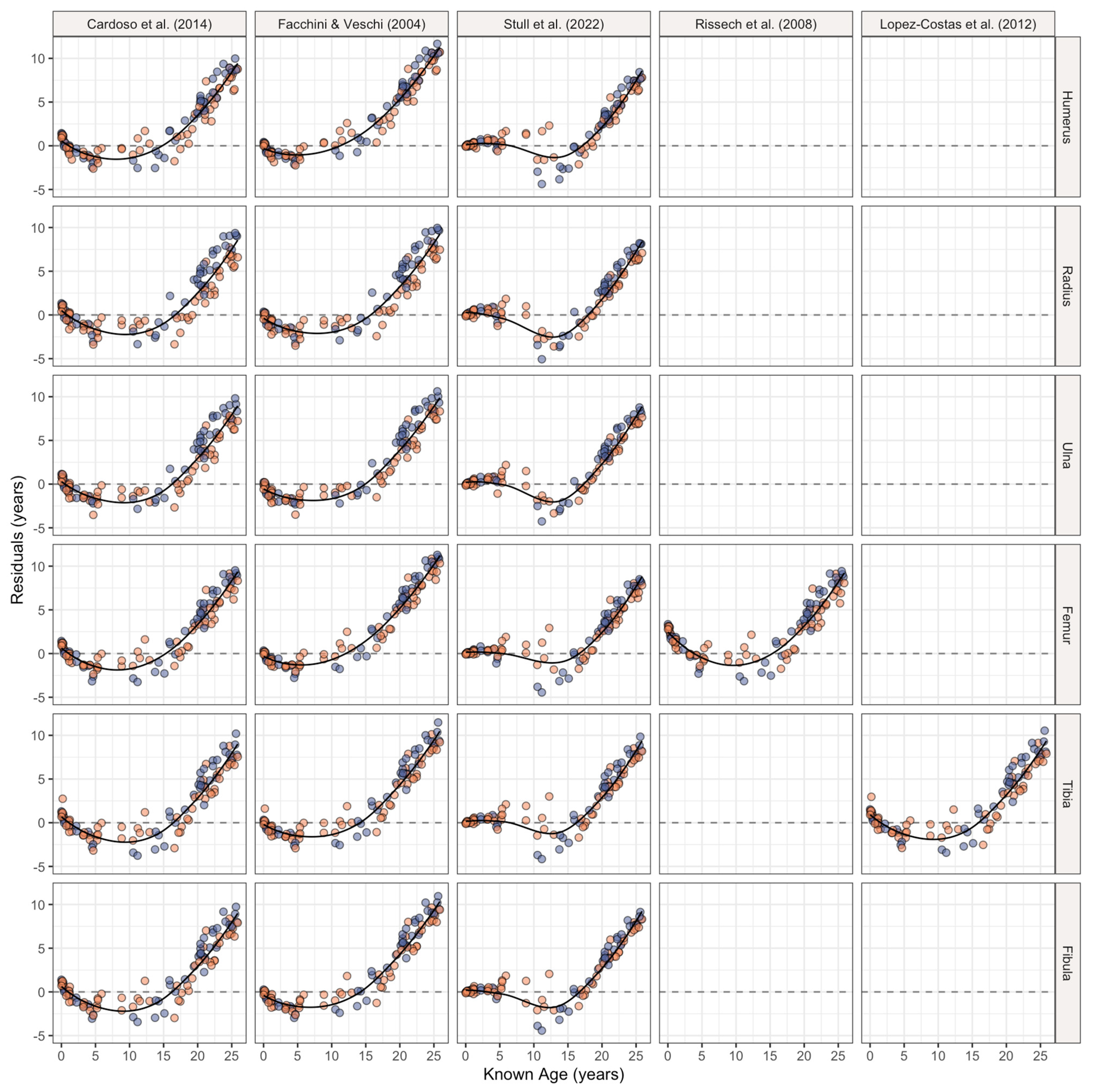
| Cardoso et al. [50] | 0–13 | F: 72 M: 122 | Portuguese and English | Linear | x |
| Facchini & Veschi [52] | 0–12 | F: 70 M: 79 | Italian | Linear | |
| Lόpez-Costas et al. [53] | 0–17 | F: 26 M: 31 | Portuguese and English | Linear | |
| Rissech et al. [54] | 0–16 | F: 52 M: 35 | Portuguese, English, Spanish, and Scottish | Linear | |
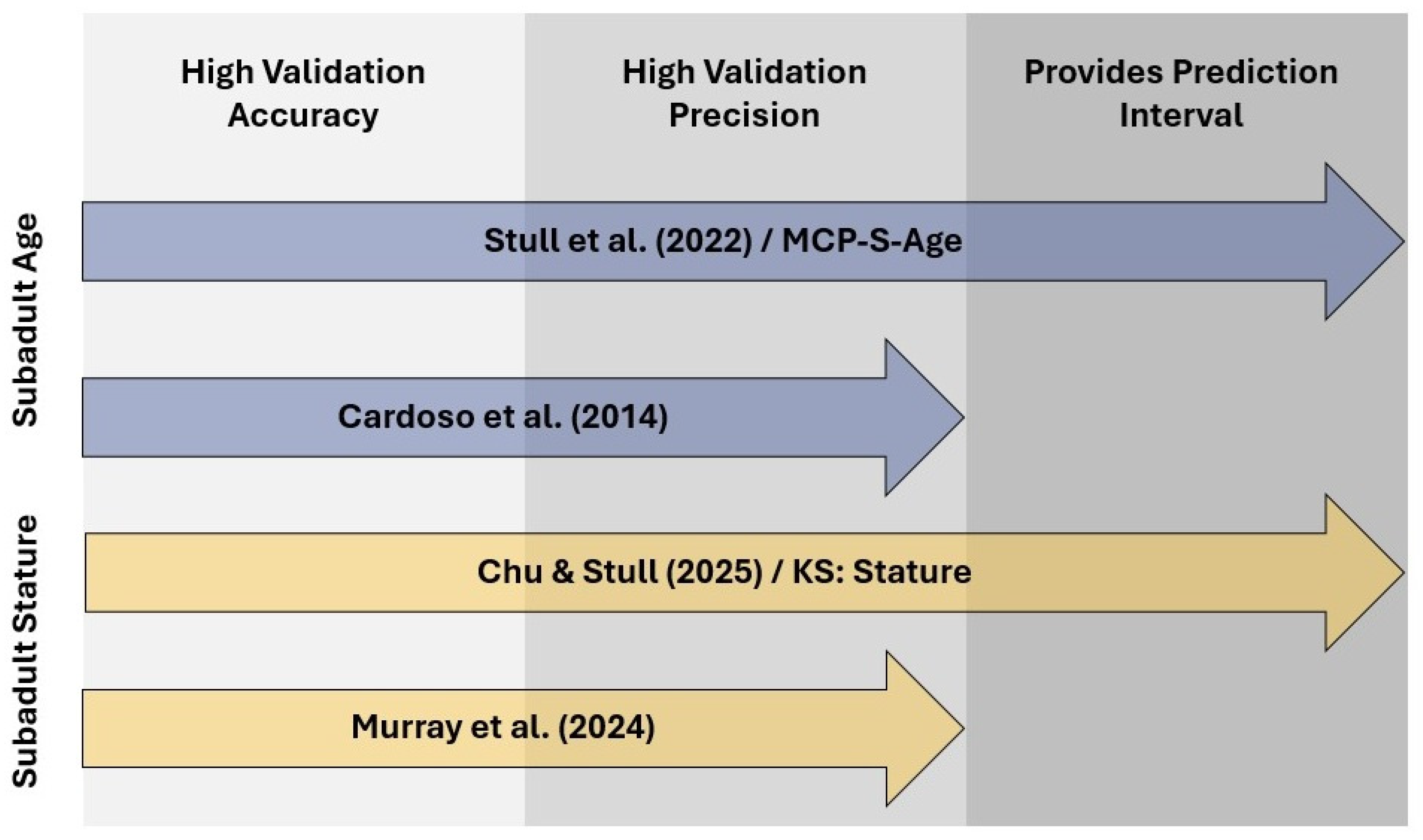
| Stull et al. [56]/ MCP-S-Age | 0–16 | F: 405 M: 526 | American (U.S.) | Nonlinear | x |
| Stature Estimation Methods | |||||
| Brits et al. [1] | 10–17 | F: 30 M: 29 | Black South African | Linear | x |
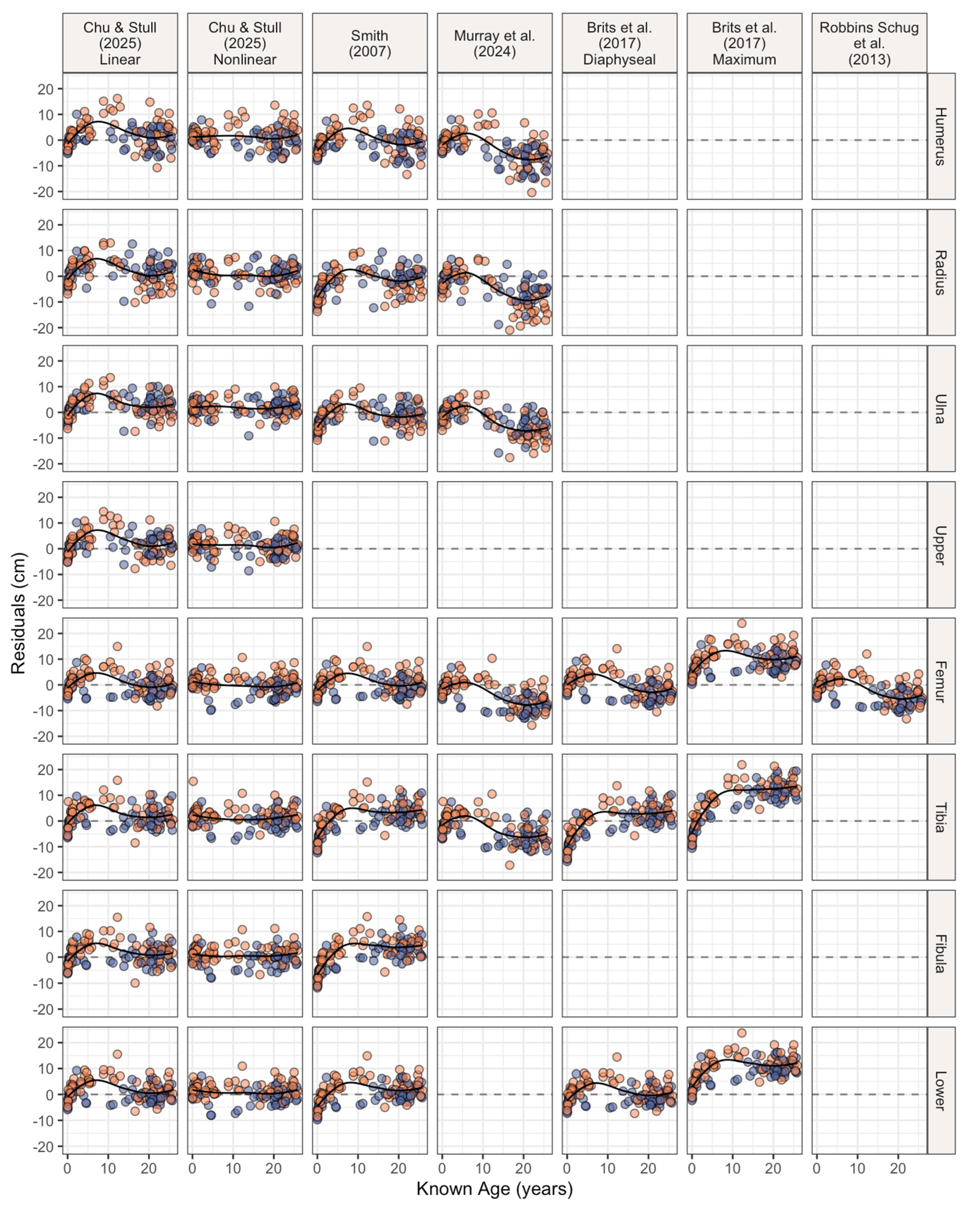
| Chu & Stull [51]/ KS: Stature | 0–20 | F: 401 M: 589 | American (U.S.) | Linear and Nonlinear | x |
| Murray et al. [13] | 0–12 | F: 79 M: 73 | American (U.S.) | Linear | x |
| Robbins Schug et al. [55] | 0.5–11.5 | 20 individuals, sex unknown | American (U.S.) | Linear | |
| Smith [14] | 3–10 | F: 36 M: 31 | American (U.S.) | Linear | x |
| Method | Long Bone | Truncated Sample Size | Accuracy (%) | MSE (in Years) | Full Sample Size | Accuracy (%) | MSE (in Years) |
|---|---|---|---|---|---|---|---|
| Cardoso et al. [50] | Humerus | 55 | 94.55 | 1.23 | 117 | 55.56 | 17.60 |
| Radius | 55 | 92.73 | 1.78 | 115 | 60 | 14.28 | |
| Ulna | 55 | 96.36 | 1.62 | 116 | 59.48 | 15.65 | |
| Femur | 55 | 90.91 | 1.59 | 117 | 52.99 | 17.12 | |
| Tibia | 55 | 89.09 | 1.92 | 115 | 54.78 | 14.97 | |
| Fibula | 55 | 90.91 | 1.80 | 114 | 56.14 | 15.05 | |
| Facchini & Veschi [52] | Humerus | 53 | - | 0.95 | 117 | - | 28.40 |
| Radius | 53 | 2.12 | 115 | 17.36 | |||
| Ulna | 53 | 1.89 | 116 | 20.23 | |||
| Femur | 53 | 1.16 | 117 | 27.65 | |||
| Tibia | 53 | 1.46 | 115 | 22.04 | |||
| Fibula | 53 | 1.70 | 114 | 21.59 | |||
| Lόpez-Costas et al. [53] | Tibia | 64 | - | 1.82 | 115 | - | 16.38 |
| Rissech et al. [54] | Femur | 60 | - | 3.19 | 117 | - | 17.23 |
| Stull et al. [56]/ MCP-S-Age | Humerus | 61 | 93.44 | 1.50 | 117 | 71.79 | 12.18 |
| Radius | 60 | 85.00 | 2.10 | 115 | 66.96 | 11.72 | |
| Ulna | 60 | 91.67 | 1.54 | 116 | 69.83 | 13.28 | |
| Femur | 60 | 88.33 | 1.40 | 117 | 63.25 | 13.23 | |
| Tibia | 61 | 90.16 | 1.35 | 115 | 61.74 | 14.97 | |
| Fibula | 61 | 96.72 | 1.44 | 114 | 70.18 | 14.22 |
| Method | Long Bone | Truncated Sample Size | Accuracy (%) | MSE (in cm) | Full Sample Size | Accuracy (%) | MSE (in cm) |
|---|---|---|---|---|---|---|---|
| Brits et al. [1] Diaphyseal | Femur | 15 | 26.67 | 38.14 | 117 | 53.85 | 20.96 |
| Tibia | 16 | 43.75 | 33.14 | 115 | 45.22 | 37.57 | |
| Lower (Femur + Tibia) | 15 | 26.67 | 35.19 | 114 | 56.14 | 16.41 | |
| Brits et al. [1] Maximum | Femur | 15 | 0.00 | 167.90 | 117 | 6.83 | 108.20 |
| Tibia | 16 | 0.00 | 163.59 | 115 | 20.00 | 117.84 | |
| Lower (Femur + Tibia) | 15 | 0.00 | 175.61 | 114 | 8.77 | 114.20 | |
| Chu & Stull [51]/ KS: Stature Linear | Humerus | 73 | 91.78 | 29.52 | 117 | 94.02 | 28.81 |
| Radius | 72 | 95.83 | 30.33 | 115 | 97.39 | 26.42 | |
| Ulna | 73 | 95.89 | 25.33 | 116 | 97.41 | 24.49 | |
| Upper (Humerus + Radius) | 72 | 94.44 | 27.62 | 115 | 95.65 | 23.62 | |
| Femur | 73 | 98.63 | 19.76 | 117 | 99.15 | 17.91 | |
| Tibia | 73 | 97.26 | 25.48 | 115 | 98.26 | 23.21 | |
| Fibula | 73 | 98.63 | 22.59 | 114 | 98.25 | 20.32 | |
| Lower (Femur + Tibia) | 72 | 98.61 | 21.42 | 114 | 99.12 | 18.32 | |
| Chu & Stull [51]/ KS: Stature Nonlinear | Humerus | 73 | 95.89 | 15.10 | 117 | 94.87 | 18.15 |
| Radius | 72 | 97.22 | 16.85 | 115 | 98.26 | 14.96 | |
| Ulna | 73 | 98.63 | 12.57 | 116 | 98.28 | 14.36 | |
| Upper (Humerus + Radius) | 72 | 98.61 | 13.34 | 115 | 98.26 | 13.62 | |
| Femur | 73 | 95.89 | 12.71 | 117 | 96.58 | 13.21 | |
| Tibia | 73 | 97.26 | 15.85 | 115 | 97.39 | 15.75 | |
| Fibula | 73 | 98.63 | 12.70 | 114 | 98.25 | 12.63 | |
| Lower (Femur + Tibia) | 72 | 98.61 | 12.22 | 114 | 97.37 | 12.30 | |
| Murray et al. [13] | Humerus | 53 | 94.34 | 12.85 | 117 | 73.50 | 49.51 |
| Radius | 53 | 96.23 | 16.11 | 115 | 71.30 | 64.56 | |
| Ulna | 53 | 100.00 | 11.31 | 116 | 81.90 | 40.47 | |
| Femur | 53 | 92.45 | 15.09 | 117 | 69.23 | 44.48 | |
| Tibia | 53 | 96.23 | 16.45 | 115 | 78.26 | 36.79 | |
| Robbins Schug et al. [55] | Femur | 37 | - | 15.92 | 117 | - | 29.38 |
| Smith [14] | Humerus | 17 | 82.35 | 24.54 | 117 | 75.21 | 25.84 |
| Radius | 17 | 82.35 | 19.27 | 115 | 72.17 | 31.40 | |
| Ulna | 17 | 82.35 | 15.24 | 116 | 81.03 | 20.51 | |
| Femur | 17 | 52.94 | 28.04 | 117 | 78.63 | 17.57 | |
| Tibia | 17 | 64.71 | 21.29 | 115 | 57.39 | 30.38 | |
| Fibula | 17 | 58.82 | 22.23 | 114 | 49.12 | 32.86 | |
| Lower (Femur + Tibia) | 17 | 47.06 | 22.47 | 114 | 61.14 | 20.12 |
Disclaimer/Publisher’s Note: The statements, opinions and data contained in all publications are solely those of the individual author(s) and contributor(s) and not of MDPI and/or the editor(s). MDPI and/or the editor(s) disclaim responsibility for any injury to people or property resulting from any ideas, methods, instructions or products referred to in the content. |
© 2025 by the authors. Licensee MDPI, Basel, Switzerland. This article is an open access article distributed under the terms and conditions of the Creative Commons Attribution (CC BY) license (https://creativecommons.org/licenses/by/4.0/).
Share and Cite
Chu, E.Y.; Treviño, A.J.; Vasquez, M.E. Validation of Subadult Age and Stature Estimation Methods Using a Contemporary Japanese Sample. Forensic Sci. 2025, 5, 41. https://doi.org/10.3390/forensicsci5030041
Chu EY, Treviño AJ, Vasquez ME. Validation of Subadult Age and Stature Estimation Methods Using a Contemporary Japanese Sample. Forensic Sciences. 2025; 5(3):41. https://doi.org/10.3390/forensicsci5030041
Chicago/Turabian StyleChu, Elaine Y., Amariah J. Treviño, and Marissa E. Vasquez. 2025. "Validation of Subadult Age and Stature Estimation Methods Using a Contemporary Japanese Sample" Forensic Sciences 5, no. 3: 41. https://doi.org/10.3390/forensicsci5030041
APA StyleChu, E. Y., Treviño, A. J., & Vasquez, M. E. (2025). Validation of Subadult Age and Stature Estimation Methods Using a Contemporary Japanese Sample. Forensic Sciences, 5(3), 41. https://doi.org/10.3390/forensicsci5030041


