Article

Development of a Fluorescent Ionic Liquid Nanosensor for the Onsite Detection of Gamma-Hydroxybutyrate

by
Joel E. R. Moss
1,
Kathryn Hamory
2,
Robert Moreland
3,
Carolyn B. Oakley
3,
David K. Bwambok
1,* and
Vivian E. Fernand Narcisse
3,4,*
1
Departmentof Chemistry, Ball Sate University, Muncie, IN 47306, USA
2
Department of Biology and Kinesiology, LeTourneau University, Longview, TX 75607, USA
3
Department of Chemistry and Physics, LeTourneau University, Longview, TX 75607, USA
4
Department of Chemistry, Forensic Science, and Oceanography, Palm Beach Atlantic University, West Palm Beach, FL 33401, USA
*
Authors to whom correspondence should be addressed.
Forensic Sci. 2025, 5(3), 28; https://doi.org/10.3390/forensicsci5030028
Submission received: 17 March 2025 / Revised: 11 May 2025 / Accepted: 2 July 2025 / Published: 7 July 2025

Abstract

Background/Objectives: Over the past few decades, controlled substance abuse in drug-facilitated sexual assaults (DFSAs) has significantly increased worldwide, leading to an urgency to develop rapid and selective drug detection methods for field use (i.e., on-spot detection). Currently, techniques for detecting DFSA drug-associated samples are laborious and require skilled personnel to analyze/interpret the results. Moreover, most DFSA-associated drugs have a short half-life, making them more challenging to detect promptly. For instance, the timely detection of gamma-hydroxybutyrate (GHB) has been of ultimate concern for decades due to its fast elimination from the body. This study describes the development of a fluorescent ionic liquid nanosensor that can be used to rapidly detect GHB in the field. Methods: Trihexyltetradecylphosphonium fluorescein (THP2FL) ionic liquid was synthesized and evaluated for its potential application in detecting GHB. THP2FL nanoparticles in deionized water were synthesized with a size of 199 nm by a reprecipitation method. Results: The addition of GHB to THP2FL nanoparticles resulted in up to a 60% increase in fluorescence intensity and a 79% increase in absorbance. These results suggest potential applications for using the fluorescent THP2FL nanoparticles to detect GHB. The sensor’s selectivity was tested on compounds structurally similar to GHB, and the results showed that 1,4-butanediol (a precursor of GHB) is a potentially interfering species. Conclusion: This fluorescent technique allows for field deployable sensors, which would benefit screening GHB onsite.

1. Introduction

The number of cases of controlled substance abuse has increased over the past several decades. Gamma-hydroxybutyrate (GHB) is a Schedule I psychoactive drug that has seen a dramatic increase since the 1990s in abuse and recreational purposes [1,2,3]. This illicit drug is often used in liquid form in drug-facilitated crimes (DFCs), mainly during drug-facilitated sexual assaults (DFSAs) [4,5]. Clinically, GHB was used in the 1960s as an anesthetic drug in surgeries, and currently, the sodium salt of GHB (Xyrem®) is used to treat narcolepsy and alcohol dependence [3,6,7,8,9]. GHB naturally occurs in minute concentrations (0.5–1.0 mg/L) in mammals, where it is both a precursor and metabolite of the neurotransmitter gamma-aminobutyric acid (GABA) and acts as a central nervous system depressant [1,2,3,10,11]. Using low amounts of GHB causes depressant effects such as anxiety reduction, drowsiness, euphoria, and visual distortion. However, when ingested at high doses or taken with alcohol, GHB can result in a variety of intoxicating effects, such as unconsciousness, seizures, vomiting, coma, or even fatal overdose [1,2,3,11]. Because of GHB’s euphoric and sedative properties and unique physical properties (i.e., colorless, odorless, somewhat salty-tasting, and soluble in beverages), this drug is an excellent candidate for DFSA [3,12,13]. Therefore, GHB is referred to as a date rape drug as it can be spiked (usually between 1 and 5 g) in individuals’ drinks at bars and clubs, leaving victims more susceptible to sexual assaults [3,14,15]. Moreover, GHB causes momentary memory loss, a type of retrograde amnesia, so the victim cannot recall any harm [3,16].
Upon ingestion, the body readily absorbs GHB, which has a half-life of 30–50 min. It becomes active within 15 to 30 min, and, depending upon the administered dosage, the effects can last for 3 to 6 h. Approximately 1–5% of the ingested dose is found in urine within 3–10 h and in blood plasma within 5 h, making it challenging to easily detect GHB in victims and, therefore, requiring expedited sample collection and analysis. Additionally, a baseline sample must be collected from victims about 12 h after the incident to determine their endogenous GHB concentration, which should be between 0.5 and 2.0 µg/mL in urine [3,10,17]. In contrast, the overdose GHB concentration in urine is generally greater than 1 mg/mL [18]. All of the above-mentioned factors make it problematic to analyze collected biological samples from victims for the presence of GHB, especially since they typically do not come forward directly due to unawareness of their assault, fear, shame, and/or guilt. Moreover, the detection of GHB is tedious and time-consuming as it requires various analytical steps and the use of proper instrumentation by skilled forensic scientists. For example, samples suspected of containing GHB are often analyzed by gas chromatography–mass spectroscopy (GC-MS), which requires a derivatization step in the sample preparation [1,14,18]. In addition, the analysis of GHB in biological samples, including urine, blood, and saliva, must be performed quickly due to its short half-life, which can impact the accuracy and the limit of detection [1,11,19,20,21,22]. Due to the time-consuming process of GHB detection, there is a need to develop quick and reliable (i.e., selective and sensitive) screening techniques for the presumptive identification of this controlled substance in suspected beverages and biological samples. Sensor-based detection methods are a unique technique that may overcome these limitations.
Various dye-based colorimetric and fluorescence methods have been developed to detect GHB [15,22,23,24,25]. However, the use of these methods remains limited since their fabrication requires several preparation steps, and using portable/field spectrophotometers might be less accurate than benchtop instruments [26]. Developments of colorimetric or fluorometric sensors for GHB would be helpful in solving this problem. This study explores using a fluorescein-based ionic liquid to detect GHB. Ionic liquids are salts whose melting points are generally below 100 °C and have tunable properties dependent on the cation and anion pairing [27,28]. This study investigated the use of trihexyltetradecylphosphonium fluorescein (THP2FL and THPFL) ionic liquid nanoparticles that have demonstrated pH sensing capability [29]. The rapid detection of GHB would help address the cost and time limitations of identification techniques widely used in the forensic field. In addition, the selectivity of the sensor to GHB will be tested to reduce potential false-positive results. To the best of our knowledge, this study is the first to report the use of ionic liquids in detecting GHB.

2. Experimental Section

Materials

Ethanol, methanol, dichloromethane, and 4-hydroxybutyric acid sodium salt solution (1 mg/mL) in methanol, disodium fluorescein, 1,4-butanediol, N-butyric acid, and propionic acid, and deuterated dimethyl sulfoxide (DMSO-d6) were obtained from Sigma Aldrich (St Louis, MO, USA) and used without further purification. Trihexyltetradecylphosphonium chloride was obtained from Strem Chemicals Inc. (Newburyport, MA, USA) and used without further purification. Proton NMR was measured using a JEOL spectrometer (400 MHz, JEOL, Tokyo, Japan). UV–vis absorption was measured using a JASCO V-740 spectrophotometer (JASCO, Easton, MD, USA) equipped with a Deuterium and Xenon Arc lamp and a Silicon photodetector. Fluorescence intensity was measured using a JASCO FP-8350 spectrophotometer (JASCO, Easton, MD, USA) equipped with a Xenon Arc lamp and photomultiplier tube with an extended near-infrared detector. The size and zeta potential of the synthesized nanoparticles were measured by dynamic light scattering (DLS) using a Malvern Zetasizer Nano ZS (Malvern Instruments, Malvern, Worcestershire, UK) at 25 °C and at a scattering angle of 173°.

3. Experimental Procedures

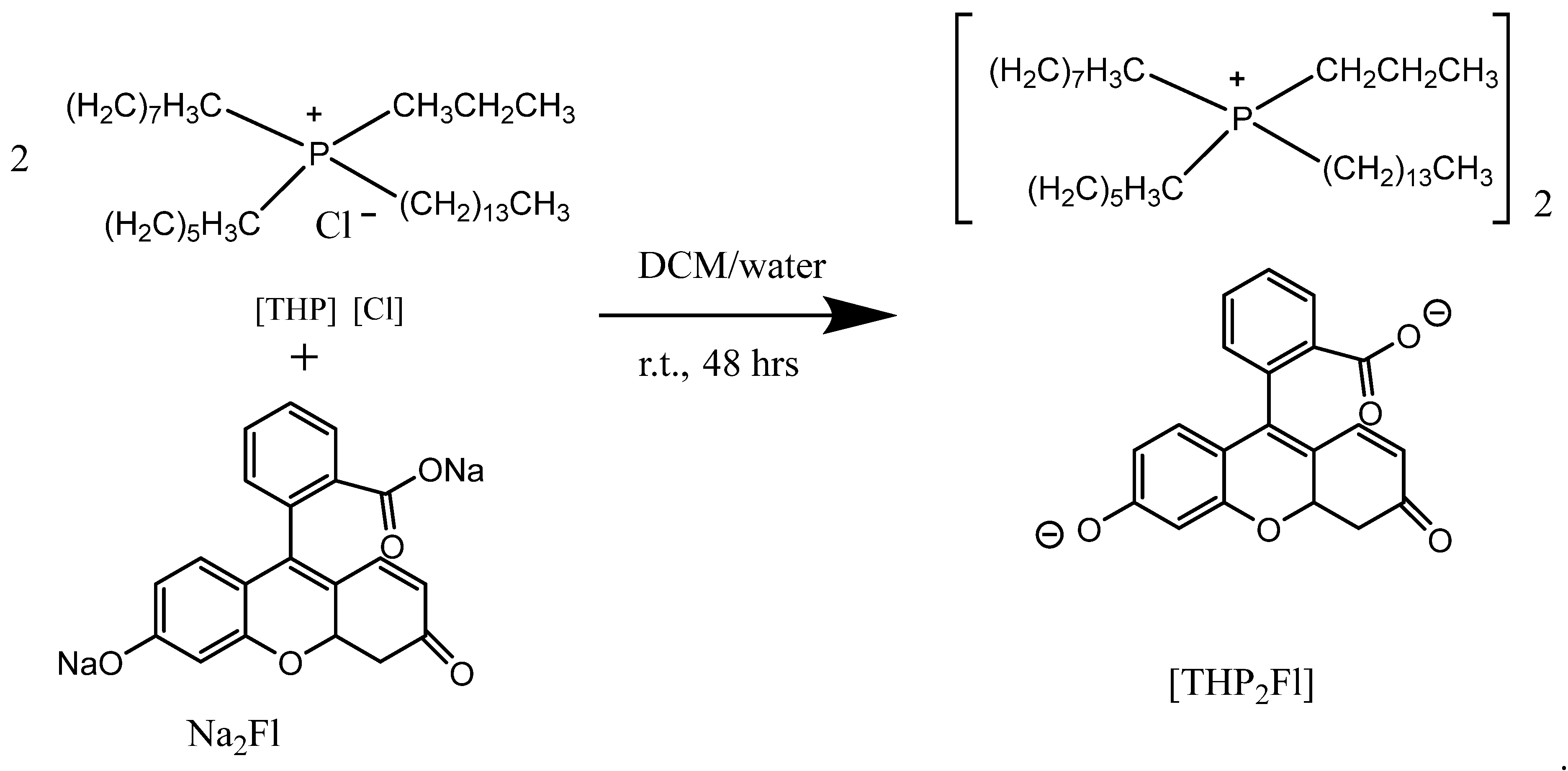
3.1. Synthesis and Characterization of THP2FL Ionic Liquid

The THP2FL ionic liquid was synthesized following a reported anion exchange procedure [29]. The synthesis was performed by mixing a 2:1 molar ratio of THPCl and Na2FL in a mixture of dichloromethane (DCM) and water (2:1, v/v) and stirring for 48 h (Scheme 1). The organic layer, containing the product, was separated from the aqueous layer using a separatory funnel. The separated DCM layer was washed with water three times, then the solvent was removed using a rotary evaporator and dried in a vacuum oven to obtain the ionic liquid product, THP2FL, as a dark red viscous liquid. Proton NMR was carried out on the product, and the results of the chemical shifts obtained were consistent with the chemical structure of the ionic liquid product [29]. The proton NMR spectrum (using d6-DMSO solvent) is provided in the Supporting Information (Figure S1).

3.2. Synthesis of THP2FL Nanoparticles

A reprecipitation method was used to synthesize the nanoparticles by dispersing in water [29]. A 10 mL of 1 mM THP2FL stock solution was prepared in ethanol. Next, 250 μL of the stock solution (1 mM THP2FL in ethanol) was injected into 5 mL of deionized water and sonicated for 2 min. The THP2FL nanoparticles appeared as a pink dispersion.

3.3. Preparation of THP2FL Samples for Absorbance and Fluorescence Measurements

For THP2FL in ethanol, a stock solution of 1 mM THP2FL was prepared in ethanol and diluted to 10 μM for UV–vis and fluorescence measurements. Absorbance was measured using a JASCO spectrophotometer (V-730) with a 1 cm-quartz cuvette. Fluorescence was measured using a JASCO fluorometer (FP-8350) with the bandwidth set at 1 nm.
For THP2FL nanoparticles, the dispersion was diluted in water (50 μL of the THP2FL nanoparticles in 1 mL using DI water) before the absorbance and fluorescence measurements. The excitation wavelength used for measuring fluorescence intensity was the maximum wavelength found in the absorbance spectrum of the nanoparticles and the bandwidth was set at 2.5 nm.

3.4. Characterization of Size and Zeta Potential of the Nanoparticles

A dynamic light scattering (DLS) technique was used to determine the size distribution and zeta potential of the THP2FL nanoparticles. A freshly prepared THP2FL nanoparticle dispersion (5 μM) in DI water was briefly vortexed and then transferred to respective sample cells for f size and zeta potential measurement using the Malvern Nano DLS system.

3.5. GHB Detection Using THP2FL Nanoparticles

For GHB detection experiments, various amounts of GHB (1 mg/mL in methanol) were added to 50 μL of freshly prepared THP2FL nanoparticle solution and then diluted to 1 mL using methanol in centrifuge tubes. The added amounts of GHB ranged from 10 μL to 100 μL, in increments of 10 μL. This maintained the concentration of the THP2FL nanoparticles, which was constant in each sample, to determine the effect of increasing amounts of GHB on the nanoparticles. For the control experiment, 50 μL of the THP2FL nanoparticle solution was diluted to 1 mL using methanol, and incremental amounts of methanol were added (instead of GHB). This control experiment was used to determine whether the methanol solvent has an effect on the absorbance and fluorescence, since the GHB stock sample was obtained as 1 mg/mL in methanol. The following concentrations of GHB were used for the measurements: 10, 20, 30, 40, 50, 60, 70, 80, 90, and 100 µg/mL.

3.6. Testing Selectivity of the GHB Nanosensor Using Potential Interfering Species with Similar Chemical Structures

Potential interfering species for GHB detection (including 1,4-butanediol, N-butyric acid, and propionic acid) were tested using the THP2FL nanosensor. These compounds have a similar structure to GHB as shown in Scheme 2. In centrifuge tubes, 50 μL of an interfering species was added to 50 μL of THP2FL nanoparticle solution and diluted to 1 mL using methanol. An additional test was conducted using 50 μL of the nanoparticle solution and diluted to 1 mL using ethanol, instead of methanol. This was performed to determine if the THP2FL nanoparticle sensor would be affected by alcoholic beverages. Absorbance and fluorescence of the samples were used to determine the effect of ethanol on the THP2FL nanoparticle sensor.

3.7. Testing Selectivity of the GHB Nanosensor Using Potential Interfering Species in Drinks and Beverages

Potential interfering species for GHB detection found in drinks (including glucose, sucrose, citric acid, and sodium citrate) were tested with the THP2FL nanosensor. In centrifuge tubes, 50 μL of potential interfering species was added to 50 μL of THP2FL nanoparticle dispersion and 50 μL of GHB, and diluted to 1 mL using methanol.
For the control, 50 μL of the THP2FL nanoparticle solution was added to 50 μL GHB and diluted to 1 mL using methanol. Absorbance and fluorescence of the samples were measured to determine the effect of interfering species on GHB detection using the THP2FL nanoparticle sensor.

4. Results and Discussion

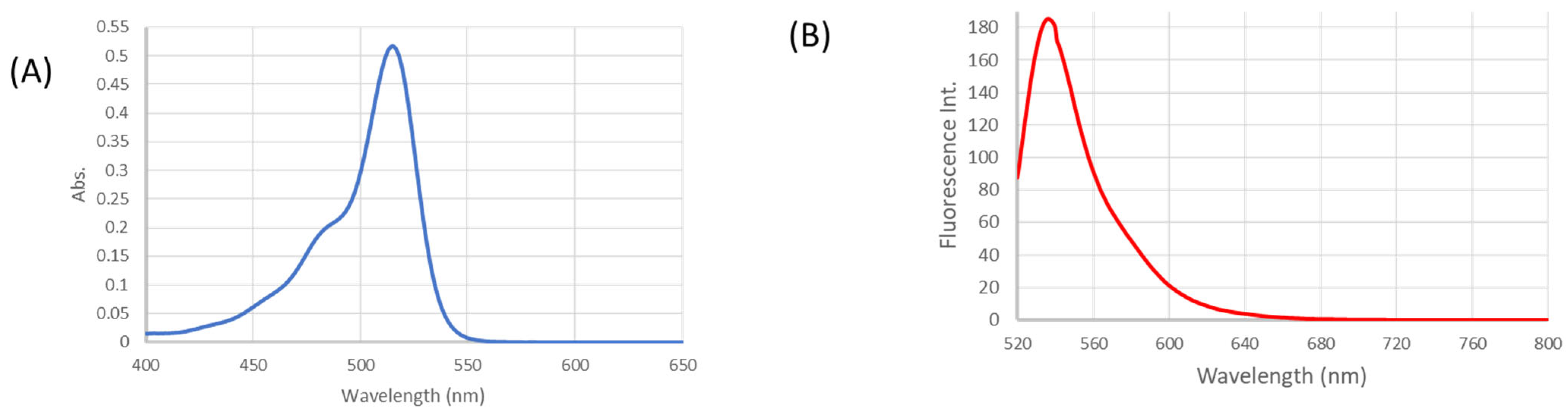
4.1. Photophysical Characterization of THP2FL

The absorbance maximum wavelength of THP2FL (10 μM) in ethanol was determined to be 515 nm. The fluorescence emission of THP2FL (10 μM) in ethanol at an excitation wavelength of 515 nm showed that the emission maxima was 536 nm (Figure 1).

4.2. Characterization of THP2FL Nanoparticles

Dynamic light scattering (DLS) was used to measure the size of the synthesized THP2FL nanoparticles in water. The size of the nanoparticles was 199 nm ± 76 nm, and the zeta potential was 25 mV (Figure 2). The polydispersity index (PDI) was 0.100, indicating a uniform distribution of the nanoparticles’ size in the aqueous solution (Figure 2). In addition, a high positive value of zeta potential (+25 mV) indicating that the nanoparticle dispersion is stable to aggregation due to the repulsion of positive surface charges.
The THP2FL nanoparticle dispersion exhibited a bathochromic shift with a maximum absorbance at a longer wavelength of 524 nm compared with the absorbance of 515 nm in the ethanol solution. Similarly, there was a red shift in the fluorescence emission of the nanoparticles with a maximum wavelength of 546 nm (at 524 nm excitation) compared with 540 nm (at 515 nm excitation) for the dye in solution (Figure 3). This red shift in the absorbance and emission of THP2FL nanoparticle dispersion in water has been attributed to the J-aggregation [30].

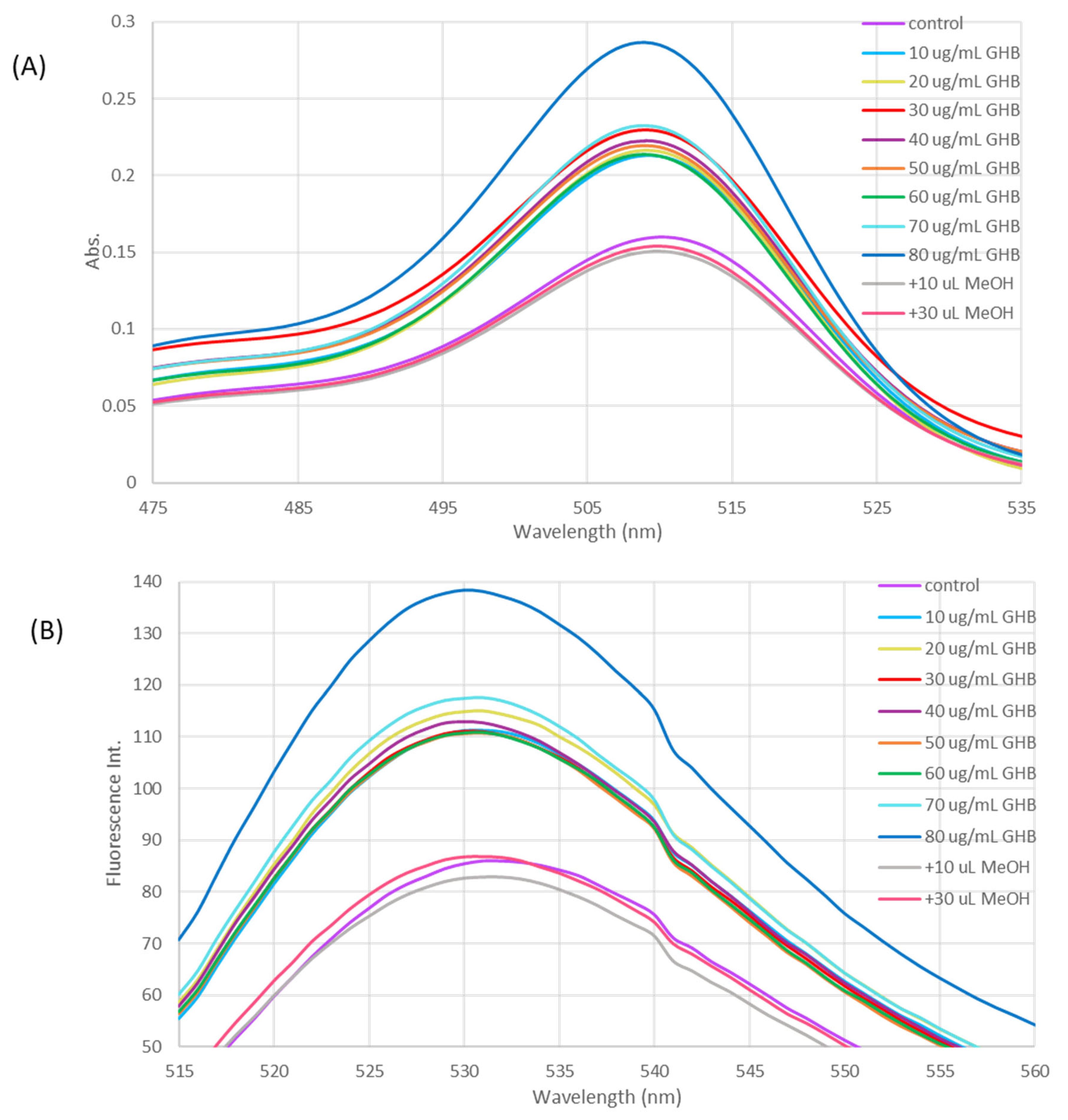
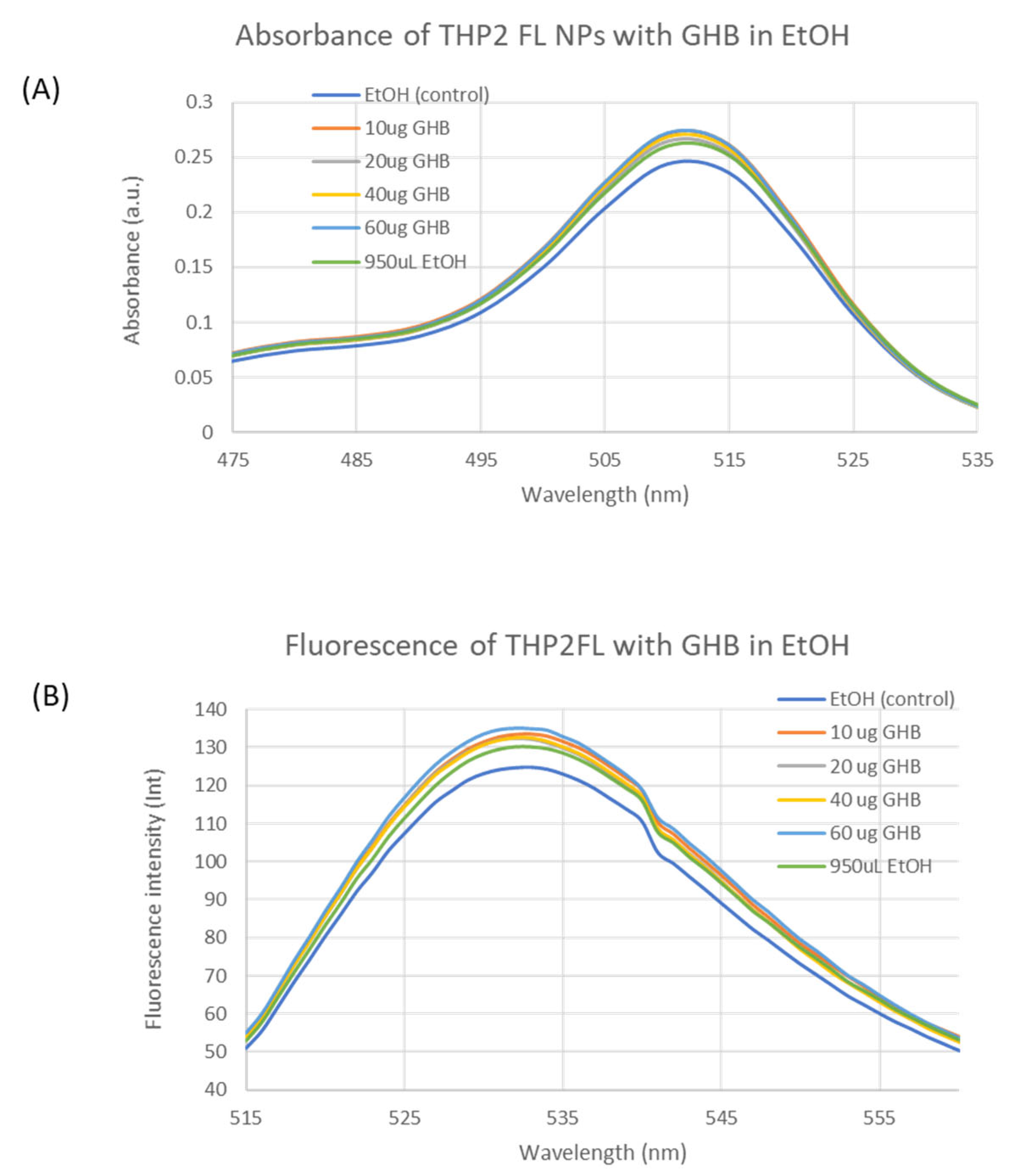
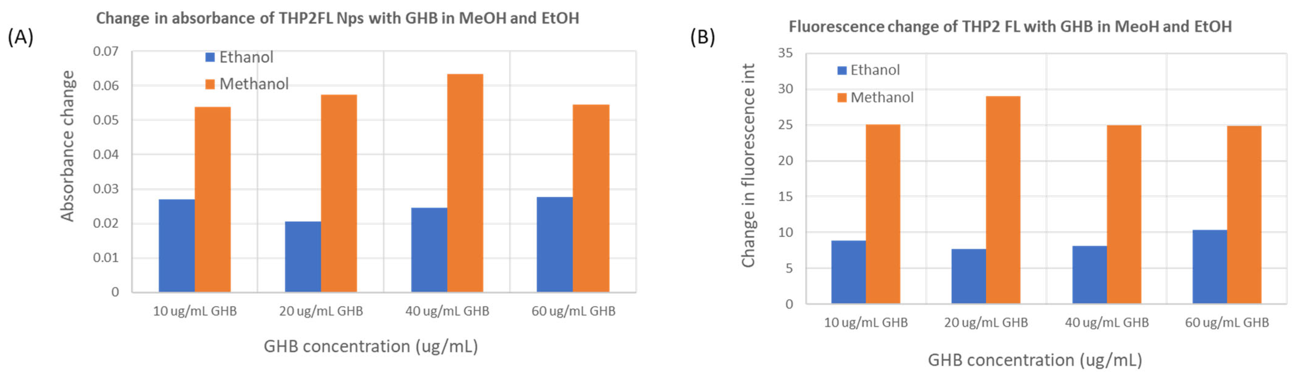
4.3. GHB Sensing Using THP2FL Nanoparticles

For the sensing experiments, various amounts of GHB were added to 50 μL of THP2FL nanoparticle dispersion and then diluted to 1 mL in methanol. Since the concentration of THP2FL nanoparticles was kept the same in all experiments, the change in absorbance and fluorescence signals is due to the changes in GHB concentrations (Figure 4). These results suggest that THP2FL nanoparticles can serve as potential sensors for detecting GHB in the concentration range tested (10–80 μg/mL). These concentrations are well within the range in which GHB is reported to cause severe problems when ingested (75 mM in a 330 mL drink) [31]. The addition of GHB (at the range 10–80 μg/mL) to the nanoparticles displayed an increase, then a plateau followed by an increase in the intensity of fluorescence and absorbance (Figure S2). The cause for this trend is unclear, but the decreased absorbance and fluorescence may be attributed to the reported formation of weakly fluorescent H-type or randomly oriented aggregates of these nanoparticles at higher analyte concentrations [29]. The experiments with additional methanol solvent added indicated a minimal effect of methanol on the absorbance and fluorescence signal of the THP2FL nanoparticles, supporting GHB as the cause of the change in signal intensity (Figure 4).
The change in the color of the nanoparticles with increasing amounts of GHB is shown (Figure S3, Supporting Information). There is a color change with GHB concentration. In addition, the emission intensity increases with GHB concentration under UV light illumination (Figure S3, Supporting Information). This provides an opportunity for the practical application of THP2FL nanoparticles for the presumptive screening of GHB in the field with UV light illumination.

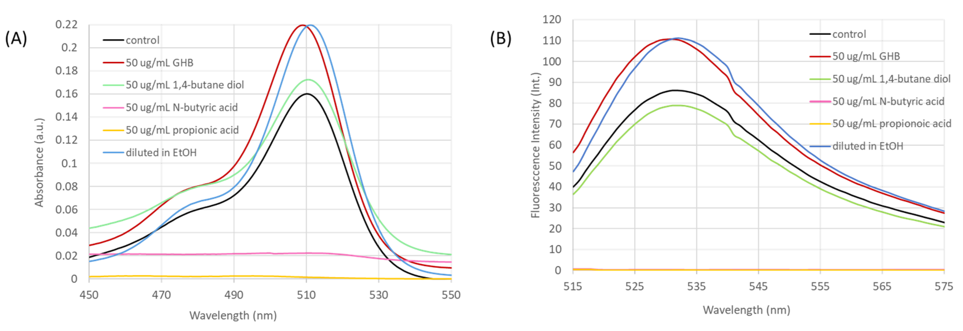
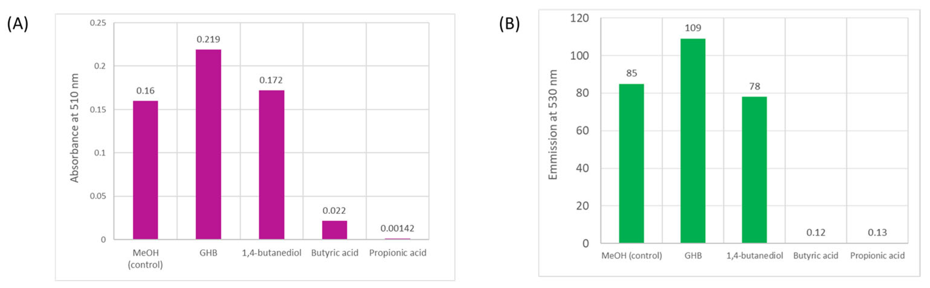
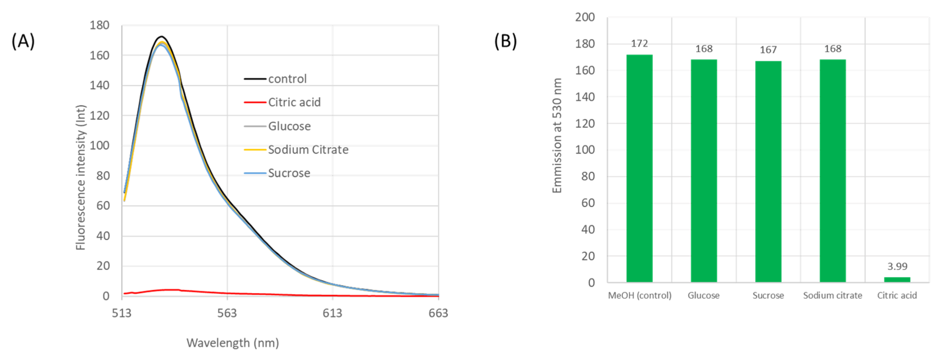
4.4. Testing for Interfering Species

The selectivity of the THP2FL nanoparticle sensor was investigated with three commonly used compounds with similar molecular structures to GHB including 1,4-butanediol, N-butyric acid, and propionic acid [15]. Each of the three compounds was added at equal concentrations of 50 μg/mL to the THP2FL nanoparticles. This allowed for their effect on the THP2FL nanoparticles at this concentration to be comparable to that of 50 μg/mL GHB. Both N-butyric acid and propionic acid resulted in a decreased absorbance and fluorescence intensity at the maximum wavelengths (Figure 5). However, adding 1,4-butanediol to the THP2FL nanoparticles resulted in an increase in absorbance but a decrease in fluorescence compared with the experiment with no GHB present (Figure 5). Given that GHB is spiked in alcoholic beverages, we conducted an experiment in which the nanoparticles were diluted in ethanol instead of methanol to evaluate the impact of ethanol on GHB testing with THP2FL nanoparticles. The dilution of the nanoparticles in ethanol resulted in an increase in both absorbance and fluorescence intensity, similar to the experiment with 50 μg/mL GHB (Figure 5). The maximum absorption wavelength of the samples diluted in methanol was 509 nm; when the THP2FL nanoparticles were tested in ethanol, the maximum absorption wavelength was determined to be at a slightly longer wavelength of 512 nm.
Plotting the absorbance at 510 nm and the fluorescence emission at 530 nm of the THP2FL nanoparticles in the presence of potential interfering species provides a clearer comparison (Figure 6). These graphs show that the species with a hydroxyl group show fluorescence emission, while the carboxylic groups quench the fluorescence of the THP2FL sensor (Figure 6). This information may be useful in elucidating the mechanism of interaction between the THP2FL nanosensor and GHB.
The selectivity of the THP2FL nanosensor was tested with other potential interfering species, including glucose, sucrose, sodium citrate, and citric acid. The results showed that the citric acid caused the quenching of fluorescence, while all other species did not have an effect on the THP2FL nanosensor (Figure 7).
Given the potential interference of ethanol for GHB detection using THP2FL nanoparticles, we tested various concentrations of GHB with ethanol as the solvent. The GHB sample was diluted with ethanol and added to varying concentrations of the THP2FL nanoparticle sensor. Interestingly, the addition of increasing amounts of GHB to THP2FL in ethanol did not result in a significant increase in absorbance and fluorescence intensity compared with the GHB samples in methanol (Figure 8). These results suggest that the THP2FL nanoparticle sensor works well in methanol, and ethanol will not be a major problem in GHB detection due to its smaller change in signal intensity across the concentration range tested (Figure 8). Some strategies have been used to overcome interferences, such as using metal complexes with the dye sensors, e.g., BODIPY- iron complexes [20].
Based on these observations, the plausible interaction between the nanoparticles and GHB is probably through hydrogen bonding. An electrostatic interaction through the carboxylate anion may occur, but it does not appear to alter the fluorescence properties. This is based on the observation that butyric acid and propionic acid did not cause potential interference compared with 1,4-butanediol, which showed an increase in the absorbance of the THP2FL nanosensor. Dispersion forces may contribute due to the slight effect of ethanol on the emission of the nanosensor, compared with methanol, which did not appear to have an effect. The shift in emission wavelength with ethanol may be due to aggregation-induced emission, which can aid in differentiating the sensor response to GHB.
A comparison of the performance of this THP2FL nanoparticle sensor for GHB using methanol or ethanol as a solvent is depicted by the change in absorbance at 526 nm and the change in fluorescence intensity at 531 nm (Figure 9). The results show that there is a greater change in signal (approximately three times in fluorescence and two times in absorbance) of the THP2FL nanoparticles with GHB in methanol compared with ethanol (Figure 9). The methanol provides better sensitivity for GHB detection using the THP2FL nanoparticle sensor.

5. Conclusions

In summary, nanoparticles from a fluorescein-based ionic liquid were successfully synthesized and evaluated for the detection of GHB. These THP2FL nanoparticles in water, with a size of 199 nm, were synthesized by a reprecipitation method. The addition of GHB to the THP2FL nanoparticles resulted in about a 79% increase in absorbance and a 60% increase in fluorescence intensity. Both the absorbance and the fluorescence intensity demonstrated a general increase as a function of the amount of GHB added to the THP2FL nanoparticle suspension. These results suggest potential applications for using these fluorescein-based ionic liquid nanoparticles to detect GHB, a controlled substance. The selectivity of the THP2FL nanoparticles was evaluated using compounds with similar chemical structures to GHB. It was determined that 1,4-butanediol and ethanol are potential interfering species to the THP2FL nanoparticle sensor for GHB. Although ethanol may interfere with the sensor, the absorbance and fluorescence intensity change in ethanol is much lower than in methanol.
Future studies will include testing the sensor in ethanol and alcoholic beverages with GHB present and other types of beverages. In addition, the use of the sensor should be evaluated for detecting GHB in biological fluids such as saliva and urine. The results from this study may be used to develop a portable sensor for the colorimetric screening of GHB in the field and at crime scenes. Accurate onsite forensic presumptive screening can reduce the initial detection and identification time associated with the analysis and confirmatory tests of a large number of drug samples at laboratories. The THP2FL nanosensor is useful for presumptive screening and has the potential to be developed for the onsite detection of GHB in the field.

Supplementary Materials

The following supporting information can be downloaded at: https://www.mdpi.com/article/10.3390/forensicsci5030028/s1, Figure S1: Proton NMR of THP2FL in d6-DMSO; Figure S2: Change in (A) absorbance at 526 nm and (B) fluorescence intensity at 531 nm of 50 μL THP2FL nanoparticles as a function of GHB concentration; Figure S3: Colorimetric images of 50 μL of the THP2FL nanoparticle sensor with increasing amounts (0, 10, 20, 30, 40, 50 μg/mL) of GHB from left to right: (A) without UV-illumination and (B) with UV-lamp illumination.

Author Contributions

Conceptualization, D.K.B. and V.E.F.N.; methodology, D.K.B., V.E.F.N., J.E.R.M., K.H., R.M. and C.B.O.; validation, D.K.B. and V.E.F.N.; formal analysis, D.K.B., V.E.F.N., J.E.R.M. and K.H.; investigation, D.K.B., V.E.F.N., J.E.R.M., K.H., R.M. and C.B.O.; resources, D.K.B. and V.E.F.N.; data curation, D.K.B. and V.E.F.N.; writing—original draft preparation, D.K.B., V.E.F.N., J.E.R.M. and K.H.; writing—review and editing, D.K.B. and V.E.F.N.; visualization, D.K.B. and V.E.F.N.; supervision, D.K.B. and V.E.F.N.; funding acquisition, D.K.B. and V.E.F.N. All authors have read and agreed to the published version of the manuscript.

Funding

D.K. Bwambok acknowledges Ball State University start-up funding for the support of this research. This research was partially conducted at Letourneau University, and V.E. Fernand Narcisse acknowledges the Welch Foundation for funding.

Institutional Review Board Statement

Not applicable.

Informed Consent Statement

Not applicable.

Data Availability Statement

The data that support the findings of this study are available from the corresponding author upon reasonable request.

Acknowledgments

The authors extend their gratitude to Sonja Sosa-Saenz for sharing her knowledge of ionic liquids and to Will Mawhorr for his contributions to the study of interfering species for the nanosensor.

Conflicts of Interest

The authors declare no conflicts of interest.

References

  1. Cooper, F.J.; Marinetti, L.J. γ-hydroxybutyrate (GHB)—Effects on human performance and behavior. Forensic Sci. Rev. 2002, 14, 101–121. [Google Scholar]
  2. Berretta, P.; Varì, M.R. The overwhelming issue of drug facilitated sexual assaults (DFSA): The case of GHB. Clin Ter. 2020, 170, e44–e45. [Google Scholar] [PubMed]
  3. Drug Enforcement Administration, Diversion Control Division, Drug & Chemical Evaluation Section. Gamma Hydroxybutyric Acid. Available online: https://www.deadiversion.usdoj.gov/drug_chem_info/ghb.pdf (accessed on 3 January 2025).
  4. García, M.G.; Pérez-Cárceles, M.D.; Osuna, E.; Legaz, I. Drug-facilitated sexual assault and other crimes: A systematic review by countries. J. Forensic Leg. Med. 2021, 79, 102151. [Google Scholar] [CrossRef]
  5. Kapitány-Fovény, M.; Zacher, G.; Posta, J.; Demetrovics, Z. GHB-involved crimes among intoxicated patients. Forensic Sci. Int. 2017, 275, 23–29. [Google Scholar] [CrossRef]
  6. Vickers, M. Gammahydroxybutyric acid. Int. Anesthesiol. Clin. 1969, 7, 75–89. [Google Scholar] [CrossRef] [PubMed]
  7. Galloway, G.P.; Frederick, S.L.; Staggers, F.E.; Gonzales, M.; Stalcup, S.A.; Smith, D.E. Gamma-hydroxybutyrate: An emerging drug of abuse that causes physical dependence. Addiction 1997, 92, 89–96. [Google Scholar]
  8. Robinson, D.; Keatin, G.M. Sodium oxybate: A review of its use in the management of narcolepsy. CNS Drugs 2007, 21, 337–354. [Google Scholar] [CrossRef]
  9. Boscolo-Berto, R.; Viel, G.; Montagnese, S.; Raduazzo, D.I.; Ferrara, S.D.; Dauvilliers, Y. Narcolepsy and effectiveness of gammahydroxybutyrate. (GHB): A systematic review and meta-analysis of randomized controlled trials. Sleep Med. Rev. 2012, 16, 431–443. [Google Scholar] [CrossRef]
  10. Busardò, F.P.; Jones, A.W. GHB pharmacology and toxicology: Acute intoxication, concentrations in blood and urine in forensic cases and treatment of the withdrawal syndrome. Curr. Neuropharmacol. 2015, 13, 47–70. [Google Scholar] [CrossRef]
  11. Busardo, F.P.; Jones, A.W. Interpreting γ-hydroxybutyrate concentrations for clinical and forensic purposes. Clin. Toxicol. 2019, 57, 149–163. [Google Scholar] [CrossRef]
  12. Németh, Z.; Kun, B.; Demetrovics, Z. Review: The involvement of gamma-hydroxybutyrate in reported sexual assaults: A systematic review. J. Psychopharmacol. 2010, 24, 1281–1287. [Google Scholar] [CrossRef] [PubMed]
  13. Carter, L.; Richards, B.; Mintzer, M.; Griffiths, R.R. Relative abuse liability of GHB in humans: A comparison of psychomotor, subjective, and cognitive effects of supratherapeutic doses of Triazolam, Pentobarbital, and GHB. NPP 2006, 31, 2537–2551. [Google Scholar] [CrossRef]
  14. Hughes, R.R.; Walker, G.S. Rapid screening for the detection and differentiation of gamma-hydroxybutyrate using ion-chromatography. J. Forensic Sci. 2011, 56, 1256–1260. [Google Scholar] [CrossRef]
  15. Baumes, L.A.; Buaki Sogo, M.; Montes-Navajas, P.; Corma, A.; Garcia, H. A colorimetric sensor array for the detection of the date-rape drug γ-hydroxybutyric acid (GHB): A supramolecular approach. Chemistry 2010, 16, 4489–4495. [Google Scholar] [CrossRef] [PubMed]
  16. Casey, T.; Lesar, C.T.; Decatur, J.; Lukasiewicz, E.; Champeil, E. Report on the analysis of common beverages spiked with gamma-hydroxybutyric acid (GHB) and gamma-butyrolactone (GBL) using NMR and the PURGE solvent-suppression technique. Forensic Sci. Int. 2011, 212, e40–e45. [Google Scholar]
  17. Frison, G.; Tedeschi, L.; Maietti, S.; Ferrara, S.D. Determination of gamma-hydroxybutyric acid (GHB) in plasma and urine by headspace solid-phase microextraction and gas chromatography/positive ion chemical ionization mass spectrometry. Rapid Commun. Mass. Spectrom. 2000, 14, 2401–2407. [Google Scholar] [CrossRef]
  18. Crookes, C.E.; Faulds, M.C.; Forrest, A.R.; Galloway, J.H. A reference range for endogenous gamma-hydroxybutyrate in urine by gas chromatography-mass spectrometry. J. Anal. Toxicol. 2004, 28, 644–649. [Google Scholar] [CrossRef]
  19. Kintz, P.; Goullé, J.P.; Cirimele, V.; Ludes, B. Window of detection of gamma-hydroxybutyrate in blood and saliva. Clin. Chem. 2001, 47, 2033–2034. [Google Scholar] [CrossRef]
  20. Ryu, J.; Kim, Y. Overcoming interferences in the colorimetric and fluorimetric detection of γ-hydroxybutyrate in spiked beverages. Sensor Actuat. B Chem. 2022, 364, 131861. [Google Scholar] [CrossRef]
  21. Tay, E.; Lo, W.K.W.; Murnion, B. Current insights on the impact of gamma-hydroxybutyrate (GHB) abuse. Subst. Abuse Rehabil. 2022, 13, 13–23. [Google Scholar] [CrossRef]
  22. Alston II, W.C.; Ng, K. Rapid colorimetric screening test for γ-hydroxybutyric acid (liquid X) in human urine. Forensic Sci. Int. 2002, 126, 114–117. [Google Scholar] [CrossRef]
  23. Zhai, D.; Tan, Y.Q.; Xu, W.; Chang, Y.T. Development of a fluorescent sensor for illicit date rape drug GHB. Chem. Commun. 2014, 50, 2904–2906. [Google Scholar] [CrossRef] [PubMed]
  24. Rodríguez-Nuévalos, S.; Costero, A.M.; Parra, M.; Gil, S.; Arroyo, P.; Sáez, J.A.; Gaviña, P.; Ceroni, P.; Fermi, A. Colorimetric and fluorescent hydrazone-BODIPY probes for the detection of γ-hydroxybutyric acid (GHB) and cathinones. Dyes Pigm. 2022, 207, 110757. [Google Scholar] [CrossRef]
  25. Dahiwadkar, R.; Kumar, H.; Kanvah, S. Detection of illicit GHB using AIE active fluorene containing α-Cyanostilbenes. JPPA 2022, 427, 113844. [Google Scholar] [CrossRef]
  26. Alonzo, M.; Alder, R.; Clancy, L.; Fu, S. Portable testing techniques for the analysis of drug materials. WIREs Forensic Sci. 2022, 4, e1461. [Google Scholar] [CrossRef]
  27. Lei, Z.; Chen, B.; Moo, Y.; MacFarlane, D.R. Introduction: Ionic liquids. Chem. Rev. 2017, 117, 6633–6635. [Google Scholar] [CrossRef]
  28. Nordness, O.; Brennecke, F.J. Ion dissociation in ionic liquids and ionic liquid solutions. Chem. Rev. 2020, 120, 12873–12902. [Google Scholar] [CrossRef]
  29. Das, S.; Magut, P.K.S.; de Rooy, S.L.; Hasan, F.; Warner, I.M. Ionic liquid-based fluorescein colorimetric pH nanosensors. RSC Adv. 2013, 3, 21054–21061. [Google Scholar] [CrossRef] [PubMed]
  30. Das, S.; Bwambok, D.; El-Zahab, B.; Monk, J.; de Rooy, S.L.; Challa, S.; Li, M.; Hung, F.R.; Baker, G.A.; Warner, I.M. Nontemplated approach to tuning the spectral properties of cyanine-based fluorescent nanoGUMBOS. Langmuir 2010, 26, 12867–12876. [Google Scholar] [CrossRef]
  31. Huller, C.; Thai, D.; Jacob III, P.; Dyer, J.E. GHB urine concentrations after single-dose administration in humans. J. Anal. Toxicol. 2006, 30, 360–364. [Google Scholar] [CrossRef]
Scheme 1. Synthesis of THP2FL by anion exchange.
Forensicsci 05 00028 sch001
Scheme 2. Chemical structures of (A) GHB, (B) 1,4-butanediol, (C) N-butyric acid, and (D) propionic acid.
Forensicsci 05 00028 sch002
Figure 1. (A) Absorbance spectrum of THP2FL (10 μM) in ethanol showing an absorbance wavelength maximum at 515 nm. (B) Fluorescence spectrum of THP2FL (10 μM) in ethanol at an excitation wavelength 515 nm. The emission wavelength maximum is at 540 nm.
Forensicsci 05 00028 g001
Figure 2. Dynamic light scattering data showing THP2FL nanoparticles with (A) size of 199 nm and (B) zeta potential of 25 mV. The data was collected in triplicate as indicated by the different colors.
Forensicsci 05 00028 g002
Figure 3. (A) Absorbance spectra of the THP2FL nanoparticle suspension in water showing an absorbance wavelength maximum at 524 nm. (B) Fluorescence spectra of the THP2FL nanoparticle dispersion in DI water at excitation wavelength 524 nm. The emission wavelength maxima are at 546 nm.
Forensicsci 05 00028 g003
Figure 4. (A) Absorption and (B) fluorescence spectra of control (THP2FL nanoparticles diluted in methanol), 10–80 μg/mL GHB samples, and nanoparticles diluted to 1 mL using methanol with additional methanol added (+10 μL and +30 μL).
Forensicsci 05 00028 g004
Figure 5. (A) Absorbance and (B) fluorescence spectra of the THP2FL nanoparticles in the presence of 50 μL of potential interfering species for GHB detection including 1,4-butanediol, N-butyric acid, propionic acid, and ethanol. The THP2FL nanoparticles diluted in methanol were used as a control.
Forensicsci 05 00028 g005
Figure 6. (A) Absorbance at 510 nm and (B) fluorescence emission at 530 nm of the THP2FL nanoparticles in the presence of 50 μL of potential interfering species for GHB detection including 1,4-butanediol, N-butyric acid, and propionic acid. The THP2FL nanoparticles diluted in methanol were used as a control.
Forensicsci 05 00028 g006
Figure 7. (A) Fluorescence spectra and (B) emission at 530 nm of the THP2FL nanoparticles in the presence of potential interfering species, including glucose, sucrose, sodium citrate, and citric acid.
Forensicsci 05 00028 g007
Figure 8. (A) Absorption and (B) fluorescence spectra of control (THP2FL nanoparticles diluted in ethanol), 10–60 μg/mL GHB samples, and nanoparticles (50 μL) diluted to 1 mL using 950 μL ethanol.
Forensicsci 05 00028 g008
Figure 9. (A) Change in absorbance at 526 nm, and (B) change in fluorescence intensity at 531 nm of 50 μL THP2FL nanoparticles as a function of GHB concentration in ethanol and methanol.
Forensicsci 05 00028 g009
Disclaimer/Publisher’s Note: The statements, opinions and data contained in all publications are solely those of the individual author(s) and contributor(s) and not of MDPI and/or the editor(s). MDPI and/or the editor(s) disclaim responsibility for any injury to people or property resulting from any ideas, methods, instructions or products referred to in the content.

