
Article

Comparative Morphology and Generic Classification of Catfishes of the Trichomycterus Lineage (Siluriformes: Trichomycteridae) †

by
Wilson J. E. M. Costa
Laboratory of Systematics and Evolution of Teleost Fishes, Institute of Biology, Federal University of Rio de Janeiro, Rio de Janeiro 21941-971, Brazil
urn:lsid:zoobank.org:act:D5CFFD3B-9146-4100-A7AD-106A9ABD2AA5; F92D593B-C398-4486-895B-32DE660AAFFA; B0D91992-E6E9-448C-AA19-3DA339334DCA; 36D853E1-A508-4CB2-BDCD-AE2AFEAF35D9; 492399DB-EC3C-4AFB-B68E-43884B38AE48; urn:lsid:zoobank.org:pub:CDF1C5F9-E95F-489D-9ADE-1030A0539154.
Taxonomy 2026, 6(1), 20; https://doi.org/10.3390/taxonomy6010020
Submission received: 4 January 2026 / Revised: 26 February 2026 / Accepted: 27 February 2026 / Published: 4 March 2026

Abstract

Recent genomic phylogenies have generated new robust classifications of actinopterygian fishes, making possible greater nomenclatural stability, but genus-level classifications of groups like the diverse catfish subfamily Trichomycterinae are still unclear, containing ill-defined paraphyletic taxa. The focus of the present study is the Trichomycterus Lineage (TL), a clade with great morphological diversity, containing about 170 species widely distributed in South America, occurring in the most important biodiversity hotspots of the world, such as the Atlantic Forest, Cerrado, and the Tropical Andes. Most species are small, but at least one reaches about 400 mm of total length, being used as food and depicted in pre-Hispanic Andean ceramics. Based on a comparative morphological analysis, mainly using osteological characters, supported by concordant genomic phylogenies, a new classification at the genus level is here provided. Many morphological features delimiting TL genera seem to be related to ecological adaptations. Nine genera are here recognised of which five are new. Recognition of the new genera will allow easier descriptions of new species and consequently better biodiversity estimates.

1. Introduction

Genomic phylogenies have provided unprecedented and robust information on the internal relationships of Actinopterygii fishes, the largest group of vertebrates in the world, leading to structural changes in their classification [1]. These changes tend towards more stable phylogenetic classification systems over time, fundamental for a better understanding of biodiversity and, consequently, for its conservation. New classification proposals range from new arrangements for orders and families (e.g., [1,2,3]) to intrafamilial revisions at the genus level (Refs. [2,4,5] as examples of Neotropical groups). However, the genus-level classification of some groups continues to have very low phylogenetic resolution, including paraphyletic genera with ambiguous or uninformative diagnoses.
With about 340 valid species [6] occurring in a vast region between southern Central America and southern South America, trichomycterine catfishes (Trichomycteridae: Trichomycterinae) are an example of genus-level classifications that still have an unsatisfactory resolution [7,8]. For many years, the vast majority of trichomycterines were allocated in a single paraphyletic genus, Trichomycterus Valenciennes, 1832, whereas others were included in monotypic genera that were described based on their unique characteristics of the external morphology without considering their relationships (see [8,9] for a recent overview). In recent decades, advances in the taxonomy of the group have resulted from greater depth studies on morphology [10,11,12,13,14,15,16] and phylogenetic relationships [7,8,9,17,18,19,20], but there are still important gaps to be filled.
The two main milestones on the way to resolving the historical taxonomic problems of Trichomycterinae were studies supporting its monophyly [7,12,17,18] and the establishment of the identity of the type species of Trichomycterus, Trichomycterus nigricans Valenciennes, 1832 [21]. Previously, Trichomycterinae was considered a paraphyletic group [22], and T. nigricans was mistakenly identified [23]. Trichomycterus was then restricted to a monophyletic group endemic to eastern South America, containing the type species of the genus and sister to a clade comprising other genera (i.e., Cambeva Katz, Barbosa, Mattos & Costa, 2018 and Scleronema Eigenmann, 1917) from eastern South America [18]. However, the many other species of other lineages formally allocated in ‘Trichomycterus’ remain with an uncertain status [8].
Phylogenetic studies have corroborated two more inclusive monophyletic groups of Trichomycterinae, both with wide geographical distribution in South America [7,8,19], the Eremophilus Lineage and the Trichomycterus Lineage [7]. The Eremophilus Lineage (EL) comprises Eremophilus Humboldt, 1805, a monotypic genus from the Andean region of Colombia; Bullockia Arratia, Chang, Menu-Marque & Rojas, 1978, a monotypic genus from trans-Andean Chile; Hatcheria Eigenmann, 1909, and Silvinichthys Arratia, 1998, two genera with poorly defined boundaries, endemic to the southern Andes region; Ituglanis Costa & Bockmann, 1993, endemic to a vast area including northern, eastern, central, northeastern, and southeastern South America; and Oreiadoglanis Costa & Katz, 2025, endemic to central South America [7,8]. Another likely member of EL is Rhizosomichthys Miles, 1943, a monotypic genus from the Colombian Andes, supposedly extinct since the 1920s [14]. In addition, numerous species allocated in ‘Trichomycterus’ from western and northwestern South America belong to different EL clades [8].
The Trichomycterus Lineage (TL), the focus of this study, includes a lineage from eastern South America comprising three genera, Cambeva, Scleronema, and Trichomycterus (the CST clade [15]), with 164 valid species, corresponding to about 50% of all trichomycterines, and two unnamed lineages from western South America, with at least about 10 species, presently allocated in ‘Trichomycterus’ [7,19]. Species of TL occur in most South American tropical river basins and typically inhabit mountain rivers and lakes, but some lineages are found in lowland fast-flowing streams [18]. The great majority of TL taxa are endemic to areas within biodiversity hotspots, including the Atlantic Forest of eastern South America, the savanna-like Cerrado and the Tropical Andes. They are small, usually reaching between about 50 and 120 mm of standard length (SL), but one species, Scleronema auromaculatus Costa, Sampaio, Giongo, de Almeida, Azevedo-Santos & Katz, 2022, does not surpass 40 mm of total length [9], and at least one species, ‘Trichomycterusrivulatus Valenciennes, 1846, reaches about 400 mm of total length and is used as food by people from the Lago Titicaca region [24], having been depicted in Incaic ceramics, thus revealing an old local socio-cultural importance [25].
The CST clade concentrates most of the trichomycterine phylogenetic studies (e.g., Refs. [8,9,18,20] and included references). Cambeva and Trichomycterus have a larger number of species (64 and 89, respectively), and although both genera are recovered as monophyletic in all molecular analyses (e.g., [7,16,18]), they have not yet been diagnosed by exclusive morphological characteristics or a combination of external morphological features, making their identification without molecular data difficult. Scleronema comprises 10 species in three lineages differing from each other by numerous morphological characters [9,26]. One of these lineages (i.e., subgenus Plesioscleronema Costa, Sampaio, Giongo, de Almeida, Azevedo-Santos & Katz, 2022) exhibits great morphological similarity with a lineage of Cambeva (i.e., the Cambeva variegata group; [27]) that occurs in the same region (upper Rio Paraná drainage, southeastern Brazil), generating difficulties in discerning the two genera with diagnoses currently available. The western South American lineages have poorly defined boundaries and scarce available information about the morphology of included species (see results below).
The first objective of this study is to conduct a comparative morphological analysis based on a large sample of all known TL lineages, searching for informative characters diagnosing TL clades. The second objective is to provide a new classification at the genus level reflecting the phylogeny of the group, allowing better identification of TL genera and reducing the number of incertae sedis species in Trichomycterinae.

2. Materials and Methods

This study was based on the examination of 154 nominal species of TL and many still undescribed species, representing all lineages and 91.6% of all nominal species of TL herein recognised (i.e., 168 species). A list of examined specimens appears in Appendix A; lists of other non-TL Trichomycterinae examined appear in Costa et al. [16,27]. In species lists after diagnoses, * indicates species that were not examined in this study; morphological data about these species were taken from the literature.
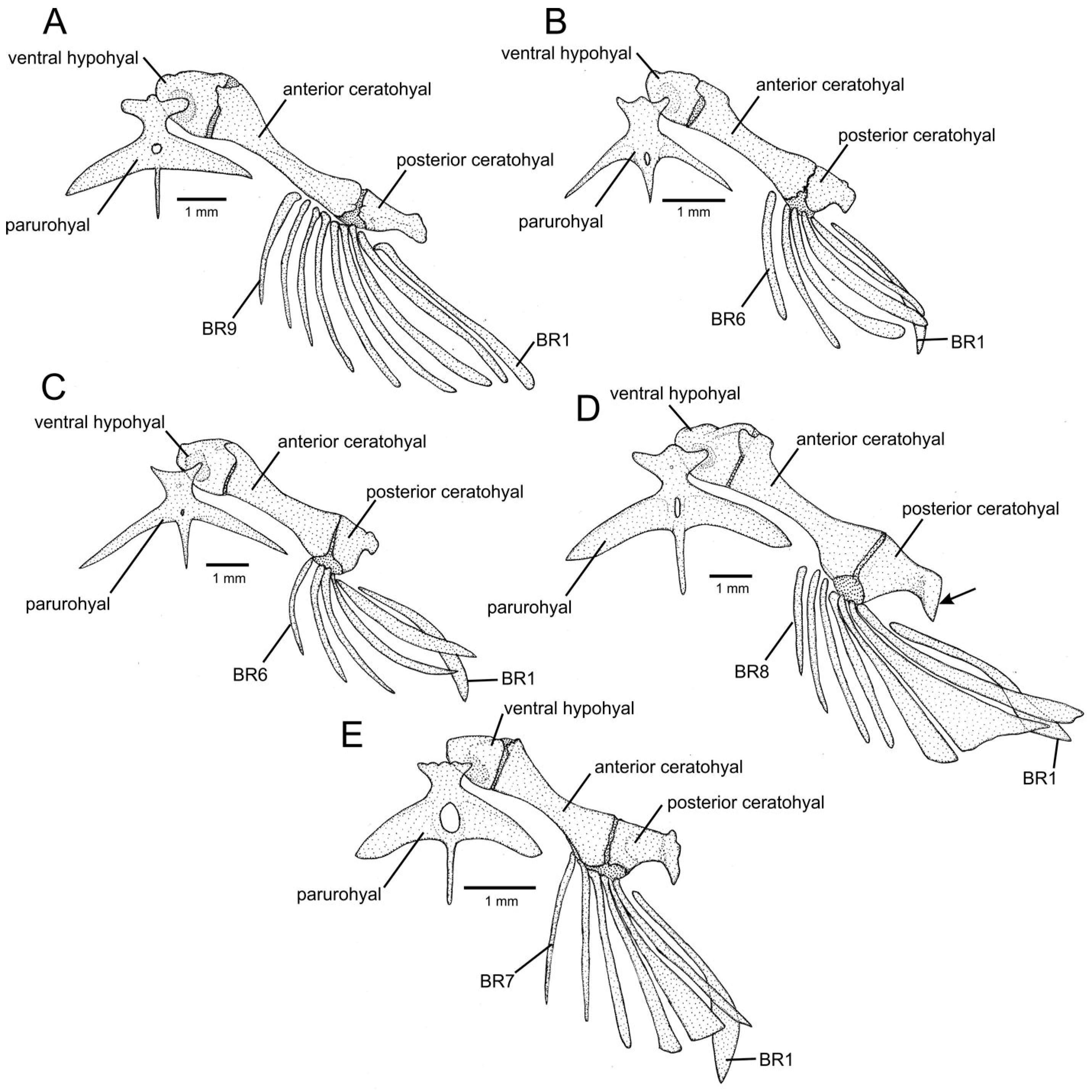
Osteological preparations followed Taylor & Van Dyke’s protocol [28]. Osteological illustrations were made using a Zeiss Stemi SV 6 stereomicroscope (Microscope Centra, Willow Grove, PA, USA) with camera lucida. The osteological nomenclature primarily follows that described in Costa [15], which was based on previous studies [29,30,31,32,33,34,35,36,37]. Modifications were those proposed by Kubicek [38] based on developmental osteology of siluroids. As observed in this study, variations in the number of branchiostegal rays among trichomycterine taxa only occur through addition or subtraction of the anterior rays. The posterior rays, which are the first to appear in developmental series [38], always remain in the same position and maintain approximately the same shape, supporting recognising them as homologous in different taxa. Consequently, numbering rays starting from the most anterior backward, as is commonly done (e.g., Refs. [14,38,39]), may result in using different numbers for the same homologous posterior ray in cases when addition or subtraction of anterior rays occurs. To maintain numbering always associated with the same homologous rays (same position and shape), here numbering is done from the most posterior ray (i.e., ray 1) forward (Figure 1).
The nomenclature of the pores of the cephalic latero-sensory system followed the proposal of Arratia & Huaquin [40], which basically followed the nomenclature of cephalic canals described by Northcutt [41], with some modifications to optimise the recognition of homologous pores. In Trichomycteridae, except for the basal trichomycterids Copionodontinae and Trichogeninae, the cephalic latero-sensory system has a considerable reduction in canals and pores, making it difficult to recognise pore homology. Among the basal trichomycterid groups, the cephalic laterosensory system of Trichogenes longipinnis Britski & Ortega, 1983, represents the least reduced system pattern among the Trichomycteridae. This pattern is here considered plesiomorphic for trichomycterids due to its great similarity in both position and number of pores with representatives of the family Diplomystidae [40], a basal group of Siluriformes [42], which in turn represents the latero-sensory morphology compatible with the generalised pattern of other teleost fishes (e.g., [43,44]). Thus, the pore pattern exhibited by T. longipinnis and diplomystids is here used as a basic standard to infer pore homologies within Trichomycterinae. On the other hand, uniquely Nematogenys inermes (Guichenot, 1848), often supported as the sister group to a clade comprising all other Loricarioidei [28,45], has a larger number of pores and is not used here as a model for pore nomenclature.
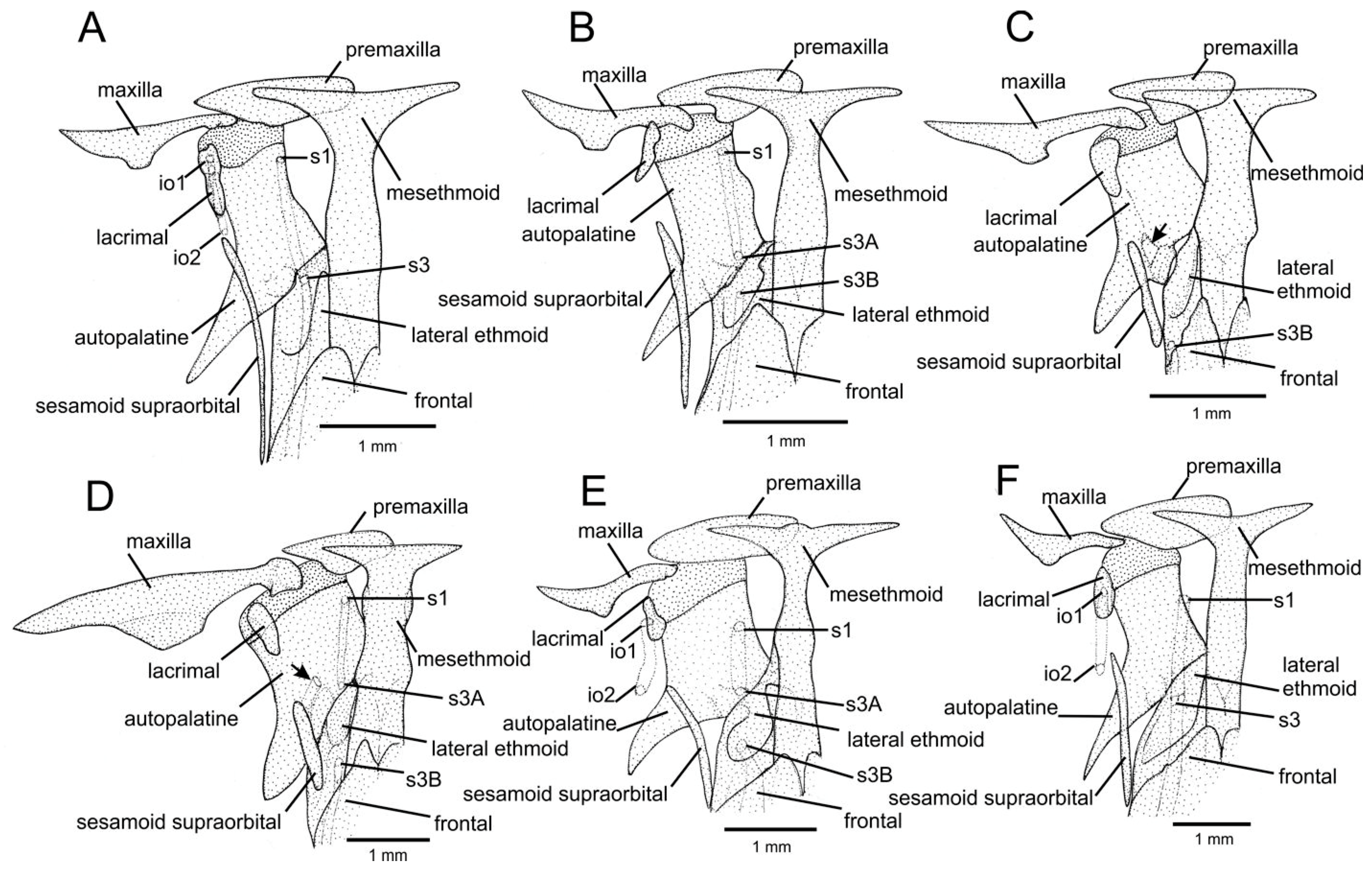
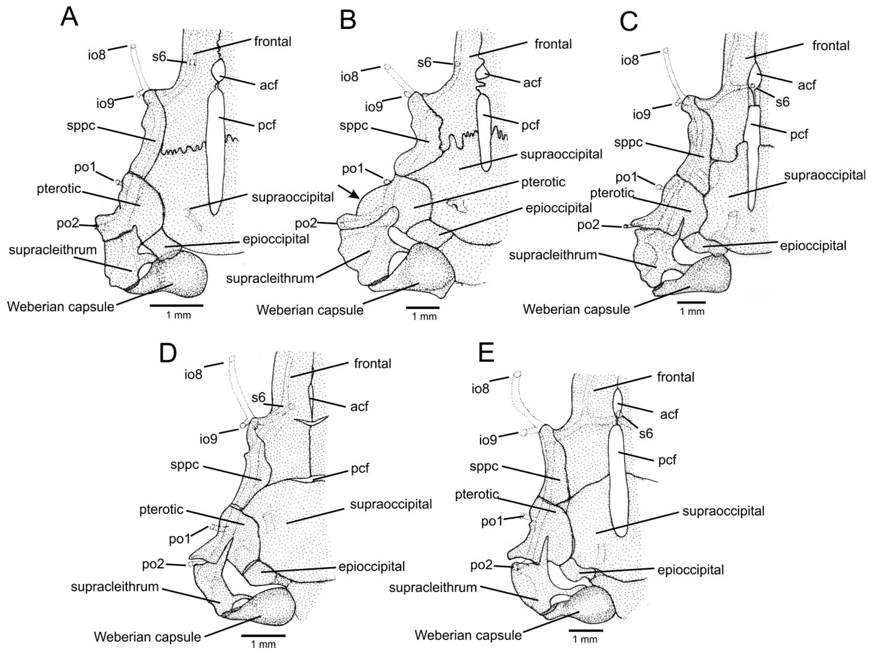
The infraorbital canal of Trichomycterinae is reduced and is represented by two segments separated by a wide interspace, each with two infraorbital pores (io pores), thus differing from the continuous canal with eight or nine pores in T. longipinnis and diplomystids. The anterior-most pore of the anterior segment is positioned near the snout, externally near the anterior nostril, internally close to the lacrimal bone canal, and the posterior pore is situated slightly posterior to the lacrimal, near the posterior nostril (Figure 2A,E,F). These pores correspond in position to pores io1 and io2 of diplomystids (figures 4B, 6 and 8A in [40]) and not to pores io1 and io3 as proposed by Arratia & Huaquin [40]. The posterior segment comprises two pores externally near the posterior portion of the orbit, internally close to the pterotic (Figure 3), corresponding to pores io8 and io9 of diplomystids, not io10 and io11 as in Arratia & Huaquin [40], who based their nomenclatural model on the autapomorphic pattern of N. inermes.
The most common morphological pattern of the supraorbital canal in Trichomycterinae, also present in basal lineages of Trichomycteridae (e.g., T. longipinnis) and thus considered the plesiomorphic condition for Trichomycteridae, is a continuous canal with only three supraorbital pores. The most anterior pore is located near the snout tip, externally adjacent to the anterior nostril, internally above the anterior portion of the autopalatine (Figure 2A,B,D–F). The median pore is adjacent to the posterior nostril and located above the anterior portion of the lateral ethmoid (Figure 2A,F). The most posterior pore is located at the level of the posterior portion of the orbit and internally associated with the frontal canal at the level of the anterior fontanel (Figure 3). These pores correspond in position, respectively, to pores s1, s3, and s6 of diplomystids, thus corroborating the nomenclature proposed by Arratia & Huaquin [40]. However, in different lineages of Trichomycterinae, the supraorbital canal is secondarily broken into two segments in its median region, with each segment having a pore at the extremity of each fragment (Figure 2B,D,E) instead of a single s3 pore in the median portion of a continuous canal. Therefore, in the interrupted canal pattern, there is one pore more than in the general plesiomorphic continuous pattern. The additional pore is here interpreted as a duplication of the pore located in the median position where the fracture occurs, the s3 pore. In these cases, it would not be appropriate to name the additional pore by its relative position as in other non-trichomycterid catfishes (i.e., s4 in [39]), since it would not be homologous to the pore of the same name in other catfish lineages. The median pores are here called s3A and s3B following a similar interpretation on canal breaking made by Gosline [46] for Cyprinodontiformes. The pores associated with the postorbital canal are here called po1 and po2, following Bockmann & Sazima [47].
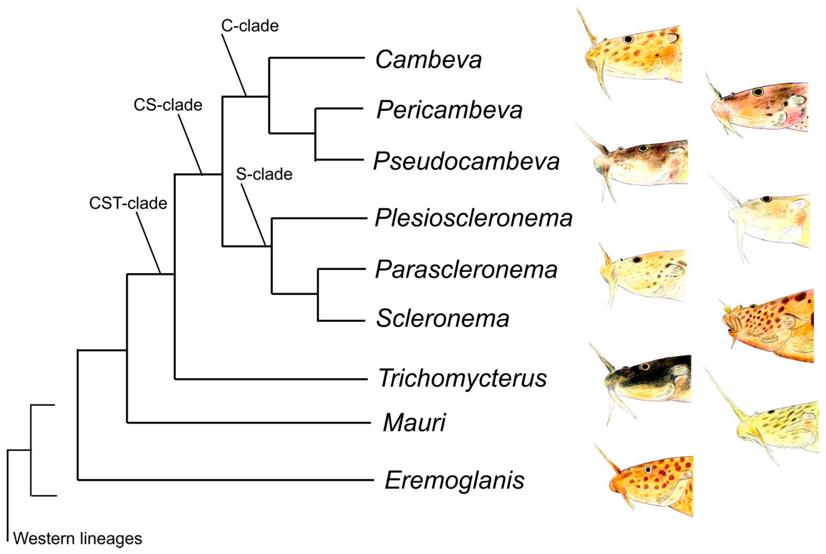
The classification here proposed is according to molecular phylogenies that are concordant and complementary among themselves, including the analysis using genomic ultra-conserved elements by Ochoa et al. [7], which constitutes the most comprehensive analysis of Trichomycteridae and the one with the highest support values at all nodes, and other phylogenetic trees generated by multigene datasets [8,9,16,19,20]. The classification followed a subordinate phylogenetic trajectory according to Figure 4. Each subsection of results begins with a brief introduction containing data on phylogenetic position and diagnostic character states (sense Sereno [48]), justifying the delimitation of taxa in the new proposed classification, followed by formal taxonomical accounts containing diagnoses, included taxa, and geographical distributions. The diagnoses are objective and include character states that occur in all members of each diagnosed group. Characters with ambiguous states or incomplete distribution among species of monophyletic units herein focused on were not included in diagnoses.

3. Results

3.1. The Trichomycterus Lineage (TL)

The Trichomycterus Lineage supported in Ochoa et al. [7] comprises taxa exhibiting high morphological diversity. No unique character state was found to diagnose TL. In addition to the absence of those character states uniquely diagnosing EL genera (see below), each TL genus is diagnosable by either unique morphological features or by unique combination of character states (see diagnoses below). TL comprises a single diverse sublineage endemic to eastern South America, the CST clade, and two sublineages endemic to western South America, the Western Trichomycterus Sublineages (WTSs).
The genera of TL are distinguished from other trichomycterine genera by the following: caudal peduncle not distinctively slenderer than the anterior portion of the trunk (vs. distinctively slenderer in Bullockia and Hatcheria [49,50]); dorsal-fin base relatively short, slightly longer than the anal-fin base, with the distal margin nearly straight to slightly convex (vs. dorsal-fin base relatively long, twice longer or more than the anal-fin base, distal margin slightly concave in Bullockia and Hatcheria [49,50]); having the anal-fin origin at a vertical approximately through the middle of the dorsal-fin base or posterior to it (vs. through the dorsal-fin base or immediately posterior to it in Ituglanis and Silvinichthys [11]); two or four infraorbital pores (vs. six in Bullockia [40]); having the supraorbital canal when continuous with three pore pairs and the lateral line canal of the trunk with one to three pores (vs. supraorbital always continuous and with five pore pairs and the lateral line canal of the trunk with four to seven pores in Bullockia and Hatcheria [40]); the absence of a thick layer of adipose tissue between the skin and the body musculature and the absence of two pillow-like masses of adipose tissue on the nape (vs. presence in Rhizosomichthys [14]); the absence of a conspicuously perforated skin surface of the body by pores of ampullary organs (vs. body skin surface highly perforated by pores of ampullary organs in Silvinichthys [11]); having 33–41 vertebrae (vs. 43 or 44 vertebrae in Eremophilus); having a laterally directed sphenotic process (Figure 3; vs. anteriorly directed in Ituglanis (figure 3 in [10]); the absence of a robust osseous ridge on the dorsolateral surface of the autopalatine and absence of a crescent-shaped transverse crest on the supraoccipital (vs. presence of both structures in Rhizosomichthys (figures 9 and 10 in [14]); the absence of a sharp posteroventral process on the lateral ethmoid (vs. presence in Eremophilus [51]); autopalatine never with a deep rounded concavity on the medial margin of the autopalatine (vs. always with a peculiar deep rounded concavity on the medial margin of the autopalatine in Ituglanis (figure 6 in [10]); and the metapterygoid never with a rounded dorsal margin to form an approximately semicircular structure (with a rounded dorsal margin to form an approximately semicircular structure in Ituglanis (figure 4 in [10]).

3.2. The CST Clade

This clade is supported in all molecular analyses [7,9,16,17,18] but not corroborated by unique morphological character states. It comprises the greatest diversity of trichomycterines in both number of species and external morphological features (e.g., Figure 5A–D).
Included taxa. The CST clade contains two more inclusive clades, each distributed in a vast region of eastern South America: the CS clade and the T clade (Figure 4), as described below.

3.3. The CS Clade

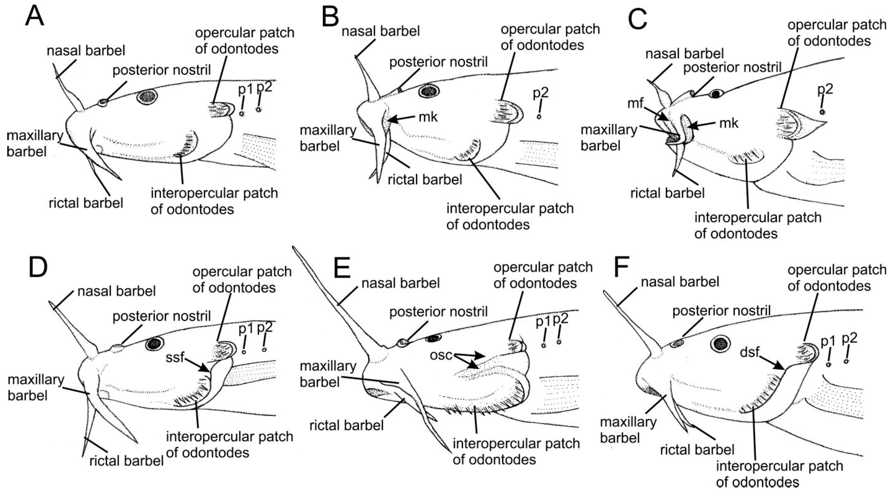
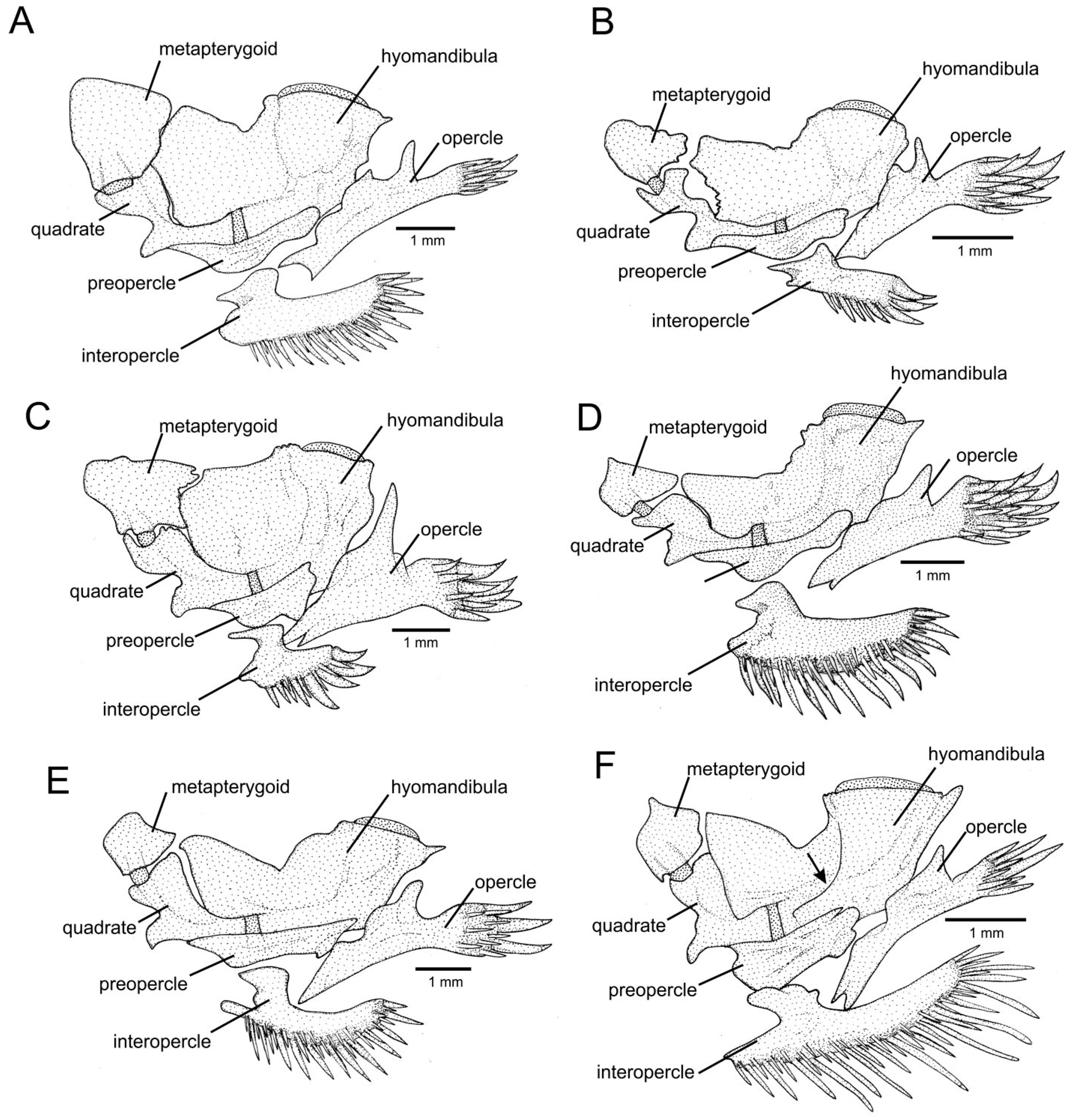
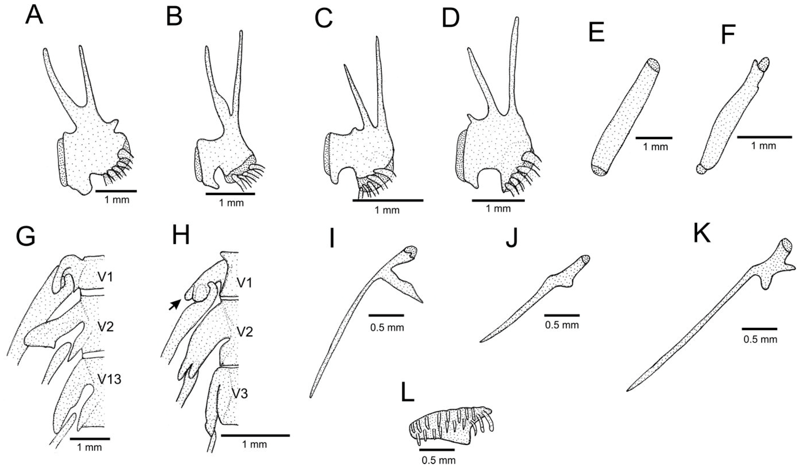
The CS clade contains different lineages exhibiting a great diversity of morphological patterns and occupying a wide geographical area [7,9,16,17,18,20]. Despite the great morphological divergence found among included lineages, this clade is here corroborated by a series of morphological character states that are unique among taxa of TL: skin surface between opercular and interopercular odontode patches continuous with the branchial membrane (Figure 5A–C; vs. separated by a skin fold or crests forming a gap, Figure 5D–F); the posterior nostril orifice smaller than the anterior nostril orifice; the largest axis of the opercular patch of odontodes horizontally aligned (Figure 5A–C; vs. slightly perpendicularly aligned, Figure 5D–F); the distal extremity of the branchiostegal ray 3 not distinctively widened, about as wide as the extremity of neighbouring rays (Figure 1A–C; vs. branchiostegal ray 3 extremity distinctively wider than neighbouring rays, Figure 1D,E); dorsal process of the interopercle with a rudimentary or absent laminar bone expansion overlapping the ventral portion of the preopercle, resulting in a nearly flat dorsal profile of the process (Figure 6A–C; vs. with a well-developed laminar bone expansion, dorsal profile convex, Figure 6D–F); quadrate narrow, with a deep concavity in its anteroventral margin (Figure 6A–C; vs. relatively broad, concavity moderate or absent, Figure 6D–F); the parapophysis of the first free vertebra with a process projecting over the dorsal surface of the adjacent rib (Figure 7H; vs. without that process, Figure 7G); and the articulation zone between the frontal and the supraoccipital comprising numerous, small prominent undulations (Figure 3A,B; vs. weak or no undulation, Figure 3C–E).
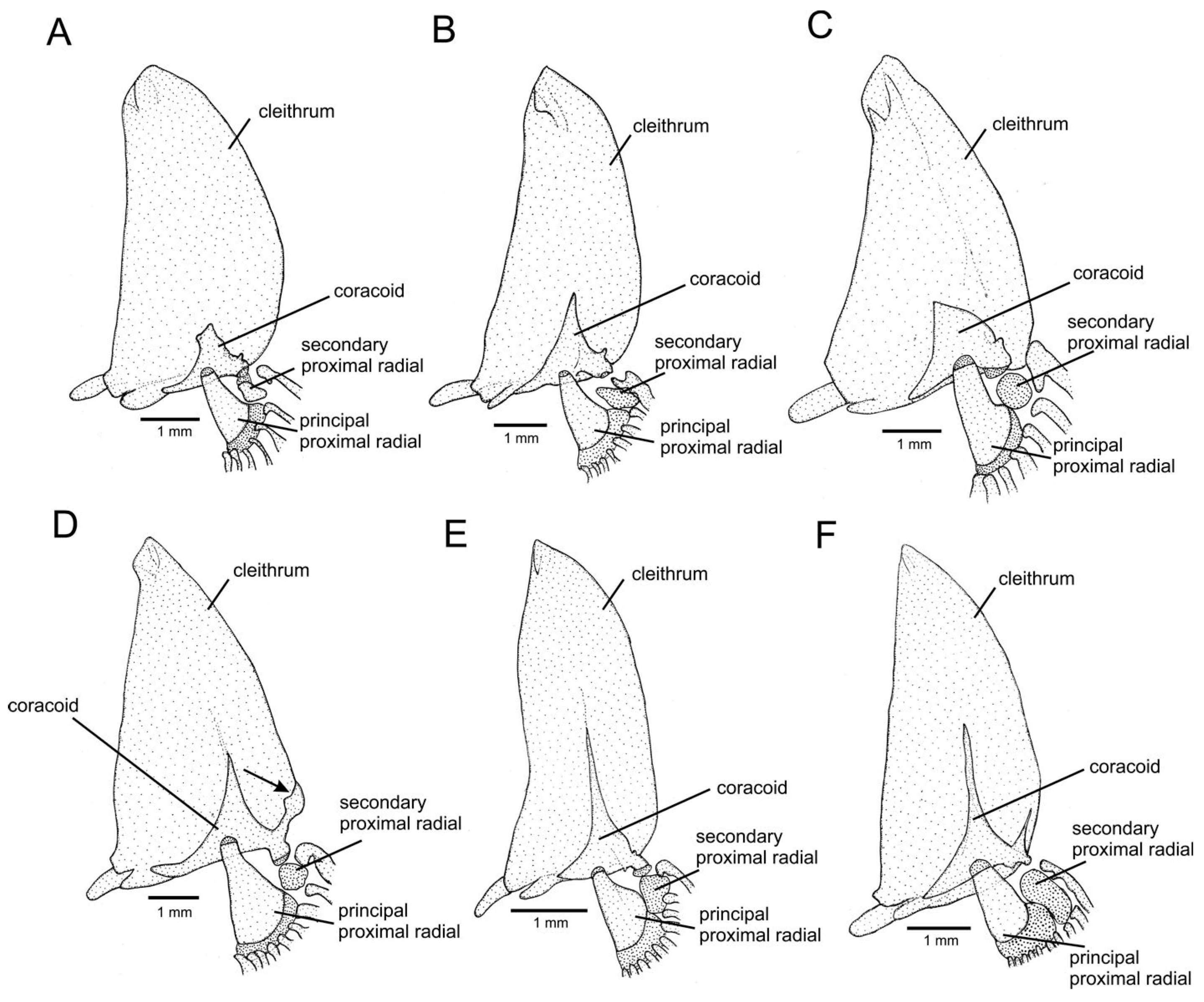
Other morphological features consisting of potential synapomorphies of the CS clade but that cannot be used as diagnostic features by homoplastically occurring in a few taxa of other TL lineages or being variable in taxa of the CS clade include the following: a rudimentary or absent coracoid supra-lateral process, occurring in all species of the CS clade (Figure 8A–C; vs. well-developed, Figure 8D), but variably developed or sometimes absent in other lineages of TL (e.g., Figure 8E); undulating margin of jaw suspensorium bones (Figure 6B,C), a condition inconspicuous or absent in some Cs clade members (Figure 6A); absence of a curved expansion on the posterior region of the posterior ceratohyal (Figure 1A–C, vs. presence, Figure 1D,E), which is also absent in some lineages within the genus Trichomycterus (e.g., subgenera Cryptocambeva Costa, 2021 and Paracambeva Costa, 2021).
Included taxa. The CS clade comprises two monophyletic groups, the C clade and the S clade (Figure 4).

3.4. The C Clade

This clade has been always corroborated as monophyletic [7,17,18,20] and is presently represented by a single nominal genus, Cambeva, not diagnosable by unique morphological features [15] but consistently differing from the S clade by a combination of morphological features (see diagnosis below). Molecular phylogenies [17,18,20] have concordantly supported four monophyletic groups: the Cambeva clade alpha that contains two subclades, the Cambeva iheringi species group and the Cambeva variegata species group, the Cambeva clade beta, and the Cambeva clade gama.
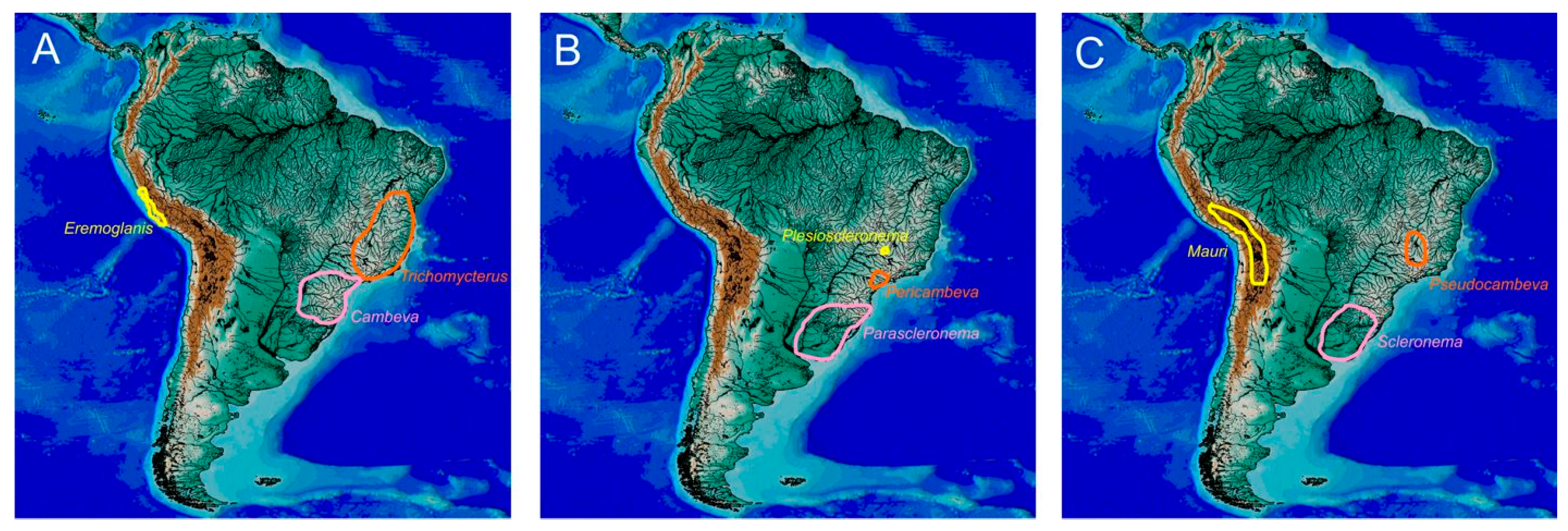
The Cambeva iheringi species group and the C. variegata species group have been supported as sister taxa (e.g., [20]), but no potential morphological synapomorphy shared by them was found. On the other hand, these two groups greatly diverge morphologically and are geographically disjunct. The Cambeva iheringi group is endemic to an area of south-eastern Brazil encompassing the upper Rio Tietê drainage of the upper Rio Paraná basin and coastal basins between the Rio Ribeira de Iguape and rivers draining into the northern extremity of the Baía de Paranaguá (Figure 9B). Species of the Cambeva iheringi group possess an exceptionally long sesamoid supraorbital, unique among TL taxa (Figure 2A), and a typical pelvic bone morphology, in which the anterior processes are situated near its medial margin (Figure 7A), not closer to the lateral margin as in other lineages of the C clade (Figure 7B) and most taxa of the S clade (Figure 7C,D).
The C. variegata species group inhabits an area comprising the Rio Grande drainage of the upper Rio Paraná basin and the upper course of the Rio São Francisco basin, southeastern Brazil (Figure 5C). This group is easily distinguished from other C clade lineages by having a skin crest similar to an adipose fin on the dorsal margin of the caudal peduncle, which is shared with all taxa of the S clade (see below). In both the C. variegata species group and in the S clade this crest is well-developed and similar in shape and position, thus not confirming Bockmann at al.’s [39] assumption that it would be well-developed in Scleronema (=S clade of the present study) and rudimentary in the C. variegata species group. The C. variegata species group shares other morphological characteristics with the S clade that are not present in any other lineage of the Cambeva clade, including both external (i.e., supraorbital canal interrupted, Figure 2B) and osteological features (premaxilla morphology, Figure 2B).
The Cambeva clade beta and the Cambeva clade gama together form a diversified monophyletic group supported in molecular phylogenies (e.g., [20]) occurring in a vast region of southern Brazil and in an adjacent area of Argentina (Figure 9A). Taxa belonging to this monophyletic group share reduced paired fins, with the pelvic-fin bases being medially placed in close proximity or in contact, besides having osteological character states unique among taxa of the Cambeva-Scleronema clade, including a large metapterygoid (Figure 6A) and a small coracoid (Figure 8A). Species of this clade typically have a high number of branchiostegal rays (8–10), but a few species have seven rays (e.g., Cambeva alphabelardense Costa, Feltrin & Katz, 2022, and Cambeva pascuali (Ochoa, Silva, Costa e Silva, Oliveira & Datovo, 2017), and eight rays may be present in some species of other genera of the CS clade.
The morphological divergence found among lineages of the Cambeva clade, mainly regarding the similar general appearance of the Cambeva variegata group with members of the S clade (see below), impedes a clear morphological identification of Cambeva among other TL genera. To improve classification with diagnosable genera, new genera are described here for the C. variegata species group and its sister group, the Cambeva iheringi species group (i.e., Pseudocambeva gen. nov. and Pericambeva gen. nov., respectively), and the genus Cambeva is restricted to the clade containing the Cambeva clade beta and the Cambeva clade gama (see below).
Included taxa. Three genera, Cambeva, Pericambeva gen. nov., and Pseudocambeva gen. nov.
Cambeva Katz, Barbosa, Mattos & Costa, 2018, new usage
Cambeva Katz et al. [18]: 560 (original description; type species: Pygidium davisi Haseman, 1911).
Diagnosis. Cambeva differs from the remaining genera of the CS clade by having the pelvic-fin bases medially in close proximity or in contact (vs. separated by an interspace), a hypertrophied metapterygoid in specimens above about 60 mm SL (Figure 6A; vs. metapterygoid small to moderate in length, Figure 6B–F), branchiostegal rays anteriorly extending to about the middle of the hyoid bar (Figure 1A; vs. slightly anteriorly surpassing the ventral cartilage of the hyoid bar, Figure 1B–E), and a relatively small coracoid (Figure 8A; vs. relatively large, Figure 8B–F). Cambeva is also distinguished from Pseudocambeva gen. nov. by the absence of a skin crest similar to an adipose fin on the dorsal margin of the caudal peduncle (vs. presence); by having the supraorbital canal continuous (vs. interrupted); the pelvic fin tip barely or not reaching the anus (vs. surpassing the urogenital papilla); and the premaxilla not distally terminating in a narrow extremity and from Pericambeva gen. nov. by having a shorter sesamoid lacrimal, shorter to slightly longer than the longitudinal length of the autopalatine excluding the posterolateral process (vs. distinctively longer).
Included species. Fifty-three species: Cambeva alphabelardense Costa, Feltrin & Katz, 2022, Cambeva atrobrunnea Costa, Feltrin & Katz, 2024, Cambeva balios (Ferrer & Malabarba, 2013), Cambeva barbosae Costa, Feltrin & Katz, 2021, Cambeva betabelardense Costa, Feltrin & Katz, 2022, Cambeva brachykechenos (Ferrer & Malabarba, 2013), Cambeva biseriata Costa, Feltrin, Mattos, Dalcin, Abilhoa & Katz, 2023, Cambeva botuvera Costa, Feltrin & Katz, 2021, Cambeva castroi (de Pinna, 1982), Cambeva crassicaudata (Wosiacki & de Pinna, 2008), Cambeva chrysornata Costa, Feltrin, Mattos, Dalcin, Abilhoa & Katz, 2023, Cambeva cauim dos Reis, Ferrer & da Graça, 2021, Cambeva cubataonis (Bizerril, 1994), Cambeva davisi (Haseman 1911), Cambeva diatropoporus (Ferrer & Malabarba, 2013), Cambeva diabola (Bockmann, Casatti & de Pinna, 2004), Cambeva diffusa Costa, Feltrin & Katz, 2021, Cambeva duplimaculata Costa, Feltrin & Katz, 2021, Cambeva flavopicta Costa, Feltrin & Katz, 2020, Cambeva galactica Costa, Feltrin & Katz, 2024, Cambeva gamabelardense Costa, Feltrin & Katz, 2022, Cambeva grisea Costa, Feltrin & Katz, 2021, Cambeva guaratuba Costa, Feltrin, Mattos, Dalcin, Abilhoa & Katz, 2023, Cambeva guareiensis Katz & Costa, 2020, *Cambeva horacioi dos Reis, Frota, Fabrin & da Graça, 2019, *Cambeva igobi Wosiacki & de Pinna, 2008), Cambeva imaruhy Costa, Feltrin & Katz, 2021, Cambeva longipalata Costa, Feltrin & Katz, 2021, Cambeva luteoreticulata Costa, Feltrin & Katz, 2024, Cambeva mboycy (Wosiaski & Garavello, 2004), Cambeva melanoptera Costa, Abilhoa, Dalcin & Katz, 2022, Cambeva naipi (Wosiaski & Garavello, 2004), Cambeva notabilis Costa, Feltrin & Katz, 2021, Cambeva orbitofrontalis Costa, Feltrin & Katz, 2021, Cambeva perkos (Datovo, Carvalho & Ferrer, 2012), Cambeva panthera Costa, Feltrin & Katz, 2021, *Cambeva papillifera (Wosiaski & Garavello, 2004), Cambeva pascuali (Ochoa, Silva, Costa e Silva, Oliveira & Datovo, 2017), Cambeva pericoh Costa, Feltrin & Katz, 2021, Cambeva piraquara dos Reis, Wosiacki, Ferrer, Donin & da Graça, 2023, *Cambeva plumbeus (Wosiaski & Garavello, 2004), Cambeva podostemophila Costa, Feltrin & Katz, 2023, Cambeva poikilos (Ferrer & Malabarba, 2013), Cambeva rotundipinna Costa, Feltrin & Katz, 2024, Cambeva stawiarski (P. Miranda-Ribeiro, 1968), Cambeva taroba (Wosiaski & Garavello, 2004), Cambeva tourensis Costa, Feltrin & Katz, 2023, Cambeva tropeira (Ferrer & Malabarba, 2011), Cambeva urubici Costa, Feltrin & Katz, 2021, Cambeva ventropapilata Costa, Feltrin, Mattos, Dalcin, Abilhoa & Katz, 2023, *Cambeva ytororo (Terán, Ferrer, Benitez, Alonso, Aguilera & Mirande, 2017), and Cambeva zonata (Eigenmann, 1918).
Geographical distribution. The Rio Paraná system between the Rio Paranapanema and upper Rio Uruguai basin in Brazil and Argentina and smaller coastal basins of southern Brazil between the Rio Ribeira de Iguape basin and the Lagoa dos Patos system (Figure 9A).
Pericambeva gen. nov.
LSID:urn:lsid:zoobank.org:act:D5CFFD3B-9146-4100-A7AD-106A9ABD2AA5
Type species. Cambeva difficilis Costa, Feltrin & Katz, 2024.
Diagnosis. Pericambeva is distinguished from the other TL genera by having a long sesamoid lacrimal, its length distinctively longer than the longitudinal length of the autopalatine excluding the posterolateral process (Figure 2A; vs. shorter to slightly longer, Figure 2B–F). Pericambeva also differs from other genera of the CS clade by having the anterior processes of the pelvic bone situated nearer the medial margin of the bone (Figure 7A; vs. nearer the lateral margin, Figure 7B–D). Pericambeva also differs from Cambeva by having the pelvic-fin bases medially separated by a broad interspace (vs. in close proximity or in contact), a moderately sized metapterygoid (vs. hypertrophied, Figure 6A), branchiostegal rays anteriorly just surpassing the ventral cartilage of the hyoid bar (vs. anteriorly extending to the middle of the hyoid bar, Figure 1A), and a relatively large coracoid (vs. relatively small, Figure 8A) and from Pseudocambeva gen. nov. by the absence of a filament at the tip of the first pectoral-fin (vs. presence), absence of a skin crest similar to an adipose fin on the dorsal margin of the caudal peduncle (vs. presence), a continuous supraorbital canal (Figure 2A; vs. interrupted, Figure 2B), a shorter pelvic fin, its extremity barely reaching urogenital papilla (vs. relatively long, surpassing urogenital papilla), and the premaxilla not distally terminating in a narrow extremity (vs. distally terminating in a narrow extremity, Figure 2B).
Etymology. From the Greek prefix peri (near) and cambeva (a popular name for trichomycterines in southeastern Brazil and also a trichomycterine genus), an allusion to the geographical proximity between the new genus and the genus Cambeva. Gender feminine.
Included species. Four species: Pericambeva difficilis (Costa, Feltrin & Katz, 2024) comb. nov., *Pericambeva guaraquessaba (Wosiacki, 2005) comb. nov., Pericambeva iheringi (Eigenmann, 1917) comb. nov., and Pericambeva tupinamba (Wosiacki & Oyakawa, 2005) comb. nov.
Geographical distribution. Upper Rio Tietê drainage and coastal basins between the Rio Ribeira de Iguape basin and river basins associated with the northern portion of the Baía de Paranaguá (Figure 9B).
Pseudocambeva gen. nov.
LSID: urn:lsid:zoobank.org:act: F92D593B-C398-4486-895B-32DE660AAFFA
Type species. Trichomycterus variegatus Costa, 1992.
Diagnosis. Pseudocambeva is distinguished from all other genera of the CS clade by having a broad lateral process of the pterotic (Figure 3A; vs. narrow, Figure 3B,C). Pseudocambeva is also distinguished from other genera of the C clade by the presence of a skin crest similar to an adipose fin on the dorsal margin of the caudal peduncle (vs. absence), having the supraorbital canal interrupted (Figure 2B; vs. continuous, Figure 2A), a relatively long pelvic fin, its extremity surpassing urogenital papilla (vs. shorter, barely or not reaching urogenital papilla), and the premaxilla distally terminating in a narrow extremity (Figure 2B; vs. not distally terminating in a narrow extremity, Figure 2A); from Cambeva by having the pelvic-fin bases medially separated by a broad interspace (vs. in close proximity); and from Pericambeva by the absence of the anterior infraorbital canal (Figure 2B; vs. presence, Figure 2A) and the presence of a filament at the tip of the first pectoral-fin (vs. absence).
Etymology. From the Greek pseudes (false) and cambeva (a popular name for trichomycterines in southeastern Brazil and also a trichomycterine genus), referring to its greater morphological similarity with genera of the S clade than to the genus Cambeva, in which their species were previously classified. Gender feminine.
Included species. Seven species: Pseudocambeva babilonica (Costa, Azevedo-Santos & Katz, 2025) comb. nov., Pseudocambeva capetinga (Costa, Azevedo-Santos, Uzeda & Katz, 2025) comb. nov., Pseudocambeva concolor (Costa, 1992) comb. nov., Pseudocambeva damnata (Costa, Azevedo-Santos, Ottoni, Vilardo & Katz, 2024) comb. nov., Pseudocambeva capitoliensis (Costa, Azevedo-Santos, Uzeda, Vilardo & Katz, 2025) comb. nov., Pseudocambeva occidentalis (Costa, Azevedo-Santos & Katz, 2025) comb. nov. and Pseudocambeva variegata (Costa, 1992) comb. nov.
Geographical distribution. Rio Grande drainage of the Rio Paraná basin and upper Rio São Francisco basin, southeastern Brazil (Figure 9C).

3.5. The S Clade

The S clade, previously considered as a single genus, Scleronema, is supported as monophyletic in all molecular analyses [7,9,16,17,18]. Three lineages have been supported within the S clade: Plesioscleronema, until now considered as a subgenus, and the Scleronema minutum and Scleronema operculatum species groups of the subgenus Scleronema [9,39]. Only one exclusive morphological character state was confirmed here for the S clade, consisting of the first free vertebra being more posteriorly directed, making them closer to the vertebral centra and overlapping them in a ventral view (Figure 7H).
Bockmann et al. [39] considered the presence of a skin crest similar to an adipose fin on the dorsal margin of the caudal peduncle as a synapomorphy of the S clade. However, a similar skin crest is also present in Pseudocambeva (see above) and in Trichomycterus rubbioli Rizzato & Bichuette, 2012, a basal taxon in its genus (Costa et al., unpublished), possibly comprising an ancestral condition for the CST clade, not a synapomorphy of the S clade. All taxa of the S clade also have a very short interopercle, making its dorsal process proportionally broader (Figure 6B,C), but a short interopercle is present in some species of Cambeva (e.g., Cambeva naipi (Wosiaski & Garavello, 2004)), making this character state unsuitable for use in diagnoses.
The monotypic Plesioscleronema has been always supported as sister to a clade containing the remaining taxa of the S clade [9,39]. Plesioscleronema is endemic to a small area of the Rio Paranaíba drainage of the Rio Paraná basin, uplands of south-eastern Brazil (Figure 9A), separated by about 1000 km from the distribution area occupied by the clade comprising the other lineages of the S clade (the Parascleronema-Scleronema subclade, Figure 9B,C) in the lowlands of southern Brazil and adjacent areas of Argentina, Paraguay, and Uruguay [9] and markedly differs from other taxa of the S clade in both the external morphology and osseous traits (see below). The great morphological divergence between Plesioscleronema and the clade comprising the other members of the S clade supports recognition of Plesioscleronema as a separate genus.
Two unique character states of the caudal skeleton described by Costa et al. [9] for Plesioscleronema, the presence of a broad hemal spine on the preural centrum 2, its width about twice or slightly more than twice the width of the anteriorly adjacent hemal spine (figure 5L in [9]), and the uroneural separated from the dorsal hypural plate by an interspace (figure 5L in [9]), are here confirmed as autapomorphies for the monotypic Plesioscleronema. Another supposedly unique character state, the presence of an expansion on the posterodorsal portion of the quadrate directed towards a concavity on the anterior outgrowth of the hyomandibula (figure 5L in [9]), is similar to the condition found in small specimens of other S clade lineages (Figure 6B), therefore not useful to diagnose Plesioscleronema. The anterior dorsocranial fontanel of Plesioscleronema that is rudimentary or absent contrasts with the well-developed fontanel in other members of the S clade and basal taxa of the C clade as well as in Trichomycterus, supporting the condition as autapomorphic for the monotypic Plesioscleronema, although there is an absence or extreme reduction of cranial fontanels homoplastically occurring in Cambeva (e.g., [52,53]).
The two other lineages of the S clade, the Scleronema minutum species group and the Scleronema operculatum species group [26], together form a clade corresponding to the genus Scleronema in Ferrer & Malabarba [26] and subgenus Scleronema in Costa et al. [9] and Bockmann et al. [39]. This clade is supported in molecular analyses (e.g., [9]) and corroborated by a series of unique morphological character states. These two species groups have highly overlapped distribution areas (Figure 9B,C) but greatly diverge in morphological features. Only the Scleronema operculatum group exhibits the main diagnostic features used by Eigenmann [54] to diagnose Scleronema, including ‘opercle with a long dermal flap’ (Figure 5C) and ‘maxillary barbel with a large osseous base (i.e., maxillary bone, Figure 2D)’. The great morphological divergence exhibited by these two lineages (see below), both in external and osteological features, justifies their obvious separation into two genera. Scleronema is here restricted to the two nominal species of the Scleronema operculatum group, thus restoring the original delimitation of Scleronema as proposed by Eigenmann [54,55]. A new genus (Parascleronema gen. nov.) is described below to contain all species of the Scleronema minutum group.
Unique character states previously described as synapomorphic for the Parascleronema-Scleronema clade and herein corroborated include the following: the presence of a fleshy keel on the posterior margin of the basal portion of the maxillary barbel (Figure 5B,C; previously described as a fleshy flap at the base of the maxillary barbel, e.g., [39]); an arched pectoral fin as a consequence of a distinctively short first ray, followed by progressively longer rays [9]; a reduced number of ventral principal caudal-fin rays (five vs. six, [39]); the presence of hypertrophied odontodes on the posterior portion of the opercular and interopercular patches of odontodes (figure 5g,h in [9]); maxilla elongate (Figure 6B,C); the presence of a prominent nerve canal on the autopalatine surface, close to the articulation for the lateral ethmoid (Figure 2C,D; modified from [26]); the autopalatine articulatory socket for vomer advancing medially, making it mostly located under the mesethmoid (modified from [26]; vs. not or slightly under vomer); and the metapterygoid elongated and slightly curved (Figure 6B,C, modified from [9]).
New unique character states corroborating the Parascleronema-Scleronema clade are as follows: a reduced number of principal anal-fin rays (i.e., six, vs. seven to nine); the basal portion of the maxillary barbel distinctively broad, abruptly tapering distally (Figure 5B,C); and the anterior-most pore of the lateral line of the trunk situated below the opercle (Figure 6B,C; vs. posterior to opercle, Figure 6A,D–F).
Species of the Parascleronema-Scleronema clade typically have six branchiostegal rays [39], instead of seven to ten as in other members of TL. However, specimens of most species may have a seventh ray, which when present is small and not directly attached to the hyoid bar. The reduction in the number of branchiostegal rays is considered here as a synapomorphy of this clade, but it is not useful to diagnose it due to the occurrence of the small seventh ray in Plesioscleronema and some species of Pseudocambeva. The narrow and sharply pointed lateral process of the parurohyal occurring in all species of the Parascleronema-Scleronema clade is not found elsewhere among species of the CS clade, except in Cambeva panthera (Costa, Feltrin & Katz, 2021). A similar condition was also observed in Trichomycterus mimonha (Costa, 1992), thus impeding it from being used as diagnostic. In addition, the absence of a laminar osseus expansion on the posterior portion of the autopalatine, ventral to the articular facet for the lateral ethmoid described by Costa et al. [9] and confirmed by Bockmann et al. [39] as synapomorphic for this clade is here refuted. This condition does not occur in larger specimens of both species of the Scleronema operculatum group (i.e., genus Scleronema in the present sense) and is therefore not diagnostic for the new genus (see below).
Scleronema in the present sense is diagnosable by 12 character states that are unique among trichomycterines. Unique character states previously described and here confirmed include the following: opercular odontode patch membrane posteriorly elongated, forming a distinctive skin flap [54] (Figure 5C), and a hypertrophied maxilla, its length about three to four times the length of the premaxilla (modified from [54], Figure 2D). The presence of a broad dark grey to black bar on the posterior portion of the caudal fin present in both species of Scleronema [26] (here confirmed) distinguishes Scleronema from the other taxa of the S clade, supporting it as a synapomorphy of this genus, but a similar colour pattern occurs in some species of Cambeva [56], although this condition is useful to distinguish Scleronema from the other taxa of the S clade.
Firstly described character states uniquely found in Scleronema among TL taxa include the following: the presence of a skin fold on the dorsal surface of the premaxillary barbel base (Figure 5C); the maxillary barbel base abruptly terminating in a narrow and short barbel filament antero-laterally directed (Figure 5C); a short nasal barbel, not or barely reaching the anterior naris, with its extremity abruptly narrowing distally (Figure 5C); orbits medially in close proximity, projecting above the dorsal surface of the head (Figure 5C); a distinctive narrowing in the anterior portion of the neurocranium (Figure 3B); a long thin osseous rim on the lateral margin of the pterotic, just anterior to the lateral process, supporting ligaments connecting it to the lateral surface of the opercle (Figure 3B); a compact preopercle, posteriorly widening (Figure 6C); and the postero-medial cleithrum extremity distinctively widened (Figure 8C). Species of Scleronema also have a broad pelvic bone (Figure 7D), contrasting with the narrower pelvic bone of other taxa of the S clade (Figure 7C), but this condition is variable in the C clade and T clade.
The new genus described below, corresponding to the Scleronema minutum species group [26], exhibits three character states that are unique among trichomycterine taxa: the presence of a long tooth, slightly curved to the mouth inside, at the corner of the distal extremity of the premaxilla, contrasting with other premaxillary teeth that are shorter and nearly straight (Figure 7L); the absence of a laminar osseous expansion on the posterior portion of the autopalatine, ventral to the articular facet for the lateral ethmoid (Figure 5D in [9]); and the presence of a short process on the proximal extremity of the fourth ceratobranchial, close to the articular cartilage (Figure 7F).
Included taxa. The genera Parascleronema gen. nov., Plesioscleronema and Scleronema.
Parascleronema gen. nov.
LSID: urn:lsid:zoobank.org:act: B0D91992-E6E9-448C-AA19-3DA339334DCA
Type species. Trichomycterus minutus Boulenger, 1891.
Diagnosis. Parascleronema is distinguished from all other genera of TL by having an elongate, slightly curved tooth at the corner of the distal extremity of the premaxilla (Figure 7L; vs. tooth on the distal extremity of the premaxilla not differing from other premaxillary teeth) and the presence of a process on the proximal extremity of the fourth ceratobranchial (Figure 7F; vs. absence, Figure 7E). Parascleronema is also distinguished from Scleronema by the absence of a skin fold on the dorsal surface of the premaxillary barbel base (Figure 5B; vs. presence, Figure 5C); the opercular odontode patch membrane not posteriorly elongated to form a distinctive skin flap (Figure 5B; vs. elongated, forming a distinctive skin flap, Figure 5C); a nasal barbel moderate in length, gradually narrowing distally (Figure 5B; vs. short, not or barely reaching the anterior naris, abruptly narrowing distally, Figure 5C); the anterior portion of the neurocranium not distinctively narrowed (like in Pseudocambeva, Figure 3A; vs. distinctively narrowed, Figure 3B); the absence of a long thin osseous rim on the lateral margin of the pterotic (vs. presence, Figure 3B); a moderately lengthened maxilla (Figure 2C; vs. hypertrophied, its length about three to four times the length of the premaxilla, Figure 2D); an elongate preopercle, not distinctively widening posteriorly (Figure 6B; vs. compact, posteriorly widening, Figure 6C); and the absence of a broad dark grey to black bar on the posterior portion of the caudal fin (vs. presence). Parascleronema is also distinguished from all other genera of TL, except Scleronema, by having the following: a fleshy keel on the posterior margin of the basal portion of the maxillary barbel (Figure 5B,C; vs. absence, Figure 5D–F); six principal anal-fin rays and ten principal caudal-fin rays (vs. seven and eleven, respectively); the basal portion of the maxillary barbel distinctively broad, abruptly tapering distally (Figure 5B,C; vs. basal portion never so broad, with gradual narrowing distally, Figure 5D–F); an arched pectoral fin as a consequence of a distinctively short first ray, followed by progressively longer rays making the first ray distinctively shorter than other rays (vs. pectoral fin subtriangular, first ray never distinctively shorter than other rays); the anterior-most pore of the lateral line of the trunk situated below the opercle (vs. away from opercle, at a vertical through the middle portion of the pectoral-fin base); the presence of hypertrophied odontodes on the posterior portion of the opercular and interopercular patches of odontodes (Figure 6B,C; vs. absence, Figure 6A,D–F); a prominent nerve canal on the autopalatine dorsal surface, close to the articulation for the lateral ethmoid (Figure 2C,D; vs. absence, Figure 2A,B,E,F); the posteromedial extremity of the autopalatine extending ventrally under the mesethmoid (vs. laterally in contact with the mesethmoid); and the lateral process of the vomer rudimentary and posteriorly directed (vs. well-developed, posterolaterally directed).
Etymology. From the Greek prefix para (resembling) and Scleronema, a nominal trichomycterine genus, of which the name means hard thread, referring to the hard base of the maxillary barbel (Eigenmann, 1917). Gender neuter.
Included species. Seven species: Parascleronema carijo (Bockmann, Ferrer, Rizzato, Esguícero, Duboc & Ingenito, 2023) comb. nov., *Parascleronema guapa (Ferrer & Malabarba, 2020) comb. nov., Parascleronema ibirapuita (Ferrer & Malabarba, 2020) comb. nov., Parascleronema mate (Ferrer & Malabarba, 2020) comb. nov., Parascleronema milonga (Ferrer & Malabarba, 2020) comb. nov., Parascleronema minutum (Boulenger, 1891), and *Parascleronema teiniagua (Ferrer & Malabarba, 2020) comb. nov.
Geographical distribution. Lower Río de La Plata system in Brazil, adjacent areas of Argentina, Paraguay, and Uruguay and coastal basins of southern Brazil, including those associated with the Lagoa dos Patos system (Figure 9B).
Plesioscleronema Costa, Sampaio, Giongo, de Almeida, Azevedo-Santos & Katz, 2022, new usage
Plesioscleronema Costa et al. [9]: 87 (original description, as a subgenus of Scleronema Eigenmann, 1917; type species: Scleronema auromaculatum Costa, Sampaio, Giongo, de Almeida, Azevedo-Santos & Katz, 2022, by monotypy and original designation).
Diagnosis. Plesioscleronema differs from the other genera of the S clade, Parascleronema and Scleronema, by the anterior dorsocranial fontanel that is absent or rudimentary (vs. well developed); the presence of a broad hemal spine on the preural centrum 2, its width about twice or slightly more than twice the width of the anteriorly adjacent hemal spine (figure 5L in [9]; vs. only slightly wider or equal in width); having the uroneural separated from the dorsal hypural plate by an interspace (figure 5L in [9]; vs. in contact); the absence of a fleshy keel on the posterior margin of the basal portion of the maxillary barbel (vs. presence); having seven principal anal-fin rays and eleven principal caudal-fin rays (vs. six and ten, respectively); the basal portion of the maxillary barbel moderately wide and gradually narrowing distally (vs. basal portion distinctively broad, abruptly tapering distally); the anterior-most pore of the lateral line of the trunk posterior to the opercle, at a vertical through the middle portion of the pectoral-fin base (vs. situated under the opercle); the absence of a distinctive canal on the dorsal surface of the autopalatine, close to the bone articulation with the lateral ethmoid (vs. presence); the posteromedial extremity of the autopalatine laterally in contact with the mesethmoid (vs. extending ventrally under the mesethmoid); the lateral process of the vomer well-developed, postero-laterally directed (vs. rudimentary and posteriorly directed); and posterior interopercular odontodes not distinctively broader than other interopercular odontodes (vs. distinctively broader).
Included species. A single species, Plesioscleronema auromaculatum (Costa, Sampaio, Giongo, de Almeida, Azevedo-Santos & Katz, 2022).
Geographical distribution. Rio Paranaiba drainage, Rio Paraná basin, southeastern Brazil (Figure 9B).
Scleronema Eigenmann, 1917
Scleronema Eigenmann [54]: 691 (original description; type species: Scleronema operculatum Eigenmann, 1917, by monotypy and original designation).
Diagnosis. Scleronema is distinguished from all other trichomycterine genera by the presence of a skin fold on the dorsal surface of the premaxillary barbel base (Figure 5C; vs. absence); the maxillary barbel base abruptly terminating in narrow and short barbel filament antero-laterally directed (Figure 5C; vs. never a similar barbel morphology); a short nasal barbel, not or barely reaching the anterior naris, abruptly narrowing distally (Figure 5C; vs. never a similar morphology); the opercular odontode patch membrane posteriorly elongated, forming a distinctive skin flap (Figure 5C; vs. never elongated); orbits medially in close proximity, projecting above the dorsal surface of the head (Figure 5C, vs. never so close, not projecting dorsally); a strong narrowing in the anterior portion of the neurocranium (Figure 3B; vs. weak narrowing, Figure 3C–E); a long thin rim on the lateral margin of the pterotic, just anterior to the lateral process, supporting ligaments connecting it to the lateral surface of the opercle (Figure 3B; vs. rim minute or absent, Figure 3C–E); a hypertrophied maxilla, its length about three to four times the length of the premaxilla (Figure 2D; vs. never hypertrophied); a compact preopercle compact, posteriorly widening (Figure 6C; not widening posteriorly, Figure 6A,B,D–F); and the medial cleithrum extremity distinctively widened (Figure 8C; vs. not distinctively widened, Figure 8A,B,D–F). Species of Scleronema are also distinguished from other trichomycterines of the S clade by the presence of a broad dark grey to black bar on the posterior portion of the caudal fin (vs. absence).
Included species. Two species, Scleronema macanuda (Ferrer & Malabarba, 2020) and Scleronema operculatum (Eigenmann, 1917).
Geographical distribution. Rio Uruguai basin in Brazil, adjacent areas of Argentina and Uruguay, and coastal drainages of southern Brazil and Uruguay including the Lagoa dos Patos system (Figure 9C).

3.6. The T Clade

This clade includes a single genus, Trichomycterus (sensum strictum, see introduction above), with intrageneric lineages exhibiting relatively high morphological diversity [15]. The T clade is distinguished from its sister group, the CS clade, by a series of morphological features that are unique for this clade. The present comparative analysis also indicated osteological character states unique for Trichomycterus, including the morphology of the last proximal radial of the dorsal and anal fins, the dorsal process of the interopercle, and the supraoccipital (see below). Most species of Trichomycterus also differ from other TL trichomycterines in possessing a sharply pointed metapterygoid, but this condition is not present in all congeners, thus not serving to diagnose the genus.
Trichomycterus Valenciennes, 1832
Trichomycterus Valenciennes in Humboldt and Bonpland [57]: 348 (original description; type species: Trichomycterus nigricans Valenciennes, 1832, by monotypy and original designation).
Diagnosis. Trichomycterus differs from other TL genera by having a bifid process on the subdistal portion of the last proximal radial of the dorsal and anal fins (Figure 7K; vs. process without a bifid termination, Figure 7I,J); a rounded expansion on the dorsal margin of the dorsal process of the interopercle, causing a bird-like lateral profile (Figure 6D; vs. absence of that expansion, Figure 6A–C,E,F); and a sinuous anterior portion of the supraoccipital (Figure 3C; vs. not sinuous, Figure 3A,B,D,E). Trichomycterus is also readily distinguished from other genera of the CST clade by having the skin surface between opercular and interopercular odontode patches separated from the branchial membrane by a skin fold (Figure 5D; vs. continuous, Figure 5A–C); the posterior nostril larger than the anterior nostril; the largest axis of the opercular patch of odontodes slightly perpendicularly aligned (Figure 5D; vs. horizontally aligned, Figure 5A–C); and the distal extremity of the branchiostegal ray 3 distinctively widened than other rays (Figure 1D; vs. branchiostegal ray 3 extremity not distinctively wider, Figure 1A–C).
Included species. Ninety species: Trichomycterus adautoleitei Costa, Azevedo-Santos & Katz, 2023, Trichomycterus albinotatus Costa, 1992, Trichomycterus alternatus (Eigenmann, 1917), Trichomycterus altipombensis Costa, Katz, Vilardo & Mattos, 2022, Trichomycterus anaisae Katz & Costa, 2021, Trichomycterus antiquus Costa, Fentrin, Mattos & Katz, 2024, Trichomycterus araxa Costa, Mattos, Sampaio, Giongo, de Almeida & Katz, 2022, Trichomycterus argos Lezama, Triques & Santos, 2012, Trichomycterus astromycterus Reis, de Pinna and Pessali 2019, Trichomycterus auroguttatus Costa, 1992, Trichomycterus bahianus Costa, 1992, Trichomycterus barrocus Reis & de Pinna, 2022, Trichomycterus brucutu Reis & de Pinna, 2022, Trichomycterus berthalutzae Costa, Barbosa & Katz, 2024, Trichomycterus brasiliensis Lütken, 1874, Trichomycterus brigadeirensis Costa, Katz and Vilardo, 2023, Trichomycterus brunoi Barbosa & Costa, 2005, Trichomycterus caipora Lima, Lazzarato & Costa, 2008, Trichomycterus candidus (P. Miranda-Ribeiro, 1949), Trichomycterus caparaoensis Costa, Barbosa and Katz, 2023, Trichomycterus caratinguensis Costa, Katz and Vilardo, 2023, Trichomycterus castelensis Costa, Katz and Vilardo, 2023, Trichomycterus caudofasciatus Alencar & Costa, 2004, Trichomycterus claudiae Barbosa & Costa, 2005, Trichomycterus coelhorum Costa, Azevedo-Santos & Katz, 2023, Trichomycterus diamantinensis Costa, Feltrin, Mattos & Katz, 2024, Trichomycterus espinhacensis Costa and Katz, 2023, Trichomycterus fabioheppi Costa, Barbosa & Katz, 2024, Trichomycterus fuliginosus Barbosa & Costa, 2005, Trichomycterus funebris Katz & Costa, 2021, Trichomycterus garbei Costa, Azevedo-Santos & Katz, 2023, Trichomycterus gasparinii Barbosa, 2013, Trichomycterus giarettai Barbosa & Katz, 2016, Trichomycterus giganteus Lima & Costa, 2004, Trichomycterus goeldii Boulenger, 1896, Trichomycterus humboldti Costa & Katz, 2021, Trichomycterus illuvies Reis & de Pinna, 2022, Trichomycterus immaculatus (Eigenmann & Eigenmann, 1889), Trichomycterus ingaiensis Katz & Costa, 2021, Trichomycterus ipatinga Reis & de Pinna, 2022, *Trichomycterus itacarambiensis Trajano & de Pinna, 1996, *Trichomycterus itacambirussu Triques & Vono, 2004, Trichomycterus itatiayae A. Miranda-Ribeiro, 1906, Trichomycterus jacupiranga Wosiacki & Oyakawa, 2005, Trichomycterus jequitinhonhae Triques & Vono, 2004, *Trichomycterus landinga Triques & Vono, 2004, Trichomycterus largoperculatus Costa & Katz, 2022, Trichomycterus lauryi Donin, Ferrer & Carvalho, 2020, Trichomycterus listruroides Costa, Katz & Azevedo-Santos, 2023, Trichomycterus longibarbatus Costa, 1992, Trichomycterus luetkeni Katz & Costa, 2021, Trichomycterus macrophthalmus Barbosa & Costa, 2012, Trichomycterus macrotrichopterus Barbosa & Costa, 2005, Trichomycterus maculosus Barbosa & Costa, 2010, *Trichomycterus maracaya Bockmann & Sazima, 2004*, Trichomycterus mariamole Barbosa & Costa, 2005, Trichomycterus melanopygius Reis, dos Santos, Britto, Volpi & de Pinna, 2020, Trichomycterus mimonha Costa, 1992, Trichomycterus mimosensis Barbosa, 2013, Trichomycterus mirissumba Costa, 1992, Trichomycterus mutabilicolor Costa, 2022, Trichomycterus nigricans Valenciennes, 1832, Trichomycterus nigroauratus Barbosa & Costa, 2008, Trichomycterus novalimensis Barbosa & Costa, 2005, Trichomycterus pantherinus Alencar & Costa, 2004, Trichomycterus paquequerensis (P. Miranda-Ribeiro, 1943), Trichomycterus pauciradiatus Alencar & Costa, 2006, Trichomycterus paulence (Eigenmann, 1917), Trichomycterus pirabitira Barbosa & Azevedo-Santos, 2012, Trichomycterus potschi Barbosa & Costa, 2003, Trichomycterus pradensis Sarmento-Soares, Matins-Pinheiro, Aranda & Chamon, 2005, Trichomycterus puriventris Barbosa & Costa, 2012, Trichomycterus quintus Costa, 2020, Trichomycterus reinhardti (Eigenmann, 1917), Trichomycterus rubiginosus Barbosa & Costa, 2005, Trichomycterus rubbioli Bichuette & Rizzato, 2012, Trichomycterus ruficaudatus Costa, Mattos, Amorim, Gago & Katz, 2025, Trichomycterus sainthilairei Katz & Costa, 2021, Trichomycterus santaeritae (Eigenmann, 1918), Trichomycterus saquarema Costa, Katz, Vilardo & Amorim, 2022, Trichomycterus saturatus Costa, Katz & Azevedo-Santos, 2023, Trichomycterus septemradiatus Katz, Barbosa & Costa, 2013, Trichomycterus tantalus Reis, Vieira & de Pinna, 2022*, Trichomycterus tete Barbosa & Costa, 2011, Trichomycterus travassosi (Miranda Ribeiro, 1949), *Trichomycterus trefauti Wosiacki, 2004, Trichomycterus uberabensis Costa, Azevedo-Santos & Katz, 2023, Trichomycterus vermiculatus (Eigenmann, 1917), Trichomycterus vinnulus Reis & de Pinna, 2022, and Trichomycterus vitalbrazili Vilardo, Katz and Costa, 2020.
Geographical distribution. River basins of eastern Brazil, between Rio Paraguaçu and streams draining the northern portion of the Baía de Paranaguá, including the upper and middle Rio Paraná and Rio São Francisco basins (Figure 9A).
Remarks. The genus Trichomycterus was divided into six subgenera by Costa [15]. With the inclusion of numerous taxa and more morphological variation recorded in recent years, limits of those subgenera will be revised elsewhere.

3.7. The Western Trichomycterus Sublineages

Two sublineages of TL from western South America have been recognised in molecular phylogenies. The first one, the Andean Trichomycterus Lineage (ATS), is supported in different molecular phylogenies as a sister to the CST clade [7,8,16,19] and includes taxa both from cis- and trans-Andean areas. The second lineage, the Pacific Trichomycterus Lineage (PTS), is supported in the most taxon-inclusive analysis by Ochoa et al. [7] as a sister to all other TL taxa and includes at least two species from the coastal Pacific river basins.
No apomorphic features shared by ATS, PTS, and other TL genera were found to corroborate the relationships strongly supported by molecular data [7]. On the other hand, two features shared by members of both ATS and PTS are unique among trichomycterine genera: the posterolateral process of the autopalatine is slightly curved (Figure 2E,F; vs. straight, Figure 2A–D), and the coracoid anteriorly terminates in a long keel (Figure 8E,F; vs. anterior keel short when present, Figure 8A–D).

3.8. The Andean Trichomycterus Sublineage (ATS)

The Andean Trichomycterus Lineage was supported by Ochoa et al. [7] as a sister to the CST clade, based on two species collected in the upper Amazon basin, Central Andes of Peru. They were identified as ‘Trichomycterus’ quechuorus (Steindachner, 1900) and ‘Trichomycterus’ cf. oroyae (Eigenmann & Eigenmann, 1889). These two nominal species have similar general appearance (e.g., head weakly flattened dorso-ventrally, nearly rounded in dorsal view; small eye), which was recognised by Eigenmann [55] when placing them together in its identification key. However, whereas specimens of ‘Trichomycterus’ cf. oroyae used by Ochoa et al. [7] were collected in the same area of the type locality of ‘T.’ oroyae (i.e., Río Mantaro drainage), therefore confirming its identification as belonging to this species, specimens of ‘T’. quechuorus were collected in the Río Urubamba basin, Río Paucartambo drainage, cis-Andean region, therefore distant from the type locality of ‘T’. quechuorus in the trans-Andean region of Arequipa, making species identification improbable. A similar species occurring in the Río Paucartambo is ‘Trichomycterus’ megantoni Fernández & Chuquihuamani, 2007 [58], but the present examination of photographs of the specimens identified as ‘T’. quechuorus and used in Ochoa et al. [7] have clearly shown that this species it not ‘T’. megantoni but another closely related species from the same drainage, apparently still undescribed and herein also examined. The present examination of ‘T’. megantoni, ‘T’. oroyae, and the unidentified species from the Río Urubamba basin revealed some unique morphological features shared by these three species, including structures in the region involving the opercular and jaw suspensorium apparatus, which are diagnostic for a new genus, Mauri gen. nov. (see below).
Fernandez et al. [19] also found phylogenetic support for a monophyletic ATS sister to the CST clade using a different approach (multigene analysis instead of ultraconserved elements as in Ochoa et al. [7]). In this analysis, ATS included that species identified as ‘T’. quechuorus using DNA sequences extracted from the same material used in Ochoa et al. (2020), five undescribed species from Bolivia, ‘Trichomycterus’ rivulatus Valenciennes, in Cuvier & Valenciennes, 1846, from the Lago Titicaca basin, ‘Trichomycterus’ spegazzinii (Berg, 1897) and ‘Trichomycterus’ roigi (Arratia & Menu-Marque, 1984) from the high Andean drainages of northern Argentina. Morphological structures of ‘T’. roigi illustrated in its original description (figures 4A and 9A in [59]) and an illustration of ‘T.’ rivulatus (figure 41 in Baskin [28]) clearly show unique features shared by ‘T’. megantoni, ‘T’. oroyae, and the unidentified species from the Río Urubamba basin, justifying their inclusion in the new genus described below.
Mauri gen. nov.
LSID: urn:lsid:zoobank.org:act: 36D853E1-A508-4CB2-BDCD-AE2AFEAF35D9
Type species. Trichomycterus megantoni Fernández & Chuquihuamani, 2007.
Diagnosis. Mauri differs from all other trichomycterine genera by a series of structures in the region involving the opercular and jaw suspensorium apparatus, externally including two longitudinally converging skin crests between the opercle and the interopercle (Figure 5E; vs. no skin crest on the opercular region, Figure 5A–D,F) and internally including a shortened longitudinal axis of the quadrate (Figure 6F; vs. never a similar morphology, Figure 6A–E); the presence of a distinctive keel in the middle portion of the lateral surface of the hyomandibula (Figure 6F; vs. absence); a short and anteriorly widened preopercle (Figure 6F; vs. never a similar morphology, Figure 6A–E); a prominent anterodorsal expansion of the dorsal process of the interopercle (Figure 6F; vs. never a similar expansion, Figure 6A–E); a relatively long interopercular odontode patch, with long internal odontodes (Figure 6F; vs. interopercular odontode patch and internal odontodes never so long, Figure 6A–E); and a uniquely elongated opercle (Figure 6F; vs. longitudinal axis of the quadrate not shortened, preopercle never anteriorly expanded, dorsal process of the interopercle never with a prominent anterodorsal expansion, Figure 6A–E).
Etymology. Mauri is a generic popular name for species of this new genus in the Andes of Bolivia and Peru. The origin of the name is unknown, possibly derived from the Quechua or Aymara languages. Gender masculine.
Included species. Five species: Mauri megantoni (Fernández & Chuquihuamani, 2007) comb. nov., Mauri oroyae (Eigenmann & Eigenmann, 1889) comb. nov., *Mauri rivulatus (Valenciennes, 1846) comb. nov., *Mauri roigi (Arratia & Menu-Marque, 1984) comb. nov., and Mauri sp.
Geographical distribution. Lago Titicaca and Andean tributaries of the Río Amazonas and Río Paraguay basins between central Peru and northern Argentina (Figure 9C).
Remarks. Fernández & Chuquihuamani [58] diagnosed M. megantoni by the presence of ‘interopercle thickened integument’, indicated by an arrow in the ventral border of the interopercular patch of odontodes (figure 2 in [58]) and a ‘foramen frontal-supraoccipital reduced’. Examination of paratypes of this species (MUSM 3115) did not reveal any distinctive morphological feature in the ventral border of the interopercular patch of odontodes and confirmed an extreme reduction of dorsal cranial fontanels (Figure 3D). In the other members of Mauri, both dorsal cranial fontanels are well-developed as in most other trichomycterine taxa.

3.9. The Pacific Trichomycterus Sublineage (PTS)

PTS was supported as a sister to a clade comprising all other taxa of TL [7], including ‘Trichomycterus’ punctulatus (Valenciennes, 1846) from the Pacific coastal basins of eastern Peru, ‘Trichomycterus’ cf. knerii (Steindachner, 1882) from the Ecuadorian Amazon, and ‘Trichomycterus’ cf. taenia (Kner, 1863) from the western slope of the Ecuadorian Andes. Specimens of ‘Trichomycterus’ cf. knerii and ‘Trichomycterus’ cf. taenia used to extract DNA by Ochoa et al. [7] and collected in areas of great trichomycterine diversity were not available for study, thus it was not possible to confirm their identity. The coordinates of the material identified as ‘T.’ punctulatus, the only group occurring in that region of Peru (i.e., coastal Pacific river basins crossing deserts of central Peru), confirms its identity. Examination of material of ‘T.’ punctulatus and a closely related species from the same region revealed apomorphic character states unique among TL taxa, including a peculiar subconical head morphology, superficially remembering the Asiatic cypriniform Dojo loach (Misgurnus anguillicaudatus (Cantor, 1842)) and other details of the cephalic morphology, diagnosing a new genus, Eremoglanis gen. nov., described below.
Eremoglanis gen. nov.
LSID: urn:lsid:zoobank.org:act: 492399DB-EC3C-4AFB-B68E-43884B38AE48
Type species. Trichomycterus punctulatus Valenciennes, 1846
Diagnosis. Eremoglanis is distinguished from all other trichomycterine genera by having a relatively deep head, subconical, with eye situated laterally (Figure 5F; vs. never a similar head morphology, eye situated laterodorsally or dorsally on head, Figure 5A–E); the presence of a deep skin fold separating opercular-interopercular skin and branchial membrane (Figure 5F; vs. skin fold superficial or absent); and a rudimentary posterior nostril membrane (Figure 5F; vs. well-developed, Figure 5A–E). Eremoglanis is also distinguished from all other members of TL, except Trichomycterus, by having a single median S6 pore (Figure 3E; vs. paired S6 pore, Figure 3A,B,D).
Etymology. From the Greek eremos (desert) and glanis, name of uncertain origin used by Aristotle for a catfish and frequently used to compose generic names in Siluriformes.
Included species. Two species: Eremoglanis punctulatus (Valenciennes, 1846) comb. nov. and Eremoglanis sp.
Geographical distribution. Pacific coastal river basins, Peru (Figure 9A).

3.10. Key to Identification of TL Genera Using External Morphology

1A. No longitudinal skin crest between opercle and interopercle; head subtrapezoidal or sub-rectangular in dorsal view ………………………………………………………. 2
1B. Two longitudinal converging skin crests between opercle and interopercle (Figure 5E); head rounded in dorsal view ………….…………………………………….. Mauri
2A. Head flattened, with eye situated laterodorsally or dorsally on head (Figure 5A–E); posterior nostril membrane well-developed (Figure 5A–E) ………………….………. 3
2B. Head subconical, with eye situated laterally on top of head (Figure 5F); posterior nostril membrane rudimentary (Figure 5F) …………………………………….. Eremoglanis
3A. Skin surface between opercular and interopercular odontode patches continuous with the branchial membrane; posterior nostril smaller than the anterior nostril; opercular patch of odontodes horizontally aligned (Figure 5A–C) …………………………………… 4
3B. Skin surface between opercular and interopercular odontode patches separated by a skin fold forming a gap; posterior nostril larger than the anterior nostril; opercular patch of odontodes slightly perpendicularly aligned (Figure 5D) ……… Trichomycterus
4A. A fleshy keel on the posterior margin of the basal portion of the maxillary barbel (Figure 5B,C); pectoral fin arched, first ray short, followed by progressively longer rays; five ventral principal caudal-fin rays; six principal anal-fin rays; anterior-most pore of the lateral line of the trunk situated below the opercle ………………………………………… 5
4B. No fleshy keel on the posterior margin of the basal portion of the maxillary barbel; a subtriangular or rounded pectoral, first ray never distinctively shorter than other rays; six ventral principal caudal-fin rays; seven to nine principal anal-fin rays; anterior-most pore of the lateral line of the trunk situated posteriorly to the opercle …………………… 6
5A. Opercular odontode patch membrane posteriorly elongated, forming a distinctive pointed skin flap (Figure 5C); a skin fold on the dorsal surface of the premaxillary barbel base (Figure 5C); maxillary barbel base abruptly terminating in narrow and short barbel filament antero-laterally directed (Figure 5C); no distinctive tooth at the corner of the distal extremity of the premaxilla; a broad dark grey to black bar on the posterior portion of the caudal fin ………………………………………………………………. Scleronema
5B. Opercular odontode patch membrane short and rounded (Figure 5B); no skin fold on the dorsal surface of the premaxillary barbel base (Figure 5B); maxillary barbel base not abruptly terminating in narrow and short barbel filament antero-laterally directed (Figure 5B); a long tooth, slightly curved to mouth inside, at the corner of the distal extremity of the premaxilla (Figure 7L); no broad dark grey to black bar on the caudal fin …………………………………………………………………….……………. Parascleronema
6A. Anterior section of the supraorbital canal present ………………………………. 7
6B. Anterior section of the supraorbital canal absent …………………. Plesioscleronema
7A. Pelvic-fin bases medially separated by a broad interspace …………………… 8
7B. Pelvic-fin bases medially in close proximity or in contact ………………… Cambeva
8A. No skin crest similar to an adipose fin on the dorsal margin of the caudal peduncle; supraorbital canal continuous; anterior section of the infraorbital canal present; pelvic fin extremity barely or not reaching urogenital papilla ………………………. Pericambeva
8B. A skin crest similar to an adipose fin on the dorsal margin of the caudal peduncle; supraorbital canal interrupted; anterior section of the infraorbital canal absent; pelvic fin extremity surpassing urogenital papilla ……………………………………. Pseudocambeva

4. Discussion

4.1. Morphological Diversity in the CST Clade

The present study provided a series of new data on the morphology of lineages of the CST clade previously supported only by molecular data or a few morphological synapomorphies. Whereas the T clade exhibits a moderate morphological diversity, supporting a classification containing a single genus, Trichomycterus, with six subgenera [15], the CS clade exhibits the most remarkable morphological diversity among TL members, justifying the six genera herein recognised. At least part of this morphological diversity is coincident with ecological specialisations like semi-fossorial habits and psammophily, as below discussed.
Among the two main clades included within the CS clade, the C clade and the S clade exhibit different patterns of morphological diversification. Field studies indicate that species of Cambeva, the most diverse lineage of the CS clade, occur in a wide range of fluvial environments from mountain rivers at about 1,325 m asl [53] to lowland streams slightly above sea level [56]. Even occupying this broad range of habitats, all species of Cambeva share some reduction of paired fins, including reduction of osseous fin support (i.e., a small coracoid, see results above), occurring with four independent evolutionary events of pelvic fin loss [20]. In addition, the largest vertebrae counts among taxa of the CST clade occur in Cambeva (i.e., 40–42 vs. rarely having more than 37 vertebrae).
Tendencies toward fin reduction and increasing of vertebrae number are typical of species with fossorial or semi-fossorial habits [60,61,62,63], occurring independently in other trichomycterid lineages (i.e., microcambevines and glanapterygines [62,64] and the trichomycterine subgenus Cryptocambeva Costa, 2021 [15]). Most species of Cambeva live associated with gravel bottom of streams, being frequently found buried under pebbles, whereas some other species have been found buried into plant debris deposited on stream banks, in amphibious plants in marginal areas with strong currents, or even specifically within dense mats of Podostemaceae plants (e.g., [20,65,66,67]). A possible exception is Cambeva crassicaudata (Wosiacki & de Pinna, 2008) supposedly having pelagic habits [68].
The greatest morphological shift among lineages of the CS clade occurs in the S clade. Field observations have indicated that Plesioscleronema auromaculatum, from the uplands of southeastern Brazil, is found associated with areas of fine gravel and sand bottom [9]. A similar condition is found in the upper land genus Pseudocambeva, a basal lineage of the C clade, therefore presumably a plesiomorphic condition for the S clade. Contrastingly, in the lowland sister genera Parascleronema and Scleronema, included species are always found associated with sandy substratum [26], being considered the only true strict psammophilic taxa [39]. Some morphological features uniquely shared by Parascleronema and Scleronema are typical of psammophilic species of other trichomycterid subfamilies. The paired fins are elliptical and relatively large in these genera, a condition frequently found in other psammophilic taxa [69], including species of the microcambevine subgenus Pterocambeva Costa & Katz, 2021 [62,70]. The basal portion of the maxillary barbel is broad (Figure 5B,C) and associated with an elongated maxillary bone that is approximately straight (Figure 2C,D), conditions also occurring in other psammophilic taxa, including the microcambevine genus Microcambeva Costa & Bockmann, 1994 [62] and the sarcoglanidine genus Stauroglanis de Pinna, 1989 [71].
Scleronema concentrates the most noteworthy morphological features among TL taxa, only paralleled by those traits present in specialised psammophilic trichomycterids. The hypertrophied maxillary bone (Figure 5C), not found elsewhere among trichomycterines and which has induced Weitzman & Myers [72] and de Pinna [71] to consider Scleronema as closely related to Amazon sarcoglanidines, is present in a few psammophilic trichomycterids non trichomycterines [28,73]. Similarly, the peculiar position of eyes that are dorsally placed and medially close to each other is a condition often occurring in psammophilic teleost fishes [69], including Stauroglanis as an example among trichomycterids [74]. Some morphological traits uniquely occurring in Scleronema among trichomycterids, including the presence of a long skin flap on the posterior margin of the opercle, a skin fold on the dorsal surface of the premaxillary barbel base, and a minute anterolaterally directed maxillary barbel filament (Figure 5C), still have their function unknown and may also be related to psammophily.

4.2. Distribution Patterns of WTLs

The distribution of TL genera illustrates historical biogeographical patterns where the Andes occupy a pivotal role, as recently discussed [8]. With an estimated origin in Central Andes during the late Eocene (about 50 Ma), TL occupies a large portion of South America, with the known distribution areas of the western and eastern lineages separated by a broad geographical gap. This intermediate area without known records of TL taxa nearly corresponds to an extensive flooded lowland region between eastern Bolivia, central-western Brazil, and northwestern Argentina. The only trichomycterine occurring in this region with uncertain generic status is ‘Trichomycterus’ dali Rizzato, Costa-Jr., Trajano & Bichuette, 2011, a troglomorphic species from central-western Brazil [75], never included in molecular phylogenies. However, examination of a specimen of this species did not reveal any characteristics allowing it to be placed in any TL group.
According to a recent time-calibrated phylogeny [8], the split between the Mauri lineage and the CST clade occurred during the Eocene (about 40 Ma), supporting an ancient origin for WTLs. Mauri, as delimited here, includes at least four nominal species (see above) occurring in a large Andean region extending over 1000 km between central Peru and northern Argentina, whereas Eremoglanis includes at least two species from Pacific river basins crossing an arid region of central Peru (Figure 9A,C). Data on species of WTLs is nearly limited to the few morphological data presented in their original descriptions, and the number of species in each genus is probably higher since many incertae sedis species in the region are poorly known and are presently allocated in ‘Trichomycterus’ (e.g., [76]). According to Fernandez et al. [19], five undescribed species from Bolivia belong to the same lineage of the herein described genus Mauri. Furthermore, in the distribution area of Mauri, there are some nominal species that are probably valid but that have been synonymised based on superficial comparative analysis without a detailed morphological study (e.g., Ref. [77], based on morphometric data), contributing to the low taxonomic resolution of Andean trichomycterines.

5. Conclusions

Previous studies have shown a positive relationship between the delimitation and formal description of monophyletic genera with the increased recognition and formal descriptions of new species, a fundamental step for biodiversity estimates and more consistent conservation strategies [8]. Therefore, the classification proposed here tends to improve the taxonomy of Trichomycterinae, facilitating the recognition of new species and their conservation. This step is particularly important, since the greatest majority of species from the nine genera delimited here occur in the most important biodiversity hotspots of the world, such as the Atlantic Forest, Cerrado, and the Tropical Andes [78], thus contributing to a better understanding of the global biodiversity.

Funding

This research was funded by Conselho Nacional de Desenvolvimento Científico e Tecnológico (CNPq), grant number 304755/2020-6, and Fundação Carlos Chagas Filho de Amparo à Pesquisa do Estado do Rio de Janeiro (FAPERJ), grant number E-26/204.305/2024.

Data Availability Statement

Data is contained within the article.

Acknowledgments

The completion of this study would not be possible without collections made over decades by several students and biologists associated with the current Laboratory of Systematics and Evolution of Teleost Fishes, but it would not be possible to mention all of them here. So, I extend thanks to several students and collaborators who have dedicated their efforts to enriching the fish collection and nominally thank the most recent students, technicians, and collaborators for sending material, participating in field studies, and collaborating in laboratory activities to generate DNA sequences used in previous phylogenetic studies supporting the present review: Anais Barbosa, Axel Katz, Beatrizz Mesquita, Caio Feltrin, Felipe Ottoni, Filipe Rangel, Isadora Gago, José Mattos, Orlando Conceição, Pedro Bragança, Pedro Amorim, Pedro Uzeda, Roger Dalcin, Valter Azevedo Santos, and Vinicius Abilhoa. Special thanks to Claudia Bove and Bruno Bove da Costa (in memoriam), for their assistance in numerous expeditions to collect trichomycterines. Special thanks are also due to Hernan Ortega for kindly sending by loan and giving permission to study an important collection of Peruvian trichomycterines. I am grateful to Mark Sabaj Perez for sending me field information and photos of specimens used in [7,17] and to Eline Bichuette for the loan of a specimen of ‘T’. dali.

Conflicts of Interest

The author declares no conflicts of interest.

Appendix A. List of Material Examined of TL

Below is a list of material examined, in which catalogue numbers are followed by number of specimens (C&S, specimens cleared and stained for osteology; H, holotype; P, paratypes). Data on localities include state or department, followed by river basin.
Clade TClade TCST clade (all material from Brazil, deposited in the ichthyological collection of the Institute of Biology, Federal University of Rio de Janeiro, Brazil, UFRJ). State acronyms: BA, Bahia; ES: Espírito Santo; GO: Goiás; MG: Minas Gerais; PR: Paraná; RJ: Rio de Janeiro; RS: Rio Grande do Sul; SC: Santa Catarina; SP: São Paulo. River basin acronyms: DB: Rio Doce basin; EB: small coastal basins of eastern Brazil; GR: Rio Grande basin; IG: Rio Iguaçu; JB: Rio Jequitinhonha basin; LP: Lagoa dos Patos basin; PA: Rio Paranapanema basin; PN: Rio Paranaiba basin; PG: Rio Paraguaçu basin; PS: Rio Paraíba do Sul basin; RC: Rio de Contas basin; SF: Rio São Francisco basin; SS: small coastal basins of southern Brazil; UB: Rio Uruguai basin).
Cambeva alphabelardense Costa, Feltrin & Katz, 2022: UFRJ 6990, 1 H, UFRJ 6991, 11 P, UFRJ 6992, 1 P (C&S), UFRJ 6993, 4 P, UFRJ 6994, 2 P (C&S); SC: UB; Cambeva atrobrunnea Costa, Feltrin & Katz, 2024: UFRJ 14066, 1 H, UFRJ 13821, 7 P, UFRJ 14067, 2 P (C&S), UFRJ 13553, 2 P, UFRJ 13554, 3 P; SC: IG; Cambeva balios (Ferrer & Malabarba, 2013): UFRJ 6935, 2 (C&S); RS: LP; Cambeva barbosae Costa, Feltrin & Katz, 2021: UFRJ 10000, 1 H, UFRJ 9503, 10 P, UFRJ 9848, 5 P (C&S), UFRJ 12629, 6 P, UFRJ 6924, 10 P, UFRJ 6925, 25 P, UFRJ 11717, 11 P, UFRJ 6921, 3 P (C&S), UFRJ 11872, 2 P; Cambeva betabelardense Costa, Feltrin & Katz, 2022: UFRJ 6995, 1 H, UFRJ 6996, 10 P, UFRJ 6997, 3 P (C&S), UFRJ 6998, 4 P; SC: UB; Cambeva biseriata Costa, Feltrin, Mattos, Dalcin, Abilhoa & Katz, 2023: UFRJ 13265, 1 H, UFRJ 13263, 3 P (C&S), UFRJ 13264, 2 P, UFRJ 11320, 3 P (C&S), UFRJ 12305, 8 P, UFRJ 12878, 4 P, UFRJ 12979, 6 P, UFRJ 13266, 6 P, UFRJ 12872, 2 P; SC: SS; Cambeva botuvera Costa, Feltrin & Katz, 2021: UFRJ 6911, 1 H, UFRJ 12196, 14 P, UFRJ 6912, 3 P (C&S), UFRJ 12200, 8 P, UFRJ 12202, 9 P, UFRJ 11918, 15 P, UFRJ 12195, 7 P, UFRJ 12201, 9 P; SC: SS; Cambeva brachykechenos (Ferrer & Malabarba, 2013): UFRJ 10586, 1 (C&S); RS: LP; Cambeva castroi (de Pinna, 1982): UFRJ 9760, 3, UFRJ 10670, 2 C&S, UFRJ 10197, 1, UFRJ 10857, 3): PR: IG; Cambeva cauim dos Reis, Ferrer & da Graça, 2021, Cambeva chrysornata Costa, Feltrin, Mattos, Dalcin, Abilhoa & Katz, 2023: UFRJ 13011, 1H, UFRJ 12899, 3P, UFRJ 13017, 2 P (C&S), UFRJ 12860, 1 P; SC: SS. Cambeva crassicaudata (Wosiacki & de Pinna, 2008): UFRJ 11851, 1 (C&S); PR: IG; Cambeva cubataonis (Bizerril, 1994): UFRJ 13213, 1 (C&S), UFRJ 13167, 8, UFRJ 13267, 2 ex., UFRJ 12978, 4, UFRJ 12972, 2 (C&S), SC:SS; Cambeva davisi (Haseman 1911): UFRJ 9759, 44, UFRJ 9762, 6, UFRJ 10713, 8 (C&S), UFRJ 10853,15, UFRJ 10201, 2, UFRJ 10205, 14; PR: IG; Cambeva diatropoporus (Ferrer & Malabarba, 2013), Cambeva diabola (Bockmann, Casatti & de Pinna, 2004): UFRJ 7009, 8; UFRJ 7010, 2 C&S: SP: PA; Cambeva diffusa Costa, Feltrin & Katz, 2021: UFRJ 6968, 1 H, UFRJ 6980, 4 P, UFRJ 6981, 3 P (C&S), UFRJ 6978, 4, UFRJ 6977, 6 P, UFRJ 6982, 1 P, UFRJ 6979, 7 P; SC: UB; Cambeva duplimaculata Costa, Feltrin & Katz, 2021: UFRJ 6949, 1 H, UFRJ 6950, 2 P, UFRJ 6951, 2 P, UFRJ 6952, 3 P (C&S); SC: UB; Cambeva flavopicta Costa, Feltrin & Katz, 2020: UFRJ 12665, 1 H, UFRJ 12234, 61 P, UFRJ 12664, 5 P (C&S), UFRJ 12235, 8 P, UFRJ 12662, 10 P, UFRJ 12663, 3 P (C&S); SC: UB; Cambeva galactica Costa, Feltrin & Katz, 2024: UFRJ 14064, 1 H, UFRJ 13516, 4 P, UFRJ 14065, 3 P (C&S), UFRJ 13517, 2 P, UFRJ 13847, 5 P; SC: IG; Cambeva gamabelardense Costa, Feltrin & Katz, 2022: UFRJ 7003, 1 H, UFRJ 7004, 7 P, UFRJ 7005, 4 P (C&S), UFRJ 7017, 5 P; SC: UB; Cambeva grisea Costa, Feltrin & Katz, 2021: 6936, 1 H, UFRJ 6937, 2 P, UFRJ 6938, 4 P (C&S), UFRJ 1219, 4 P, UFRJ 6962, 1 P (C&S), UFRJ 10698, 4 P (C&S); Cambeva guaratuba Costa, Feltrin, Mattos, Dalcin, Abilhoa & Katz, 2023: UFRJ 13296, 1 H, UFRJ 13007, 6 P, UFRJ 12302, 1 P, UFRJ 12303, 2 P (C&S); SC:SS; Cambeva guareiensis Katz & Costa, 2020: UFRJ 12173, I H, UFRJ 12170, 19, UFRJ 12171, P, 6 P (C&S), UFRJ 12172, 3 P; SP: PA; Cambeva imaruhy Costa, Feltrin & Katz, 2021: UFRJ 6939, 1 H, UFRJ 6964, 6 P, UFRJ 6940, 7 P, UFRJ 6941, 5 P (C&S); SC: SS; Cambeva longipalata Costa, Feltrin & Katz, 2021: UFRJ 6944, 1 H, UFRJ 6945, 6 P, UFRJ 6947, 1 P (C&S), UFRJ 6946, 11 P, UFRJ 6948, 4 P (C&S); SC: UB; Cambeva luteoreticulata Costa, Feltrin & Katz, 2024: UFRJ 14068, 1 H, UFRJ 13562, 7 P, UFRJ 13563, 3 P, UFRJ 13828, 15 P, UFRJ 14069, 4 P (C&S); SC: IG; Cambeva mboycy (Wosiaski & Garavello, 2004), Cambeva melanoptera Costa, Abilhoa, Dalcin & Katz, 2022: UFRJ 7006, 1 P., UFRJ 7008, 2 P, UFRJ 7007, 2 (C&S), UFRJ 7040, 2; BR: IG; Cambeva naipi (Wosiaski & Garavello, 2004): UFRJ 9761,5, UFRJ 10202, 1, UFRJ 10548, 2, UFRJ 10199,1, UFRJ 10549, 1, UFRJ 10551, 6, UFRJ 10855, 1, UFRJ 10841, 1 (C&S), UFRJ 10842, 2 (C&S), UFRJ 10843, 1 (C&S): PR: IG; Cambeva notabilis Costa, Feltrin & Katz, 2021: UFRJ 6965, 1 H, UFRJ 6966, 1 P (C&S), UFRJ 10513, 1 P; SC: SS; Cambeva orbitofrontalis Costa, Feltrin & Katz, 2021: UFRJ 6953, 1 H, UFRJ 6954, 2 P, UFRJ 6955, 3 ex. (C&S), UFRJ 6956, 1 P, UFRJ 6957, 1 P, UFRJ 6958, 2 P, UFRJ 6987, 1 P, UFRJ 6988, 9 P; SC: SS; Cambeva perkos (Datovo, Carvalho & Ferrer, 2012): UFRJ 7025, 3 C&S; RS: LP; Cambeva panthera Costa, Feltrin & Katz, 2021: UFRJ 6984, 1 H, UFRJ 6985, 6 P, UFRJ 6986, 4 P (C&S); SC: SS; Cambeva pascuali (Ochoa, Silva, Costa e Silva, Oliveira & Datovo, 2017), Cambeva pericoh Costa, Feltrin & Katz, 2021: UFRJ 6969, 1 H, UFRJ 6970, 3 P, UFRJ 6971, 2 P, UFRJ 6972, 2 P (C&S); SC: UB; Cambeva piraquara dos Reis, Wosiacki, Ferrer, Donin & da Graça, 2023, Cambeva plumbeus (Wosiaski & Garavello, 2004), Cambeva podostemophila Costa, Feltrin & Katz, 2023: UFRJ 7026, 1 H, UFRJ 7027, 3 P, UFRJ 7028, 3 P (C&S), UFRJ 13325, 2 P; RS: UB; Cambeva poikilos (Ferrer & Malabarba, 2013), Cambeva rotundipinna Costa, Feltrin & Katz, 2024: UFRJ 14070, 1 H, UFRJ 13820, 2 P, UFRJ 14071, 3 P (C&S), UFRJ 13559, 1 P, UFRJ 13560, 1 P; SC: IG; Cambeva stawiarski (P. Miranda-Ribeiro, 1968): UFRJ 11847, 3 (1 C&S); UFRJ 11850, 1 (C&S); SC: IG; Cambeva taroba (Wosiaski & Garavello, 2004), Cambeva tourensis Costa, Feltrin & Katz, 2023: UFRJ 13361, 1 H, UFRJ 13362, 2 P, UFRJ 13363, 3 P (C&S), UFRJ 13283, 1 P; RS: UB; Cambeva tropeira (Ferrer & Malabarba, 2011): UFRJ 6935, 2 (C&S): SC: UR; Cambeva urubici Costa, Feltrin & Katz, 2021: UFRJ 6967, 1 H, UFRJ 6973, 4 P, UFRJ 6975, 2 P, UFRJ 6976, 3 P (C&S), UFRJ 6983, 4 P; SC: UB; Cambeva ventropapilata Costa, Feltrin, Mattos, Dalcin, Abilhoa & Katz, 2023: UFRJ 13013, 1 H, UFRJ 12910, 5 P, UFRJ 13016, 3 P (C&S), UFRJ 12859, 2 P, UFRJ 13295, 1 P, UFRJ 13203, 2 P, UFRJ 13220, 5 P, UFRJ 13217, 1 P (C&S), UFRJ 13207, 1 P, UFRJ 13199, 4 P, UFRJ 13206, 2 P; SC: SS; Cambeva zonata (Eigenmann,1918): UFRJ 11849, 1, UFRJ 11900, 1 (C&S). Parascleronema carijo (Bockmann, Ferrer, Rizzato, Esguícero, Duboc & Ingenito, 2023): UFRJ 13273, 14 (2 C&S), UFRJ 14259, 1; Parascleronema ibirapuita (Ferrer & Malabarba, 2020): UFRJ XX, 1 (C&S); RS: UR; Parascleronema mate (Ferrer & Malabarba, 2020): UFRJ 10645, 214632, 1 (C&S); UFRJ 12897, 13; UFRJ 12892, 8; UFRJ 14695, 11, Parascleronema milonga (Ferrer & Malabarba, 2020): UFRJ 5826, 214632, 1 (C&S), UFRJ 14689, 2, UFRJ 10591, 14, UFRJ 14703, 4, UFRJ 4165, 27; Parascleronema minutum (Boulenger, 1891): UFRJ 13423, 16, 14632, 1 (C&S), UFRJ 14805, 3 14632, 1 (C&S), UFRJ 11548, 114632, 1 (C&S), UFRJ 14809, 3 (C&S), UFRJ 14808, 3 (C&S), UFRJ 14590, 3, UFRJ 14693, 8, UFRJ 13278, 3, UFRJ 14692, 11, UFRJ 14116, 41, UFRJ 4216, 15, UFRJ 11548, 8, UFRJ 11549, 3, UFRJ 14691, 7, UFRJ 14261, 16, UFRJ 12619, 51, UFRJ 14688, 2; RS: UR/LP. Pericambeva difficilis (Costa, Feltrin & Katz, 2024): UFRJ 13962, 1 H, UFRJ 5772, 1 P, UFRJ 5773, 1 P (C&S), UFRJ 13963, 1 P; SP: TR; Pericambeva iheringi (Eigenmann, 1917), Pericambeva tupinamba (Wosiacki & Oyakawa, 2005), Plesioscleronema auromaculatum (Costa, Sampaio, Giongo, de Almeida, Azevedo-Santos & Katz, 2022): UFRJ 7018, 1 H, UFRJ 7019, 2 P, UFRJ 7022, 1 P, UFRJ 7020, 1 P (C&S), UFRJ 7021, 2 P (C&S); MG: GR; Pseudocambeva babilonica (Costa, Azevedo-Santos & Katz, 2025): UFRJ 14050, 1 H, UFRJ 14051, 11 P, UFRJ 14052, 4 P (C&S); UFRJ 14042, 2 P; MG: GR; Pseudocambeva capetinga (Costa, Azevedo-Santos, Uzeda & Katz, 2025): UFRJ 14326, 1 H, UFRJ 14223, 6 P, UFRJ 14200, 3 P, UFRJ 14059, 5 P, UFRJ 14060, 2 P (C&S); MG: GR; Pseudocambeva damnata (Costa, Azevedo-Santos, Ottoni, Vilardo & Katz, 2024): UFRJ 12967, 1 H, UFRJ 12967, 6 P, UFRJ 12968, 22 P, UFRJ 12983, 9 P, UFRJ 13654, 4 P (C&S), UFRJ 12965, 7 P, UFRJ 12966, 15 P, UFRJ 12982, 3 P; MG: SF; Pseudocambeva capitoliensis (Costa, Azevedo-Santos, Uzeda, Vilardo & Katz, 2025): UFRJ 14326, 1 H, UFRJ 14222, 2 P, UFRJ 13637, 2 P, UFRJ 14329, 9 P, UFRJ 14056, 4 P, UFRJ 14330, 5 P (C&S); MG: GR; Pseudocambeva occidentalis (Costa, Azevedo-Santos & Katz, 2025): UFRJ 14327, 1 H, UFRJ 14224, 1 P, UFRJ 14328, 2 P (C&S), UFRJ 14201, 3 P; MG: GR; Pseudocambeva variegata (Costa, 1992): UFRJ 584, 9, UFRJ 585, 2 (C&S); MG: SF; Scleronema macanuda Ferrer & Malabarba, 2020: UFRJ 13425, 7, UFRJ 11547, 6, UFRJ 13425, 7, UFRJ 14806, 3 (C&S), UFRJ 11914, 6, UFRJ 13342, 3, UFRJ 13427, 3, UFRJ 12905, 18, UFRJ 11550, 6, UFRJ 14696, 14, UFRJ 13432, 3, UFRJ 14807, 2 (C&S), UFRJ 11856, 1 (C&S), UFRJ 14694, 34 (3 C&S), UFRJ 14810, 4 (C&S); RS: LP; Scleronema operculatum Eigenmann, 1917: UFRJ 14471, 1 (C&S); Trichomycterus adautoleitei Costa, Azevedo-Santos & Katz, 2023: UFRJ 13406, 1 H, UFRJ 13407, 3 P, UFRJ 13408, 2 P (C&S), UFRJ 13409, 6 P; MG: GR; Trichomycterus albinotatus Costa, 1992: UFRJ 10476, 2, UFRJ 11062, 4; UFRJ 11976, 5; UFRJ 11661, 1, UFRJ 11662, 1, UFRJ 11668, 6, UFRJ 9873, 2, UFRJ 10485, 3, UFRJ 11667, 5, UFRJ 11659, 3, UFRJ 12035, 12; UFRJ 12060, 7, UFRJ 11932, 3, UFRJ 12064, 7, UFRJ 11983, 1, UFRJ 11665, 5, UFRJ 11666, 15, UFRJ 11931, 3, UFRJ 12061, 7, UFRJ 12059, 3, UFRJ 12063, 4, UFRJ 11664, 1, UFRJ 11670, 3, UFRJ 11663, 1, UFRJ 11658, 4, UFRJ 11964, 3, UFRJ 11963, 4; RJ/MG: PS; Trichomycterus alternatus (Eigenmann, 1917): UFRJ 0080, 9, UFRJ 0080, 1 (C&S), UFRJ 0556, 2 (C&S), UFRJ 0836, 2; MG: DB; Trichomycterus altipombensis Costa, Katz, Vilardo & Mattos, 2022: UFRJ 13211, 1 H, UFRJ 12784, 23 P, UFRJ 12770, 5 P, UFRJ 10283, 15 P, UFRJ 12475, 2 P (C&S), UFRJ 10264, 3 P; MG: PS; Trichomycterus anaisae Katz & Costa, 2021: UFRJ 12678, 1 H, UFRJ 10289, 6 P, UFRJ 9655, 5 P, UFRJ 12679, 2 P (C&S), UFRJ 10287, 8 P, UFRJ 10266, 1 P, UFRJ 10287, 8 P, UFRJ 10266, 1 P; MG: SF; Trichomycterus antiquus Costa, Fentrin, Mattos & Katz, 2024: UFRJ 13674, 1 H, UFRJ 13673, 1 P, UFRJ 13681, 1 P, UFRJ 14201, 1 P (C&S); SP: PS; Trichomycterus araxa Costa, Mattos, Sampaio, Giongo, de Almeida & Katz, 2022: UFRJ 7029, 1 H, UFRJ 7030, 18 P, UFRJ 7031, 3 P (C&S); MG: PN; Trichomycterus argos Lezama, Triques & Santos, 2012: UFRJ XX, 5 (2 C&S); MG: RD; Trichomycterus astromycterus Reis, de Pinna and Pessali 2019: UFRJ 12788, 5, UFRJ 13230, 2 (C&S); MG: DB; Trichomycterus auroguttatus Costa, 1992: UFRJ 0640, 2 P, UFRJ 610, 3, UFRJ 1302, 2, UFRJ 3365, 1, UFRJ 4556, 3 (C&S); UFRJ 10478, 3, UFRJ 10684, 6, UFRJ 12023, 8, UFRJ 12062, 4, UFRJ 1637, 1, UFRJ 10487, 5, UFRJ 11845, 1, UFRJ 4101, 7, UFRJ 4558, 2 (C&S), UFRJ 11673, 6, UFRJ 11669, 7; MG/RJ: PS; Trichomycterus bahianus Costa, 1992: UFRJ XX 5 (2 C&S); BA: EB; Trichomycterus barrocus Reis & de Pinna, 2022: UFRJ 13248, 1 (C&S), UFRJ 13249, 1; MG: DB; Trichomycterus berthalutzae Costa, Barbosa & Katz, 2024: UFRJ 12930, 1 H, UFRJ 7546, 3 P, UFRJ 12932, 2 P (C&S), UFRJ 13252, 9 P, UFRJ 13650, 1 P (C&S); ES: EB; Trichomycterus brasiliensis Lütken, 1874: UFRJ 4833, 5, UFRJ 4834, 3 (C&S), UFRJ 4923, 2; MG: SF; Trichomycterus brigadeirensis Costa, Katz and Vilardo, 2023: UFRJ 13480, 1 H, UFRJ 13239, 10 P, UFRJ 13655, 4 P (C&S), UFRJ 13169, 4 P; MG: DB; Trichomycterus brucutu Reis & de Pinna, 2022: UFRJ 13125, 1, UFRJ 13133, 1 (C&S); MG: DB; Trichomycterus brunoi Barbosa & Costa, 2005: UFRJ 6030, 1 H, UFRJ 5649, 11 P, UFRJ 1158, 5 P (C&S); MG: EB; Trichomycterus caipora Lima, Lazzarato & Costa, 2008: UFRJ 6583, 1 H, UFRJ 7247, 12 P, UFRJ 7614, 3 (C&S), UFRJ 7257, 2 P; RJ: EB; Trichomycterus candidus (P. Miranda-Ribeiro, 1949): UFRJ 4926, 31, UFRJ 4928, 5, (C&S); MG: RG; Trichomycterus caparaoensis Costa, Barbosa and Katz, 2023: UFRJ 6006, 1 H, UFRJ 5676, 8 P, UFRJ 7881, 2 P, UFRJ 5682, 4 P (C&S), UFRJ 7044, 1 P, UFRJ 13172, 2 P; MG: DB; Trichomycterus caratinguensis Costa, Katz and Vilardo, 2023: UFRJ 13815, 1 H, UFRJ 13816, 3 P (C&S), UFRJ 13817, 4 P, UFRJ 13238, 11 P: MG: DB; Trichomycterus castelensis Costa, Katz and Vilardo, 2023: UFRJ 13481, 1 H, UFRJ 13250, 17 P, UFRJ 13482, 4 P (C&S), UFRJ 13251, 3 P, UFRJ 13108, 1 P; MG: DB; Trichomycterus caudofasciatus Alencar & Costa, 2004: UFRJ 6002, 1 H, UFRJ 5655, 10 P, UFRJ 5656, 5 P (C&S), UFRJ 5657, 10 P, UFRJ 4070, 5 P; MG: EB; Trichomycterus claudiae Barbosa & Costa, 2005: UFRJ 6027, 1 H, UFRJ 5684, 9 P, UFRJ 5685, 3 P (C&S); RJ: PS; Trichomycterus coelhorum Costa, Azevedo-Santos & Katz, 2023: UFRJ 13410, 1 H, UFRJ 12916, 5 P, UFRJ 12961, 3 P (C&S); MG: RG; Trichomycterus diamantinensis Costa, Feltrin, Mattos & Katz, 2024: UFRJ 13688, 1 H, UFRJ 13686, 3 P, UFRJ 13689, 3 P (C&S), UFRJ 13690, 2 P; BA: PG; Trichomycterus espinhacensis Costa and Katz, 2023: UFRJ 13483, 1 H, UFRJ 13118, 11 P, UFRJ 13484, 3 P (C&S), UFRJ 13105, 3 P (DNA); MG: DB; Trichomycterus fabioheppi Costa, Barbosa & Katz, 2024: UFRJ 12931, 1 H, UFRJ 7939, 8 P, UFRJ 12933, 3 P (C&S); ES: EB; Trichomycterus fuliginosus Barbosa & Costa, 2005: UFRJ 6029, 1 H, UFRJ 718, 5 P, UFRJ 5207, 2 P (C&S); UFRJ 3248, 1 P; RJ: PS; Trichomycterus funebris Katz & Costa, 2021: UFRJ 10216, 1 H, UFRJ 9856, 1 P, UFRJ 9904, 2 P, UFRJ 9975, 1 P (C&S), UFRJ 9813, 1 P, UFRJ 9829, 1 P (C&S); MG: RG; Trichomycterus garbei Costa, Azevedo-Santos & Katz, 2023: UFRJ 12917, 1 H, UFRJ 12918, 11 P, UFRJ 12960, 3 P, UFRJ 12896, 9 P; SP: GR; Trichomycterus gasparinii Barbosa, 2013: UFRJ 8157, P, UFRJ 7542, 13 P, UFRJ 8158, 7 P (C&S), UFRJ 7543, 2 P, UFRJ 8184, 1 P (C&S); ES: EB; Trichomycterus giarettai Barbosa & Katz, 2016: UFRJ 10109, 1 H, UFRJ 9676, 8 P, UFRJ 9739, 3 P (C&S); GO: PR; Trichomycterus giganteus Lima & Costa, 2004: UFRJ 5999, 1 H, UFRJ 5730, 10 P, UFRJ 5732, 2 P (C&S), UFRJ 5399, 4 P, UFRJ 5733, 2 P (C&S); RJ: EB; Trichomycterus goeldii Boulenger, 1896: UFRJ 9434, 1, UFRJ 1114, 1ex, UFRJ 1121, 1, UFRJ 1123, 1, UFRJ 1064, UFRJ 0263, 8, UFRJ 3386, 10, UFRJ 3387, 6, UFRJ 4448, 1 (C&S), UFRJ 4560, 2 (C&S), UFRJ 6078, 2, UFRJ 6113, 1, UFRJ 7271, 1, UFRJ 7705, 9, UFRJ 7706, 8, UFRJ 7708, 16, UFRJ 7717, UFRJ 7759, 2, 8, UFRJ 7760, 1, UFRJ 7803, 1, UFRJ 7847, 3, UFRJ 7879, 6, UFRJ 7938, 8, UFRJ 12468, 6, UFRJ 12668, 2 (C&S), UFRJ 12734, 1, UFRJ 0721, 9,UFRJ 4562, 3 (C&S), UFRJ 5465, 6; RJ: PS; Trichomycterus humboldti Costa & Katz, 2021: UFRJ 10218, 1 H, UFRJ 1146, 1 P, UFRJ 1297, 2 P, UFRJ 1309, 3 P, UFRJ 1313, 3 P, UFRJ 4555, 1 P (C&S), UFRJ 4557, 1 P (C&S), UFRJ 9963, 4 P, UFRJ 9981, 1P, UFRJ 10008, 1 P (C&S), UFRJ 10018, 1 P (C&S); MG: GR; Trichomycterus illuvies Reis & de Pinna, 2022: UFRJ XX, 2 (1 C&S); MG: DB; Trichomycterus immaculatus (Eigenmann & Eigenmann, 1889): UFRJ 13128, 2, UFRJ 557, 1 (C&S), UFRJ 0082, 2, UFRJ 422, 1, UFRJ 429, 2, UFRJ 420, 6, UFRJ 557, 3 (C&S), UFRJ 7734, 1, UFRJ 7732, 4, UFRJ 7733, 1, UFRJ 7734, 1, UFRJ 7732, 4, UFRJ 7733, 1; UFRJ 6053, 4, ES/MG: DB/EB; Trichomycterus ingaiensis Katz & Costa, 2021: UFRJ 10214, 1 H, UFRJ 7290, 5 P, UFRJ 9334, 2 P (C&S), UFRJ 9997, 1 P (C&S), UFRJ 9285, 1 P, UFRJ 9882, 4 P; MG: GR; Trichomycterus ipatinga Reis & de Pinna, 2022: UFRJ 13127, 2; UFRJ 13244, 2 (C&S); MG: DB; Trichomycterus itatiayae A. Miranda-Ribeiro, 1906: UFRJ 5139, 3, UFRJ 5155, 2 (C&S), UFRJ 5222, 1 ex. (C&S), UFRJ 5667, 5, UFRJ 5668, 4 ex., UFRJ 5669, 2 ex.; RJ: PS; Trichomycterus jacupiranga Wosiacki & Oyakawa, 2005: UFRJ 5321, 8, UFRJ 4549, 5 (C&S), UFRJ 7626, 20, UFRJ 12550, 7, UFRJ 12757 19; SP: RI; Trichomycterus jequitinhonhae Triques & Vono, 2004: UFRJ 8229, 6; UFRJ 13138, 1 (C&S); MG: JB; Trichomycterus largoperculatus Costa & Katz, 2022: UFRJ 6987, 1 H, UFRJ 6988, 7 P, UFRJ 3P (C&S); RJ: PS; Trichomycterus lauryi Donin, Ferrer & Carvalho, 2020: UFRJ 12801, 3 (2 C&S); Trichomycterus listruroides Costa, Katz & Azevedo-Santos, 2023: UFRJ 11845, 1 H, UFRJ 10020, 4 P, UFRJ 13345, 3 P (C&S); MG: GR; Trichomycterus longibarbatus Costa, 1992: UFRJ 629, 1 P; UFRJ 3368, 8 ex, UFRJ 5674, 3 ex (C&S), UFRJ 7236, 6 ex, UFRJ 9578, 2 ex; ES: DB; Trichomycterus luetkeni Katz & Costa, 2021: UFRJ 12680, 1 H, UFRJ 10290, 4 P, UFRJ 12681, 3 P (C&S), UFRJ 10268, 2 P, UFRJ 9658, 1 P; MG: SF; Trichomycterus macrophthalmus Barbosa & Costa, 2012: UFRJ 6003, P, UFRJ 5683, 6 P, UFRJ 5675, 3 P (C&S), UFRJ 12101, 1, UFRJ 12104, 1; RJ: PS; Trichomycterus macrotrichopterus Barbosa & Costa, 2005: UFRJ 6031, 1 H, UFRJ 5775, 3 P, UFRJ 5776, 2 P (C&S); UFRJ 8354, 1 P; MG: SF; Trichomycterus maculosus Barbosa & Costa, 2010: UFRJ 6033, 1 H, UFRJ 5693, 2 P, UFRJ 677, 6 P, UFRJ 5168, 1 P, paratype (C&S), UFRJ 5169, 2 P, UFRJ 13023, 1, UFRJ 13367, 3, UFRJ 13368, 5, UFRJ 13677, 2, UFRJ 13369, 9, UFRJ 13676, 3; SP: PS; Trichomycterus mariamole Barbosa & Costa, 2005: UFRJ 6026, 1 H, UFRJ 5666, 17 P, UFRJ 5142, 15 P, UFRJ 5400, 3 P (C&S), UFRJ 5401, 3 P (C&S), UFRJ 5247, 15 P, UFRJ 5688, 6 P, UFRJ 7609, 4, UFRJ 7604, 6, UFRJ 1147, 2; RJ/SP: PS; Trichomycterus melanopygius Reis, dos Santos, Britto, Volpi & de Pinna, 2020: UFRJ 13130, 1, UFRJ 13136, 8, UFRJ 13242, 8, UFRJ 13226, 1 (C&S), UFRJ 13221, 1; ES/MG: DB; Trichomycterus mimonha Costa, 1992: UFRJ 641, 7 P, UFRJ 5209, 1 (C&S), UFRJ 4731, 22, UFRJ 5665, 2; SP: PS; Trichomycterus mimosensis Barbosa, 2013: UFRJ 8156, 1 H, UFRJ 7545, 11 P, UFRJ 7792, 5 (C&S); ES: EB; Trichomycterus mirissumba Costa, 1992: UFRJ 642, 3 P, UFRJ 4729,12, UFRJ 4730, 5 (C&S), UFRJ 1300, 5, UFRJ 3391, 2, UFRJ 4729, 12, UFRJ 10486, 3, UFRJ 11843, 1, UFRJ 11656, 1, UFRJ 3864, 1, UFRJ 11677, 4, UFRJ 1638, 1, UFRJ 3366, 7, UFRJ 4100, 2; RJ/MG: PS; Trichomycterus mutabilicolor Costa, 2022: UFRJ 12650, 1 H, UFRJ 12649, 1 P, UFRJ 5697, 1 P, UFRJ 5652, 2 P, UFRJ 5696, 1 P, (C&S), UFRJ 5698, 7 P, UFRJ 5699, 3 P (C&S), UFRJ 5700, 8 P, UFRJ 5625, 4 P, (C&S); SP: PS; Trichomycterus nigricans Valenciennes, 1832: UFRJ 5322, 2, UFRJ 10996, 18, UFRJ 10989, 5, UFRJ 11897, 3 (C&S), UFRJ 11898, 1 (C&S), UFRJ 11899, 2 (C&S), UFRJ 8433, 3, UFRJ 8498, 4, UFRJ 8499, 1, UFRJ 8500, 2; RJ: PS; Trichomycterus nigroauratus Barbosa & Costa, 2008: UFRJ 6034, 1 H, UFRJ 5689, 7 P, UFRJ 7585, 6 P, UFRJ 5248, 36 P, UFRJ 5691, 10 P (C&S), UFRJ 5692, 7 P, UFRJ 5694, 1 P, UFRJ 5695, 1 P; SP/RJ: PS; Trichomycterus novalimensis Barbosa & Costa, 2005: UFRJ XX, 8 (2 C&S); MG: SF; Trichomycterus pantherinus Alencar & Costa, 2004: UFRJ 6001, 1 H, UFRJ 5659, 22 P, UFRJ 5660, 6 P (C&S); ES: EB; Trichomycterus paquequerensis (P. Miranda-Ribeiro, 1943): UFRJ 3593, 1, UFRJ 7265, 1 (C&S), UFRJ 10001, 1, UFRJ 12051, 1 (C&S), UFRJ 12050, 1, UFRJ 609, 3, UFRJ 706, 3, UFRJ 10489, 1 (C&S), UFRJ 10680, 1, UFRJ 11969, 1, UFRJ 7595, 1, UFRJ 7272, 2 (1 C&S), UFRJ 12602, 1; MG/RJ: PS; Trichomycterus pauciradiatus Alencar & Costa, 2006: UFRJ 5831, 1 H, UFRJ 5830, 1 P, UFRJ 5807, 22 P, UFRJ 5808, 6 P (C&S); MG: GR; Trichomycterus pirabitira Barbosa & Azevedo-Santos, 2012: UFRJ 8335, 1 H, UFRJ 8140, 6 P, UFRJ 8264, 3 P (C&S); MG: GR; Trichomycterus potschi Barbosa & Costa, 2003: UFRJ 4727, 11 P, UFRJ 4728, 5 P (C&S), UFRJ 1636, 2, UFRJ 11002, 17, UFRJ 719, 10; RJ: EB; Trichomycterus pradensis Sarmento-Soares, Matins-Pinheiro, Aranda & Chamon, 2005: UFRJ 10921, 3 (C&S), UFRJ 8208, 59, UFRJ 9743, 17, UFRJ 9745, 6, UFRJ 10920, 1 (C&S); MG/BA: EB; Trichomycterus puriventris Barbosa & Costa, 2012: UFRJ 6005, 1 H, UFRJ 5644, 1 P, UFRJ 5677, 1 P (C&S), UFRJ 5397, 23 P, UFRJ 5398, 28 P, UFRJ 5405, 6 P, UFRJ 5624, 3 P, UFRJ 8432, 5 P (C&S), UFRJ 5687, 10 P; RJ: PS; Trichomycterus quintus Costa, 2020: UFRJ 12601, 1 H, UFRJ 609, 3 P, UFRJ 12600, 2 P, UFRJ 12597, 1 P, UFRJ 12599, 2 P, UFRJ 11844, 3 P (C&S); MG/RJ: PS; Trichomycterus reinhardti (Eigenmann, 1917): UFRJ 9489, 6, UFRJ 9995, 2, (C&S); UFRJ 9497, 12, UFRJ 10217, 1, UFRJ 9902, 4, UFRJ 9972, 2 (C&S), UFRJ 9874, 4, UFRJ 9921, 5, UFRJ 9973, 1 (C&S); MG: SF; Trichomycterus rubiginosus Barbosa & Costa, 2005: UFRJ XX, 4 (2 C&S); MG: SF; Trichomycterus rubbioli Bichuette & Rizzato, 2012: 14097, 1, UFRJ 14247, 2, UFRJ 14299, 1 (C&S), UFRJ 14221, 1; BA: SF; Trichomycterus ruficaudatus Costa, Mattos, Amorim, Gago & Katz, 2025: UFRJ 14437, 1 H, UFRJ 14347, 15 P, UFRJ 14438, 4 P (C&S), UFRJ 10059, 2 ex; RJ: EB; Trichomycterus sainthilairei Katz & Costa, 2021: UFRJ 10215, 1 H, UFRJ 9886, 6 P, UFRJ 9911, 6 P, UFRJ 9971, 5 P (C&S); MG: GR; Trichomycterus santaeritae (Eigenmann, 1918): UFRJ 5436, 1, UFRJ 12592, 3, UFRJ 12408, 2, UFRJ 12589, 3 (C&S), UFRJ 12409, 3, UFRJ 595, 8, UFRJ 646, 1 (C&S); MG/RJ: PS; Trichomycterus saquarema Costa, Katz, Vilardo & Amorim, 2022: UFRJ 13019, 1 H, UFRJ 13237, 2 P (C&S), UFRJ 13236, 2 P, UFRJ 13059, 3 P, UFRJ 13020, 2 P (C&S), UFRJ 12999, 2 P; RJ: EB; Trichomycterus saturatus Costa, Katz & Azevedo-Santos, 2023: UFRJ 13378, 1 H, UFRJ 9912, 3 P, UFRJ 13463, 3 P (C&S), UFRJ 12827, 1 P; MG: GR; Trichomycterus septemradiatus Katz, Barbosa & Costa, 2013: UFRJ 8576, 1 H, UFRJ 7278, 11 P, UFRJ 8385, 3 P (C&S); MG: GR; Trichomycterus tete Barbosa & Costa, 2011: UFRJ 8062, 1 H, UFRJ 7775, 9 P, UFRJ 7774, 3 (C&S); BA: RC; Trichomycterus travassosi (Miranda Ribeiro, 1949): UFRJ 596, 8, UFRJ 4554, 3 (C&S), UFRJ 4563, 1 (C&S), UFRJ 5140, 1, UFRJ 5354, 4 (C&S), UFRJ 5671, 8, UFRJ 12603, 2, UFRJ 5670, 3, UFRJ 5246, 13; RJ/SP: PS; Trichomycterus uberabensis Costa, Azevedo-Santos & Katz, 2023: UFRJ 13366, 1 H, UFRJ 12921, 8 P, UFRJ 12957, 3 P (C&S), UFRJ 12894, 4 P; MG: GR; Trichomycterus vermiculatus (Eigenmann, 1917): UFRJ 11787, 21, UFRJ 6095, 3, UFRJ 5462, 8, UFRJ 5465, 3 (C&S), UFRJ 5463, 3 (C&S), UFRJ 12564, 1, UFRJ 12563, 1, UFRJ 5464, 4, UFRJ 582, 12, UFRJ 7241, 2, UFRJ 3592, 16, UFRJ 571, 1, UFRJ 1143, 4, UFRJ 1131, 5, UFRJ 720, 3; MG: PS; Trichomycterus vinnulus Reis & de Pinna, 2022: UFRJ 12790, 19, UFRJ 13240, 11, UFRJ 13241, 9, UFRJ 12998, 3 (C&S); MG: DB; Trichomycterus vitalbrazili Vilardo, Katz and Costa, 2020: UFRJ 12151, 1 H, UFRJ 10924, 2 P, UFRJ 10923, 1 P, UFRJ 12128, 1 P (C&S), UFRJ 12125, 3 P, UFRJ 5979, 8 P, UFRJ 12150, 2 P (C&S), UFRJ 7210, 10 P; RJ: PS.
Western lineages (all material from Peru, deposited in the ichthyological collection of the Museo Nacional Universidad Mayor de San Marcos, MUSM). Departamento acronyms: CS: Cusco; IC: Ica; JU: Junin; LI: Lima. River basin acronyms: CA: Río Cañete basin; HU: Río Huaura basin; MA: Río Mala basin; MT: Río Mantaro basin; RG: Rio Grande basin; UB: Río Urubamba basin.
Eremoglanis punctulatus (Valenciennes, 1846): MUSM 1707, 4; LI: CA; MUSM 3117, 3; LI: HU; MUSM 1702, 4 (1 C&S); LI: MA; Eremoglanis sp.: MUSM 2017, 8; IC: RG. Mauri megantoni (Fernández & Chuquihuamani, 2007): MUSM 3115, 5 P (1 C&S): CS: UR; Mauri oroyae (Eigenmann & Eigenmann, 1889): MUSM 2542, 4 (1 C&S); JU: MT; Mauri sp. 1: MUSM 2012, 10 (1 C&S): CS: UR.
Incertae sedis taxon: ‘Trichomycterus’ dali Rizzato, Costa-Jr., Trajano & Bichuette, 2011: LESCI 00321, 1 (C&S): Rio Paraguai basin: BR.

References

  1. Near, T.J.; Thacker, C. Phylogenetic Classification of Living and Fossil Ray-finned Fishes (Actinopterygii). Bul. Peabody Mus. Nat. Hist. 2024, 65, 3–302. [Google Scholar] [CrossRef]
  2. Melo, B.F.; Ota, R.P.; Benine, R.C.; Carvalho, F.R.; Lima, F.C.T.; Mattox, G.M.T.; Souza, C.S.; Faria, T.C.; Reia, L.; Roxo, F.F.; et al. Phylogenomics of Characidae, a hyper-diverse Neotropical freshwater fish lineage, with a phylogenetic classification including four families (Teleostei: Characiformes). Zool. J. Linn. Soc. 2024, 202, zlae101. [Google Scholar] [CrossRef]
  3. Thacker, C.E.; Near, T.J. Phylogeny, biology, and evolution of acanthopterygian fish clades. Rev. Fish Biol. Fish. 2025, 35, 805–845. [Google Scholar] [CrossRef]
  4. Alonso, F.; Teran, G.E.; Alanis, W.S.S.; Calvino, P.; Montes, M.M.; Garcia, I.D.; Barneche, J.A.; Almiron, A.; Ciotek, L.; Giorgis, P.; et al. From the mud to the tree: Phylogeny of Austrolebias killifishes, new generic structure and description of a new species (Cyprinodontiformes: Rivulidae). Zool. J. Linn. Soc. 2023, 199, 280–309. [Google Scholar] [CrossRef]
  5. Dias, A.C.; Tencatt, L.F.C.; Roxo, F.F.; da Costa Silva, G.S.; Santos, S.A.; Britto, M.R.; Taylor, M.I.; Oliveira, C. Phylogenomic analyses in the complex Neotropical subfamily Corydoradinae (Siluriformes: Callichthyidae) with a new classification based on morphological and molecular data. Zool. J. Linn. Soc. 2024, 203, zlae053. [Google Scholar] [CrossRef]
  6. Fricke, R.; Eschmeyer, W.N.; Van der Laan, R. Eschmeyer’s Catalog of Fishes: Genera, Species, References. 2022. Available online: http://researcharchive.calacademy.org/research/ichthyology/catalog/fishcatmain.asp (accessed on 3 December 2025).
  7. Ochoa, L.E.; Datovo, A.; DoNascimiento, C.; Roxo, F.F.; Sabaj, M.H.; Chang, J.; Melo, B.F.; Silva, G.S.C.; Foresti, F.; Alfaro, M.; et al. Phylogenomic analysis of trichomycterid catfshes (Teleostei: Siluriformes) inferred from ultraconserved elements. Sci. Rep. 2020, 10, 2697. [Google Scholar] [CrossRef]
  8. Costa, W.J.E.M.; Mattos, J.L.O.; Amorim, P.F.; Katz, A.M. Phylogeny and biogeography of a new South American catfish genus (Siluriformes: Trichomycteridae) providing insights about role of Andean uplift in trichomycterine diversification. Zool. Res. Divers. Conserv. 2025, 2, 88–98. [Google Scholar] [CrossRef]
  9. Costa, W.J.E.M.; Sampaio, W.M.S.; Giongo, P.; de Almeida, F.B.; Azevedo-Santos, V.M.; Katz, A.M. An enigmatic interstitial trichomycterine catfish from south-eastern Brazil found at about 1000 km away from its sister group (Siluriformes: Trichomycteridae). Zool. Anz. 2022, 297, 85–96. [Google Scholar] [CrossRef]
  10. Costa, W.J.E.M.; Bockmann, F.A. Un nouveau genre néotropical de la famille des Trichomycteridae (Siluriformes: Loricarioidei). Rev. Fr. Aquariol. Herpetol. 1993, 20, 43–46. [Google Scholar]
  11. Arratia, G. Silvinichthys, a new genus of trichomycterid catfishes from the Argentinian Andes, with redescription of Trichomycterus nigricans. Ichthyol. Explor. Freshw. 1998, 9, 347–370. [Google Scholar]
  12. Datovo, A.; Bockmann, F.A. Dorsolateral head muscles of the catfish families Nematogenyidae and Trichomycteridae (Siluriformes: Loricarioidei): Comparative anatomy and phylogenetic analysis. Neotrop. Ichthyol. 2010, 8, 193–246. [Google Scholar] [CrossRef]
  13. Rizzato, P.P.; Bichuette, M.E. The laterosensory canal system in epigean and subterranean Ituglanis (Siluriformes: Trichomycteridae), with comments about troglomorphism and the phylogeny of the genus. J. Morphol. 2017, 278, 4–28. [Google Scholar]
  14. Schaefer, S.A.; Fernández, L. Redescription of the Pez Graso, Rhizosomichthys totae (Trichomycteridae) of Lago Tota, Colombia, and aspects of cranial osteology revealed by microtomography. Copeia 2009, 2009, 510–522. [Google Scholar] [CrossRef]
  15. Costa, W.J.E.M. Comparative osteology, phylogeny and classification of the eastern South American catfish genus Trichomycterus (Siluriformes: Trichomycteridae). Taxonomy 2021, 1, 160–191. [Google Scholar] [CrossRef]
  16. Costa, W.J.E.M.; Mattos, J.L.O.; Katz, A.M. Two new catfish species from central Brazil comprising a new clade supported by molecular phylogeny and comparative osteology (Siluriformes: Trichomycteridae). Zool. Anz. 2021, 293, 124–137. [Google Scholar] [CrossRef]
  17. Ochoa, L.E.; Roxo, F.F.; DoNascimiento, C.; Sabaj, M.H.; Datovo, A.; Alfaro, M.; Oliveira, C. Multilocus analysis of the catfish family Trichomycteridae (Teleostei: Ostariophysi: Siluriformes) supporting a monophyletic Trichomycterinae. Mol. Phyl. Evol. 2017, 115, 71–81. [Google Scholar] [CrossRef]
  18. Katz, A.M.; Barbosa, M.A.; Mattos, J.L.O.; Costa, W.J.E.M. Multigene analysis of the catfish genus Trichomycterus and description of a new South American trichomycterine genus (Siluriformes, Trichomycteridae). Zoosyst. Evol. 2018, 94, 557–566. [Google Scholar] [CrossRef]
  19. Fernandez, L.; Arroyave, J.; Schaefer, S.A. Emerging patterns in phylogenetic studies of trichomycterid catfishes (Teleostei, Siluriformes) and the contribution of Andean diversity. Zool. Scr. 2021, 50, 318–336. [Google Scholar] [CrossRef]
  20. Costa, W.J.E.M.; Feltrin, C.R.M.; Mattos, J.L.M.; Amorim, P.F.A.; Katz, A.M. Phylogenetic relationships of new taxa support repeated pelvic fin loss in mountain catfishes from southern Brazil (Siluriformes: Trichomycteridae). Zool. Anz. 2023, 305, 82–90. [Google Scholar] [CrossRef]
  21. Costa, W.J.E.M.; Katz, A.M.; Mattos, J.L.O.; Amorim, P.F.; Mesquita, B.O.; Vilardo, P.J.; Barbosa, M.A. Historical review and redescription of three poorly known species of the catfish genus Trichomycterus from south-eastern Brazil (Siluriformes: Trichomycteridae). J. Nat. Hist. 2020, 53, 2905–2928. [Google Scholar] [CrossRef]
  22. de Pinna, M.C.C. Phylogenetic relationships of neotropical Siluriformes (Teleostei: Ostariophysi): Historical overview and synthesis of hypotheses. In Phylogeny and Classification of Neotropical Fishes; Malabarba, L.R., Reis, R.E., Vari, R.P., Lucena, Z.M.S., Lucena, C.A.S., Eds.; Edipucrs: Porto Alegre, Brazil, 1998; pp. 279–330. [Google Scholar]
  23. de Pinna, M.C.C.; Wosiacki, W. Trichomycteridae. In Check List of the Freshwater Fishes of South and Central America; Reis, R.E., Kullander, S.O., Ferraris, C.J., Jr., Eds.; Edipucrs: Porto Alegre, Brazil, 2003; pp. 270–290. [Google Scholar]
  24. Vila, I.; Pardo, R.; Scott, S. Freshwater fishes of the Altiplano. Aqu. Ecosyst. Health Manag. 2007, 10, 201–211. [Google Scholar] [CrossRef]
  25. Rivera-Dorado, M. La cerámica inca de Chinchero, Perú. Indiana 1977, 4, 139–170. [Google Scholar]
  26. Ferrer, J.; Malabarba, L.R. Systematic revision of the Neotropical catfish genus Scleronema (Siluriformes: Trichomycteridae), with descriptions of six new species from Pampa grasslands. Neotrop. Ichthyol. 2020, 18, e190081. [Google Scholar] [CrossRef]
  27. Costa, W.J.E.M.; Mattos, J.L.O.; Azevedo-Santos, V.M.; Feltrin, C.R.M.; Amorim, P.F.; Ottoni, F.P.; Vilardo, P.J.; Katz, A.M. A Poorly Known Catfish Clade in an Endangered Neotropical Biodiversity Hotspot: Relationships and Distribution Patterns of the Cambeva variegata Group (Siluriformes: Trichomycteridae). Fishes 2024, 9, 116. [Google Scholar] [CrossRef]
  28. Taylor, W.R.; Van Dyke, G.C. Revised procedures for staining and clearing small fishes and other vertebrates for bone and cartilage study. Cybium 1985, 9, 107–119. [Google Scholar]
  29. Baskin, J.N. Structure and relationships of the Trichomycteridae. Supplementary material in de Pinna, M. Neotrop. Ichthyol. 2016, 14, S1–S62. [Google Scholar]
  30. Fink, S.V.; Fink, W.L. Interrelationships of the ostariophysan fishes (Teleostei). Zool. J. Linn. Soc. 1981, 72, 297–353. [Google Scholar] [CrossRef]
  31. Arratia, G. Description of the primitive family Diplomystidae (Siluriformes, Teleostei, Pisces): Morphology, taxonomy and phylogenetic implications. Bonn. Zool. Monogr. 1987, 24, 1–120. [Google Scholar]
  32. Arratia, G. Development and diversity of the suspensorium of trichomycterids and comparison with loricarioids (Teleostei: Siluriformes). J. Morphol. 1990, 205, 193–218. [Google Scholar] [CrossRef]
  33. Arratia, G.; Schultze, H.-P. The urohyal: Development and homology within Osteichthyans. J. Morphol. 1990, 203, 247–282. [Google Scholar] [CrossRef]
  34. Arratia, G.; Schultze, H.P. Palatoquadrate and its ossifications: Development and homology within osteichthyans. J. Morphol. 1991, 208, 1–81. [Google Scholar] [CrossRef]
  35. Arratia, G. Development and variation of the suspensorium of primitive Catfishes (Teleostei: Ostariophysi) and their phylogenetic relationships. Bonn. Zool. Monogr. 1992, 32, 1–149. [Google Scholar]
  36. Britz, R.; Bartsch, P. The myth of dorsal ribs in gnathostome vertebrates. Proc. R. Soc. Lond. Biol. Sci. Ser. B 2003, 270, S1–S4. [Google Scholar] [CrossRef] [PubMed]
  37. Carvalho, M.; Bockmann, F.A.; de Carvalho, M.R. Homology of the fifth epibranchial and accessory elements of the ceratobranchials among Gnathostomes: Insights from the development of Ostariophysans. PLoS ONE 2013, 8, e62389. [Google Scholar] [CrossRef] [PubMed]
  38. Kubicek, K.M. Developmental osteology of Ictalurus punctatus and Noturus gyrinus (Siluriformes: Ictaluridae) with a discussion of siluriform bone homologies. Verteb. Zool. 2022, 72, 661–727. [Google Scholar] [CrossRef]
  39. Bockmann, F.A.; Ferrer, J.; Rizzato, P.P.; Esguícero, A.L.H.; Duboc, L.F.; Ingenito, L.F.S. Anatomy, ecology, and behavior of a new species of Scleronema Eigenmann, 1917 (Siluriformes: Trichomycteridae) from coastal drainages in the southern Brazilian Atlantic Rainforest, with comments on the monophyly and phylogeny of the genus. Zootaxa 2023, 5297, 1–47. [Google Scholar] [CrossRef]
  40. Arratia, G.; Huaquin, L. Morphology of the lateral line system and of the skin of diplomystic and certain primitive loricarioid catfishes and systematic and ecological considerations. Bonn Zool. Monogr. 1995, 36, 1–110. [Google Scholar]
  41. Northcutt, G. The phylogenetic distribution and innervation of craniate mechanoreceptive lateral lines. In The Mechanosensory Lateral Line; Coombs, S.P., Görner, P., Münz, H., Eds.; Springer: New York, NY, USA, 1989; pp. 17–18. [Google Scholar]
  42. Sullivan, J.P.; Lundberg, J.G.; Hardman, M. A phylogenetic analysis of the major groups of catfishes (Teleostei: Siluriformes) using rag1 and rag2 nuclear gene sequences. Mol. Phylog. Evol. 2006, 41, 636–662. [Google Scholar] [CrossRef]
  43. Nelson, G.J. Cephalic sensory canals, pitlines and the classification of esocoid fishes, with notes on galaxiids and other teleosts. Am. Mus. Nov. 1972, 2492, 1–49. [Google Scholar]
  44. Pastana, M.N.L.; Bockmann, F.A.; Datovo, A. The cephalic lateral-line system of Characiformes (Teleostei: Ostariophysi): Anatomy and phylogenetic implications. Zool. J. Linn. Soc. 2020, 189, 1–46. [Google Scholar] [CrossRef]
  45. Betancur-R, R.; Ortí, G.; Pyron, R.A. Fossil-based comparative analyses reveal ancient marine ancestry erased by extinction in ray-finned fishes. Ecol. Lett. 2015, 18, 441–450. [Google Scholar] [CrossRef]
  46. Gosline, W.A. The sensory canals of the head in some cyprinodont fishes, with particular reference to the genus Fundulus. Occas. Pap. Mus. Zool. Univ. Michigan 1949, 519, 1–24. Available online: https://deepblue.lib.umich.edu/handle/2027.42/56957 (accessed on 26 February 2026).
  47. Bockmann, F.A.; Sazima, I. Trichomycterus maracaya, a new catfish from the upper rio Paraná, southeastern Brazil (Siluriformes: Trichomycteridae), with notes on the T. brasiliensis species-complex. Neotrop. Ichthyol. 2004, 2, 61–74. [Google Scholar] [CrossRef]
  48. Sereno, P.C. Logical basis for morphological characters in phylogenetics. Cladistics 2007, 23, 565–587. [Google Scholar] [CrossRef]
  49. Arratia, G.; Chang, A.; Menu-Marque, S.; Rojas, G. About Bullockia gen. nov., Trichomycterus mendozensis n. sp. and revision of the family Trichomycteridae (Pisces, Siluriformes). Stud. Neotrop. Fauna Environ. 1978, 13, 157–194. [Google Scholar] [CrossRef]
  50. Arratia, G.; Menu-Marque, S. Revision of the freshwater catfishes of the genus Hatcheria (Siluriformes, Trichomycteridae) with commentaries on ecology and biogeography. Zool. Anz. 1981, 207, 88–111. [Google Scholar]
  51. Arratia, G. The South American Trichomycterinae (Teleostei: Siluriformes), a problematic group. In Vertebrates in the Tropics: Proceedings of the International Symposium on Vertebrate Biogeography and Systematics in the Tropics; Peters, G., Hutterer, R., Eds.; Alexander Koenig Zoological Research Institute and Zoological Museum: Bonn, Germany, 1990; pp. 395–403. [Google Scholar]
  52. Ferrer, J.; Malabarba, L.R. Taxonomic review of the genus Trichomycterus Valenciennes (Siluriformes: Trichomycteridae) from the laguna dos Patos system, Southern Brazil. Neotrop. Ichthyol. 2013, 11, 217–246. [Google Scholar] [CrossRef]
  53. Costa, W.J.E.M.; Feltrin, C.R.M.; Katz, A.M. Field inventory reveals high diversity of new species of mountain catfishes, genus Cambeva (Siluriformes: Trichomycteridae), in south-eastern Serra Geral, southern Brazil. Zoosystema 2021, 43, 659–690. [Google Scholar] [CrossRef]
  54. Eigenmann, C.H. Descriptions of sixteen new species of Pygidiidae. Proc. Am. Phil. Soc. 1917, 56, 690–703. [Google Scholar]
  55. Eigenmann, C.H. The Pygidiidae, a subfamily of South American catfishes. Mem. Carnegie Mus. 1918, 7, 259–398. [Google Scholar]
  56. Costa, W.J.E.M.; Abilhoa, V.; Dalcin, R.H.; Katz, A.M. A new catfish species of the genus Cambeva (Siluriformes: Trichomycteridae) from the Rio Iguaçu drainage, southern Brazil, with a remarkable unique colour pattern. J. Fish Biol. 2022, 101, 69–76. [Google Scholar] [CrossRef]
  57. Valenciennes, A. Nouvelles observations sur le Capitan de Bogota, Eremophilus mutisii. In Recueil D’observations de Zoologie et d’Anatomie Comparée, Faites dans l’Ocean Atlantique, dans L’interieur du Nouveau Continent et dans la Mer du Sud Pendant les Annés 1799, 1800, 1801, 1802 et 1803, Deuxième Volume. Observations de Zoologie et d’Anatomie Comparée; Humboldt, A., Bonpland, A., Eds.; J. Smith & Gide: Paris, France, 1832; pp. 341–348. [Google Scholar]
  58. Fernández, L.; Chuquihuamaní, R.Q. A new species of Trichomycterus (Siluriformes: Trichomycteridae) from the Andean Cordillera of Perú, with comments on relationships within the genus. Zootaxa 2007, 1545, 49–57. [Google Scholar] [CrossRef]
  59. Arratia, G.; Menu Marque, S. New catfishes of the genus Trichomycterus from the high Andes of South America (Pisces, Siluriformes) with remarks on distribution and ecology. Zool. Jahrb. Abt. Anat. Ontog. Tiere 1984, 111, 493–520. [Google Scholar]
  60. Yamada, T.; Sugiyama, T.; Tamaki, N.; Kawakita, A.; Kato, M. Adaptive radiation of gobies in the interstitial habitats of gravel beaches accompanied by body elongation and excessive vertebral segmentation. BMC Evol. Biol. 2009, 9, 145. [Google Scholar] [CrossRef]
  61. Adriaens, D.; Baskin, J.N.; Coppens, H. Evolutionary morphology of trichomycterid catfishes: About hanging on and digging in. In Origin and Phylogenetic Interrelationships of Teleosts; Nelson, J.S., Schultze, H.P., Wilson, M.V.H., Eds.; Verlag Dr. Friedrich Pfeil: München, Germany, 2010; pp. 337–362. [Google Scholar]
  62. Costa, W.J.E.M.; Katz, A.M. Comparative Morphology, Phylogeny, Classification and Evolution of Interstitial Habits in Microcambevine Catfishes (Siluriformes: Trichomycteridae). Taxonomy 2021, 1, 313–344. [Google Scholar] [CrossRef]
  63. Anoop, V.; Britz, R.; Arjun, C.; Dahanukar, N.; Raghavan, R. Pangio bhujia, a new, peculiar species of miniature subterranean eel loach lacking dorsal and pelvic fins from India (Teleostei: Cobitidae). Zootaxa 2019, 4683, 144–150. [Google Scholar] [CrossRef]
  64. Schaefer, S.A.; Provenzano, F.; de Pinna, M.; Baskin, J.N. New and noteworthy Venezuelan glanapterygine catfishes (Siluriformes, Trichomycteridae), with discussion of their biogeography and psammophily. Am. Mus. Novit. 2005, 3496, 1–27. [Google Scholar] [CrossRef]
  65. Costa, W.J.E.M.; Feltrin, C.R.M.; Katz, A.M. Two new remarkable and endangered catfish species of the genus Cambeva (Siluriformes, Trichomycteridae) from southern Brazil. Europ. J. Taxon. 2022, 794, 140–155. [Google Scholar] [CrossRef]
  66. Costa, W.J.E.M.; Feltrin, C.R.M.; Mattos, J.L.O.; Dalcin, R.H.; Abilhoa, V.; Katz, A.M. Morpho-Molecular Discordance? Re-Approaching Systematics of Cambeva (Siluriformes: Trichomycteridae) from the Guaratuba-Babitonga-Itapocu Area, Southern Brazil. Fishes 2023, 8, 63. [Google Scholar] [CrossRef]
  67. Wosiacki, W.B.; Garavello, J.C. Five new species of Trichomycterus from the Iguaçu (rio Paraná Basin), southern Brazil (Siluriformes: Trichomycteridae). Ichthyol. Explor. Freshw. 2004, 15, 1–16. [Google Scholar]
  68. Wosiacki, W.B.; de Pinna, M. A new species of the Neotropical catfi sh genus Trichomycterus (Siluriformes: Trichomycteridae) representing a new body shape for the family. Copeia 2008, 2008, 273–278. [Google Scholar] [CrossRef]
  69. Zuanon, J.; Bockmann, F.A.; Sazima, I. A remarkable sand-dwelling fish assemblage from central Amazonia, with comments on the evolution of psammophily in South American freshwater fishes. Neotrop. Ichthyol. 2006, 4, 107–118. [Google Scholar]
  70. de Medeiros, L.S.; Moreira, C.R.; de Pinna, M.C.C.; Lima, S.M.Q. A new catfish species of Microcambeva Costa & Bockmann 1994 (Siluriformes: Trichomycteridae) from a coastal basin in Rio de Janeiro State, southeastern Brazil. Zootaxa 2020, 4895, 111–123. [Google Scholar] [CrossRef]
  71. de Pinna, M.C.C. A new sarcoglandine catfish, phylogeny of its subfamily, and an appraisal of the phyletic status of the Trichomycterinae (Teleostei, Trichomycteridae). Am. Mus. Novit. 1989, 2950, 1–39. [Google Scholar]
  72. Myers, G.S.; Weitzman, S.H. Two remarkable new trichomycterid catfishes from the Amazon basin in Brazil and Colombia. J. Zool. 1966, 149, 277–287. [Google Scholar] [CrossRef]
  73. Claeson, K.M.; Hagadorn, J.W.; Luckenbill, K.; Lundberg, J.G. Anatomy of the Very Tiny: First Description of the Head Skeleton of the Rare South American Catfish Sarcoglanis Simplex (Siluriformes: Trichomycteridae). Palaeontol. Electr. 2008, 11, 11.2.6A. Available online: http://palaeo-electronica.org/2008_2/130/index.html (accessed on 26 February 2026).
  74. Zuanon, J.; Sazima, I. Natural history of Stauroglanis gouldingi (Siluriformes: Trichomycteridae), a miniature sand-dwelling candiru from central Amazonia streamlets. Ichthyol. Explor. Freshw. 2004, 15, 201–208. [Google Scholar]
  75. Rizzato, P.P.; Costa-Jr, E.P.D.; Trajano, E.; Bichuette, M.E. Trichomycterus dali: A new highly troglomorphic catfish (Silurifomes: Trichomycteridae) from Serra da Bodoquena, Mato Grosso do Sul State, Central Brazil. Neotrop. Ichthyol. 2011, 9, 477–491. [Google Scholar] [CrossRef]
  76. Ortega, H.; Guerra, H.; Ramirez, R. The Introduction of Nonnative Fishes into Freshwater Systems of Peru. In Ecological and Genetic Implications of Aquaculture Activities; Bert, T.M., Ed.; Springer: New York, NY, USA, 2007; pp. 247–278. [Google Scholar] [CrossRef]
  77. Tchernavin, V. A revision of some Trichomycterinae based on material preserved in the British Museum (Natural History). Proc. Zool. Soc. 1944, 114, 234–275. [Google Scholar] [CrossRef]
  78. Myers, N.; Mittermeir, R.A.; Mittermeir, C.G.; da Fonseca, G.A.B.; Kent, J. Biodiversity hotspots for conservation priorities. Nature 2000, 403, 853–858. [Google Scholar] [CrossRef] [PubMed]
Figure 1. Hyoid arch, middle and left portions, ventral view, of the following: (A) Cambeva chrysornata; (B) Paracambeva mate; (C) Scleronema macanuda; (D) Trichomycterus nigricans; (E) Mauri megantoni. Larger stippling represents cartilage. BR1–9: branchiostegal rays 1–9. Arrow indicates the curved expansion in the posterior region of the posterior ceratohyal.
Taxonomy 06 00020 g001
Figure 2. Mesethmoidal region, middle and left portions, dorsal view, of the following: (A) Pericambeva iheringi; (B) Pseudocambeva capitoliensis; (C) Paracambeva mate; (D) Scleronema macanuda; (E) Mauri megantoni; (F) Eremoglanis punctulatus. Larger stippling represents cartilage. BR1–9: branchiostegal rays 1–9. Arrows indicate the autopalatine nerve canal.
Taxonomy 06 00020 g002
Figure 3. Neurocranium and adjacent structures, middle and left portions, dorsal view, of the following: (A) Pseudocambeva damnata; (B) Paracambeva mate; (C) Scleronema macanuda; (D) Mauri megantoni; (E) Eremoglanis punctulatus. Larger stippling represents cartilage. BR1–9: branchiostegal rays 1–9. Arrow indicates the pterotic lateral rim.
Taxonomy 06 00020 g003
Figure 4. Relationships among TL genera following molecular phylogenies [7,8,9,16,19,20] and classification here adopted.
Taxonomy 06 00020 g004
Figure 5. Schematic representation of the head, left lateral view, of the following: (A) Pericambeva difficilis; (B) Paracambeva minutum; (C) Scleronema macanuda; (D) Trichomycterus nigricans; (E) Mauri megantoni; (F) Eremoglanis punctulatus. Abbreviations: mf, maxillary skin fold; mk, maxillary fleshy keel; p1–2: lateral line pores 1–2.
Taxonomy 06 00020 g005
Figure 6. Left jaw suspensorium and opercular series, lateral view, of the following: (A) Cambeva galactica; (B) Paracambeva mate; (C) Scleronema macanuda; (D) Trichomycterus ruficaudatus; (E) Eremoglanis punctulatus; (F) Mauri megantoni. Larger stippling represents cartilage. Arrow indicates the hyomandibular keel.
Taxonomy 06 00020 g006
Figure 7. Osteological structures: (AD) left pelvic girdle, ventral view; (E,F) left fourth ceratobranchial, dorsal view; (G,H) first three free vertebra, right portion, ventral view; (IK) last proximal radial; (L) left premaxilla, ventral view. (A) Pericambeva difficilis; (B) Pseudocambeva damnata; (C) Parascleronema minutum; (D,H,J) Scleronema macanuda; (E,G) Trichomycterus nigricans; (F) Parascleronema mate; (I) Eremoglanis punctulatus; (K) Trichomycterus nigroauratus. Larger stippling represents cartilage. Arrow indicates the parapophysis dorsal process.
Taxonomy 06 00020 g007
Figure 8. Left shoulder girdle, ventral view, of the following: (A) Cambeva davisi; (B) Pericambeva iheringi; (C) Scleronema macanuda; (D) Trichomycterus nigricans; (E) Mauri megantoni; (F) Eremoglanis punctulatus. Larger stippling represents cartilage. Arrow indicates the coracoid supra-lateral process.
Taxonomy 06 00020 g008
Figure 9. Geographical distribution of TL genera: (A) Cambeva, Eremoglanis gen. nov. and Trichomycterus; (B) Parascleronema gen. nov., Pericambeva gen. nov. and Plesioscleronema; (C) Mauri gen. nov., Pseudocambeva gen. nov. and Scleronema. Source: Map produced using QGis 2.14.5. Reproduced with permission of the author, Axel Katz–Universidade Federal do Rio de Janeiro. The map was generated using QGis version 2.14.5 (QGIS Development Team. (2016). QGIS Geographic Information System. Open Source Geospatial Foundation. [https://www.osgeo.org/]). The Shape, rivers topography) data were obtained from IBGE.
Taxonomy 06 00020 g009
Disclaimer/Publisher’s Note: The statements, opinions and data contained in all publications are solely those of the individual author(s) and contributor(s) and not of MDPI and/or the editor(s). MDPI and/or the editor(s) disclaim responsibility for any injury to people or property resulting from any ideas, methods, instructions or products referred to in the content.