3. Results
3.1. Molecular Phylogenetic Analysis
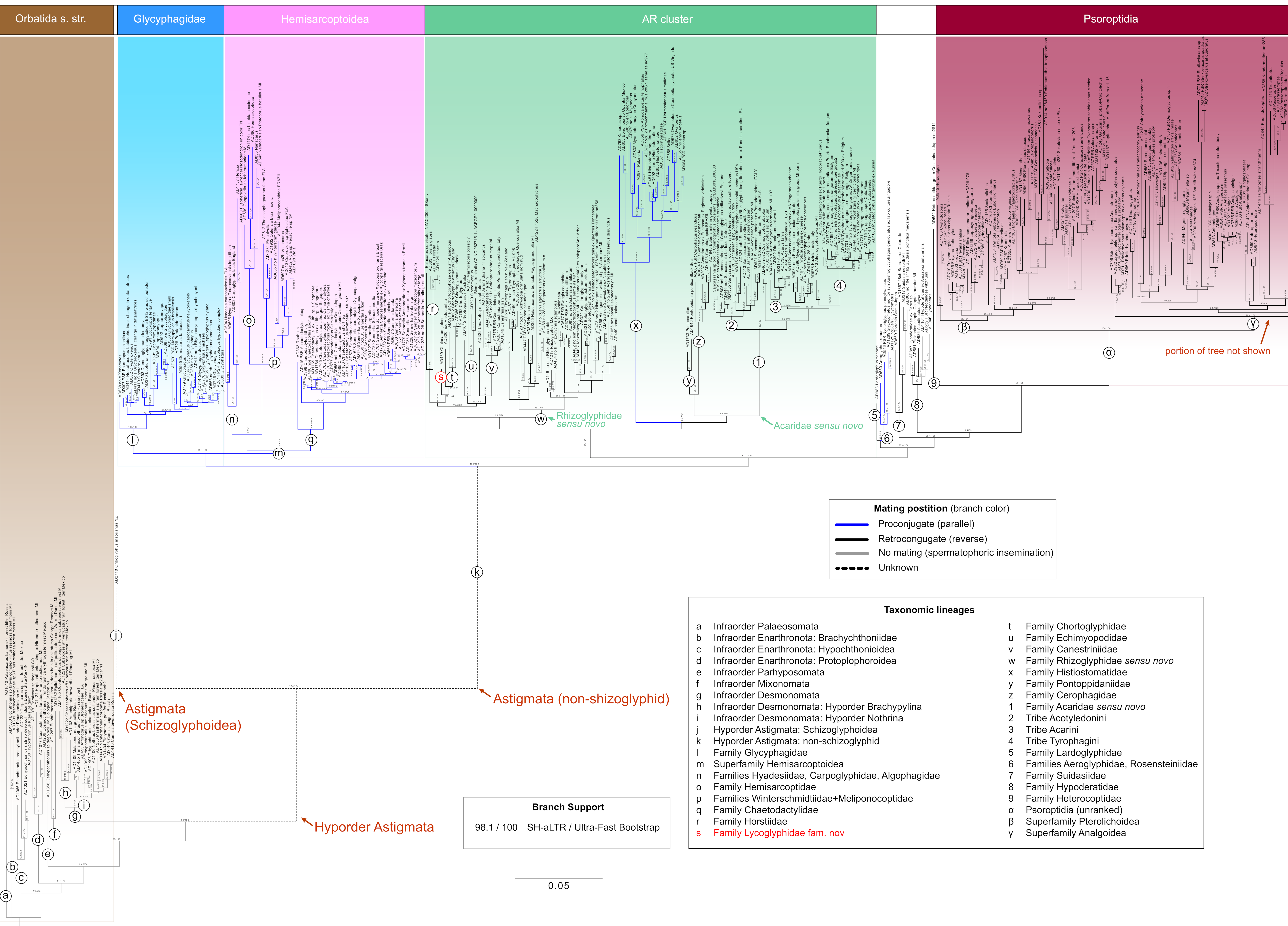
Our phylogenetic tree recovered Astigmata as an internal lineage within Oribatida
sensu stricto, forming a well-supported sister relationship with the infraorder Desmonomata (Nothrina + Brachypylina) (SH-aLRT/ultrafast bootstrap = 99/100;
Figure 1). Within Astigmata, several major lineages previously recognized based on morphology were recovered, including the superfamilies Schizoglyphoidea, Hemisarcoptoidea, Hypoderatoidea, and Psoroptidia—an unranked lineage composed largely of vertebrate parasites and including the superfamilies Pterolichoidea and Analgoidea (including Sarcoptoidea). These lineages occupied their expected positions in the tree (
Figure 1).
The previously unresolved lineage Heterocoptidae (incertae sedis) was robustly resolved as sister to Psoroptidia, with maximal support. In contrast, the superfamilies Glycyphagoidea, Acaroidea, and Histiostomatoidea showed substantial rearrangements. Specifically, (i) Glycyphagidae was recovered as closely related to Hemisarcoptoidea, whereas other glycyphagoid lineages were placed elsewhere in the tree; (ii) within a large clade here termed the AR cluster, Acaroidea was broadly redefined to include only one former core family, Acaridae, which itself was split into several well-supported independent lineages—Rhizoglyphidae
sensu novo, Acaridae
sensu novo, Pontoppidaniidae, Cerophagidae, Horstiidae, and Lycoglyphidae fam. nov.—while additional families were also placed within the AR cluster (Histiostomatidae, Chortoglyphidae, Echimyopodidae, and Canestriniidae); and (iii) a distinct group of families clustered around Psoroptidae, including Lardoglyphidae, Aeroglyphidae, Rosensteiniidae, Hypoderatidae, and Heterocoptidae (
Figure 1).
The economically important family Acaridae
sensu novo comprised the tribes Acarini, Tyrophagini, and Acotyledonini; while the family Rhizoglyphidae
sensu novo largely corresponds to the traditional morphology-based concept, with the exclusion of Acotyledonini (
Figure 1).
Figure 1.
Maximum likelihood tree of astigmatid mites (Astigmata) and outgroups (Oribatida s. str.) inferred in IQ-TREE. The input phylogenetic matrix had 619 taxa and alignments of six genes (
6861 sites total aligned): 18S (1675 nt), 28S (3379 nt), EF1A (363 aa), SRP54 (456 aa), HSP70 (569 aa), and COX1 (419 aa). Missing data: 6.1%. Following model selection and partition merging (-m MFP+MERGE), IQ-TREE used a three-partition scheme: TIM2+F+R7 for 18S, GTR+F+I+R8 for 28S, and Q.INSECT+R6/I for all protein-coding genes. This partition scheme was then used in the downstream maximum likelihood phylogenetic analysis with 1000 ultrafast bootstrap replicates and SH-aLRT branch tests to assess node support. A high-resolution tree is also available in
Supplementary Materials (Figure S1).
Figure 1.
Maximum likelihood tree of astigmatid mites (Astigmata) and outgroups (Oribatida s. str.) inferred in IQ-TREE. The input phylogenetic matrix had 619 taxa and alignments of six genes (
6861 sites total aligned): 18S (1675 nt), 28S (3379 nt), EF1A (363 aa), SRP54 (456 aa), HSP70 (569 aa), and COX1 (419 aa). Missing data: 6.1%. Following model selection and partition merging (-m MFP+MERGE), IQ-TREE used a three-partition scheme: TIM2+F+R7 for 18S, GTR+F+I+R8 for 28S, and Q.INSECT+R6/I for all protein-coding genes. This partition scheme was then used in the downstream maximum likelihood phylogenetic analysis with 1000 ultrafast bootstrap replicates and SH-aLRT branch tests to assess node support. A high-resolution tree is also available in
Supplementary Materials (Figure S1).
Mapping the two character states associated with mating position revealed that proconjugate (parallel) mating originated independently in three monophyletic clades: (i) Hemisarcoptoidea + Glycyphagidae, (ii) Histiostomatidae, and (iii) Aeroglyphidae + Rosensteiniidae. In contrast, all other non-schizoglyphid Astigmata exhibited retroconjugate (reverse) mating (
Figure 1).
3.2. GenBank Sequence Misidentification
We conducted the following analyses for three suspect and critical GenBank sequences.
For “
Rhizoglyphus robini” (NC_038058.1; mitochondrial genome) [
80], a COX1 search (BLASTx using translation Table 5) returned the closest matches to
Sancassania (amino acid identity 98.6–99.8%), followed by
Aleuroglyphus and numerous
Tyrophagus sequences (95.7–96.3%), consistent with phylogenetic expectations (Acaridae
sensu novo). We therefore classify this sequence as
Sancassania cf.
berlesei (closest COX1 match: 99.8% amino acid identity to
Sancassania berlesei, YP_009050190.1).
“Cosmoglyphus oudemansi” (JBNXMS010000000) is represented by a whole-genome shotgun assembly. Because this assembly contains only nucleotide sequences, we extracted the 18S rRNA gene and conducted a BLAST (nt) (2.17.0) search, which returned six closest matches to Sancassania (nucleotide identity 97.7–99.4%). A 96.5% match to Cosmoglyphus sp. AD702 (JQ000105.1) was also recovered, as expected given the close phylogenetic relationship between these genera. To further validate these similarity-based BLAST results, we recovered our five standard nuclear genes from this genome and performed a phylogeny-based identification. This analysis placed the sequence unambiguously within Sancassania, among several accurately identified species. We therefore classify this genomic sequence as Sancassania (closest 18S rRNA match: 99.4% nucleotide identity to Sancassania berlesei, GQ864331.1).
“Rhizoglyphus sp.
” SJD-2001 (AF287236.1) [
48] is represented by a single 18S rRNA sequence. As above, we conducted a BLAST (nt) search, which returned
Cosmoglyphus sp. as the closest match (99.7% nucleotide identity), followed by numerous sequences belonging to Acaridae
sensu novo (e.g.,
Acarus,
Tyrophagus and
Sancassania). In contrast, true
Rhizoglyphus is expected to cluster with Rhizoglyphidae
sensu novo. We therefore classify this sequence as
Cosmoglyphus sp. (closest 18S rRNA match: 99.7% nucleotide identity to
Cosmoglyphus sp. AD702, JQ000105.1).
3.3. Systematics
Order Sarcoptiformes Reuter, 1909.
Suborder Oribatida van der Hammen, 1968.
Hyporder Astigmata Canestrini, 1891.
Superfamily Acaroidea Latreille, 1802.
Family Lycoglyphidae fam. nov.
Type genus Lycoglyphus gen. nov.
urn:lsid:zoobank.org:act:1B40EBDE-5904-494B-A2F5-5C5E5687E1FF
Diagnosis. Adults. Chelicerae chelate with few teeth and with two setae, cha and chb; cha large, lanceolate, situated anterior to chb. Palp reflexed inward, bearing two (sup and cm) or three (a, sup and cm) setae, terminal solenidion ω and terminal eupathidium ul″ and ul′; palp tarsus has a rounded lateral protrusion with lanceolate cm. Subcapitulum without ventral external ridges and with setae h and elcp. Idiosomal cuticle smooth. Grandjean’s organ with one (sometimes with additional tongue-shaped outgrowth) trunk and numerous small spines. Supracoxal setae pinnate. Prodorsal shield vestigial (Lycoglyphus and Obelacarus) or well developed (Viedebanttia), not divided. Setae ve present (short or vestigial in Viedebanttia). All idiosomal and coxal setae smooth, thin, and filiform. Setae si, se, c1, c2, cp, c3, d1, d2, e1, e2, f2, h1, h2, h3, ps1, ps2, ps3 present. Anterior coxal apodemes I–IV and posterior coxal apodemes II present. Female oviporus longitudinal with genital valves fused to body anteriorly, free posteriorly. Epigynal apodeme absent. Genital papillae conoidal associated with genital opening. Opisthosoma rounded posteriorly, without opisthosomal shield or sclerotization. Anus positioned near posterior margin of body. Males with para-anal suckers. Legs ventrally positioned. Pretarsi with short strong condylophores. Pretarsal ambulacrum not greatly expanded. Empodial claws present on all legs, simple. Males with sucker-like setae d and e on tarsus IV. Otherwise, all tarsi with long filiform setae d, f and short spiniform e. Unguinal setae u, v larger and stouter than proral setae p, q, or absent. Setae aa I, ba II present. Tarsus I with solenidion ω2 basal to ω1. Genu I with two solenidion (σ″ and σ′), ratio of the lengths of which differs among genera. Heteromorphic males present, with enlarged leg III with vestigial ambulacrum, rudimentary condylophores and not enlarged claw, spiniform setae e, r, w, s, filiform d, f and rudimentary v and q, without an enlarged seta similar in form to empodial claw.
Heteromorphic deutonymph. Gnathosoma short, with filiform setae cm, sup and alveoli h. Palps separated, with two segments. Body ovoid, smoothly punctate. Propodosoma not overhanging gnathosoma anteriorly. Setae vi present on dorsal propodosoma, ve present (Lycoglyphus, Obelacarus) or absent (Viedebanttia). Propodosomal ocelli absent. Hysterosoma with 11 pairs of simple filiform setae on hysterosomal shield (c1, c2, cp, d1, d2, e1, e2, f2, h1, h2, h3). Sejugal furrow present. Coxal setae 1a and 3a present (conoidal or filiform). Coxal apodemes I and anterior apodemes II not extending to level of coxal apodemes III. Coxal apodemes II end freely; III–IV fused medially. Attachment organ with anterior and median suckers (ad3 and ad1+2) well developed and setae ps1 and ps2 in the form of conoids. All segments of legs free. Legs III–IV posteriorly or laterally directed in repose. Pretarsi without membranous ambulacra, empodia arising from tarsal apices. Empodial claw present on all legs. Setae nG III present. Setae aa I, wa I and ba II present. Solenidion ω2 I present.
Genera included: Lycoglyphus gen. nov; Obelacarus gen. nov.; Viedebanttia Oudemans, 1929.
Remarks. Lycoglyphidae fam. nov. shares several characteristics with Acaroidea [
5,
49]: chelicerae are chelate, the subcapitulum and palps with completed chaetome, idiosoma with cuticle smooth, dorsal and paraproctal chaetome complete, female oviporus in the form of an inverted V, internally with strongly folded pseudovipositor, male with paranal suckers and with modified suckerlike setae on tarsus IV, all legs with free segments and complete chaetome, empodial claws present, and pretarsal ambulacrum not greatly expanded. Lycoglyphidae and Bulacaridae share the presence of two cheliceral setae (
cha and
chb).
Lycoglyphidae differs from Bulacaridae in the lanceolate shape and terminal arrangement cha (vs. filiform, situated in basal part of fixed digit); lateral protrusion on palpal tarsi present (vs. lateral protrusion absent); lanceolate cm (vs. filiform); setae ve present (vs. absent); Grandjean’s organ well developed (vs. absent); supracoxal setae scx pinnate (vs. slender, barbed at their terminal half); prodorsal shield vestigial or distinct, whole (vs. divided into paired parallel sclerites); dorsal setae smooth, filiform (vs. dorsal setae lanceolate, barbed and curved); setae 4a filiform (vs. lanceolate); epigynal apodeme absent (vs. present); setae aa I present (vs. absent); solenidion ω1 situated near ε (vs. far from ε); two solenidia σ I (vs. one σ I).
Lycoglyphidae can be distinguished from Acaridae by the combination of the following characters, including: two cheliceral setae (vs. one only cha); palp tarsi with large rounded lateral protrusion on which the lanceolate setae cm are situated (vs. without lateral protrusion, cm typically filiform); Grandjean’s organ with numerous small spines (vs. usually smooth).
Lycoglyphidae can be distinguished from Gaudiellidae and Suidasiidae by asymmetrical tarsal setae e and f–e spiniform, f–filiform (vs. setae e and f filiform, similar in length).
Lycoglyphidae can be distinguished from Glycacaridae by prodorsal shield entire, not being in the form of pair of parallel sclerites, vestigial (vs. being in the form of pair of well-developed parallel sclerites); smooth dorsal setae (vs. elongate and heavily barbed); smooth cuticle (vs. cuticle with small triangular microtrichae).
Lycoglyphidae can be differentiated from most Lardoglyphidae by simple empodial claws in both sexes (vs. bifurcate in females and sometimes males).
Lycoglyphidae can be differentiated from Scatoglyphidae by the anus situated near posterior margin of body (vs. anus positioned directly behind genital opening, displaced from the posterior edge of the body by a distance greater than the length of the anus); well-developed rounded para-anal suckers in males (vs. vestigial or absent in males).
Heteromorphic deutonymphs of Lycoglyphidae are difficult to distinguish from Acaridae due to the great morphological diversity of the latter. Lycoglyphidae, like some Acaridae, have two-segmented palps, setae cm, sup are present on the gnathosoma, leg setae aa I, wa I–II, hT I–II, gT I–II, ba II, nG III are present, and one solenidion is present on genu I. Heteromorphic deutonymphs of Viedebanttia clearly distinguished from all others by their long tibiae I–II (much longer than the corresponding genua) (tibia I–II no longer than genua in other genera of new family).
Heteromorphic deutonymphs of Lycoglyphidae differ from Suidasiidae and Lardoglyphidae by presence of empodial claw on leg IV (vs. absent). Deutonymphs of Lycoglyphidae differ from Gaudiellidae by having coxal apodemes III–IV fused medially and coxal fields III–IV closed (vs. coxal apodemes III–IV ending freely and coxal fields III–IV open).
Biology. Species of this family are closely associated with puffball fungi of the genus
Lycoperdon and
Apioperdon (
Agaricales and
Lycoperdaceae). Adult mites and other feeding stages live inside old sporocarps of their fungal hosts, where they feed on fungal spores. Phoretic deutonymphs of
Lycoglyphus and
Obelacarus disperse on endomychid beetles (Coleoptera and Endomychidae), whose larvae are biologically associated with the same fungal host and feed on fungal spores in puffballs (genus
Lycoperdina Latreille, 1807) [
81]. In some cases, mite species of two genera (
Lycoglyphus and
Viedebanttia) co-occur within the same host specimen. In contrast, deutonymphs of
Viedebanttia are mostly phoretic on unspecialized hosts (e.g., Carabidae, Formicidae and Myriapoda). Although this type of phoresy is largely a “one-way ticket” for the mites, who frequently fail to survive or reach a suitable sporocarp, it can nonetheless be ecologically significant for the fungus. Mites carrying spores are more likely to deposit them in habitats suitable for germination than spores dispersed passively by wind, thereby enhancing the fungus’s probability of successful colonization, even if the mites themselves perish. An additional reason why
Viedebanttia is found phoretic on a wide range of unspecialized hosts is that this genus is not exclusively restricted to puffballs but may also occur in other common fungal hosts, such as
Daldinia (Xylariales and Hypoxylaceae). This broader habitat preference likely explains why
Viedebanttia exhibits less host specificity in phoretic associations than
Lycoglyphus and
Obelacarus.
Lycoglyphus gen. nov.
Type species: Lycoglyphus kurosai sp. nov.; monotypic.
urn:lsid:zoobank.org:act:12619B10-7AF8-4AF0-8561-2BD422D15F5A
Diagnosis. Adult. Central trunk of supracoxal setae scx lanceolate with numerous lateral processes. Grandjean’s organ with one pointed trunk and numerous small spines. Prodorsal shield vestigial. Seta ve well developed, located on the same transverse line as vi. Posterior coxal apodeme II absent, and sclerite present. Unguinal setae u and v vestigial. Solenidia φ I–II reach the apices of their respective tarsi without ambulacra. Solenidion σ″ I longer than σ′. Solenidion σ III absent. Inseminatory canal of spermatheca with relatively large diameter. Paired sclerites of efferent ducts elongated, with very short stem.
Heteromorphic deutonymph. Seta ve present. Ratio hysterosomal shield/prodorsal shield length = 1.9–2.1. Coxal fields I closed. Coxal setae 1a and 3a filiform. External conoidal setae ps2 of attachment organ slightly posterior to median sucker ad1+2. Tibiae I–II similar in length or only slightly longer than corresponding genua. Seta aa I weakly spiniform, short (no longer than ω3). Setae e I–II cylindrical with sucker-like apex, different in form from simply foliate seta f. Setae d I–II shorter than e I–II. Seta ba II shorter than ω1 II. Setae p and q I–IV small spiniform. Seta f IV slightly longer than leg IV. Solenidion σ III absent.
Etymology. The generic name Lycoglyphus is a contraction of Lycoperdon (the fungal host) and the Greek verb γλύφω (glýphō, “to carve, engrave, or gnaw out”), alluding to the mite’s life-history and trophic association with puffball fungi. Gender masculine.
Remarks. The differences between the three genera of Lycoglyphidae fam. nov. are presented in the key below.
Lycoglyphus kurosai sp. nov.
Unnamed acarid mite—Kurosa, 2000 [
82]: 940, fig. 27.
urn:lsid:zoobank.org:act:4276E81C-24A1-438C-A5E9-95B3F1C7DD80
Material. Holotype: female—RUSSIA: Primorsky Krai, Ussuriysky District, Kaimanovka, ex Lycoperdon sp. (Agaricales: Lycoperdaceae), 21 Jun 1998 (ZISP). Paratypes: 13 HDN—same data; 5 HDN—same data, ex Lycoperdina koltzei Reitter (Coleoptera, Endomychidae) from Lycoperdon sp., 21 Jun 1998 (ZISP).
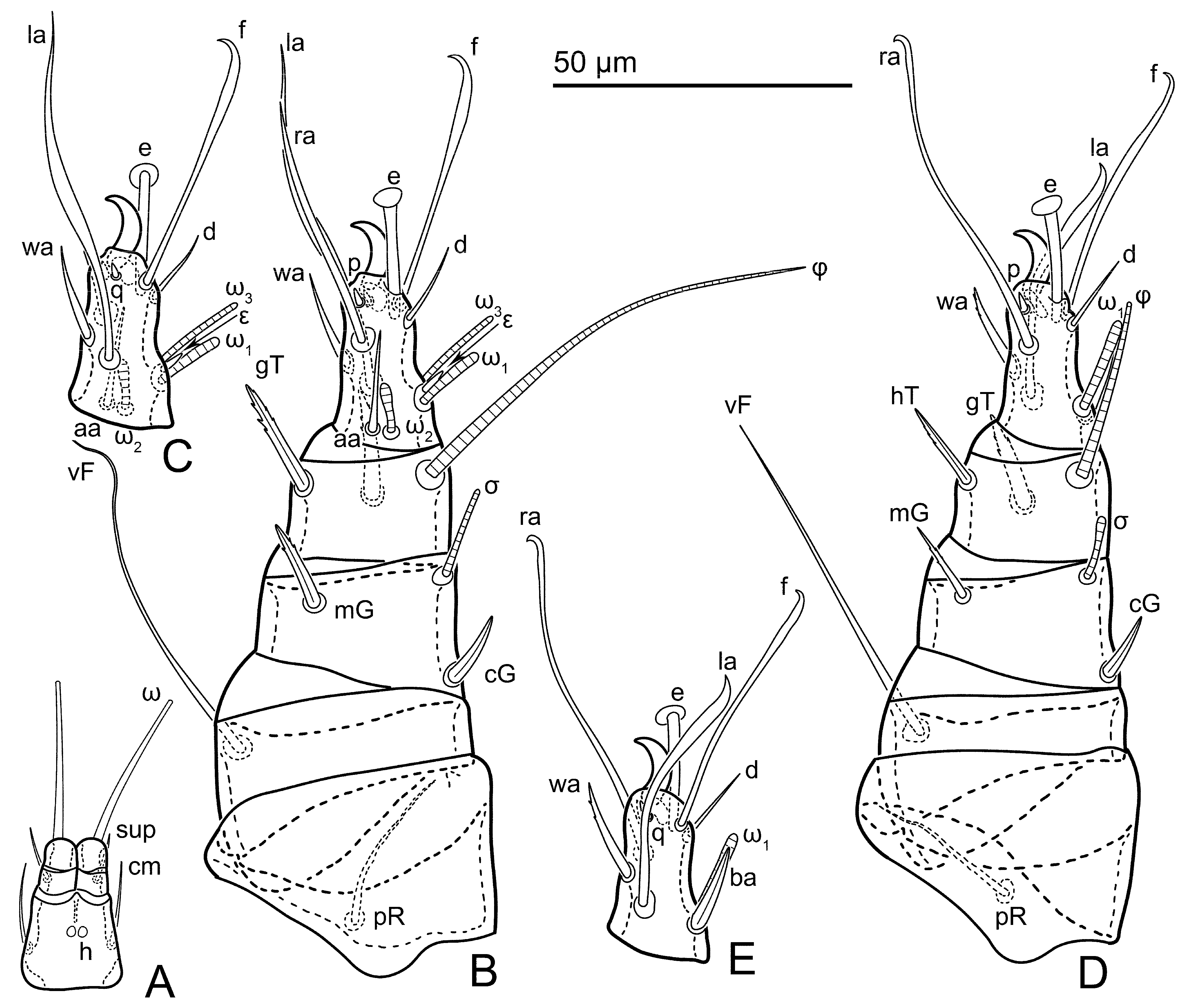
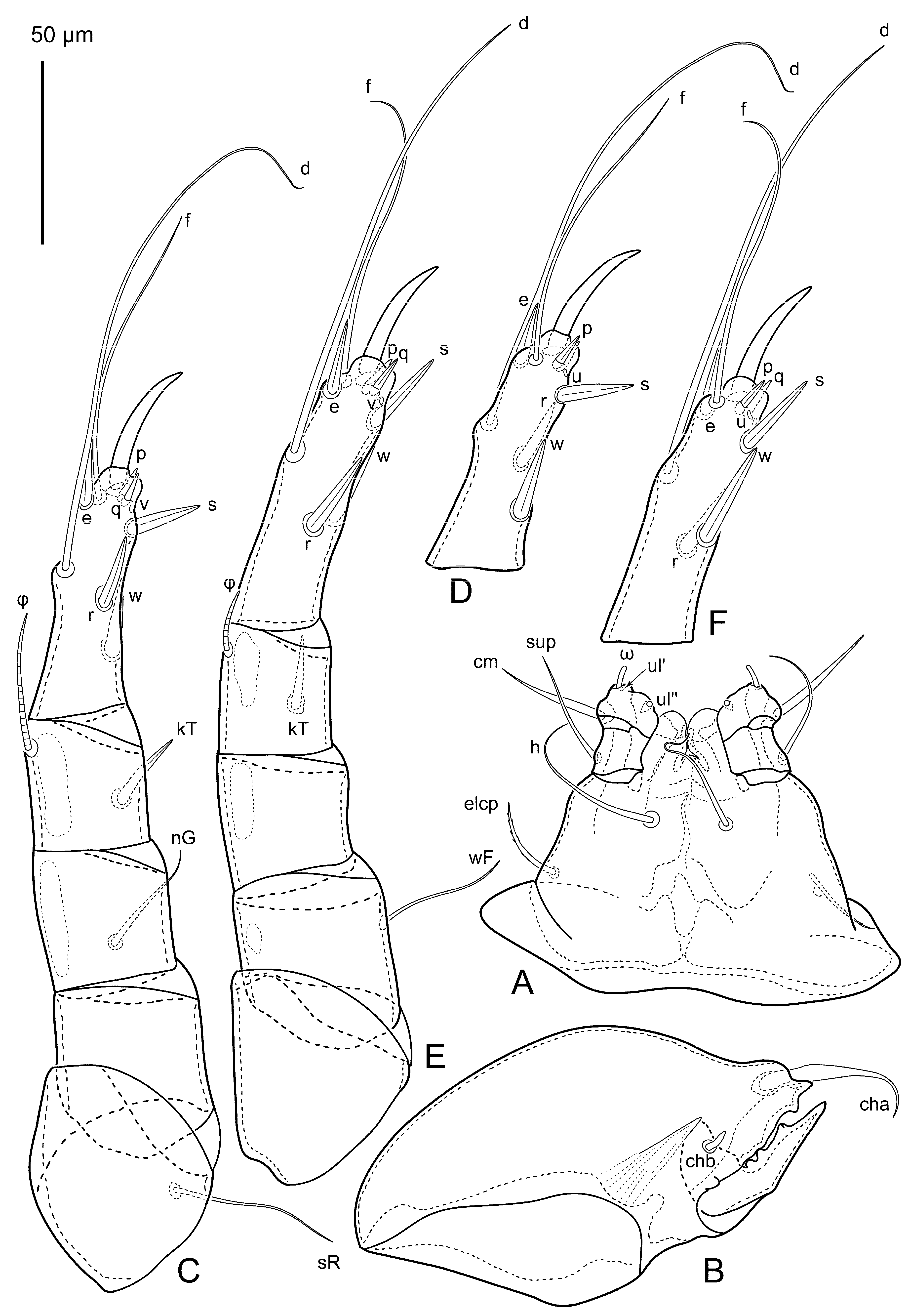
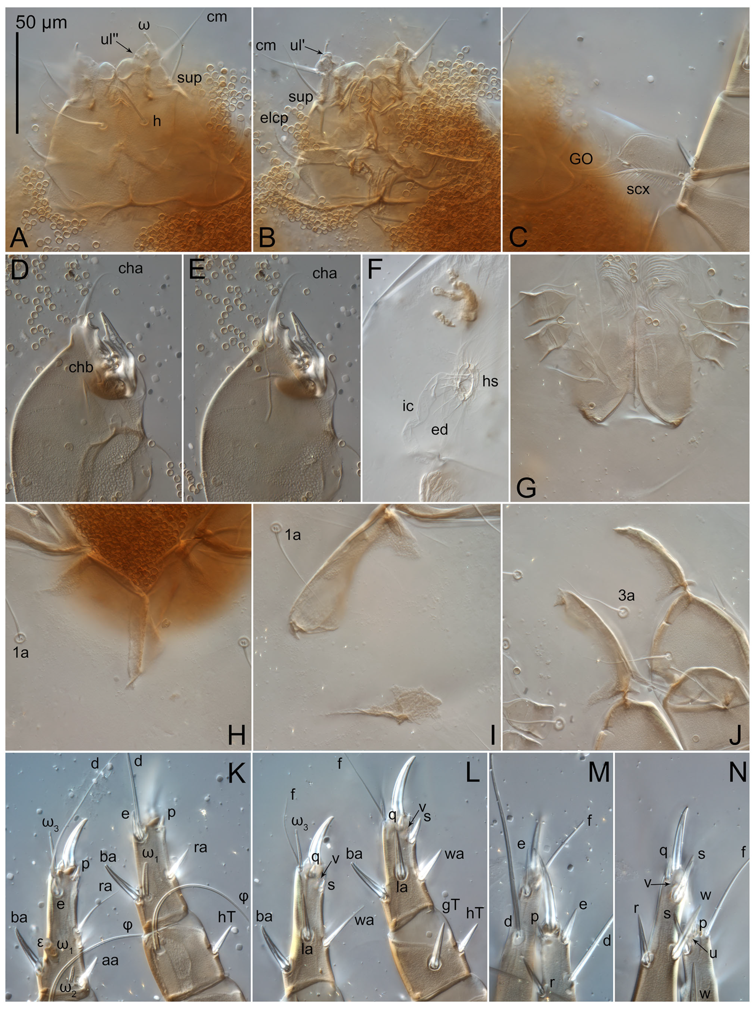
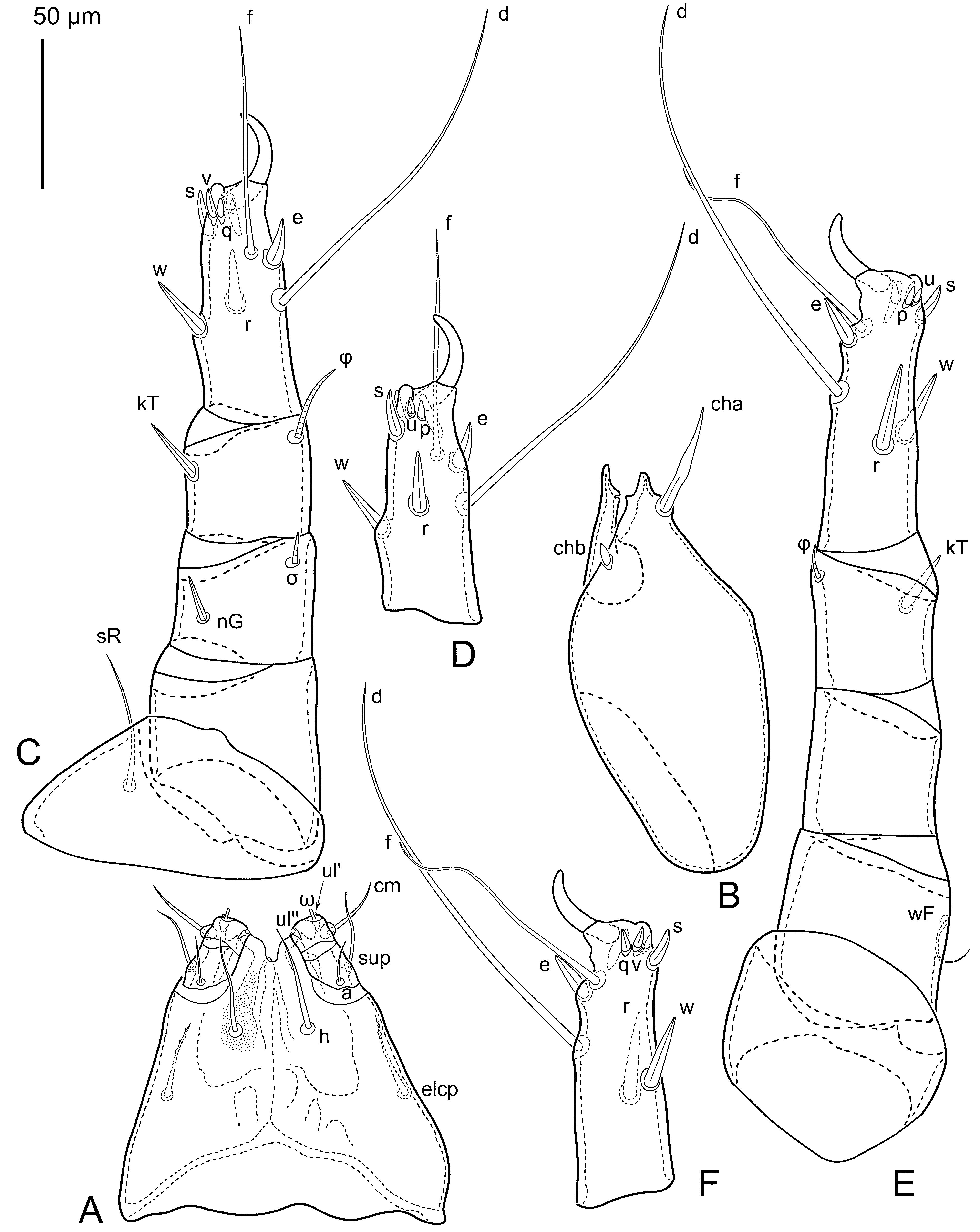
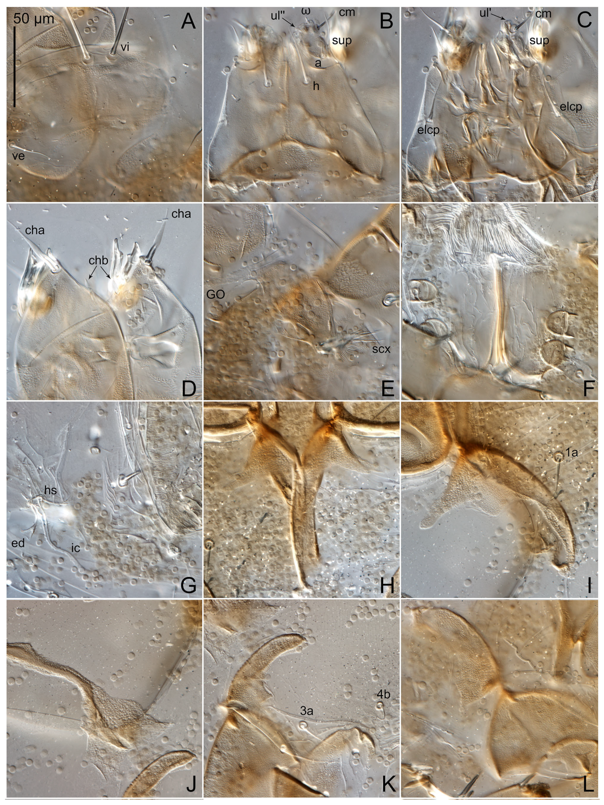
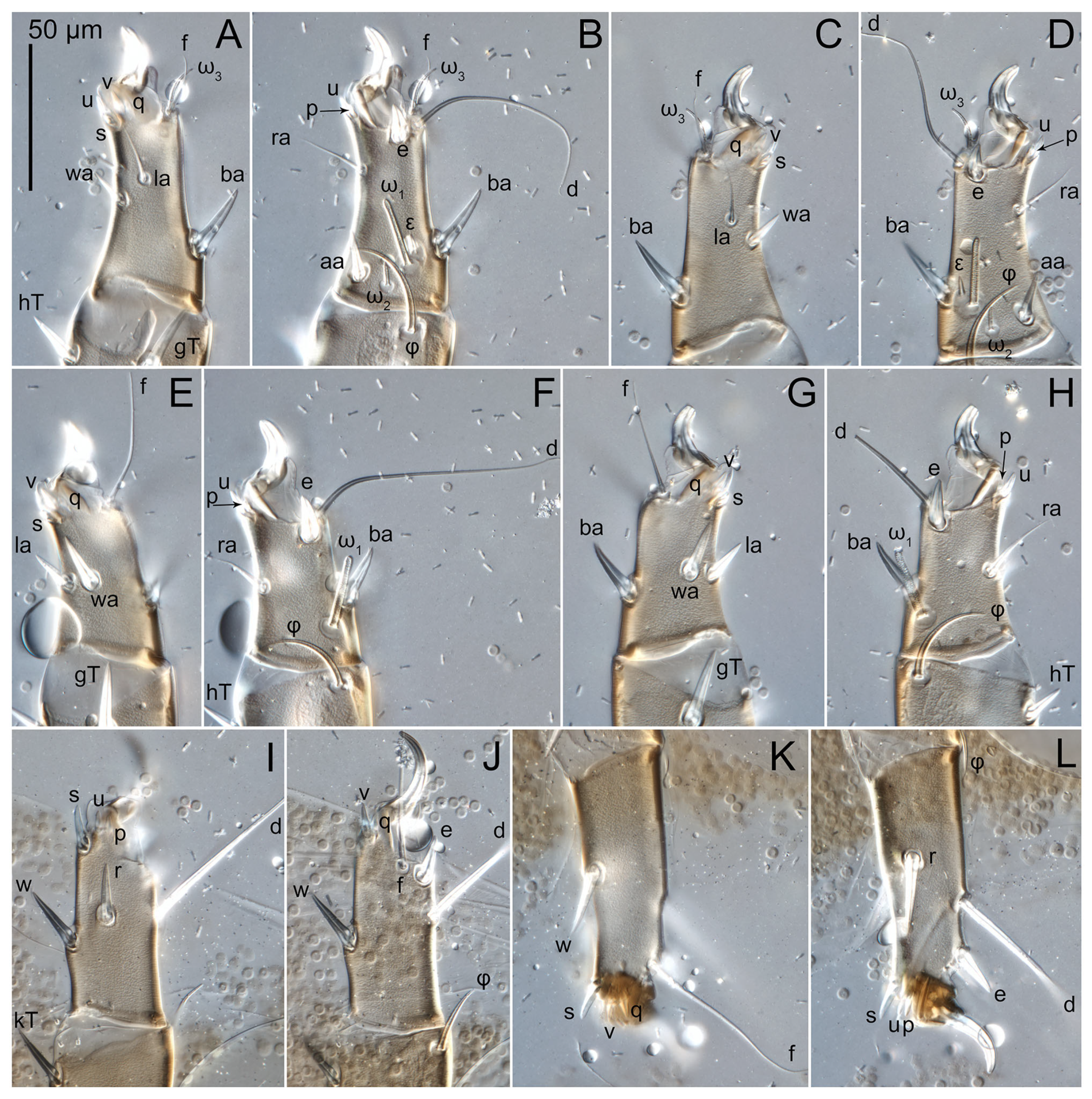
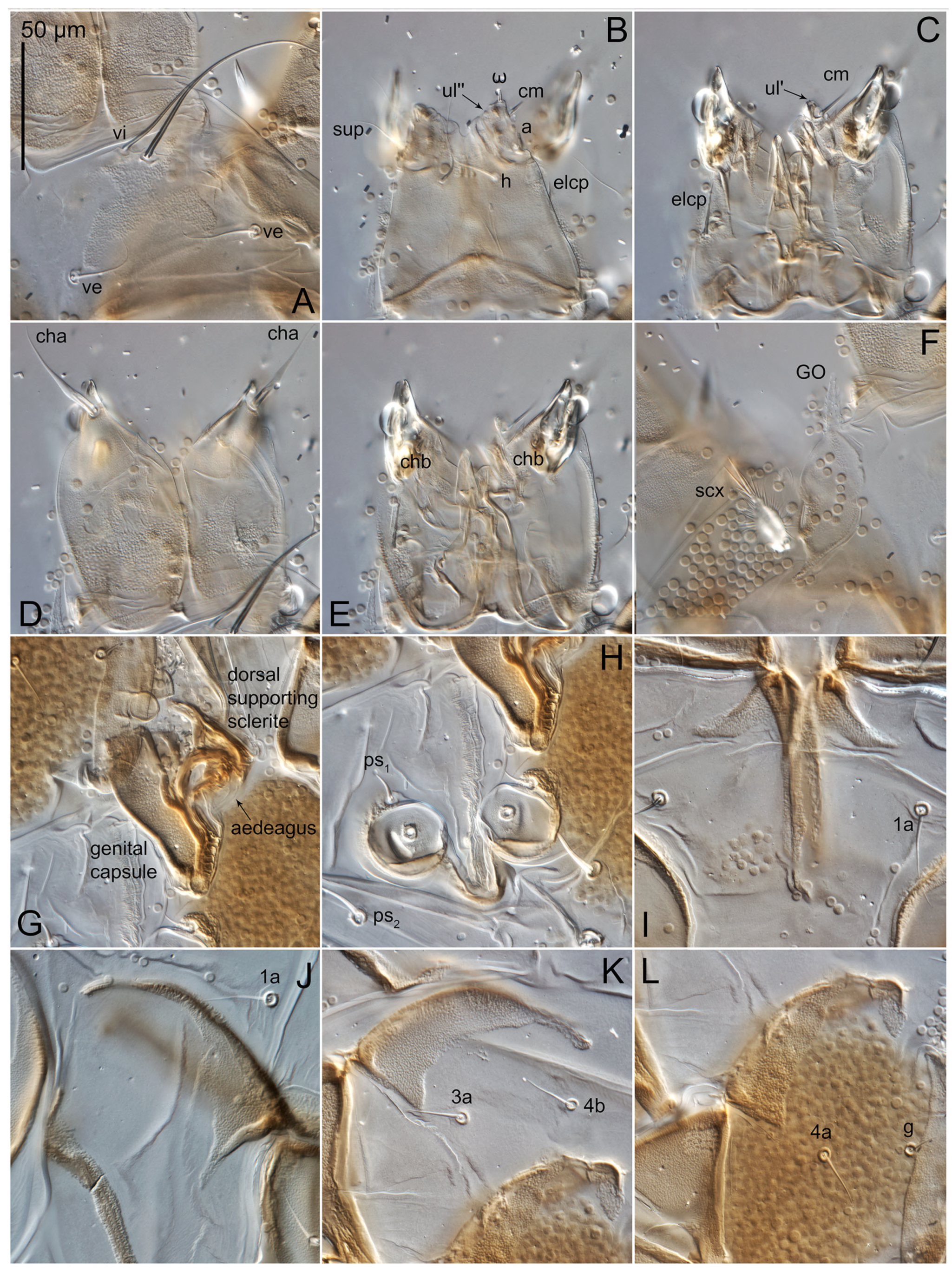
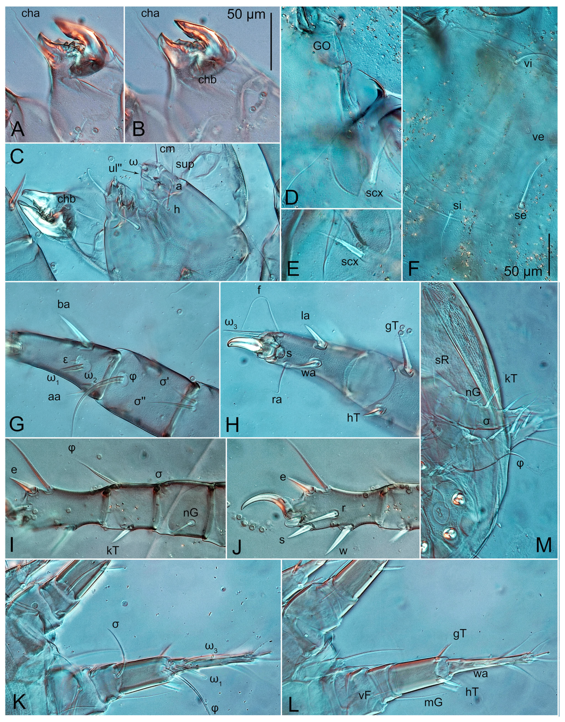
Gnathosoma (
Figure 5A,B and
Figure 6A,B,D,E): subcapitulum (
Figure 5A and
Figure 6A,B) wide, 77 × 110, evenly punctated; subcapitular setae (
h) long, widened basally; palpal supracoxal seta
elcp present, weakly barbed; palps wide; lateral dorsal palp tibial setae (
sup) filiform; palp tibial setae (
a) absent; dorsal palp tarsal seta (
cm) lanceolate, its base located on a large rounded lateral protrusion of palp tarsus; terminal palp tarsal solenidion ω short; terminal eupathidium
ul″ small, dome-shaped; terminal eupathidium
ul′ small, rounded. Chelicera (
Figure 5B and
Figure 6D,E) with two setae:
cha large, lanceolate, situated distally on fixed digit,
chb short, and spiniform. Fixed digit of chelicera with one tooth; movable digit with three teeth (anterior double).
Prodorsal shield vestigial, represented by a short weakly sclerotized area with punctation behind bases of setae
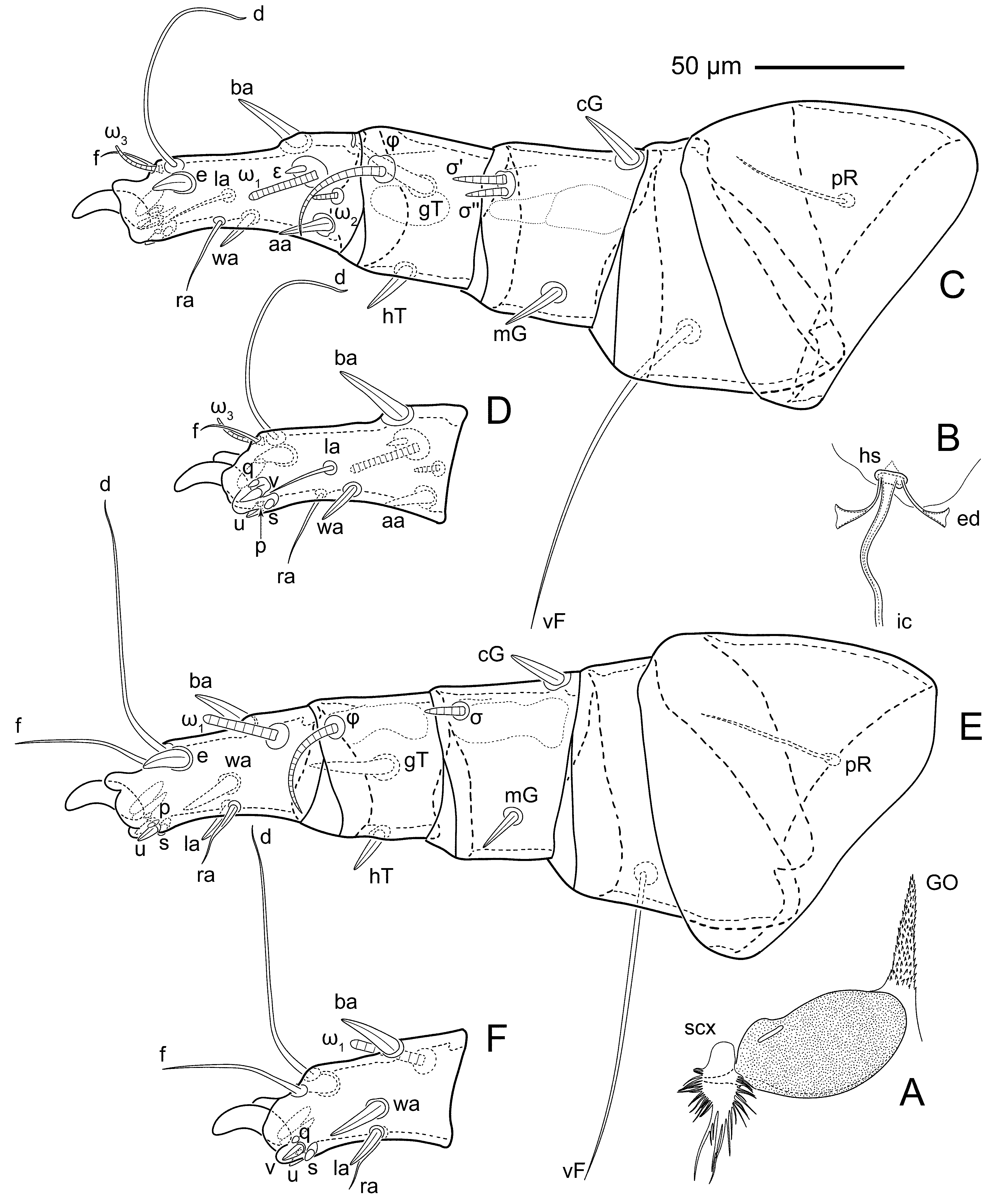
vi. Grandjean’s organ (
GO) with one pointed trunk and numerous small spines (
Figure 4A and
Figure 6C). Supracoxal setae (
scx) pinnate—its central trunk lanceolate with numerous (24–25) long lateral processes pointed at tip (
Figure 4A and
Figure 6C). Supracoxal shield oval, punctate, with a small pointed projection near base of
scx. Idiosomal setae (
vi,
ve,
si,
se,
c1,
c2,
cp,
c3,
d1,
d2,
e1,
e2,
f2,
h1,
h2,
h3,
ps1,
ps2,
ps3) smooth, filiform, setae
h3,
se,
vi longer than other setae;
ve and
si shorter than other setae. Opisthosomal gland openings between setal bases
e1 and
e2. Four pairs of fundamental cupules (
ia,
im,
ip and
ih) present. Ventral idiosoma with four pairs of coxal setae (
1a,
3a,
4a and
4b) and one pair of genital setae (
g). Coxal sclerites: I—short, narrow, triangular (
Figure 6H); anterior II—narrowly extended along anterior apodeme II, with a slight expansion at base (
Figure 6I); posterior apodeme II absent, but present as shapeless sclerite (
Figure 6I); III and IV—narrowly extended along apodemes, with a slight expansion at base (
Figure 6J). Genital region (
Figure 6G) situated between coxal fields III and IV; genital valves form an inverted Y; epigynal apodeme absent. Genital papillae (
Figure 6G) wide, with pointed ends. Anal opening ventral, long; distance between ovipore and anal opening less than half length of anal opening. Copulatory tube absent. Inseminatory canal of spermatheca wide, ribbon-like, slightly widened at junction with base of spermatheca (
Figure 4B and
Figure 6F). Base of spermatheca flat, ring-shaped. Paired efferent ducts (
Figure 4B and
Figure 6F) elongated, with very short stem (difficult to see). Three pairs adanal (
ad1–3) setae.
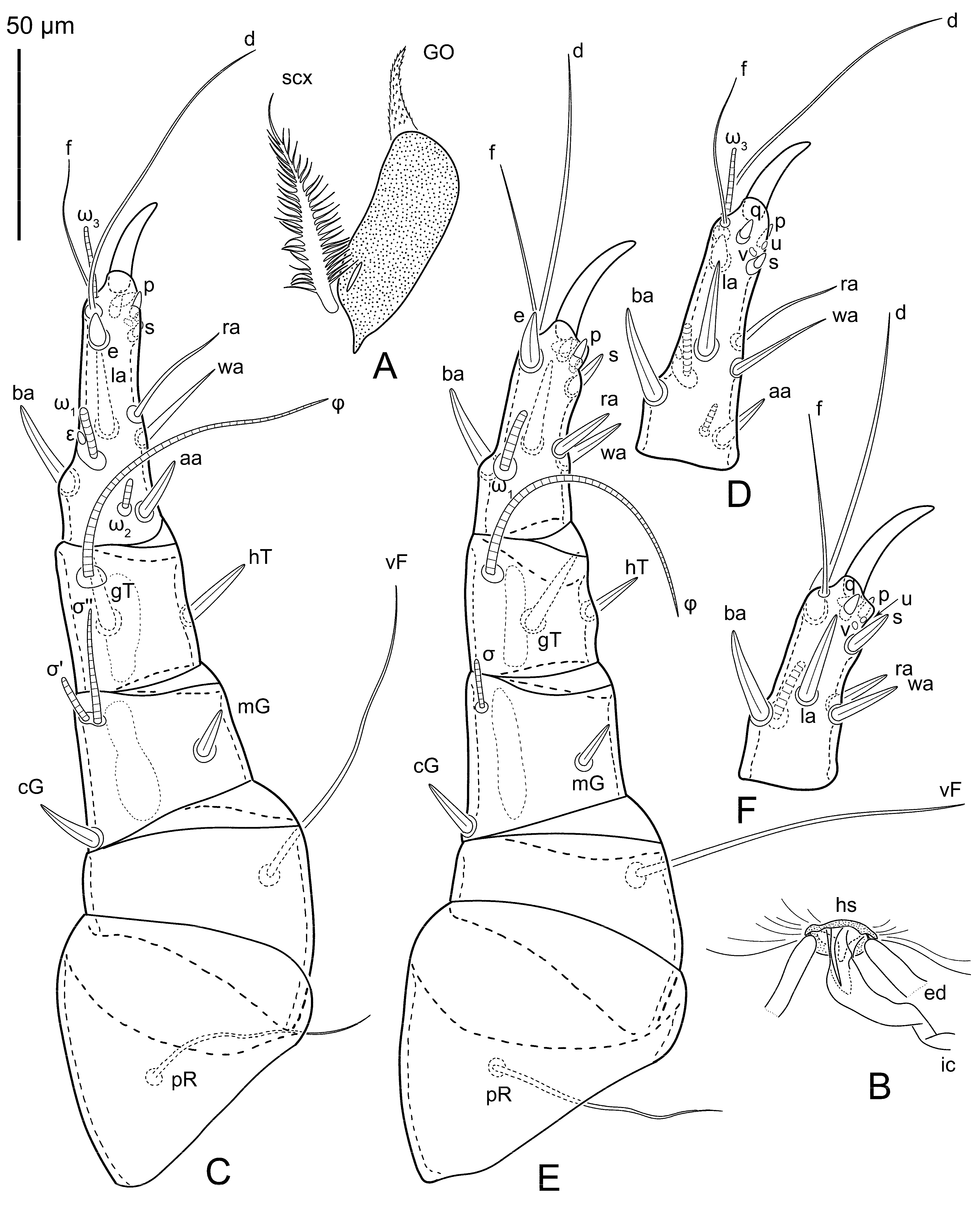
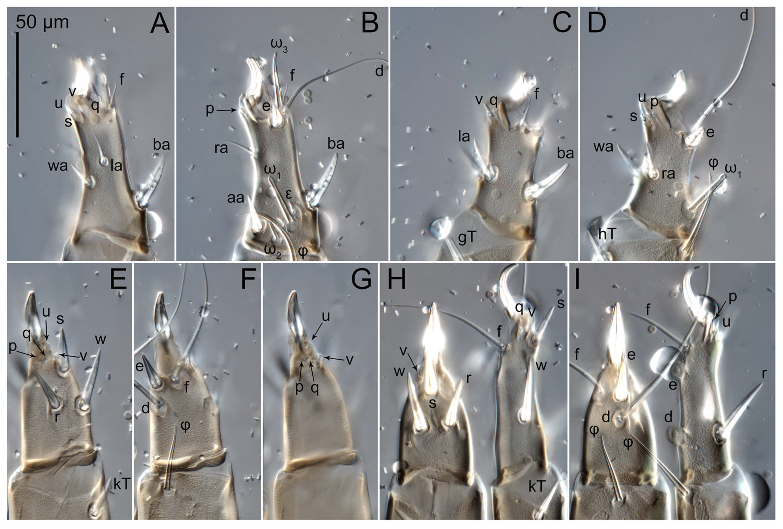
All legs shorter than body; all segments free (
Figure 4C–F,
Figure 5C–F and
Figure 6K–N). Trochanters I–III each with long filiform seta,
pR I–II,
sR III; trochanter IV without setae. Femoral setation 1-1-0-1; setae
vF I–II and
wF IV long, filiform. Genual setation 2-2-1-0; setae
mG and
cG I–II spiniform; seta
nG III filiform. Tibial setation 2-2-1-1; setae
hT I–II,
gT I–II and
kT III–IV spiniform,
hT slightly shorter than
gT. Tarsal setation 13-12-10-10; pretarsi consist of slightly curved and long empodial claws attached to short paired condylophores. Tarsus I with setae
ra,
f and
d filiform,
aa,
ba,
e,
wa,
la larger spiniform,
s,
p and
q short spiniform, and
u and
v represented by very small remnants. Tarsus II with same setae except
ra spiniform and
s larger, spiniform, and
aa absent. Tarsi III and IV with setae
f and
d filiform,
r,
w,
e and
s larger spiniform,
p and
q small spiniform, and
u and
v represented by very small remnants. Solenidia ω
1 on tarsi I and II cylindrical, with clavate apex, not bent. Solenidion ω
2 on tarsus I shorter than ω
1, cylindrical, with rounded apex, situated distinctly posterior to ω
1, beside
aa. Solenidion ω
3 distal on tarsus I, cylindrical, with rounded tip, longer than ω
1. Famulus (ε) of tarsus I wide, spiniform, with broadly rounded apex. Solenidia φ of tibiae I–II elongate, tapering, reaching the apices of their respective tarsi without ambulacra but not protruding beyond apex of empodial claws; solenidia φ III–IV tapering, shorter than respective tarsi (without ambulacra), φ III reaches level of bases setae
w and
r, and φ IV not reaching level of these setae. Solenidia σ′ and σ″ on genu I elongate, tapering, σ″ longer than σ′, σ″ not quite reaching bases of φ I. Solenidion σ on genu II more than 9–10 times longer than its width, with rounded tip. Solenidion σ of genu III absent.
Male unknown.
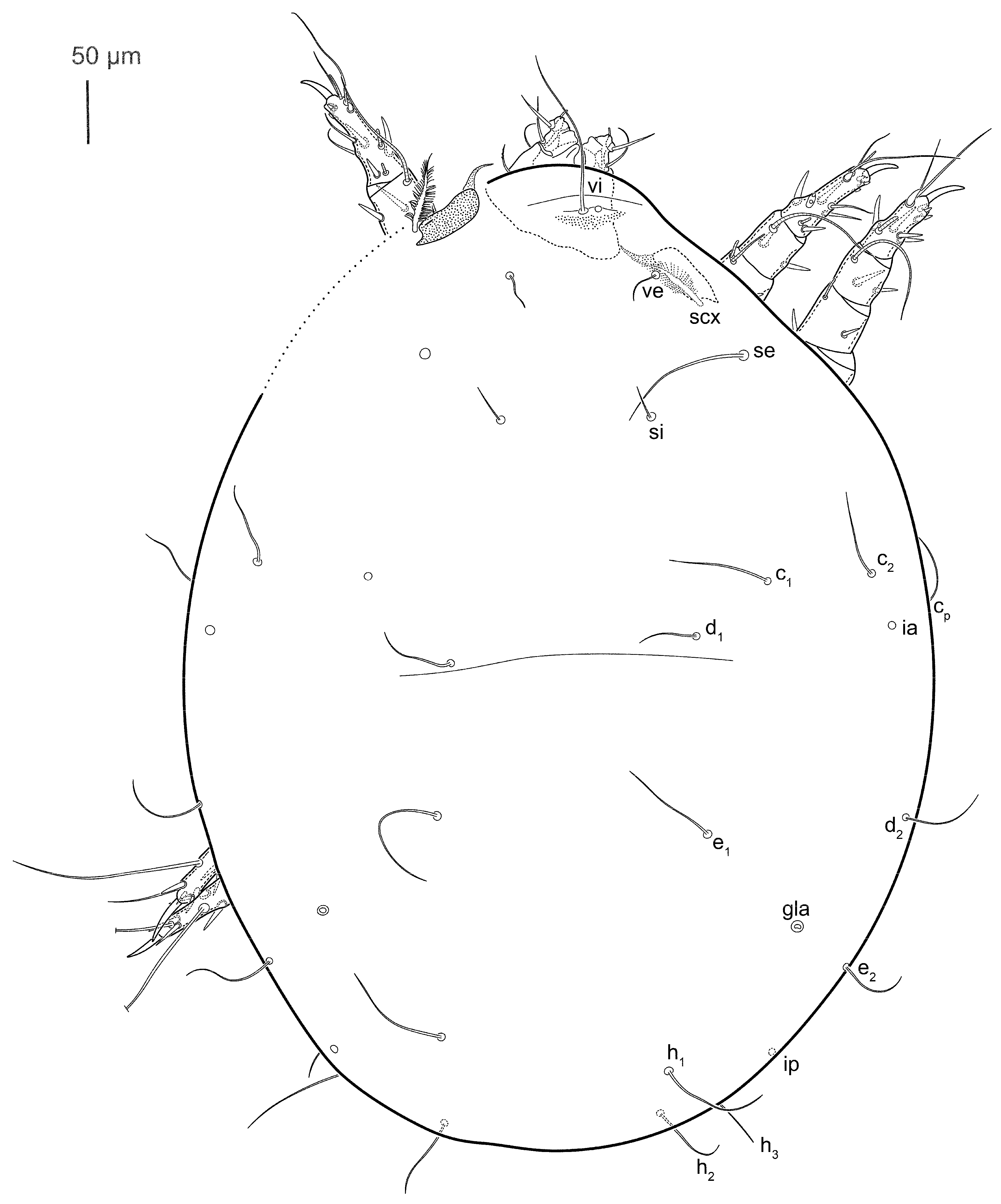
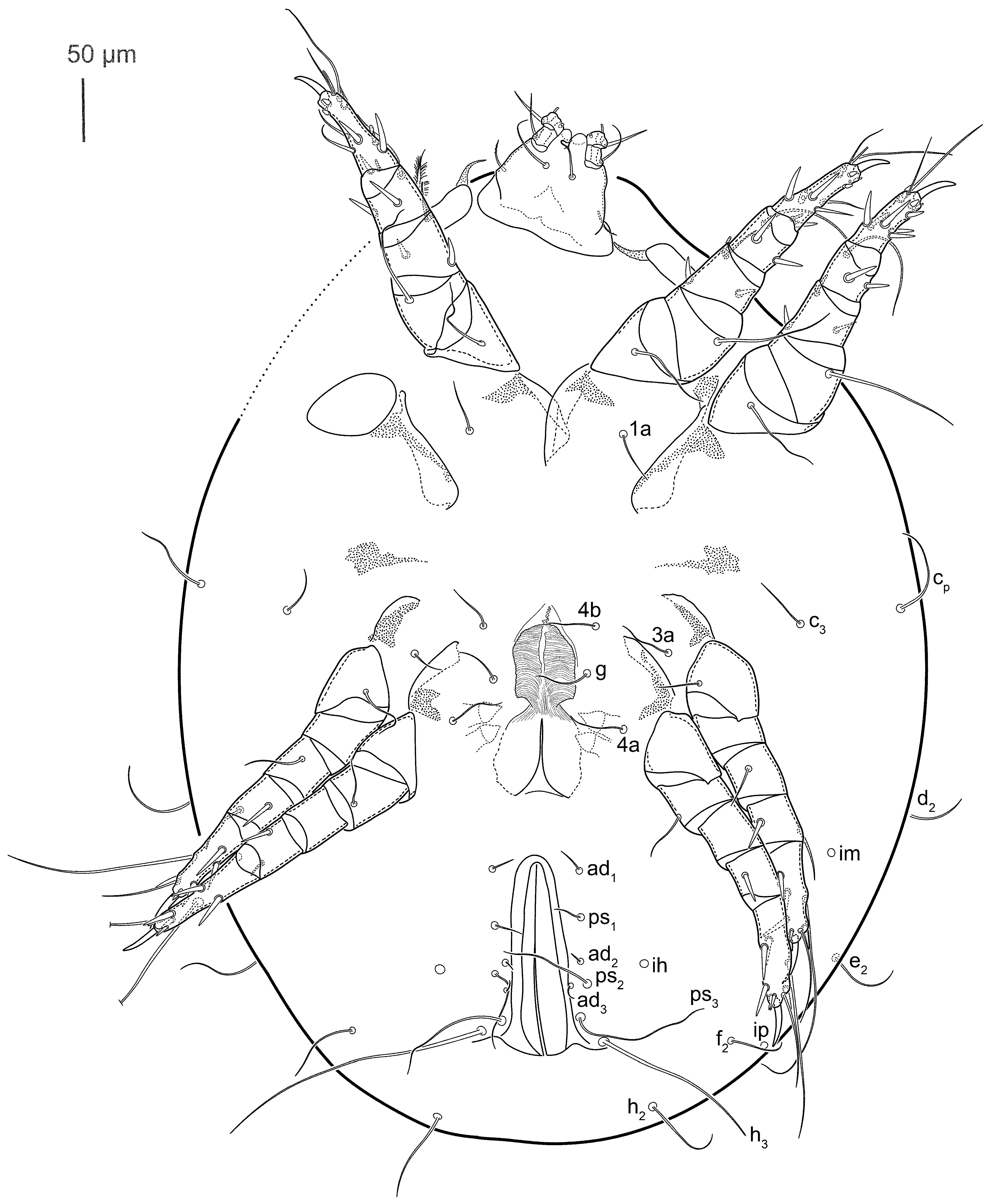
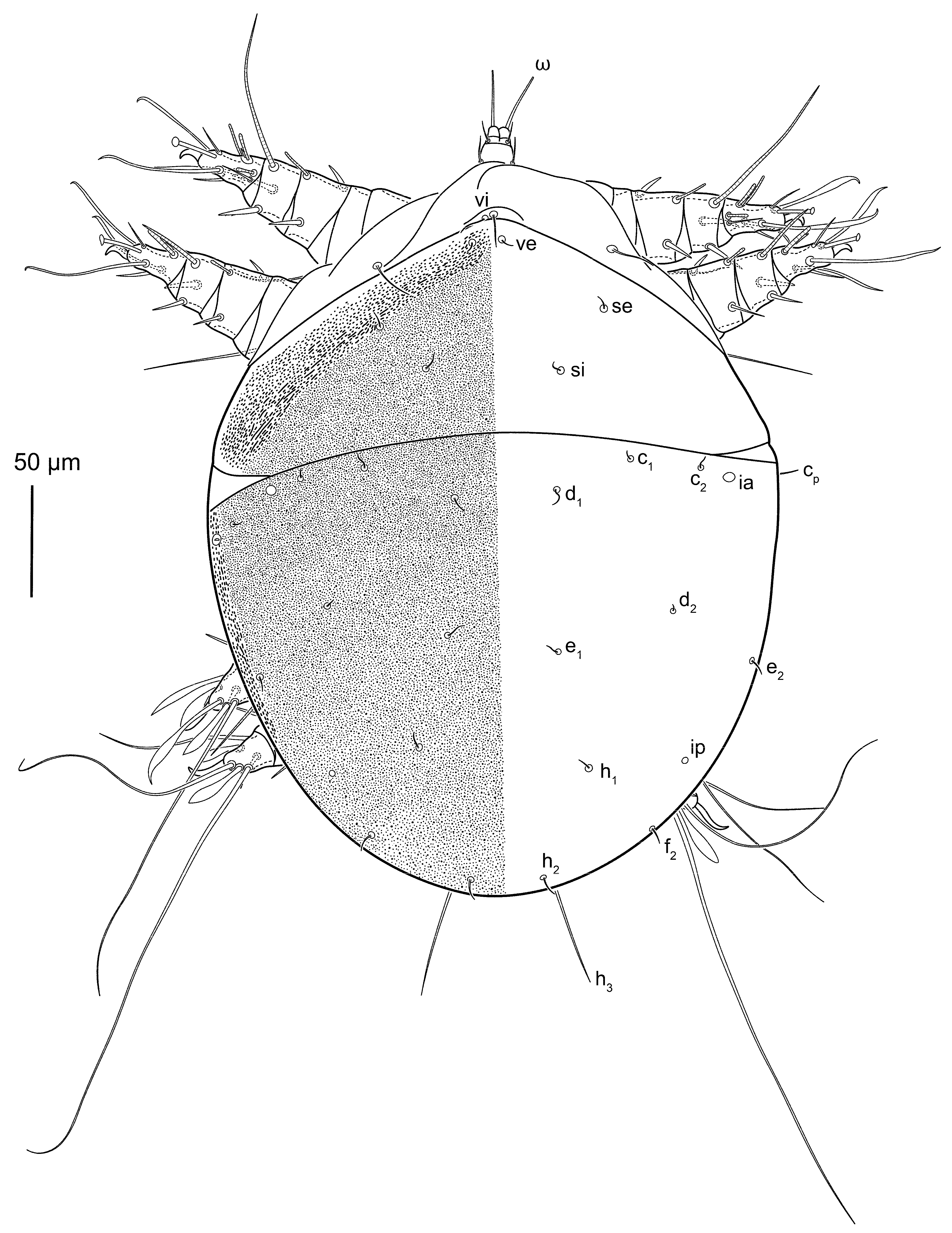
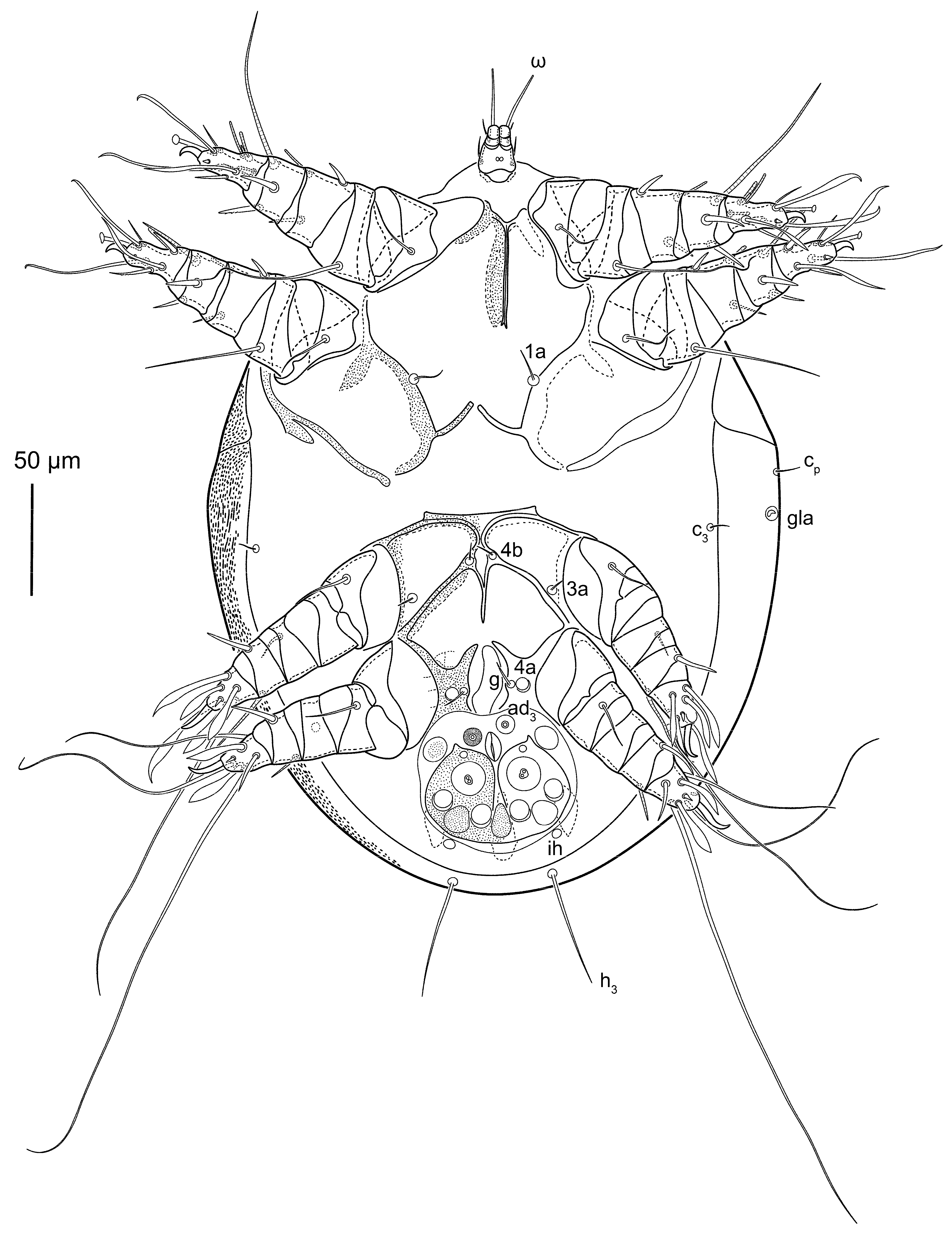
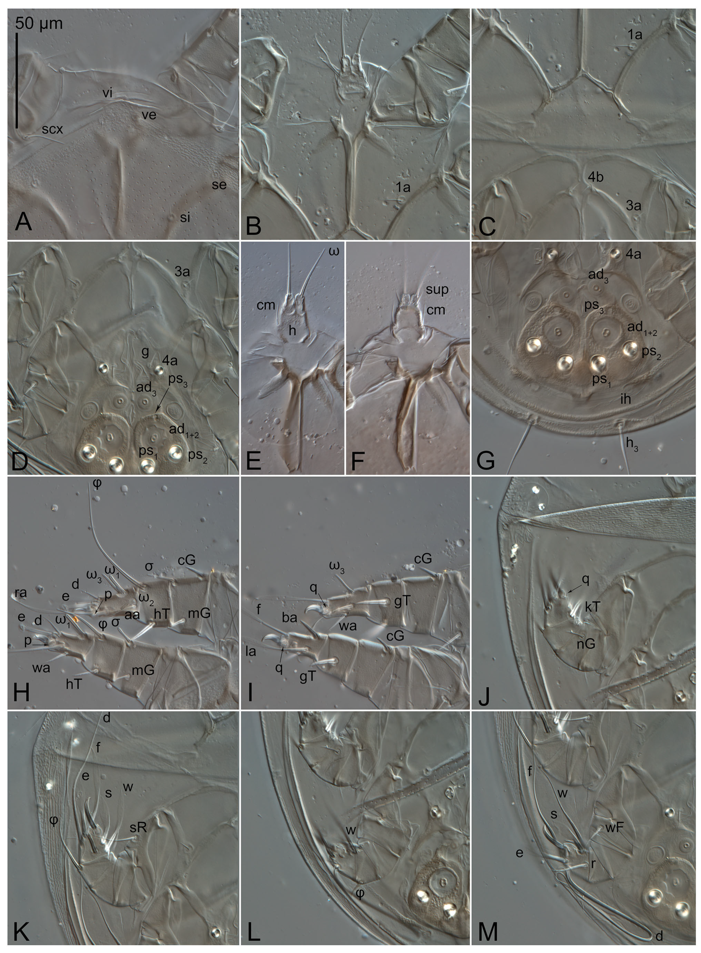
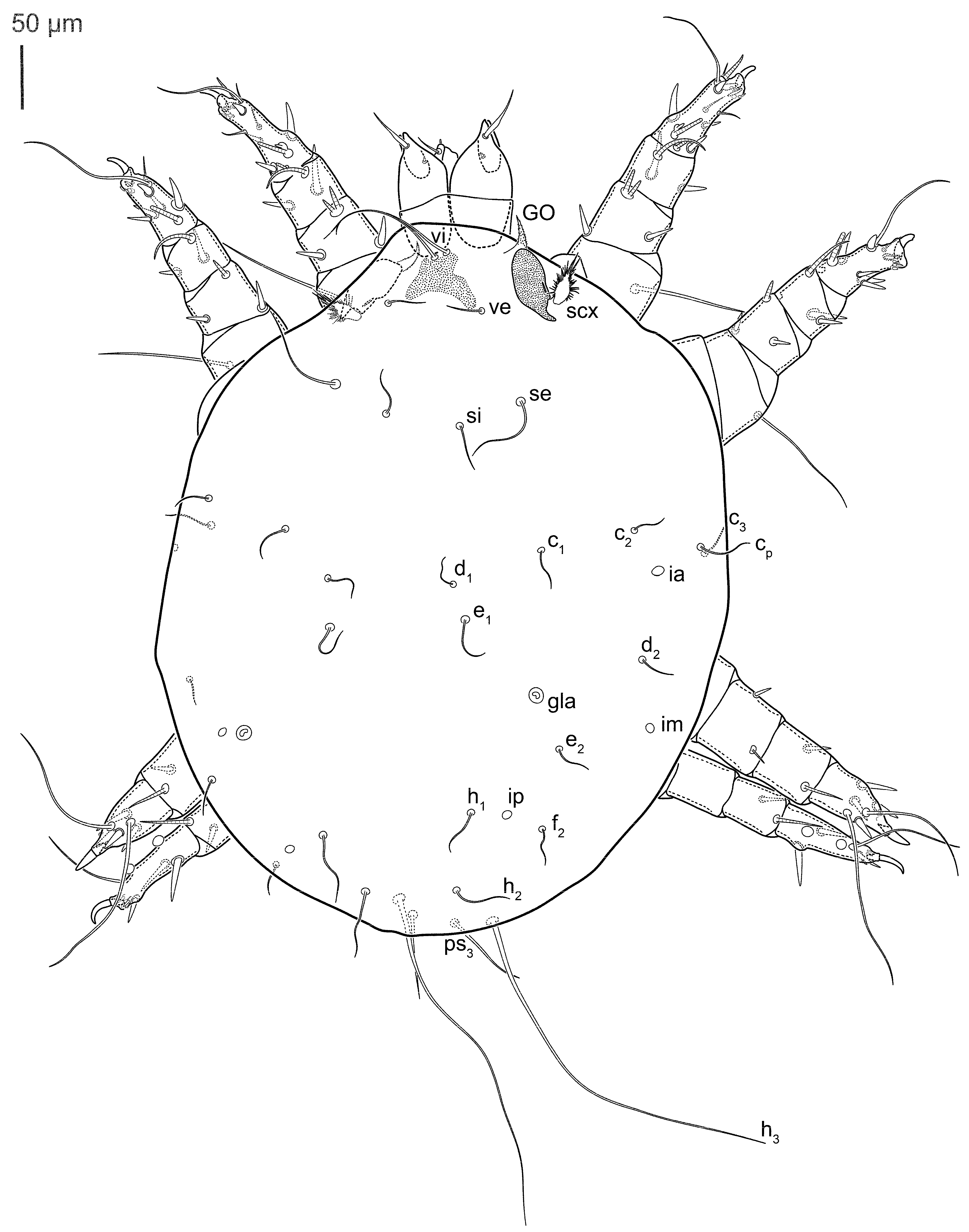
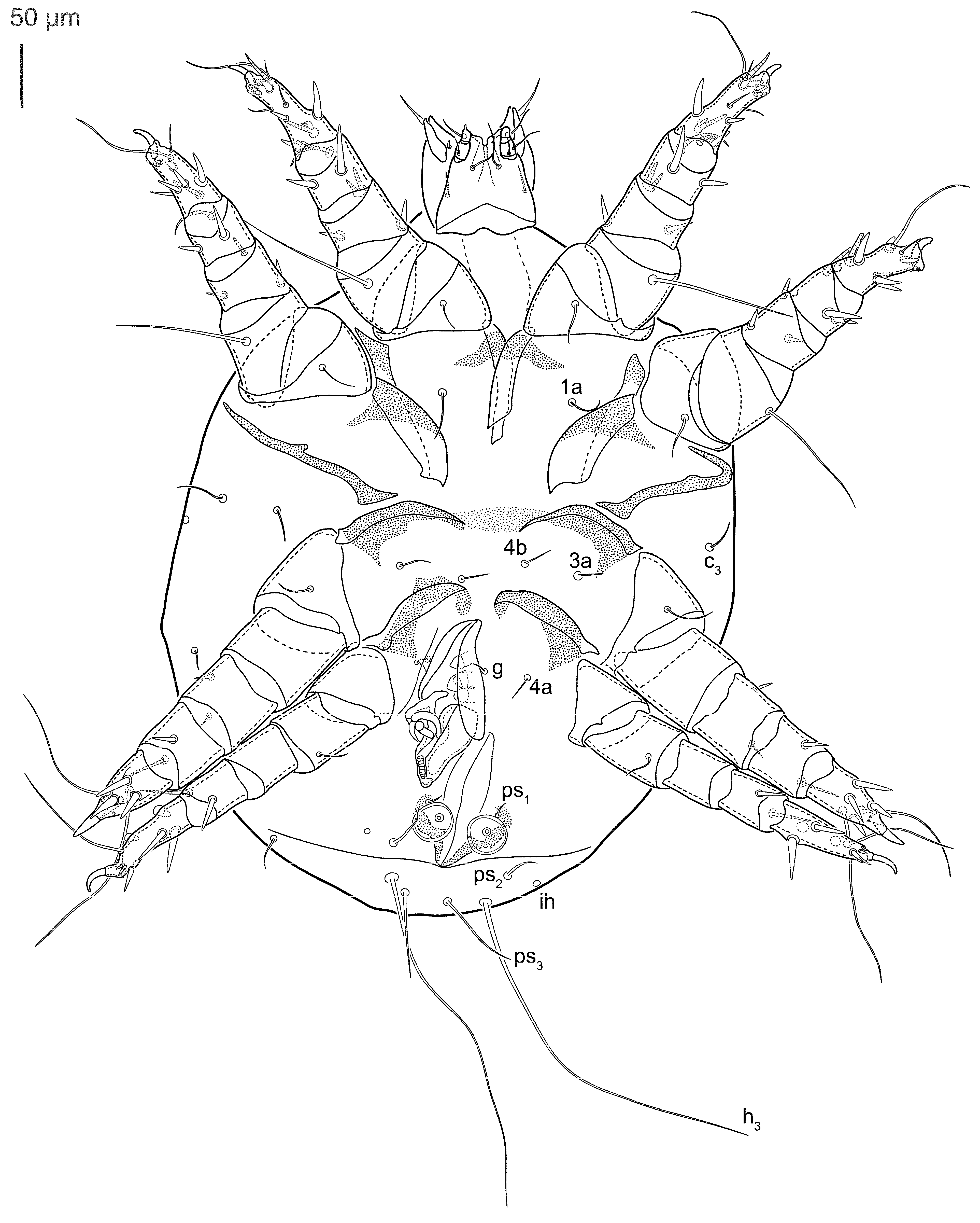
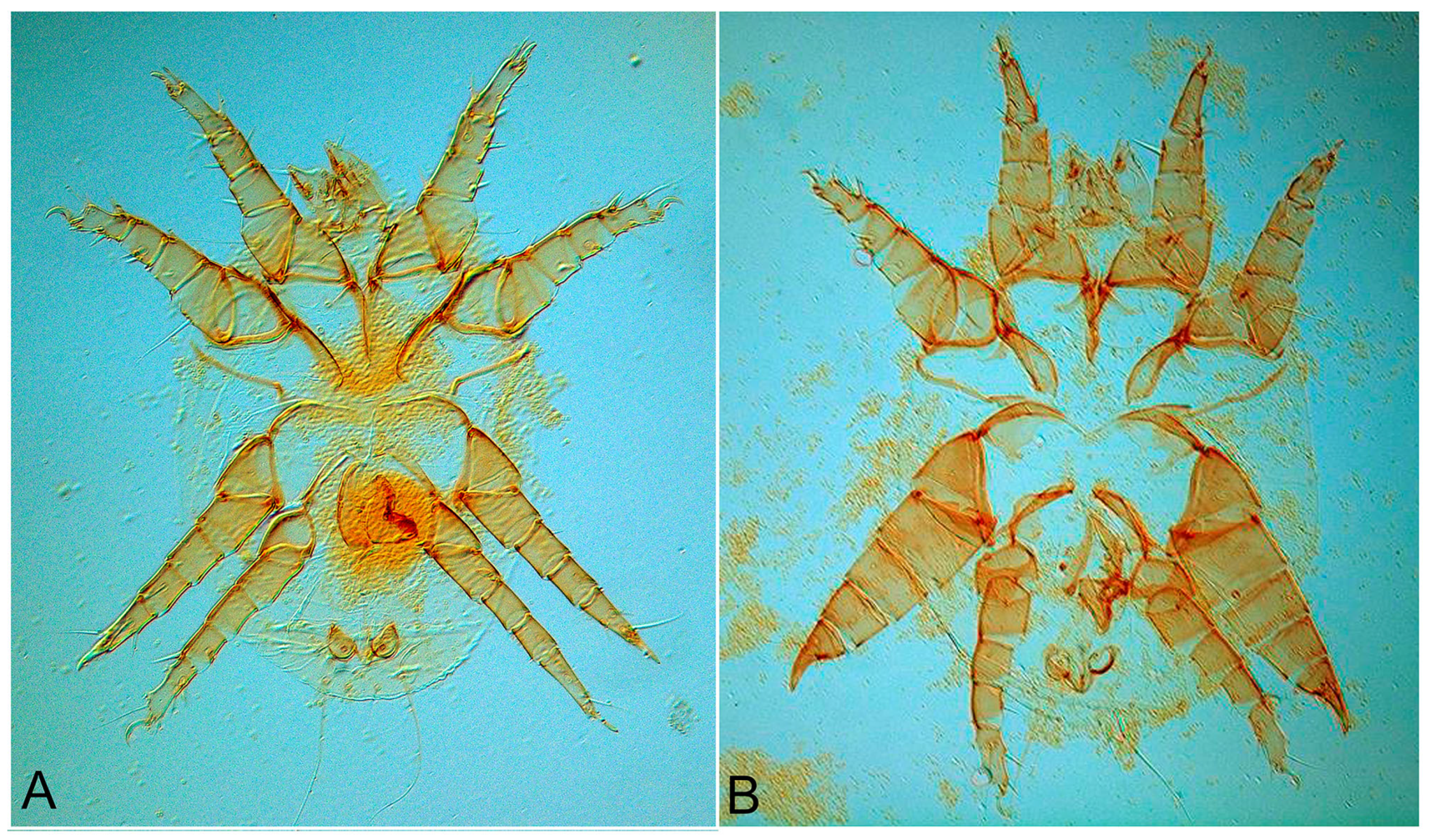
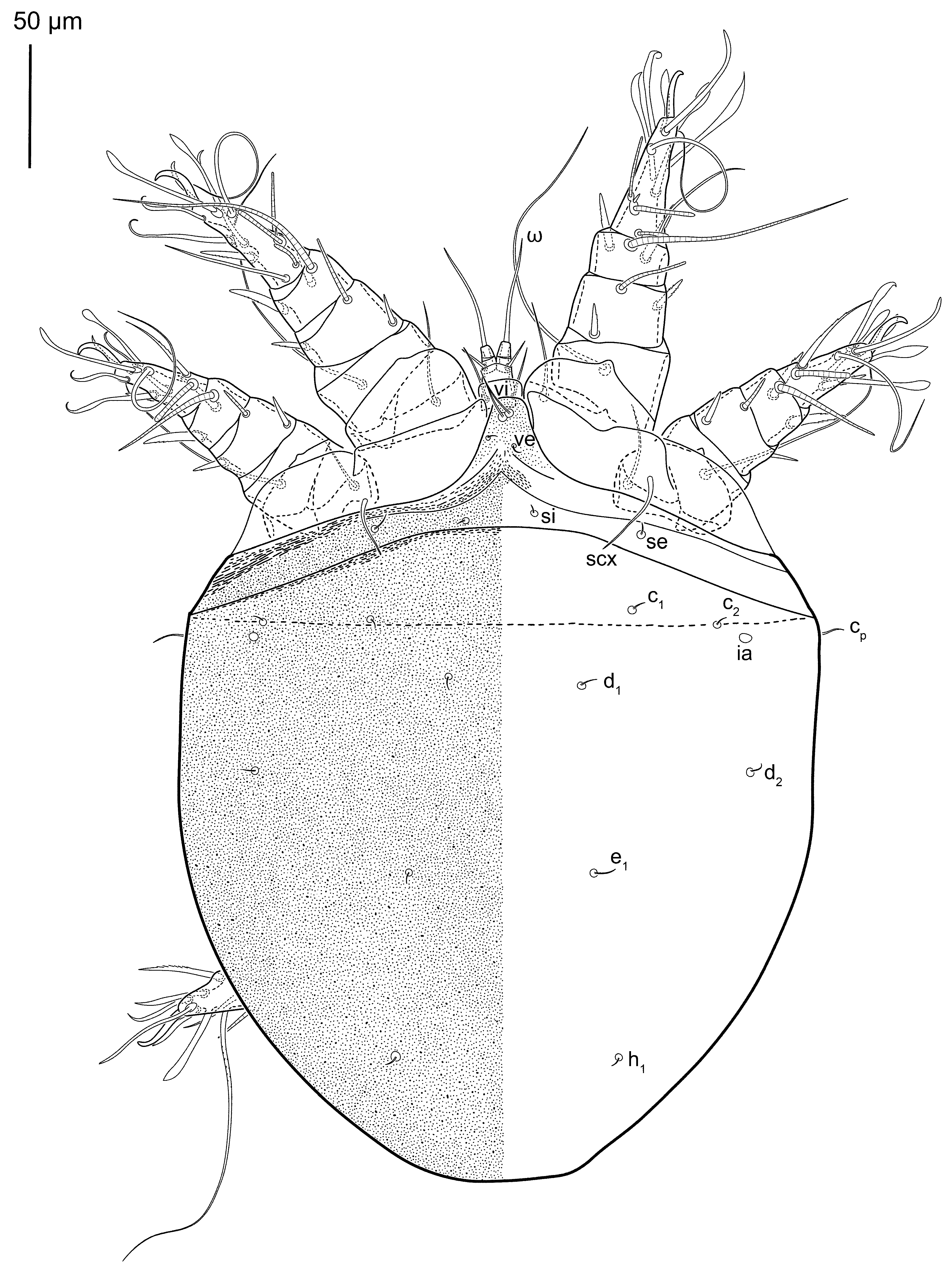
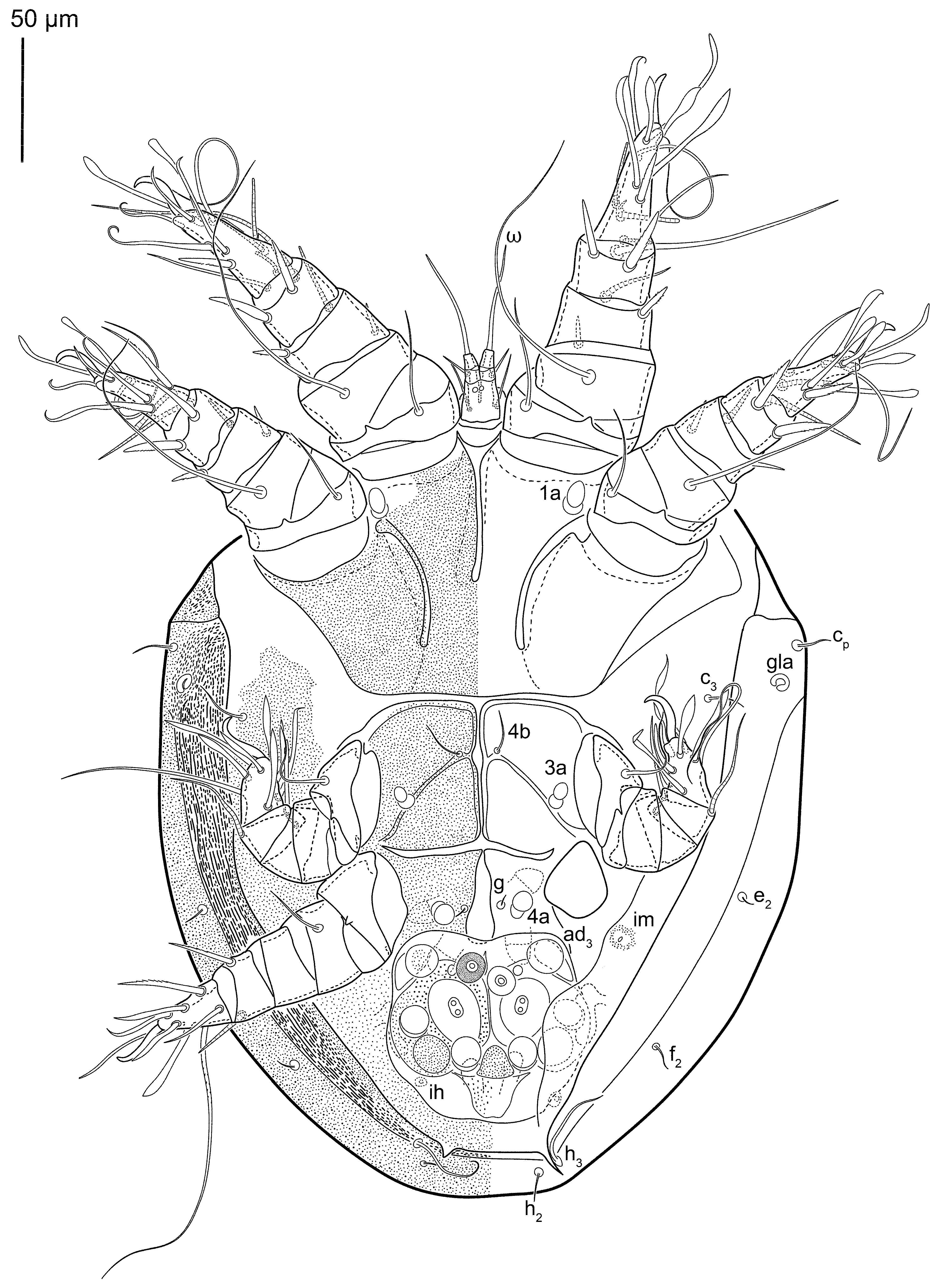
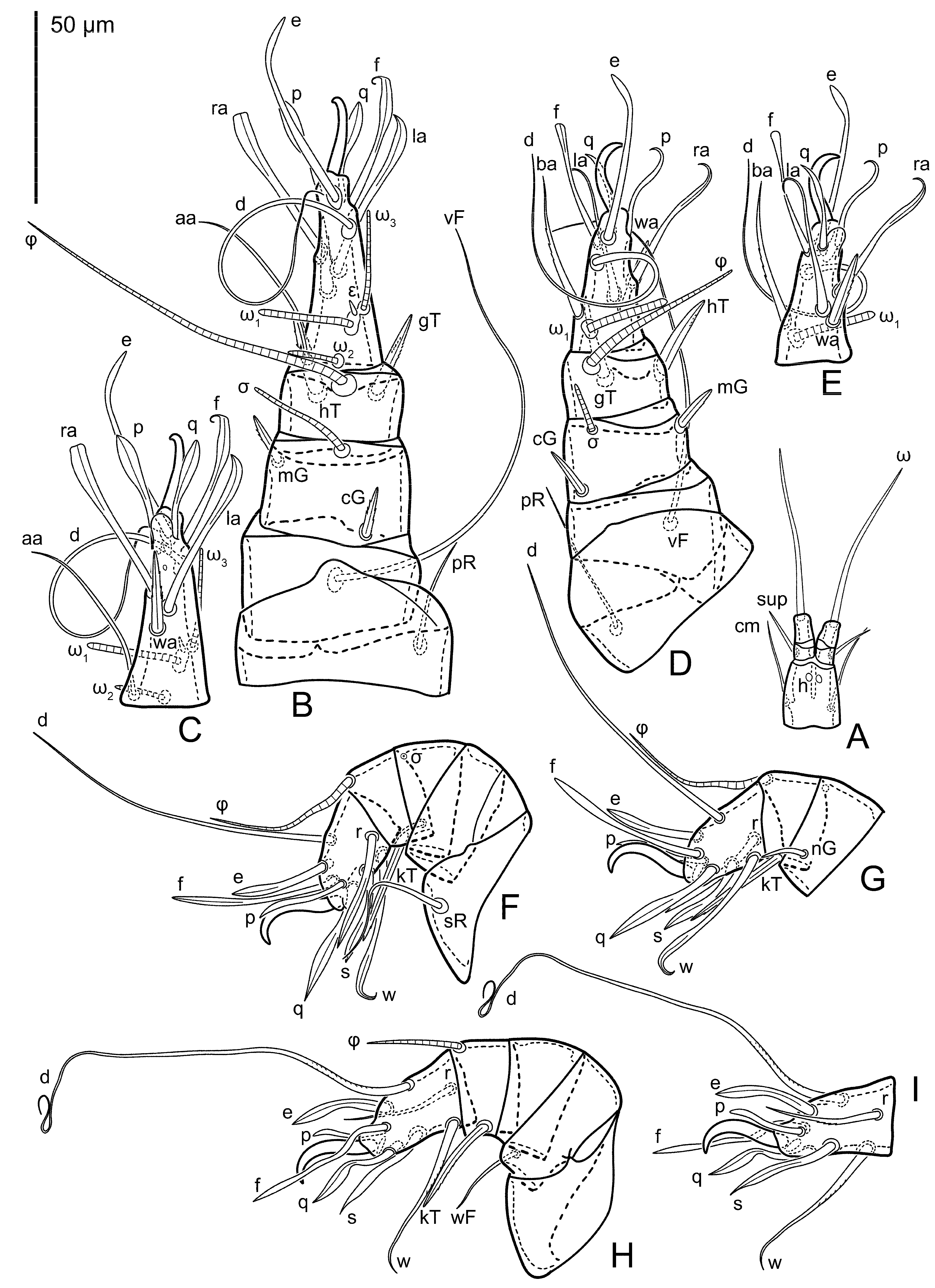
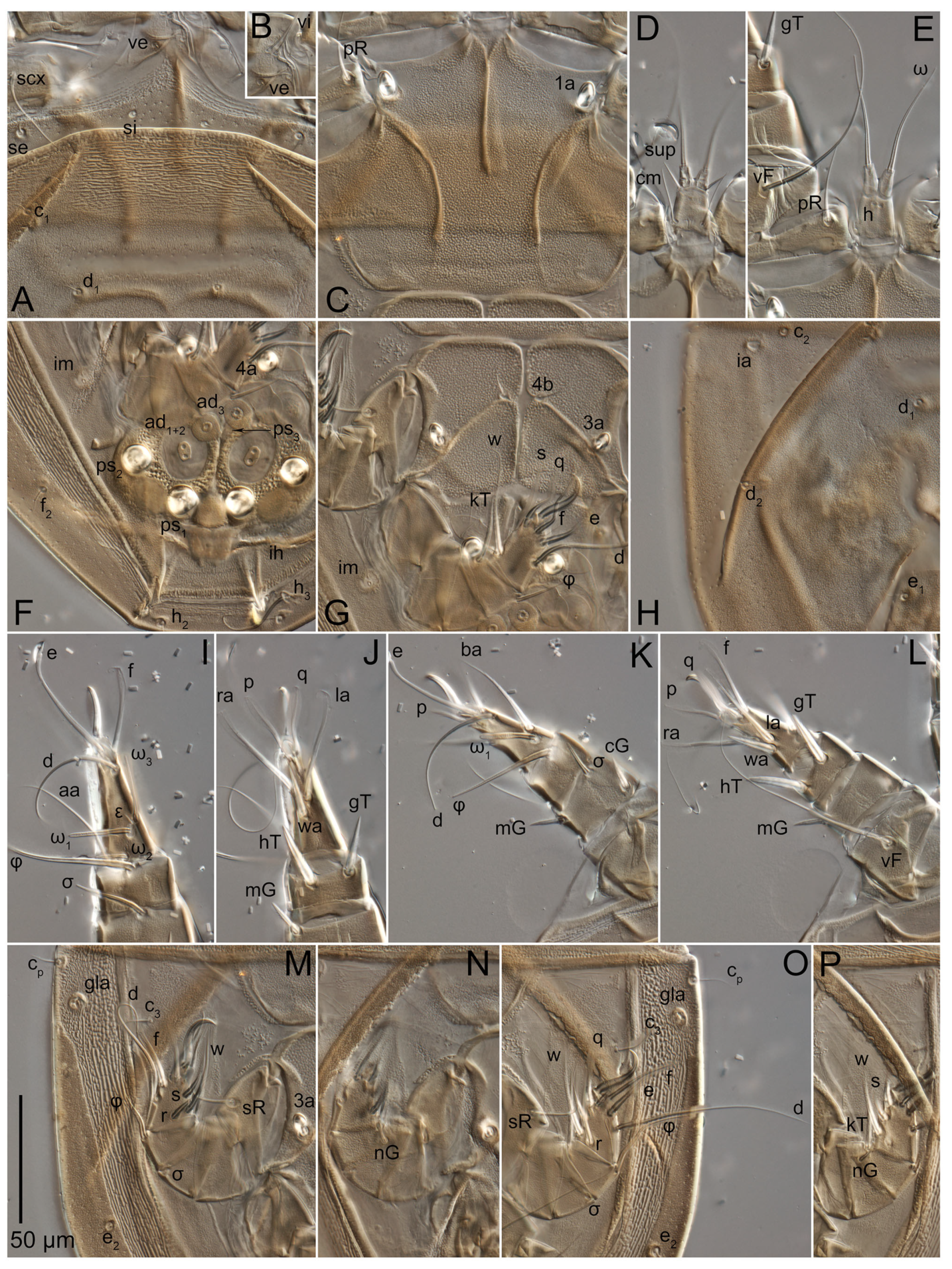
Heteromorphic deutonymph (n = 3) (
Figure 7,
Figure 8,
Figure 9,
Figure 10 and
Figure 11). Body rounded (
Figure 7 and
Figure 8), 1.2–1.3 times longer than wide, widest in sejugal region; idiosomal length 330–380, width 260–290. Gnathosoma (
Figure 9A and
Figure 11E,F) short, subcapitulum 160 × 170, trapezoidal, with filiform setae
cm, and setae
h absent (their positions marked by somewhat refractile spots); palps 90, two-segmented, with filiform setae
sup and palpal solenidia (ω); palp length slightly exceeds the length of the gnathosoma.
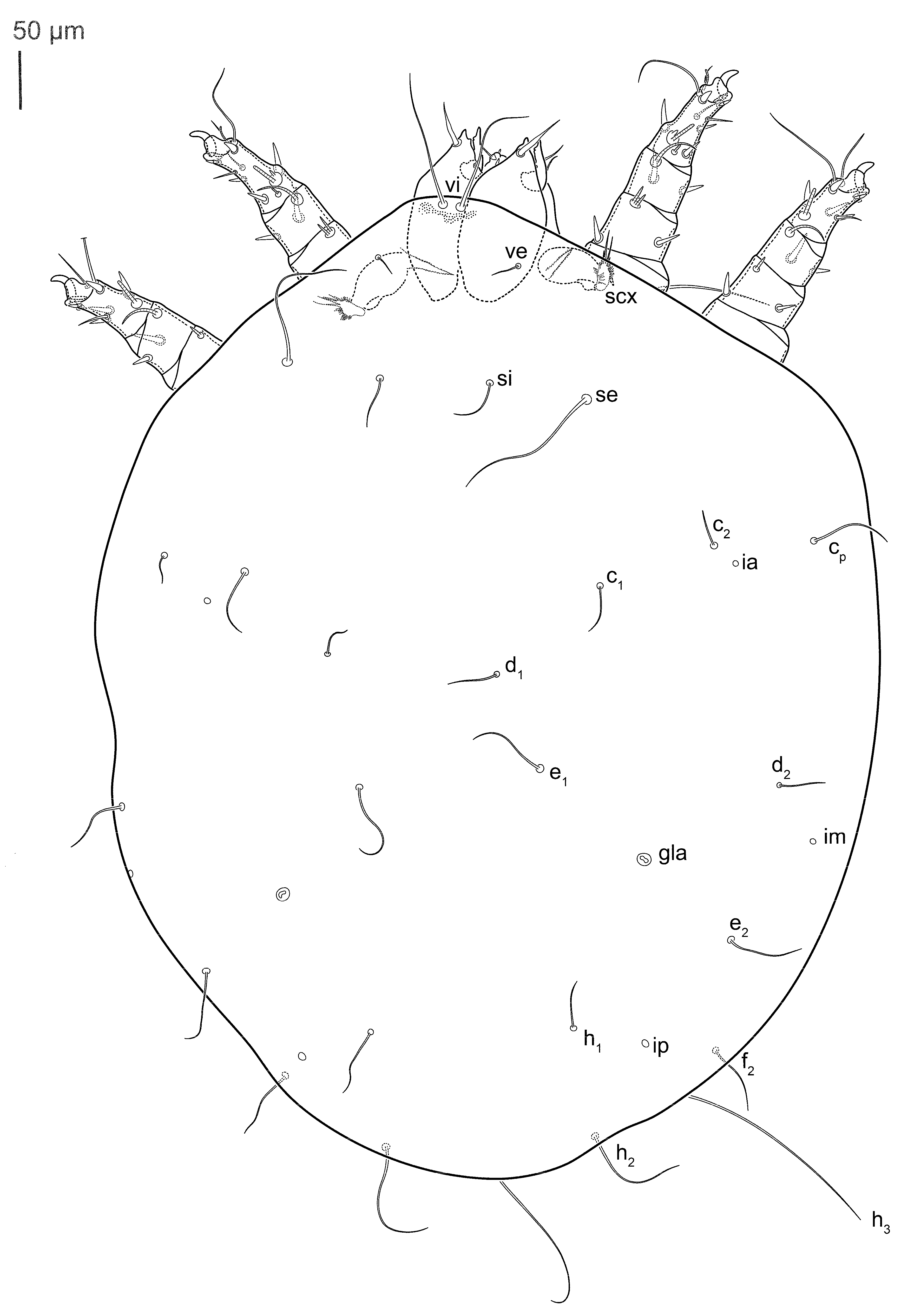
Dorsum (
Figure 7 and
Figure 11A). Idiosoma smoothly punctate (there are two types of punctation—rare but large pits and numerous but very small dots between them); distinct linear pattern present on anterior and lateral sides of prodorsal sclerite and hysterosomal shield. Apex of propodosoma anterior to anterior border of prodorsal shield, with apical internal vertical setae (
vi) (bases separated). Anterior border of prodorsal shield rounded triangular, with setae
ve, ocelli absent. External scapular setae
se and internal scapular setae
si situated on prodorsal shield;
si distinctly posterior
se. Supracoxal setae of legs I (
scx) filiform. Sejugal furrow well developed. Prodorsal shield 100–116, hysterosomal shield 210–220, ratio hysterosomal shield/prodorsal shield length = 1.9–2.1. Hysterosoma with 11 pairs of simple filiform setae on hysterosomal shield (
c1,
c2,
cp,
d1,
d2,
e1,
e2,
f2,
h1,
h2,
h3); setae
h3 distinctly longer than other setae. Opisthonotal gland openings (
gla) situated laterally on hysterosomal shield, slightly anterior setae
c3, much closer to seta
cp than to
c3. Four pairs of fundamental cupules (
ia,
im,
ip and
ih) present.
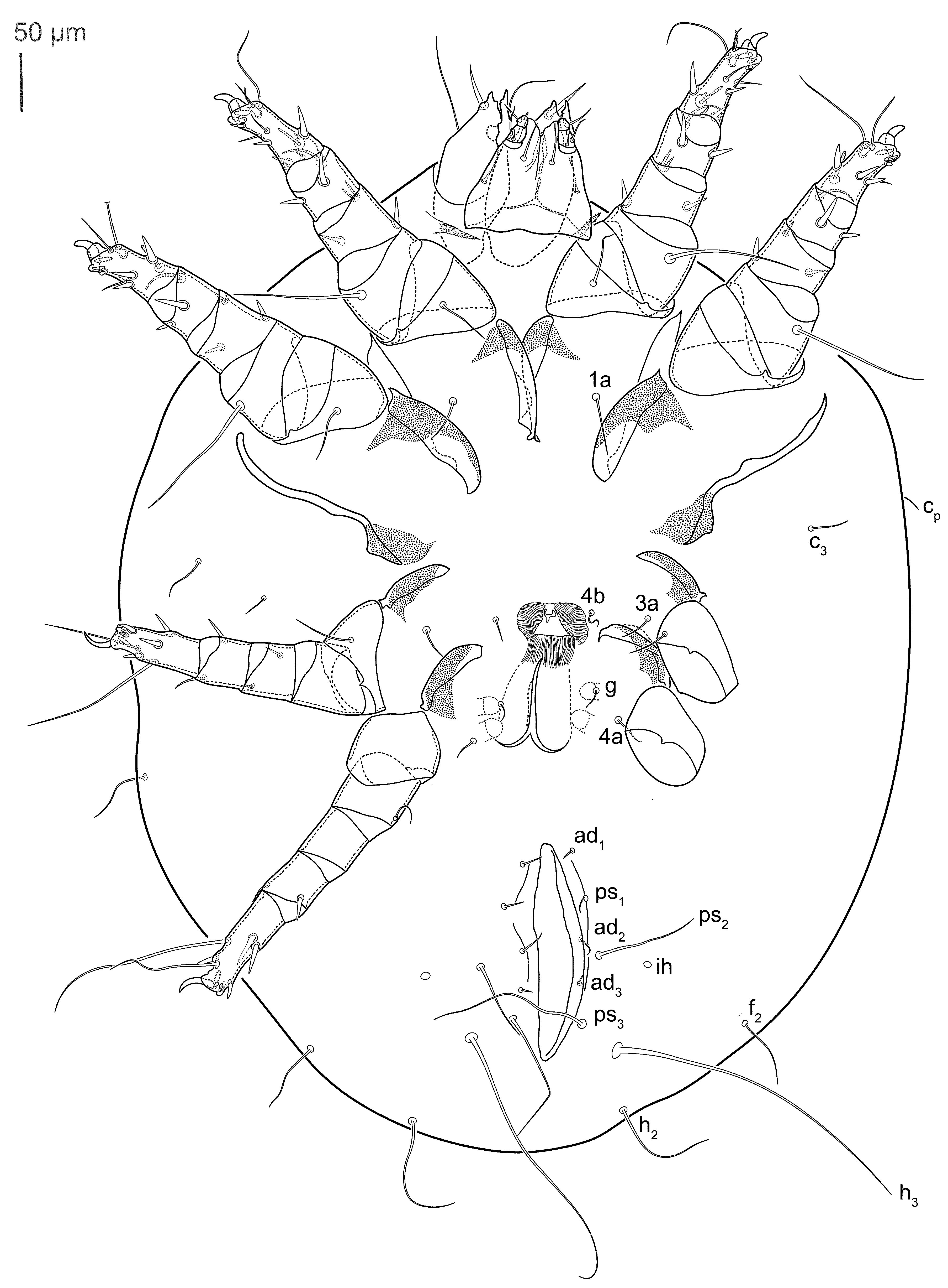
Venter (
Figure 8 and
Figure 11B–D,G). Coxal fields smooth. Anterior apodemes of coxal fields I fused forming sternum; coxal fields I closed, sternum fused with processes of anterior apodemes of coxal fields II (in one paratype, the connection was broken during preparation); coxal fields II closed, anterior and posterior apodemes of coxal fields II curved medially. Sternal and ventral shield contiguous. Anterior apodemes of coxal fields III connected to the anterior apodemes of coxal fields IV but not forming a common medial apodeme III (medial apodeme III divided into two parallel apodemes with setal pair
4b); coxal fields III closed. Ventrum present, connected to the medial apodemes III, but well separated from genital opening. Setae
c3 filiform, situated on ventral surface between legs II and III, near region separating sternal and ventral shields. Coxal setae
1a,
3a,
4b and genital setae
g filiform;
4a in form of rounded conoids. Genital region in posterior portion of coxisternal fields IV; progenital opening elongated, with two pairs of genital papillae within genital atrium; genital papillae two-segmented, with cone-shaped rounded apices. Genital setae laterad of genital opening. Attachment organ (
Figure 11D,G) posterior to coxal fields IV. Anterior suckers (
ad3) round, median suckers (
ad1+2) distinctly larger, with paired vestigial alveoli (situated on common sclerite); pair of small refractile spots anterolaterad median suckers (
ps3); lateral conoidal setae of attachment organ (
ps2) situated slightly posterior to line joining centers of median suckers; anterior and posterior lateral and posterior median cuticular conoids well developed; anus situated between anterior suckers (
ad3).
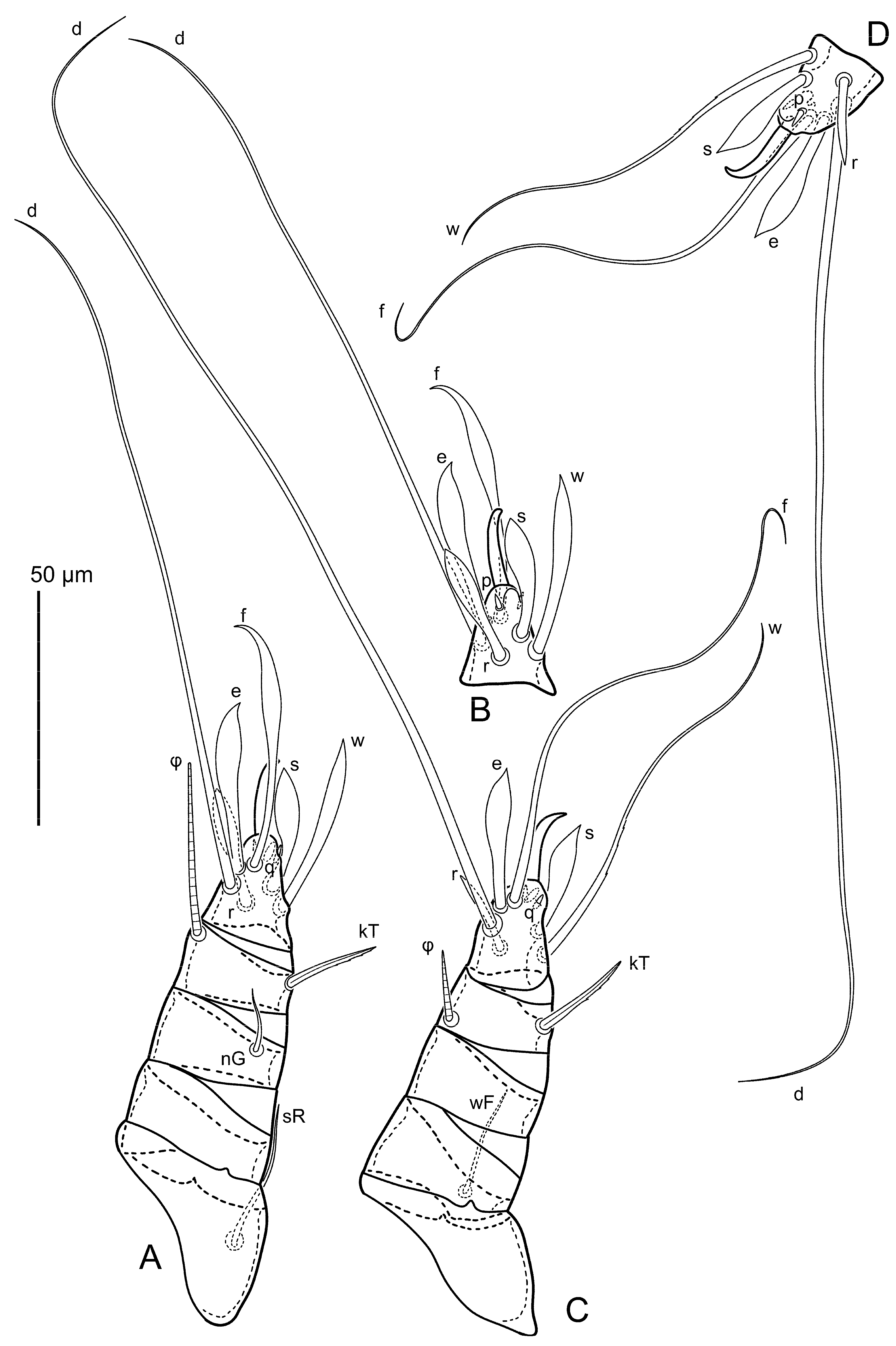
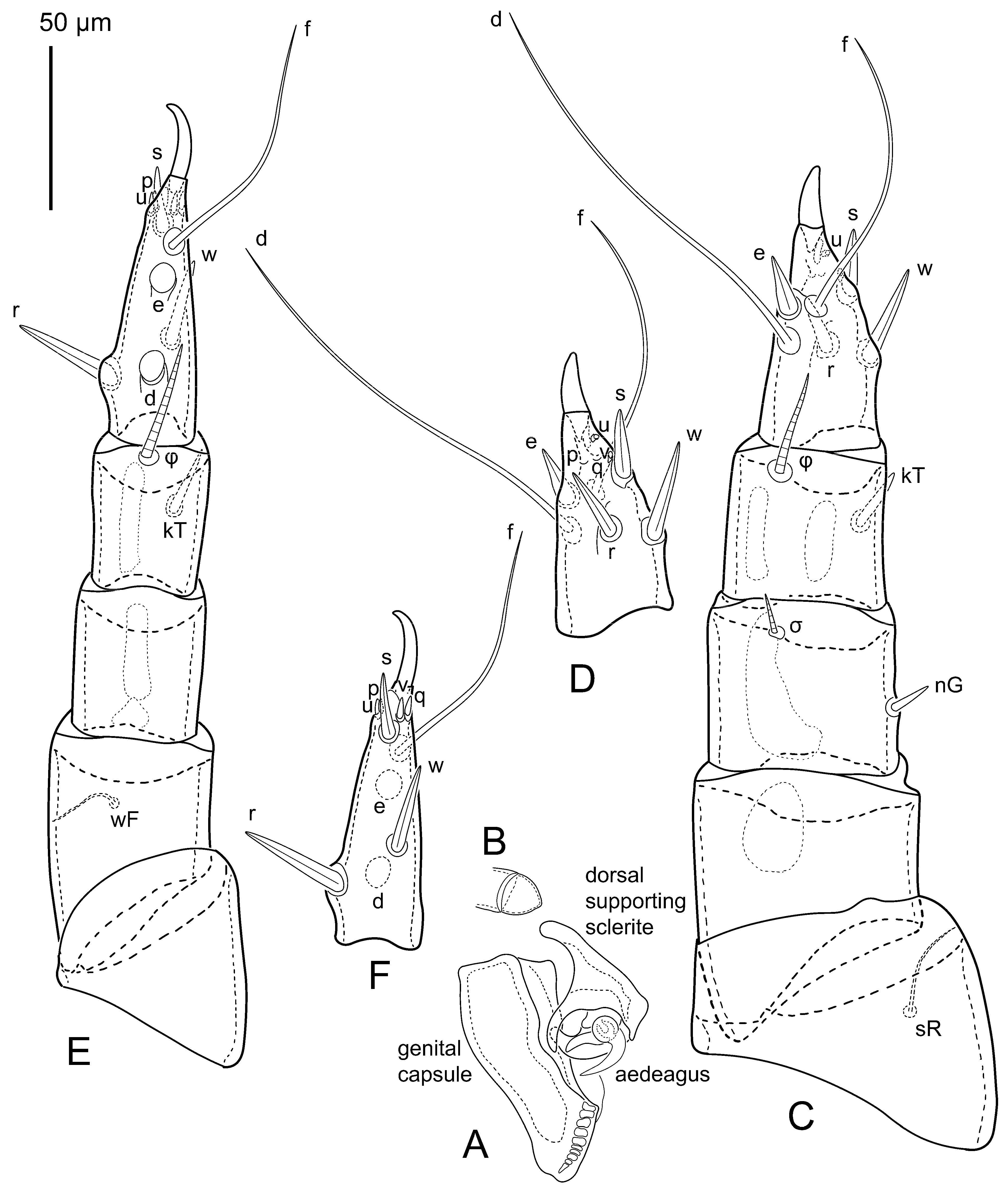
Legs (
Figure 9B–E,
Figure 10 and
Figure 11H–M). Legs short; all segments free. Trochanters I–III each with long filiform seta,
pR I–II,
sR III. Femoral setation 1-1-0-1; setae
vF I–II and
wF IV long (
vF I longer than
vF II), filiform. Genual setation 2-2-1-0; setae
mG I–II spiniform, finely serrated,
cG I–II spiniform,
nG III weakly spiniform. Tibial setation 2-2-1-1; setae
gT,
hT I–II and kT III–IV spiniform, finely serrated; setae
gT and
hT approximately the same length. Tarsal setation 10-9-8-8. All pretarsi consisting of empodial claws attached to short paired condylophores; claws I–II hooked, III–IV slightly curved and long. Tarsus I with setae
ra,
la,
f foliate; seta
d filiform, shorter than
e; seta
e wide, cylindrical with sucker-like apex, different in form from simply foliate seta
f; setae
wa spiniform; seta
aa weakly spiniform;
p and
q small spiniform;
ba I absent. Tarsus II similar to tarsus I except spiniform seta
ba present;
aa absent and
wa finely serrated. Tarsus III with setae
w,
r,
s,
e and
f smooth and foliate; seta
d filiform, longer than leg; setae
p and
q small spiniform. Tarsus IV similar to tarsus III except seta
r weakly foliate,
w filiform, finely serrated and
f smooth filiform. Solenidia ω
1 on tarsi I–II cylindrical, with slightly clavate apices; ω
3 on tarsus I longer than ω
1, with rounded apex, situated slightly distal to ω
1; ω
1 and ω
3 separated by spiniform famulus (ε); solenidion ω
2 of tarsus I slightly expanding apically, situated somewhat more basal and posterior to ω
1 + ε + ω
3 group. Solenidia φ of tibiae I–IV elongate, tapering; φ I longer than tarsus I with ambulacra; solenidion φ II shorter than tarsus II; solenidion φ III longer than tarsus III without ambulacra; solenidion φ IV short (in one specimen, on one leg, it is attached at base of tibia). Solenidion σ of genu I elongate, cylindrical, nearly reaching tip of tibia I; solenidion σ of genu II much shorter, slightly expanding apically, not reaching midlength of tibia II; solenidion σ of genu III absent.
Etymology. The new species is named in honor of the Japanese acarologist Dr. Kazuyoshi Kurosa, who independently discovered it in Japan [
82].
Type deposition. Holotype and all paratypes are deposited in the collection of the Zoological Institute, Russian Academy of Sciences, St. Petersburg, Russia (ZISP).
Biology. Mites live in sporocarps of old puffballs Lycoperdon spp. (Agaricales, Lycoperdaceae), feeding on spores (as evidenced by gut contents). Heteromorphic deutonymphs are phoretic on beetles Lycoperdina koltzei, larvae of which are also associated with the same fungal species. Co-occurring mite species: Viedebanttia egorovi Klimov, 1998.
Distribution. Russia: Primorskiy Kray; Japan.
Obelacarus gen. nov.
Type species: Obelacarus gulosus sp. nov.
“AD469_puffball_mite” Klimov and OConnor, 2013: 413 [
6]
urn:lsid:zoobank.org:act:AD96C317-7757-47DA-B0E9-A308F946A926
Diagnosis. Adult. Central trunk of supracoxal setae scx broadly flattened with numerous lateral processes and terminal long processes. Grandjean’s organ with one pointed trunk and numerous small spines. Prodorsal shield vestigial. Seta ve well developed, located on the same transverse line as vi. Posterior coxal apodeme II present, with sclerite located at end of apodeme. Tarsal setae u and v developed, spiniform. Solenidia φ I–II short, not reaching the apices of their respective tarsi without ambulacra. Solenidion σ″ I shorter than σ′. Solenidion σ III present. Inseminatory canal of spermatheca narrow. Paired efferent ducts cone-shaped, with long stem.
Heteromorphic deutonymph. Seta ve present. Ratio hysterosomal shield/prodorsal shield length = 4.7. Coxal field I free. Coxal setae 1a and 3a conoids. External conoidal setae ps2 of attachment organ lateral to median sucker ad1+2. Tibiae I–II similar in length or only slightly longer than corresponding genua. Seta aa I filiform, long (longer than ω3). Setae e I–II cylindrical with sucker-like apex, different in form from simply foliate seta f. Setae d I–II longer than e I–II. Seta ba II longer than ω1 II. Setae p and q I–IV foliate. Seta f IV shorter than leg IV. Solenidion σ III represented refractile spots.
Etymology. The generic name Obelacarus is derived from the Latinized form of Obelix, a corpulent fictional Gaulish character from French comics and films, in reference to the robust body shape of the mite.
Remarks. The differences between both genera of Lycoglyphidae fam. nov. are presented in the key below.
Obelacarus gulosus sp. nov.
urn:lsid:zoobank.org:act:09506039-C499-4B86-8427-D2468804FC82
“AD469_puffball_mite” Klimov and OConnor, 2013: 413 [
6]
Figure 12,
Figure 13,
Figure 14,
Figure 15,
Figure 16,
Figure 17,
Figure 18,
Figure 19,
Figure 20,
Figure 21,
Figure 22,
Figure 23,
Figure 24,
Figure 25,
Figure 26 and
Figure 27 Material. Holotype: female—USA: New York, Chautauqua County, Westfield, ex Lycoperdon piriforme (Agaricales: Lycoperdaceae), 8 October 1981, BMOC #81-1008-15 (UMMZ). Paratypes: 1HDN—same data; 1 heteromorphic male—same data (UMMZ).
Additional materials. 26 females, 10 males, 12 deutonymphs, 5 protonymphs, 5 larvae + more unmounted—BMOC 81-1008-015—USA: New York, Chatauqua Co., Westfield (42°19′20″ N, 079°34′40″ W); 8 October 1981; B.M. OConnor; reared from tritonymphs in Apioperdon pyriforme (Schaeff.) Vizzini (=Lycoperdon pyriforme) (Agaricales: Lycoperdaceae); 3 females, 2 males, 7 deutonymphs—BMOC 81-0407-001—Tompkins Co., Ringwood Preserve, 7.6 km E Ithaca (42°26′58″ N, 076°22′25″ W); October 1980; J. Pakaluk; reared from deutonymphs under elytra of Lycoperdina ferruginea LeConte, 1824 (Coleoptera: Endomychidae); 6 females 7 males, 6 deutonymphs—BMOC 82-0729-001—Tompkins Co., Ithaca (42°26′26″N, 076°29′49″ W); summer 1981; J. Pakaluk; ex A. pyriforme; 12 deutonymphs—BMOC 80-0506-009—Cortland Co., McLean Reserve (now Lime Hollow Nature Center), 42°34′07″ N, 076°15′02″ W; 6 May 1980; B.M. OConnor; ex Fomitopsis betulina (Bull.) B.K.Cui, M.L.Han & Y.C.Dai (2016); 25 deutonymphs—BMOC 81-0407-003—Tompkins Co., Ringwood Preserve, 7.6 km E Ithaca (42°26′58″ N, 076°22′25″ W); October 1980; J. Pakaluk; ex A. pyriforme; 3 deutonymphs—BMOC 86-0407-003—Tompkins Co., Ringwood Preserve, 7.6 km E Ithaca (42°26′58″ N, 076°22′25″ W); October 1980; J. Pakaluk; ex L. ferruginea larva in A. pyriforme; 10 deutonymphs—BMOC 81-1008-017—Chatauqua Co., Westfield (42°19′20″ N, 079°34′40″ W); 8 October 1981; B.M. OConnor; ex 2 L. ferruginea in rotting stump with mycelia of A. pyriforme; 4 tritonymphs, 7 deutonymphs, 4 larvae—BMOC 96-0809-036—USA: Michigan, Lake Co., Elk twp. 6.9 km S Irons, (44°05′39″ N, 085°58′24″ W); 9 August 1996; K. Okabe; ex A. pyriforme; 2 deutonymphs—BMOC 17-1026-001—Washtenaw Co., Ann Arbor (42°16′40″ N, 83°44′08″ W); 25 October 2017; T. McKnight; ex L. ferruginea; 13 deutonymphs—BMOC 81-1010-001—Washtenaw Co., Stinchfield Woods, 6.4 km SSE Pinckney (42°24′04″ N, 083°55′50″ W); 10 October 1981; B.M. OConnor; ex 3 L. ferruginea in A. pyriforme; 24 deutonymphs on 7 beetles; 3 females, 1 male, abundant protonymphs and larvae reared—BMOC 83-1001-001—Washtenaw Co., Hudson Mills Metropark, 5 km NW Dexter (42°22′43″ N, 083°54′45″ W); 1 October 1983; B.M. OConnor; reared ex 7 L. ferruginea on Lycoperdon perlatum Pers. (1796) (Agaricales: Lycoperdaceae); 38 deutonymphs—BMOC 00-1005-005, 7, 8, 9—Livingston Co., E.S. George Reserve, 5 km E Pinckney (42°26′55″ N, 84°01′19″ W); 5 October 2000; B.M. OConnor, P. Klimov, A. Dowling & M. Lekveishvili; ex 9 L. ferruginea on L. perlatum; 2 deutonymphs—BMOC 00-1005-010—Livingston Co., E.S. George Reserve, 5 km E Pinckney (42°26′55″ N, 84°01′19″ W); 5 October 2000; B.M. OConnor, P. Klimov, A. Dowling & M. Lekveishvili; ex 9 L. ferruginea on A. pyriforme (Agaricales: Lycoperdaceae); 10 deutonymphs—BMOC 81-1220-001, 2, 3—Washtenaw Co., Ann Arbor (42°16′40″ N, 83°44′08″ W); 21 October 1928; W. Clanton; ex 3 L. ferruginea; 14 deutonymphs—BMOC 81-1220-004, 5—Schoolcraft Co., Manistique (45°57′28″ N, 86°14′57″ W); 10 September 1923; S. Moore; ex 2 L. ferruginea; 8 deutonymphs—81-1220-006, 7—Mackinac Co., Naubinway (46°05′32″ N, 085°26′51″ W); 7 October 1920; S. Moore; ex 2 L. ferruginea; 1 deutonymph—BMOC 81-1220-008—Emmet Co., McGulpin Point (45°46′49″ N, 084°47′34″ W); 8 September 1921; S. Moore; ex L. ferruginea; 4 deutonymphs—BMOC 81-1220-009—Charlevoix Co., High Island (45°43′13″ N, 084°47′34″ W); 4 September 1923; S. Moore; ex L. ferruginea; 3 deutonymphs—BMOC 81-1220-010—Gogebic Co., Hughitt-Rawson Preserve,. south shore Thousand Island Lake (46°13′13″ N, 89°23′57″ W); 10 August 1919; T.H. Hubbell; ex L. ferruginea in Betula alleganiensis Britt. log.; 4 deutonymphs—BMOC 81-1220-012—Livingston Co., E.S. George Reserve, 5 km E Pinckney (42°26′55″ N, 84°01′19″ W); 14 May 1933; A. W. Andrews; ex L. ferruginea; 2 males and 2 females—BMOC 04-1209-003—Michigan: Livingston Co., E.S. George Reserve, 42°27′03.9″ N 084°45′53″ W; 9 December 2004; A. Dowling & P. Klimov; ex Lycoperdon pyriforme; 3 deutonymphs—BMOC 81-1220-013—USA: Pennsylvania: Washington Co., Canonsburg, Mt. Blain (40°15′43″ N, 80°11′06″ W); 20 July 1920; G.A. Ackerlind; ex L. ferruginea; 17 deutonymphs—BMOC 81-1220-014—USA: Illinois, Cook Co., Willow Springs (41°44′30″ N, 87°52′40″ W); 11 July 1916; no collector; ex L. ferruginea; 2 females, 2 males, 11 tritonymphs, 15 deutonymphs (including 8 pharate tritonymphs)—BMOC 86-0928-006—CANADA: Ontario, Parry Sound Co., Sequin Twp., Portage Lake (45°13′12″ N, 079°48′38″ W); 28 September 1986; B.M. OConnor; reared from deutonymphs on L. ferruginea on A. pyriforme.
Gnathosoma (
Figure 15A,B and
Figure 16B–D): subcapitulum (
Figure 14A and
Figure 15B,C) wide, 96 × 112, evenly punctated; subcapitular setae (
h) long, widened basally; supracoxal seta
elcp present, weakly barbed; palps wide; lateral dorsal palp tibial setae (
sup) and palp tibial setae (
a) filiform; dorsal palp tarsal seta (
cm) weakly lanceolate, base located on large rounded lateral protrusion of palp tarsus; terminal palp tarsal solenidion ω short; terminal eupathidium
ul″ small, dome-shaped; terminal eupathidium
ul′ small, rounded. Chelicera (
Figure 15B and
Figure 16D) with two setae:
cha large, lanceolate, distal on fixed digit,
chb short, spiniform. Fixed finger of chelicera with one tooth, movable with three teeth (anterior double).
Prodorsal shield (
Figure 16A) vestigial, represented by a short weakly sclerotized area with punctation behind bases of setae
vi. Grandjean’s organ (
GO) with one pointed trunk and numerous small spines (
Figure 14A and
Figure 16E). Supracoxal setae (
scx) pinnate—central trunk broadly flattened with numerous (15–18) long pointed lateral processes and three terminal long processes (
Figure 14A and
Figure 16E). Supracoxal shield oval, punctate, with small pointed projection near base of
scx. Idiosomal setae (
vi,
ve,
si,
se,
c1,
c2,
cp,
c3,
d1,
d2,
e1,
e2,
f2,
h1,
h2,
h3,
ps1,
ps2,
ps3) smooth, filiform, setae
h3,
se,
vi longer than other setae. Opisthosomal gland openings between setal bases
e1 and
e2. Four pairs of fundamental cupules (
ia,
im,
ip and
ih) present. Ventral idiosoma with four pairs of coxal setae (
1a,
3a,
4a and
4b) and one pair of genital setae (
g). Coxal sclerites: I—short, triangular (
Figure 16H); anterior II –extended along anterior apodeme II, expanded at base (
Figure 16I); sclerite posterior coxal apodeme II shapeless, located at end posterior apodeme (
Figure 16J); III and IV –extended along apodemes, expanded at base (
Figure 16K,L). Genital region situated between coxal fields III and IV; genital valves form an inverted Y; epigynal apodeme absent (
Figure 16F). Genital papillae (
Figure 16F) wide, with rounded ends. Anal opening ventral, long; distance between ovipore and anal opening less than length of anal opening. Copulatory tube absent. Inseminatory canal of spermatheca narrow, slightly widened at junction with base of spermatheca (
Figure 14B and
Figure 16G). Base of spermatheca flat, ring-shaped. Paired efferent ducts cone-shaped, with long stem (same length as funnel). Three pairs adanal (
ad1–3) setae.
All legs shorter than body; all segments free (
Figure 14C–F,
Figure 15C–F and
Figure 17). Trochanters I–III each with long filiform seta,
pR I–II,
sR III; trochanter IV without setae. Femoral setation 1-1-0-1; setae
vF I–II and
wF IV long filiform. Genual setation 2-2-1-0; setae
mG and
cG I–II spiniform; seta
nG III filiform. Tibial setation 2-2-1-1; setae
hT I–II,
gT I–II and
kT III–IV spiniform,
hT slightly shorter than
gT. Tarsal setation 13-12-10-10; pretarsi I–II consist of hooked, III–IV slightly curved and long empodial claws attached to short paired condylophores. Tarsus I with setae
ra,
la,
f and
d filiform,
aa,
ba,
e,
wa larger spiniform,
s,
v,
u,
p and
q short spiniform (
u and
v longer than
s,
p and
q). Tarsus II has the same setae except that
la spiniform, ra spiniform with filiform ends and absent
aa. Tarsus III and IV with setae
f and
d filiform,
r,
w,
e and
s larger spiniform,
u,
v,
p and
q small spiniform. Solenidia ω
1 on tarsus I and II cylindrical, with clavate apex, not bent. Solenidion ω
2 on tarsus I shorter than ω
1, cylindrical, with rounded apex, situated distinctly posterior to ω
1, at equal distance between ω
1 and
aa. Solenidion ω
3 on tarsus I cylindrical, with rounded tip, shorter than ω
1. Famulus (ε) of tarsus I spiniform. Solenidia φ of tibiae I–IV elongate, tapering, not reaching apices of their respective tarsi without ambulacra. Solenidia σ′ and σ″ on genu I elongate, tapering, not reaching bases of φ I, σ′ longer than σ″. Solenidion σ on genu II 5 times longer than its width, tapering. Solenidion σ of genu III present, 6 times longer than its width, tapering.
Male (
Figure 18,
Figure 19,
Figure 20,
Figure 21,
Figure 22 and
Figure 23) with apparent size-based allometric development of legs III. Individuals of body size intermediate between large and small forms (below) with development of legs III intermediate between largest and smallest forms.
Gnathosoma (
Figure 21B–E): subcapitulum (
Figure 21B,C) wide, 74 × 80, other characteristics are same as in females.
Prodorsal shield (
Figure 21A) vestigial, represented by a short weakly sclerotized area with punctation behind bases of setae
vi. Grandjean’s organ (
GO), supracoxal setae (
scx) and supracoxal shield as female (
Figure 21F). Idiosomal setae (
vi,
ve,
si,
se,
c1,
c2,
cp,
c3,
d1,
d2,
e1,
e2,
f2,
h1,
h2,
h3,
ps1 and
ps2) smooth, filiform, setae
h3,
se,
vi longer than other setae (
h3 approximately half length of body). Opisthosomal gland openings between setal bases
e1 and
e2. Four pairs of fundamental cupules (
ia,
im,
ip and
ih) present. Ventral idiosoma with four pairs of coxal setae (
1a,
3a,
4a and
4b) and one pair of genital setae (
g). Coxal sclerites: I—short, triangular (
Figure 21I); anterior II –extended along anterior apodeme II, expanded at base (
Figure 21J); posterior apodeme II narrow, extending along entire apodeme (
Figure 21J); III and IV –extended along apodemes, expanded at base, between anterior apodemes III area of weak sclerotization present (
Figure 21K,L). Genital region situated between coxal fields IV. Genital papillae (
Figure 20B and
Figure 21G) wide, with cone-shaped rounded apeces. Aedeagus short, not protruding beyond anterior edge of genital capsule (
Figure 20A and
Figure 21G). Anal opening ventral, long, located posterior to genital region. Paranal suckers rounded (
Figure 21H).
Legs I–II as in female except solenidion ω
3 wider and longer than female’s and
ra II without filiform ends. Leg III (
Figure 20C,D and
Figure 22E–I) wider and more robust than other legs, its chaetotaxy like that of female except for strong reduction of setae
u,
v,
p and
q (
u and
v—small vestigial remnants,
p and
q represented only by cuticular traces). Leg III with segments wider than long. Tarsus III shortened, conical, with strong empodial claw fused with tarsus apically, condylophores absent. Tarsal setae
r and
w adjacent to spine-like seta
s. Leg IV as in female except setae
d and
e represented by suckers and solenidion φ longer (
Figure 20E,F and
Figure 22H,I).
Small male. Similar to large male but smaller. Leg III of different form: leg segments longer than wide. Pretarsal empodial claw smaller, not fused with tarsal apex, and small condylophores visible. Tarsus IV distinctly longer with setae r and w well posterior of seta s, and setae p, q, u & v present.
Heteromorphic deutonymph (n = 1) (
Figure 24,
Figure 25,
Figure 26 and
Figure 27). Body rounded (
Figure 24 and
Figure 25), 1.3 times longer than wide, widest in sejugal region; idiosomal length 420 width 330. Gnathosoma (
Figure 26A and
Figure 27D,E) short, subcapitulum 170 × 150, trapezoidal, with filiform setae
cm, setae
h absent (their positions marked by somewhat refractile spots); palps 120, two-segmented, with filiform setae
sup and palpal solenidia (ω), whose length significantly exceeds gnathosomal length.
Dorsum (
Figure 24 and
Figure 27A,B,H). Idiosoma smoothly punctate (two types of punctation—relatively few large pits and numerous but very small dots between them); distinct linear pattern present on anterior and lateral sides of prodorsal sclerite and hysterosomal shield. Anterior border of prodorsal shield trapezoidal elongated, with setal pairs
ve and
vi, ocelli absent. Externalscapular setae
se and internal scapular setae
si situated on prodorsal shield,
si slightly anterior
se. Supracoxal setae of legs I (
scx) filiform. Sejugal furrow well developed. Prodorsal shield 70, hysterosomal shield 330, ratio hysterosomal shield/prodorsal shield length = 4.7. Hysterosoma with 11 pairs of simple filiform setae on hysterosomal shield (
c1,
c2,
cp,
d1,
d2,
e1,
e2,
f2,
h1,
h2,
h3), setae
h3 distinctly longer than other setae. Opisthonotal gland openings (
gla) situated ventrally on hysterosomal shield, slightly anterior setae
c3, much closer to seta
cp than to
c3. Four pairs of fundamental cupules (
ia,
im,
ip and
ih) present.
Venter (
Figure 25 and
Figure 27C,F,G). Coxal fields sclerotized, smoothly punctate. Anterior apodemes of coxal fields I fused forming sternum; sternum not reaching posterior border of sternal shield by distance not exceeding its length; coxal fields I and II open; anterior and posterior apodemes of coxal fields II curved medially. Sternal and ventral shield contiguous. Anterior apodemes of coxal fields III connected to anterior apodemes of coxal fields IV but not forming a common medial apodeme III (medial apodeme III divided into two parallel communicating apodemes with setal pairs
4b), coxal fields III closed. Ventrum present, divided into two parallel apodeme connected to two parallel medial apodemes III but separated from genital opening. Subhumeral setae (
c3) filiform, situated on ventral surface between legs II and III, near region separating sternal and ventral shields. Coxal setae
1a,
3a and
4a conoids,
4b and genital setae
g filiform. Genital region in posterior portion of coxisternal fields IV; genital opening elongated, with two pairs of genital papillae within genital atrium; genital papillae two-segmented, with cone-shaped rounded apices. Genital setae laterad of genital opening. Attachment organ (
Figure 27F) posterior to coxal fields IV. Anterior suckers (
ad3) round, median suckers (
ad1+2) distinctly larger, with paired vestigial alveoli (situated on common sclerite); pair of small refractile spots anterolaterad median suckers (
ps3); lateral conoidal setae of attachment organ (
ps2) situated lateral to line joining centers of median suckers; posterior conoidal setae (
ps1) directly posterior to median suckers; anterior and posterior lateral and posterior median cuticular suckers well developed; large trapezoidal cuticular sclerite (larger than diameter of cuticular conoids) situated behind posterior median cuticular suckers; anus situated between anterior suckers (
ad3).
Legs (
Figure 26B–I and
Figure 27I–P). Legs short; all segments free. Trochanters I–III each with long filiform seta,
pR I–II,
sR III. Femoral setation 1-1-0-1; setae
vF I–II and
wF IV long (
vF I longer than
vF II), filiform. Genual setation 2-2-1-0; setae
mG I–II spiniform, finely serrated,
cG I–II spiniform,
nG III weakly spiniform. Tibial setation 2-2-1-1; setae
gT,
hT I–II and kT III–IV spiniform, finely serrated; setae
gT and
hT approximately the same length. Tarsal setation 10-9-8-8. All pretarsi consisting of long hooked and thin empodial claws attached to short paired condylophores. Tarsus I with setae
ra,
la,
f,
p and
q foliate; seta
d filiform, long; seta
e long, cylindrical with sucker-like apex, different in form from simply foliate seta
f; setae
wa spiniform, finely serrated; seta
aa long, filiform;
ba I absent. Tarsus II similar to tarsus I except long and finely serrated seta
ba present and
aa absent. Tarsus III with setae
w,
r,
s,
e,
f,
p and
q smooth and foliate; seta
d filiform, longer than leg. Tarsus IV similar to tarsus III except seta
r and
w finely serrated. Solenidia ω
1 on tarsi I–II cylindrical, with slightly clavate apices; ω
3 on tarsus I equal in length ω
1, with rounded apex, situated slightly distal to ω
1; ω
1 and ω
3 separated by spiniform famulus (ε); solenidion ω
2 of tarsus I slightly expanding apically, situated somewhat more basal and posterior to ω
1 + ε + ω
3 group. Solenidia φ of tibiae I–IV elongate, tapering; φ I longer than tarsus I with ambulacra; solenidion φ II shorter than tarsus II without ambulacra; solenidion φ III longer than tarsus III without ambulacra; solenidion φ IV short. Solenidion σ of genu I elongate, cylindrical, reaching apex of tibia I; solenidion σ of genu II much shorter, slightly expanding apically, not reaching midlength of tibia II; solenidion σ of genu III represented by refractile spots.
Etymology. The species name is derived from the Latin gulosus—gluttonous, greedy for food. Gender masculine.
Type deposition. Holotype and paratypes are deposited in UMMZ. Additional paratypes and non-paratype specimens deposited in CNC, OSAL, USNMHN, and ZISP.
Biology. Feeding stages of this species live in the sporocarps of puffballs, Apioperdon (=Lycoperdon) pyriforme (Basidiomycetes, Agaricales, and Lycoperdaceae), feeding on spores. Adult body form globular, appearing to “swim” through spores. When removed from sporocarps, adult mites were unable to walk on a flat surface. Phoretic deutonymphs occur on Lycoperdina ferruginea LeConte, 1824 (Coleoptera: Endomychidae), the larvae of which also feed in fungal sporocarps. Adult beetles carrying mites were found on A. pyriforme and Lycoperdon perlatum Pers. (1796); however, no post-deutonymphal stages of O. gulosus were found in sporocarps of L. perlatum. Similarly, one collection of O. gulosus was obtained from L. ferruginea on Fomitopsis betulina (Bull.) B.K.Cui, M.L.Han & Y.C.Dai (2016), but, again, no post-deutonymphal stages were found in the sporocarp. Based on our collections this species may overwinter as deutonymphs on host beetles or in fungal mycelia in the woody substrate.
Distribution. CANADA: Ontario; USA: Illinois, Michigan, New York, Pennsylvania.
Remarks. Males of
Obelacarus exhibit polymorphism in body size and development of legs III. Although not directly tested, this polymorphism may reflect allometric enlargement of leg III related to body size. Males of
Obelacarus, like some Acaridae, have modified legs III, the armament of which has a number of significant differences from Acaridae (for other families of Acaroidea male polymorphism is unknown). In Acaridae, two types of polymorphism in males are known, continuous and discrete. In
Schwiebea,
Thyreophagus and
Rhizoglyphus, polymorphism is more or less continuous, while in
Sancassania it is discrete, with distinct homeomorphic and heteromorphic forms. In this genus, two morphological features may vary (enlargement of legs III and dorsal setal length). These can vary independently, yielding four distinct morphs [
17,
69,
83,
84,
85]. In large males of
Obelacarus legs III have thickened empodial claws (vs. greatly thickened in
Schwiebea,
Thyreophagus and
Rhizoglyphus or absent or fused with tarsus in
Sancassania). Condylophores are present in small males but absent or not visible in large males (vs. condylophores absent in other genera). Unlike
Sancassania and
Rhizoglyphus, large males of the new genus have setae
e,
r and
w spiniform (vs. filiform) and rudimentary
p and
q (vs. absent except some
Sancassania with spiniform
v and
q). In the form of the tarsal setae, the new genus resembles
Schwiebea, which also has spiniform
e and
w (
w in same species) and spiniform or rudimentary
p and
q. But in
Obelacarus, setae
r are spiniform (vs. filiform in
Schwiebea and
Thyreophagus) and rudimentary
p,
q (vs. spiniform in
Schwiebea and
Thyreophagus).
Heteromorphic deutonymphs of Obelacarus are very similar to Sancassania but differ in the following character states: seta aa on tarsus I situated at the level of solenidion ω2 (more apical in Sancassania except S. geotruporum (Zachvatkin, 1941)); insertion alveoli of solenidia ω1 and ω3 are merged (separated in Sancassania), seta ba II is close to ω1, spiniform (ba II and ω1 well –separated; ba II filiform in Sancassania).
Viedebanttia Oudemans, 1929
Viedebanttia Oudemans, 1929: 485 [
86]; Zachvatkin, 1941: 218 [
87]; Eyndhoven, 1944: 41 [
88]; Baker and Wharton, 1952: 332 [
89]; Zachvatkin, 1953: 74 [
44]; Fain and Schwan, 1984: 93 [
90]; Fain, 1985: 276 [
91]; OConnor and Pfaffenberger, 1987: 1189 [
92]; Fan and Beaucournu, 1993: 79 [
93]; Oconnor, 1994: 144 [
41]; Klimov, 1998a: 24 [
94]; Klimov, 1998b: 251 [
95]; Klimov, 2000: 106 [
96]; Oconnor, 2001: 78 [
97]; Okabe and Oconnor, 2001: 495 [
98]; Klimov et al., 2003: 470 [
69]; Szilman et al., 2006: 377 [
99]; Klimov and Tolstikov, 2011: 255 [
100]; Haitlinger, 2008: 117 [
101]; Swafford and Bond, 2009: 448 [
102]; Siepel et al., 2016: 87 [
103]; Fujii et al., 2021: 2 [
104]; Haitlinger, 2025: 72 [
105].
Robinisca (non Zachvatkin, 1941): Zachvatkin, 1941: 215 (part.) [
87]; Volgin, 1951: 36 [
106]; Sevastianov and Marroch, 1993: 149 [
107]; Bugrov, 1997: 152 (part.) [
108].
Acotyledon (non Oudemans, 1903): Türk and Türk, 1957: 93 (HDN but not adults; part.) [
70]; Černý and Samšińák, 1971: 508 (part.) [
109]; Haitlinger, 1991: 81 [
110] (part.).
Caloglyphus (non Berlese, 1923): Mahunka, 1979: 326 (part.) [
111].
Viedebantia Eyndhoven, 1944: 54 [
88] (lapsus, corrected in Errata); Buitendijk, 1945: 353 [
112] (lapsus); Radford, 1950: 151 (lapsus); Haitlinger, 2008: 116 [
101] (lapsus); Haitlinger and Łupicki, 2012: 39 [
113] (lapsus); Haitlinger, 2025: 93 [
105] (lapsus).
Type species: Viedebanttia schmitzi Oudemans, 1929, by monotypy.
Gender: feminine.
Diagnosis. Adult. Central trunk of supracoxal setae lanceolate with numerous lateral processes. Grandjean’s organ with additional tongue-shaped process and numerous small spines. Prodorsal shield well developed, not divided. Seta ve vestigial. Sclerite of posterior medial apodemes II irregular, situated at end apodemes. Tarsal setae u and v present, spiniform. Solenidia φ I–II reach apices of their respective tarsi without ambulacra. Solenidion σ” I longer than σ’. Solenidion σ III present. Female inseminatory canal of spermatheca narrow, conically expanded at entrance of spermatheca. Paired efferent ducts (sclerites) elongated, with very short stems.
Heteromorphic deutonymph. Seta ve absent. Ratio hysterosomal shield/prodorsal shield length = 6.0–7.0. Coxal field I free. Coxal setae 1a and 3a conoids. External conoidal setae ps2 of attachment organ lateral to median sucker ad1+2. Tibiae I–II much longer than corresponding genua. Seta aa I present. Setae e I–II with relatively small foliate end. Setae d I–II shorter than e I–II. Seta ba II longer than ω1 II. Setae p and q I–IV filiform. Seta f IV shorter than leg IV. Solenidion σ III present.
Species included: Viedebanttia coniferae (Sevastianov & Marroch, 1993), V. diamanus Fain & Schwan, 1984, V. egorovi Klimov, 1998, V. fuscipes (Vitzthum, 1924), V. longipes (Volgin, 1951), V. macrocnemis (Zachvatkin, 1941), V. schmitzi Oudemans, 1929, V. vitzthumi (Mahunka, 1979).
3.4. Key to Families of Non-Psoroptidan Astigmatid Mites (Adults)
(Modified from OConnor [
5] based on an older classification; a revised classification will be published separately. See
Figure 1 for guidance.)
1 Chelicerae laterally compressed, often attenuate, usually with numerous finely divided teeth, non-chelate; palp with a terminal solenidion and usually at least one elongate, eupathidial seta, normal setae strongly reduced, palp usually reflexed outward; female genital valves fused to body posteriorly, open anteriorly forming a transverse oviporus; genital papillae of both sexes often in the form of large rings on ventral body surface…….2
– Chelicerae usually chelate, with few teeth, rarely chelicerae vestigial or fixed digit absent; palp reflexed inward, usually bearing 3 filiform setae and terminal solenidion; female oviporus longitudinal with genital valves fused to body anteriorly, free posteriorly; genital papillae always associated with genital opening and never in the form of large rings………………………………………………………………………………………………..3
2 Genital papillae small, arising from a common furrow on either side of genital opening or apparently absent in males; body strongly sclerotized, dorsoventrally flattened and with legs laterally positioned; very small species. Two genera in bat guano; Afrotropical, Neotropical……………………………………………………Guanolichidae Fain, 1968
– Genital papillae ring-like, usually large, not arising from a common furrow and not directly associated with the genital opening; body relatively larger and not dorsoventrally flattened. Many genera in a wide variety of wet habitats; cosmopolitan…………………………Histiostomatidae Berlese, 1897 (=Anoetidae Oudemans, 1904)
3 Female with a sub-globular terminal hysterosomal pad-like organ bearing five pairs of setae and copulatory opening; solenidion ω1 on tip of tarsus I. One genus, Glaesacarus; fossil (Baltic amber)…………………………….Glaesacaridae Sidorchuk and Klimov, 2011
– Female without a sub-globular terminal hysterosomal pad-like organ bearing five pairs of setae and copulatory opening; solenidion ω1 on base of tarsus I………………….4
4 Pretarsal ambulacrum usually greatly expanded; legs laterally positioned; commensals or parasites of adult insects or myriapods, or nidicoles in social insect nests……..5
– Pretarsal ambulacrum not greatly expanded; legs ventrally positioned or rarely dorsal or lateral; not associated with adult insects in all instars (Linobia coccinellae (Scopoli, 1763), a hemisarcoptid parasite of chrysomelid beetles is exceptional; this genus has reduced ambulacra and lacks the fixed cheliceral digit)………………………………………10
5 Dorsum almost entirely covered by 1–2 large sclerites…………………………………6
– Dorsum with only a rectangular propodosomal sclerite; supracoxal sclerites and rarely a small, median, opisthosomal sclerite………………………………………………….7
6 Body rounded, dorsum bearing a large domed sclerite with deep punctations; legs tapering; tarsi with proral setae foliate, partially fused to pretarsal ambulacrum. One genus in ant nests, Holarctic………………………………………Lemanniellidae Wurst 2001
– Body flattened, dorsum with 1–2 large sclerites, smooth or with linear or transverse grooves, never with deep punctations; legs cylindrical; tarsi with proral setae simple or absent. External commensals or parasites of Coleoptera; Afrotropical, Oriental, Australian, Nearctic, Neotropical………………………………………..…Heterocoptidae Fain, 1967
7 Supracoxal seta of leg I displaced laterally away from supracoxal gland opening; tibiae I–II with 0–1 ventral seta (rarely 2), femur IV usually without seta. Associates of many beetle families; cosmopolitan except Nearctic………….Canestriniidae Berlese, 1884
– Supracoxal seta of leg I closely associated with supracoxal gland opening; tibiae I–II usually with 2 ventral setae; femur IV usually with 1 ventral seta………………………….8
8 Body cuticle smooth, striate or scaly; solenidion ω2 of tarsus I apical or absent; male without para-anal suckers…………………………………………………………………….…..9
– Body cuticle mammillate, at least in female; solenidion ω2 of tarsus I basal in position; male with para-anal suckers and sucker-like setae on tarsus IV. Associates of Diplopoda; Afrotropical…………………………………………….Chetochelacaridae Fain, 1987
9 Body small, most dorsal hysterosomal setae with a shaft and numerous lateral rounded lobes; male with 1 sucker-like seta on tarsus IV. One species in ant nests, Palaearctic…………………………………………..Myrmolichidae Türk and Türk, 1957 stat. nov.
– Body larger, dorsal hysterosomal setae simple or otherwise modified; male without sucker-like setae on tarsus IV. Associates of Blattaria, Dermaptera and possibly Diplopoda; Afrotropical, Neotropical, Nearctic…….Rosensteiniidae Cooreman, 1954 (part) (=Lophonotacaridae Fain, 1987 syn. nov., =Troglotacaridae Fain, 1977 syn. nov.)
10 Prodorsum with setae ve absent; pretarsi with long thin condylophores or condylophores fused or absent; empodial claws present or absent; dorsal setae may be elongate but never heavily barbed; males without para-anal suckers or sucker-like setae on tarsus IV…………………………………………………………………………………………………25
– Prodorsum with setae ve present, or, if absent, then pretarsi with short strong condylophores, or some dorsal setae long and heavily barbed, or empodial claws absent and opisthosoma bilobed posteriorly; males with or without para-anal suckers and sucker-like setae on tarsus IV………………………………………………………………………………..11
11 Anus positioned directly behind genital opening, displaced from posterior edge of body by a distance greater than length of anus; without ventral ridges on subcapitulum; male with para-anal suckers vestigial or absent…………………………12
– Anus positioned near posterior margin of body, or, if more anterior, then subcapitulum with a distinct pattern of ventral ridges (some Glycyphagidae); male with or without para-anal suckers………………………………………………………………………………..13
12 Dorsal setae relatively short heavily barbed; leg segments with strong ridges; female with copulatory opening surrounded by a large round sclerite; male with a pair of modified sucker-like setae on tarsus IV; male with 4 pairs of setae in anal region (3 p, 1 ad). One genus, Scatoglyphus Berlese, 1913, in bird nests; Holarctic …………………………………………………….Scatoglyphidae Zachvatkin & Volgin, 1956
– Dorsal setae very long and barbed (Euglycyphagus Fain & Philips, 1977) or short and nude (Lomelacarus Fain, 1978); female copulatory opening not surrounded by a large sclerite; male without sucker-like setae on tarsus IV and with only 3 pairs of setae in anal region (3 ps). Two genera in bird nests and house dust; Holarctic, Afrotropical……………………………………………………….Euglycyphagidae Fain & Philips, 1977
13 Female and sometimes male with empodial claws bifurcate; male with legs III enlarged, terminating in a large straight empodial claw and an enlarged straight spiniform seta similar in length and form to empodial claw; male with para-anal suckers and sucker-like setae on tarsus IV. One genus Lardoglyphus in vertebrate nests, carrion, and stored products; cosmopolitan……………………………………Lardoglyphidae Oudemans, 1927
– Both sexes with empodial claws simple or absent; male with legs III similar to legs IV, or, if legs III enlarged, then empodial claw shorter and curved and without an enlarged seta similar in form to empodial claw…………………………………………………………14
14 Ventral subcapitulum with a prominent pattern of external transverse and oblique ridges (not to be confused with internal pharyngeal sclerotization—ridges reduced in heavily sclerotized Fusacarus Michael, 1903); empodial claws small or absent; condylophores very thin or absent; female usually with an epigynal apodeme and often with an external copulatory tube; male without para-anal suckers or modified setae on tarsus IV. In nests of vertebrates, stored products, house dust, or occasionally in soil, litter, or plant foliage; cosmopolitan……………………………………………Glycyphagidae Berlese, 1897
– Ventral subcapitulum without external ridges; other characters variable………….15
15 Body cuticle at least partially striated, often in dorsal propodosomal region or postero-ventrally, or with a pattern of striations broken into scale-like structures; if striations or scales absent, then with all dorsal setae elongate and heavily barbed except c1, which is long and nude; solenidion ω2 of tarsus I more distal than ω1; tarsus IV of both sexes with tectal setae absent……………………………………………………………………………….16
– Body cuticle without striations; small rounded protuberances or small triangular microtrichae may be present; solenidion ω2 variable in position, usually more basal than ω1; tectal setae present on all tarsi or one member of pair may be absent on all tarsi………17
16 Opisthosoma with a row of small closely spaced microtrichae extending between the bases of most lateral body setae; most dorsal setae elongate and heavily barbed; pretarsal ambulacra short, relatively simple; empodial claws reduced or absent. In bat roosts, vertebrate nests, stored products, house dust; cosmopolitan……………………………………………………………Aeroglyphidae Zakhvatkin, 1941
– Opisthosoma without microtrichae running between the bases of lateral body setae; body often ornamented with scale-like protuberances; dorsal setae often highly modified, rarely elongate and heavily barbed; gnathosoma usually with a pair of enlarged rutellar processes; pretarsal ambulacrum usually large, divided into three distinct regions (reduced in Troglotacarus Fain, 1977); empodial claws well developed. In bat roosts or in fur of bats; cosmopolitan………………………………Rosensteiniidae Cooreman, 1954 (part)
17 Discrete coxal apodemes III and sometimes IV absent; discrete propodosomal sclerite absent……………………………………………………………………………………..…18
– Discrete coxal apodemes III and IV present, projecting obliquely from bases of trochanters; propodosomal sclerite usually present…………………………………………….19
18 Body cuticle covered by small microtrichae; male without para-anal suckers or modified sucker-like setae on tarsus IV (a basal sucker may be present in tibio-tarsal articulation. In nests of mammals, stored products, and house dust; Australian, Nearctic, Neotropical; 1 genus (Blomia Oudemans, 1928) introduced cosmopolitan ………………………………………………………………………Echimyopodidae Fain, 1967
– Body without microtrichae; male with para-anal suckers and often with sucker-like setae on tarsus IV. In nests of mammals, stored products, and house dust; Afrotropical, Oriental, Australian, Nearctic, 1 species, Chortoglyphus arcuatus (Troupeau, 1879), introduced cosmopolitan…………………………………………Chortoglyphidae Berlese, 1897
19 Tarsi with setae f and e asymmetrical, e usually spiniform, rarely filiform; unguinal setae usually larger and stouter than proral setae……………………………………………23
– Tarsi with both setae f and e filiform, similar in length; proral setae spiniform or enlarged and claw-like; unguinal setae spiniform, reduced or absent, never distinctly larger than proral setae…………………………………………………………………………20
20 Chelicera with two setae (cha and chb). One genus Bulacarus Fan and Zhao, 2024 ……………………………………………………………..Bulacaridae Fan and Zhao, 2024
– Chelicera with only seta cha…………………………………………………………….21
21 Propodosoma with setae ve absent; empodial claws absent, tibiae I–II with two ventral setae; body cuticle usually smooth or with scale-like ornamentation; opisthosoma usually bilobed posteriorly; if rounded, then chelicerae vestigial or greatly enlarged (Hypodectes de Filippi, 1862). In nests of birds or rarely mammals; cosmopolitan but few genera described as adults…………………………………….Hypoderatidae Murray, 1877
– Propodosoma with setae ve present; empodial claws present, or, if absent, then tibiae I–II with only l ventral seta; body cuticle often with rounded protuberances; opisthosoma not bilobed posteriorly………………………………………………………………………….22
22 Body outline round, length similar to width; some dorsal setae often heavily pectinate or plumose, occasionally all short and simple (Platyglyphus Kurosa, 1976); tarsi with proral and unguinal setae similar in form. In nests of social bees; Holarctic, Neotropical, Oriental………………………………………Gaudiellidae Atyeo, Baker & Delfinado, 1974
– Body distinctly longer than wide; dorsal setae filiform, unbarbed; tarsi with proral setae enlarged and claw-like; unguinal setae reduced or absent. In vertebrate or insect nests, stored products, house dust, rarely in soil; cosmopolitan…………………………Suidasiidae Hughes, 1948 (=Sapracaridae Fain, 1988 syn. nov.)
23 Chelicera with two setae (cha and chb), seta cha large, lanceolate, situated distally on fixed digit…………………………………………………………Lycoglyphidae fam. nov.
– Chelicera with only one small and simple seta cha, basally on fixed digit…………24
24 Prodorsal sclerotization in the form of two thin elongate parallel sclerites; dorsal setae elongate and heavily barbed; cuticle ornamented with small triangular microtrichae; male with leg III much larger than leg IV but otherwise unmodified. One genus, Glycacarus Griffiths, 1977, in bird nests; subantarctic islands………………………………………………………………..Glycacaridae Griffiths, 1977
– Prodorsal sclerotization in the form of a rectangular shield that may be incised posteriorly, or prodorsal sclerotization absent; dorsal setae variable but not usually both elongate and heavily barbed; cuticle smooth or rarely with small rounded protuberances; male with leg III similar to leg IV, or leg III enlarged and bearing a large empodial claw and reduced tarsal setation. In a wide variety of habitats; cosmopolitan………………………………………………………………………..Acaridae Latreille, 1802
25 Tarsi I–II very short, bearing a greatly enlarged, apical, claw-like seta; pretarsi I–II elongate, bearing a very small empodial claw; pretarsi III–IV shorter, empodial claws very large; supracoxal setae greatly elongate and heavily barbed; with a pair of lyriform organs posterior to the prodorsal sclerite. Marine, intertidal to subtidal, cosmopolitan in coastal areas……………………………………………………….Hyadesiidae Halbert, 1915
– Tarsi I–II more elongate, without a greatly enlarged apical seta; pretarsi variable in form but similar on all legs; supracoxal setae short; without lyriform organs on prodorsum although ocelli may be present…………………………………………………………………26
26 Supracoxal gland opening on, or adjacent to, a large sclerotized region directly above legs I………………………………………………………………………………………27
– Supracoxal gland opening not associated with a large sclerotized region………….28
27 Supracoxal gland opening on an elongate, oval, sclerotized region, restricted to dorsal area above legs I; female pretarsus with paired elongate condylophores; male with condylophores fused along their length and emerging ventrobasally as a hook-like structure in the base of the pretarsus. In bee nests; cosmopolitan ……………………………………………………………..Chaetodactylidae Zachvatkin, 1941
– Supracoxal gland opening hidden by a large strap-like sclerotized region (axillary organ) that usually extends ventrally between legs I and II and may extend posteriorly over ventral surface; condylophores normally developed or absent in both sexes. In sap fluxes, phytotelmata, or fully aquatic habitats; Holarctic, Neotropical, Oriental, subantarctic islands…………………………………………………………………Algophagidae Fain, 1974
28 Prodorsal sclerite absent; setae vi situated about half-way between anterior edge of propodosoma and setae si; with a pair of large ocelli in propodosomal region; coxal apodemes I fused medially with coxal apodemes II closing coxal fields I in both sexes; condylophores elongate and separate in female, asymmetrical in male; male with genital setae (g) and coxal setae 4b present. One genus, Carpoglyphus Robin, 1869, in stored products, vertebrate nests, bee nests, flowers; cosmopolitan……………………………………………………………Carpoglyphidae Oudemans, 1923
– Prodorsal sclerite well developed; setae vi at anterior edge of propodosoma or absent; ocelli present or absent; apodemes I not fused medially with apodemes II, coxal fields I open; condylophores fused to each other or absent; male with genital setae vestigial or absent, setae 4b filiform, or absent but with alveoli modified into a sucker………………29
29 Empodial claws present; condylophores usually fused into a V-shaped sclerite or a more elongate sclerite in base of ambulacral stalk; tibiae I–II with 1–2 ventral setae; male with all traces of genital setae and coxal setae 4b absent; male usually with pretarsal ambulacra I–II arising from ventral apex of tarsus, often modified as a sucker. In a wide variety of habitats; cosmopolitan……………………Winterschmidtiidae Oudemans, 1923 (=Saproglyphidae Oudemans, 1927)
– Empodial claws absent, condylophores apparently absent; tibiae I–II with 0–1 ventral setae………………………………………………………………………………………….30
30 Both sexes with genital opening between or posterior to coxal fields IV, female oviporus often confluent with anal opening; male with alveoli of setae 4b fused medially forming a sucker anterior to aedeagus. In a variety of habitats; cosmopolitan ………………………………………………………………Hemisarcoptidae Oudemans, 1904
– Both sexes with genital opening between coxal fields III–IV; male with setae 4b filiform, alveoli not fused to form a sucker. In nests of stingless bees (Apidae: Meliponini), Neotropical, Oriental……………………………………………Meliponocoptidae Fain, 1983
3.5. Key to Families of Non-Psoroptidian Astigmata (Phoretic Deutonymphs)
(modified from OConnor [
5])
1 Palp with 3 segments; chelicera present or rudimentary remnant of chelicera present; seta aa II present…………………………………………………………………………….2
– Palp with 1–2 segments or absent; chelicera absent; seta aa II absent………………..3
2 Genital valves with four or five pairs of minute setae; rudimentary remnant of chelicera present; tarsus II with 2 solenidia (ω1 and ω3). Associated with termites and tenebrionid beetle; New Guinea and New Zealand…………….Schizoglyphidae Mahunka, 1978
– Genital valves with one pair of setae; chelicera present; tarsus II with 1 solenidia (ω1). Fossil (Cretaceous Amber), 1 genus with 1 species………….Levantoglyphidae Klimov, Vorontsov, Azar, Sidorchuk, Braig, Khaustov and Tolstikov, 2021*
3 Legs III–IV with femur–genu–tibia–tarsus partially or completely fused; legs III–IV flexed at trochanter–femur articulation and directed anteriorly in repose; rostral and lamellar pairs of setae absent; setae 4b absent. Cosmopolitan on a variety of hosts………………………Histiostomatidae Berlese, 1897 (= Anoetidae Oudemans, 1904)
– Legs III–IV usually without this degree of fusion of segments; legs III–IV posteriorly or laterally directed in repose, or, if anteriorly directed, then attachment organ vestigial or absent and living in hair follicles of mammals; at least rostral setae usually present; setae 4b usually present…………………………………………………………………………………4
4 Attachment organ with anterior and median suckers vestigial or absent; setae ps1 and ps2 in the form of ribbed claspers, small vestigial conoids, or absent…………………..5
– Attachment organ with anterior and median suckers (ad3 and ad1+2) well developed and setae ps1 and ps2 in the form of conoids; if attachment organ is reduced, at least one pair of suckers remains…………………………………………………………………………..9
5 Coxal setae 3a and 4a present, 1a present or absent; attachment organ vestigial or absent………………………………………………………………………………………………6
– Coxal setae 1a, 3a, and 4a absent (occasionally a vestigial 1a present); attachment organ with ps1 and ps2 as claspers or vestigial…………………………………………………8
6 Genital region terminal or sub-terminal, with genital papillae projecting posteriorly; ventral setae of tibiae III–IV in the form of strongly toothed combs; dorsal opisthosomal setae spiniform, often bifurcate apically. Endofollicular parasites of Rodentia, Eulipotyphla and Primates; Nearctic, Afrotropical, Australasian……………………………………………………………Chortoglyphidae Berlese, 1897
– Genital region ventral with papillae in normal position; genital region often surrounded by a sclerotized ring; tibial setae of legs III–IV unmodified; dorsal setae variously formed……………………………………………………………………………………………..7
7 Coxal fields II closed; setae vi situated on a strong hooked anterior protuberance; setae ps1 and ps2 present as very small conoids; body elongate. One genus, Pedetopus, with 1 species in hair follicles of Pedetes; Afrotropical………………Pedetopodidae Fain, 1969
– Coxal fields II open (closed in one undescribed species); setae vi near anterior apex of propodosoma; setae ps1 and ps2 absent; body elongate or rounded. In hair follicles and subcutaneous tissues of various marsupial orders, Rodentia, and Xenarthra; Neotropical, Nearctic, Australian………………………………………………Echimyopodidae Fain, 1967
8 Attachment organ with setae ps1 and ps2 in the form of ribbed claspers, or, if absent, then vestiges of their alveoli present as small sclerotized rings (Baloghella), or attachment organ reduced and tibiae III–IV with large ventral, comb-like setae; posterior coxal apodemes with distinct edges. On hairs or in hair follicles of Didelphimorphia, Paucituberculata, Xenarthra, Eulipotyphla, Afrosoricida and Rodentia, 1 species in ears of Carnivora; or sedentary or wind dispersed; cosmopolitan…..Glycyphagidae Berlese, 1897
– All traces of attachment organ completely absent; legs well developed; tibiae III–IV without comb-like setae; posterior coxal apodemes often with indistinct edges. In subcutaneous tissues of birds and rodents; cosmopolitan………….Hypoderatidae Murray, 1877
9 Legs I–II without pretarsi; coxal apodemes II–IV end freely………………………………………………………………………………………………10
– Pretarsal ambulacrum and/or empodial claw present on legs I–II; coxal apodemes II–IV end freely or fuse with other apodematal elements……………………………………11
10 Dorsal sclerites smooth; propodosomal sclerite very short, setae se and si apparently absent; genua I–II without dorsal solenidion. One genus, Lemanniella, associated with ants; Holarctic…………………………………………….Lemanniellidae Wurst, 2001
– Dorsal sclerites smooth or with rounded protuberances; propodosomal sclerite normally developed, bearing interlamellar and exobothridial setae; genu I–II each with dorsal solenidion……………………………..Suidasiidae Hughes, 1948 (in part, genus Sapracarus)
11 Leg IV with empodial claw absent; leg IV often shorter than leg III, with some fusion of segments and usually with one or more long apical setae……………………….12
– Leg IV with empodial claw present, or, if absent, then empodial claw III also absent, but claws I–II present; leg IV generally similar in form to leg III……………………………16
12 Empodial claws I–III borne on membranous ambulacra (small in the Chaetodactylidae where claws are very large and hooked)…………………………………………….13
– Empodial claws I–III arising directly from tarsal apices, or some or all claws absent. Phoretic on Coleoptera and Hymenoptera; cosmopolitan……Suidasiidae Hughes, 1948 (part)
13 Pretarsal ambulacra with a pair of very thin symmetrical condylophores extending from tarsal apices to empodial claws; dorsal hysterosomal sclerite usually reticulate anteriorly, smooth or striate posteriorly. One genus, Lardoglyphus phoretic on Dermestes beetles; cosmopolitan…………………………….……………Lardoglyphidae Oudemans, 1927
– Condylophores either restricted to tarsal apices, or, if extending through ambulacral stalk, then very strong and asymmetrical……………………………………………………………….…………………….………..14
14 Empodial claws greatly enlarged and hook-like, ocelli absent; body relatively circular in outline and with 2 dorsal sclerites, or ellipsoid and with 1 dorsal sclerite. Phoretic on bees: Apidae (s. lat.) and Megachilidae; cosmopolitan………………………………………………………….Chaetodactylidae Zachvatkin, 1941
– Empodial claws usually small and simple; if enlarged and hook-like, then body fusiform and with 2 dorsal sclerites; ocelli often present…………………………………….15
15 Tarsus I with solenidia widely separated, solenidion ω3 apical; coxal apodemes III usually free apically, rarely weakly fused to median ventral apodeme; ocelli present on apex of propodosoma, pigment spots underlying ocelli fused medially; coxal setae 4a well developed; tibiae I–II with 0–1 ventral seta. On a wide variety of insects; cosmopolitan………………………………………….……………….Hemisarcoptidae Oudemans, 1904
– Tarsus I with solenidia closely associated, usually in the basal half of the tarsus; coxal apodemes III either fused together, to apodemes of coxal fields IV or to median apodeme, rarely free; ocelli present on apex of propodosoma with separate pigment spots, rarely widely separated, or occasionally absent; tibiae I–II with 1–2 ventral setae. On a variety of insect groups; cosmopolitan ……….……..Winterschmidtiidae Oudemans, 1923
16 Pretarsi with membranous ambulacra……………………………………………….17
– Pretarsi without membranous ambulacra, empodia arising from tarsal apices…..18
17 Coxal fields III closed but not fused to each other medially; posterior median ventral apodeme absent; ocelli present, widely separated on propodosoma. One genus, Carpoglyphus, phoretic on Lepidoptera and Coleoptera; cosmopolitan……………………………………………………………Carpoglyphidae Oudemans, 1923
– Coxal fields III open or closed, but coxal apodemes III always fused medially; posterior median ventral apodeme present; ocelli, when present, at anterior apex of propodosoma. Phoretic on Diptera, Coleoptera, Lepidoptera; Holarctic, Neotropical, Orienta…………………………………………………………………….Algophagidae Fain, 1974
18 Coxal apodemes III–IV ending freely; all coxal fields completely open; propodosomal ocelli absent. Associates of social bees; Holarctic, Neotropical, Oriental……………………………………………….Gaudiellidae Atyeo, Baker & Delfinado, 1974
– Coxal apodemes III–IV usually fused medially; if unfused (some Acaridae), then posterior apodemes II contiguous or fused with apodemes III, or propodosomal ocelli present……………………………………………………………………………………………….19
19 Body very large, broadly tear-drop shaped, coxal fields III narrowing medially, connected by a short apodeme. One genus, Megacanestrinia, on Tefflus spp. (Coleoptera: Carabidae), Afrotropical…………………………………………Canestriniidae Berlese, 1884
– Body not greatly widened posteriorly; coxal fields variable but coxal fields III usually quadrate or longer than wide……………………………………………………………..20
20 Setae vi situated on a ventral tubercle between gnathosoma and overhanging propodosoma. One genus, Euglycyphagus, on trogid beetles; Holarctic………………………………………………………..Euglycyphagidae Fain & Philips, 1977
– Setae vi otherwise; if propodosoma overhangs gnathosoma anteriorly, then rostral setae inserted on its ventral surface…………………………………Acaridae Latreille, 1802 and Lycoglyphidae fam. nov.
*Published in an electronic-only journal; according to ICZN, here we provide ZooBank accession numbers (description by indication, with the same original authors): Levantoglyphidae Klimov, Vorontsov, Azar, Sidorchuk, Braig, Khaustov and Tolstikov, 2021 (urn:lsid:zoobank.org:act:5866BE62-1C06-4B01-870A-383CEA30E6EC); Levantoglyphus Klimov, Vorontsov, Azar, Sidorchuk, Braig, Khaustov and Tolstikov, 2021 urn:lsid:zoobank.org:act:0B899DC5-C0C4-451A-8748-C533C9F4624B.
3.6. Key to Genera of Acaroidea (Adults)
1 Anus positioned directly behind genital opening, displaced from posterior edge of body by distance greater than length of anus; male with para-anal suckers vestigial or absent. In bird nests; Holarctic………………Scatoglyphus Berlese, 1913 (Scatoglyphidae)
– Anus positioned near posterior margin of body; male with or without para-anal suckers…………………………………………………………………………………………….2
2 Female and sometimes male with empodial claws bifurcate; male with legs III enlarged, terminating in a large straight empodial claw and an enlarged straight spine-like seta similar in length and form to empodial claw; male with para-anal suckers and sucker-like setae on tarsus IV. In vertebrate nests, carrion, and stored products; cosmopolitan………………………………………………………Lardoglyphus Oudemans, 1927 (=Hoshikadania Sasa and Asanuma, 1951, =Sinolardoglyphus Jiang, 1991 syn. nov.) (Lardoglyphidae)
– Both sexes with empodial claws simple or absent; male with legs III similar to legs IV, or, if legs III enlarged, then empodial claw shorter and curved and without an enlarged seta similar in form to empodial claw………………………………………………………….3
3 Tarsi with both setae f and e filiform, similar in length; proral setae spiniform or enlarged and claw-like; unguinal setae spiniform, reduced or absent, never distinctly larger than proral setae…………………………………………………………………………..4
– Tarsi with setae f and e asymmetrical, e usually spiniform, rarely filiform; unguinal setae usually larger and stouter than proral setae……………………………………………13
4 Chelicera with two setae cha and chb. One species, B. curvisetus … ……………………………………………………Bulacarus Fan and Zhao, 2024 (Bulacaridae)
– Chelicera with only setae cha…………………………………………………………….5
5 Body outline round, length similar to width; some dorsal setae often heavily pectinate or plumose, occasionally all short and simple; tarsi with proral and unguinal setae similar in form (Gaudiellidae)…………………………………………………………………..6
– Body distinctly longer than wide; dorsal setae filiform, unbarbed; tarsi with proral setae enlarged and claw-like; unguinal setae reduced or absent (Suidasiidae)……………9
6 Dorsal body surface smooth, without mammilations…………………………………7
– Dorsal body surface with rounded mammilations…………………………………….8
7 Pretarsi with empodial claws well developed; all dorsal setae short and filiform. One species from a “bee nest”; Oriental…………………………Platyglyphus Kurosa, 1976
– Pretarsi without empodial claws; some dorsal setae stout and strongly barbed. Associated with meliponine bees; Neotropical…Gaudiella Atyeo, Baker and Delfinado, 1974 (=Trigonacoptes Fain and Rosa, 1983)
8 Opisthosoma shortened, distance between genital region and anus less than length of anus; adult without prodorsal sclerotization; palps normally formed. Associated with meliponine bees; Neotropical………………………Partamonacoptes Fain and Rosa, 1983
– Opisthosoma normally developd, distance between genital region and anus greater than length of anus; adult with well-developed prodorsal sclerite; palps attenuated distally, palpal solendion as long as basal palpal segment. Associated with Bombus, Holaractic……………………………………………………………………Cerophagus Oudemans, 1902
9 Pretarsi with empodial claws and condylophores reduced or absent; tibiae I–II with 1 ventral barbed seta. In soil, vertebrate nests, stored products; widespread………………………………………………………Sapracarus Fain and Philips, 1978
– Pretarsi with well-developed empodial claws and condylophores; tibiae I–II with 2 ventral setae………………………………………………………………………………………10
10 External scapular setae at least twice as long as internal scapular setae; solenidion ω2 of tarsus I distinctly distal to solenidion ω1……………………………………………….11
– External scapular setae only slightly longer than internal scapulars; solenidion ω2 of tarsus I at same level or only slightly more distal than ω1. Two species in stored products; India, Africa, Europa…………Neosuidasia Ranganath and ChannaBasavanna, 1983
11 Female with oviporus about as wide posteriorly as long; male with para-anal suckers completely absent. In nests of Hymenoptera……………..Tortonia Oudemans, 1911 (=Donnadieuia Zachvatkin, 1941, Neottiglyphus Volgin, 1974 syn. nov.; =Ebertia Oudemans, 1924 syn. nov.)
– Female oviporus at least twice as long as posterior width; male with para-anal suckers present, well developed or vestigial………………………………………………………12
12 Supracoxal seta scx simply barbed; may be apically bifurcate but not divided in a Y-shape. In stored products, vertebrate nests, bat guano, often associated with dead insects; probably cosmopolitan…Suidasia Oudemans, 1905 (=Sinosuidasia Jiang, 1996 syn. nov.)
– Supracoxal seta scx barbed and divided into a Y-shape. One species from desert sands, Namibia………………………………Namibacarus Fain, Coineau and André, 1993
13 Prodorsal sclerotization in the form of two thin elongate parallel sclerites; dorsal setae elongate and heavily barbed; cuticle ornamented with small triangular microtrichae; male with leg III much larger than leg IV but otherwise unmodified. In bird nests; sub-antarctic islands……………………………..……Glycacarus Griffiths, 1977 (Glycacaridae)
– Prodorsal sclerotization in the form of a rectangular shield that may be incised posteriorly, or prodorsal sclerotization absent; dorsal setae variable but not usually both elongate and heavily barbed; cuticle smooth or rarely with small rounded protuberances; male with leg III similar to leg IV, or leg III enlarged and bearing a large empodial claw and reduced tarsal setation………………………………………………………………………….14
14 Chelicera with two setae (cha and chb), seta cha large, lanceolate, situated distally on fixed digit (Lycoglyphidae fam. nov.)…………………………………………………….15
– Chelicera with one unmodified seta cha, situated on proximally to digit (Acaridae)……………………………………………………………..….………………………………17
15 Prodorsal shield well developed. Setae ve vestigial or very short, distinctly shorter than other short dorsal setae. Grandjean’s organ with a main trunk and an auxiliary tongue-shaped process………………………………………Viedebanttia Oudemans, 1929
– Prodorsal shield small or vestigial. Setae ve well developed, subequal with other short dorsal setae. Grandjean’s organ with a main trunk, auxiliary process absent…………………….16
16 Central trunk of supracoxal setae lanceolate with numerous lateral processes. Sclerite of posterior coxal apodeme II present, but apodeme absent. Tarsal setae u and v vestigial. Solenidia φ I–II reach the apices of their respective tarsi without ambulacra, solenidion σ” I longer than σ′, solenidion σ III absent…………………………Lycoglyphus gen. nov.
– Central trunk of supracoxal setae broadly flattened with numerous lateral processes and terminal long processes. Sclerite of posterior apodeme II located at end apodemes. Tarsal setae u and v developed, spiniform. Solenidia φ I–II not reaching the apices of their respective tarsi without ambulacra, solenidion σ” I shorter than σ′, solenidion σ III present ………………………………………………………………………………Obelacarus gen. nov.
17 External vertical setae ve located on the same transverse line as internal vertical setae vi or displaced posteriorly at most one-third the distance to scapular setae se and si; hysterosoma with 12 pairs of setae, some of which are often elongate and finely barbed……………………………………………………………………………………………18
– External vertical setae ve located approximately midway between internal vertical setae vi and scapular setae se and si or external vertical setae ve absent; hysterosoma sometimes with fewer than 12 pairs of setae; setae rarely elongate and barbed……………….35
18 Tarsus I with solenidion ω2 about midway between base and apex; proral (p, q) and unguinal (u, v) setae at lateroventral apex of tarsi thin; dorsal hysterosomal setae e2 and f2 very closely associated; cheliceral digits thin with only small apical teeth………..19
– Tarsus I with solenidion ω2 near base of segment near ω1 or ω2 absent; proral and unguinal setae thin or stout spines; dorsal hysterosomal setae e2 and f2 not closely associated (except Paulacarellus); cheliceral digits robust, usually with teeth along length of digits…………………………………………………………………………………………………20
19 Palps elongate, longer than length of subcapitulum. One species, D. maculatus from salted hides; Argentina………………………………Diphtheroglyphus Nesbitt, 1950
– Palps normally developed, shorter than length of subcapitulum. In beach wrack; Holarctic………………………………………………………Pontoppidania Oudemans, 1923
20 Genu I with solenidion σ” at least three times longer than σ′; ventral apex of tarsus with proral (p, q) and unguinal (u, v) setae thin, not short, stout spines; male with leg I enlarged and bearing a ventral apophysis on femur; in stored products and vertebrate nests…Acarus Linnaeus, 1758 (=Tyroglyphus Latreille, 1796, =Aleurobius Canestrini, 1888)
– Genu I with solenidion σ” no more than three times longer than σ′; ventral apex of tarsi with proral and unguinal setae usually in the form of short and stout spines, occasionally one or both pairs strongly reduced or absent; male without modifications of leg I………21
21 External vertical setae ve barbed, more than half the length of internal vertical setae vi, and usually positioned at or near anterior lateral corners of prodorsal shield…….…..22
– External vertical setae ve generally smooth, less than 1/4 the length of internal vertical setae vi and positioned along edge of prodorsal shield somewhat posterior to anterior lateral corners……………………………………………………………………………………29
22 Internal scapular setae si similar in length or shorter than external scapular setae se…………………………………………………………………………………………………..23
– Internal scapular setae si distinctly longer than external scapular setae se…………26
23 At least some anterior hysterosomal setae rounded at tips. In nests of ants and honey bees……..Forcellinia Oudemans, 1924 (=Hungaroglyphus Mahunka, 1962 syn. nov., =Paraforcellinia Kadzhaya, 1974 syn. nov.)
– All dorsal setae thin and tapering to fine points………………………………………24
24 Scapular setae se and si very long, extending beyond tip of palp; all dorsal hysterosomal setae except c1 longer than length of hysterosoma; setae c1 very short and smooth. One species, V. talyshiana, in forest litter and moss; Caucasus……………………………………………………………………Volginia Kadzhaya, 1969
– Scapular setae se and si short, not extending to anterior edge of propodosoma; most dorsal idiosomal setae shorter than distance to next posterior seta; setae c1 barbed………25
25 Tarsi with unguinal setae in the form of simple spines generally similar to proral setae. Two species in stored products, mainly tropical………………………………………Madaglyphus Fain, 1971 (=Comerinia Ferguson, 1985)
– Tarsi with unguinal setae stout and claw-like, distinctly larger and thicker than proral setae. Three species in stored products; widespread…………………………………………………………Aleuroglyphus Zachvatkin, 1940
26 All hysterosomal setae except c1 elongate, longer than the distance to the next posterior seta…………………………………………………………………………………………27
– Hysterosomal setae c1 and at least one other pair of setae (typically d1 or d2) shorter than the distance to the next posterior seta…………………………………………………..28
27 Hysterosomal setae c1 unbarbed, positioned often very close to seta d1; tarsi tanned, with a distinct dorsal ridge; some paraproctal setae of female shorter than distance between them. In nests of Bombus bees (Apidae)…………………….Kuzinia Zachvatkin, 1941
– Setae c1 barbed, positioned more anteriorly; tarsi without dorsal ridge; all paraproctal setae of female longer than the distance between them. One species, T. casei, in a variety of habitats often involving sugar-based substrates………………………………………………………………Tyrolichus Oudemans, 1924
28 Tarsi I–II short, approximately twice as long as basal width; tarsi with proral setae (p, q) very reduced; Grandjean’s organ flattened and fimbriate. Three species in stored products…………………………………………………………….Tyroborus Oudemans, 1924
– Tarsi I–II more than twice as long as basal width; proral setae thinner than unguinal setae but similar in length; Grandjean’s organ finger-like. In a variety of field and storage habitats……………………………………………………………Tyrophagus Oudemans, 1924
29 Supracoxal seta scx of leg I long, thick and strongly barbed. In nests of small sphecid wasps (Psenulus, Stigmus)………………………………………Sphecacarus Klimov, 2011
– Supracoxal seta scx of leg I thin, smooth or very weakly barbed……………………30
30 Male with bilobed posterior hysterosomal projection bearing flattened fan-like setae h3 and p1, female unknown. In beach wrack, Far Eastern Russia, NW North America; subantarctic islands…………………………………………………Paulacarellus Fain, 1976
– Male without posterior terminal modifications as above……………………………31
31 External scapular setae se positioned distinctly anterior to level of internal scapular setae si. In nests of Megachile (Chalicodoma and related subgenera) bees (Megachilidae), occasionally in honey-bee hives. One species, C. indicus, in stored products…………………………Cerophagopsis Zachvatkin, 1941 and Megachilopus Fain, 1974
– External and internal scapular setae positioned on the same transverse line…32
32 Posterior edge of prodorsal sclerite incised; tarsi with unguinal setae (u, v) thin or absent. In nests of Anthophora and Diadasia bees (Apidae)…………………………………33
– Posterior edge of prodorsal sclerite entire; tarsi with unguinal setae well developed………………………………………………………………………………………………34
33 Pretarsi with empodial claws present; tarsi relatively short, at most three times longer than wide; tarsi with ventro-terminal seta s shorter than apical width of tarsus; coxal seta 3a absent. Primarily associated with Anthophora bees (Apidae), occasionally with Diadasia; Holarctic……………………………………………………Medeus Volgin, 1974
– Pretarsi lacking empodial claws; tarsi at least six times longer than wide; tarsi with ventro-terminal seta s strongly enlarged, hooked, longer than apical width of tarsus; coxal seta 3a present; primarily associated with Diadasia bees, occasionally with Anthophora; New World………………………………………………………Diadasiopus OConnor, 1997
34 Female paraproctal region with ps1–3 and ad1–3 setal pairs, spermatheca large; male with aedeagus long, longer than diameter of para-anal suckers. In stored products, vertebrate nests…………………………………………………….Mycetoglyphus Oudemans, 1932
– Female paraproctal region with only ps1–3 setal pairs, ad1–3 setae absent; spermatheca small, some species thelytokus; known males with aedeagus shorter than diameter of para-anal suckers. In nests of termites………………..Australhypopus Fain et Friend, 1984 (includes “Acotyledon” formosani, and other species)
35 Legs IV and/or III of both sexes strongly modified; empodial claws IV and/or III greatly enlarged, hook-like, opposable with structures at base of tarsus. Associated with Crustacea (Coenobitidae & Potamidae)………………………………………………………36
– Legs III–IV of female with tibiae-tarsi freely articulated; male as female or with legs III enlarged; empodial claws not opposable with structures on base of tibia……………..39
36 Tibiae and tarsi III–IV not fused; leg III with tarsus normally developed, without empodial claw opposing structures at base of tarsus; male with fan-like projections posteriorly. One species on gills of river crab, Geothelphusa dehaani; Japan…………………………………Kanekobia Fain, Yunker, van Goethem et Johnston, 1982
– Tibiae and tarsi III–IV fused; tibio-tarsi III–IV similiarly developed, with empodial claw opposing structures at base of tarsus; male without fan-like posterior projections…37
37 Empodial claws III–IV opposable to a large hypertrophied seta arising from the base of tibio-tarsus. On body setae behind gills of Coenobita; pantropical………………………………………………………………………Askinasia Yunker, 1970
– Empodial claws III–IV opposable to a large, strong cuticular tubercle on the basal portion of tibio-tarsus; tubercle bearing 3 modified setae at its apex……………………….38
38 Body globose, both sexes rounded posteriorly, cuticle smooth. One species attached to shaft of gills of Coenobita clypeatus; Neotropical……………..Ewingia Pearse, 1929
– Body somewhat dorso-ventrally flattened, female bilobed posteriorly, cuticle wrinkled. One species attached to gill lamellae of Coenobita rugosus; Afrotropical………………………………………………………………Hoogstraalacarus Yunker, 1970
39 External vertical setae ve present on lateral sides of prodorsal shield approximately halfway between internal vertical setae vi and scapular setae, length of ve setae greater than 10 μm; tarsi I–II each with seta ba present, filiform, not associated with solenidion ω1…………………………………………………………………………………………………40
– External vertical setae ve absent, or, if present (some Rhizoglyphus and Acarotalpa), then 10 μm long or less and tarsi I–II with seta ba spiniform and closely associated with solenidion ω1 or ba absent………………………………………………………………………45
40 Tibiae I–II with 1 ventral seta; tibiae IV without ventral seta; prodorsal shield weakly developed. In nests of Xylocopa spp. (Apidae), 1 species, H. longa, in house dust or stored food products…………………………………………………Horstia Oudemans, 1905
– Tibiae I–II with 2 ventral setae; tibiae IV with 1 ventral seta; prodorsal shield well developed………………………………………………………………………………………..41
41 Tarsi I–II with 2–3 foliate setae, seta ra always foliate and positioned posteroapically; tibial setae often spine-like; supracoxal seta scx of leg I unbarbed, sometimes greatly reduced or absent……………………………………………Sancassania Oudemans, 1916 (=Caloglyphus Berlese, 1923, =Rhizoglyphoides Volgin, 1978, =Mycetosancassania Klimov, 2000, =Ctenocolletacarus Fain, 1984 syn. nov.)
– Tarsi I–II with one foliate seta (f) or none; seta ra filiform, positioned medially or apically; supracoxal setae scx variously formed………………………………………………42
42 Tarsi I–II with seta ra posteroapical in position; supracoxal setae scx of leg I inflated basally and heavily barbed; tibial setae spiniform. In field and storage habitats, often associated with ants or termites………………………………Cosmoglyphus Oudemans, 1932
– Tarsi I–II with seta ra posteromedial in position; supracoxal setae scx variously formed, long or short, inflated basally or thin, nude or barbed; tibial setae filiform……..43
43 Female with internal spermatheca large, bearing sclerotized ramifications, openings to oviducts widely separated; tarsi I–II with seta f thinly foliate or filiform; supracoxal seta scx of leg I longer than 10 μm, thin or inflated basally. In vertebrate nests, tree holes and storage habitats……………………………………………Acotyledon Oudemans, 1903
– Female with spermatheca simple, openings to oviducts close together……………44
44 Supracoxal seta scx of leg I thin and nude or slightly barbed, length >20 μm; length of posterior hysterosomal setae less than or equal to hysterosomal width. One species, N. rhizoglyphoides, in stored products, insect and vertebrate nests; widespread…………………………………………………………Neoacotyledon Samsinak, 1980
– Supracoxal seta scx of leg I a tiny spine < 5 μm long; posterior hysterosomal setae distinctly longer than maximum hysterosomal width. Two species from litter and a rodent nest; Central Asia……………………………………………………….Apiacarus Volgin, 1974
45 Dorsal cuticle ornamented with raised protuberances; hysterosomal setae long and heavily barbed. In decaying wood; Holarctic………….Hortacarus Mahunka, 1978 (=Fagacarus Fain & Norton, 1979 syn. nov.)
– Dorsal cuticle smooth; hysterosomal setae simple……………………………………46
46 Tarsi I–II with seta ba absent, or, if present on tarsus I, then filiform………………47
– Tarsi I–II with seta ba in the form of a spine directly adjacent to solenidion ω1…….52
47 Tarsus I with ba present, filiform……………………………………………………..48
– Tarsus I with ba absent…………………………………………………………………..49
48 Body rounded; seta ba absent from tarsus II; tibiae I–II with 1 ventral seta; legs long, tarsi more than four times longer than wide; body rounded; male with para-anal suckers vestigial. Phytophagous or fungivorous in tree foliage; tropical and subtropical…………………………………………………………………….Neotropacarus Baker, 1985
– Body elongate; tarsi I–II with seta ba present; tarsi shorter, less than four times longer than wide; body elongate; male with para-anal suckers well developed. In polypore fungi; Holarctic…………………………………………………Umakefeq Klimov, 2000
49 Female with one pair of paraproctal setae situated in distince ovoid pits anterior lateral to anus; male without sclerotized posterior projection. In nests of honey bees and ants; Palaearctic, Oriental……………………Lasioacarus Kadzhyaya et Sevastianov, 1967
– Female lacking ovoid pits bearing paraproctal setae; male with sclerotized projection extending from posterior opisthosoma…………………………………………………..50
50 Inner scapular setae si and hysterosomal setae c1, c2, and d1 present, long, filiform; male with posterior opisthosomal sclerotization bilobed……….…..Horstiella Turk, 1949
– Inner scapular setae si and hysterosomal setae c1, c2, and d1 present as microsetae or absent; male with not bilobed posterior opisthosomal sclerotization…………………….51
51 Hysterosomal setae c1, c2, and d1 present as microsetae; seta f2 present; male with posterior opisthosomal sclerotization narrow, setae h1 not on sclerite, bases of setae h2, h3 and ps1 closely associated on edge of posterior sclerotized projection. Associated with subcortical habitats and fungal fruiting bodies; Holarctic………Reckiacarus Kadzhaya, 1972
– Hysterosomal setae c1, c2, and d1 absent; seta f2 absent; male with posterior opisthosomal sclerotization broad, setae h1 on sclerite; bases of setae h2, h3 and ps1 arranged in a triangle on ventral surface of posterior sclerotized projection. Often associated with dead insects, subcortical habitats, fungi, plant habitats or stored products; widespread…………………………………………………………….Thyreophagus Rondani, 1874 (= Fumouzea Zachvatkin, 1953, =Michaelopus Fain et Johnston, 1974, =Moniziella Berlese, 1897)
52 Both sexes with posterior hysterosoma sclerotized or tanned; supracoxal seta scx elongate and filiform; tibiae I–II with only one ventral seta (hT absent); all males homeomorphic with legs III normally developed. In spore tubes of woody bracket fungi (Polyporaceae)………………………………………………………………………………………53
– Female lacking posterior hysterosomal sclerite male often with posterior hysterosomal sclerite but female always without; supracoxal seta scx often very short or absent; tibiae I–II with one or two ventral setae………………………………………………………54
53 Body elongate 2.5–3 times longer than wide; setae si short, much shorter than setae se; female epigynal apodeme Y-shaped; tarsi I–II with setae wa present; Palaearctic, Nearctic, Oriental, Australian………………………………………….Boletoglyphus Volgin, 1953 (=Fantovia Samsinak, 1957, =Ellipsopus Fain et Ide, 1976, =Lindquistia Mahunka, 1977)
– Body approximately twice as long as wide; setae si long, equal in length to se; female epigynal apodeme straight, transverse; tarsi I–II with setae wa absent; Eastern Palaearctic, Oriental………………………………………………………Capillaroglyphus Klimov, 1999
54 Hysterosoma with maximum complement of dorsal setae (12 pairs); internal scapular setae si present although usually short (absent in 1 species)……………………………55
– Hysterosoma with at most 7 pairs of dorsal setae; setae c1 and f2 always absent; internal scapular setae si and setae c2 and d1 usually absent………………………………..58
55 Tarsus I with seta aa present in the form of a large, recurved spine. In soil; Europe……………………………………………………………………Acarotalpa Volgin, 1966
– Tarsus I with seta aa absent……………………………………………………………..56
56 Body globose; prodorsal shield extending posteriorly beyond insertions of external scapular setae se; genital papillae elongate. In fungal fruiting bodies…………………………………………………………Mezorhizoglyphus Kadzhaya, 1966 (=Boletacarus Volgin et Mironov, 1980)
– Body ovoid; prodorsal shield extending posteriorly at most to level of external scapular setae se; genital papillae rounded………………………………………………………..57
57 Male with sclerotized extention of posterior opisthosomal sclerite, sometimes bearing modified setae with enlarged, flattened bases. Associated with epiphytic Bromeliaceae; Neotropical……………………………………………Bromeliaglyphus Nesbitt, 1985
– Male with posterior opisthosoma without sclerotized extension; all posterior setae unmodified. Widespread in soil, decaying vegetable material, or in intact bulbs and corms……………………………………………………………Rhizoglyphus Claparède, 1869
58 Tarsi less than twice as long as wide; hysterosomal setae short, not usually longer than the distance to the next posterior seta; male without posterior projection…………..59
– Tarsi longer than 2 times the basal width; most dorsal hysterosomal setae longer than the distance to the next posterior seta; in phytotelmata………………………………60
59 Male with posterior opisthosomal sclerite extending posteriorly and bearing 4 modified fan-like setae. Associated with wood-boring insects, 1 species, H. carpio, occasionally in stored products and fermented beverages……………Histiogaster Berlese, 1883
– Male without a terminal posterior expansion of posterior opisthosomal sclerite, all posterior setae filiform. Associated with phytotemlata………Naiadacarus Fashing, 1974 (=Naiacus Nesbitt, 1990)
60 Dorsal surface entirely sclerotized, without distinct prodorsal sclerite; anterior hysterosomal cupules ia greatly enlarged into dorsolateral lyriform organs; legs I–II short, stout; tarsal setae ba and e I–II greatly enlarged spines; tarsi I–II with only 2 filiform/foliate setae (d, ra—setae la short, spiniform); only homeomorphic males known. In caves and decaying organic matter……………Stereoglyphus Berlese, 1923 (=Troglocoptes Fain, 1966)
– Dorsal surface usually unsclerotized except for prodorsal sclerite (rarely totally sclerotized); anterior hysterosomal cupules ia small, rounded; legs I–II variable in form; tarsi I–II with 3 filiform/foliate setae (d, ra, la); heteromorphic males common. In soil, litter, decaying wood, fungi, tree holes, or in semiaquatic to aquatic habitats…………………………………………………………………Schwiebea Oudemans 1916
Notes: Adults of the following genera are unknown: Armacarus Mahunka, 1979, Bembidioglyphus Klimov, 1998, Calvoliella Samsinak, 1969, Carabidobius Volgin, 1953, Froriepia Vitzthum, 1919, Garsaultia Oudemans, 1916, Ghanacarus Mahunka, 1973, Halictacarus Mahunka, 1975, Irianopus Fain, 1986, Kadzhajania Sevastianov, 1969, Konoglyphus Delfinado et Baker, 1974, Lackerbaueria Zachvatkin, 1941, Lamtoglyphus Fain, 1975, Lowreyacarus Fain, 1986, Machadoglyphus Mahunka, 1963, Mahunkallinia Eraky, 1999, Neohorstia Zachvatkin, 1941, Notiopsyllopus Fain, 1977, Paraceroglyphus Fain et Beaucournu, 1973, Passaloglyphus Mahunka et Samsinak, 1972, Pinoglyphus Mahunka, 1978, Psyllopus Fain et Beaucournu, 1993, Schulzea Zachvatkin, 1941, Spinacaropus Fain et Camerik, 1978, Terglyphus Samsinak, 1965, Thectochloracarus Fain, Engel, Flechtmann et OConnor, 1999, Trichopsyllopus Fain et Baker, 1983, Meliponopus Fain and Flechtmann, 1985
Notes: Genera Heteroglyphus Foa, 1897, Podoglyphus Oudemans, 1937, Valmontia Oudemans, 1923 described very superficially, their position in the family is unknown.
3.7. Key to Genus of Phoretic Deutonymphs Acaroidea
1 Legs I–II without pretarsi; coxal apodemes II–IV end freely…………………………………………Sapracarus Fain and Philips, 1978 (Suidasiidae)
– Pretarsal ambulacrum and/or empodial claw present on legs I–II; coxal apodemes II–IV end freely or fuse with other apodematal elements……………………………………..2
2 Leg IV with empodial claw absent; leg IV often shorter than leg III, with some fusion of segments and usually with one or more long apical setae…………………………..3
– Leg IV with empodial claw present, or, if absent, then empodial claw III also absent, but claws I–II present; leg IV generally similar in form to leg III…………………………….4
3 Empodial claws I–III borne on membranous ambulacra………………………………………Lardoglyphus Oudemans, 1927 (Lardoglyphidae)
– Empodial claws I–III arising directly from tarsal apices, or some or all claws absent. Phoretic on Coleoptera and Hymenoptera; cosmopolitan………Tortonia Oudemans, 1911 (Suidasiidae)
4 Coxal apodemes III–IV ending freely; all coxal fields completely open; propodosomal ocelli absent. Associates of social bees; Holarctic, Neotropical, Oriental…………….5
– Coxal apodemes III–IV usually fused medially; if unfused (some Acaridae), then posterior apodemes II contiguous or fused with apodemes III, or propodosomal ocelli present………………………………………………………………………………………………..6
5 Dorsal sclerites strongly punctate; empodial claws I–IV well developed, strongly hooked, with basal flange. Associated with Bombus; Holarctic………………………………………………………………….Cerophagus Oudemans, 1903
– Dorsal sclerites smooth; empodial claws I–II small and thin; claws III IV absent. Associated with meliponine bees; Neotropical…..Meliponopus Fain and Flechtmann, 1985
6 Posterior apodeme of coxal field II fused with anterior apodeme of coxal field III or running parallel for much of its length, or divided, with medial element parallel to coxal field III; empodial claws often enlarged and hook-like; genital papillae often elongate and tapering to a fine point, rarely rounded; coxal setae I, III filiform or absent. Associates of bees and wasps (Hymenoptera)………………………………………………………………..7
– Posterior apodeme of coxal field II free posteriorly, although it may end near the anterior apodeme of coxal field III (if fused with apodeme III, then coxal setae conoidal); empodial claws small; if enlarged then not hook-like; genital papillae short, broadly rounded apically; coxal setae often conoidal. Most genera not associated with bees and wasps (exceptions occur)……………………………………………………………………….12
7 Tarsus IV with 2 or 3 long setae, each longer than the length of leg IV……………..8
– Tarsus IV with at most one seta longer than the length of leg IV…………………..10
8 Empodial claws I–IV large, twisted, and hook-like; claws I–III distinctly larger than claws IV. Associated with Anthidium and related genera of bees (Megachilidae)…………………………………………………………….Sennertionyx Zachvatkin, 1941
– Empodial claws variously formed, but never twisted…………………………………9
9 Empodial claws I–IV short, strong, barely hooked apically; posterior apodeme of coxal field II entire, running obliquely along anterior apodeme III; tibiae I–II with 1 ventral seta. Associated with Xylocopa bees (Apidae)……………………..Horstia Oudemans, 1900
– Empodial claws I–IV with distinct apical hook; posterior apodeme of coxal field II divided, with only medial portion parallel to anterior apodeme III; tibiae I–II with 2 ventral setae. Associated with Diadasia and Anthophora bees (Apidae)………………………………………………………………Diadasiopus OConnor, 1997
10 Coxal setae 3a and 4a present, filiform, setae 4b absent; posterior apodeme of coxal field II divided; posterior dorsum with well-developed longitudinal apodemes. Associated with Anthophora and Diadasia (Apidae); Holarctic………………Medeus Volgin, 1974
– Coxal setae 3a and 4a absent, setae 4b present, filiform; posterior apodeme of coxal field II continuous; posterior dorsum without longitudinal apodemes; associated with Megachile (Chalicodoma) (Megachilidae)……………………………………………………….11
11 Empodial claws I–IV with a rounded basal flange. Cosmopolitan……………………………………………………………Cerophagopsis Zachvatakin, 1941
– Empodial claws I–IV simply claw-like. Afrotropical……….Megachilopus Fain, 1974
12 Propodosoma with a pair of pigmented eye-spots, with or without distinct lenses……………………………………………………………………………………………..13
– Propodosoma without eye-spots………………………………………………………22
13 Propodosoma with unpaired eye-spot on anterior part of propodosoma; genu I with 2 solenidia (σ″ and σ′)…………………………………………………………………….14
– Propodosoma with paired eye-spots on anterior or lateral part of propodosoma; genu I with 1 solenidion…………………………………………………………………………15
14 Palp supracoxal setae elcp present; lanceolate; supracoxal seta scx not bifurcate apically; tarsal seta aa I present; solenidia σ″ and σ′ approximately the same size. Associated with Ips typographus (Coleoptera: Curculionidae: Scolytinae), one species ………………………………………………………….Ipsoglyphus Klimov & Khaustov, 2018
– Palp supracoxal setae elcp absent; supracoxal seta scx bifurcate apically; tarsal seta aa I absent; solenidion σ′ longer than σ″, σ″ very short. Associated with Mycetina marginalis (Coleoptera: Endomychidae), one species ………Mycetinopus Klimov & Kolesnikov, 2023
15 Eyes widely separated in middle of propodosoma…………………………………16
– Eyes contiguous or closely separated on anterior edge of propodosoma………….20
16 Eyes without distinct lenses; conoidal setae ps1 and ps2 of attachment organ in a transverse row or external conoids ps2 only slightly anterior to internal conoids ps1…….17
– Eyes with distinct lenses; conoidal setae ps1 and ps2 of attachment organ in a concave pattern, external conoids ps2 distinctly anterior to internal conoids ps1……………………18
17 Propodosomal shield with rostrum; gnathosomal solenidia not longer than gnathosoma; setae sup spiniform or knife-like; famulus clavate; setae e I–II, p and q I–IV short, spiniform; attachment organ not shifted from posterior edge of body (distance between them much shorter than attachment organ length). In polypore fungi, phoretic on Bolitophagini (Tenebrionidae) and Ciidae ……………………Capillaroglyphus Klimov, 1998
– Propodosomal shield rounded, without rostrum; gnathosomal solenidia distinct longer than gnathosoma; setae sup filiform; famulus solenidion-like, with rounded tip, not clavate; setae e I–II, p and q I–IV filiform; attachment organ shifted from posterior edge of body (distance between them equal or more the attachment organ length). In polypore fungi, phoretic on Bolitophagini (Tenebrionidae) and Ciidae…Boletoglyphus Volgin, 1953
18 Coxal apodemes III–IV ending freely, median apodeme absent. Often on wood-associated insects………………………………………………Thyreophagus Rondani, 1874
– Coxal apodemes fused to each other medially and to posterior median apodeme…………………………………………………………………………………………19
19 Sternal apodeme ending freely far anterior to ends of coxal apodemes II. Palaearctic, Afrotropical………………………………………………………Lamtoglyphus Fain, 1974
– Sternal apodeme extending to ends of coxal apodemes II and fused with them. One species, G. endroedii, from undetermined Coleoptera………….Ghanacarus Mahunka, 1973
20 Coxal apodemes III–IV ending freely, not fused to each other medially…………………………………………………………………….Histiogaster Berlese, 1883
– Coxal apodemes III–IV fused to each other medially…………………………………………………………………………………………..……21
21 Dorsal hysterosomal setae c1 present. Associated with subcortical insects, Holarctic, Oriental…………………………………………………………Calvoliella Samšiňák, 1969
– Dorsal hysterosomal setae c1 absent. Associated with polypore fungi; Holarctic………………………………………………………………………Umakefeq Klimov, 1999
22 Attachment organ vestigial, with 1 pair of suckers; gnathosomal solenidia long………………………………………………………………Acotyledon Oudemans, 1903
– Attachment organ well developed with 2 pairs of suckers and 2 pairs of conoidal setae, or, if vestigial (“inert” deutonymphs), then gnathosomal solenidia vestigial…………………………………………………………………………………………….…..23
23 Coxal setae 4a in the form of large striate conoids; supracoxal setae scx absent. Associated with Epicharis bees (Apidae); Neotropical………………Horstiella Turk, 1948
– Coxal setae 4a not large, if there are conoids, then not striate; supracoxal setae scx present……………………………………………………………………………………………24
24 External conoidal setae ps2 of attachment organ completely anterior to median sucker ad1+2……………………………………………………………………………………….25
– External conoidal setae ps2 of attachment organ lateral or posterior to median sucker ad1+2……………………………………………………………………………………………….30
25 Coxal apodemes I fused into a shorter sternal apodeme that does not extend as far posteriorly as apodemes II…………………………………………………………………….26
– Coxal apodemes I fused into a sternal apodeme that extends posteriorly approximately as far as coxal apodemes II…………………………………………………………….27
26 Body elongate, oval; attachment organ shifted anteriorly such that distance from posterior end of organ to posterior end of body greater than length of attachment organ; anterior and posterior coxal apodemes II elongate, fused to each other posteriorly; some dorsal setae may be elongate. Associated with termites; Palaearctic……………………………………..New Genus for “Acotyledon” lamiai and similar species
– Body rounded or ovoid, not elongate; attachment organ positioned near posterior end of body; coxal apodemes II variously developed but not extremely elongate; associated with bees and wasps…………………………………………Schulzea Zachvatkin, 1941
27 Attachment organ displaced anteriorly from end of body by a distance greater than its length; a distinct median apodeme present posterior to attachment organ. Associated with ants; Holarctic…………………………………………Garsaultia Oudemans, 1916
– Attachment organ normally positioned near posterior end of body; without median apodeme posterior to attachment organ………………………………………………………28
28 Dorsal body setae elongate and pectinate or stiffened and spineform; tarsus III with 1 and tarsus IV with 2 setae longer than their respective legs. Associated with Sphecidae wasps……Sphecacarus Klimov, 2011 (includes some Lackerbaueria Zachvatkin, 1941)
– Dorsal body setae simple; tarsus III and IV with no setae longer than legs…29
29 Coxal setae 4a in the form of clearly bilobed conoids. Associated with ants………Forcellinia Oudemans, 1924 (=Dorylacarus Mahunka, 1979, =Ocellacarus Mahunka, 1979) and Tyrophagus Oudemans, 1924 (these genera are not separable morphologically)
– Coxal setae 4a in the form of simple conoids. In soil, decaying wood and stored products………………………………………………………Mycetoglyphus Oudemans, 1932
30 Coxal apodemes I fused to form sternum, which extends along with anterior apodemes II to level of coxal apodemes III………………………………………………….31
– Coxal apodemes I and anterior apodemes II not extending to level of coxal apodemes III…………………………………………………………………………………….37
31 Legs I–II short, tarsi very short, only slightly longer than wide…………………..36
– Legs I–II longer, tarsi at least 3 times longer than basal width……………………..32
32 Coxal fields heavily sclerotized, with rounded fovea or scale-like reticulation; coxal apodemes weakly developed beneath coxal sclerites…………………………………33
– Coxal fields not as heavily sclerotized, usually simply punctate; coxal apodemes distinct……………………………………………………………………………………………35
33 Coxal fields I–II heavily sclerotized only in medial area (sclerotization is shaped like a “bottle”—wide at the base and narrowing towards the gnathosoma). Afrotropical… ………………………Sandoroglyphus nom. nov. pro Fainoglyphus Mahunka, 1979 (preoc.)
urn:lsid:zoobank.org:act:07025D27-E4A1-49CC-A386-B9D782167E01
– Coxal fields I–II entirely heavily sclerotized. Associated with ants…………………34
34 Propodosoma very long and broad, extending anteriorly approximately as far as tips of legs I; at least some dorsal hysterosomal setae very long and heavily pectinate; coxal fields with scale-like reticulation; subcapitular remnant elongate, at least 4 times longer than basal width; associated with army ants; Neotropical…………………………………………………………………Pinoglyphus Mahunka, 1978
– Propodosoma extending anteriorly over gnathosomal remnant but not to tips of legs I; dorsal hysterosomal setae very short and simple; coxal fields with rounded fovea; subcapitular remnant about as long as wide. Associated with ants; Nearctic, Neotropical…………………………………………………………………….Armacarus Mahunka, 1979
35 Posterior lateral cuticular suckers of attachment organ laterally displaced, much closer to conoidal setae ps2 than to ps1; associated with termites; widespread……………………………………………..Australhypopus Fain & Friend, 1984 (including “Acotyledon” formosani and related species)
– Posterior lateral cuticular suckers of attachment organ normally positioned between conoidal setae ps1 and ps2. Associated with termites; Palearctic, Nearctic, Neotropical…………………………………………………Mahunkallinia Eraky, 1999
36 Tarsus I with 3–4 setae thin and attenuate, longer than tarsus I; empodial claws I–II very elongate or not distinct. Associated with ants and termites; Palearctic, Nearctic, Neotropical……………………………………………………………Froriepia Vitzthum, 1919
– Tarsus I with some foliate setae very broad, no setae attenuate; empodial claws large. Associated with termites; Afrotropical, Australian……………………………………………………………Machadoglyphus Mahunka, 1963
37 Attachment organ with all 4 conoidal setae in a transverse row completely posterior to median suckers…………………………………………………………………………..38
– Attachment organ with lateral conoidal setae more anterior than posterior conoidal setae, not completely posterior to median suckers…………………………………………..39
38 Coxal setae 1a, 3a, 4a, 4b represented by vestigial alveoli; external and internal scapular setae similar in length. In stored products, vertebrate nests; often phoretic in fur of mammals; widespread……………………………………Neoacotyledon Samsinak, 1980
– Coxal setae 1a, 3a, 4a in the form of conoids, 4b filiform; external scapular setae at least 5 times longer than internal scapulars. Associated with passalid beetles; Afrotropical, Neotropical……………………………………Passaloglyphus Mahunka & Samsinak, 1972
39 Genua III–IV distinctly longer than tibiae III–IV; tarsi III–IV often projecting at an angle from tibiae III–IV………………………………Cosmoglyphus Oudemans, 1932 (=Stunkardacarus Fain, 1979, =Rettacarus Mahunka, 1978)
– Genua III–IV subequal to or shorter than tibiae III–IV; tarsi III–IV not projecting at an angle from tibiae III–IV……………………………………………………………………..40
40 Genu III with a short dorsal solenidion (σ) in addition to lateral seta……………41
– Genu III without solenidion……………………………………………………………45
41 Tibiae I–II much longer than corresponding genua. Holarctic…………………………………Viedebanttia Oudemans, 1929 (Lycoglyphidae fam. nov.)
– Tibiae I–II similar in length or only slightly longer than corresponding genua……42
42 Genu I with 2 solenidia, one long, the other very short. One species, T. neotropicalis. Associated with Thectochlora bees (Halictidae); Neotropical…………………………….Thectochloracarus Fain, Engel, Flechtmann & OConnor, 1999
– Genu I with only 1 solenidion…………………………………………………………43
43 Coxal fields III closed, nearly contiguous medially; tarsi at least three times longer than wide; subcapitular remnant of gnathosoma at least twice as long as wide. Associated with phytotelmata, often phoretic on Syrphidae flies; Nearctic, Neotropical, Palearctic, Australian…………………………………………………………Naiadacarus Fashing, 1974
– Coxal fields closed but widely separated medially; tarsi at most twice as long as wide; subcapitular remnant shorter, about as wide as long. Associated with Hymenoptera………………………………………………………………………………………………..44
44 Conoidal setae ps2 of attachment organ positioned anterior to median suckers; posterior apodemes of coxal fields II well developed, connecting laterally with transverse median groove; dorsal and ventral body surfaces heavily sclerotized, smooth or with fine punctations. Associated with Bombus bees (Apidae); widespread…………………………………………………………………Kuzinia Zachvatkin, 1941
– Conoidal setae ps2 of attachment organ positioned lateral to median suckers; posterior apodemes of coxal fields II absent; without transverse median ventral groove; body weakly sclerotized, dorsum with rounded fovea. One species, S. brasiliensis; Neotropical…………………………………………………………Spinacaropus Fain & Camerik, 1978
45 Empodial claws of pretarsi IV much smaller than claws of pretarsi I–III. One species associated with Halictus bees (Halictidae); South Africa…………………………………………………………………Halictacarus Mahunka, 1975
– Empodial claws of pretarsi I–IV similar in form………………………………………46
46 Propodosoma with external scapular setae se at least 4 times longer than internal scapular setae. Associated with termites; Palaearctic………Terglyphus Samšiňák, 1965 (?=Mahunkaglyphus Eraky, 1998)
– External scapular setae se no more than 3 times longer than internal scapular setae; associations varied………………………………………………………………………………47
47 Solenidion ω2 of tarsus I absent; posterior ventral setae gT of tibiae I–II and sometimes genua cG I–II usually filiform and distinctly barbed; internal vertical setae si usually long and barbed. Phoretic on fleas or carrion or grain insects……………………………..48
– Solenidion ω2 of tarsus I present; posterior setae gT of tibiae I–II and genua cG I–II stout and spineform, smooth or barbed, or thin and unbarbed, never both filiform and barbed; internal vertical setae si unbarbed……………………………………………………53
48 Setae of coxal fields I and III in the form of conoids………………………………..50
– Setae of coxal fields I and III very fine and short or absent…………………………49
49 Dorsal body surface with rounded depressions; setae of coxal fields I and III present but very short; solenidion ω3 of tarsus I situated in middle of tarsus. On fleas, grain and carrion beetles, in grain, cosmopolitan………………………….Acarus Linnaeus, 1758
– Dorsal body surface finely punctate, without rounded depressions; setae of coxal fields I & III absent; solenidion ω3 positioned in basal third of tarsus. One species, P. gerbillicola, phoretic on fleas; North Africa………….……Psyllopus Fain & Beaucournu, 1993
50 Coxal apodemes III–IV ending freely; coxal fields III widely open medially; no posterior median longitudinal apodeme anterior to genital region. One species, T. oregonensis from fleas associated with mountain beavers; western Nearctic…………………………………………………………Trichopsyllopus Fain & Baker, 1981
– Coxal apodemes III–IV fused to each other on both sides, closing coxal fields III; posterior median apodeme present anterior to genital opening……………………………51
51 Dorsal idiosomal setae somewhat elongate and flattened; opisthonotal gland openings situated laterally at level of setae d1. One species, B. acinacisetosus, from a carabid beetle; far eastern Palaearctic………………………………Bembidioglyphus Klimov, 1998
– Dorsal idiosomal setae very short, filiform; opisthonotal gland openings situated laterally at level of setae e1………………………………………………………………………52
52 Dorsal surface smooth. Phoretic on fleas; Palaearctic, Nearctic, Afrotropical…………………………………………………Paraceroglyphus Fain & Beaucournu, 1973
– Dorsal surface with distinct punctations. One species associated with Crabronidae wasps, Ectemnius spp.; Palaearctic…………………………Lackerbaueria Zachvatkin, 1941
53 External vertical setae ve present; bases of internal vertical setae vi usually separate, rarely contiguous………………………………………………………………………….54
– External vertical setae ve absent; bases of internal vertical setae vi usually contiguous, rarely separate………………………………………………………………………………63
54 Tarsus I with 9 setae (aa present at level of solenidion ω2 or more apical)……….58
– Tarsus I with 8 setae (aa absent)………………………………………………………..55
55 Internal vertical setae vi elongate, at least 4 times longer than external verticals ve; propodosomal shield with larger punctations at apex than posteriorly; coxal apodemes III meet in midline………………………………………………………………………………….56
– Internal vertical setae vi very short, only slightly longer than external verticals ve; propodosomal shield with uniform small punctations; coxal apodemes III fused with apodemes IV, widely separate medially. One species, I. brevis, from small marsupial; New Guinea………………………………………………………………………Irianopus Fain, 1986
56 Seta ba II absent…………………………………………Reckiacarus Kadzhaya, 1972
– Seta ba II present…………………………………………………………………………57
57 Gnathosomal remnant elongate, more than 2.5 times longer than basal width; distance between trochanters I more than twice the maximum width of the gnathosomal remnant……………………………………………………………Rhizoglyphus Claparede, 1869
– Gnathosomal remnant wider, not more than 1.5 times longer than wide; distance between trochanters I only slightly greater than maximum width of the gnathosomal remnant……………Mezorhizoglyphus Kadzhaya, 1966 (=Boletacarus Volgin et Mironov, 1980)
58 Seta e of tarsus I simply foliate, similar in form to seta f; anterior cuticular suckers of attachment organ vestigial. One species, N. segermanae, phoretic on fleas; subantarctic islands………………………………………………………………Notiopsyllopus Fain, 1977
– Seta e of tarsus I cylindrical with sucker-like apex, different in form from simply foliate seta f; anterior suckers of attachment organ normally developed…………………59
59 Tarsus I attenuate, at least 6 times longer than basal width; proral setae (p, q), and ventral setae la and ra very thinly foliate; gnathosomal remnant elongate, subcapitular remnant longer than femur I. One species, L. longipes, from caves; Australia……………………………………………………………………Lowreyacarus Fain, 1986
– Tarsus I normally developed, less than 5 times longer than basal width; proral setae and setae la and ra more broadly foliate (or proral setae small spines); gnathosomal remnant not elongate, subcapitular remnant generally shorter than femur I…………………..60
60 Coxal apodemes III–IV completely fused with median apodeme; attachment organ large, with striated posterior membrane; tarsus I with seta aa postioned between levels of solenidia ω2 and ω3. Palaearctic……………………………………Carabidobius Volgin, 1953
– Coxal apodemes III–IV fused with each other but usually slightly separated medially; attachment organ without striated posterior membrane………………………………61
61 Proral setae (p, q) on all legs are small spines……………….Lycoglyphus gen. nov. (Lycoglyphidae fam. nov.)
– Proral setae (p, q) on all legs foliate, long……………………………………………..62
62 Seta aa on tarsus I more apical than solenidion ω2, positioned approximately at level of solenidion ω3 (exception S. geotruporum); fields of solenidia ω1 and ω3 are separated; seta ba II distant from ω1, filiform. Widespread in many habitats………………………………Sancassania Oudemans, 1916 (=Caloglyphus Berlese, 1923), Psyllacarus Fain, Bartholomaeus, Cooke & Beaucournu, 1990 (=Ctenocolletacarus Fain, 1984 syn. nov., =Dynastopus Fain, 1978 syn. nov. and =Omentopus Fain, 1978 syn. nov.)
– Seta aa on tarsus I at level of solenidion ω2; fields of solenidia ω1 and ω3 connected; seta ba II closed to ω1, spiniform…………..Obelacarus gen. nov. (Lycoglyphidae fam. nov.)
63 Dorsal hysterosomal setae in the form of very large spines or long and somewhat lanceolate; conoidal coxal setae large and somewhat elongate. Associated with bees……64
– Dorsal hysterosomal setae usually short and filiform, rarely elongate and filiform; coxal setae small, rounded conoids or filiform……………………………………………….65
64 Gnathosoma deeply cleft into a V-shape; dorsal setae in the form of very large spines. One species, K. mexicanus, from Hymenoptera; Mexico……………………………………………………….Konoglyphus Delfinado & Baker, 1974
– Gnathosomal remnant small, not cleft into a V-shape; scapular setae and anterior hysterosomal setae long and somewhat lanceolate. One species, N. mammilata, associated with Archianthidium (Megachilidae); SE Europe, W Asia……Neohorstia Zachvatkin, 1941
65 Supracoxal seta scx of leg I thick, bifurcate apically; spoon-like seta e of tarsus I greatly expanded apically, wider than tarsal apex; tarsus I less than 4 times longer than wide………………………………………………………………………………………………66
– Supracoxal seta of leg I simple, filiform, not bifurcate apically; seta e of tarsus I usually only slightly expanded apically, or, if wider than tarsal apex, then tarsus 4–5 times longer than basal width…………………………………………………………………………67
66 Coxal apodemes III fused with apodemes IV and with median apodeme; empodial claws short, at most equal to 1/5 length of tarsus I; seta e of tarsus II short, filiform; tibiae I–II with 2 ventral setae. In decaying wood, phoretic on Coleoptera; Holarctic………………………………………………………………Hortacarus Mahunka, 1978 (=Fagacarus Fain & Norton, 1979 syn. nov.)
– Coxal apodemes III fused with apodemes IV on each side but not fused together medially; empodial claws long, at least half the length of tarsus I; seta e of tarsus II foliate or spoon-like; tibiae I–II with 1 ventral seta. Associated with honeybees (Apis spp.); Old World……………………………………………Lasioacarus Kadzhaya & Sevastianov, 1969
67 Tarsus I 4–5 times longer than wide, with ventral seta wa strongly thickened, heavily barbed and extending to tarsal apex; seta hT of tibiae I–II also strongly thickened, and heavily barbed; on temperate forest insects and myriapods; Holarctic………………………………………………………………Kadzhajania Sevastianov, 1969
– Tarsus I less than 4 times longer than wide, with ventral seta wa short, simple, unbarbed; seta hT of tibiae I–II also simple, unbarbed; widespread in many habitats………………………………………………………………Schwiebea Oudemans, 1916 (probably =Aellenella Manunka, 1977, =Mauracarus Mahunka, 1978, =Robinisca Zachvatkin, 1941, =Rodionovia Zachvatkin, 1941, =Setoglyphus Mahunka, 1979, =Troupeauia Zachvatkin, 1941)
Notes: “Inert” deutonymphs of Acarus and Stereoglyphus not included in key.
Deutonymphs are unknown in the genera Acarotalpa Volgin, 1966, Aleuroglyphus Zachvatkin, 1940, Apiacarus Volgin, 1974, Askinasia Yunker, 1970, Bromeliaglyphus Nesbitt, 1985, Diphtheroglyphus Nesbitt, 1950, Ewingia Pearse, 1929, Hoogstraalacarus Yunker, 1970, Kanekobia Fain, Yunker, van Goethem et Johnston, 1982, Madaglyphus Fain, 1971, Neotropacarus Baker, 1985, Paulacarellus Fain, 1976, Pontoppidania Oudemans, 1923, Tyroborus Oudemans, 1924, Tyrolichus Oudemans, 1924, Volginia Kadzhaya, 1969, Platyglyphus Kurosa, 1976, Gaudiella Atyeo, Baker and Delfinado, 1974, Partamonacoptes Fain and Rosa, 1983, Bulacarus Fan and Zhao, 2024, Suidasia Oudemans, 1905, Namibacarus Fain, Coineau and André, 1993, Neosuidasia Ranganath and ChannaBasavanna, 1983, Glycacarus Griffiths, 1977, Scatoglyphus Berlese, 1913.
The genus Troxocoptes Fain & Philips, 1983, originally described in the Acaridae, warrants separate family placement.
Fainoglyphus Mahunka, 1979 is a junior homonym of Fainoglyphus Atyeo & Gaud, 1977 and is replaced here by Sandoroglyphus nom. nov. The new name is derived from the original author name Sándor Mahunka.