Article

Genomic Insights into the Taxonomy and Metabolism of the Cyanobacterium Pannus brasiliensis CCIBt3594

by
Mauricio Junior Machado
1,†,
Natalia Betancurt Botero
2,†,
Ana Paula Dini Andreote
1,
Anderson Miguel Teixeira Feitosa
1,
Rafael Vicentini Popin
3,
Kaarina Sivonen
3 and
Marli F. Fiore
1,*
1
Center for Nuclear Energy in Agriculture (CENA), University of São Paulo, Piracicaba 13416-000, SP, Brazil
2
School of Applied Sciences and Engineering, EAFIT University, Medellin 050022, Colombia
3
Department of Microbiology, Faculty of Agriculture and Forestry, University of Helsinki, 00014 Helsinki, Finland
*
Author to whom correspondence should be addressed.
These authors contributed equally to this work.
Taxonomy 2024, 4(1), 184-198; https://doi.org/10.3390/taxonomy4010010
Submission received: 5 January 2024 / Revised: 27 January 2024 / Accepted: 31 January 2024 / Published: 5 February 2024

Abstract

:
The freshwater Pannus genus comprises cyanobacterial unicellular species with a particular morphology, forming free-floating rounded colonies with thin, homogenous, and colorless colonial mucilage. There is little literature on the taxonomy of the Pannus and none on its metabolism. This study presents the first genomic characterization of a Pannus strain isolated from Pantanal Biome, Brazil. The genome was assembled into 117 contigs with a total size of 5.1 Mb and 99.12% completeness. It contained 4988 protein-encoding genes, including some involved in secondary metabolite biosynthesis, such as cyanobactin and terpenes. Interestingly, P. brasiliensis CCIBt3594 has a complete set of nitrogen fixation genes and is a non-heterocytou unicellular cyanobacterium. Finally, the phylogenomic analyses revealed the lack of information on closely related strains and anchored the genus Pannus within the order Chroococcales, Microcystaceae family, closest to Microcystis spp. representatives. This work presents novel evidence concerning a sparsely characterized genus of the Cyanobacteria phylum and contributes to elucidating taxonomic and systematic issues within the group of unicellular cyanobacteria.

1. Introduction

Cyanobacteria is a group of ancient oxygenic photoautotrophic bacteria, considered the predecessors of plant chloroplasts and responsible for the shift to an oxygen-rich atmosphere about 2 billion years ago [1,2]. These organisms’ distribution encompasses aquatic and terrestrial ecosystems [3,4,5] due to their diverse morphological and metabolic diversity, including extreme habitats like cold polar regions [6] and hot thermal springs [7]. Cyanobacteria can also establish symbiotic relationships with plants [8,9,10], diatoms [11], fungi [12], and marine animals [13,14]. They influence life on earth as important primary producers, capable of fixing atmospheric carbon and, sometimes, nitrogen [15,16]. Known for producing toxins in freshwater, cyanobacteria constitute a fascinating group for bioprospection as producers of various secondary metabolites with commercial applications [17,18,19].
Historically, cyanobacterial systematics and classification relied on morphological characteristics. The genus Pannus was first described using morphological features by Hickel [20] in natural material collected from plankton in a brackish cove in the Baltic Sea, in Northern Germany. However, recognizing the constraints of morphological classification, a polyphasic approach now integrates ecological, molecular, and ultrastructural characteristics to enhance the comprehension of their phylogenetic relationships [21]. Twenty-one years after the description of the Pannus genus, a new species, Pannus brasiliensis CCIBt3594, was isolated and characterized from a macroscopic greenish biofilm collected in a puddle in Mato Grosso do Sul State, Brazil [22]. Currently, the genus Pannus adheres to the original concept and includes only three species: P. spumosus (the type species), P. punctiferus, and the newly discovered P. brasiliensis. A phylogenetic analysis placed the Pannus spp. in a clade closely related to Microcystis spp. representatives. As far as we know, the CCIBt3594 strain is the only Pannus representative isolated and maintained in culture collections nowadays, and a recent floristic survey has found representatives of the genus in Southern Brazil [23].
The Pannus brasiliensis CCCIBt3594 strain discovered thriving within a biofilm composed of Oscillatoriacean filaments and green algae in a water pool in the Brazilian Pantanal distinguishes itself from the type species (P. spumosus). Notable differences include a larger cell size (up to three times), the absence of individual mucilage, and a distinct habitat. Malone et al. [22] conducted a detailed characterization of the morphology of the type strain of P. brasiliensis CCIBT3594 from the molecular and ultrastructural point of view. The cellular arrangement of strain CCIBt3594 is a single-layered structure at the mucilage surface. The developmental progression of the species commences with rounded clathrate colonies characterized by densely arranged cells. Then, colony expansion begins through a protrusion containing multiple cells, in line with the observations made by Hickel [20]. Mature colonies attain a considerable size, display an irregular morphology, and frequently adopt open and undulating configurations. In aging colonies, cells assume a loosely organized arrangement, forming lines or ribbons. Remarkably, gas vesicles have never been observed in the cells of the strain CCIBt3594. Cell division consistently occurs in two planes. An ultrastructural analysis of P. brasiliensis CCIBt3594 revealed the presence of fasciculated thylakoids initiating along the cell wall and extending towards the center of the protoplast, occasionally forming a loop.
Intra-specific variations are observed in cyanobacteria due to their morphological and genomic plasticity, contributing to their adaptability. This inherent flexibility, however, poses a challenge in the taxonomic identification of the clade [24]. Nevertheless, studies involving genomic sequences have assisted researchers in this process [25,26,27]. There are only 43 cyanobacterial genomes, out of more than 120 species and 3000 strains belonging to the order Chroococcales, available in the National Center for Biotechnology Information (NCBI) databases. Despite observing a disparity between the described species and the quantity of genomes, whole-genome sequencing and annotation can significantly enhance species identification. This approach elucidates the functional profiles of taxonomic groups, offering a better resolution to distinguish between closely related species when compared to single-gene approaches. Additionally, it facilitates the discovery of unique and novel features among different groups of cyanobacteria [25,28,29].
Given the significant impact of Cyanobacteria on ecosystems and the taxonomic complexities within this phylum, this study aimed to provide genomic insights on the P. brasiliensis CCIBt3594 in order to enhance our comprehension of cyanobacterial diversity and their ecological importance.

2. Materials and Methods

2.1. Culture and DNA Extraction

The freshwater unicellular cyanobacterium P. brasiliensis CCIBt3594 was isolated and described by Malone et al. [22]. The strain is maintained in the Molecular and Cellular Biology Laboratory collection (CENA/USP), Piracicaba, São Paulo State, Brazil. The strain was grown into 125 mL Erlenmeyer flasks containing 50 mL of BG-11 and kept under a 14:10 h light:dark (L:D) light cycle and under constant light (3–15 µmol·m−2·s−1) at 20 to 25 °C for 30 days. The culture was concentrated by centrifugation at 5000× g for 10 min. The concentrated biomass was used for total DNA extraction using the AxyPrep Bacterial Genomic DNA Miniprep Kit (Axygen Biosciences, Union City, CA, USA) according to the manufacturer’s requirements. The quality of the DNA was confirmed using 1% (m/w) of agarose gel. DNA quantification was performed with a Qubit 2.0 Fluorometer, using a Qubit dsDNA BR Assay Kit (Life Technologies, Carlsbad, CA, USA).

2.2. Genome Sequencing and Assembly

An amount of 1 µg of DNA was used to prepare paired-ends libraries with the Nextera XT Sample Prep Kit (Illumina, San Diego, CA, USA), which was sequenced in a platform MiSeq (Illumina) using the MiSeq Reagent Kit v3 600 cycle (Illumina) following the manufacturer’s instructions.
The quality of the reads was verified with FastQC 0.11.8 (www.bioinformatics.babraham.ac.uk/projects/fastqc/). Sequences with qualities lower than Phred 20, lengths shorter than 50 bp, and adapters were removed with Trimmomatic v.0.38 [30]. De novo genome assembly was performed with SPAdes v.3.15.1 [31] with meta parameters according to Alvarenga et al. [32]. Sequences presenting similarity to cyanobacterial references were identified among the assemblies with Kraken2 v.2.1.2 [33], and contamination sequences were removed with KrakenTools v.1.2 [34]. Assembly statistics were obtained with QUAST v.5.0.2 [35] and CheckM v1.0.18 [36]. The assembled sequence obtained in the study was deposited in NCBI under the following BioProject ID: PRJNA1068625.

2.3. Functional Annotation

Prokka 1.13 [37] and the NCBI Prokaryotic Genome Annotation Pipeline [38] were used for automatic annotation. Functional analyses and metabolic pathways were identified with the BlastKOALA [39] web interface querying against the genus of prokaryotes database. The genome was analyzed with PHASTER to identify the presence of prophages regions [40]. Also, the antiSMASH 7.0.1 [41] software was used for predicting biosynthetic gene clusters (BGCs) of secondary metabolism. Geneious Prime 2022.2.2 (https://www.geneious.com) was used for manual curation of the putative genes.

2.4. Potential for Nitrogen Fixation

A comparative analysis was made upon the identification of genes associated with biological nitrogen fixation through automatic annotation. Primarily, nucleotide sequences of the nifH gene were curated against in the database, and the sequences exhibiting robust identification were used for constructing the phylogenetic tree. The sequences were aligned with MUSCLE v.5 [42] in Geneious Prime 2022.2.2 (https://www.geneious.com). Additionally, the operon of nif sequences from genomes more closely related was compared with deposited sequences through the Clinker v.1 software using the default settings [43].

2.5. 16S rRNA Gene Phylogeny

The 16S rDNA gene extracted from the P. brasiliensis CCIBt3594 genome was compared with 16S rDNA gene sequences deposited in the databases to update the phylogenetic tree initially presented by Malone et al. [22]. A maximum-likelihood tree was constructed using 74 nucleotide sequences (>1100 bp) obtained from cyanobacterial strains of the orders Chroococcales and Synechococcales retrieved from NCBI (https://www.ncbi.nlm.nih.gov/genome/; accessed on 20 September 2023), which were aligned with MUSCLE v.5 [42] in Geneious Prime 2022.2.2 (https://www.geneious.com). Bayesian inferences were performed with MrBayes 3.2 [44] using two separate runs with four chains each and 5,000,000 Markhov Chain Monte Carlo generations. The tree was visualized with iTOL v.6.8.1 (http://itol.embl.de/; accessed on 22 September 2023) and edited with Inkscape v.1.3 (https://inkscape.org/).

2.6. Phylogenomic Analysis

A data set covering Chroococcales and Synechococcales reference genomes was created using publicly available genomes from NCBI (https://www.ncbi.nlm.nih.gov/genome/; accessed on 2 October 2023). A set of 45 genomes, supplemented with an outgroup cyanobacterial genome, was used for the phylogenomic analysis based on the alignment of 120 bacterial single-copy conserved marker proteins with GTDB-tk v.0.32 [45]. A maximum-likelihood tree was generated using IQ-TREE v.2 [46], with the LG + F + R5 model selected by ModelFinder v.1 [47] and 1000 bootstraps. The tree was visualized with iTOL v.6.8.1 (http://itol.embl.de/; accessed on 3 October 2023) [48] and edited with Inkscape v.1.3 (https://inkscape.org/).
Average nucleotide identity (ANI) by orthology was calculated between the genome of Pannus brasiliensis and the reference genomes of the Microcystis spp. using OAT v.1 software [49,50], and the average amino acids identity (AAI) was estimated by an AAI calculator (http://enve-omics.ce.gatech.edu/aai/; accessed on 23 January 2024). Digital DNA–DNA hybridization (DDH) values were calculated using the Genome-To-Genome Distance Calculator (GGDC 3.0) server [51,52]. Identification and visualization of orthologous clusters of the genomes were carried out using OrthoVenn3 v.3 [53].

3. Results and Discussion

3.1. Genome Assembly and Annotation

Genome sequencing confers numerous advantages to the taxonomic identification of species, particularly microorganisms that are difficult to describe by morphological traits or that exhibit polyphyletic clades. Using complete genomes creates opportunities for the development of innovative tools in phylogenetic reconstruction and evolutionary analyses [54]. The P. brasiliensis genome was sequenced for the first time, providing a glimpse of its complexity and allowing for its comparison with the closest-related species. The draft genome assembled of P. brasiliensis CCIBt3594 had a 5.1 Mb total size and 117 contigs, a completeness of 99.12% and, in the quality control, presented a low contamination (0.29%) and a genomic GC content of 52.04% (Table 1). Fragmented assemblies represent a large part of the currently deposited cyanobacterial genomes [55]. The G + C content of Pannus brasiliensis was higher than that observed for the most phylogenetically related genus, Microcystis (approximately 42%) [56,57,58]. The automatic annotation of genes using Prokka (1.13) predicted 4833 protein-encoding genes. Conversely, the NCBI Prokaryotic Genome Annotation Pipeline (PGAP) presented a broad annotation, identifying 5040 total genes, including 4988 protein-encoding genes, 38 pseudogenes, and 51 RNA sequences comprising three rRNA, 44 tRNA, and four ncRNA.
Functional annotation by the BlastKOALA tool on the KEGG Orthology (KO) database revealed energy metabolism (10.2%), genetic information processing (19.6%), carbohydrate metabolism (9.2%), signaling and cellular processes (8.7%), metabolism of cofactors and vitamins (7.7%), and environmental processing (7.6%) as the most represented categories (Figure S1). The existence of unclassified categories (14.4%) may indicate an uncharted and untapped potential within the metabolism and genetic processing of P. brasiliensis. Remarkably, the PHASTER web server identified a prophage region in the contig 17, with 25.1 Kb and G + C content of 51.83%; genes of Phage-like protein, integrase, and a tail shaft were annotated (Figure S2). Bacteriophages play a crucial role in the evolution and ecology of the Cyanobacteria group, serving not only as a means of population control but also facilitating the transfer of genes between species [59]. The integrase gene is responsible for phage integration and excision of the host’s chromosome [60,61]. The presence of this gene may suggest the potential ability of the prophage region identified in the genome to establish lysogenic relationships with P. brasiliensis CCIBt3594, with further analyses required to elucidate this relationship.

3.2. Identification of Genes Involved in Secondary Metabolites Products

AntiSMASH (7.0.1) predicted five secondary metabolite gene clusters, two clusters for terpene compounds corresponding to the genes of phytoene synthase (PSY) and terpene cyclase (TPS), in addition to BGCs encoding for T1PKS: Type 1 polyketide synthase, NRPS, RiPPs, and ribosomal cyclic peptides produced by cyanobacteria (cyanobactins) (Table 2).
Cyanobacteria synthesize a diverse array of natural products, which include photoprotective mycosporine-like amino acids, complex cyanotoxins, alkaloids, polyketides, flavonoids, peptides, and more [62], exhibiting neurotoxic, antibacterial, anticancer, and immunosuppressive properties [63,64]. This metabolic versatility stems from remarkable morphological, biochemical, and physiological diversity. They are often produced in response to environmental stressors, providing cyanobacteria the means to thrive in diverse and extreme habitats [65]. Among the natural products synthesized by Cyanobacteria, peptides are the most representative class. These molecules are biosynthesized through the activities of nonribosomal peptide synthetases (NRPSs) and ribosomally synthesized and post-translationally modified peptides (RiPPs) [17,66].
The polyketide 1-Heptadecene is a prevalent alkane produced by cyanobacteria and is regarded as crucial for biofuel production [67,68,69]. Heptadecene is commonly studied for alkane production through intermediate pathway genes’ overexpression in cyanobacteria, such as Synechocystis sp. PCC6803 [70]. Moreover, cyanobactins, prevalent peptides within this taxon, exhibit diverse natural functions, ranging from chelating metals to demonstrating cytotoxic activity [71].
For the pathway of terpene compounds, the gene squalene-hopene cyclase (shc) responsible for the biosynthesis of hopanoids was predicted. These compounds are associated with the previously predicted terpene cyclase BCG. Squalene synthesis by cyanobacteria has previously been reported in the Thermosynechococcus sp. in marine environments [72,73], in the Synechocystis sp. PCC6803 in freshwater [74], and in Phormidium autumnale in industrial wastewater [75]. The latter two have been studied to provide a new source for squalene production [76,77], as, currently, the primary source for the high commercial demand of this product is shark liver, endangering marine ecosystems [78]. These genes may be particularly important for P. brasiliensis because hopanoids also play a crucial role in bacterial stress resistance, enabling bacteria to adapt to adverse environmental conditions, including high temperatures, pH levels, and salinity gradients through the modulation of the cell membrane [79,80].
Phytoene synthase-related genes, cruA, crtB, crtH, crtO, crtP, crtQ, crtR, and crtW for the synthesis of pigments were predicted through the reconstruction of metabolic pathways using the BlastKoala tool, with the potential of producing beta-carotene, beta-cryptoxanthin, echinenone, canthaxanthin, antheraxanthin, and zeaxanthin, as well as compounds from the myxol group. This variety of accessory pigments extends the range of absorbed wavelengths, allows for their survival under variable light environments [81], and contributes to their ecological success [82]. Additionally, as active oxy-photosynthetic organisms, cyanobacteria generate reactive oxygen species (ROS), damaging cellular components like DNA, proteins, and lipids [83]. Pigments act as antioxidants to mitigate oxidative stress’s harmful effects by neutralizing ROS [84]. Thus, the broad pigment repertoire predicted in the P. brasiliensis CCIBt3594 genome not only helps to prevent the detrimental repercussions of ROS but also enhances light absorption efficiency. It may also reduce competition for light within the aquatic environment, given their absence of gas vesicles, benthic growth habits, and fluctuations in the water levels, resulting in varying wavelengths capable of penetrating water in the Pantanal region [22,85].

3.3. Genes Involved in Nitrogen Fixation

After the automatic prediction of some genes related to nitrogen fixation in the previous analyses, a genomic screening of the nitrogen fixation operon (nif) was performed on P. brasiliensis CCIBt3594. We report here, for the first time, this genus as a potential diazotrophic unicellular cyanobacterium. Some groups of cyanobacteria carry out nitrogen fixation. The best-known is the Nostocales order, which has specialized cells called heterocytes to isolate the N2 fixation process from possible damage caused by the oxygen released during the photosynthesis process in vegetative cells [86,87]. Non-heterocytous cyanobacteria can perform atmospheric nitrogen fixation through spatial (microzones with low levels of oxygen) and temporal (in the night) mechanisms [87,88,89,90]. This mechanism is particularly significant for oceanic cyanobacteria, allowing them to conduct nitrogen fixation simultaneously with photosynthesis [91,92]. The first species of unicellular non-heterocytous cyanobacteria described as nitrogen-fixing was the Gloeothece sp. [93], followed by the Synechococcus sp. [94].
The 16 nif genes in cyanobacteria can be identified as forming distinct operons (nifBSU, nifENXW, nifHDK, and nifVZT). These operons exhibit a high degree of conservation, with minimal translocations and insertions [95,96]. Furthermore, it is usual to find more than one operon in some strains [97]. In the P. brasiliensis CCIBt3594 genome, the genes nifK, nifH, nifD, nifB, nifS, and nifJ were identified, as well as iscU and fdx within the nif gene cluster, in the same order previously reported for Anabaena variabilis ATCC29413 [98] and other strains (Figure 1). The gene fdx plays a crucial role in heterocytous-forming cyanobacteria. It encodes a distinct [2Fe-2S]-type ferredoxin that is particularly adapted for electron donation to nitrogenase [99]. Remarkably, iscU translation showed high identity scores with the nifU protein from Rippkaea orientalis PCC8801 (77.7%) and the Crocosphaera sp. (78.4%). The other genes in the cluster showed a high similarity to sequences deposited in the database (Table S1). In addition, nitrogen-fixing related genes iscS, hesA, hesB, and nifW [100] and genes involved in environmental nitrogen absorption and metabolism were also identified (Table S1). The central enzyme in nitrogen fixation is nitrogenase, which has two parts: dinitrogenase, a FeMo protein encoded by the genes nifD (α-subunit) and nifK (β-subunit), which is organized in a α2β2 tetramer associated with two FeMo cofactors (FeMo-co) and two P-clusters; and dinitrogenase reductase, a homodimer with one [4Fe-4S] cluster (Fe Protein) encoded by nifH [95,101,102].
Considering the conservation pattern of nif genes, a phylogenetic tree was constructed based on nifH (Figure S3), regarded as one of the most conserved genes and a structural protein [88]. Through a phylogenetic analysis, it was observed that, despite Pannus forming an exclusive clade, the closest genus to P. brasiliensis was the Myxosacina sp. and Chroococcidiopsis thermalis, as seen in Figure 1 for the operon. However, this gene did not provide conclusive evolutionary inferences, suggesting the possibility of the absence of a monophyletic clade for this gene among unicellular lineages, in contrast to the established pattern for heterocytic Cyanobacteria [103].

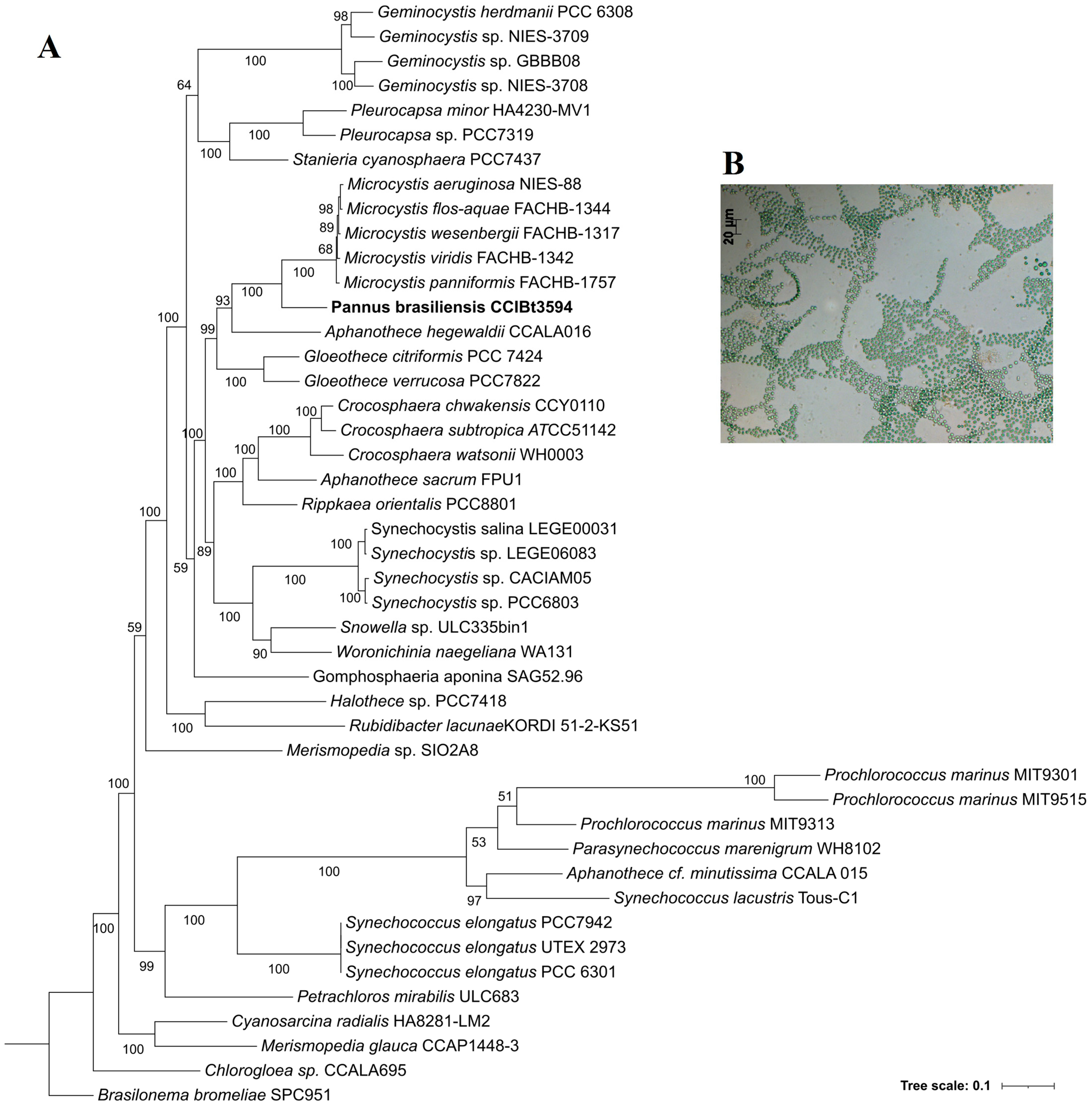
3.4. Phylogenetic Analyses

The evaluation of 16S rDNA phylogeny was performed to update the previous one observed by Malone et al. [22] in the description of this species in 2014 (Figures S4 and S5). In both trees, P. brasiliensis CCIBt3594 anchored isolated, with the nearest genus being Microcystis, sharing a maximum 16S sequence identity of 94.53% with the cyanobacterium Microcystis ichthyoblabe VN213. Morphologically, Microcystis and Pannus are unicellular, colony-forming, mucilage-producing freshwater genera. However, different from Pannus, Microcystis is known as cosmopolitan and bloom-forming and, mainly, for its toxigenic potential, for producing toxins like microcystin [104]. The genomic comparisons between Pannus and Microcystis revealed a 73.49% similarity to the cyanobacterium Microcystis aeruginosa NIES-88, determined through the average nucleotide identity (ANI). Additionally, the average nucleotide identity (AAI) analysis indicated a 72% similarity across all genomes within the Microcystis genera (Figure S6). In silico, the DDH values with reference genomes from the Microcystis genera ranged from 20.8 to 21.2%, and the G + C content ranged from 9.31 to 9.69%, indicating that the genomes do not belong to the same species (Table S2). The analyses revealed that the strain closest to P. brasiliensis CCIBt3594 is M. aeruginosa NIES-88, showing a similarity of less than 75%. M. aeruginosa NIES-88 was isolated from a freshwater lake in Japan and assembled into 262 contigs with remarkable completeness (99.47%) and minimal contamination (0.35%). These characteristics closely resemble the genomic attributes of P. brasiliensis CCIBT3594.
The phylogenomic assessment applied to validate phylogenetic positions was based on the alignment of 120 conserved bacterial single-copy marker proteins (Figure 2). A similar result to the 16S rDNA trees was obtained, with Microcystis being the genus most closely related to the Pannus sp., grouping in a monophyletic clade Microcystis, Pannus, Aphanothece, and Gloeothece, all members of the Microcystaceae family [105] with 99% bootstrap. The Pannus genus was originally described as a member of the family Microcystaceae, of the Chroococcales order [20]. Afterwards, Komárek and Anagnostidis [106] transferred this genus to the family Merismopediaceae due to its typical two planes of cell division. In 2005, the proposal for a cyanobacterial classification system by Hoffmann et al. [107] placed the genus Pannus in the family Synechococcaceae based on phenotypic features. In 2014, Komárek et al. [21] returned the Pannus genus into the Merismopediaceae, a classification which is used nowadays by the NCBI and CyanoDB databases. However, the most recent proposal for a cyanobacterial classification system considers the genera as belonging to the order Chroococcales, of the Microcystaceae family [105]. The phylogenomic evaluation presented corroborates the most recent proposal for Pannus placement into cyanobacterial taxonomy.
The primary challenge in analyzing whole genomes is the smaller presence of genome data in public databases. Known cyanobacterial genomes deposited in the databases correspond to cultivated strains belonging to more than 100 genera [108], as opposed to the broader representation found in known 16S rDNA sequences. The most represented genera in the genome database correspond to Nostoc, Leptolyngbya, Prochlorococcus, Synechococcus, Planktothrix, Microcystis, and Calothrix [108]. While this is the first genome of a Pannus representative, there are around 200 genomes from different Microcystis morphospecies released into public databases [109]. The low identities and phylogenetic distribution obtained showed that there are no described lineages closely related to the genus Pannus and emphasized that generating new sequences for type species and recently isolated species of Cyanobacteria will significantly advance the field of taxonomy within this clade. Furthermore, it illustrated the need for efforts to isolate and characterize traditionally uncultivated Cyanobacteria strains by sampling underexplored habitats and locations [110,111,112].
Assessing homological relationships between sequences has considerable meaning in genome annotation and phylogenetic inference. In the orthology analyses, when considering the genomes of five Microcystis sp. and the genome of P. brasiliensis, 2406 clusters of orthologous genes were identified. This set represents the core genome shared between the species, and the large number of shared genes suggests a common evolutionary history with conserved functions between the genera (Figure 3). These analyses aid in discerning whether a pair of homologous genes are orthologues (speciation) or paralogs (gene duplication) [113]. The stable core is a collection of genes characterized by comparable evolutionary trajectories exhibiting high conservation. This core encompasses genes responsible for main metabolic functions and the ribosomal apparatus, and it is resistant to horizontal gene transfer (HGT) events [24].
The accessory genome of P. brasiliensis CCIBt3594 includes 91 singletons, representing genes encoding proteins exclusive to this lineage. Among these, genes associated with biological nitrogen fixation stand out, an attribute absent in the other evaluated genomes. This observation suggests that this characteristic originated from an evolutionary path distinct from the most closely related lineages. Selective pressures predominantly shape the evolution of the accessory genome. Diversity within this genome is maintained through selection, acting on the acquisition of genes via horizontal transfer and the loss of genes possessing metabolic advantages [114,115]. The differences between genomes can be used to distinguish and separate closely related species, thereby identifying phylogenetic patterns [116]. This is especially relevant because there is a tendency for even the same species to have similar core and accessory genomes, supporting the hypothesis that the evaluated species are phylogenetically distinct.

4. Conclusions

This study presented the first genome of a Pannus genus representative and identified P. brasiliensis CCIBT 3594 as a potential diazotrophic cyanobacterium. The phylogenomic analyses anchored Pannus to the order Chroococcales, of the family Microcystaceae. These findings contribute to the taxonomy of this poorly reported genus. The presence of multiple secondary metabolite gene clusters in the genome of Pannus brasiliensis CCIBT3594 adds to the organism’s metabolic versatility and ecological adaptability, including the synthesis of pigments with photoprotective and antioxidant properties, which may have applications in the industry. Further studies are needed to evaluate the nitrogen-fixing mechanisms associated with P. brasiliensis CCIBt3594, adding to the understanding of nutrient cycling and primary production in the natural environment.

Supplementary Materials

The following supporting information can be downloaded at https://www.mdpi.com/article/10.3390/taxonomy4010010/s1, Figure S1: Functional classification of the genes annotated in the genome of Pannus brasiliensis CCIBt3594; Figure S2: Prophage region identified in the contig number 17 in the genome of Pannus brasiliensis CCIBt3594. PLP: Phage-like Protein; Sha: Tail Shaft; Hyp: Hypothetical Protein; Int: Integrase; Figure S3: Maximum-likelihood Phylogenetic tree using nifH gene displaying the relationship of Pannus brasiliensis CCIBt3594 (in bold). 1000 resampling bootstrap test was performed. Bootstrap values 50% are displayed at nodes; Figure S4: Phylogenetic tree using 16S rDNA gene displaying the relationship of Pannus brasiliensis CCIBt3594 (in bold) with other species of order Synechococcales and Chroococcales available in NCBI. Maximum-likelihood and 1000 resampling bootstrap test was performed. Bootstrap values 50% are displayed at nodes; Figure S5: Phylogenetic tree reconstructed by the Bayesian Inference method displaying the relationship of Pannus brasiliensis CCIBt3594 (in bold) with other species of order Synechococcales and Chroococcales available in NCBI. Bayesian posterior probabilities are represented on each node; Figure S6: Heatmap showing the Average Nucleotide Identity (ANI) and the Average Amino acids Identity (AAI) among the genomes of Pannus brasiliensis CCIBT3594 and related species of the genus Microcystis spp.; Table S1: Identity of the amino acid sequences of the of nif, isc, fdx and hsc genes in Pannus brasiliensis CCIBt3594 genome; Table S2: The in silico DDH values for Pannus brasiliensis CCIBt3594 and selected reference genomes of Microcystis genera.

Author Contributions

Conceptualization, M.J.M., N.B.B., A.P.D.A. and M.F.F.; data curation, M.J.M. and N.B.B.; formal analysis, M.J.M. and N.B.B.; funding acquisition, K.S. and M.F.F.; methodology, M.J.M., N.B.B., A.M.T.F. and R.V.P.; software, M.J.M., N.B.B. and A.M.T.F.; supervision, M.F.F.; visualization, A.P.D.A. and R.V.P.; writing—original draft, M.J.M., N.B.B. and A.P.D.A.; writing—review and editing, A.M.T.F., R.V.P., K.S. and M.F.F. All authors have read and agreed to the published version of the manuscript.

Funding

The State of São Paulo Research Foundation (FAPESP, 2016/14227-5) supported this research.

Data Availability Statement

The raw data supporting the conclusions of this article will be made available by the authors on request.

Acknowledgments

M.J.M. received a doctoral fellowship from the Brazilian National Council for Scientific and Technological Development (CNPq, 140991/2022-0). Both M.J.M. and A.M.T.F. received scholarships from the Brazilian Federal Agency for the Support and Evaluation of Graduate Education (CAPES, Finance code 001). R.V.P. was funded by the Doctoral Programme in Microbiology and Biotechnology of the University of Helsinki. M.F.F. received a research fellowship (#309476/2022-4) from CNPq. We would like to thank Rafael Dextro for the critical reading.

Conflicts of Interest

The authors declare no conflicts of interest.

References

  1. Archibald, J.M. The Puzzle of Plastid Evolution. Curr. Biol. 2009, 19, R81–R88. [Google Scholar] [CrossRef]
  2. Schirrmeister, B.E.; Gugger, M.; Donoghue, P.C.J. Cyanobacteria and the Great Oxidation Event: Evidence from genes and fossils. Palaeontology 2015, 58, 769–785. [Google Scholar] [CrossRef]
  3. Maya, Y.; López-Cortés, A.; Soeldner, A. Cyanobacterial Microbiotic Crusts in Eroded Soils of a Tropical Dry Forest in the Baja California Peninsula, Mexico. Geomicrobiol. J. 2002, 19, 505–518. [Google Scholar] [CrossRef]
  4. Kobos, J.; Błaszczyk, A.; Hohlfeld, N.; Toruńska-Sitarz, A.; Krakowiak, A.; Hebel, A.; Sutryk, K.; Grabowska, M.; Toporowska, M.; Kokociński, M.; et al. Cyanobacteria and Cyanotoxins in Polish Freshwater Bodies. Oceanol. Hydrobiol. Stud. 2013, 42, 358–378. [Google Scholar] [CrossRef]
  5. Costa, M.; Garcia, M.; Costa-Rodrigues, J.; Costa, M.S.; Ribeiro, M.J.; Fernandes, M.H.; Barros, P.; Barreiro, A.; Vasconcelos, V.; Martins, R. Exploring Bioactive Properties of Marine Cyanobacteria Isolated from the Portuguese Coast: High Potential as a Source of Anticancer Compounds. Mar. Drugs 2014, 12, 98–114. [Google Scholar] [CrossRef]
  6. Mataloni, G.; Komárek, J. Gloeocapsopsis aurea, a New Subaerophytic Cyanobacterium from Maritime Antarctica. Polar Biol. 2004, 27, 623–628. [Google Scholar] [CrossRef]
  7. Bravakos, P.; Kotoulas, G.; Skaraki, K.; Pantazidou, A.; Economou-Amilli, A. A Polyphasic Taxonomic Approach in Isolated Strains of Cyanobacteria from Thermal Springs of Greece. Mol. Phylogenet. Evol. 2016, 98, 147–160. [Google Scholar] [CrossRef]
  8. Bergman, B.; Johansson, C.; Soderback, E. Tansley Review No. 42: The Nostoc—Gunnera Symbiosis. Rev. Lit. Arts Am. 1992, 42, 379–400. [Google Scholar] [CrossRef]
  9. Baker, J.A.; Entsch, B.; McKay, D.B. The Cyanobiont in an Azolla Fern is Neither Anabaena nor Nostoc. FEMS Microbiol. Lett. 2003, 229, 43–47. [Google Scholar] [CrossRef]
  10. Gutierrez-Garcıa, K.; Bustos-Dıaz, E.D.; Corona-Gomez, J.A.; Ramos-Aboites, H.E.; Selem-Mojica, N.; Cruz-Morales, P.; Perez-Farrera, M.A.; Barona-Gomez, F.; Cibrian-Jaramillo, A. Cycad Coralloid Roots Contain Bacterial Communities Including Cyanobacteria and Caulobacter spp. That Encode Niche-Specific Biosynthetic Gene Clusters. Genome Biol. Evol. 2019, 11, 319–334. [Google Scholar] [CrossRef]
  11. Hilton, J.A.; Foster, R.A.; James Tripp, H.; Carter, B.J.; Zehr, J.P.; Villareal, T.A. Genomic Deletions Disrupt Nitrogen Metabolism Pathways of a Cyanobacterial Diatom Symbiont. Nat. Commun. 2013, 4, 1767. [Google Scholar] [CrossRef]
  12. Rikkinen, J. Algal and Cyanobacteria Symbioses. In Symbiotic Cyanobacteria in Lichens; World Scientific: London, UK, 2017; Chapter 5; pp. 147–167. [Google Scholar]
  13. Konstantinou, D.; Gerovasileiou, V.; Voultsiadou, E.; Gkelis, S. Sponges-Cyanobacteria Associations: Global Diversity Overview and New Data from the Eastern Mediterranean. PLoS ONE 2018, 13, e0195001. [Google Scholar] [CrossRef] [PubMed]
  14. Oswald, F.; Schmitt, F.; Leutenegger, A.; Ivanchenko, S.; D’Angelo, C.; Salih, A.; Maslakova, S.; Bulina, M.; Schirmbeck, R.; Nienhaus, G.U.; et al. Contributions of Host and Symbiont Pigments to the Coloration of Reef Corals. FEBS J. 2007, 274, 1102–1122. [Google Scholar] [CrossRef] [PubMed]
  15. Berman-Frank, I.; Lundgren, P.; Falkowski, P. Nitrogen Fixation and Photosynthetic Oxygen Evolution in Cyanobacteria. Res. Microbiol. 2003, 154, 157–164. [Google Scholar] [CrossRef]
  16. Hartmann, M.; Gomez-Pereira, P.; Grob, C.; Ostrowski, M.; Scanlan, D.J.; Zubkov, M.V. Efficient CO2 Fixation by Surface Prochlorococcus in the Atlantic Ocean. ISME J. 2014, 8, 2280–2289. [Google Scholar] [CrossRef]
  17. Dittmann, E.; Gugger, M.; Sivonen, K.; Fewer, D.P. Natural Product Biosynthetic Diversity and Comparative Genomics of the Cyanobacteria. Trends Microbiol. 2015, 23, 642–652. [Google Scholar] [CrossRef]
  18. Vijayakumar, S.; Menakha, M. Pharmaceutical Applications of Cyanobacteria—A Review. J. Acute Med. 2015, 5, 15–23. [Google Scholar] [CrossRef]
  19. Zahra, Z.; Choo, D.H.; Lee, H.; Parveen, A. Cyanobacteria: Review of Current Potentials and Applications. Environments 2020, 7, 13. [Google Scholar] [CrossRef]
  20. Hickel, B. Two New Chroococcal Cyanophytes from a Brackish Environment (Schlei-Fjord) Germany. Algol. Stud./Arch. Hydrobiol. 1991, 64, 97–104. [Google Scholar]
  21. Komárek, J.; Kaštovský, J.; Mareš, J.; Johansen, J.R. Taxonomic Classification of Cyanoprokaryotes (Cyanobacterial genera) 2014, Using a Polyphasic Approach. Preslia 2014, 86, 295–335. [Google Scholar]
  22. Da Silva Malone, C.F.; Alvarenga, D.O.; Fiore, M.F.; Sant’Anna, C.L. Towards a Phylogenetic Position for the Morphologically-Defined Genus Pannus (Cyanobacteria). Nova Hedwig. 2014, 99, 511–524. [Google Scholar] [CrossRef]
  23. Werner, V.R.; Martins, M.D.; da Silva, F.O. Cyanobacteria from a Brazilian Subtropical Freshwater Water Body. Braz. J. Bot. 2018, 41, 901–921. [Google Scholar] [CrossRef]
  24. Willis, A.; Woodhouse, J.N. Defining Cyanobacterial Species: Diversity and Description Through Genomics. Crit. Rev. Plant Sci. 2020, 39, 101–124. [Google Scholar] [CrossRef]
  25. Shih, P.M.; Wu, D.; Latifi, A.; Axen, S.D.; Fewer, D.P.; Talla, E.; Calteau, A.; Cai, F.; de Marsac, N.T.; Rippka, R.; et al. Improving the Coverage of the Cyanobacterial Phylum Using Diversity-Driven Genome Sequencing. Proc. Natl. Acad. Sci. USA 2013, 110, 1053–1058. [Google Scholar] [CrossRef] [PubMed]
  26. Abreu, V.A.; Popin, R.V.; Alvarenga, D.O.; Schaker, P.D.; Hoff-Risseti, C.; Varani, A.M.; Fiore, M.F. Genomic and Genotypic Characterization of Cylindrospermopsis raciborskii: Toward an Intraspecific Phylogenetic Evaluation by Comparative Genomics. Front. Microbiol. 2018, 9, 306. [Google Scholar] [CrossRef]
  27. Pérez-Carrascal, O.M.; Terrat, Y.; Giani, A.; Fortin, N.; Greer, C.W.; Tromas, N.; Shapiro, B.J. Coherence of Microcystis Species Revealed Through Population Genomics. ISME J. 2019, 13, 2887–2900. [Google Scholar] [CrossRef] [PubMed]
  28. Whitman, W.B. Genome Sequences as the Type Material for Taxonomic Descriptions of Prokaryotes. Syst. Appl. Microbiol. 2015, 38, 217–222. [Google Scholar] [CrossRef]
  29. Ramasamy, D.; Mishra, A.K.; Lagier, J.C.; Padhmanabhan, R.; Rossi, M.; Sentausa, E.; Raoult, D.; Fournier, P.E. A Polyphasic Strategy Incorporating Genomic Data for the Taxonomic Description of Novel Bacterial Species. Int. J. Syst. Evol. Microbiol. 2014, 64 Pt 2, 384–391. [Google Scholar] [CrossRef]
  30. Bolger, A.M.; Lohse, M.; Usadel, B. Trimmomatic: A Flexible Trimmer for Illumina Sequence Data. Bioinformatics 2014, 30, 2114–2120. [Google Scholar] [CrossRef]
  31. Bankevich, A.; Nurk, S.; Antipov, D.; Gurevich, A.A.; Dvorkin, M.; Kulikov, A.S.; Lesin, V.M.; Nikolenko, S.I.; Pham, S.; Prjibelski, A.D.; et al. SPAdes: A New Genome Assembly Algorithm and Its Applications to Single-Cell Sequencing. J. Comput. Biol. 2012, 19, 455–477. [Google Scholar] [CrossRef]
  32. Alvarenga, D.O.; Fiore, M.F.; Varani, A.M. A Metagenomic Approach to Cyanobacterial Genomics. Front. Microbiol. 2017, 8, 809. [Google Scholar] [CrossRef]
  33. Wood, D.E.; Salzberg, S.L. Kraken: Ultrafast Metagenomic Sequence Classification Using Exact Alignments. Genome Biol. 2014, 15, R46. [Google Scholar] [CrossRef] [PubMed]
  34. Lu, J.; Rincon, N.; Wood, D.E.; Breitwieser, F.P.; Pockrandt, C.; Langmead, B.; Salzberg, S.L.; Steinegger, M. Metagenome Analysis Using the Kraken Software Suite. Nat. Protoc. 2022, 17, 2815–2839. [Google Scholar] [CrossRef] [PubMed]
  35. Gurevich, A.; Saveliev, V.; Vyahhi, N.; Tesler, G. QUAST: Quality Assessment Tool for Genome Assemblies. Bioinformatics 2013, 29, 1072–1075. [Google Scholar] [CrossRef]
  36. Parks, D.H.; Imelfort, M.; Skennerton, C.T.; Hugenholtz, P.; Tyson, G.W. CheckM: Assessing the Quality of Microbial Genomes Recovered from Isolates, Single Cells, and Metagenomes. Genome Res. 2015, 25, 1043–1055. [Google Scholar] [CrossRef] [PubMed]
  37. Seemann, T. Prokka: Rapid Prokaryotic Genome Annotation. Bioinformatics 2014, 30, 2068–2069. [Google Scholar] [CrossRef] [PubMed]
  38. Tatusova, T.; DiCuccio, M.; Badretdin, A.; Chetvernin, V.; Nawrocki, E.P.; Zaslavsky, L.; Lomsadze, A.; Pruitt, K.D.; Borodovsky, M.; Ostell, J. NCBI Prokaryotic Genome Annotation Pipeline. Nucleic Acids Res. 2016, 44, 6614–6624. [Google Scholar] [CrossRef] [PubMed]
  39. Kanehisa, M.; Sato, Y.; Morishima, K. BlastKOALA and GhostKOALA: KEGG Tools for Functional Characterization of Genome and Metagenome Sequences. J. Mol. Biol. 2016, 428, 726–731. [Google Scholar] [CrossRef] [PubMed]
  40. Arndt, D.; Grant, J.R.; Marcu, A.; Sajed, T.; Pon, A.; Liang, Y.; Wishart, D.S. PHASTER: A Better, Faster Version of the PHAST Phage Search Tool. Nucleic Acids Res. 2016, 44, W16–W21. [Google Scholar] [CrossRef]
  41. Blin, K.; Shaw, S.; Augustijn, H.E.; Reitz, Z.L.; Biermann, F.; Alanjary, M.; Fetter, A.; Terlouw, B.R.; Metcalf, W.W.; Helfrich, E.J.N.; et al. AntiSMASH 7.0: New and Improved Predictions for Detection, Regulation, Chemical Structures and Visualisation. Nucleic Acids Res. 2023, 51, W46–W50. [Google Scholar] [CrossRef]
  42. Edgar, R.C. MUSCLE: A Multiple Sequence Alignment Method with Reduced Time and Space Complexity. BMC Bioinform. 2004, 5, 113. [Google Scholar] [CrossRef]
  43. Gilchrist, C.L.; Chooi, Y.H. Clinker & Clustermap.js: Automatic Generation of Gene Cluster Comparison Figures. Bioinformatics 2021, 37, 2473–2475. [Google Scholar] [CrossRef]
  44. Ronquist, F.; Huelsenbeck, J.P. MrBayes 3: Bayesian phylogenetic inference under mixed models. Bioinformatics 2003, 19, 1572–1574. [Google Scholar] [CrossRef] [PubMed]
  45. Chaumeil, P.A.; Mussig, A.J.; Hugenholtz, P.; Parks, D.H. GTDB-Tk: A Toolkit to Classify Genomes with the Genome Taxonomy Database. Bioinformatics 2020, 36, 1925–1927. [Google Scholar] [CrossRef] [PubMed]
  46. Nguyen, L.T.; Schmidt, H.A.; Von Haeseler, A.; Minh, B.Q. IQ-TREE: A Fast and Effective Stochastic Algorithm for Estimating Maximum-Likelihood Phylogenies. Mol. Biol. Evol. 2015, 32, 268–274. [Google Scholar] [CrossRef] [PubMed]
  47. Kalyaanamoorthy, S.; Minh, B.Q.; Wong, T.K.F.; Von Haeseler, A.; Jermiin, L.S. ModelFinder: Fast Model Selection for Accurate Phylogenetic Estimates. Nat. Methods 2017, 14, 587–589. [Google Scholar] [CrossRef] [PubMed]
  48. Letunic, I.; Bork, P. Interactive Tree of Life (iTOL) v5: An Online Tool for Phylogenetic Tree Display and Annotation. Nucleic Acids Res. 2021, 49, W293–W296. [Google Scholar] [CrossRef]
  49. Lee, I.; Kim, Y.O.; Park, S.C.; Chun, J. OrthoANI: An Improved Algorithm and Software for Calculating Average Nucleotide Identity. Int. J. Syst. Evol. Microbiol. 2016, 66, 1100–1103. [Google Scholar] [CrossRef]
  50. Yoon, S.H.; Ha, S.M.; Kwon, S.; Lim, J.; Kim, Y.; Seo, H.; Chun, J. Introducing EzBioCloud: A Taxonomically United Database of 16S rRNA Gene Sequences and Whole-Genome Assemblies. Int. J. Syst. Evol. Microbiol. 2017, 67, 1613–1617. [Google Scholar] [CrossRef]
  51. Meier-Kolthoff, J.P.; Klenk, H.P.; Göker, M. Taxonomic Use of DNA G+C Content and DNA–DNA Hybridization in the Genomic Age. Int. J. Syst. Evol. Microbiol. 2014, 64 Pt 2, 352–356. [Google Scholar] [CrossRef]
  52. Meier-Kolthoff, J.P.; Carbasse, J.S.; Peinado-Olarte, R.L.; Göker, M. TYGS and LPSN: A Database Tandem for Fast and Reliable Genome-Based Classification and Nomenclature of Prokaryotes. Nucleic Acids Res. 2022, 50, D801–D807. [Google Scholar] [CrossRef]
  53. Sun, J.; Lu, F.; Luo, Y.; Bie, L.; Xu, L.; Wang, Y. OrthoVenn3: An Integrated Platform for Exploring and Visualizing Orthologous Data Across Genomes. Nucleic Acids Res. 2023, 51, W397–W403. [Google Scholar] [CrossRef]
  54. De Las Rivas, J.; Lozano, J.J.; Ortiz, A.R. Comparative Analysis of Chloroplast Genomes: Functional Annotation, Genome-Based Phylogeny, and Deduced Evolutionary Patterns. Genome Res. 2002, 12, 567–583. [Google Scholar] [CrossRef] [PubMed]
  55. Cornet, L.; Meunier, L.; Van Vlierberghe, M.; Léonard, R.R.; Durieu, B.; Lara, Y.; Misztak, A.; Sirjacobs, D.; Javaux, E.J.; Philippe, H.; et al. Consensus Assessment of the Contamination Level of Publicly Available Cyanobacterial Genomes. PLoS ONE 2018, 13, e0200323. [Google Scholar] [CrossRef] [PubMed]
  56. Frangeul, L.; Quillardet, P.; Castets, A.M.; Humbert, J.F.; Matthijs, H.C.; Cortez, D.; Tolonen, A.; Zhang, C.-C.; Gribaldo, S.; Kehr, J.-C.; et al. Highly Plastic Genome of Microcystis aeruginosa PCC 7806, a Ubiquitous Toxic Freshwater Cyanobacterium. BMC Genom. 2008, 9, 274. [Google Scholar] [CrossRef]
  57. Zhang, J.-Y.; Guan, R.; Zhang, H.-J.; Li, H.; Xiao, P.; Yu, G.-L.; Du, L.; Cao, D.-M.; Zhu, B.-C.; Li, R.-H.; et al. Complete Genome Sequence and Genomic Characterization of Microcystis panniformis FACHB 1757 by Third-Generation Sequencing. Stand. Genom. Sci. 2016, 11, 11. [Google Scholar] [CrossRef]
  58. Lefler, F.W.; Barbosa, M.; Berthold, D.E.; Laughinghouse IV, H.D. Genome Sequences of Two Microcystis aeruginosa (Chroococcales, Cyanobacteria) Strains from Florida (United States) with Disparate Toxigenic Potentials. Microbiol. Resour. Announc. 2020, 9, 10–1128. [Google Scholar] [CrossRef]
  59. Shestakov, S.V.; Karbysheva, E.A. The Role of Viruses in the Evolution of Cyanobacteria. Biol. Bull. Rev. 2015, 5, 527–537. [Google Scholar] [CrossRef]
  60. Melnikov, O.; Zaritsky, A.; Zarka, A.; Boussiba, S.; Malchin, N.; Yagil, E.; Kolot, M. Site-specific Recombination in the Cyanobacterium Anabaena sp. Strain PCC 7120 Catalyzed by the Integrase of Coliphage HK022. J. Bacteriol. 2009, 191, 4458–4464. [Google Scholar] [CrossRef]
  61. Zhong, K.X.; Suttle, C.A.; Baudoux, A.C.; Derelle, E.; Colombet, J.; Cho, A.; Caleta, J.; Six, C.; Jacquet, S. A New Freshwater Cyanosiphovirus Harboring Integrase. Front. Microbiol. 2018, 9, 2204. [Google Scholar] [CrossRef]
  62. Kultschar, B.; Llewellyn, C. Secondary metabolites in cyanobacteria. In Secondary Metabolites—Sources and Applications; Vijayakumar, R., Ed.; IntechOpen: London, UK, 2018. [Google Scholar] [CrossRef]
  63. Burja, A.M.; Banaigs, B.; Abou-Mansour, E.; Grant Burgess, J.; Wright, P.C. Marine Cyanobacteria—A Prolific Source of Natural Products. Tetrahedron 2001, 57, 9347–9377. [Google Scholar] [CrossRef]
  64. Demay, J.; Bernard, C.; Reinhardt, A.; Marie, B. Natural Products from Cyanobacteria: Focus on Beneficial Activities. Mar. Drugs 2019, 17, 320. [Google Scholar] [CrossRef]
  65. Yadav, P.; Singh, R.P.; Rana, S.; Joshi, D.; Kumar, D.; Bhardwaj, N.; Gupta, R.K.; Kumar, A. Mechanisms of Stress Tolerance in Cyanobacteria under Extreme Conditions. Stresses 2022, 2, 531–549. [Google Scholar] [CrossRef]
  66. Micallef, M.L.; D’Agostino, P.M.; Al-Sinawi, B.; Neilan, B.A.; Moffitt, M.C. Exploring Cyanobacterial Genomes for Natural Product Biosynthesis Pathways. Mar. Genom. 2015, 21, 1–12. [Google Scholar] [CrossRef]
  67. Coates, R.C.; Podell, S.; Korobeynikov, A.; Lapidus, A.; Pevzner, P.; Sherman, D.H.; Allen, E.E.; Gerwick, W.H. Characterization of Cyanobacterial Hydrocarbon Composition and Distribution of Biosynthetic Pathways. PLoS ONE 2014, 9, e85140. [Google Scholar] [CrossRef] [PubMed]
  68. Gibbons, J.; Gu, L.; Zhu, H.; Gibbons, W.; Zhou, R. Identification of Two Genes Required for Heptadecane Production in a N2-Fixing Cyanobacterium Anabaena sp. Strain PCC 7120. AMB Express 2018, 8, 167. [Google Scholar] [CrossRef] [PubMed]
  69. Nitnaware, K.M.; Raskar, K.B.; Agarwal, G.; Montes, R.A.C.; Chopra, R.; López-Arredondo, D.L.; Nikam, T.D.; Patil, G.B. Whole-Genome Characterization and Comparative Genomics of a Novel Freshwater Cyanobacteria Species: Pseudanabaena punensis. Mol. Phylogenet. Evol. 2021, 164, 107272. [Google Scholar] [CrossRef] [PubMed]
  70. Kudo, H.; Hayashi, Y.; Arai, M. Improving Hydrocarbon Production by Engineering Cyanobacterial Acyl-(Acyl Carrier Protein) Reductase. Biotechnol. Biofuels 2019, 12, 291. [Google Scholar] [CrossRef] [PubMed]
  71. Gu, W.; Dong, S.H.; Sarkar, S.; Nair, S.K.; Schmidt, E.W. The Biochemistry and Structural Biology of Cyanobactin Pathways: Enabling Combinatorial Biosynthesis. Methods Enzymol. 2018, 604, 113–163. [Google Scholar] [CrossRef] [PubMed]
  72. Pearson, A.; Rusch, D.B. Distribution of Microbial Terpenoid Lipid Cyclases in the Global Ocean Metagenome. ISME J. 2009, 3, 352–363. [Google Scholar] [CrossRef] [PubMed]
  73. Lee, S.; Poulter, C.D. Cloning, Solubilization, and Characterization of Squalene Synthase from Thermosynechococcus elongatus BP-1. J. Bacteriol. 2008, 190, 3808–3816. [Google Scholar] [CrossRef]
  74. Englund, E.; Pattanaik, B.; Ubhayasekera, S.J.K.; Stensjö, K.; Bergquist, J.; Lindberg, P. Production of Squalene in Synechocystis sp. PCC 6803. PLoS ONE 2014, 9, e90270. [Google Scholar] [CrossRef]
  75. Fagundes, M.B.; Alvarez-Rivera, G.; Vendruscolo, R.G.; Voss, M.; da Silva, P.A.; Barin, J.S.; Jacob-Lopes, E.; Zepka, L.Q.; Wagner, R. Green microsaponification-based method for gas chromatography determination of sterol and squalene in cyanobacterial biomass. Talanta 2021, 224, 121793. [Google Scholar] [CrossRef]
  76. Pattanaik, B.; Englund, E.; Nolte, N.; Lindberg, P. Introduction of a Green Algal Squalene Synthase Enhances Squalene Accumulation in a Strain of Synechocystis sp. PCC 6803. Metab. Eng. Commun. 2020, 10, e00125. [Google Scholar] [CrossRef]
  77. Fagundes, M.B.; Falk, R.B.; Facchi, M.M.X.; Vendruscolo, R.G.; Maroneze, M.M.; Zepka, L.Q.; Jacob-Lopes, E.; Wagner, R. Insights in Cyanobacteria Lipidomics: A Sterols Characterization from Phormidium autumnale Biomass in Heterotrophic Cultivation. Food Res. Int. 2019, 119, 777–784. [Google Scholar] [CrossRef]
  78. Jiang, Y.; Fan, K.W.; Wong, R.T.Y.; Chen, F. Fatty Acid Composition and Squalene Content of the Marine Microalga Schizochytrium mangrovei. J. Agric. Food Chem. 2004, 52, 1196–1200. [Google Scholar] [CrossRef] [PubMed]
  79. Carvalho, C.C.; Fernandes, P. Production of Metabolites as Bacterial Responses to the Marine Environment. Mar. Drugs 2010, 8, 705–727. [Google Scholar] [CrossRef] [PubMed]
  80. Belin, B.J.; Busset, N.; Giraud, E.; Molinaro, A.; Silipo, A.; Newman, D.K. Hopanoid Lipids: From Membranes to Plant–Bacteria Interactions. Nat. Rev. Microbiol. 2018, 16, 304–315. [Google Scholar] [CrossRef] [PubMed]
  81. Takaichi, S.; Mochimaru, M. Carotenoids and Carotenogenesis in Cyanobacteria: Unique Ketocarotenoids and Carotenoid Glycosides. Cell. Mol. Life Sci. 2007, 64, 2607–2619. [Google Scholar] [CrossRef] [PubMed]
  82. Gan, F.; Bryant, D.A. Adaptive and Acclimative Responses of Cyanobacteria to Far-Red Light. Environ. Microbiol. 2015, 17, 3450–3465. [Google Scholar] [CrossRef]
  83. Latifi, A.; Ruiz, M.; Zhang, C.C. Oxidative Stress in Cyanobacteria. FEMS Microbiol. Rev. 2009, 33, 258–278. [Google Scholar] [CrossRef] [PubMed]
  84. Foyer, C.H.; Noctor, G. Redox Homeostasis and Antioxidant Signaling: A Metabolic Interface between Stress Perception and Physiological Responses. Plant Cell 2005, 17, 1866–1875. [Google Scholar] [CrossRef] [PubMed]
  85. Averina, S.; Velichko, N.; Senatskaya, E.; Pinevich, A. Far-red light photoadaptations in aquatic cyanobacteria. Hydrobiologia 2018, 813, 1–17. [Google Scholar] [CrossRef]
  86. Gallon, J.R. The oxygen sensitivity of nitrogenase: A problem for biochemists and micro-organisms. Trends Biochem. Sci. 1981, 6, 19–23. [Google Scholar] [CrossRef]
  87. Stal, L.J. Nitrogen fixation in cyanobacteria. eLS 2015, 1–9. [Google Scholar] [CrossRef]
  88. Colón-López, M.S.; Sherman, D.M.; Sherman, L.A. Transcriptional and translational regulation of nitrogenase in light-dark- and continuous-light-grown cultures of the unicellular cyanobacterium Cyanothece sp. strain ATCC 51142. J. Bacteriol. 1997, 179, 4319–4327. [Google Scholar] [CrossRef]
  89. Lin, S.; Henze, S.; Lundgren, P.; Bergman, B.; Carpenter, E.J. Whole-cell immunolocalization of nitrogenase in marine diazotrophic cyanobacteria, Trichodesmium spp. Appl. Environ. Microbiol. 1998, 64, 3052–3058. [Google Scholar] [CrossRef]
  90. Fujita, Y.; Uesaka, K. Nitrogen fixation in cyanobacteria. In Cyanobacterial Physiology: From Fundamentals to Biotechnology; Kageyama, H., Waditee-Sirisattha, R., Eds.; Academic Press: London, UK, 2022; pp. 29–45. [Google Scholar] [CrossRef]
  91. Zehr, J.P.; Waterbury, J.B.; Turner, P.J.; Montoya, J.P.; Omoregie, E.; Steward, G.F.; Hansen, A.; Karl, D.M. Unicellular cyanobacteria fix N2 in the subtropical north Pacific Ocean. Nature 2001, 412, 635–638. [Google Scholar] [CrossRef]
  92. Zehr, J.P.; Capone, D.G. Unsolved mysteries in marine nitrogen fixation. Trends Microbiol. 2023, 31, 1–14. [Google Scholar] [CrossRef] [PubMed]
  93. Wyatt, J.T.; Silvey, J.K.G. Nitrogen fixation by Gloeocapsa. Science 1969, 165, 908–909. [Google Scholar] [CrossRef] [PubMed]
  94. Mitsui, A.; Cao, S.; Takahashi, A.; Arai, T. Growth synchrony and cellular parameters of the unicellular nitrogen-fixing marine cyanobacterium, Synechococcus sp. strain Miami BG043511, under continuous illumination. Physiol. Plant 1987, 69, 1–8. [Google Scholar] [CrossRef]
  95. Esteves-Ferreira, A.A.; Cavalcanti, J.H.F.; Vaz, M.G.M.V.; Alvarenga, L.V.; Nunes-Nesi, A.; Araújo, W.L. Cyanobacterial Nitrogenases: Phylogenetic Diversity, Regulation and Functional Predictions. Genet. Mol. Biol. 2017, 40, 261–275. [Google Scholar] [CrossRef] [PubMed]
  96. Watanabe, T.; Horiike, T. The evolution of molybdenum-dependent nitrogenase in cyanobacteria. Biology 2021, 10, 329. [Google Scholar] [CrossRef] [PubMed]
  97. Nowruzi, B.; Hutarova, L. Structural and Functional Genes, and Highly Repetitive Sequences Commonly Used in the Phylogeny and Species Concept of the Phylum Cyanobacteria. Cryptogam. Algol. 2023, 44, 59–84. [Google Scholar] [CrossRef]
  98. Thiel, T.; Pratte, B.S. Regulation of Three Nitrogenase Gene Clusters in the Cyanobacterium Anabaena variabilis ATCC 29413. Life 2014, 4, 944–967. [Google Scholar] [CrossRef]
  99. Schrautemeier, B.; Cassing, A.; Böhme, H. Characterization of the Genome Region Encoding an fdxH-Type Ferredoxin and a New 2 [4Fe-4S] Ferredoxin from the Nonheterocystous, Nitrogen-Fixing Cyanobacterium Plectonema boryanum PCC 73110. J. Bacteriol. 1994, 176, 1037–1046. [Google Scholar] [CrossRef]
  100. Dos Santos, P.C.; Johnson, D.C.; Ragle, B.E.; Unciuleac, M.C.; Dean, D.R. Controlled Expression of nif and isc Iron-Sulfur Protein Maturation Components Reveals Target Specificity and Limited Functional Replacement Between the Two Systems. J. Bacteriol. 2007, 189, 2854–2862. [Google Scholar] [CrossRef] [PubMed]
  101. Böhme, H. Regulation of Nitrogen Fixation in Heterocyst-Forming Cyanobacteria. Trends Plant Sci. 1998, 3, 346–351. [Google Scholar] [CrossRef]
  102. Latysheva, N.; Junker, V.L.; Palmer, W.J.; Codd, G.A.; Barker, D. The Evolution of Nitrogen Fixation in Cyanobacteria. Bioinformatics 2012, 28, 603–606. [Google Scholar] [CrossRef]
  103. Zehr, J.P.; Bench, S.R.; Carter, B.J.; Hewson, I.; Niazi, F.; Shi, T.; Tripp, H.J.; Affourtit, J.P. Globally Distributed Uncultivated Oceanic N2-Fixing Cyanobacteria Lack Oxygenic Photosystem II. Science 2008, 322, 1110–1112. [Google Scholar] [CrossRef]
  104. Dick, G.J.; Duhaime, M.B.; Evans, J.T.; Errera, R.M.; Godwin, C.M.; Kharbush, J.J.; Nitschky, H.S.; Powers, M.A.; Vanderploeg, H.A.; Schmidt, K.C.; et al. The Genetic and Ecophysiological Diversity of Microcystis. Environ. Microbiol. 2021, 23, 7278–7313. [Google Scholar] [CrossRef]
  105. Strunecký, O.; Ivanova, A.P.; Mareš, J. An Updated Classification of Cyanobacterial Orders and Families Based on Phylogenomic and Polyphasic Analysis. J. Phycol. 2023, 59, 12–51. [Google Scholar] [CrossRef]
  106. Komárek, J.; Anagnostidis, K. Cyanoprokaryota. 1. Teil Chroococcales. In Süsswasserflora von Mitteleuropa; Gärtner, E.H., Heying, H., Möllenhauer, D., Eds.; G. Fischer: Jena, Germany, 1998. [Google Scholar]
  107. Hoffmann, L.; Komárek, J.; Kaštovský, J. System of Cyanoprokaryotes (Cyanobacteria) State in 2004. Algol. Stud./Arch. Hydrobiol. 2005, 117, 95–115. [Google Scholar] [CrossRef]
  108. Dextro, R.B.; Delbaje, E.; Cotta, S.R.; Zehr, J.P.; Fiore, M.F. Trends in Free-access Genomic Data Accelerate Advances in Cyanobacteria Taxonomy. J. Phycol. 2021, 57, 1392–1402. [Google Scholar] [CrossRef]
  109. Zhang, X.; Xiao, L.; Liu, J.; Tian, Q.; Xie, J. Trade-off in Genome Turnover Events Leading to Adaptive Evolution of Microcystis aeruginosa Species Complex. BMC Genom. 2023, 24, 462. [Google Scholar] [CrossRef] [PubMed]
  110. Thompson, A.W.; Foster, R.A.; Krupke, A.; Carter, B.J.; Musat, N.; Vaulot, D.; Kuypers, M.M.M.; Zehr, J.P. Unicellular Cyanobacterium Symbiotic with a Single-Celled Eukaryotic Alga. Science 2012, 337, 1546–1550. [Google Scholar] [CrossRef] [PubMed]
  111. Temraleeva, A.D.; Dronova, S.A.; Moskalenko, S.V.; Didovich, S.V. Modern Methods for Isolation, Purification, and Cultivation of Soil Cyanobacteria. Microbiology 2016, 85, 389–399. [Google Scholar] [CrossRef]
  112. Velichko, N.; Smirnova, S.; Averina, S.; Pinevich, A. A Survey of Antarctic Cyanobacteria. Hydrobiologia 2021, 848, 2627–2652. [Google Scholar] [CrossRef]
  113. Ullah, I.; Sjöstrand, J.; Andersson, P.; Sennblad, B.; Lagergren, J. Integrating Sequence Evolution into Probabilistic Orthology Analysis. Syst. Biol. 2015, 64, 969–982. [Google Scholar] [CrossRef]
  114. Bobay, L.M.; Ochman, H. Factors Driving Effective Population Size and Pan-Genome Evolution in Bacteria. BMC Evol. Biol. 2018, 18, 153. [Google Scholar] [CrossRef]
  115. Goyal, A. Metabolic Adaptations Underlying Genome Flexibility in Prokaryotes. PLoS Genet. 2018, 14, e1007763. [Google Scholar] [CrossRef] [PubMed]
  116. Cao, H.; Xu, D.; Zhang, T.; Ren, Q.; Xiang, L.; Ning, C.; Zhang, Y.; Gao, R. Comprehensive and Functional Analyses Reveal the Genomic Diversity and Potential Toxicity of Microcystis. Harmful Algae 2022, 113, 102186. [Google Scholar] [CrossRef] [PubMed]
Figure 1. Synteny analysis showing the nifHDK and nifBSU operon in Pannus brasiliensis CCIBt3594 genome and the most related sequences in the database.
Taxonomy 04 00010 g001
Figure 2. (A) Maximum-likelihood phylogenomic tree based on the alignment of 120 bacterial single-copy conserved marker proteins using GTDB-tk. A 1000 resampling bootstrap test was performed. Bootstrap values > 50 are displayed at nodes. (B) Photomicrograph of P. brasiliensis CCIBt3594 cells grouped by a mucilaginous structure.
Taxonomy 04 00010 g002
Figure 3. Venn diagrams for gene orthology between Pannus brasiliensis CCIBt3594 and Microcystis spp., emphasizing the number of genes that structure the core genome (2406) shared among the genera as well as highlighting the exclusive characteristics within the accessory genomes (91) of P. brasiliensis CCIBt3594.
Taxonomy 04 00010 g003
Table 1. Features of Pannus brasiliensis CCIBt3594 genome assembly.
AttributesValue
Genome size (bp)5,152,646
G + C content (%)52.04
Completeness (%)99.12
Contamination (%)0.29
Contigs117
Largest contig (bp)248,692
N50 length (bp)83,624
Protein encoding gene4988
Total tRNAs44
16S rRNA copies2
Table 2. Identification of secondary metabolite biosynthesis gene clusters in P. brasiliensis CCIBT3594 genome sequence.
ContigLocation (nt)Product TypeMost Similar Known ClusterSimilarity
880,421–126,963T1PKS; NRPS1-heptadecene100%
225103–76,035TerpenePhytoene synthase31%
511–19,543TerpeneTerpene cyclase11%
5712,658–28,163RRE-ContainingRiPP12%
761–13,978CyanobactinRiPP13%
Disclaimer/Publisher’s Note: The statements, opinions and data contained in all publications are solely those of the individual author(s) and contributor(s) and not of MDPI and/or the editor(s). MDPI and/or the editor(s) disclaim responsibility for any injury to people or property resulting from any ideas, methods, instructions or products referred to in the content.

