Article

Four New Caddisfly Species of Marilia Müller, 1880 (Trichoptera: Odontoceridae) from a Tailings Dam Disaster Area, Rio Doce basin, Brazil †

by
Pedro Bonfá Neto
1,*,
Frederico Falcão Salles
1 and
Albane Vilarino
2,*
1
Departamento de Entomologia, Museu de Entomologia, Universidade Federal de Viçosa, Av. P.H. Rolfs, s.n, Campus Universitário, Viçosa 36570-900, Brazil
2
Instituto de Biologia, Universidade Federal da Bahia, Rua Barão de Geremoabo, 147, Ondina, Salvador 40170-115, Brazil
*
Authors to whom correspondence should be addressed.
urn:lsid:zoobank.org:pub:8B3E3282-FC2D-47A7-BA82-D8E582C9B6D3; urn:lsid:zoobank.org:act:68B0EEF2-37D3-480A-A3D5-3D5362AEF95F; urn:lsid:zoobank.org:act:E49DA5EA-B249-4E50-AD7C-B81CDB4D2886; urn:lsid:zoobank.org:act:4C694833-2FC8-4C7B-A0F4-209A554B4699; urn:lsid:zoobank.org:act:45A73225-FEA2-497B-8D16-28173ADEE67F.
Taxonomy 2023, 3(3), 381-400; https://doi.org/10.3390/taxonomy3030022
Submission received: 25 May 2023 / Revised: 24 July 2023 / Accepted: 26 July 2023 / Published: 28 July 2023

Abstract

:
Marilia is the most diverse genus of the caddisfly family Odontoceridae, being distributed throughout the Oriental, Australasian and Neotropical regions. Herein, we describe four new Neotropical species from the Atlantic Forest, Rio Doce basin, Brazil. This basin was partly affected by a flood of 50 million m3 of mud and mining debris in 2015. Due to this disaster, numerous monitoring activities have been carried out to assess the impact generated. Material from one of these attempts was used to describe the new species: Marilia aranan sp. nov., Marilia krenak sp. nov., M. maxakali sp. nov. and M. mukurin sp. nov. Additionally, we provide a new state record of M. guaira from Minas Gerais. Marilia aranan sp. nov. was collected in tributaries as well as in the impacted area 7 years after the disaster. Marilia guaira and M. krenak sp. nov. were collected only in the impacted area. The other species were found only in tributaries. Odontoceridae are considered highly sensitive to disturbance. The presence of Marilia species suggests that parts of the riverbed have recovered from the fine tailing sediments and also highlights the importance of tributaries in the recolonization process.

1. Introduction

Odontoceridae (Trichoptera: Integripalpia) has 177 extant species in 15 genera [1,2], having the greatest diversity in the Oriental and Neotropical regions. Three genera have been recorded in the Neotropics: two endemic monotypic genera, both from southeastern Brazil, Anastomoneura Huamantinco & Nessimian 2004 [3] and Barypenthus Burmeister 1839 [4], and the highly diverse and worldwide-distributed Marilia Müller 1880 [5]. Marilia has a disjunct distribution with 73 extant species, 44 of which are Neotropical, 20 Oriental, 2 East Palearctic, 3 Australasian, 2 Nearctic and 2 Nearctic–Neotropical [2]. There are also three fossil species from Western Palearctic Baltic amber [2]. So far, 22 species of Marilia have been recorded in Brazil, 6 of them occurring in the Minas Gerais state [6].
Marilia larvae favor slow-flowing areas or depositional zones; however, they can be found in a variety of freshwater environments [7,8,9]. Odontocerid larvae are omnivorous, feeding on organic detritus, vascular plants, algae and aquatic arthropods [8,9]. Larval cases are formed of sand grains or bigger mineral particles and are extremely tough to crush [8,9]. The presence of a sexual dimorphism in the eyes, which are very large in males, distinguishes Marilia adults from other Neotropical Odontoceridae [7,10,11,12]. Males also differ from females by the wing venation, in which males present the forewing forks I, II and V and the hindwing fork I [13], while females have the forewing forks I, II, III and V and the hindwing forks I and V [10]. Marilia species can have a different tibial spur formula (2,4,4 or 2,4,2), which is used to circumscribe species groups [14]. However, a cladistic analysis revealed that the distinct tibial spurs lacked a phylogenetic signal for the group [15]. Diagnostic characteristics of the Oriental species of the genus are discussed by Oláh and Johanson [16] and Yang et al. [17]. Neotropical species are discussed mainly by Flint [14,18] and Bueno-Soria and Rojas-Ascencio [19], among other studies. A revision and phylogeny of the Neotropical species of Marilia were provided by Costa [15].
The Rio Doce basin is located in Brazil between the states of Minas Gerais and Espirito Santo and is part of the Southeast Atlantic watershed. It is Brazil’s fifth largest basin, with a drainage area of approximately 83,400 km2 [20]. The Rio Doce basin was strongly impacted by iron ore tailings after the Fundão dam collapse in 2015, releasing roughly 50 million m3 of tailings into the river system [21,22]. This mudflow traveled more than 650 km before reaching the Atlantic Ocean [23,24]. It is regarded as one of Brazil’s worst environmental disasters [25,26].
Given the catastrophic consequences, fauna and flora monitoring was carried out along the Rio Doce to comply with Brazilian government environmental regulations. Macroinvertebrates such as mayflies (Ephemeroptera), stoneflies (Plecoptera) and caddisflies (Trichoptera) have been shown to be, on average, more intolerant to environmental disturbance than other freshwater taxa [27], and thus alterations in abundance and diversity in these taxa are used in freshwater biomonitoring programs. The new species of Marilia described here were discovered as part of collections through numerous monitoring activities that were performed to assess the environmental impacts caused by the dam rupture.
Herein, we describe four new species of Marilia and provide a new record of M. guaira Flint, 1983 [14] from the Minas Gerais state.

2. Materials and Methods

The analyzed specimens were mostly collected within the Rio Doce basin, except for a single male collected in the São Mateus basin (Figure 1). The specimens were collected through light attraction methods using Pennsylvania traps [28]. The light traps used fluorescent lamps with UV light and/or white light connected to 12 V batteries using electronic converters. Pennsylvania traps were active from dusk until dawn. The material analyzed was preserved in ethanol 80%, and was deposited at the Museu de Entomologia, Universidade Federal de Viçosa (UFVB) and Museu Nacional, Universidade Federal do Rio de Janeiro (MNRJ), both in Brazil.
Species identifications were carried out with the aid of a stereomicroscope, with a magnification of 50×, and an optical microscope, with a magnification of 400×; the observed specimens were compared with the species described in the taxonomic literature, e.g., [3,9,13,14,15,18,19,29].
The structures used for the species delimitation were from the morphology of male genitalia. A diaphanization process through lactic acid was used for the observation of the genitalia, following procedures detailed by Blahnik and Holzenthal [30]. Later, the genitalia were placed in excavated slides with a drop of glycerin or alcohol gel and observed with microscopy. The wings were mounted dry, on permanent slides. The wings and body parts were photographed using a Leica Camera (MC170 HD) coupled to a Leica stereomicroscope (M205 A) and edited using Adobe Photoshop CC®. The genitalia were photographed using a Motic Camera (Moticam A5) attached to an Olympus microscope (CX31); the focus stacking was made using Helicon Focus® software. These photographs were used as templates for illustrations, which were made by tracing the structures digitally using Adobe Illustrator CC®. The terminology used in the species descriptions is modified from Oláh and Johanson [16] and Bueno-Soria and Rojas-Ascencio [19]. Terminology for the wing venation is modified from Mosely and Kimmins [31].
The distribution map was made using a Quantum GIS program using the 2017 IBGE cartographic databases and raster images for a digital elevation model (DEM) with a resolution of 75 and 30 m, provided by the Global Multi-resolution Terrain Elevation Data (GMTED) and TOPODATA of the National Institute for Space Research (INPE).

3. Results

3.1. Taxonomy

Type material. Holotype male. Brazil, Minas Gerais, Ferros, Santo Antônio river, 19°14′55.40″ S, 42°56′27.40″ W, 425 m a.s.l., 20–21.viii.2022, Pennsylvania trap, Viana, Bonfá & Ataide col. (UFVB TR00275). Paratypes. Brazil, Espírito Santo: São Mateus, Santa Maria, São Mateus river, 18°39′17.10″ S, 39°59′36.00″ W, 16 m a.s.l., 07–08.ix.2012, Pennsylvania trap, CEUNES col. 1♂ (UFVB TR00274). Minas Gerais: Matias Lobato, Suaçuí Grande river, 18°34′23.50″ S, 41°56′52.30″ W, 197 m a.s.l., 05–06.ix.2022, Pennsylvania trap, Viana, Bonfá & Ataide col. 1♂ (UFVB TR00276). Aimorés, Manhuaçu river, 19°29′29.10″ S, 41°11′13.30″ W, 97 m, 10–11.ix.2022, Pennsylvania trap, Viana, Bonfá & Ataide col. 1♂ (MNRJ). Tumiritinga. Rio Doce river, 18°58′07.56″ S, 41°39′49.36″ W, 132 m, 07–08.i.2022, Pennsylvania trap, Viana, Bonfá, Rodrigues & Rothe-Neves col. 2♂, 2 ♀ (UFVB TR00278).
Diagnosis. Marilia aranan sp. nov. is most similar to Marilia humerosa Flint, 1983 [14], sharing segment IX with two lateral sutures, segment X sclerotized sides with a subapical angle, coxopodite with a strong basomesal angle and phallic endotheca with spines. The new species can be differentiated from M. humerosa and other congeners mainly by (1) segment X presenting a long, narrow, spine-like dorsal projection, and (2) by the coxopodite with a basomesal lobe wider than M. humerosa.
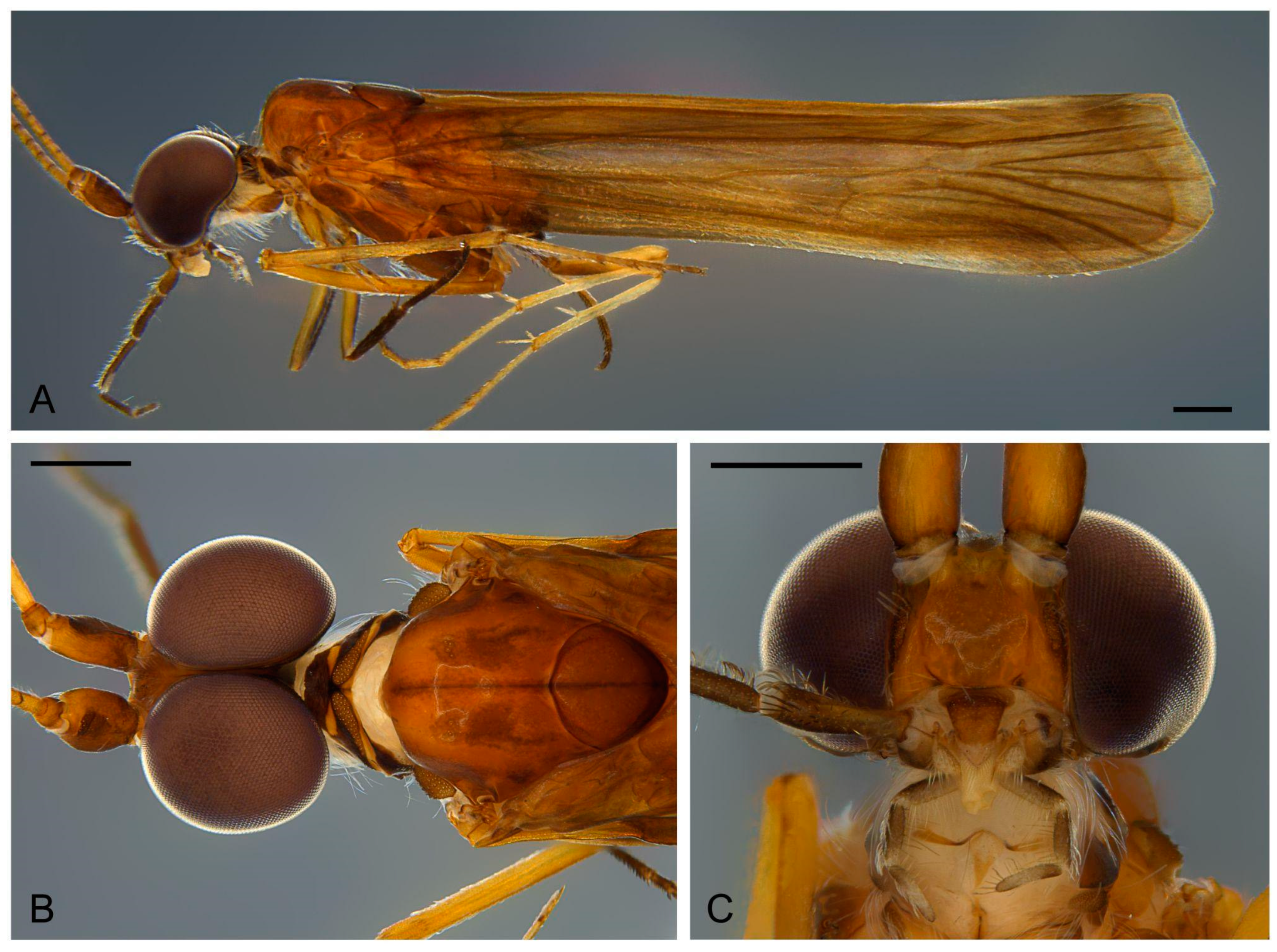
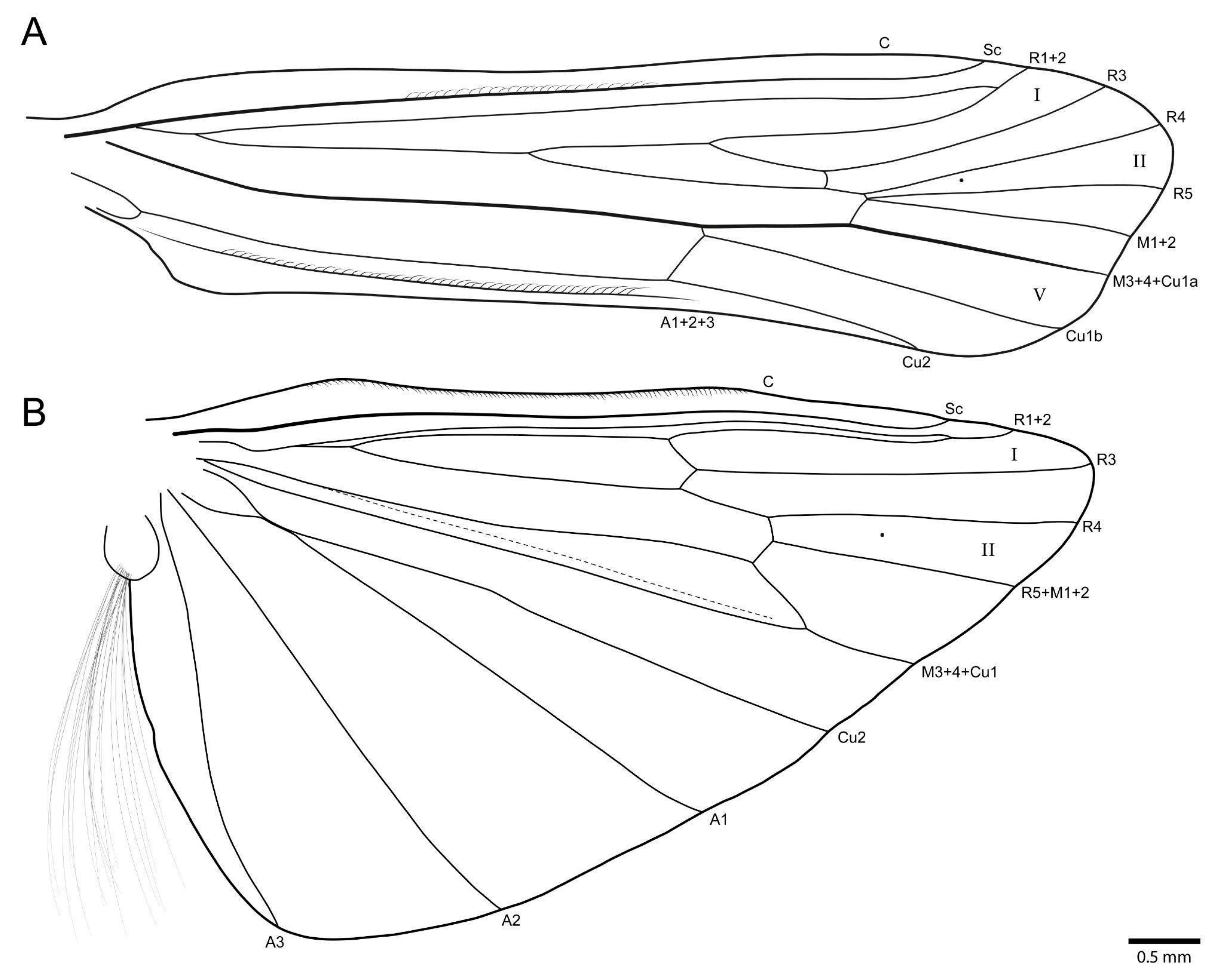
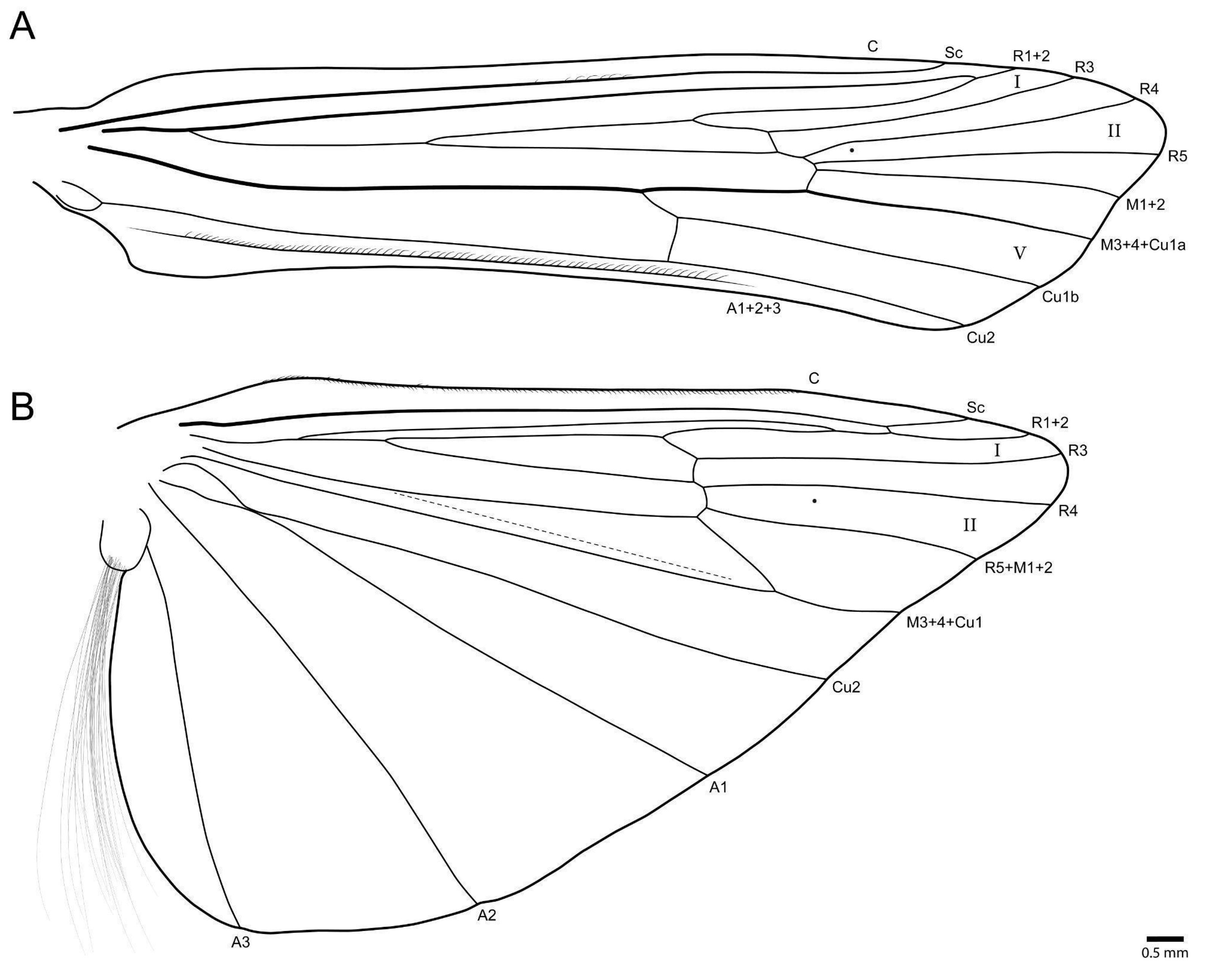
Description. Adult male. Forewing 7.4–8.0 mm (7.78 mm, n = 5). Color (in alcohol): Body and forewings brown, foreleg tibia and tarsus dark brown, midleg tibia and tarsus brown, hindleg tibia and tarsus light brown (Figure 2A). Head: Eyes large, almost touching each other at vertex (Figure 2B). Antennae long, about 2.2 times body length, with narrow annuli; scapes wide, brown. Maxillary palps five-articulated, articles formula (I = IV = V) < (II = III). Labial palpi three-articulated, articles subequal. Thorax: Pronotum narrow, with transversely elongate setal wart. Mesonotum wide, with pair of small setal warts; mesoscutellum almost circular, anterior margin slightly acute, without setal warts (Figure 2B). Tibial spur formula 2-4-4. Wings, forewing forks I, II, V present; hindwing fork I and II present (Figure 3). Abdomen: Simple, without differentiated structures.
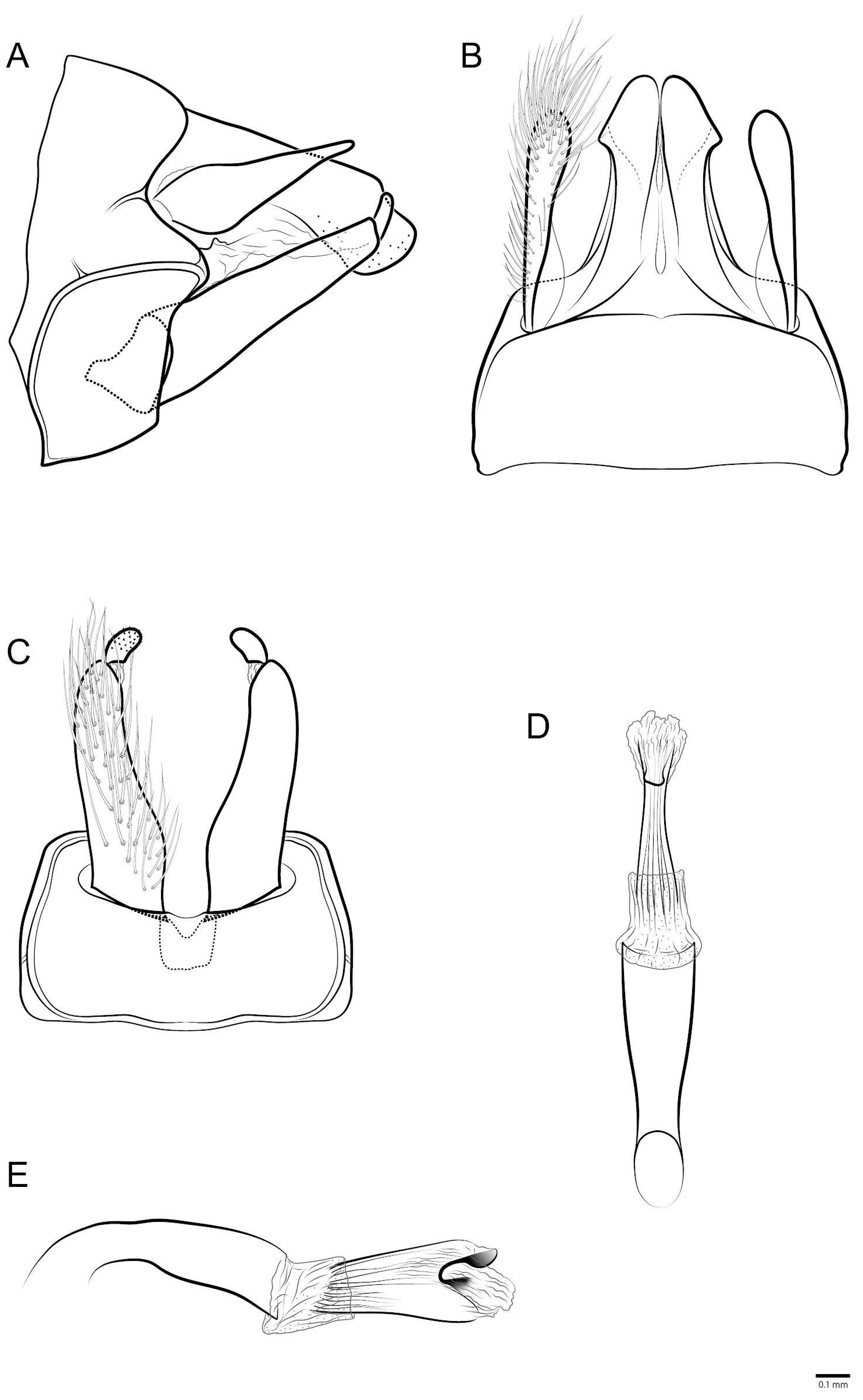
Male genitalia (Figure 4A–E): Segment IX, in lateral view with anterior margin slightly projected subventrally, posterior margin projected dorsally and mesally, with sutures separating each side of segment IX into three subequal parts (Figure 4A); in dorsal view, anterior margin straight, membranous, posterior margin with concave suture, lateroapical corners weakly produced (Figure 4B); in ventral view, segment IX not separated by suture, anterior margin straight, posterior margin concave. Preanal appendage, in lateral view, shorter than segment X, wider sub-basally, slightly tapering apically, apex rounded (Figure 4A); in dorsal view, clavate, with numerous setae and base, narrower than apex (Figure 4B). Segment X, in lateral view, wider at base and narrowing at apex, ventral margin straight, apex rounded, dorsal margin with long, narrow, digitate projection (Figure 4A); in dorsal view, wider at base, narrowing subapically, sclerotized laterally, each side with strong subapical angle and acute apex, membranous mesally with small subapical sclerotized lobe (Figure 4B). Inferior appendage bi-articulated: in lateral view, coxopodite cylindrical with base broader than apex (Figure 4A); in ventral view, with basomesal lobe forming strong angle (Figure 4C); harpago short in lateral view, about 3× shorter than coxopodite, with small conical spines apically (Figure 4A); in ventral view, nearly straight (Figure 4C). Phallus tubular slightly curved near base in lateral view (Figure 4E), straight in ventral view (Figure 4D); endotheca membranous with subtle small spines; in lateral view, phallotremal sclerite with C-shaped dorsal structure and tube-shaped ventral structure (Figure 4E).
Etymology. The specific epithet, aranan, refers to the name of the Aranã indigenous people, who are currently dispersed in several urban and rural areas of the states of São Paulo and Minas Gerais, with a greater concentration in the Jequitinhonha Valley, northeast of Minas Gerais.
Distribution. Atlantic Forest (Espírito Santo and Minas Gerais state, Brazil).
Type material. Holotype male. Brazil, Espírito Santo, Linhares, Fazenda Maria Bonita, Rio Doce river, 19°26′50.80″ S, 39°56′29.20″ W, 9 m a.s.l., 24–25.xi.2014, Pennsylvania trap, CEUNES col. (UFVB TR00280). Paratypes. Brazil, Espírito Santo, Linhares, same data as holotype 3♀, 1♂ (UFVB TR00281). Linhares, near street of Lagoa Palminhas, Rio Doce river, 19°29′53.10″ S, 40°16′40.90″ W, 22 m a.s.l., 09–10.ix.2014, Pennsylvania trap, CEUNES col. 1♂ (MNRJ). Minas Gerais, Tumiritinga, Rio Doce river, 18°58′07.56″ S, 41°39′49.36″ W, 132 m a.s.l., 06–07.ix.2022, Pennsylvania trap, Viana, Bonfá & Ataide col. 1♀, 1♂ (UFVB TR00279).
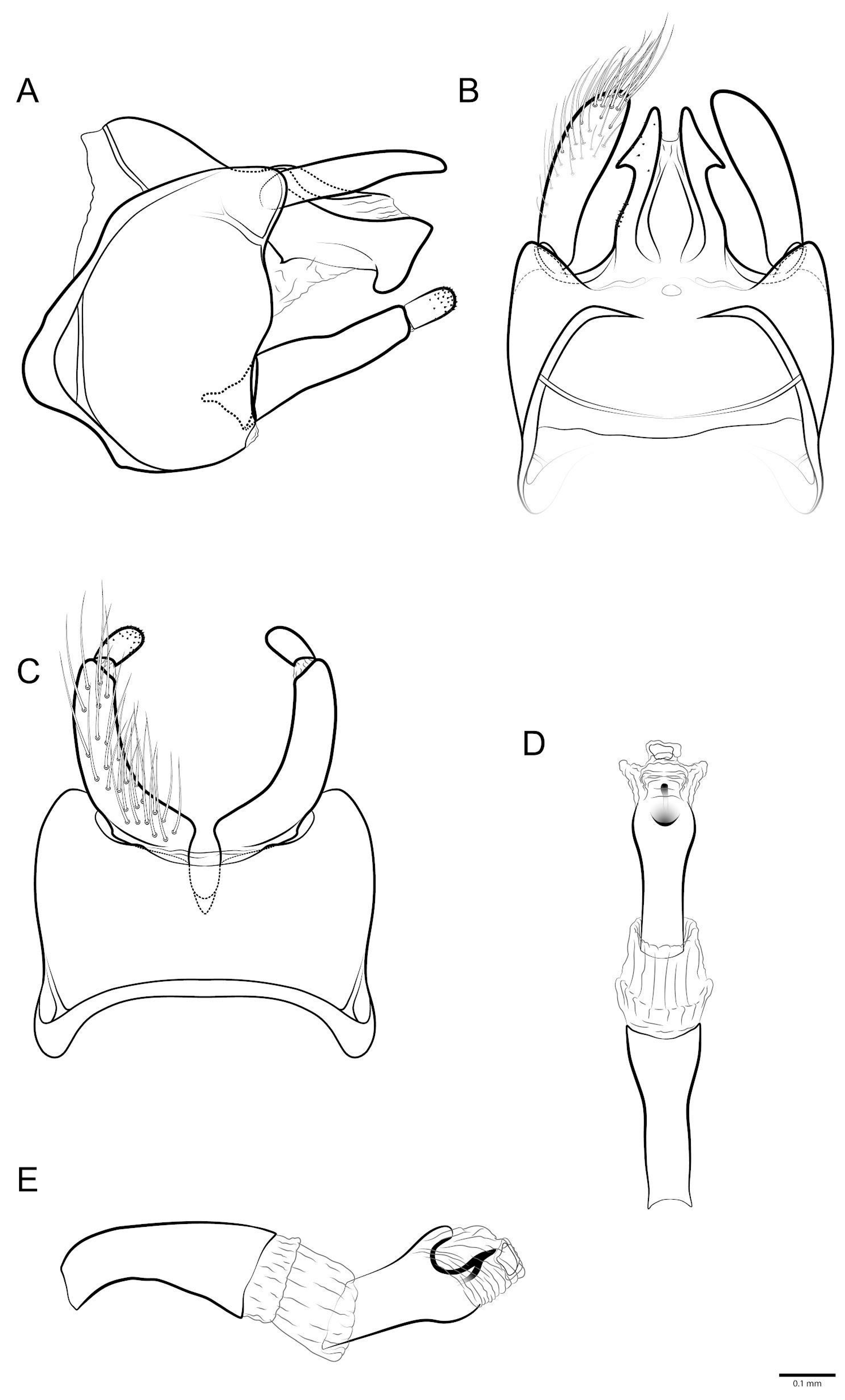
Diagnosis. The new species is most similar to M. misionensis Flint, 1983 [14] by the shape of segment X. The new species can be differentiated from this and other species mainly by (1) segment X in the dorsal view with a pronounced and acute subapical projection; (2) the anterior margin of segment IX in lateral with a strong subventral projection; (3) the sternum IX in the lateral view without midlateral sutures and with an anterodorsal transversal suture; and (4) the narrow preanal appendage in the lateral view.
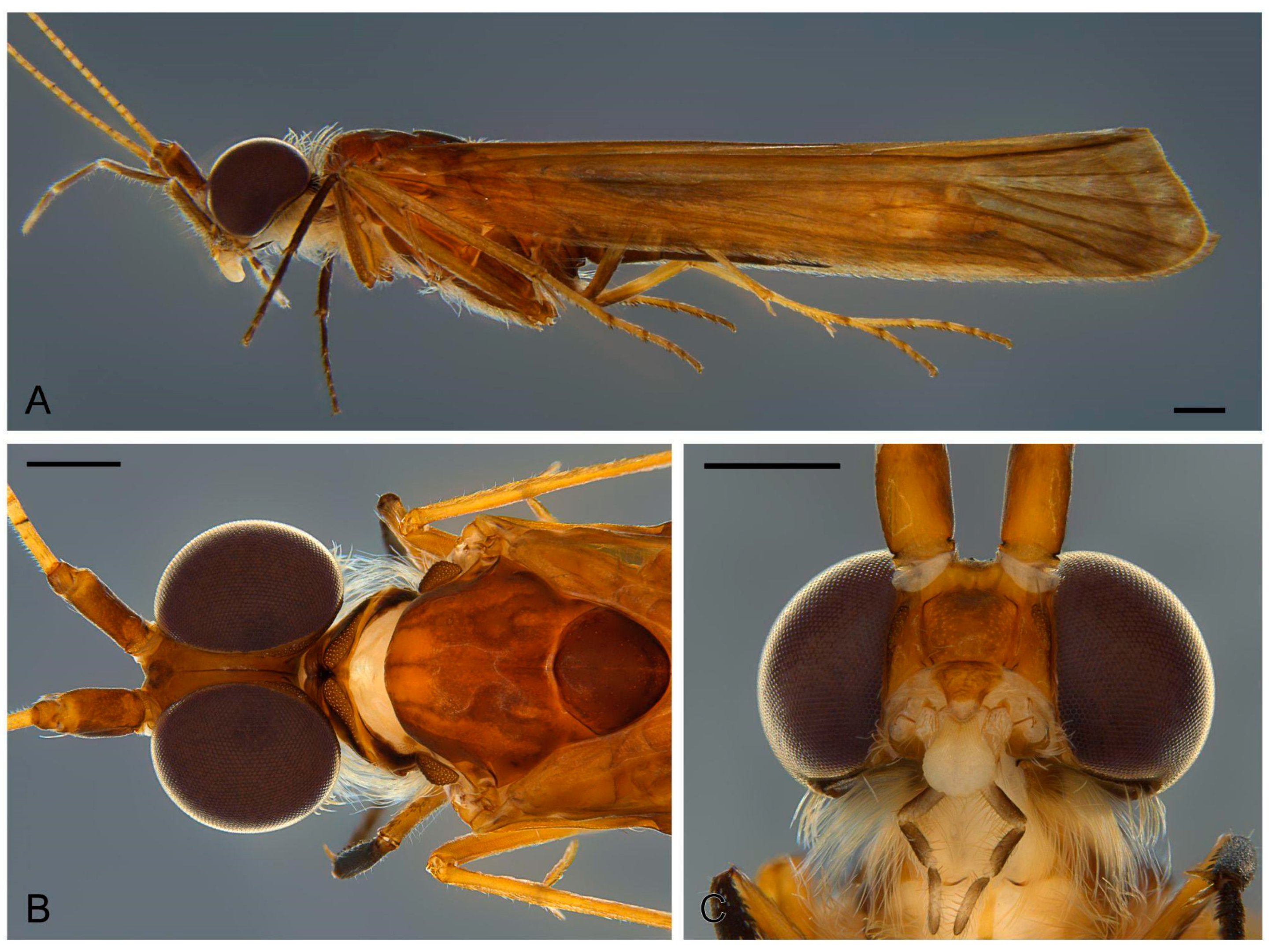
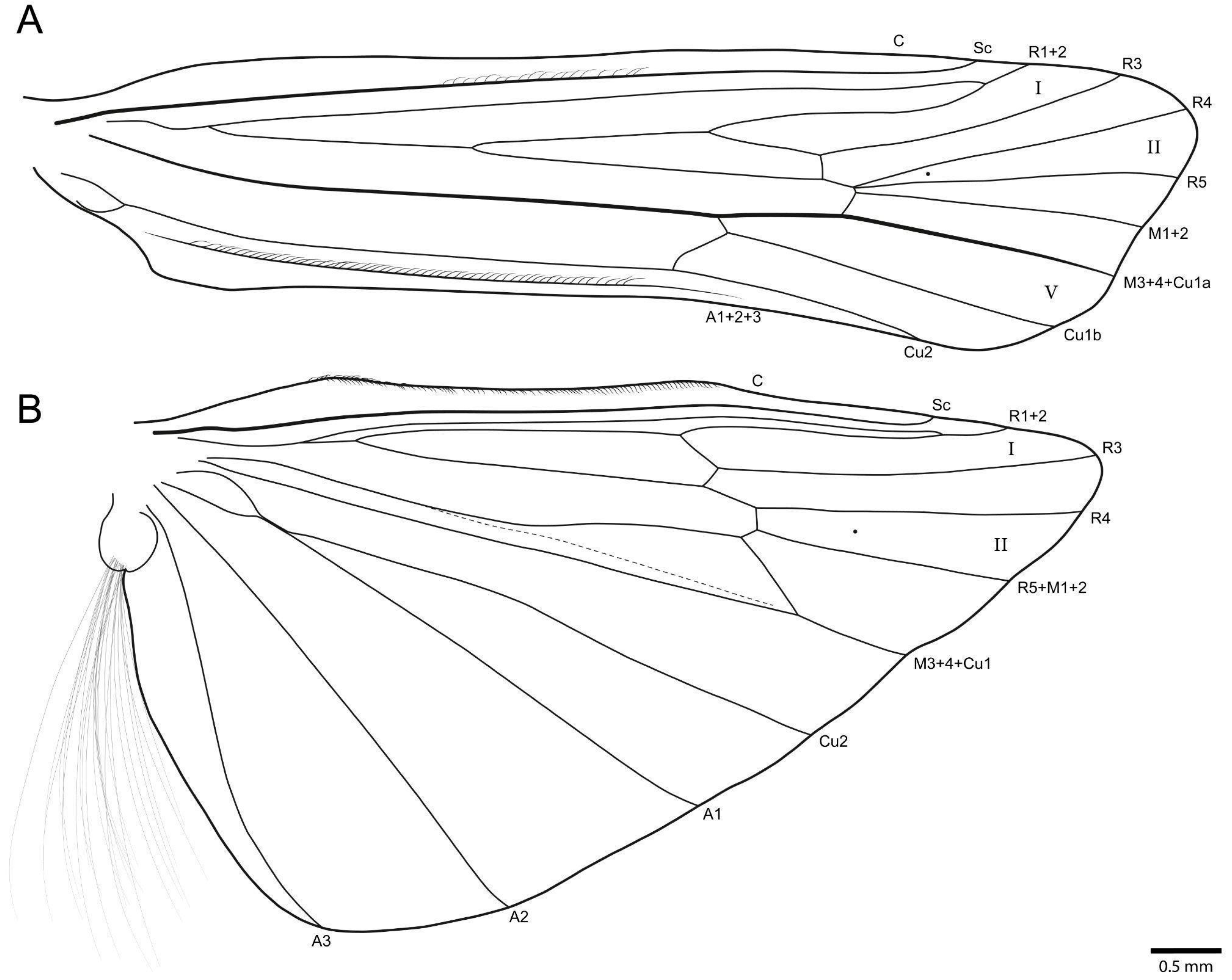
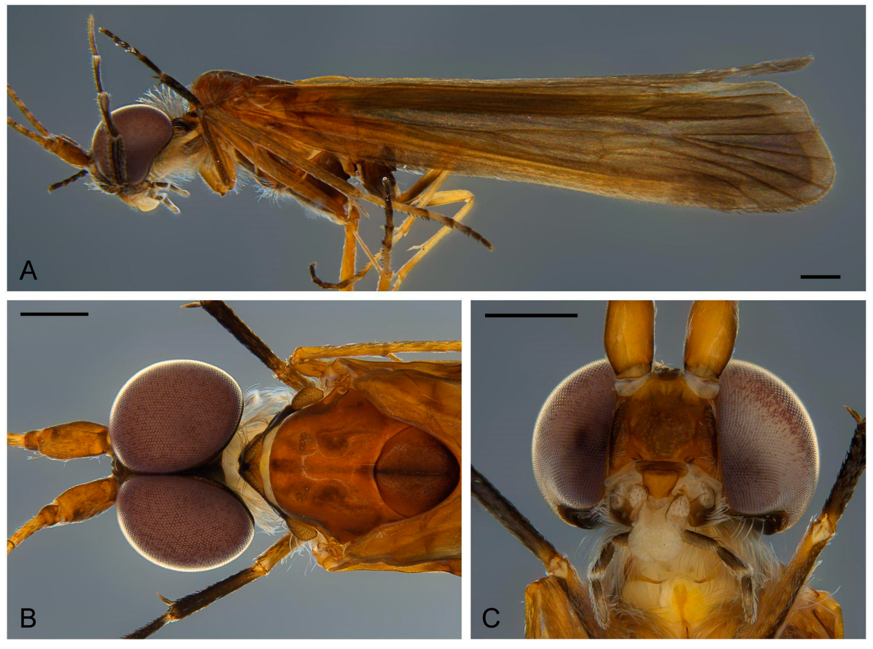
Description. Adult male. Forewing 7.9–8.1 mm (7.98 mm, n = 4). Color (in alcohol): Body and forewings brown in alcohol, foreleg tibia and tarsus dark brown, midleg tibia and tarsus brown, hindleg tibia and tarsus light brown (Figure 5A). Head: Eyes large, far from each other at vertex, intraocular distance as long as scapus width (Figure 5B). Antennae long, about 2.2 times body length, with narrow annuli; scapes wide, brown. Maxillary palps five-articulated, articles formula (I = IV = V) < II < III. Labial palpi three-articulated, articles subequal. Thorax: Pronotum narrow, with transversely elongate setal wart. Mesonotum wide, with pair of small setal warts; mesoscutellum almost circular, anterior margin rounded, without setal warts (Figure 5B). Tibial spur formula 2-4-2. Wings, forewing forks I, II, V present; hindwing fork I and II present (Figure 6). Abdomen: Simple, without differentiated structures.
Male genitalia (Figure 7A–E): Segment IX, in lateral view, anterior margin strongly projected subventrally, posterior margin slightly projected dorsally and mesally, with sutures separating each side of segment IX into four parts: one narrow anteroventral, one wide posterior partially divided posterodorsally and two anterodorsal parts (Figure 7A); in dorsal view, anterior margin nearly straight, membranous, with transversal suture and lateral oblique suture not meeting at middle, posterior margin concave, lateroapical corners conspicuous, deltoid (Figure 7B); in ventral view, segment IX with anterolateral deltoid projection delimited by suture, anterior margin concave, posterior margin concave (Figure 7C). Preanal appendage, in lateral view, long, about as long as segment X, slightly tapering apically, apex rounded (Figure 7A); in dorsal view, oblong, base narrower than apex (Figure 7B). Segment X, in lateral view, wider at base, narrowing subapically, apex truncate, with apicoventral projection, dorsal margin slightly sigmoid (Figure 7A); in dorsal view, wider at base, narrowing towards apex, sclerotized laterally, each side with subapical pointed deltoid projection, apex pointed, arrow-shaped, segment membranous mesally with deep, squared incision, with mesal sclerotized lanceolate region (Figure 7B). Inferior appendage bi-articulated: in lateral view, coxopodite cylindrical with base broader than apex, darkly sclerotized (Figure 7A); in ventral view, with basomesal lobe weakly produced mesad (Figure 7C); harpago short in lateral view, about 3.5× shorter than coxopodite, with small conical spines apically (Figure 7A); in ventral view, nearly straight (Figure 7C). Phallus tubular slightly curved near base in lateral view (Figure 7E), straight in ventral view (Figure 7D); endotheca membranous; in lateral view, phallotremal sclerite with C-shaped dorsal structure and S-shaped ventral structure (Figure 7E).
Etymology. The specific epithet, Krenak, is the name of the indigenous people who currently inhabit the banks of the Rio Doce, in the eastern region of Minas Gerais.
Distribution. Atlantic Forest (Espírito Santo and Minas Gerais state, Brazil).
Type material. Holotype male. Brazil, Minas Gerais: Rio Doce, Piranga river 20°19′45.20″ S, 42°53′55.00″ W, 372 m a.s.l., 10–11.viii.2022, Pennsylvania trap, Viana, Bonfá & Rodrigues col. (UFVB TR00283).
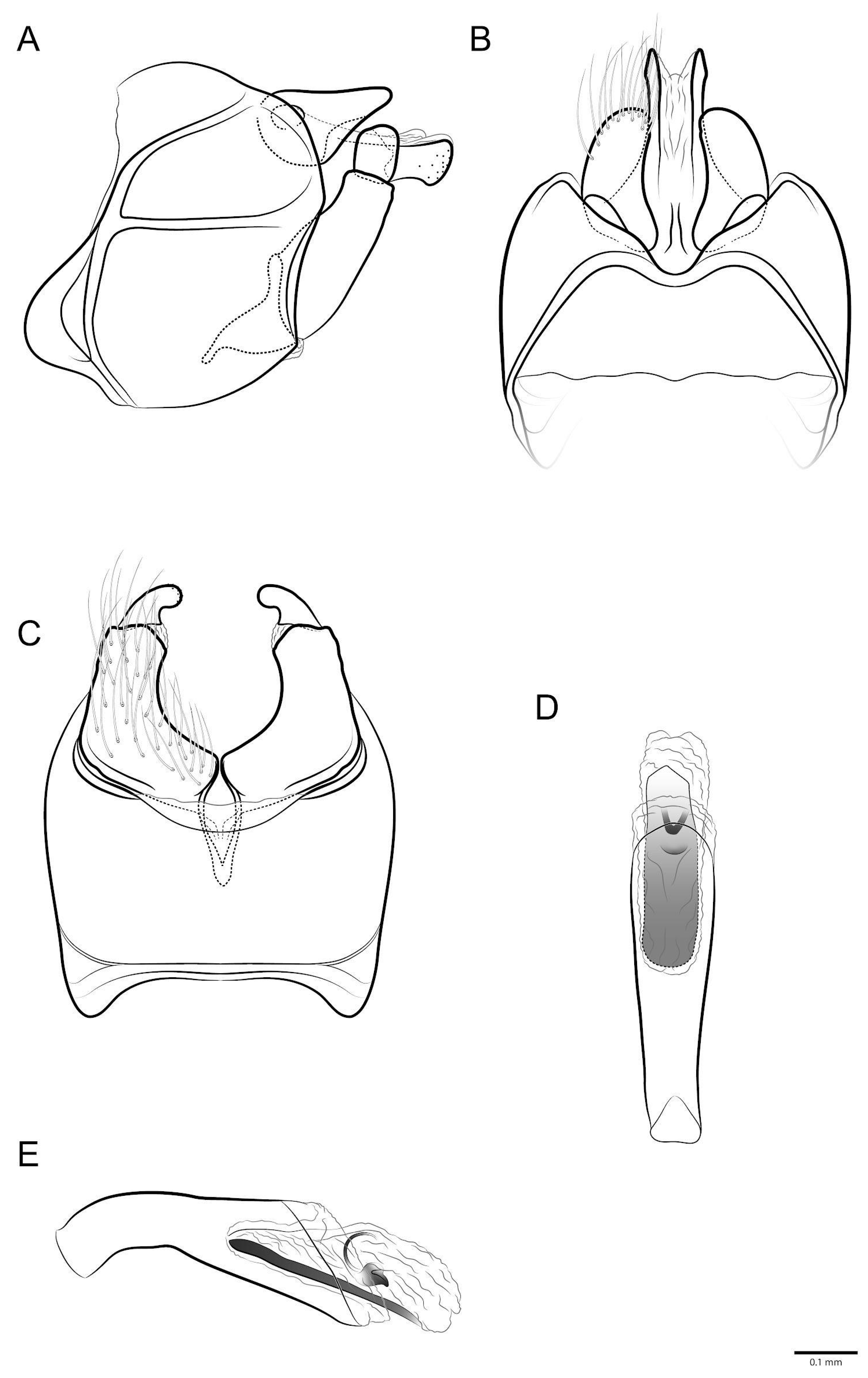
Diagnosis. The new species is similar to M. salta Flint, 1983 [14] by the straight segment X and the M-shaped dorsal suture. The new species can be differentiated from other congeners mainly by (1) the presence of a deep incision on the posterior margin of segment IX in the dorsal view; (2) the preanal kidney-shaped appendage, displaced mesally; and (3) the club-shaped segment X apex in the lateral view.
Description. Adult male. Forewing 7.6 mm (holotype). Color (in alcohol): Body brown, forewings grayish brown, foreleg tibia and tarsus dark brown, midleg tibia and tarsus brown, hindleg tibia and tarsus light brown (Figure 8A). Head: Eyes large, touching each other anterad at vertex (Figure 8B). Antennae long, about 2.1 times body length, with narrow annuli; scapes wide, brown. Maxillary palps five-articulated, articles formula (I = IV = V) < (II = III), articles subequal. Labial palpi three-articulated, articles subequal. Thorax: Pronotum narrow, with transversely elongate setal wart. Mesonotum wide, with pair of small setal warts; mesoscutellum almost circular, anterior margin rounded, without setal warts (Figure 8B). Tibial spur formula 2-4-4. Wings, forewing fork I, II, V present; hindwing fork I and II present (Figure 9). Abdomen: Simple, without differentiated structures.
Male genitalia (Figure 10A–E): Segment IX, in lateral view, anterior margin strongly projected subventrally, posterior margin slightly projected subdorsally, with sutures separating each side of segment IX into four parts: one narrow anteroventral, one wide posteroventral and two dorsal parts (Figure 10A); in dorsal view, anterior margin straight, membranous, posterior margin with M-shaped suture meeting wide, V-shaped mesal incision, lateroapical corners conspicuous, deltoid (Figure 10B); in ventral view, segment IX with anterolateral deltoid projection delimited by suture, anterior margin concave, posterior margin concave (Figure 10C). Preanal appendage, in lateral view, shorter than segment X, very wide basally, tapering apically, apex rounded (Figure 10A); in dorsal view, displaced mesad over segment X, base wider than apex, kidney-shaped (Figure 10B). Segment X, in lateral view, wider at base, narrowing at apex, ventral margin straight, apex rounded, club-shaped, dorsal margin nearly straight, with small membranous lobe subapically (Figure 10A); in dorsal view, wider at base, narrowing subapically, sclerotized laterally, each side mostly straight, slightly wider subapically, apex rounded, membranous mesally with V-shaped incision, base with mesal sclerotized longitudinal ridge (Figure 10B). Inferior appendage bi-articulated: in lateral view, coxopodite cylindrical with base broader than apex, in ventral view, with basomesal lobe forming strong angle (Figure 10C); harpago short, in lateral view, about 2.5× shorter than coxopodite, with small conical spines apically (Figure 10A); in ventral view, strongly curved mesad (Figure 10C). Phallus tubular slightly curved near base in lateral view (Figure 10E), straight in ventral view (Figure 10D); endotheca membranous; in lateral view, phallotremal sclerite with sclerotized rod, C-shaped dorsal structure and seed-shaped ventral structure (Figure 10E).
Etymology. The specific epithet, Maxakali, is the name of the indigenous people who currently inhabit four small areas in the northeastern region of Minas Gerais.
Distribution. Atlantic Forest (Minas Gerais state, Brazil).
Type material. Holotype male. Brazil, Minas Gerais, Araponga, Pousada Fazenda do Remanso, 20°39′28.00 S, 42°27′05.00 W, 1124 m a.s.l., 22–23.x.2022, Pennsylvania trap, Salles & ENT666 students col. (UFVB TR00284). Paratypes. Brazil, Minas Gerais, Araponga, same data as holotype 4♂ (UFVB TR00285). Same data except Pousada Fazenda do Remanso, 20°39′18.00″ S, 42°27′12.00″ W, 1062 m a.s.l., 15–16.ii.2023, Rippel, Freitas & Costa col. 1♂ (MNRJ). Same data except 20°39′19.90″ S, 42°27′11.50″ W, 1064 m a.s.l., 17–18.xi.2021. Bonfá, Gonçalves, Velásquez & Rezende col. 1♂ (UFVB TR00287).
Diagnosis. The new species is most similar to M. major Müller, 1880 [5] by the general shape of the genitalia and the phallus subapical constriction, and to M. huamantincoae Dumas & Nessimian, 2009 [13] by having a single lateral suture on segment IX. The new species can be differentiated from its congeners mainly by the combination of the following characteristics: (1) the presence of a single lateral suture on segment IX; (2) segment X with a shallow, narrow apical incision, and a lateral subapical angle; (3) the preanal appendage tapering to the acute apex in the lateral view; and (4) the phallus with a subapical constriction in the ventral view.
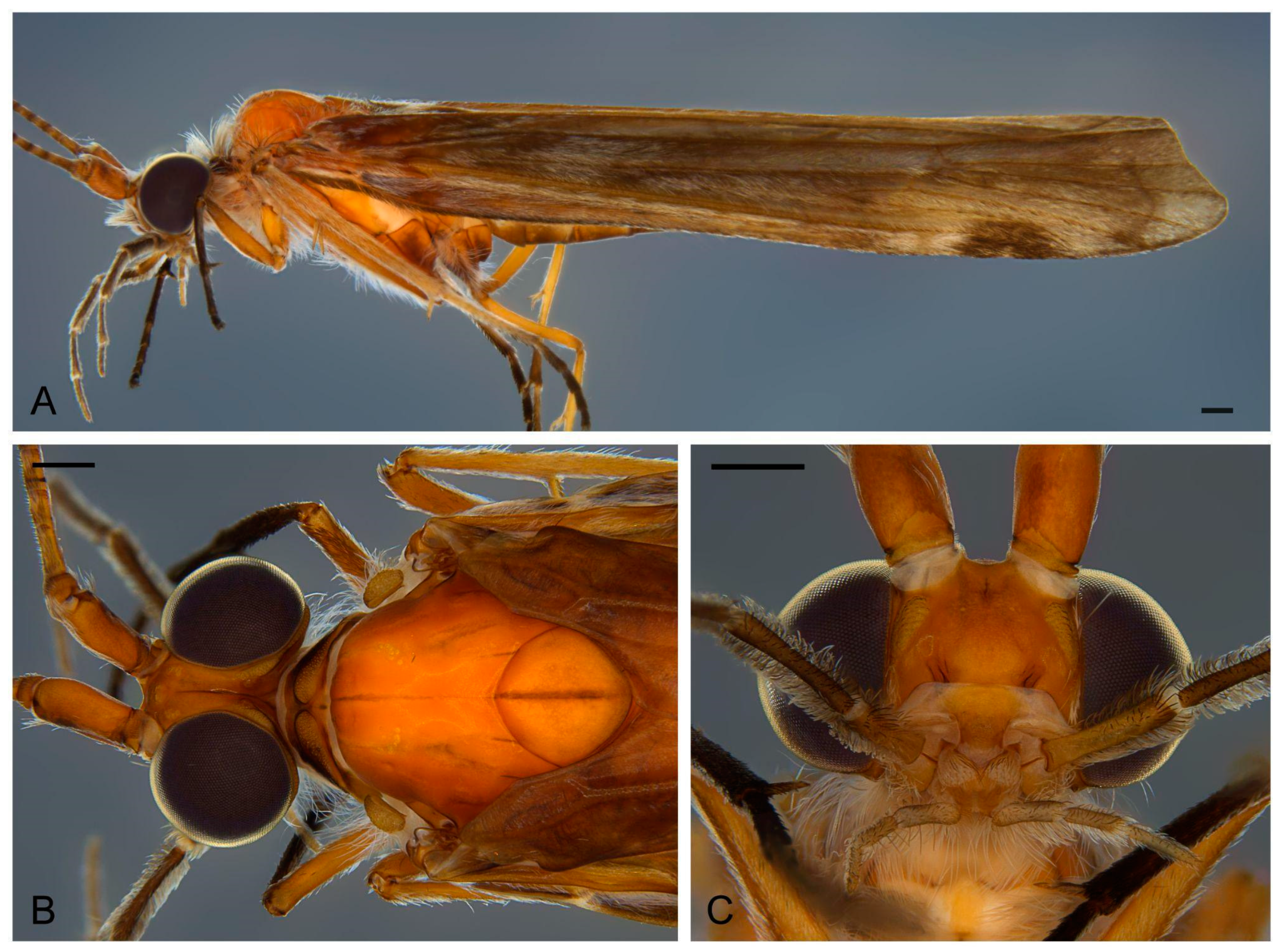
Description. Adult male. Forewing 15.1–15.8 mm (15.46 mm, n = 7). Color (in alcohol): Body brownish orange, forewings brown with whitish scattered pattern; foreleg tibia and tarsus dark brown, midleg tarsus dark brown, hindleg tarsus dark brown (Figure 11A). Head: Eyes large, far from each other at vertex, intraocular distance greater than scapus width (Figure 11B). Antennae long, about 2.9 times length of body, with narrow annuli; scapes wide, brown. Maxillary palps five-articulated, articles formula V < (I = IV) < (II = III). Labial palpi three-articulated, articles subequal (Figure 11C). Thorax: Pronotum narrow, with transversely elongate setal wart. Mesonotum wide, with pair of small setal warts; mesoscutellum almost circular, anterior margin rounded, without setal warts (Figure 11B). Tibial spur formula 2-4-4. Wings, forewing forks I, II, V present; hindwing fork I and II present. (Figure 12). Abdomen: Simple, without differentiated structures.
Male genitalia (Figure 13A–E): Segment IX, in lateral view, anterior margin nearly straight, posterior margin strongly projected dorsally and mesally; with midlateral suture separating each side of segment IX into two subequal parts, dorsal part partially divided (Figure 13A); in dorsal view, anterior margin straight, posterior margin concave, without sutures, lateroapical corner weakly produced (Figure 13B); in ventral view, anterolateral corner without projection, delimited by suture, anterior margin nearly straight, posterior margin concave. Preanal appendage, in lateral view, short, shorter than segment X, deltoid, tapering apically, apex acute (Figure 13A); in dorsal view, clavate, wider apically (Figure 13B). Segment X, in lateral view, wider at base, narrowing towards apex, apex rounded, without projections, dorsal margin nearly straight (Figure 13A); in dorsal view, wider at base, narrowing towards apex, sclerotized, each side with subapical angle, apex rounded, mesally sclerotized, apex with shallow V-shaped incision (Figure 13B). Inferior appendage bi-articulated: in lateral view, coxopodite cylindrical with base broader than apex, darkly sclerotized; in ventral view, without basomesal lobe (Figure 13C); harpago short in lateral view, about 5× shorter than coxopodite, with small conical spines apically (Figure 13A); in ventral view, nearly straight (Figure 13C). Phallus tubular slightly curved near base in lateral view (Figure 13E); straight in ventral view with subapical constriction (Figure 13D); endotheca membranous; in lateral view, phallotremal sclerite with rounded dorsal structure, C-shaped mesal structure and oblong ventral structure (Figure 13E).
Etymology. The specific epithet is the name of the Mukurin or Mokuriñ indigenous people, who currently inhabit a small area in the extreme north of the Rio Doce basin, northeast of Minas Gerais.
Distribution. Atlantic Forest (Minas Gerais state, Brazil).

3.2. New Records

  • Marilia guaira Flint, 1983
Material analyzed. Brazil, Minas Gerais, Naque, Rio Doce river, 19°15′05.56″ S, 42°18′50.50″ W, 190 m a.s.l., 08–09.i.2022, Pennsylvania trap, Viana, Bonfá, Rodrigues & Rothe-Neves col. 4♀, 2♂ (UFVB TR00288). Governador Valadares, Rio Doce river, 18°55′50.27″ S, 42°01′20.23″ W, 164 m a.s.l., 04–05.ix.2022, Pennsylvania trap, Viana, Bonfá & Ataide col. 1♀, 12♂ (UFVB TR00289).
Distribution. Atlantic Forest, Amazon rainforest and Cerrado savanna (Bolivia, Brazil (Goiás, Roraima, Maranhão and Minas Gerais state), Colombia, Paraguay, Venezuela).
Remarks. This is the first record of this species in the Minas Gerais state and the northernmost record of this species in the Atlantic Forest.

4. Discussion

The inferences we present here are exclusively related to the biology and sensitivity of Marilia and the reasoning of how they would interact with the mining tailings’ impacts. The impacts of the dam break are still being investigated and extensive and detailed monitoring efforts can be found in other studies, e.g., [32,33,34,35,36,37,38].
This is the first study to identify Odontoceridae at the species level in the Rio Doce basin. Studies on water quality using benthic macroinvertebrates were performed in the basin prior to the dam disaster but specimens were identified only at the family level, some of them reporting the occurrence of Odontoceridae [39,40,41,42,43].
Most of the species in the present study were collected within the Rio Doce basin, but in tributaries not directly affected by the iron ore tailings released from the dam break (Figure 14). Marilia guaira, Marilia aranan sp. nov. and Marilia krenak sp. nov. were collected in the directly impacted area, but nearly 7 years after the accident. Marilia krenak sp. nov. was also collected at two sites in the lower parts of the impacted area, but only before the dam collapse in 2015. Interestingly, collections were recently made near these same sites during the same season and no Marilia species were found. Although this episodic collection is not conclusive, it suggests that Marilia populations may still have not recovered at the Rio Doce’s lower reaches.
One of the main impacts of the dam break is the accumulation of fine sediments on the riverbed, which directly affects Marilia and other macroinvertebrates. Sediment discharge can cause drastic reductions in lotic invertebrate populations, with a small increase of 50–80 mg/L in suspended solids, causing a 60% reduction in macroinvertebrate fauna [44,45] and discharges greater than 1000 mg/L, causing reductions of over 90% in the fauna [46]. An important consequence of an increased deposition of fine sediments is the change in the substrate composition; the average particle size becomes smaller, the interstices between rocks and larger particles become filled and the stability of the sediment in the riverbed is reduced [47]. Most species of Trichoptera and other macroinvertebrates have specific requirements for a given substrate and tend to avoid inappropriate areas [48,49]. Therefore, the alteration of the substrate has a direct impact on habitat availability, with a strong correlation between the aquatic insect community and substrate composition [50,51,52,53]. Few taxa are found in unstable sediment deposits [46]. Additionally, many species inhabit the interstices between rocks and large particles, which are used as a refuge against a strong current and predators [48,54,55]. This refuge availability has a significant influence on community composition, so that when substrate interstices are filled with sediment deposition, the caddisflies and other macroinvertebrates become more vulnerable to physical disturbances in flood events [56].
Members of the family Odontoceridae are considered highly sensitive to environmental disturbances [57,58]. Marilia species prefer slow-moving river pools with sand and pebbles, where they may dig in the sandy substrate, which they also utilize to create their cases [7,8,9]. Therefore, they appear to rely strongly on the refuges of the hyporheic interstitial zone, so the fine tailing sediments seem to be inappropriate for their biology. Consequently, it is expected that the interstitial zone was recovered in the locations where the species were collected. Although, the impact of fine sediment covering the riverbed may still be noticeable in the lower parts of the river, where no Marilia species have been found in recent collections.
Recolonization studies in lotic environments show that animal groups rapidly reappear in affected areas, e.g., [59,60,61]; however, taxonomic diversity and population density may not be re-established for many years, depending on the severity and duration of stress and the availability of undamaged areas to serve as a source for recolonization [62]. Aquatic insect recolonization of completely degraded habitats primarily occurs via a stream drift and airborne dispersal, with a contribution of 41% of recolonization through a drift and 28% by air [63]. Thus, the preservation of small tributaries is critical for the recovery of larger streams, and 7 years after the disaster, we observed that highly sensitive species are re-establishing populations in some regions of the affected area.

Author Contributions

Conceptualization, P.B.N., A.V. and F.F.S.; methodology, P.B.N. and A.V.; software, P.B.N. and A.V.; validation, P.B.N., A.V. and F.F.S.; formal analysis, P.B.N.; investigation, P.B.N.; resources, F.F.S.; data curation, P.B.N.; writing—original draft preparation, P.B.N. and A.V.; writing—review and editing, P.B.N., A.V. and F.F.S.; visualization, P.B.N. and A.V.; supervision, F.F.S.; project administration, F.F.S.; funding acquisition, F.F.S. All authors have read and agreed to the published version of the manuscript.

Funding

This study was financed by Fundação de Amparo à Pesquisa do Estado de Minas Gerais (FAPEMIG)—Finance Code (proc. APQ-05461-18), and financed in part by the Coordenação de Aperfeiçoamento de Pessoal de Nível Superior—Brasil (CAPES)—Finance Code 001 (006/2022 PROPG—PDEE) to A.V. and Conselho Nacional de Desenvolvimento Científico (CNPq, proc. 309666/2019-8) to F.F.S.

Data Availability Statement

The data analyzed in this study is available through the voucher specimens deposited at the collections as indicated in type material and material analyzed sections.

Acknowledgments

We would like to thank all the members of the Museu de Entomologia (UFV) who contributed to the collections and field trips, especially those involved in the project to assess the impact of the Rio Doce basin, mainly the project coordinator Carlos Frankl Sperber and Ana Dária Leite Viana. Thanks also to Og DeSouza and the other members of the Laboratório de Termitologia (DEE—UFV) for the availability and use of the stereomicroscope with the attached camera.

Conflicts of Interest

The authors declare no conflict of interest. The funders had no role in the design of the study; in the collection, analyses or interpretation of data; in the writing of the manuscript; or in the decision to publish the results.

References

  1. Morse, J.C.; Frandsen, P.B.; Graf, W.; Thomas, J.A. Diversity and ecosystem services of Trichoptera. Insects 2019, 10, 125. [Google Scholar] [CrossRef] [Green Version]
  2. Morse, J.C. Trichoptera World Checklist. 2023. Available online: https://trichopt.app.clemson.edu/search.php (accessed on 15 May 2023).
  3. Huamantinco, A.A.; Nessimian, J.L. A new Neotropical genus and species of Odontocerinae (Trichoptera: Odontoceridae) from southeastern Brazil. Aquat. Insects 2004, 26, 281–288. [Google Scholar] [CrossRef]
  4. Burmeister, H. Handbuch der Entomologie, Zweiter Band, Zweite Ubtheilung. Theod. Chr. Friedr. Enslin Berl. 1839, 12, 397–1050. [Google Scholar] [CrossRef]
  5. Müller, F. Sôbre as casas construídas pelas larvas de insectos Trichopteros da Província de Santa Catharina. Arch. Mus. Nac. Rio De Jan. 1880, 3, 99–134. [Google Scholar]
  6. Calor, A.R.; Santos, A.P.M. Trichoptera in: Catálogo Taxonômico da Fauna do Brazil. PNUD. 2023. Available online: http://fauna.jbrj.gov.br/ (accessed on 15 May 2023).
  7. Wiggins, G.B. Larvae of the North American Caddisfly Genera (trichoptera); University of Toronto Press: Toronto, ON, Canada, 1996; 400p. [Google Scholar] [CrossRef]
  8. Holzenthal, R.W.; Blahnik, R.J.; Prather, A.P.; Kjer, K.M. Order Trichoptera Kirby, 1813 (Insecta), Caddisflies. Zootaxa 2007, 1668, 639–698. [Google Scholar] [CrossRef]
  9. Camargos, L.M.; Pes, A.M.; Hamada, N. New Neotropical species of Marilia Müller (Trichoptera: Odontoceridae). Zootaxa 2020, 4853, 1–41. [Google Scholar] [CrossRef] [PubMed]
  10. Schmid, F. The Insects and Arachnids of Canada, Part 7. Genera of the Trichoptera of Canada and Adjoining or Adjacent United States; NRC Research Press: Ottawa, ON, Canada, 1998; 319 pp. [Google Scholar]
  11. Pes, A.M.; Holzenthal, R.W.; Sganga, J.V.; Santos, A.P.M.; Barcelos-Silva, P.; Camargos, L.M. Order Trichoptera. In Keys to Neotropical Hexapoda. Vol III. Thorp and Covich’s Freshwater Invertebrates, 4th ed.; Hamada, N., Thorp., J.H., Rogers, D.C., Eds.; Academic Press: Boston, MA, USA, 2018; pp. 237–323. [Google Scholar] [CrossRef]
  12. Pes, A.M.; Santos, A.P.M.; Barcelos-Silva, P.; Camargos, L.M. Ordem Trichoptera, In Insetos Aquáticos na Amazônia Brasileira: Taxonomia, Biologia e Ecologia; Hamada, N., Nessimian, J., Querino, R.B., Eds.; Editora do Instituto Nacional de Pesquisas da Amazônia: Manaus, Brazil, 2019; pp. 387–429.
  13. Dumas, L.L.; Nessimian, J.L. Description of two new species of Marilia Müller (Trichoptera, Odontoceridae) from southeastern Brazil, including the description of the female of Marilia major Müller. Rev. Bras. Entomol. 2009, 53, 344–348. [Google Scholar] [CrossRef] [Green Version]
  14. Flint, O.S., Jr. Studies of Neotropical Caddisflies, XXXIII: New species from austral South America (Trichoptera). Smithson. Contrib. Zool. 1983, 377, 1–100. [Google Scholar] [CrossRef]
  15. Costa, A.M. Revisão taxonômica e filogenia das espécies de Marilia Müller, 1880 (Trichoptera: Odontoceridae) da Região Neotopical. Ph.D. Thesis, Universidade de São Paulo, Ribeirão Preto, Brazil, 2016; 157p. [Google Scholar]
  16. Oláh, J.; Johanson, K.A. Description of 33 new species of Calamoceratidae, Molannidae, Odontoceridae and Philorheithridae (Trichoptera), with detailed presentation of their cephalic setal warts and grooves. Zootaxa 2010, 2457, 1–128. [Google Scholar] [CrossRef]
  17. Yang, L.F.; Yuan, H.Y.; Morse, J.C. Lannapsyche and Marilia species of China (Trichoptera: Odontoceridae). Zootaxa 2017, 4320, 81–99. [Google Scholar] [CrossRef]
  18. Flint, O.S., Jr. Studies of Neotropical caddisflies, XLV: The taxonomy, phenology, and faunistics of Trichoptera of Antioquia, Colombia. Smithson. Contrib. Zool. 1991, 520, 1–113. [Google Scholar] [CrossRef]
  19. Bueno-Soria, J.; Rojas-Ascencio, A. New species and distribution of the genus Marilia Muller (Trichoptera; Odontoceridae) in Mexico and Central America. Proc. Entomol. Soc. Wash. 2004, 106, 679–696. [Google Scholar]
  20. Coelho, A.L.N. Bacia hidrográfica do Rio Doce (MG/ES): Uma análise socioambiental integrada. Rev. Geogr. 2009, 7, 131–146. [Google Scholar] [CrossRef] [Green Version]
  21. Segura, F.R.; Nunes, E.A.; Panis, F.P.; Paulelli, A.C.C.; Rodrigues, G.B.; Braga, G.U.L.; Filho, W.R.P.; Barbosa, F., Jr.; Cerchiaro, G.; Silva, F.F.; et al. Potential risks of the residue from Samarco’s mine dam burst (Bento Rodrigues, Brazil). Environ. Pollut. 2016, 218, 813–825. [Google Scholar] [CrossRef] [PubMed]
  22. Carmo, F.F.; Kamino, L.H.Y.; Junior, R.T.; Campos, I.C.; Carmo, F.F.; Silvino, G.; Castro, K.J.S.X.; Mauro, M.L.; Rodrigues, N.U.A.; Miranda, M.P.S.; et al. Fundão tailings dam failures: The environment tragedy of the largest technological disaster of Brazilian mining in global context. Perspect. Ecol. Conserv. 2017, 15, 145–151. [Google Scholar] [CrossRef]
  23. Queiroz, H.M.; Nobrega, G.N.; Ferreira, T.O.; Almeida, L.S.; Romero, T.B.; Santaella, S.T.; Bernardino, A.F.; Otero, X.L. The Samarco mine tailing disaster: A possible time-bomb for heavy metals contamination? Sci. Total Environ. 2018, 637–638, 498–506. [Google Scholar] [CrossRef]
  24. Rudorff, N.; Rudorff, C.M.; Kampel, M.; Ortiz, G. Remote sensing monitoring of the impact of a major mining wastewater disaster on the turbidity of the Doce River plume off the eastern Brazilian coast. ISPRS J. Photogramm. Remote Sens. 2018, 145, 349–361. [Google Scholar] [CrossRef]
  25. CEPTA [Centro Nacional de Pesquisa e Conservação da Biodiversidade Aquática Continental] Nota técnica Nº. 24, de 24 de Novembro de 2015. Consequências Parciais na Biodiversidade Aquática da bacia do rio doce, Provocadas pelo Rompimento da Barragem de Rejeitos de Mineração da Samarco Mineradora S.A. no município de Mariana, MG. Available online: https://www.icmbio.gov.br/portal/publicacoes?id=7862:documentos-rio-doce (accessed on 18 April 2022).
  26. Lopes, L.M.N. O rompimento da barragem de Mariana e seus impactos socioambientais. Sinapse Múltipla 2016, 5, 1. [Google Scholar]
  27. Barbour, M.T.; Gerritsen, J.; Snyder, B.D.; Stribling, J.B. Rapid Bioassessment Protocols for Use in Streams and Wadeable Rivers: Periphyton, Benthic Macroinvertebrates and Fish, 2nd ed.; EPA 841-B-99-002; U.S. Environmental Protection Agency, Office of Water: Washington, DC, USA, 1999. Available online: https://www3.epa.gov/region1/npdes/merrimackstation/pdfs/ar/AR-1164.pdf (accessed on 31 January 2022).
  28. Frost, S.W. Light traps for insect collection, survey and control. Bull. Pa. State Coll. Agric. Exp. Stn. 1952, 550, 1–32. [Google Scholar]
  29. Rocha, I.C.; Souza, W.R.M. Marilia caramuru sp. nov. and Marilia paraguassu sp. nov. (Trichoptera: Odontoceridae), two new species from Maranhão State, Northeast Brazil. Zootaxa 2018, 4425, 393–400. [Google Scholar] [CrossRef]
  30. Blahnik, R.J.; Holzenthal, R.W. Collection and curation of Trichoptera, with an emphasis on pinned material. Nectopsyche Neotrop. Trichoptera Newsl. 2004, 1, 8–20. [Google Scholar]
  31. Mosely, M.E.; Kimmins, D.E. The trichoptera (Caddis-Flies) of Australia and New Zealand; British Museum (Natural History): London, UK, 1953; 550p. [Google Scholar] [CrossRef]
  32. Cordeiro, M.C.; Garcia, G.D.; Rocha, A.M.; Tschoeke, D.A.; Campeão, M.E.; Appolinario, L.R.; Soares, A.C.; Leomil, L.; Froes, A.; Bahiense, L.; et al. Insights on the freshwater microbiomes metabolic changes associated with the world’s largest mining disaster. Sci. Total Environ. 2019, 654, 1209–1217. [Google Scholar] [CrossRef] [PubMed]
  33. Duarte, E.B.; Neves, M.A.; de Oliveira, F.B.; Martins, M.E.; de Oliveira, C.H.R.; Burak, D.L.; Orlando, M.T.D.; Rangel, C.V.G.T. Trace metals in Rio Doce sediments before and after the collapse of the Fundão iron ore tailing dam, Southeastern Brazil. Chemosphere 2021, 262, 127879. [Google Scholar] [CrossRef] [PubMed]
  34. Gabriel, F.Â.; Ferreira, A.D.; Queiroz, H.M.; Vasconcelos, A.L.S.; Ferreira, T.O.; Bernardino, A.F. Long-term contamination of the Rio Doce estuary as a result of Brazil’s largest environmental disaster. Perspect. Ecol. Conserv. 2021, 19, 417–428. [Google Scholar] [CrossRef]
  35. Gomes, L.E.O.; Correa, L.B.; Sá, F.; Neto, R.R.; Bernardino, A.F. The impacts of the Samarco mine tailing spill on the Rio Doce estuary, Eastern Brazil. Mar. Pollut. Bull. 2017, 120, 28–36. [Google Scholar] [CrossRef]
  36. Vergilio, C.D.S.; Lacerda, D.; Souza, T.D.S.; de Oliveira, B.C.V.; Fioresi, V.S.; de Souza, V.V.; Rodrigues, G.D.R.; Barbosa, M.K.D.A.M.; Sartori, E.; Rangel, T.P.; et al. Immediate and long-term impacts of one of the worst mining tailing dam failure worldwide (Bento Rodrigues, Minas Gerais, Brazil). Sci. Total Environ. 2021, 756, 143697. [Google Scholar] [CrossRef]
  37. Wild, M.; Rouhani, S.; Oehrig, J.; Alves, P.H.G.; Odle, W.; Gaspar, D.F.A. Using Spatiotemporal Ratio Analyses to Quantitatively Estimate Water Quality Recovery of the Rio Doce. Integr. Environ. Assess. Manag. 2023. [Google Scholar] [CrossRef]
  38. Yamamoto, F.; Onishi, K.; Ralha, T.; Silva, L.; Deda, B.; Pessali, T.; Souza, C.; Ribeiro, C.O.; Abessa, D. Earlier biomarkers in fish evidencing stress responses to metal and organic pollution along the Doce River Basin. Environ. Pollut. 2023, 329, 121720. [Google Scholar] [CrossRef]
  39. Aguila, G.E.N.; Pujoni, D.G.F.; Marques, M.M.; Santos, L.G.C.; Dornelas, N.M.d.L.; Andrade, K.; Monteiro, I.M.; Maia-Barbosa, P.M.; Barbosa, F.A.R. Benthic macroinvertebrate diversity in the Middle Doce River Basin, Brazil. Data 2018, 3, 17. [Google Scholar] [CrossRef] [Green Version]
  40. Cota, L.; Goulart, M.; Moreno, P.; Callisto, M. Rapid assessment of river water quality using an adapted BMWP index: A practical tool to evaluate ecosystem health. Int. Ver. Für Theor. Und Angew. Limnol. Verhandlungen 2002, 28, 1713–1716. [Google Scholar] [CrossRef]
  41. Marques, M.G.S.M.; Ferreira, R.L.; Barbosa, F.A.R. A comunidade de macroinvertebrados aquáticos e características limnológicas das lagoas Carioca e da Barra, Parque Estadual do Rio Doce, MG. Rev. Bras. Biol. 1999, 59, 203–210. [Google Scholar] [CrossRef] [Green Version]
  42. Marques, M.M.; Barbosa, F. Biological quality of waters from an impacted tropical watershed (middle Rio Doce basin, southeast Brazil), using benthic macroinvertebrate communities as an indicator. Hydrobiologia 2021, 457, 69–76. [Google Scholar] [CrossRef]
  43. Moretti, M.S.; Callisto, M. Biomonitoring of benthic macroinvertebrates in the middle Doce River watershed. Acta Limnol. Bras. 2005, 17, 267–281. [Google Scholar]
  44. Gammon, J.R. The Effect of Inorganic Sediment on Stream Biota; U.S. Environmental Protection Agency, Water Quality Office 18050 DWC: Washington, DC, USA, 1970; 108–113p. [Google Scholar]
  45. White, D.S. The effect of inorganic limestone sediment on the macroinvertebrates of Deer Creek. Master’s Thesis, DePauw University, Greencastle, IN, USA, 1970; 135p. [Google Scholar]
  46. Herbert, D.W.; Alabaster, J.S.; Dart, M.C.; Lloyd, R. The effect of chinaclay wastes on trout streams. Int. J. Air Watsr Poll. 1961, 5, 56–74. [Google Scholar]
  47. Kaufmann, P.R.; Larsen, D.P.; Faustini, J.M. Bed stability and sedimentation associated with human disturbances in Pacific Northwest streams. J. Am. Water Resour. Assoc. 2009, 45, 434–459. [Google Scholar]
  48. Williams, D.D.; Smith, M.R. Colonization dynamics of river benthos in response to local changes in bed characteristics. Freshw. Biol. 1996, 36, 237–248. [Google Scholar]
  49. Sarriquet, P.E.; Bordenave, P.; Marmonier, P. Effects of bottom sediment restoration on interstitial habitat characteristics and benthic macro-invertebrate assemblages in a headwater stream. River Res. Appl. 2007, 23, 815–828. [Google Scholar] [CrossRef]
  50. Collier, K.J.; Wilcock, R.J.; Meredith, A.S. Influence of substrate type and physico-chemical conditions on macroinvertebrate faunas and biotic indices of some lowland Waikato, New Zealand, streams. N. Z. J. Mar. Freshw. Res. 1998, 32, 1–19. [Google Scholar]
  51. Angradi, T.R. Fine sediment and macroinvertebrate assemblages in Appalachian streams: A field experiment with biomonitoring applications. J. N. Am. Benthol. Soc. 1999, 18, 49–66. [Google Scholar] [CrossRef]
  52. Armitage, P.D.; Cannan, C.E. Annual changes in summer patterns of mesohabitat distribution and associated macroinvertebrate assemblages. Hydrol. Process. 2000, 14, 3161–3179. [Google Scholar] [CrossRef]
  53. Buffagni, A.; Crosa, G.A.; Harper, D.M.; Kemp, J.L. Using macroinvertebrate species assemblages to identify river channel habitat units: An application of the functional habitats concept to a large, unpolluted Italian river (River Ticino, northern Italy). Hydrobiologia 2000, 435, 213–225. [Google Scholar]
  54. Lancaster, J. Scaling the effects of predation and disturbance in a patchy environment. Oecologia 1996, 107, 321–331. [Google Scholar] [PubMed]
  55. Swan, C.M.; Palmer, M.A. What drives small—Scale spatial patterns in lotic meiofauna communities? Freshw. Biol. 2000, 44, 109–121. [Google Scholar]
  56. Gjerløv, C.; Hildrew, A.G.; Jones, J.I. Mobility of stream invertebrates in relation to disturbance and refugia: A test of habitat templet theory. J. N. Am. Benthol. Soc. 2003, 22, 207–223. [Google Scholar] [CrossRef]
  57. Armitage, P.D.; Moss, D.; Wright, J.F.; Furse, M.T. The performance of a new biological water quality score system based on macroinvertebrates over a wide range of unpolluted running-water sites. Water Res. 1983, 17, 333–347. [Google Scholar]
  58. Junqueira, M.V.; Amarante, M.; Dias, C.; França, E. Biomonitoramento de qualidade das águas da Bacia do Rio das Velhas (MG/Brasil) através de macroinvertebrados. Acta Limnol. Bras. 2000, 12, 7387. [Google Scholar]
  59. Muller, K. Investigations on the organic drift in north swedish streams. Rep. Inst. Freshwat. Res. Drottningholm 1954, 35, 133–148. [Google Scholar]
  60. Waters, T.F. Interpretation of invertebrate drift in streams. Ecology 1965, 46, 327–334. [Google Scholar] [CrossRef]
  61. Gray, L.J.; Ward, J.V. Effects of sediment releases from a reservoir on stream macroinvertebrates. Hydrobiologia 1982, 96, 177–184. [Google Scholar]
  62. Cairns, J.J.; Cıtossman, J.S.; Dıckson, K.L.; Herricks, E.E. The recovery of damaged Streams. Ass. South-East. Biol. Bull. 1971, 18, 79–106. [Google Scholar]
  63. Williams, D.D.; Hynes, H.B.N. The recolonization mechanisms of stream benthos. Oikos 1976, 27, 265–272. [Google Scholar] [CrossRef]
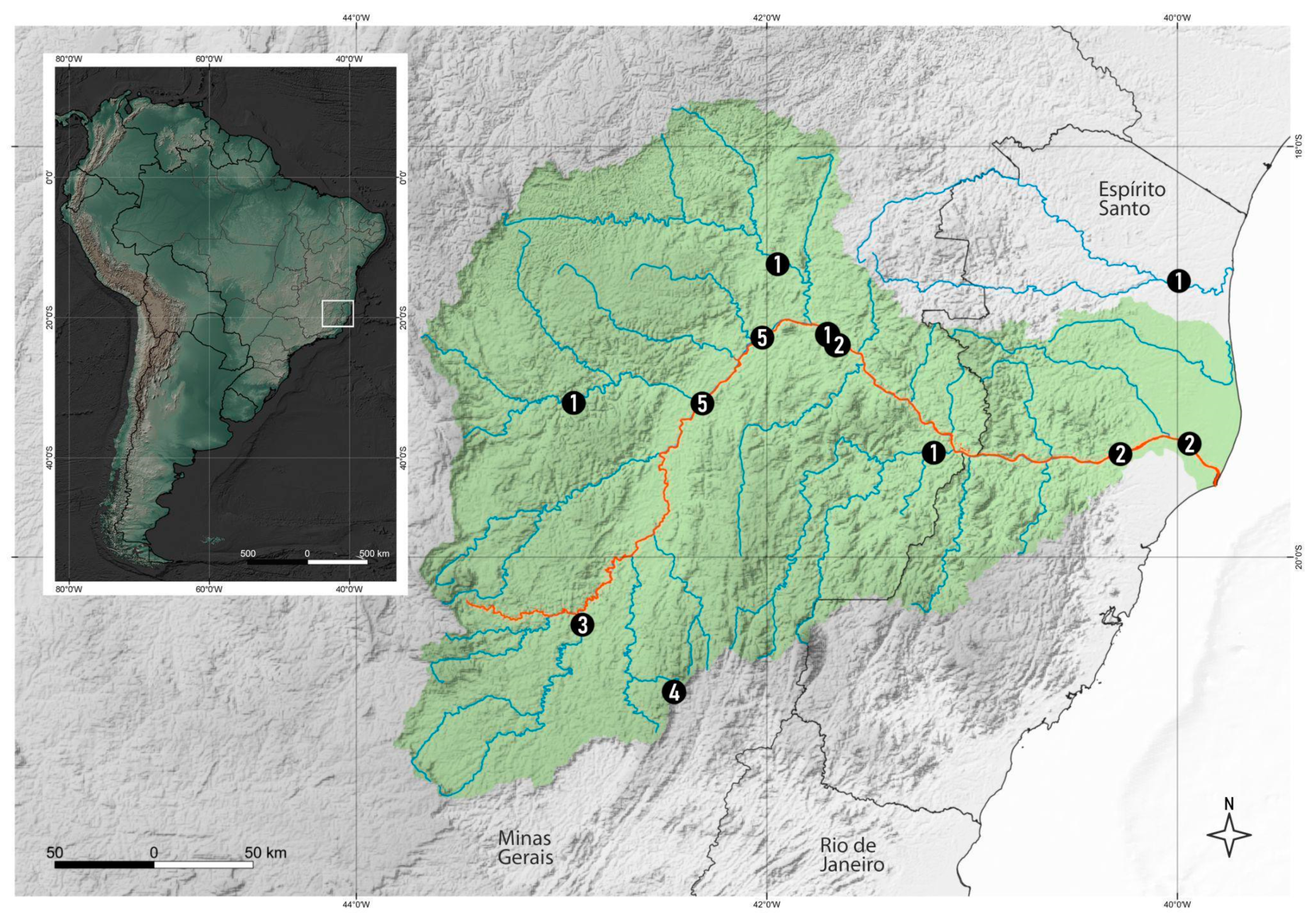
Figure 1. Collection sites of analyzed specimens. Showing Rio Doce basin in green, mud-affected area in red, main hydrography in blue. Circles with numbers refer to different species: 1—Marilia aranan sp. nov.; 2—M. krenak sp. nov.; 3—M. maxakali sp. nov.; 4—M. mukurin sp. nov.; 5—M. guaira Flint, 1983.
Taxonomy 03 00022 g001
Figure 2. Marilia aranan sp. nov.: (A) Habitus, lateral; (B) Head and thorax, dorsal; (C) Head, ventral. Scale bar: 0.5 mm.
Taxonomy 03 00022 g002
Figure 3. Marilia aranan sp. nov.: Wing venation. (A) Forewing; (B) Hindwing.
Taxonomy 03 00022 g003
Figure 4. Marilia aranan sp. nov., male genitalia: (A) Lateral; (B) Dorsal; (C) Ventral; (D) Phallus ventral; (E) Phallus lateral.
Taxonomy 03 00022 g004
Figure 5. Marilia krenak sp. nov.: (A) Habitus, lateral; (B) Head and thorax, dorsal; (C) Head, ventral. Scale bar: 0.5 mm.
Taxonomy 03 00022 g005
Figure 6. Marilia krenak sp. nov.: Wing venation. (A) Forewing; (B) Hindwing.
Taxonomy 03 00022 g006
Figure 7. Marilia krenak sp. nov., male genitalia: (A) Lateral; (B) Dorsal; (C) Ventral; (D) Phallus ventral; (E) Phallus lateral.
Taxonomy 03 00022 g007
Figure 8. Marilia maxakali sp. nov.: (A) Habitus, lateral; (B) Head and thorax, dorsal; (C) Head, ventral. Scale bar: 0.5 mm.
Taxonomy 03 00022 g008
Figure 9. Marilia maxakali sp. nov.: Wing venation. (A) Forewing; (B) Hindwing.
Taxonomy 03 00022 g009
Figure 10. Marilia maxakali sp. nov., male genitalia: (A) Lateral; (B) Dorsal; (C) Ventral; (D) Phallus ventral; (E) Phallus lateral.
Taxonomy 03 00022 g010
Figure 11. Marilia mukurin sp. nov.: (A) Habitus, lateral; (B) Head and thorax, dorsal; (C) Head, ventral. Scale bar: 0.5 mm.
Taxonomy 03 00022 g011
Figure 12. Marilia mukurin sp. nov.: Wing venation. (A) Forewing; (B) Hindwing.
Taxonomy 03 00022 g012
Figure 13. Marilia mukurin sp. nov., male genitalia: (A) Lateral; (B) Dorsal; (C) Ventral; (D) Phallus ventral; (E) Phallus lateral.
Taxonomy 03 00022 g013
Figure 14. Collection sites at the tributaries in Minas Gerais: (A,B) Rio Doce, Piranga river; (C,D) Ferros, Santo Antônio river; (E,F) Matias Lobato, Suaçuí Grande river; (G,H) Aimorés, Manhuaçu river; (I,J) Araponga, Pousada Fazenda do Remanso.
Taxonomy 03 00022 g014
Disclaimer/Publisher’s Note: The statements, opinions and data contained in all publications are solely those of the individual author(s) and contributor(s) and not of MDPI and/or the editor(s). MDPI and/or the editor(s) disclaim responsibility for any injury to people or property resulting from any ideas, methods, instructions or products referred to in the content.