2. Materials and Methods
Repository institution. The holotype and two paratypes were deposited in the Coleção Entomológica do Laboratório de Coleoptera, at the Universidade Federal de Viçosa (CELC, collection abbreviation follows Evenhuis [
17]), Minas Gerais, Brazil. Specimens from other species of
Strumigenys which were studied comparatively (including the specimens of all male morphospecies) were also deposited in this collection, some of them loaned from the following Brazilian institutions (all databased at Antweb.org with their institution of origin specified): MZSP, Museu de Zoologia da Universidade de São Paulo; MPEG, museu Paraense Emílio Goeldi; and INPA, Instituto Nacional de Pesquisas da Amazonia.
Morphological data set. The terminology used in the descriptions follows Bolton [
9], except for the terms “alitrunk” (mesosoma used instead), anepisternum and katepisternum (upper and lower mesopleura used instead [
18]). I used “basigastral ventral setae” instead of “basigastral ventral pad” (upon verifying that the structure, when present, was composed of highly modified setae in all examined specimens (+100 spp.) of a study, Chaul et al., in prep.). Small pale patches on the body were treated simply as glands throughout the description [
19,
20,
21]. I used “apicoventral scape gland” rather than “ventral scape gland”. The mesothoracic “hairwheels”, considered glands in some studies [
1], were treated as mesopleural excavations [
22].
I followed Brassard et al. [
23] for body measurements and their abbreviations, except for the following additions:
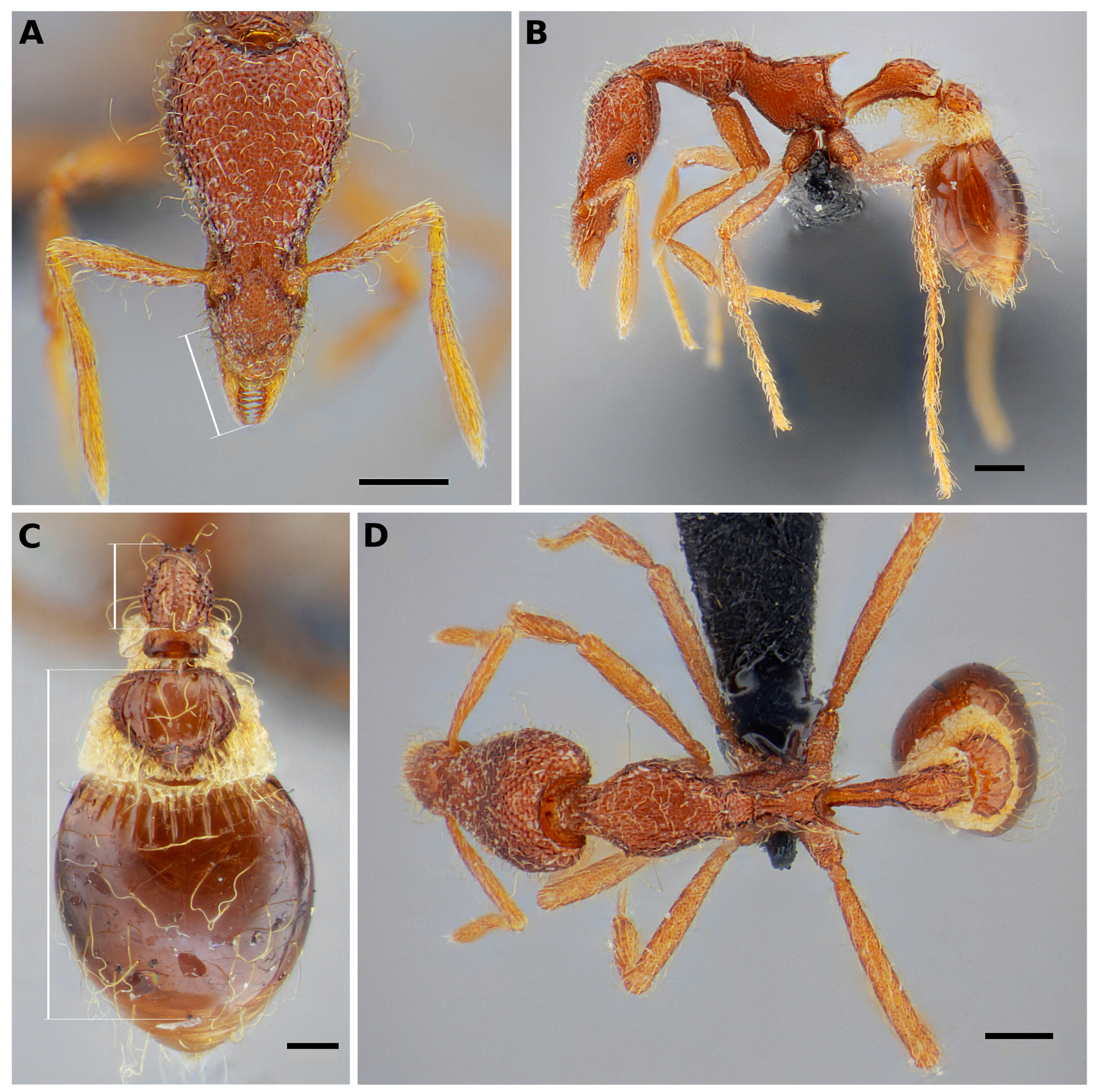
ML2. Mandible length 2. In full-face view, the distance from basalmost point of the outer mandible margin to the visible apex of the mandible (not necessarily the apical tooth, as it might be downcurved) (
Figure 1A). Measuring ML in short-mandibulate
Strumigenys often subestimates the real length of the mandibles, as mandible insertion on the lateral clypeus can be significantly posterior to the anteriormost point of the anterior clypeal margin, where ML starts to be measured. I suggest that ML2 should be an auxiliary measurement, taken in conjunction with ML. This new measurement will probably be suitable in the context of revisions for comparisons between closely related species.
ON. Ommatidia number.
HD. Head depth: in lateral view of the head, the distance between the lines touching the dorsalmost and the ventralmost points of the head, each line being parallel to the longitudinal head axis.
MtbtL. Metabasitarsus maximum length.
DPetndL. The maximum length of the petiole node in dorsal view of the metasoma (as in
Figure 1C), including the posterior spongiform collar.
PosPetW. The maximum width of the postpetiole disc in dorsal view, excluding lateral spongiform lobes.
A3+A4L. The distance from the anteriormost point of the postpetiole (spongiform collar not considered) to the posteriormost point of the first gastral tergite (
Figure 1C). This differs from Brassard et al. [
23], as in this study, the measurement was taken as a straight line rather than a separate measurement from each of the two sclerites. It can be taken in lateral or dorsal view. Deflection of the gaster in relation to the postpetiole in
Strumigenys is usually mild, thus not interfering significantly in the measurement.
GW. The maximum width of first gastal tergite in dorsal view of the metasoma (A4).
TL. A proxy for the total length of the body, given by the sum of ML, HL, WL, PetL and A3+A4L.
LegL. A proxy for the length of hind leg, given by the sum of MtfmL, Mttbl and MtbtL.
LegI. Leg index (WL/LegL × 100).
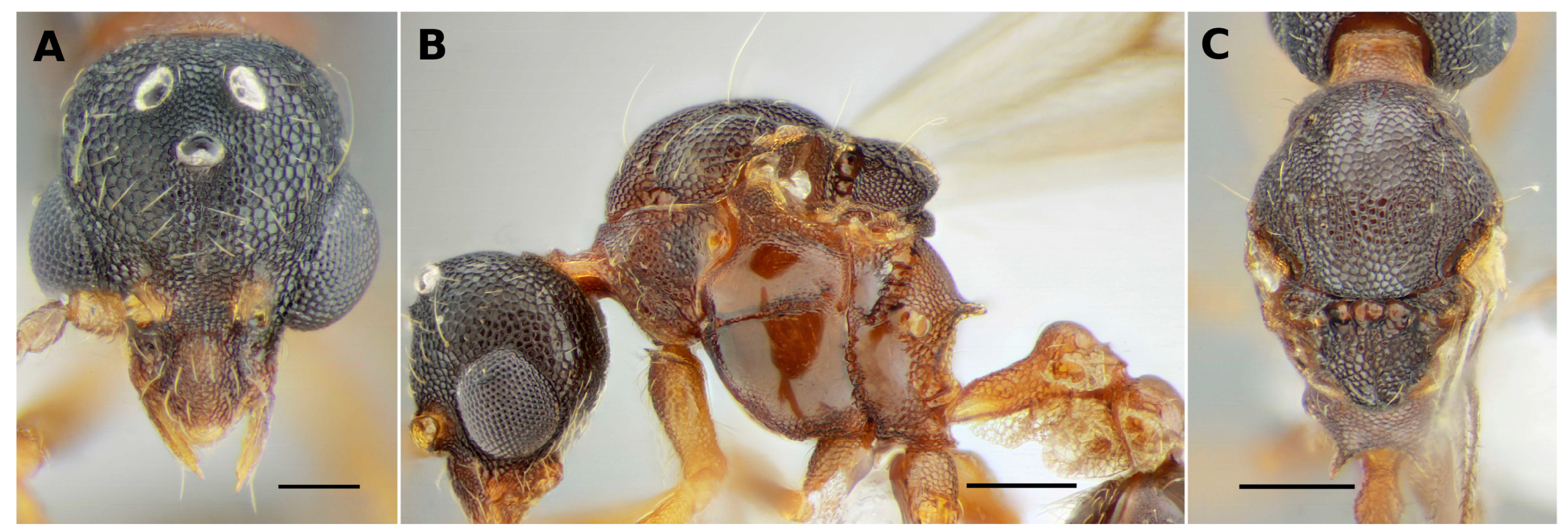
Figure 1.
Strumigenys flavianae, holotype worker (ANTWEB1032460). (A) Full-face view of head; (B) profile of the body; (C) dorsum of metasoma; (D) dorsal view of the body. Scale bars are 0.2 mm in (A,B,D) and 0.1 mm in (C). The measurement ML2 is indicated in (A), and measurements DPetndL and A3+A4L in (C).
Imaging. Images were captured using a Canon 1100D camera (Canon Inc., Tokyo, Japan) attached to a Leica S8APO stereomicroscope (Leica Camera AG, Wetzlar, Germany) coupled with a 2× auxiliary objective lens. Illumination was produced by a slightly modified version of the set presented in Kawada and Buffington [
24]. The series of images acquired at each photographed angle were combined using Zerene Stacker software, version 1.04 (Zerene Systems LLC, Richland, WA, USA). The resulting montage was edited using Gimp software [
25] for the purpose of enhancing sharpness and adjusting the rotation and light intensity. Scale bars were added to the ImageJ software [
26] by calibrating the edited image via a body measurement taken during the acquisition of the images (usually head width, Weber’s length, or pronotal width). Mouthparts and left antennae of the paratype ANTWEB1032422 were dissected and mounted in polyvinyl alcohol–lactic acid–glycerol (PVLG) medium [
27]. The dissected sclerites were mounted beneath the specimen in between a pair of round cover glasses.
The new species had the morphospecies code “
Strumigenys ufv-11” for a few years on AntWeb before this publication. The images of
S. flavianae composing
Figure 1,
Figure 2 and
Figure 3 as well as additional images of all types and the putative male (morphospecies
Strumigenys ufv-66), were uploaded to the website [
28].
A map was made in Qgis software [
29], and point coordinates of the species were taken from imaged specimens in Antweb or from specimens that I examined physically.
Species delimitation method. The studied specimens were considered members of a new species, as they shared a set of morphological characters that, when compared to all other species in the genus, comfortably isolates them morphologically, indirectly indicating reproductive isolation.
3. Results
The difficulty of classifying the new species into existing species groups made it necessary to create a group containing the new taxon alone. The species group is characterized and the new species is described as follows.
Taxonomy
The flavianae-group
(i) Mandible small in relation to head, multidentate (
Figure 1A), likely performing the gripping mechanism of action [
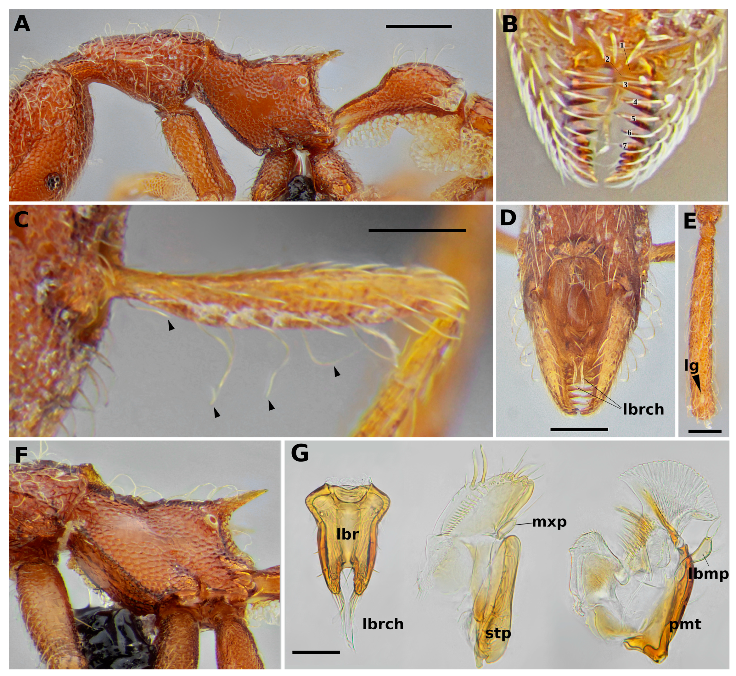
8]. (ii) Mandible inner margin (masticatory) with a small, triangular basal lamella, followed, without diastemmic gap, by a series of six–seven sharp teeth, then four–five truncate teeth and a sharp apical tooth (
Figure 2B). (iii) Mandible’s outer margin confluent with clypeal lateral margin (
Figure 1A). (iv) Labrum with basolateral expansions, followed by mildly convex lateral margins that form a pair of relatively low, robust lobes; lobes with up to four flagellate chaetae that are approximately two-thirds of labrum length, some flattened and some simple (
Figure 2G). (v) Head much longer than wide (CI 51) (
Figure 1A); mesosoma, petiole and legs also elongate (
Figure 1B,D). (vi) Flagellate, subflagellate and wire-like setae distributed on body in an apparently untidy manner; however, the apicoscrobal, humeral and mesonotal pairs stand out as larger, wire-like setae (
Figure 1). (vii) Freely projecting, wire-like setae on scape anterior margin, not inclined in a defined direction (
Figure 2C). (viii) Dorsum of mesosoma discontinuous, promesonum much higher than propodeal level. (ix) Triangular cuticular lamella on anterolateral edge of mesonotum, directly over the mesonotal spiracles (best seen in ventrolateral view,
Figure 2F). (x) Mesopleura excavation inconspicuous, but clearly discernible in specimens with dissected forecoxa, excavation filled with whitish setae. (xi) Propodeal spine long, propodeal lamella reduced. (xii) Femoral and tibial glands visible in all legs, small; some tarsomeres with glands visible as well (see description below). (xiii) Petiole node in profile long and low. (xiv) Well-developed spongiform tissue on the waist segments (
Figure 1B,C). (xv) Basigastral costulae well-marked, smaller than postpetiole disc length (
Figure 1C).
Figure 2.
Strumigenys flavianae, holotype worker (ANTWEB1032460) (A–E) and paratype worker (ANTWEB1032422) (F,G). (A) Mesosoma and petiole in profile; (B) mandible and sharp teeth indicated by numbers; truncate teeth and apical tooth not clearly visible due to mandible apical curvature; (C) scape dorsal surface, triangles indicate wire-like setae on anterior scape margin; (D) mouthparts in ventral view; (E) metafemur in dorsal view, triangle indicates femoral gland; (F) mesosoma in ventrolateral view (notice small smooth patch on meso- and metapleura); (G) mouthparts, dissected, from left to right: labrum, maxilla and labium. Scale bars: 0.2 mm in (A), 0.1 mm in (C,D) and 0.05 mm in (E) and (G). Abbreviations: lbr, labrum; lbrch, labral chaetae; lg, leg gland; lbmp, labial palp; mxp, maxilary palp; pmt, prementum; stp, stipes.
Strumigenys flavianae Chaul sp. nov.
Type material. Holotype worker. BRAZIL, Minas Gerais, Viçosa, Mata do Paraíso, -20.803959 -42.855107, 13.ii.2017, Berlesate (Borlini, P.) (CELC, unique specimen identifier: ANTWEB1032460). Paratype workers: BRAZIL, Minas Gerais, Viçosa, Fragmento Florestal, 1994 (Sperber, Lopes and Louzada) (CELC, unique specimen identifier: ANTWEB1032112); BRAZIL, Minas Gerais, Viçosa, Mata do Paraíso, -20.803146 -42.856782, 21.viii.2017, winkler sample (Moura, M. N.; Micolino, R.) (CELC, unique specimen identifier: ANTWEB1032422).
Diagnosis. Differing from all other Strumigenys by the combination of a slender body and long legs; pilosity composed of subflagellate, flagellate, or wire-like setae; basal lamella of mandible triangular, entirely covered by anterior clypeal margin when mandible is closed, and without diastemmic gap between it and the basal tooth of the masticatory margin; mandible outer margin confluent with clypeal lateral margin, not bulging; spongiform tissue ventrally on the petiole, notched at about its midlength; sides of mesosoma mostly sculptured, with only a small smooth patch in between upper mesopleuron and upper metapleuron.
Geographical Distribution. Minas Gerais, Brazil.
Description. HW 0.42–0.43, ML 0.08, ML2 0.18–0.21, HL 0.81–0.84, SL 0.39–0.4, ON 12, HD 0.28–0.31, WL 0.83–0.85, PetL 0.45–0.46, A3+A4L 0.72–0.8, PrW 0.29–0.305, DPetW 0.14–0.155, DPetndL 0.2–0.23, PosPetW 0.25–0.27, GW 0.5–0.53, MtfrL 0.635–0.68, MttbL 0.44–0.47, MtbtL 0.48–0.52, TL 3.02–3.16, LegL 1.55–1.67, CI 51.19–51.85, SI 92.86–93.02, MI 9.7, LegI 51.15–52.85 (n = 3).
Head: head elongate (
Figure 1A). In full-face view, mandibles small, elongate triangular, its dorsal surfaces smooth, covered with small, simple, appressed hairs. Outer margins of mandibles confluent with lateral clypeal margins. Basal lamellae of mandibles triangular, its basal side slightly tilted dorsally. Masticatory margin with 12 teeth, the first originating without a diastemmic gap between it and the basal lamella. The 6 basal teeth are large and acute, growing in size until tooth 3 (the largest on the mandible) and then becoming gradually smaller until tooth 6. Tooth 7 is even smaller than 6, and can be triangular or truncate. Teeth 8 to 11 are small and truncate, and tooth 12, the apical, is slightly larger than the previous, and triangular (but still smaller and less acute than the basal 7 teeth) (
Figure 2B). Tips of mandibles downcurved, so that the row of small truncated teeth are best seen with the head in anterodorsal view. Basiventral glands on mandibles not visible. Basal half of labrum (excluding the apical lobes) with margins diverging and converging back, and then mildly convex along the second half of the labrum body. Lobes short and thick, about a quarter the length of the labrum disc, converging slightly towards each other and separated by a wide notch. Each lobe has three simple hairs and a longer, flattened and sinuous seta arising from the apex (
Figure 2D,G). Clypeus longer than wide, reticulate punctate, its anterior margin projected strongly convex over mandibles and covering basal lamellae of mandibles. Antennae 6-segmented. Scape apicoventral gland present. Scape covered with appressed fine hairs and a row of freely projecting long, curved to coiled and wire-like setae on its anterior margin, most of them not obviously curved basally or apically (
Figure 1A and
Figure 2C). Apical and preapical antennomeres, taken together, slightly longer than scape; preapical antennomere as thick as apical, not swollen. Dorsal head reticulate punctuate with superimposed fainting rugulae, and ground pilosity composed of simple, irregularly coiled to subflagellate hairs. A pair of long, wire-like to flagellate apicoscrobal setae. Vertexal standing setae are absent, although ground pilosity are somewhat longer posteriorly on the head. In lateral view, scrobe not conspicuously deep, its dorsal margin well-marked, apically and ventrally poorly marked; ventral margin fades anterior to the eye level without reaching the anterior end of the head; in full-face view, dorsal margin of scrobe forms a thin translucent cuticular projection. Compound eyes with about 12 ommatidia, not seen in full-face view. Preocular carina ends much anterior than the level of the eyes in profile.
Mesosoma: slender and elongate (
Figure 2A). Dorsally, promesonotal suture a feeble ridge. Pronotum reticulate punctate with superimposed fainting rugulae, mesonotum reticulate punctate. Metanotal impression, a shallow groove, anteriorly marked by thin feeble traversal carina and with a short median longitudinal carina across it. Dorsum of propodeum, mesopleura, metapleura and lateral propodeum reticulate punctate, with only a small smooth patch in between upper mesopleuron and upper metapleuron. A pair of thin triangular lamellate projections laterally on mesonotum, on top of mesothoracic spiracles, directed laterally and upwards (
Figure 2F). Humeral pair of long, wire-like setae arising from a pair of small indented protrusions. Similar, but slightly smaller and thinner, setae are also present anterolaterally on the pronotum posterior to the humeral pair, anterolaterally on the mesonotum, and mediolaterally on the mesonotum (arising from the tip of the mesonotal lamellate projections). Ground pilosity on dorsum of mesosoma, as on the head, composed of thin, long subflagellate hairs. Propodeal teeth sharp and long, in profile posteriorly and slightly dorsally inclined, subtended by a thin propodeal lamella which descends with regular width until the level of the metapleural gland reservoir, where the lamella forms a small round lobe (
Figure 2A). Mesothoracic ventral excavation not densely hairy. In lateral view, anterior margin of mesopleuron with a translucid lamella, which is lobate on the upper section, pointing towards the mesothoracic excavation (
Figure 2F). Legs elongate, reticulate, covered in hairs similar to the head and mesosomal ground pilosity. All pairs of legs with tiny, rounded to oval, apicodorsal femoral glands; hind femur with the largest and middle femur with the smallest (
Figure 1D and
Figure 2E). Slit-shaped, well-developed apicodorsal glands on hind and middle tibiae, and tiny oval gland on protibia. Among the tarsomeres, those with visible glands are probasitarsus, mesobasitarsus and mesotarsomeres 2, and metabasitarsus and metatarsomeres 2 and 3.
Metasoma: petiole elongate, node longer than high, evenly and gently curved in profile view. The anterior face of node gradually ascending from peduncle and merging into dorsal faces, these faces not obviously distinguishable (
Figure 2A). Postpetiole disc with longitudinal rugae which are well-marked laterally and shallow to fainting medially (
Figure 1C). Spongiform tissue strongly developed ventrally on the petiole as a thin and tall curtain, notched at about its midlength (
Figure 2A). Posterior edge of node of petiole with a thin strip of spongiform tissue connecting two pyramidal spongiform posterolateral lobes. Spongiform tissue well-developed ventrally on postpetioles, with two large and dense lateral outgrowths linked in the middle by a thinner strip. Postpetiole disc with a thin, transversal, spongiform anterior strip and large spongiform lobes posterolaterally, which gradually becomes thinner where they link posteromedially. Well-developed spongiform transverse strip anteriorly on first gastral tergite. Basigastral ventral patch of specialized setae reduced. Dorsum of metasoma covered in long, wire-like to flagellate hairs. Gaster smooth and shiny. Gastral sternites as well as dorsally around the sting with thin, curved and simple hairs. Basigastral costulae well-marked but small, about half the length of postpetiole disc (
Figure 1C).
Etymology. The specific epithet honors the late Flaviana Heloisa da Silva Caetano, a young Brazilian undergraduate student from the Universidade Federal de Viçosa, whose untimely death during the COVID-19 pandemic saddened all her colleagues in the biology course. The name was created by adding the singular Latin genitive case suffix -ae to the first name of a female person. The orthography of an eponym is unchangeable and does not depend on the generic name for which the epithet is used.
Biology. Nothing is known about the biology of living colonies of S. flavianae; however, its slender body and legs suggest that it forages on the upper stratum of the litter or maybe within cavities inside rotten logs rather than amidst the deeper and denser litter and soil stratum. The new species has been found three times in secondary growth forest remnants, once in a pitfall trap (specific forest remnant not determined on label) and twice by winkler extractors (both in “Mata do Paraíso” forest remnant). Despite years of sampling in the region, the species is still rare. It might be the case that its populations are low in the most often sampled remnant, Mata do Paraíso (most sampling efforts throughout the years have been undertaken there). Further explorations of other forest remnants (dozens exist in the municipality) as well as in larger, more preserved nearby forests, such as the Parque Estadual da Serra do Brigadeiro or Parque Nacional do Caparaó, might reveal other, hopefully more abundant, populations.
Comments. The new species stands out among all species occurring in the Brazilian Atlantic Rainforest for its slenderness (long head, slender mesosoma and petiole and long legs) and wire-like pilosity. In this biome, species with short mandibles (either gripping or short trap-jaw mechanisms) are never too slender and never have the same type of pilosity. Some species of long trap-jaw mandibles in the mandibularis group are as slender as S. flavianae, but in that case, they cannot be confused with the newly discovered species because of their conspicuous linear mandibles (and also due to the lack of wire-like pilosity). A few species in the probatrix- and hyphata-groups, mostly Amazonian, are similar to S. flavianae; the ones in the probatrix-group are even more slender; however, due to other traits, they also cannot be confused with S. flavianae (see Discussion).
Figure 3.
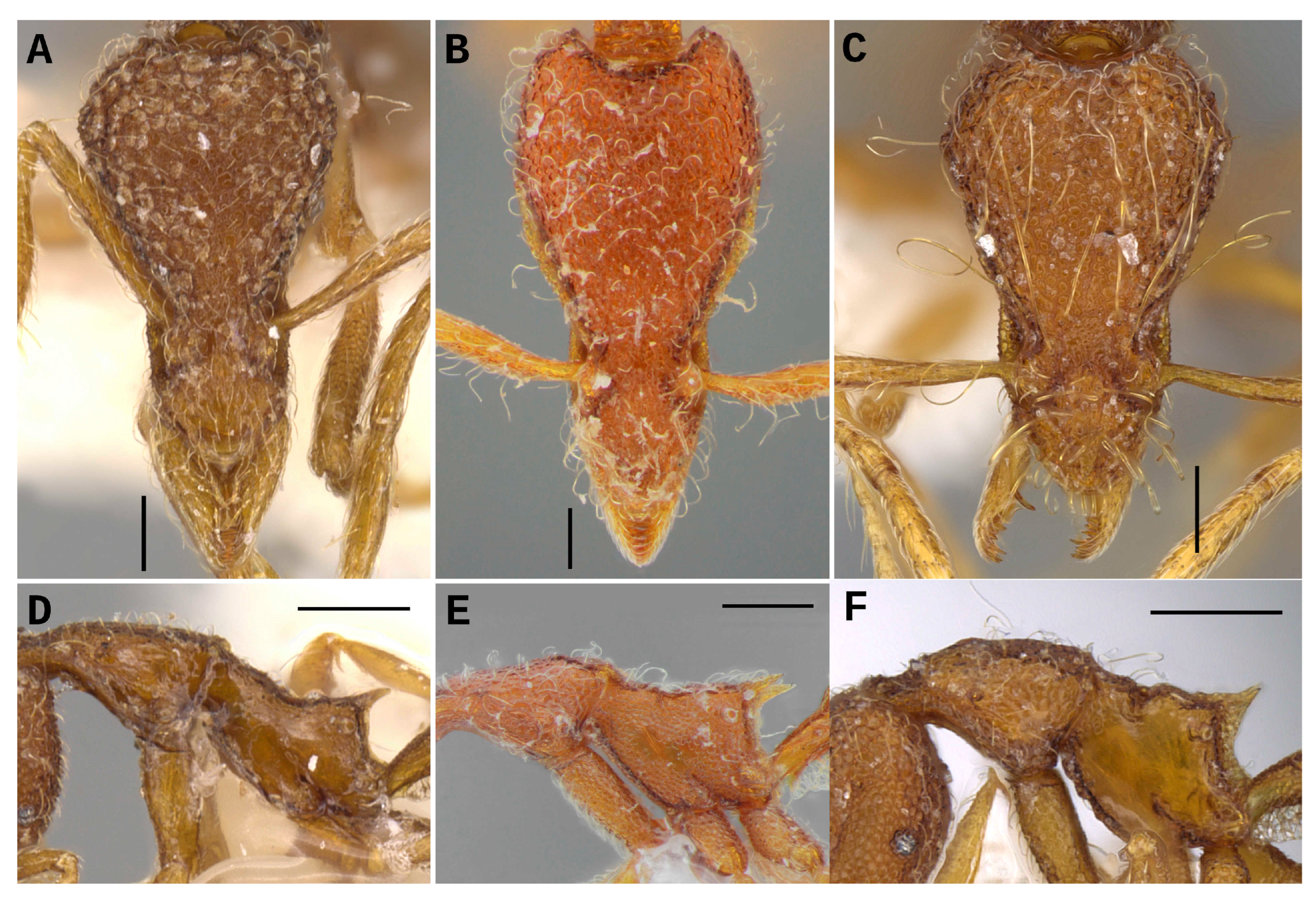
Comparison between S. flavianae (B,E) and morphologically similar species, S. doryceps (A,D) of the probatrix-group and S. hyphata (C,F) of the hyphata group. Head in full-face view in the top line and mesosoma profile in the bottom line. Strumigenys doryceps (CASENT0900198, images by Will Erichson); S. flavianae (paratype, ANTWEB1032112); S. hyphata (CASENT0914595, images by Zach Lieberman). Scale bars are 0.1 mm in (A–C) and 0.2 mm in (D–F).
4. Discussion
Identification. While keying specimens of
S. flavianae, the coiled setae on the anterior margins of the scapes make the decision ambiguous as to whether the setae curve to the base or the apex of the scapes, an important couplet in the key (couplet 31 of “Key to Neotropical
Pyramica species” [
9]). An attempt to insert
S. flavianae into the key can be made by modifying couplet 31 to a triplet, as follows (the first two lugs reproduced exactly as in Bolton, 2000):
—With the head in full-face view, the leading edge of the scape has a row of conspicuous projecting curved hairs, of which one or more, distal to the subbasal bend, distinctly curves towards the base of the scape. These hairs may be simple, spatulate, remiform, spoon-shape or wire-like .......................................................................................................... 32
—With the head in full-face view, the leading edge of the scape lacks projecting hairs that distinctly curve towards the base of the scape. The scape’s edge may have elongated, simple, straight or flagellate hairs present, may have entirely anteriorly or apically directed short hairs (hairs simple, narrowly to broadly spatulate or spoon-shaped) or may be hairless ............................................................................................................................................. 56
—With the head in full-face view, the leading edge of the scape has a row of conspicuous projecting wire-like hairs which have tips curving towards the base or the apex of the scape (or even ventrally or dorsally curved) (
Figure 2C). Head elongate (
Figure 1A). Spongiform tissue notched ventrally on the petiole (
Figure 2A) ...
S. flavianaeThe ambiguity of this species to pass smoothly through the original couplet 31 of the key, and the fact that many other species have been described since 2000 highlight the necessity of an updated identification work that incorporates all species described into a new identification tool. This could be carried out either by one large key that accounts for all Neotropical species or by a key that leads to the species groups, with each group having its own specific key updated separately.
Morphologically similar Neotropical species. Strumigenys flavianae is an odd Neotropical representant, not fitting perfectly into any of the species groups delimited for the genus [
9]; however, it is similar to the members of the
hyphata-group,
S. hyphata (Brown, 1953) [
4] and
S. cincinnata (Kempf, 1975) [
30], and to
S. doryceps (Bolton, 2000) [
9], a member of the
probatrix-group.
The new species overall slenderness suggests that it could be a member of the
probatrix-group, as it is very similar to
S. doryceps. It differs from
S. doryceps by having a short and triangular basal lamella of the mandible, which is not visible when mandibles are fully closed, rather than a long, subrectangular one that is visible with the mandibles closed. It also differs from that species due to not having the outer mandible margin basally bulging in relation to the lateral clypeal margin just above it; instead, in
S. flavianae, the margin is confluent with the lateral clypeal margin. The dentition in
S. flavianae is not confined to the apical third of the mandible (
Figure 2B) as in
S. doryceps. The habitus between the two is similar, however,
S. doryceps having a more slender mesosoma and much larger smooth pleural patches (
Figure 3A,B,D,E). Two interesting similarities between
S. flavianae and
S. doryceps are the well-developed and medially notched spongiform tissue located ventrally on the petiole and the short but well-marked basigastral costulae.
Strumigenys flavianae differs more radically from
S. probatrix, the other member of the
probatrix-group, in many obvious aspects. Except for slenderness, they do not have a similar habitus:
S. flavianae has freely projecting setae on the anterior clypeal margin, which are not tidily arranged and spatulated as in
S. probatrix; it has dense and abundant spongiform tissue located ventrally on the petiole (moderate in
S. probatrix); and
S. flavianae is ferruginous brown rather than dark brown. However, in terms of group-defining characters, all dentition differences between
S. flavianae and
S. doryceps are also present between it and
S. probatrix (while
S. probatrix matches
S. doryceps in all of these dentition traits).
When it comes to the
hyphata-group, a relationship between
S. flavianae and the two members could be hypothesized due to the overall similar habitus, but not particularly the slenderness. In this case, the shapes of the basal lamellae (small and triangular) of
S. hyphata and
S. cincinnata match that of
S. flavianae. Still, the dentition that immediately follows the basal lamella is not exactly equal; in the
hyphata-group, there is a diastema between the basal lamella and the teeth which follow it (
Figure 3C). Moreover, the labrum in the
hyphata-group is unique, with the main body being roughly hourglass-shaped (imaged under specimens ANTWEB1032490 and UFV-LABECOL-000095 on Antweb.org), not resembling that of
S. flavianae (
Figure 2G). At the species level,
S. flavianae cannot be confused with either of the two members of the
hyphata-group. It can be easily differentiated from
S. cincinnata by its much larger size, and from
S. hyphata by size and by the absence of hypertrophied specialized clypeal setae, among other aspects.
In a recent phylogenetic study of the genus, the species
S. probatrix and
S. cincinnata were included [
8]. The two, despite being recovered as members of the same subclade within a larger Neotropical clade, are not particularly closely related, with
S. probatrix being the sister of a clade containing members of the
paradoxa-group and the
rogata-group and
S. cincinnata being the sister of the exquisite
S. mirabilis Mann, 1926 [
8,
31]. With such a robust phylogenetic backbone available, investigating what position
S. flavianae would assume once incorporated into it, is surely a goal that must be attained.
Rarity. Despite relatively constant sampling efforts in the past thirty years at the type locality, only three workers are known for the new
taxon. Geographically,
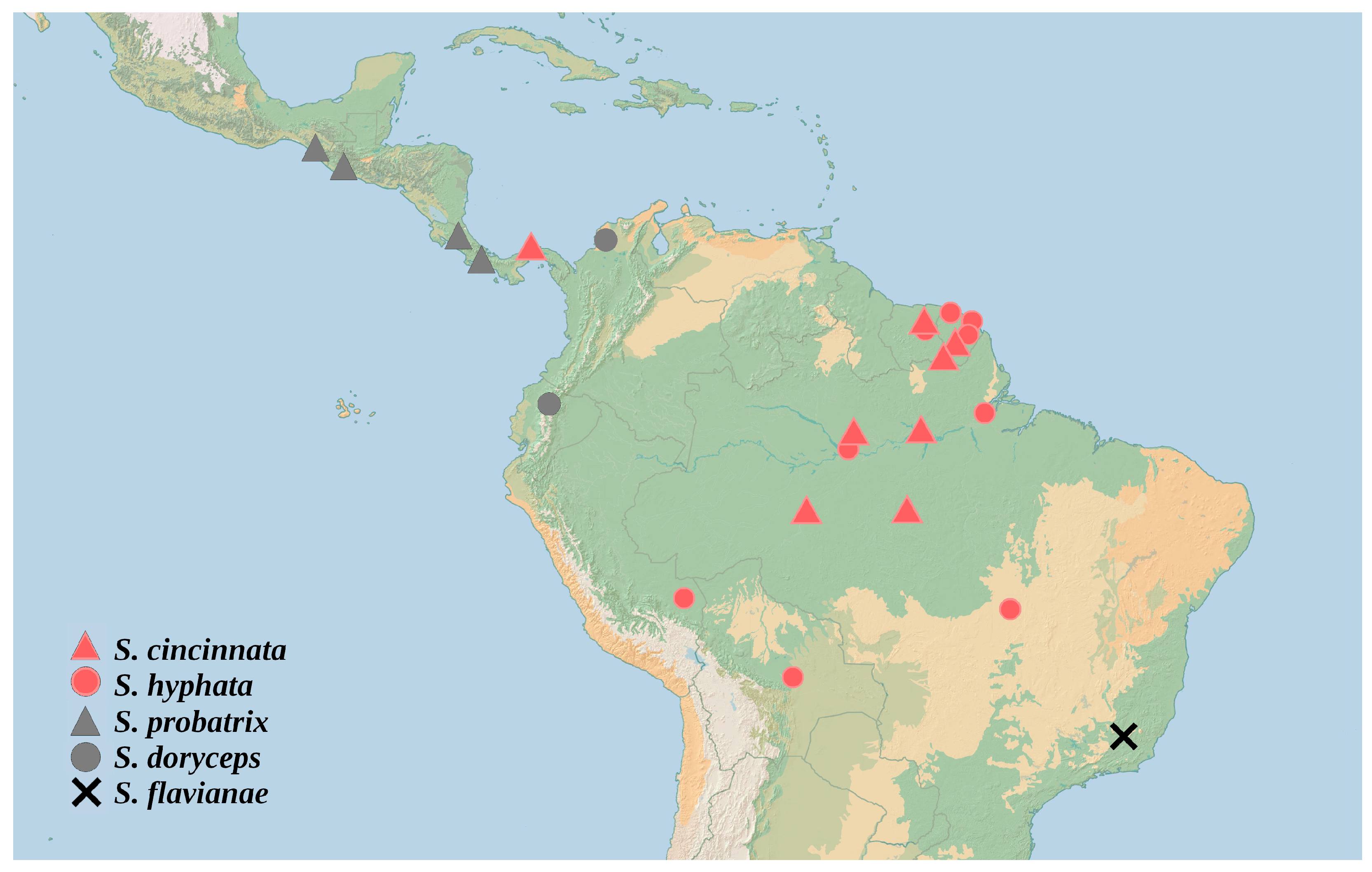
S. flavianae is far from its morphologically similar species, occurring in the southeastern to eastern South America Dry Diagonal (eSADD, sensu Luebert [
32]), while the other species in the
hyphata- and
probatrix-groups all occur northwest of the eSADD or, at most, within it (
S. hyphata occurs in the Goias state of Brazil and in Bolivia) (
Figure 4). In a large study of
Strumigenys in the Brazilian Atlantic Forest, no species similar to
S. flavianae were recorded [
33]. Moreover,
Strumigenys specimens of various collections containing Atlantic Forest ants were examined by the author and myrmecologists working on large ant collections in Brazil confirmed the absence of specimens matching
S. flavianae (Thiago R. Silva and Esperidião Neto, personal communication).
It is not uncommon that
Strumigenys species are represented by only one or a few records, even in the case of species described long ago and/or in regions which have received considerable sampling efforts since their descriptions. Some examples of such species in the Neotropics are
S. mandibularis Smith, 1860 [
34],
S. dentinasis (Kempf, 1960) [
35],
S. substricta (Kempf, 1964) [
36],
S. browni (Bolton, 2000) [
9],
S. xochipili (Bolton, 2000) [
9],
S. tlaloc (Bolton, 2000) [
9] and
S. warditeras (Bolton, 2000) [
9].
Strumigenys doryceps itself, the species discussed just above, had been known by the Ecuatorian type series for almost two decades, but was only recently recorded again in Colombia [
37].
Putative male sex. The male morphospecies
Strumigenys ufv-66 (
Figure 5 and additional images under specimen UFV-LABECOL-009731 on Antweb.org), sampled in a Malaise trap, is possibly conspecific to
S. flavianae. The morphospecies
Strumigenys ufv-66 occurs in the type locality of
S. flavianae, where six other unassociated male
Strumigenys morphospecies are known (totaling seven male morphospecies out of twenty species known by workers in that locality). All of these males are databased and photographed, and can be accessed at Antweb.org. These are: ufv-42 (UFV-LABECOL-008242), ufv-45 (ANTWEB1032841), ufv-46 (ANTWEB1032780), ufv-62 (UFV-LABECOL-009966), ufv-65 (ANTWEB1038468) and ufv-111 (ANTWEB1041072). None of them appear to better match
S. flavianae than
Strumigenys ufv-66. It is larger than other male morphospecies, which is in accordance with the new species (also a relatively large
Strumigenys). Moreover, it shows similar development of the spongiform tissue on the waist segments, including a mild notch on the ventral petiole spongiform tissue that partially matches the corresponding state seen in workers of
S. flavianae. Finally, it has a conspicuous propodeal spine, also in accordance with the workers of the new species.
