Article

Green-Synthesized Zinc Oxide Nanoparticles with Enhanced Release Behavior for Sustainable Agricultural Applications

1
National Agricultural Research Center (NARC), Baqa’a 19381, Jordan
2
Faculty of Science, University of Petra (UOP), Amman 11196, Jordan
*
Author to whom correspondence should be addressed.
Solids 2025, 6(4), 59; https://doi.org/10.3390/solids6040059 (registering DOI)
Submission received: 26 August 2025 / Revised: 15 October 2025 / Accepted: 16 October 2025 / Published: 26 October 2025

Abstract

This study presents a green and sustainable approach for synthesizing zinc oxide nanoparticles (ZnO-NPs) using Melia azedarach leaf extract as a reducing and stabilizing agent, with zinc acetate as the precursor. The synthesized nanoparticles were thoroughly characterized to assess their structural, morphological, and physicochemical properties, revealing nanoscale dimensions, enhanced crystallinity, and improved stability compared to commercial ZnO. Controlled release experiments under plant-relevant pH conditions demonstrated a gradual and sustained release of Zn2+ ions, accompanied by buffering effects and re-precipitation of Zn(OH)2, highlighting their potential for long-term nutrient availability in soil systems. Unlike conventional studies that focus mainly on synthesis or characterization, this work emphasizes the functional performance of ZnO-NPs as nanofertilizers, combining eco-friendly production with practical agricultural applications. The plant-mediated synthesis yielded nanoparticles with uniform size distribution, enhanced dispersion, and stability, which are critical for efficient nutrient delivery and persistence in soil. Overall, this study provides a cost-effective, scalable, and environmentally benign strategy for producing ZnO nanoparticles and offers valuable insights into the development of sustainable nanofertilizers aimed at improving crop nutrition, soil fertility, and agricultural productivity.

Graphical Abstract

1. Introduction

The overuse of chemical fertilizers has led to environmental pollution and economic challenges, prompting the exploration of nanotechnology-based solutions to improve nutrient use efficiency and crop yields [1,2]. There is a growing global demand for biocompatible, eco-friendly, and cost-effective materials to enhance nutrient availability and stimulate plant growth [3]. These materials should ideally be inert, less toxic, and more stable than widely used synthetic chemicals, ensuring efficient delivery to target sites [4].
The synthesis of nanoscale metal oxides such as ZnO typically requires external stabilizing agents [5]. However, Melia azedarach L. (commonly known as Chinaberry), a deciduous tree with a rich phytochemical profile, presents a sustainable alternative. Its extracts contain a variety of bioactive compounds—including terpenoids, flavonoids, polyphenols, saponins, tannins, amides, and carboxylic acids—which can act as both reducing and capping agents during nanoparticle formation [6]. This dual functionality enhances nanoparticle stability and eliminates the need for synthetic stabilizers [7,8]. The applicability of M. azedarach in green synthesis has been demonstrated in previous studies; Haq et al. [9] reported the successful biosynthesis of silver and magnesium oxide nanoparticles using its extracts, indicating the plant’s significant potential in environmentally friendly nanomaterial production.
Zinc oxide nanoparticles (ZnO-NPs) are a prominent class of metal oxide nanomaterials known for their unique physicochemical properties. These include high chemical stability, a broad absorption spectrum, strong electrochemical coupling, and excellent photostability. ZnO-NPs, with the molecular formula ZnO, are widely produced and used across commercial and industrial sectors [10]. One notable application is their use in the food industry as a zinc nutrient source [11,12].
Several studies have reported that the band gap of ZnO nanoparticles influences their Zn2+ release in soil, as it reflects particle size, crystallinity, and surface defects. Nanoparticles with a narrower band gap, indicating higher defect density, exhibit weaker Zn–O lattice stability and release Zn2+ ions more readily, particularly under acidic or neutral pH. In contrast, wider band gap, highly crystalline ZnO NPs dissolve more slowly, providing controlled, sustained ion release. Thus, band gap measurements can serve as a useful predictor of ZnO NP dissolution behavior in agricultural soils [13,14,15]. ZnO typically possesses a direct band gap of about 3.37 eV, and its physicochemical properties can be tuned through different synthesis approaches, including sol–gel, hydrothermal, and green synthesis using plant extracts. Such variations can modify the band gap, thereby influencing the solubility and controlled release of Zn2+ ions, which are vital micronutrients for plant growth [16,17,18]. Research indicates that ZnO nanoparticles can be absorbed by plants through roots or foliar pathways, affecting physiological attributes such as chlorophyll accumulation, antioxidant activity, and biomass production. The release rate of Zn2+ ions is strongly dependent on the band gap, with narrower band gaps often promoting faster dissolution and enhanced bioavailability for plant uptake. Hence, elucidating the correlation between the band gap energy of ZnO nanoparticles and their release kinetics is essential for advancing their agricultural applications, ensuring effective nutrient delivery while mitigating potential phytotoxic risks. Importantly, band gaps narrower than ~3.37 eV are frequently associated with accelerated photocorrosion and dissolution due to the upward shift in the valence band edge, which renders the material more susceptible to self-oxidation under illumination. This highlights the inherent trade-off between achieving visible-light activity through band gap narrowing and maintaining the structural stability of ZnO nanoparticles, thereby underscoring the need for careful tuning of their electronic properties to balance nutrient release efficiency with long-term stability [18,19].
Recent studies have highlighted the multifaceted roles of zinc oxide nanoparticles (ZnO-NPs) in enhancing plant physiology and stress resilience. In tea plants (Camellia sinensis), ZnO-NPs significantly improved photosynthetic efficiency, including increased levels of RubisCO (key CO2-fixing enzyme), enhanced chlorophyll fluorescence, and elevated CO2 utilization efficiency [20]. Similarly, in common bean (Phaseolus vulgaris), soil or foliar application of ZnO-NPs restored electron transport functions under stress, enhancing linear electron flow (LEF) by up to 72% compared to untreated plants, thereby supporting higher adenosine triphosphate/nicotinamide adenine dinucleotide phosphate generation suited for CO2 fixation [21]. These findings underscore the potential of ZnO-NPs in improving photosynthesis and carbon fixation processes in plants.
Despite the extensive research on ZnO nanoparticles for agricultural applications, sustainable and cost-effective synthesis methods that also ensure controlled nutrient release remain limited. This study addresses this gap by introducing a green synthesis approach using Melia azedarach L. extract to produce ZnO nanoparticles and systematically evaluating the controlled release behavior of Zn2+ ions under plant-relevant pH conditions. Unlike many previous studies that focus primarily on synthesis or characterization, our work emphasizes the functional performance of ZnO-NPs as potential nanofertilizers, specifically targeting long-term nutrient availability and persistence in soil. By integrating an eco-friendly synthesis route with practical performance assessment, this investigation provides new insights for the development of sustainable nanofertilizers that enhance crop nutrition and productivity.

2. Materials and Methods

Zinc acetate dihydrate (C4H6O4Zn·2H2O) and commercial zinc oxide nanoparticles (designated as ZnOcom) were obtained from Thermo Fisher Scientific Inc., Waltham, MA, USA. and Ishihara Sangyo Kaisha, Ltd., Osaka, Japan respectively. Analytical grade of sodium hydroxide (NaOH) with purity of 98% was supplied by Sigma Aldrich, St. Louis, MO, USA. Deionized water was used as the solvent and aqueous medium to disperse the nanoparticles. Absolute ethanol (Barcelona, Spain, 99.9%) was used as a solvent in the final washing stage. Fresh Melia azedarach (Chinaberry) leaves were collected locally, washed thoroughly with distilled water, and air-dried prior to use.

2.1. Synthesis of ZnO Nanoparticles

Fresh leaves of Melia azedarach (20 g) were cut into small pieces and boiled in 200 mL of distilled water at 100 °C for 30 min. The mixture was filtered through Whatman No. 1 filter paper to remove solid residues, and the filtrate was cooled to room temperature. The freshly prepared extract was used immediately as a reducing and stabilizing agent. An equal volume (1:1, v/v) of the extract was added dropwise to 0.1 M zinc acetate solution under continuous stirring. This ratio was chosen based on preliminary optimization experiments, in which different extract-to-precursor ratios (1:0.5, 1:1, and 1:2) were tested. The 1:1 ratio produced ZnO nanoparticles with the most uniform particle size distribution, as confirmed by SEM analysis. The pH of the reaction mixture was adjusted to 10 using 1 M NaOH with a calibrated pH meter (WTW inoLab 7310, Weilheim, Germany). The suspension was left to stand for 24 h at ambient temperature to facilitate complete precipitation and stabilization of zinc hydroxide nanoparticles (designated as Zn_OH2). The resulting precipitate was collected by centrifugation (10,000 rpm, 10 min), washed three times with distilled water to remove unreacted species, and dried at 80 °C overnight. The dried product was subsequently calcined at 500 °C for 2 h in a muffle furnace to yield crystalline ZnO nanoparticles (designated as ZnO_calc).

2.2. Physicochemical Characterizations of ZnO Nanoparticles

The particle size of ZnO_calc samples was estimated using the Malvern panalyticalzitasier (model Mastersizer 3000+ Lab, Malvern, UK). ZnO-NPs were carefully dispersed in distilled water using an ultrasonicator for 1 h before measurement. FTIR spectrometer (Lambda scientific, Guangzhou, China) and an attenuated total reflectance diamond crystal unit were used to obtain the infrared spectra. The spectra were recorded by scanning from 4000 to 650 cm−1 with a resolution of 4 cm−1. The morphology and chemical composition of synthesized ZnO were investigated using a Phenom XL G2 scanning electron microscope combined with an AXS EDS system (Thermo Fisher Scientific, Waltham, MA, USA). The kaolinitic clay and the synthesized product were coated with a ~300 Å thick platinum film under an argon atmosphere using an AGAR sputter coater machine (model AGB7340, Stansted, UK) in a high-vacuum evaporator. The SEM images were produced at 6 × 10⁻⁴ Pa and 15 kV accelerating voltage. The structural phases of the ZnO nanoparticles samples were investigated using an Anton Paar XRD diffractometer (XRDynamic 500, Graz, Austria) with a Cu Ka-radiation k = 1.54 Å, 40 kV, 40 mA at a 2 h range 2–70 with a scan rate of 2 deg/min, step scan size 0.02, and receiving slit of 0.3 mm. The data obtained from the XRD pattern was used to calculate the crystallite size using the Scherrer equation [22].
The surface area and pore size distribution were measured using N2 adsorption–desorption isotherms on a Quantachrome Instruments (Anton Paar, Autosorb-iQ, Graz, Austria) analyzer. Samples were degassed under vacuum at 250 °C for 3 h to remove adsorbed moisture and volatiles. Specific surface area was calculated using the multi-point BET method, while pore size distribution was determined with AsiQwin software (v5.21) using NLDFT and the N2 at 77 K on oxide surface kernel. This slit-pore NLDFT model, tailored for oxides, accounts for solid–fluid and fluid–fluid interactions, offering a more accurate characterization of meso- and microporous structures than classical BJH methods, which assume cylindrical macropores.
The optical band gap of ZnO_calc sample was determined using a JASCO UV–Visible spectrophotometer (Model V-730, Tokyo, Japan). The nanoparticles were dispersed in deionized water and sonicated for 15 min prior to measurement. The UV–Vis spectrum was recorded at room temperature in the wavelength range of 200–800 nm. The absorption coefficient was calculated according to the procedure described by Ayesh and Abdel-Rahem [23], taking into account the nanoparticle concentration and the 1 cm optical path length of the quartz cuvette. The band gap energy was subsequently estimated using the Tauc relation (Equation (1), [24]).
α h v 1 2 = A h v E g
here
α: absorption coefficient;
hv: photon energy (Planck’s constant × frequency);
A: proportionality constant;
Eg = optical band gap energy;
n = 1/2 → direct transition allowed (as in ZnO).

2.3. Release Behavior Study of Synthesized Green ZnO Nanoparticles

A quantity of 0.5 g of the synthesized green zinc oxide (ZnO) nanoparticles was dispersed in 1.0 L of distilled water. The pH of the prepared solution was adjusted to the desired value using acetic acid to simulate internal plant conditions that promote Zn2+ ion release. The release behavior of Zn2+ was evaluated over a period of two weeks. Electrical conductivity (EC) was measured periodically using a calibrated WTW Cond 3210 (Weilheim, Germany) conductivity meter to estimate the overall ionic concentration in the solution. Simultaneously, pH was monitored with a WTW inoLab pH 7310 m to assess changes in acidity that could influence the dissolution rate. This experimental setup was designed to provide insights into the sustained release of zinc ions under near-physiological plant conditions and to evaluate the potential of the ZnO_calc sample as a slow-release micronutrient fertilizer.

3. Results

3.1. Physiochemical Characterization of the Green-Synthesized ZnO NPs

The size distribution of the ZnO_calc sample (Figure 1) displays a dominant peak at approximately 122 nm, representing the majority of the nanoparticle population. This relatively uniform distribution indicates that nucleation and growth were effectively regulated by plant-derived compounds, which coated the nanoparticles and imparted electrostatic stabilization through negatively charged carboxyl groups. In addition, a minor peak observed near 5560 nm corresponds to a small fraction of larger aggregates, likely originating from insufficiently capped nanoparticles or residual organic constituents from the plant extract.
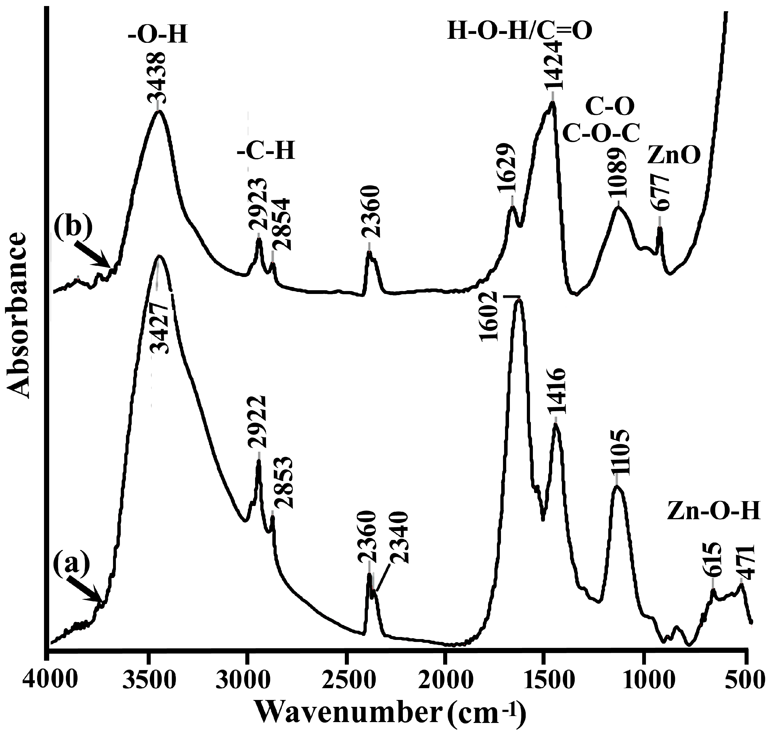
The chemical structure evolution from Zn(OH)2 to ZnO nanoparticles was investigated using FTIR spectroscopy, with spectra of Zn_OH2 and ZnO_calc samples presented in Figure 2a,b, respectively. The Zn_OH2 spectrum (Figure 2a) shows a broad O–H stretching band at 3427 cm−1, indicating surface hydroxyl groups and adsorbed water, and peaks at 2922 and 2853 cm−1 corresponding to aliphatic C–H stretching from plant-derived phytochemicals, which likely contribute to nanoparticle stabilization. Additional bands at 1602 and 1416 cm−1 are attributed to H–O–H bending and C=O/COO vibrations, while the 1105 cm−1 band reflects C–O or C–O–C stretching, confirming the presence of organic capping agents [25]. Notably, peaks at 615 and 471 cm−1 correspond to Zn–O stretching and Zn–O–H bending, verifying the formation of Zn(OH)2 nanoparticles [26].
Upon calcination, the ZnO_calc spectrum (Figure 2b) exhibits marked changes. The O–H stretching band shifts to 3438 cm−1 with reduced intensity, reflecting dehydration, while weak C–H and C–O bands indicate partial removal of residual organics. Absorptions at 1629 and 1424 cm−1 correspond to H–O–H bending and C=O/COO; stretching, suggesting minor surface-bound organics or carbonates. Sharp bands at 677 cm−1 confirm Zn–O lattice vibrations, demonstrating the successful transformation of Zn(OH)2 into ZnO nanoparticles. These results are consistent with the literature, which reports diminishing organic features and retention of characteristic lattice vibrations upon calcination of green-synthesized nanoparticles [27]. Overall, the comparison highlights the effectiveness of calcination in removing organic residues while preserving the ZnO crystalline framework.
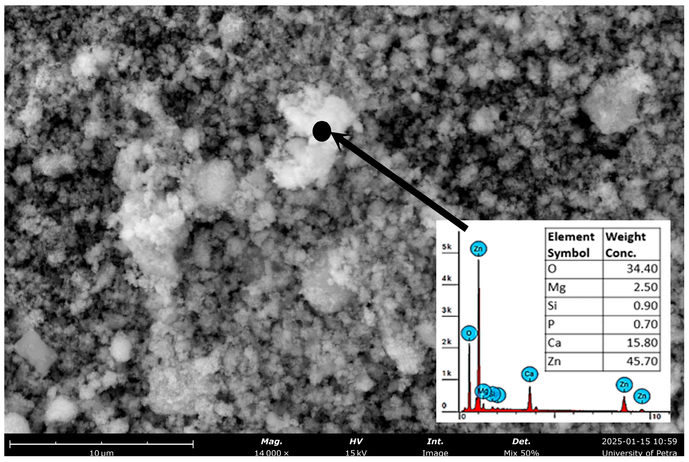
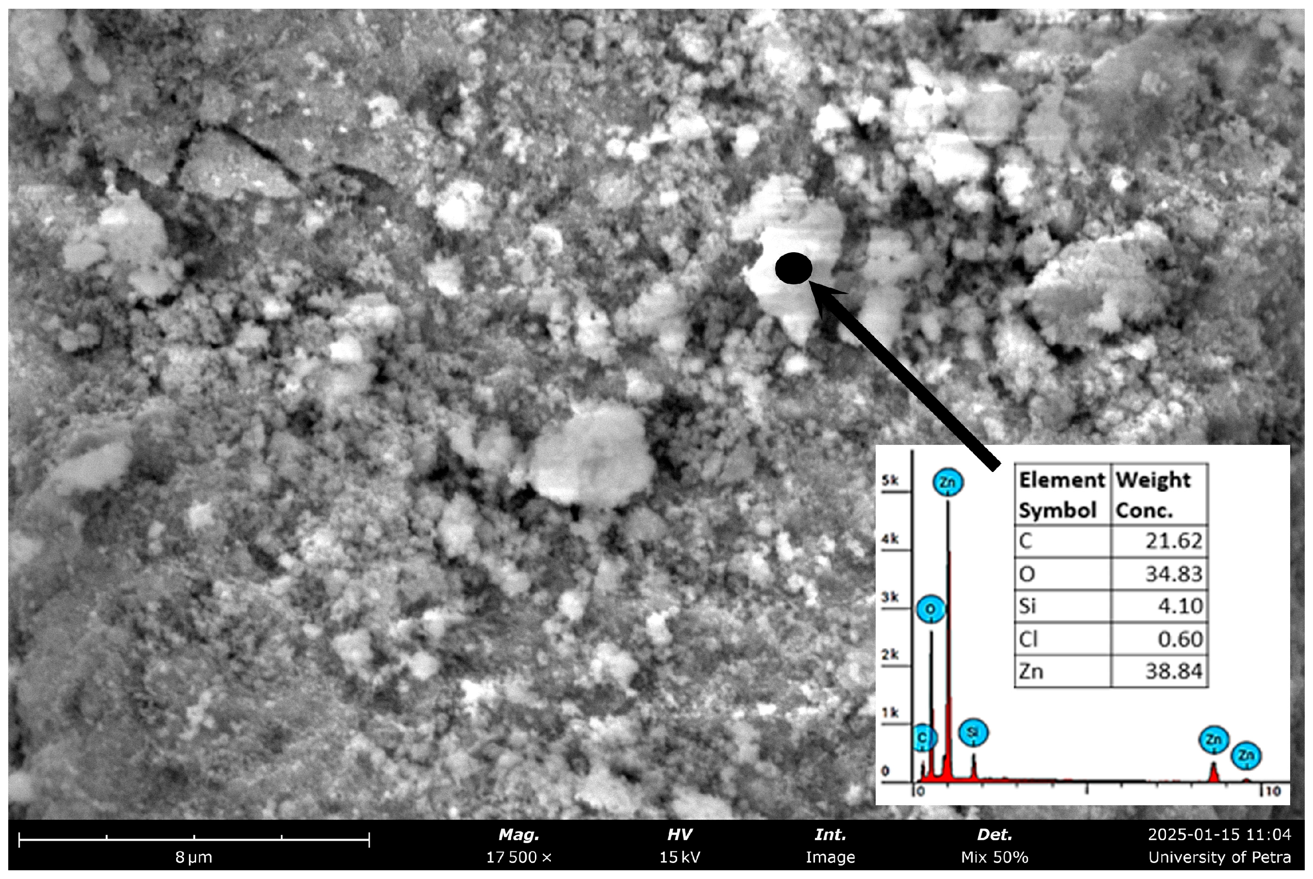
The SEM–EDS analyses of ZnO_calc and ZnO_com nanoparticles are shown in Figure 3 and Figure 4, respectively. Both samples exhibited nanosized particles with a tendency to aggregate, a common feature of high surface energy nanomaterials. The ZnO_calc sample (Figure 3) displayed nearly spherical particles with irregular clustering, likely due to interactions with phytochemicals from the plant extract, which acted as natural capping and stabilizing agents. EDS confirmed Zn (45.70 wt%) and O (34.40 wt%) as the primary elements, along with minor amounts of Ca (15.80 wt%), Mg (2.50 wt%), Si (0.90 wt%), and P (0.70 wt%), attributed to plant-derived metabolites or associated minerals, suggesting surface adsorption of bioactive compounds that aid stabilization and functionalization. In contrast, the ZnO_com sample (Figure 4) showed stronger agglomeration, irregular morphology, and a porous texture that increases the surface-to-volume ratio. Its EDS spectrum identified Zn (38.86 wt%) and O (34.83 wt%) as the dominant constituents, with Si (4.10 wt%) confirming the presence of a stabilizing silicon coating. Additional signals included C (21.62 wt%), originating from carbon tape or surface contamination, and Cl (0.60 wt%), likely from residual precursor salts. These findings confirm the successful formation of ZnO nanoparticles in both cases, with plant-mediated ZnO_calc functionalized by phytochemicals, while the commercial ZnO_com was effectively modified with a silicon coating to enhance stability and potential performance in optical and catalytic applications.
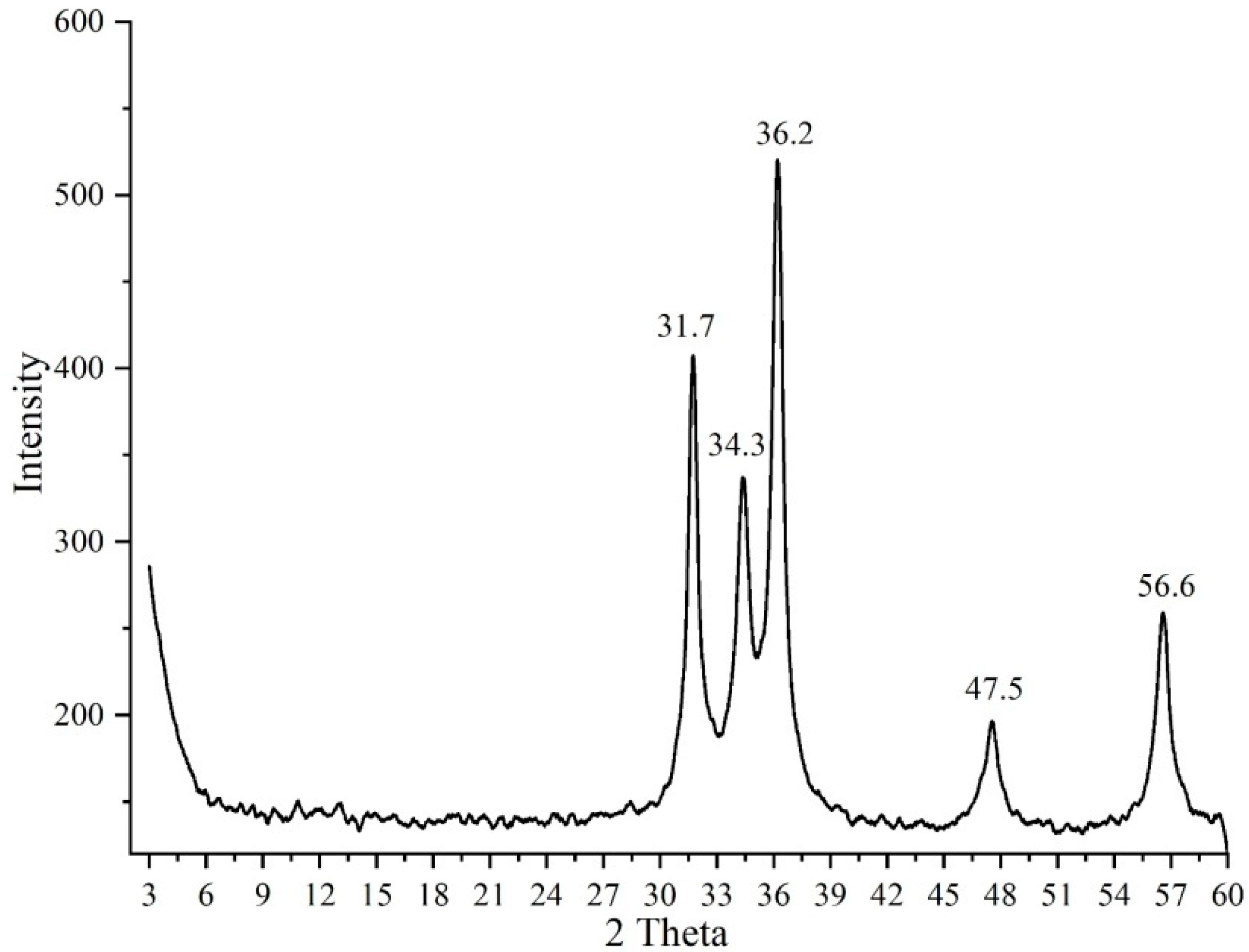
The XRD pattern of the ZnO_calc sample (Figure 5) confirms the successful formation of the hexagonal wurtzite structure, as evidenced by the characteristic diffraction peaks listed in Table 1, consistent with JCPDS card no. 36–1451. Crystallite size estimation using the Scherrer equation (Table 1) indicated an average size of ~18–20 nm, verifying the nanocrystalline nature of the sample. The noticeable broadening of the diffraction peaks can be attributed to the combined effects of nanoscale crystallite dimensions and lattice strain. Such strain is likely introduced during green synthesis, where phytochemicals present in the plant extract act as reducing and capping agents, thereby restricting crystal growth and inducing lattice distortions and surface defects that contribute to peak broadening.
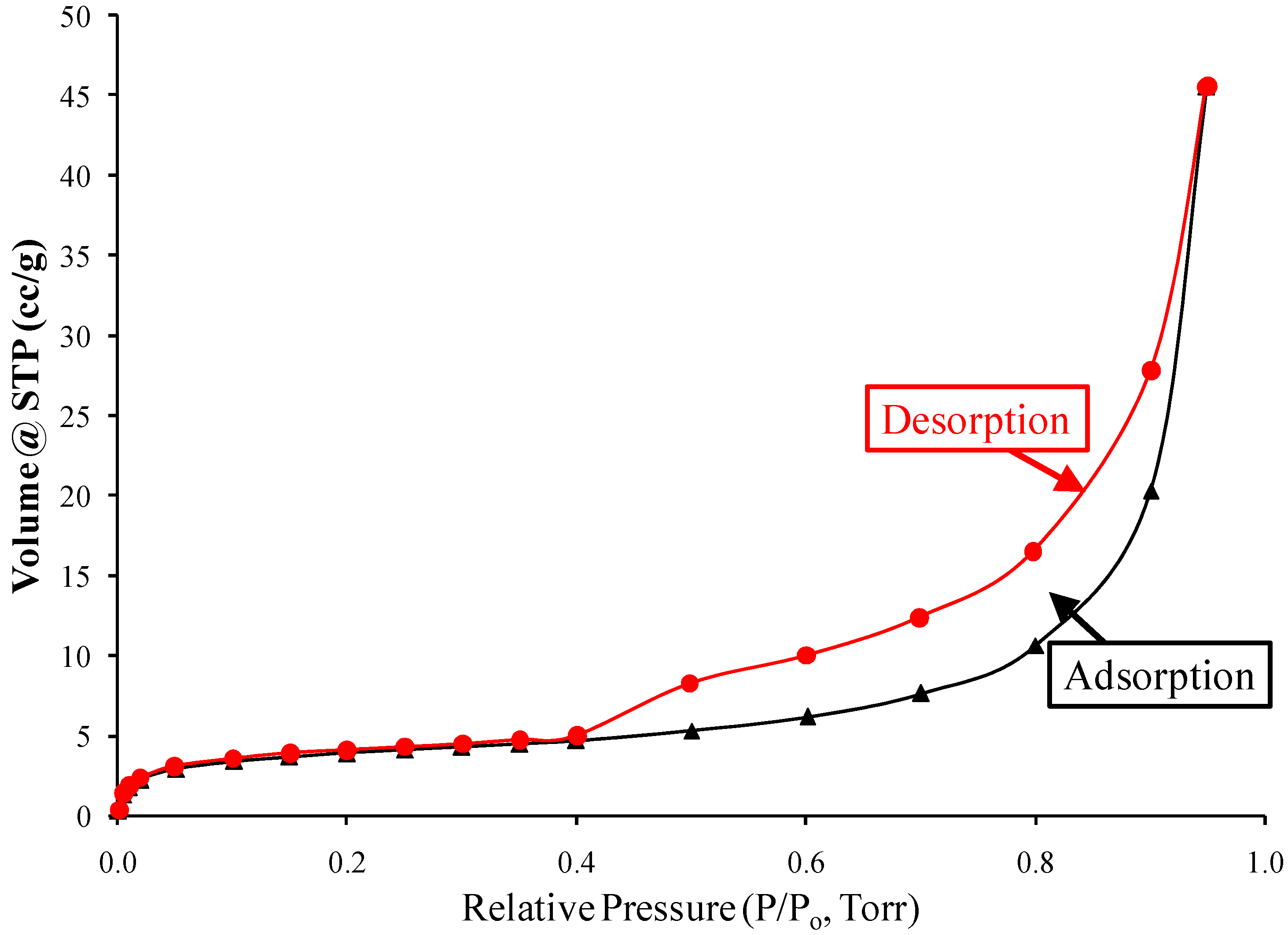
Figure 6 shows the N2 adsorption–desorption isotherm of the ZnO_calc sample. According to the IUPAC classification [28], the isotherm corresponds to type II with an H4 hysteresis loop, characteristic of mesoporous ZnO nanoparticles. This indicates a pore structure capable of accommodating substantial amounts of adsorbed gas, which is beneficial for applications requiring high surface area, such as desorption, catalysis, and controlled-release systems. The corresponding pore size distribution is presented in Figure 7, showing nanopores ranging from 4 to 24.0 nm with an average diameter of 8.5 nm. The BET surface area was measured as 12.9 m2/g, confirming the high porosity of the synthesized nanoparticles. Such mesoporosity and surface area are expected to facilitate controlled Zn2+ release, as the nanopores can act as reservoirs, supporting both immediate nutrient availability and sustained delivery over time in agricultural applications.

3.2. Optical Properties Measurement Using UV–Visible Absorption Spectroscopy and Energy Gap Calculations

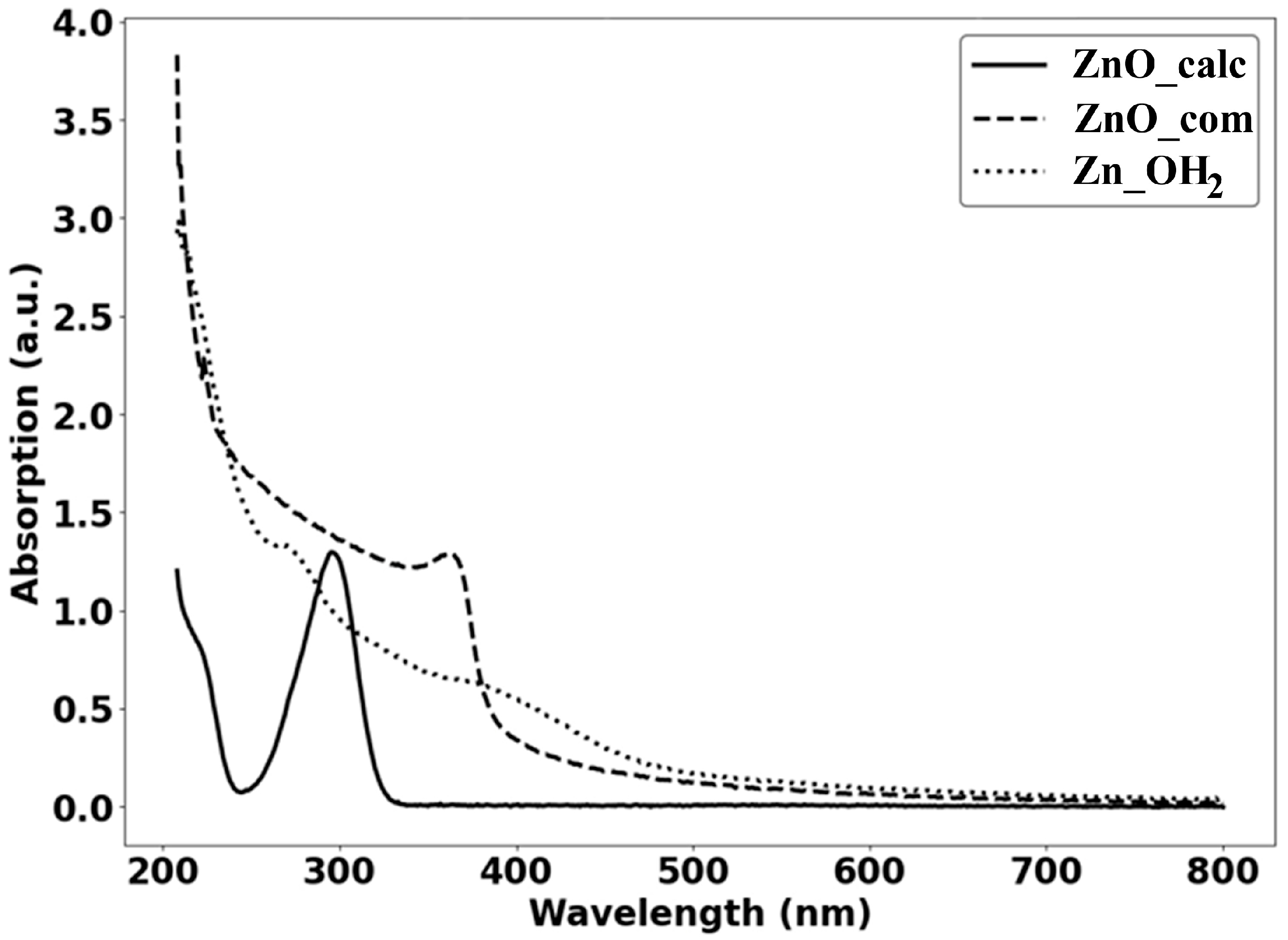
The UV–Vis spectra of the three samples (200–800 nm), shown in Figure 8, display pronounced UV absorption with clear absorption edges, enabling band-gap estimation through Tauc analysis for a direct-allowed transition (n = 1/2). The ZnO_calc sample exhibits a sharp absorption edge in the 315–335 nm region, yielding an estimated band gap of Eg = 3.6 ± 0.1 eV, which is slightly blue-shifted relative to bulk ZnO, consistent with quantum confinement effects. Previous studies have reported band gaps of approximately 3.39 eV for ZnO nanoparticles and 3.2 eV for ZnO nano-rods, highlighting the influence of nanoscale confinement on the optical properties of ZnO [29].
In contrast, the ZnO_com sample shows a broader absorption profile extending to 390–400 nm, corresponding to Eg = 3.23 ± 0.07 eV; this red shift and band-tail formation can be linked to Si-induced surface states and increased scattering effects. Meanwhile, the Zn_OH2-derived sample demonstrates a diffuse absorption edge around 330–355 nm, giving an apparent Eg = 3.45 ± 0.10 eV, reflecting its limited crystallinity and incomplete transformation to ZnO. These findings collectively indicate that phytochemical-assisted crystallization promotes the formation of well-defined ZnO with a wider band gap, whereas Si coating reduces and broadens the optical transition through surface modification, and Zn(OH)2 remains optically less distinct until fully converted. Similar trends have been reported in other green synthesis studies, where ZnO nanoparticles are produced from plant extracts such as pomegranate, beetroot, and other botanicals, often exhibiting lower band gap values than chemically synthesized counterparts, enhancing both light absorption and surface reactivity [30].

3.3. Controlled Release of Zn2+ Ions

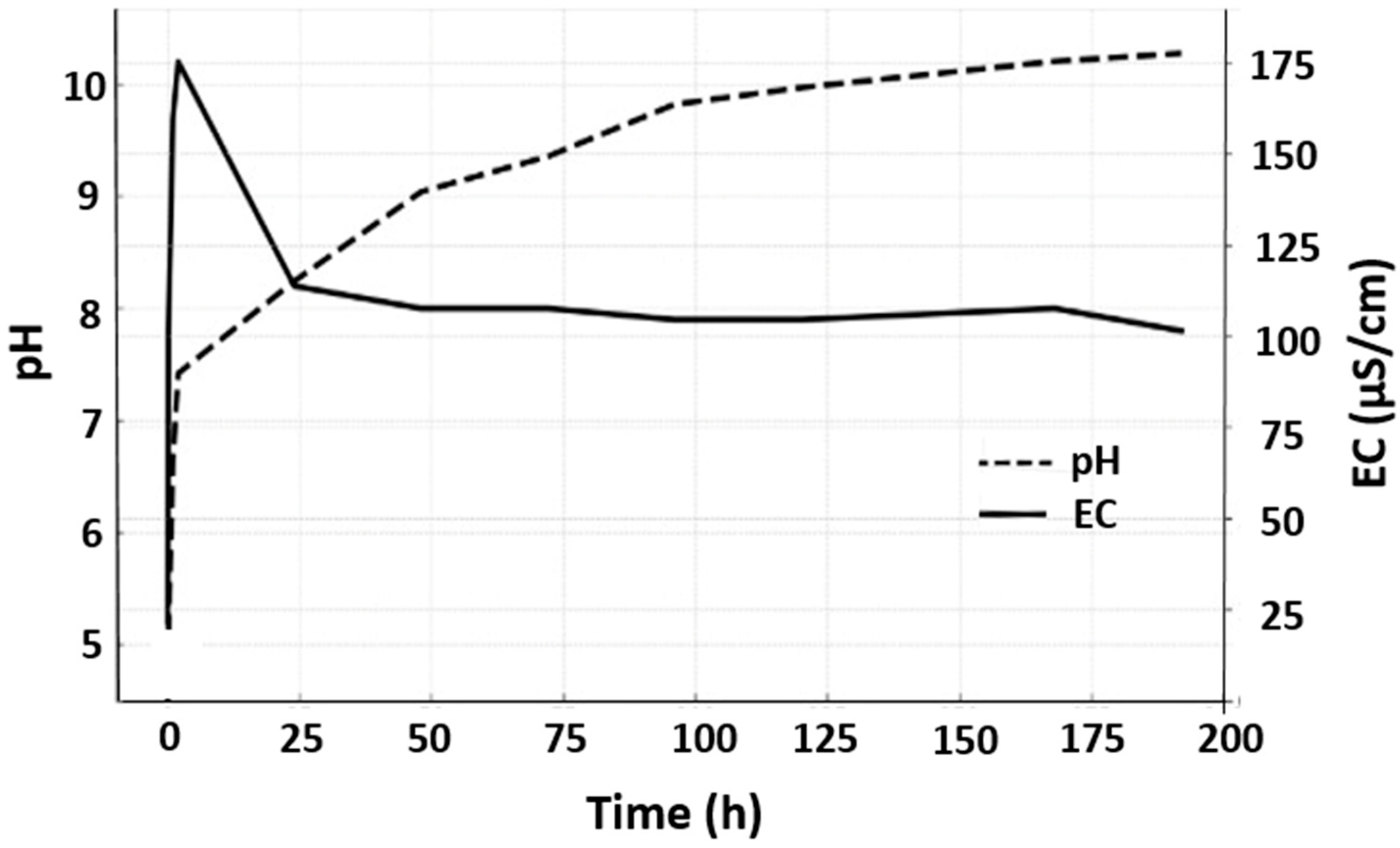
The combined pH and electrical conductivity (EC) profile (Figure 9) reflects the ionic release kinetics of the ZnO_calc sample. An initial sharp rise in pH (above 9 within hours) and concurrent EC increase indicate rapid surface hydrolysis and partial ZnO dissolution, followed by stabilization of pH (≈7.5–8.5) and EC (~100 µS/cm), reflecting a sustained, slower-release regime. This dual-phase release provides immediate nutrient availability while ensuring long-term Zn2+ supply, reducing toxicity risk. Compared to chemically synthesized ZnO nanoparticles, which often show rapid or poorly controlled release [31,32,33], our green-synthesized ZnO exhibits a more balanced and sustained release profile. These results, supported by recent field studies demonstrating improved salt tolerance and crop yield [34], indicate that the ZnO_calc nanoparticles are effective slow-release zinc sources, delivering bioavailable zinc (≤200 ppm) and potentially modulating rhizosphere pH to favor nutrient uptake. Overall, plant-mediated synthesis offers a practical and eco-friendly approach, combining controlled nutrient delivery with enhanced functional performance and environmental safety.
As summarized in Table 2, Zn2+ release from ZnO nanoparticles is strongly governed by the synthesis route. Green-synthesized ZnO exhibited a biphasic release; an initial sharp rise in pH and EC, followed by gradual stabilization, indicative of controlled dissolution. Compared with sol–gel, precipitation or hydrothermal methods, the green-synthesized ZnO nanoparticles show slow ion diffusion and moderating Zn2+ solubility under alkaline conditions dominated by Zn(OH)2. The observed EC–pH correlation supports previous findings (Table 2), highlighting EC as a practical proxy for ionic mobility in soils. These results demonstrate that green synthesis imparts favorable slow-release characteristics, making ZnO nanoparticles suitable for sustainable zinc fertilization.

4. Conclusions

This study demonstrates a sustainable and effective green synthesis route for ZnO nanoparticles using Melia azedarach leaf extract, yielding highly crystalline, uniformly dispersed, and bioavailable nanostructures with distinct advantages over commercial silicon-coated ZnO. Comprehensive characterization confirmed the presence of organic surface capping, enhanced crystallinity, and a nanoscale band gap consistent with improved stability and reactivity. The observed sustained Zn2+ release under mildly acidic conditions further supports their potential as controlled-release nanomaterials. Taken together, these results underline the suitability of green-synthesized ZnO-NPs as low-cost, eco-friendly alternatives for agricultural and environmental applications, offering functional efficiency.

Author Contributions

R.A.S.: Conceptualization, methodology, investigation, supervision, writing—original draft. A.S.A.: Conceptualization, methodology, investigation. M.E.: Investigation, formal analysis, validation, writing—original writing—review and editing. N.M.: Data curation, supervision. N.B.H.: visualization. B.A.R.: Investigation, writing—review and editing, E.R.: Investigation, writing—review and editing, M.A.S.: Investigation, writing—review and editing. All authors have read and agreed to the published version of the manuscript.

Funding

This research was funded by Abdul Hameed Shoman Foundation, grant number 230800404 and The APC was funded by Abdul Hameed Shoman Foundation.

Data Availability Statement

Data is contained within the article.

Acknowledgments

The authors gratefully acknowledge the University of Petra for granting complimentary access to advanced, modern laboratories, which enabled the successful execution of the experimental work.

Conflicts of Interest

The authors declare no conflicts of interest.

References

  1. Pudhuvai, B.; Koul, B.; Das, R.; Shah, M.P. Nano-Fertilizers (NFs) for Resurgence in Nutrient Use Efficiency (NUE): A Sustainable Agricultural Strategy. Curr. Pollut. Rep. 2024, 11, 1–29. [Google Scholar] [CrossRef]
  2. Saurabh, K.; Prakash, V.; Dubey, A.K.; Ghosh, S.; Kumari, A.; Sundaram, P.K.; Jeet, P.; Sarkar, B.; Upadhyaya, A.; Das, A.; et al. Enhancing sustainability in agriculture with nanofertilizers. Discov. Appl. Sci. 2024, 6, 1–23. [Google Scholar] [CrossRef]
  3. Arora, P.K.; Tripathi, S.; Omar, R.A.; Chauhan, P.; Sinhal, V.K.; Singh, A.; Srivastava, A.; Garg, S.K.; Singh, V.P. Next-generation fertilizers: The impact of bionanofertilizers on sustainable agriculture. Microb. Cell Factories 2024, 23, 1–8. [Google Scholar] [CrossRef]
  4. Chinnasamy, G.; Chandrasekharan, S.; Bhatnagar, S. Biosynthesis of Silver Nanoparticles from Melia azedarach: Enhancement of Antibacterial, Wound Healing, Antidiabetic and Antioxidant Activities. Int. J. Nanomed. 2019, 14, 9823–9836. [Google Scholar] [CrossRef]
  5. Ganguly, S.; Sengupta, J. Green synthesis of metal oxide nanoparticles. In Handbook of Green and Sustainable Nanotechnology; Shanker, U., Manviri, R., Chaudhury, M.H., Eds.; Springer: Berlin/Heidelberg, Germany, 2023; pp. 303–327. [Google Scholar] [CrossRef]
  6. Dinga, E.; Mthiyane, D.M.N.; Marume, U.; Botha, T.L.; Horn, S.; Pieters, R.; Wepener, V.; Ekennia, A.; Onwudiwe, D.C. Biosynthesis of ZnO nanoparticles using Melia azedarach seed extract: Evaluation of the cytotoxic and antimicrobial potency. OpenNano. 2022, 8, 100068. [Google Scholar] [CrossRef]
  7. Maciel, M.V.; Morais, S.M.; Bevilaqua, C.M.L.; Camurça-Vasconcelos, A.L.F.; Costa, C.T.C.; Castro, C.M.S. Ovicidal and larvicidal activity of Melia azedarach extracts on Haemonchuscontortus. Vet. Parasitol. 2006, 140, 98–104. [Google Scholar] [CrossRef] [PubMed]
  8. Makarov, V.V.; Love, A.J.; Sinitsyna, O.V.; Makarova, S.S.; Yaminsky, I.V.; Taliansky, M.E.; Kalinina, N.O. “Green” nanotechnologies: Synthesis of metal nanoparticles using plants. Acta Naturae 2014, 6, 35–44. [Google Scholar] [CrossRef]
  9. Haq, S.I.; Nisar, M.; Zahoor, M.; Ikram, M.; Islam, N.U.; Ullah, R.; Alotaibi, A. Green fabrication of silver nanoparticles using Melia azedarach ripened fruit extract, their characterization, and biological properties. Green Process. Synth. 2023, 12, 20230029. [Google Scholar] [CrossRef]
  10. Kołodziejczak-Radzimska, A.; Jesionowski, T. Zinc oxide—From synthesis to application: A review. Materials 2014, 7, 2833–2881. [Google Scholar] [CrossRef]
  11. Sirelkhatim, A.; Mahmud, S.; Seeni, A.; Kaus, N.H.M.; Ann, L.C.; Bakhori, S.K.M.; Hasan, H.; Mohamad, D. Review on zinc oxide nanoparticles: Antibacterial activity and toxicity mechanism. Nano-Micro Lett. 2015, 7, 219–242. [Google Scholar] [CrossRef]
  12. Eixenberger, J.E.; Anders, C.B.; Hermann, R.J.; Brown, R.J.; Reddy, K.M.; Punnoose, A.; Wingett, D.G. Rapid dissolution of ZnO nanoparticles induced by biological buffers significantly impacts cytotoxicity. Chem. Res. Toxicol. 2017, 30, 1641–1651. [Google Scholar] [CrossRef]
  13. Alhasaniah, A.H.; Aldabaan, N.A.; Reddy, D.; Alzahrani, H.A.; Shaikh, I.A.; Mahnashi, M.H.; Mannasaheb, B.A.; Muddapur, U.M.; Khan, A.A.; Bahafi, A.; et al. Harnessing Asystasia gangetica Flower Extract and Green Synthesized Zinc Oxide Nanoparticles for Combating Multidrug–Resistant Pathogens. ChemistrySelect 2025, 10, e00965. [Google Scholar] [CrossRef]
  14. Mahajan, M.; Kumar, S.; Gaur, J.; Kaushal, S.; Somvanshi, A.; Kaur, H.; Singh, G.; Hatshan, M.R.; Kumar, S.; Lotey, G.S. Role of cellulose, phenolic compounds, and water-soluble proteins in ZnO nanoparticle synthesis using Mangifera indica leaf extract for photocatalytic and antioxidant investigations. Colloids Surf. A Physicochem. Eng. Asp. 2025, 720, 137066. [Google Scholar] [CrossRef]
  15. SelvaKumar, M.; Jeyamurugan, R.; Jose, P.A.; Ponvel, K.M.; Sundarajan, M. Green Synthesis, Characterization, and Biological Evaluation of CuO–ZnO Nanoparticles Using Cyphostemma Setosum Leaf Extract. ChemistrySelect 2025, 10, e06062. [Google Scholar] [CrossRef]
  16. Aspoukeh, P.K.; Barzinjy, A.A.; Hamad, S.M. Synthesis, properties and uses of ZnO nanorods: A mini review. Int. Nano Lett. 2022, 12, 153–168. [Google Scholar] [CrossRef]
  17. Sharma, D.K.; Shukla, S.; Sharma, K.K.; Kumar, V. A review on ZnO: Fundamental properties and applications. Mater. Today: Proc. 2022, 49, 3028–3035. [Google Scholar] [CrossRef]
  18. Wang, Q.; Xu, S.; Zhong, L.; Zhao, X.; Wang, L. Effects of zinc oxide nanoparticles on growth, development, and flavonoid synthesis in Ginkgo biloba. Int. J. Mol. Sci. 2023, 24, 15775. [Google Scholar] [CrossRef]
  19. Khan, S.H.; Suriyaprabha, R.; Pathak, B.; Fulekar, M.H. Development of zinc oxide nanoparticle by sonochemical method and study of their physical and optical properties. In AIP Conference Proceedings. International Conference on Mathematics, Engineering and Computer Science, Kuala Lumpur, Malaysia, 26 February 2016; Publishing LLC: Melville, NY, USA, 2016; Volume 1724. [Google Scholar] [CrossRef]
  20. Chen, H.; Song, Y.; Wang, Y.; Wang, H.; Ding, Z.; Fan, K. Zno nanoparticles: Improving photosynthesis, shoot development, and phyllosphere microbiome composition in tea plants. J. Nanobiotechnol. 2024, 22, 1–28. [Google Scholar] [CrossRef]
  21. Gupta, A.; Bharati, R.; Kubes, J.; Popelkova, D.; Praus, L.; Yang, X.; Severova, L.; Skalicky, M.; Brestic, M. Zinc oxide nanoparticles application alleviates salinity stress by modulating plant growth, biochemical attributes and nutrient homeostasis in Phaseolus vulgaris L. Front. Plant Sci. 2024, 15, 1432258. [Google Scholar] [CrossRef]
  22. Cullity, B.D.; Stock, S.R. Elements of X-Ray Diffraction, 3rd ed.; Prentice Hall: Upper Saddle River, NJ, USA, 2001. [Google Scholar]
  23. Ayesh, A.S.; Abdel-Rahem, R.A. Optical and electrical properties of polycarbonate/MnCl2 composite films. J. Plast. Film Sheeting 2008, 24, 109–124. [Google Scholar] [CrossRef]
  24. Tauc, J. Optical properties and electronic structure of amorphous Ge and Si. Mater. Res. Bull. 1968, 3, 37–46. [Google Scholar] [CrossRef]
  25. Abdelbaky, A.S.; Abd El-Mageed, T.A.; Babalghith, A.O.; Selim, S.; Mohamed, A.M. Green synthesis and characterization of ZnO nanoparticles using Pelargonium odoratissimum (L.) aqueous leaf extract and their antioxidant, antibacterial and anti-inflammatory activities. Antioxidants 2022, 11, 1444. [Google Scholar] [CrossRef]
  26. Hoseinpour, V.; Souri, M.; Ghaemi, N.; Shakeri, A. Optimization of green synthesis of ZnO nanoparticles by Dittrichia graveolens (L.) aqueous extract. Health Biotechnol. Biopharma. 2017, 1, 39–49. [Google Scholar]
  27. Duong, T.B.N.; Pham, P.-Q.; Tran, A.T.; Bui, D.T.; Pham, A.T.T.; Nguyen, T.C.T.; Nguyen, L.H.T.; Ung, T.D.T.; Hoang, N.V.; Pham, N.K. Correlation between organic residuals of green synthesized nanoparticles and resistive switching behavior. RSC Adv. 2024, 14, 36340–36350. [Google Scholar] [CrossRef] [PubMed]
  28. Thommes, M.; Kaneko, K.; Neimark, A.V.; Olivier, J.P.; Rodriguez-Reinoso, F.; Rouquerol, J.; Sing, K.S.W. Physisorption of gases, with special reference to the evaluation of surface area and pore size distribution (IUPAC Technical Report). Pure Appl. Chem. 2015, 87, 1051–1069. [Google Scholar] [CrossRef]
  29. Musa, I.; Qamhieh, N. Study of optical energy gap and quantum confinment effects in Zinc Oxide nanoparticles and nanorods. Dig. J. Nanomater. Biostruct. 2019, 14, 119–125. [Google Scholar]
  30. Dhandapani, K.V.; Anbumani, D.; Gandhi, A.D.; Annamalai, P.; Muthuvenkatachalam, B.S.; Kavitha, P.; Ranganathan, B. Green route for the synthesis of zinc oxide nanoparticles from Melia azedarach leaf extract and evaluation of their antioxidant and antibacterial activities. Biocatal. Agric. Biotechnol. 2020, 24, 101517. [Google Scholar] [CrossRef]
  31. Ekanayake, S.A.; Godakumbura, P.I. Synthesis of a Dual-Functional Nanofertilizer by Embedding ZnO and CuO Nanoparticles on an Alginate-Based Hydrogel. ACS Omega 2021, 6, 26262–26272. [Google Scholar] [CrossRef]
  32. Yuvaraj, M.; Subramanian, K.S.; Cyriac, J. Efficiency of zinc oxide nanoparticles as controlled release nanofertilizer for rice (Oryza sativa L). J. Plant Nutr. 2023, 46, 4477–4493. [Google Scholar] [CrossRef]
  33. Rodrigues, S.; Avellan, A.; Bland, G.D.; Miranda, M.C.R.; Larue, C.; Wagner, M.; Moreno-Bayona, D.A.; Castillo-Michel, H.; Lowry, G.V.; Rodrigues, S.M. Effect of a Zinc Phosphate Shell on the Uptake and Translocation of Foliarly Applied ZnO Nanoparticles in Pepper Plants (Capsicum annuum). Environ. Sci. Technol. 2024, 58, 3213–3223. [Google Scholar] [CrossRef]
  34. Deger, A.G.; Çevik, S.; Kahraman, O.; Turunc, E.; Yakin, A.; Binzet, R. Effects of green and chemically synthesized ZnO nanoparticles on Capsicum annuum under drought stress. Acta Physiol. Plant. 2025, 47, 1–17. [Google Scholar] [CrossRef]
  35. Aalami, Z.; Hoseinzadeh, M.; Manesh, P.H.; Aalami, A.H.; Es’HAghi, Z.; Darroudi, M.; Sahebkar, A.; Hosseini, H.A. Synthesis, characterization, and photocatalytic activities of green sol-gel ZnO nanoparticles using Abelmoschus esculentus and Salvia officinalis: A comparative study versus co-precipitation-synthesized nanoparticles. Heliyon 2024, 10, e24212. [Google Scholar] [CrossRef]
  36. Irede, E.L.; Awoyemi, R.F.; Owolabi, B.; Aworinde, O.R.; Kajola, R.O.; Hazeez, A.; Raji, A.A.; Ganiyu, L.O.; Onukwuli, C.O.; Onivefu, A.P.; et al. Cutting-edge developments in zinc oxide nanoparticles: Synthesis and applications for enhanced antimicrobial and UV protection in healthcare solutions. RSC Adv. 2024, 14, 20992–21034. [Google Scholar] [CrossRef] [PubMed]
  37. David, C.A.; Galceran, J.; Rey-Castro, C.; Puy, J.; Companys, E.; Salvador, J.; Monné, J.; Wallace, R.; Vakourov, A. Dissolution Kinetics and Solubility of ZnO Nanoparticles Followed by AGNES. J. Phys. Chem. C 2012, 116, 11758–11767. [Google Scholar] [CrossRef]
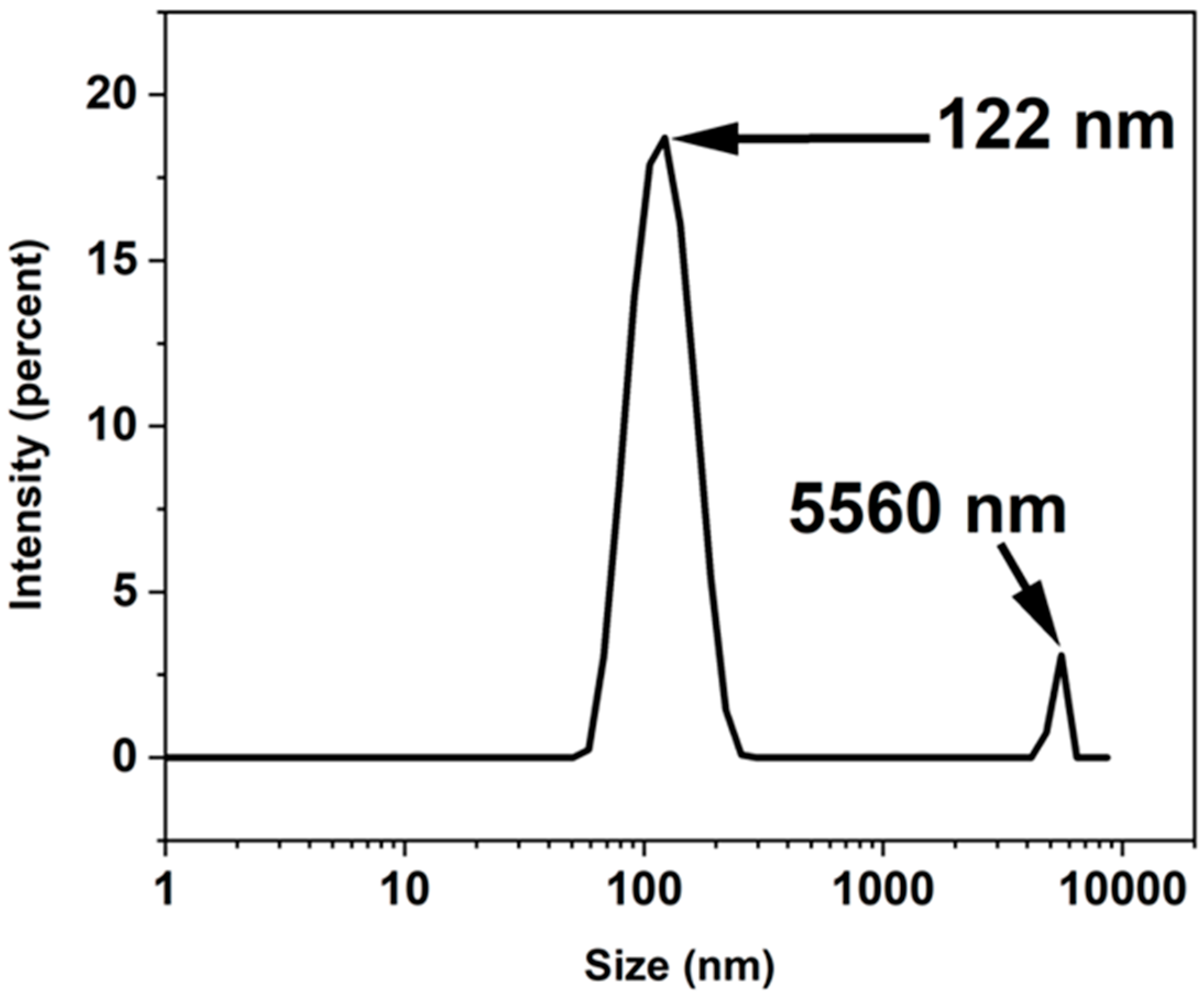
Figure 1. LSD result and size distribution of ZnO-NPs.
Solids 06 00059 g001
Figure 2. FTIR spectra of (a) Zn-OH2 and (b) ZnO_calc samples.
Solids 06 00059 g002
Figure 3. SEM-EDS micrograph of ZnO_calc sample.
Solids 06 00059 g003
Figure 4. SEM-EDS micrograph of ZnO_com sample.
Solids 06 00059 g004
Figure 5. XRD pattern of ZnO_calc sample.
Solids 06 00059 g005
Figure 6. N2 adsorption–desorption isotherm curve of ZnO_calc sample.
Solids 06 00059 g006
Figure 7. Pore size distribution curves that have been calculated by density functional theory at STP for the ZnO_calc sample.
Solids 06 00059 g007
Figure 8. UV–Visible absorption spectra of ZnO_calc, ZnO_com and Zn_OH2 samples.
Solids 06 00059 g008
Figure 9. Ionic release behavior of Zn2+ from ZnO_calc sample.
Solids 06 00059 g009
Table 1. XRD Peak Parameters of ZnO_calc sample.
Miller IndexPeak Position
(2θ Deg)
FWHMCrystallite Size D (nm)
(100)31.70.445618.5
(002)34.40.84059.9
(101)36.20.501216.7
(102)47.50.89099.7
(110)56.60.94899.5
Table 2. Comparative summary of Zn2+ ion release behavior from ZnO synthesized by different methods.
Synthesis MethodpH Behavior over TimeEC Behavior over TimeZn2+ Release CharacteristicsRef.
Sol–gelGradually increases with time as dissolution proceedsEC steadily increases over longer periodModerate-to-high release rate is attributed to the porous structure, which increases surface reactivity and facilitates ion exchange.[35]
HydrothermalStable pH profile; minimal variationLow and stable EC valuesExhibits the lowest Zn2+ release rate owing to its dense crystal structure and minimal surface defects that restrict dissolution.[36]
PrecipitationDecreases slightly with time as Zn2+ hydrolyzesRapid EC rise initially, stabilizing after a few hoursModerate release rate resulting from particle aggregation and the presence of residual ions that promote initial Zn2+ release.[37]
Present Green synthesis studyRapid early increase (peak ≈ 10 at ~few h) then stabilizes to mildly alkaline ~8–8.5 over long termEC: sharp early rise, then gradual increase and plateau (~170 µS/cm by 200 h). EC correlates with ionic release.Controlled and sustained Zn2+ release is attributed to phytochemical capping agents that decrease solubility and enable gradual dissolution.
Disclaimer/Publisher’s Note: The statements, opinions and data contained in all publications are solely those of the individual author(s) and contributor(s) and not of MDPI and/or the editor(s). MDPI and/or the editor(s) disclaim responsibility for any injury to people or property resulting from any ideas, methods, instructions or products referred to in the content.