Review

Doped Tin Dioxide (d-SnO2) and Its Nanostructures: Review of the Theoretical Aspects, Photocatalytic and Biomedical Applications

by
Alexandre H. Pinto
1,*,
Andre E. Nogueira
2,
Cleocir J. Dalmaschio
3,
Iago N. Frigini
3,
Jéssica C. de Almeida
4,5,
Mateus M. Ferrer
6,
Olivia M. Berengue
7,
Rosana A. Gonçalves
8 and
Vagner R. de Mendonça
4,9
1
School of Science, Department of Chemistry and Biochemistry, Manhattan College, 4513 Manhattan College Parkway, Riverdale, NY 10471, USA
2
Institute of Exact and Biological Sciences (ICEB), Department of Chemistry, Federal University of Ouro Preto-UFOP, Ouro Preto 35400-000, MG, Brazil
3
Center of Exact Sciences, Department of Chemistry, Federal University of Espirito Santo, Av. Fernando Ferrari, 514 Vitoria, Goiabeiras 29075-910, ES, Brazil
4
Science and Technology Center for Sustainability, Federal University of São Carlos, Rod. SP-264, km 110, Sorocaba 18052-780, SP, Brazil
5
Institute of Energy and Climate Research (IEK-14): Electrochemical Process Engineering Forschungszentrum Jülich GmbH, 52425 Jülich, Germany
6
Technological Development Center, Federal University of Pelotas, Pelotas 96010-610, RS, Brazil
7
Department of Physics, School of Engineering, São Paulo State University (UNESP), Guaratinguetá 12516-410, SP, Brazil
8
Federal Institute Baiano of Education, Science and Technology—Campus Xique-Xique, Rodovia Ba 052, Km 458, s/n Zona Rural, Xique-Xique 47400-000, BA, Brazil
9
Federal Institute of Education, Science, and Technology of São Paulo, Av. João Olímpio de Oliveira, 1561, Itapetininga 18208-000, SP, Brazil
*
Author to whom correspondence should be addressed.
Solids 2022, 3(2), 327-360; https://doi.org/10.3390/solids3020024
Submission received: 19 April 2022 / Revised: 24 May 2022 / Accepted: 27 May 2022 / Published: 2 June 2022

Abstract

:
Nanomaterials based on metal oxides are extensively studied for several applications due to their versatility. Improvements in their performances can be obtained due to specific structural modifications. One possible modification is by doping the crystal structure, which can affect the materials structure and properties, especially in nanosized particles. Electronic features are among the properties that can be modified through the doping process, consequently morphological and optical parameters can also be controlled by this process. In this sense, this review presents some modifications to tin dioxide (SnO2), one the most studied materials, mainly through the doping process and their impact on several properties. The article starts by describing the SnO2 structural features and the computational models used to explain the role of the doping process on these features. Based on those models, some applications of doped SnO2, such as photocatalytic degradation of pollutants, CO2 reduction, and desulfurization of fossil fuels are presented and discussed. Additionally, the review describes many biological applications related to antimicrobial activity for doped SnO2 and its nanostructures. Although most of the examples presented in this article are based on the doped SnO2, it also presents examples related to SnO2 composites with other nanomaterials forming heterojunctions. The metal oxides SnO2, doped-SnO2 and their nanostructures are promising materials, with results reported in many fields presented in this review, such as theoretical and computational chemistry, environmental remediation, nanoparticle morphology control, fossil fuels improvement, and biomedical applications. Although widely explored, there are still fields for innovation and advances with tin dioxide nanostructures, for example, in transparent conducting oxides, in forensics as materials for latent fingerprints visualization, and sensors in medicine for detection of exhaled volatile organic compounds. Therefore, this article aims to be a reference regarding correlating the doping processes and the properties presented by the SnO2 nanostructures.

1. Introduction

The growth of fundamental and applied research on nanomaterials in several fields in the last three decades indicates the relevance of nanostructures and their potential applications. Among metal oxide semiconductors, tin dioxide (SnO2) has been the subject of interest in numerous research projects due to its excellent physicochemical properties, such as high photosensitivity, chemical, thermal and mechanical stability, long-term stability, transparency, abundant reserve, low cost, and nontoxicity [1,2,3]. Tin dioxide is an n-type semiconductor with bandgap values between 3.6–3.8 eV; it has a low valence band potential, which gives the photogenerated holes in the valence band (VB) a high oxidation capacity [4]. In addition, SnO2 has high electron mobility (~240 cm2/V·s), indicating a faster transport of photoexcited electrons and a higher potential conduction band (CB), with great potential for application in several processes, such as gas sensing, photocatalysis, H2 generation, solar cells, supercapacitors, lithium-ion battery electrodes, optoelectronic devices and transparent conducting electrodes to modern medical applications such as cancer treatments [5,6,7], diseases diagnosis [8,9,10], and DNA biosensors [10,11,12].
Although SnO2 has promising properties for some of the mentioned applications, for instance, for photocatalysis, it still presents some limitations because of its wide bandgap and high electron-hole pair recombination rate, resulting in low efficiency.
One possibility to overcome this drawback is based on tuning parameters during the synthesis and preparation of these nanomaterials. For instance, Babu et al. demonstrated that it is possible to modify the band structure of SnO2 quantum dots by varying both the CB and VB potentials and the SnO2 bandgap value from the UV region to the visible wavelength region, simply by varying the quantum dots annealing temperature, as shown in Figure 1. This bandgap shift may have a beneficial impact on the possibility of using solar radiation to activate the semiconductor [13].
Another interesting approach to overcome the limitations related to the wide bandgap and electron-hole recombination high rate is by doping the SnO2 with metals [14,15], transition metals ions [16,17], or nonmetal elements [18]. Doping the SnO2 can extend its absorption spectrum to visible light regions, reducing the necessary energy for the photoactivation and allowing the use of solar light as an energy source during its application [19].
In general, doping can be classified as p-doping and n-doping. The n-doping happens when a doping ion donates electrons to the conduction band of the SnO2, thus increasing the number of negative charge carriers. Commonly, the n-doping is carried by antimony (generally with mixed oxidations states Sb+3/Sb+5) and fluorine ions (F) [20,21]. On the other hand, p-doping happens when the doping ion creates holes in the SnO2 VB, thus, increasing the number of positive charge carriers. Although less commonly attained than the n-doping, the SnO2 p-doping has been accomplished by using In+3, Ga+3, Al+3, Li+3 ions, and Al+3 co-doped with N [22,23,24].
Additionally, the p- and n-doping create intermediate energy levels between the VB and CB, changing the relative position of the VB and CB in relation to Fermi energy level (EF), as shown in Figure 2. In the case of a n-doped semiconductor, the intermediate level is created in the bottom of the CB, and it is called the donor level, whereas in the case of the p-doped semiconductor, the intermediate level is created on the top of the VB, and it is called the acceptor level. The doping and the creation of these intermediate states makes less likely that electrons can be directly excited from the VB to the CB, like it would happen in an intrinsic or undoped semiconductor [25].
In the case of the n-doped semiconductor, as the doping species has extra valence electrons in relation to the host material, the electrons are excited from the VB to the donor level. Then, the energy difference between the donor level and CB is equivalent to the electron biding energy, which is relatively smaller than the bandgap energy (Eg) of the undoped semiconductor. Consequently, the thermal energy at room temperature is enough to excite the electrons from the donor level to the CB. As the electrons are provided from the donor level to the CB, no counterpart hole is created in the VB. Consequently, the number of electrons present in the CB is higher than the number of holes present in the VB. For that reason, electrons are the major charge carriers present in n-doped semiconductors [25].
In the case of the p-doped semiconductor, as the doping species is electron deficient in relation to the host materials, holes are created in the acceptor level. Then, thermal excitation can transition electrons from the VB to the acceptor level, creating a counterpart hole in the VB, as the energy difference between the VB and the acceptor level is lower than the bandgap energy for the undoped semiconductor. Each hole in the acceptor level can accept or recombine with an electron thermally excited from VB to the acceptor level. This recombination makes the electron unable to be excited to the conduction band, and consequently leaves the VB with an excess of holes. For that reason, holes are the major charge carriers present in the p-doped semiconductors [25].
In summary, the creation of these intermediate energy levels can change the electronic and optical properties of the doped SnO2 (d-SnO2), compared to the undoped one [25,26].
The nanosized SnO2 can exhibit varying morphologies, such as nanoparticles, nanobelts, nanodisks, nanowires, nanoflowers, hollow nanospheres, among others [27,28,29,30]. The doping process can promote changes in morphological and crystalline aspects of this material as well. In fact, inserting a different atom or ion into the crystalline lattice can promote defects in the structure and can lead to growth patterns different from those expected to the undoped SnO2. Thus, doping can impact morphological features.
An additional consequence of doping, when using species with different oxidation states and coordination numbers from the host lattice, is associated with the preferential sites for the doping atom [31]. The strain promoted by the doping agent will lead to the insertion of the atom in sites to reduce the impact on the total free energy. A surface site will be preferred to host the doping species; in this way, a general tendency will be to reduce crystallite sizes to reduce strain and creates sites for doping at the surface. This also reflects in the percentage of dopant that SnO2 lattice will support, once the nanometric materials have higher specific surface area than bulk material. Therefore, nanometric structures that incorporate dopant in facets and edges of nanocrystals achieve superior doping amount.
In this sense, this review article presents how doping can change the structural, morphological, and theoretical aspects and properties of the SnO2 nanostructures. The review starts presenting the structural and theoretical aspects of the undoped SnO2. Then, it follows with some applications of the d-SnO2, such as photocatalytic degradation of pollutants, CO2 photoreduction, fossil fuels desulfurization, and biological applications, which are examined in depth. Finally, further applications and possible future challenges are presented. Although the focus is placed on the doped SnO2 nanostructures, the scope of this review article is not limited to d-SnO2. In fact, examples of other nanostructures, such as heterostructures and composites, are presented if they are appropriate for the topic in discussion.

2. SnO2 Structural and Theoretical Aspects

The most stable crystal structure of SnO2 at room temperature and pressure is the cassiterite, which relates to the tetragonal rutile structure, Figure 3. The rutile structure belongs to the D 4 h 14   symmetry group, with two formula-units per unit cell, and lattice parameters around 0.473 nm, for the lattice parameter a, and 0.318 nm, for the lattice parameter c [32].
An in-depth understanding of material properties and how to manipulate them is often not achieved solely by experimental methods. The use of theoretical and computational methods for the elaboration of precise models that give the basis for such discussions is being increasingly applied in conjunction with experimental studies as they are achieving estimated results increasingly closer to the experimental data [33].
An adequate theoretical model with accurate estimates is a key point to understanding the structure-property relationship of a material. Such a theoretical perspective makes possible the controlled planning of the modulation and optimization of properties, mainly dealing with multifunctional materials such as SnO2.
In particular, the study of d-SnO2 can be well evaluated theoretically by computational methods based on quantum mechanics because they can describe both the atomic and energetic structure of the system. The addition of dopants can drastically change the energy levels of the system [34]. Thus, the understanding of these levels, as well as diverse factors such as polarization, spin, and morphological effects, are essential for the elaboration of the mechanisms behind the origin of the properties.

2.1. Computational Tools and Density Functional Theory

The evaluation of molecular orbitals of solid systems for many years was considered to be a great challenge within quantum mechanics due to the “infinite” number of atoms that composes the system. In the 1960s, the Hohenberg and Kohn [35] and Kohn and Sham [36] theorems, the bases of the Density Functional Theory (DFT), initiated the new possibilities of applications of quantum mechanics in systems of many particles.
Briefly, the DFT represents an expressive simplification where the many-electron system is replaced by an equivalent set of self-consistent one-electron equations. The total energy is described as an electronic density functional ( E = E ρ ) and can be written in the form as E = T ρ + U ρ + E X C ρ , where T[ ρ ] is the Kinetic energy of non-interaction electrons, U ρ is the Coulomb’s energy of the electrons and E X C ρ is the exchange-correlation energy correction term. The correction of the Kinetic term due to the particles’ interactions is presented in the last term Exc. Therefore, it is necessary to find the energy functional in its minimum energy condition, which is done from an eigenvalue problem known as the Kohn–Sham equation. The full Kohn–Sham equation will not be presented here and, despite being an exact equation, the true form of the term Exc is not known. Thus, several functionals, or functionals elaborated from the sum of functionals, are used to generate an adequate approximation so that the data can be compared with experimental values.
However, the emergence of the DFT alone did not allow the calculation of many atoms. Therefore, it was necessary to study the crystalline periodicity and also the expected periodicity of the electronic properties, which was possible with the use of Bloch’s Theorem [37,38]. In this sense, it was possible to make a model with the number of atoms of a single unit cell that was enough to reproduce the electronic properties of a system at the bulk level.
The proper simulation of doped systems is still considered a great challenge, especially in the case of a small number of impurities. The elaboration of doped models with small percentages requires a considerable number of actual sites in the theoretical model. For example, for doping in a small percentage of a substitutional impurity at the Sn site in the SnO2, it is necessary to create supercells so that several Sn positions exist. Then, the impurity is added to one of them. This becomes even more complex in the modelling of surface structures, where many layers are often required to achieve energy balance in the models. On the other hand, the improvement of the processing hardware, together with the improvements in the approximation methods and algorithms used in the calculations, have made possible results that are closer and closer to experimental results.

2.2. SnO2 Theoretical Models

The undoped SnO2 system is a relatively theoretically well-explored system via DFT by several types of functionals [39,40]. In general, the hybrid functionals have shown a greater ability to estimate both the structural data (lattice parameters) and the bandgap values of this system [39]. A detailed evaluation of the bands generated by the theoretical models shows that SnO2 in the rutile structure presents a direct Γ-Γ transition with the VB formed mainly by oxygen states and the conduction band CB mostly by states of Sn.
In addition, theoretical models can provide information about a wide range of subjects, such as vibrational modes, dielectric constant, resistivity, phase transformation, bulk modulus, and charges [40,41,42].
As much as the bulk models are indispensable for understanding the material properties, it is often necessary to estimate the surface regions from the modelling of the expected unsaturation according to the exposed plane. Such models are indispensable when it comes to simulation involving very small particles.
The theoretical study of the surface models shows that the most stable surface is the (110) plane, followed by the (100), (101), and (001) planes [43,44,45]. However, it is already well established that the stability of surfaces during crystal growth can be modified according to the synthesis conditions. The fact of generating different types of exposed surfaces according to the control of the morphology in the experimental synthesis is a crucial tool to control the properties of the materials [46].
Among the surfaces, the (110) is the one with the largest number of studies on its structure and evaluating its properties and mechanisms, due to its greater stability. For example, Chen et al. [47] used surface models to propose the reason why SnO2 is not a good sensor for H2 in the presence of air and showed that the presence of O2 is responsible for this low performance for such an application. Maleki and Pacchioni [48] showed using DFT simulations that the surface (110) of SnO2 is reactive for the decomposition of formic acid. This study also included the addition of dopants in the matrix, which will be discussed later.
In addition to the most stable surface, other surfaces were also explored. Valdez Garcia et al. [49] used surface models to show that the (101) surface has greater efficiency for catalytic oxidation of methane than other surfaces. Thus, a study is invited to try to increase the rate of surface (101) if the material is directed to this end, which may be a DFT transition state study of the catalysed oxidation of methane on SnO2 surfaces. Therefore, it is interesting to evaluate experimental strategies so that a higher surface rate the word for (101) is achieved if the material is directed to this application.

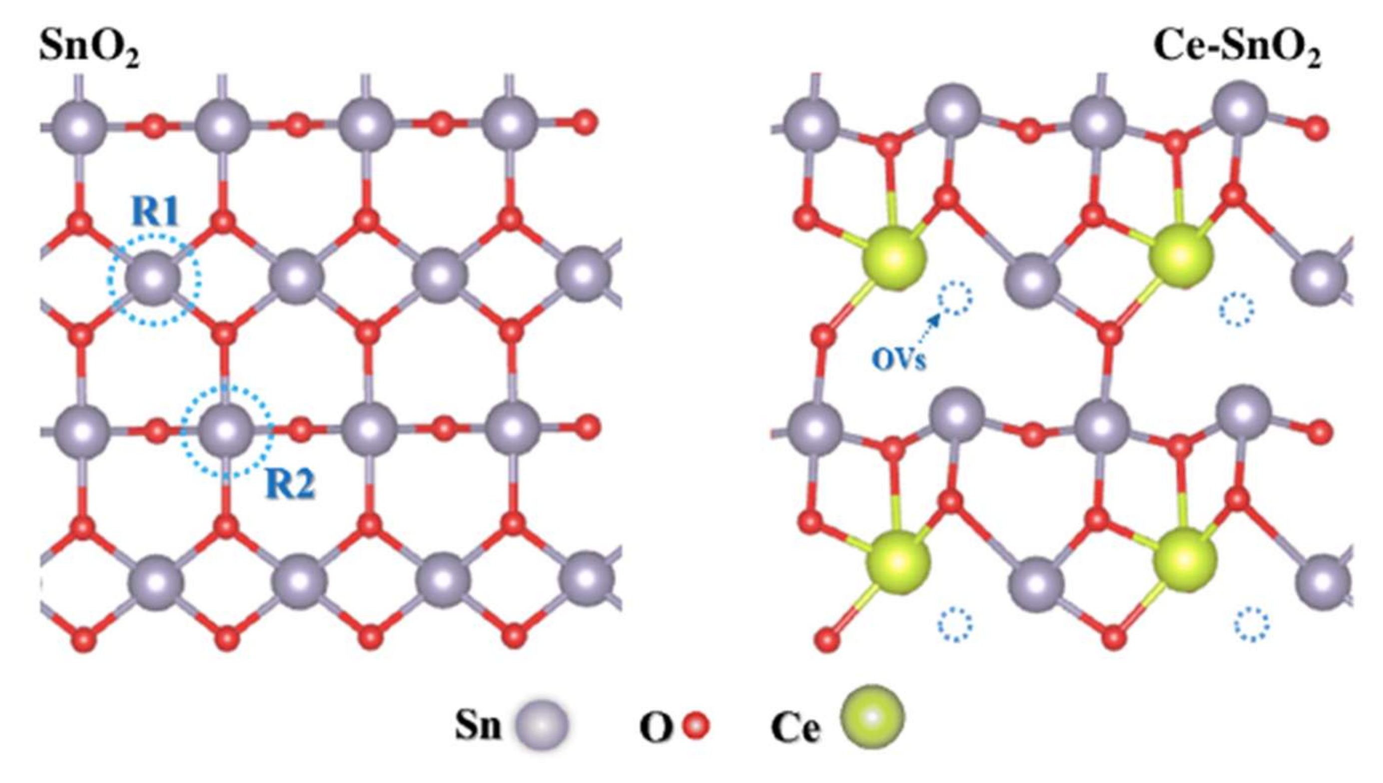
2.3. Doped SnO2 (d-SnO2)

The potential for multifunctional applications of SnO2 makes this system an incessant target of research, many of them to modify and optimize its properties. The use of dopants is a common practice in SnO2 and is constantly evaluated theoretically and experimentally. This fact is motivated by the possibility of modification of the SnO2 bandgap with the presence of dopants. However, the detailed knowledge about the energetic changes caused by the dopants at the subatomic level is only possible to be evaluated systematically from theoretical models.
The theoretical models of d-SnO2 provide information about variations in bandgaps, changes in band positions, and electronic transitions information. A detailed understanding of the relationship between the structure and the electronic characteristics is indispensable for the improvement of the devices for technological applications, such as catalysis, photocatalysis, bactericidal, and viricidal, among others [46].
An example of bandgap energy modulation caused by dopant addition was presented by Ganose and Scanlon [50]. In this study, the authors explored the variations in bandgaps caused by the substitution of Sn by Pb and the relative displacement of the bands. The band alignment scheme relative to the O 1 s state reported by the authors can be seen in Figure 4.
In this Pb doping, the bandgap clearly shows a decrease in response to the increase of the dopant percentage [50]. In certain applications, the use of smaller bandgap semiconductors improves device efficiency by facilitating electron transitions. Furthermore, even though in this example we have a simplified scheme, the band shifts are valuable information for certain applications, such as the case for the energetic understanding of electron-hole pairs.
The potential energy of the states and, in other words, of the charge carriers in an excited state are, for example, indispensable for the understanding of photocatalytic devices [51]. Therefore, changing the positions of the SnO2 bands will also change the types of reactions that the semiconductor can catalyse.
There are several theoretical models of SnO2 with the addition of dopants for the evaluation of energy changes found in literature; these are models that explore substitutions at the Sn and O sites, and also interstitial positions [34,48]. Different from the tendency observed in doping with Pb that was previously commented on, the substitution of Sn by scandium (Sc) generated an increase in the bandgap of the system, as shown by Beloufa et al. [52]. In such a study, it was estimated that the formation energy led to a thermodynamically stable doped system, and that the transition remains direct Γ-Γ even with the modifications of increasing the bandgap. Medina et al. [53] used DFT to evaluate the effects of vanadium (V) doping to understand the experimentally observed modifications. Among the results of this study, it was possible to observe that the V addition generates a unit cell compression and a decrease in the bandgap. The bandgap reduction is caused by levels generated below the Fermi level and at the bottom of the conduction band caused by the d-states of the V hybridized with p-oxygen states.
In addition to the studies with cation (Sn4+) substitution in the structure, as shown above, some papers have evaluated impurities that replace the anion O2−, including its relationship with paramagnetic properties [54,55,56]. The effects of impurity insertion also present different responses in the structural and electronic characteristics of the surfaces. The study by Zhang and co-workers [57] estimated the electronic properties of (110), (101), and (211) surfaces, of the undoped SnO2 and with the addition of nitrogen (N) as a dopant in the superficial region. The replacement of O by N on the surfaces generated a decrease in the bandgap of all surfaces. In addition, the electrochemical CO2 reduction potential of this surface was estimated according to the proposed theoretical model. The N superficial site served as a surface activation point for this purpose and the theoretical data estimated that the doped (211) surface presents the best catalytic performance for the CO2 to HCOOH conversion among the explored surfaces.
Li et al. [58], based on theoretical surface models, sought an in-depth understanding of the mechanisms of conductivity and CO sensitivity on the Sb doped SnO2 (110) and Sb, S co-doping SnO2 (110) surface. From a comparative evaluation of the state densities of the undoped and doped systems, it was observed that the addition of the two dopants simultaneously inserts several energy levels. These new levels are responsible for increasing the conductivity and can further enhance the sensitivity of the SnO2 gas sensor for CO gas.
The theoretical evaluation of the surface regions of the doped SnO2 system is still poorly explored, especially concerning details. This is mainly due to the high computational demand required in models that often require hundreds of atoms. However, it is unquestionable that the theoretical support can help in the understanding of the mechanisms and propose new strategies for modifying the properties aiming at their optimization.
Scientific/technological advances regarding processors (hardware) and algorithms that help in the simulation stages of materials are points that will increasingly leverage studies about these topics, which includes the possibility of elaborating more robust models with higher potential of estimative close to experimental data.

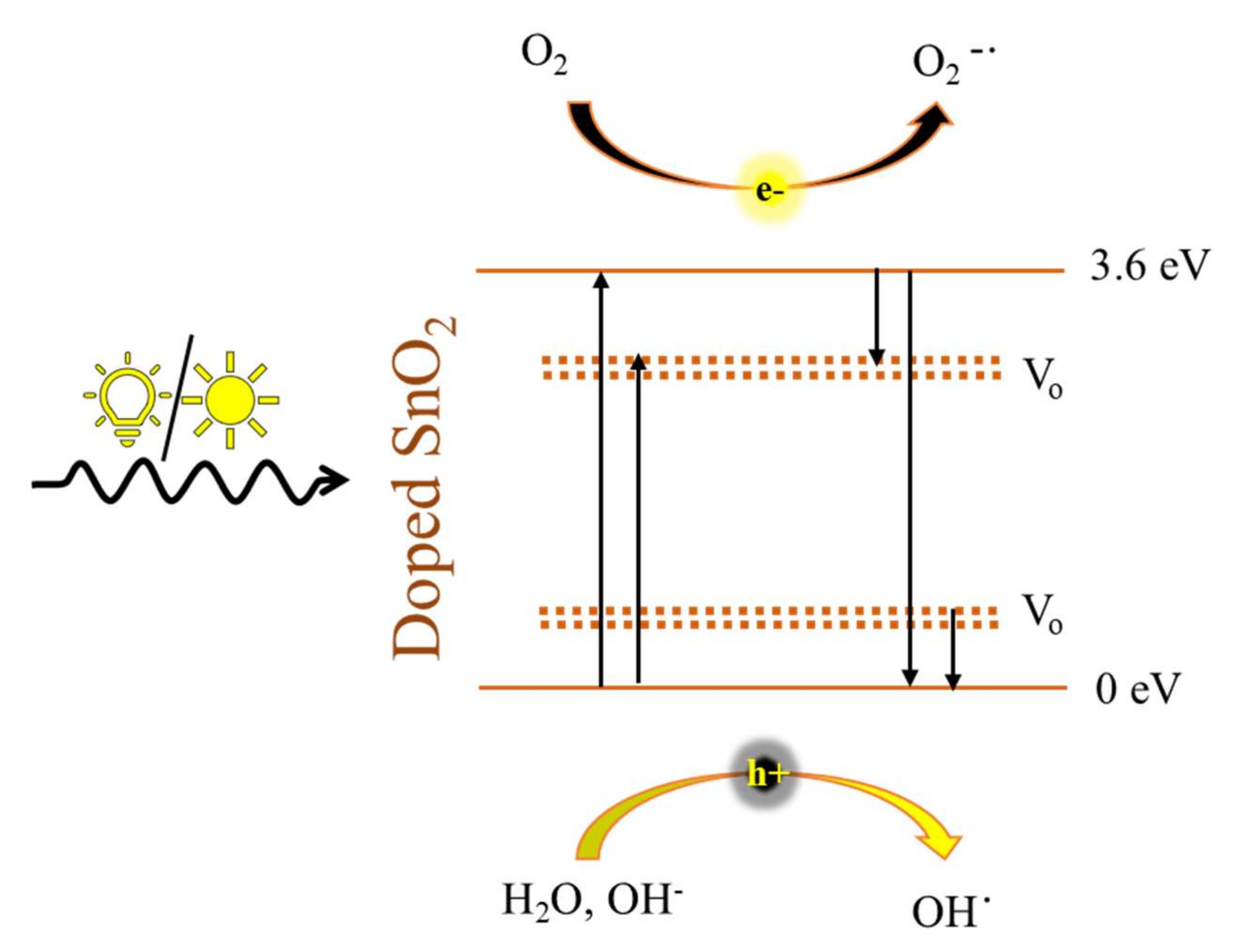
3. SnO2 and d-SnO2 Photocatalysis

Heterogeneous photocatalysis is an advanced oxidation process (AOP), which uses a solid semiconductor catalyst to promote the degradation or reduction of contaminants in the gaseous or liquid phase [59]. Heterogeneous photocatalysis is being widely investigated and may propose an attractive route for environmental remediation applications [1,60,61].
During photocatalysis, an electron-hole pair is formed by the excitation of electrons from the valence band to the conduction band of the semiconductor, as shown in Equation (1). The positive hole in the valence band (hVB+) and the electron in the conduction band (eCB) can interact with molecules in the solid interface (Equations (2)–(4)), oxidizing and reducing reagents in the medium and generating by-products by direct (Equation (2)) or indirect degradation mechanisms (Equation (5)) [60,62]. The potential of using photocatalytic processes is due to their simplicity, because the reactions take place at ambient temperature and pressure, with low energy costs, and use semiconductors as catalysts for the process. Numerous semiconductor metal oxides such as TiO2, SnO2, ZnO, WO3, CuO, and Nb2O5, have been studied to be applied as photocatalysts, either for pollutant degradation and CO2 reduction [59,62,63,64].
S n O 2     h v     S n O 2   e c b + h v b +  
S n O 2 h v b + + R X a d s o r b e d   S n O 2 + R X · +
S n O 2 h v b + + H 2 O   S n O 2 + O H · + H +
S n O 2 h v b + + O H   S n O 2 + O H ·
O H · + R X   O x i d a t i o n   b y p r o d u c t s
To perform their function as catalysts, the semiconductors need to be radiated, allowing electrons to be excited by the absorption of photons. The energy required to promote the electrons from the VB to the CB depends on the bandgap of the material. However, these bandgaps have to be greater than 1.3 eV, so that the generated electrons have enough energy to force the redox reactions of the process [65]. The presence of dopants can affect the described reactions by decreasing the material bandgap and, consequentially, dropping the necessary energy (hv) to generate the eCB and the hVB+ shown in Equation (1). The energy reduction provided by the doping allows the material to be photoactive not only under UV but also under visible radiation [66].
Furthermore, it is still relevant that these semiconductors have stability as well as a particle size that allows their catalytic activity by creating active centers that promote the adsorption of reagent molecules on their surface to provide its activation [67]. Figure 5 illustrates some of the most common semiconductor materials used in photocatalysis and the reduction potentials required for converting CO2 into different products.
Intense studies have made efforts to find and optimize the photocatalytic properties of some materials. However, not always pure materials with unsuitable properties for direct application must be discarded before the validation of some forms of structural modification, including the addition of dopants. A common example is the case of SnO2. Its high band gap makes this material in its usual undoped condition unsuitable for photocatalytic application, in addition to its fast electron-hole combination characteristics [69,70]. However, the addition of dopants in the SnO2 structure can modulate its electronic structure and even create local polarizations capable of intensifying its photocatalytic properties [71,72]. One of the main reasons why doping the SnO2 usually leads to improved photocatalytic activity is based on the creation of surface oxygen vacancies. The oxygen vacancies may be created mainly due to a mismatch in the ionic radii of the Sn4+ ion and the doping metal cation [73].
Once the oxygen vacancies are present, they can enhance the photocatalytic activity by reducing the electron-hole pair recombination rate either by trapping the electron from the valence band or transferring the electron to the conduction band of the SnO2 [73].
In the literature, some remarkable works about photocatalysis with doped SnO2 are presented. The first mention of the d-SnO2 was described in 1976 by Mark S. Wrighton et al. [74]. In the investigation, the authors used Sb-doped SnO2 for water splitting, attaining approximately 90% of the efficiency for H2 generation. They also investigated the effect of the temperature on the photoactivity of the catalyst, showing that, as expected, for higher temperatures, less energy is necessary for the doped semiconductor to be activated [74].
In another work, Asaithanbi and collaborators worked on a Co-doped SnO2 photocatalyst for brilliant green dye degradation. They compared the photoactivity of the undoped SnO2 catalyst and the SnO2 doped with 1–7% Co. The particles were synthesized by the chemical co-precipitation method and annealed at 700 °C per 5 h. In their comparison, they noticed a decrease in the bandgap value from 3.69 eV of SnO2 to 3.47 eV of the 7% Co-doped SnO2, dropping the necessary energy to activate the catalysts. The dye decolouration efficiency reached approximately 75% for pure SnO2 vs. 91% for 7% Co-doped SnO2 within 90 min under visible light irradiation. Recycling tests presented an efficiency decrease of less than 2% after three cycles, showing the good stability of the as-synthesized samples [75].
Dissimilar results were observed by Mitrovic et al. with ultrafine Co-doped SnO2. The catalyst was synthesized by microwave-assisted hydrothermal method and applied to the photodegradation of methylene blue under UV light irradiation. In their study, some considerations concerning the influence of the oxygen vacancy defects and the Co doping of SnO2 were described. A photoluminescence technique was applied to verify the luminescence intensity of the doped and undoped SnO2 samples. Their analysis suggests that the Co doping in the SnO2 structure resulted in a decrease of the in-plane oxygen vacancies, leading to a drop in the efficiency of the photocatalyst for the methylene blue degradation [2]. The last two examples show that the doping of the material is not enough alone to achieve higher performance, rather, also important is the synthesis method.
It is established in the literature that the photocatalytic efficiency of semiconductor materials can be greatly affected by the presence of intrinsic defects in their structure [76,77,78]. In the case of SnO2, oxygen vacancies have been found as the prevalent intrinsic defects [2]. The oxygen vacancies in the SnO2 play a critical role, inhibiting the recombination of the photogenerated charges. It occurs because the oxygen vacancies act as trapping centres for electrons in the conduction band or holes in the valence bands of a semiconductor, preventing them to recombine and promoting the transference of these charges to adsorbed molecules [79,80]. Figure 6 illustrates the role of the oxygen vacancies during the photocatalytic activity of SnO2.
Based on this information, some works in the literature have reported the use of the doping technique to increase the oxygen vacancy concentration in semiconductors and improve their photocatalytic activity [78,81,82]. Baig et al. studied the use of Ce dopant to improve the photoactivity of SnO2 nanoparticles. As described, Ce3+/4+ ions have a larger diameter than Sn4+ ions, consequently, their presence in the SnO2 lattice results in substitutional doping. In addition, due to the Ce ions dimensions, a higher number of oxygen vacancies are created without modifications of the crystalline structure of the material. In their photocatalysis test, 4% Ce-doped SnO2 attained 94.5% of efficiency on the decolouration of methyl orange, against 34.8% achieved by the undoped SnO2 in the same conditions (100 min under UV-Vis light illumination) [83].
Song et al. reported another interesting study exploring the positive effect of the vacancy defects promoted by the doping of SnO2 with Ce ions, as depicted in Figure 7. They applied the Ce-doped-SnO2 catalyst to NO- removal under visible light irradiation. The synthesized samples were prepared in three different conditions: pristine SnO2, Ce-doped-SnO2-Ar (calcinated in argon atmosphere), and Ce-doped-SnO2-Air (air atmosphere). A higher amount of oxygen vacancies was observed in Ce-doped-SnO2-Ar, followed by Ce-doped-SnO2-Air and SnO2. As a result, they observed that Ce-doped-SnO2-Ar presented enhanced photoactivity, being capable to oxidize 70% of the NO- flow, while the undoped SnO2 attained stabilization in 10%. The Ce-doped SnO2-Ar catalyst also remained stable in different humidity conditions [84].

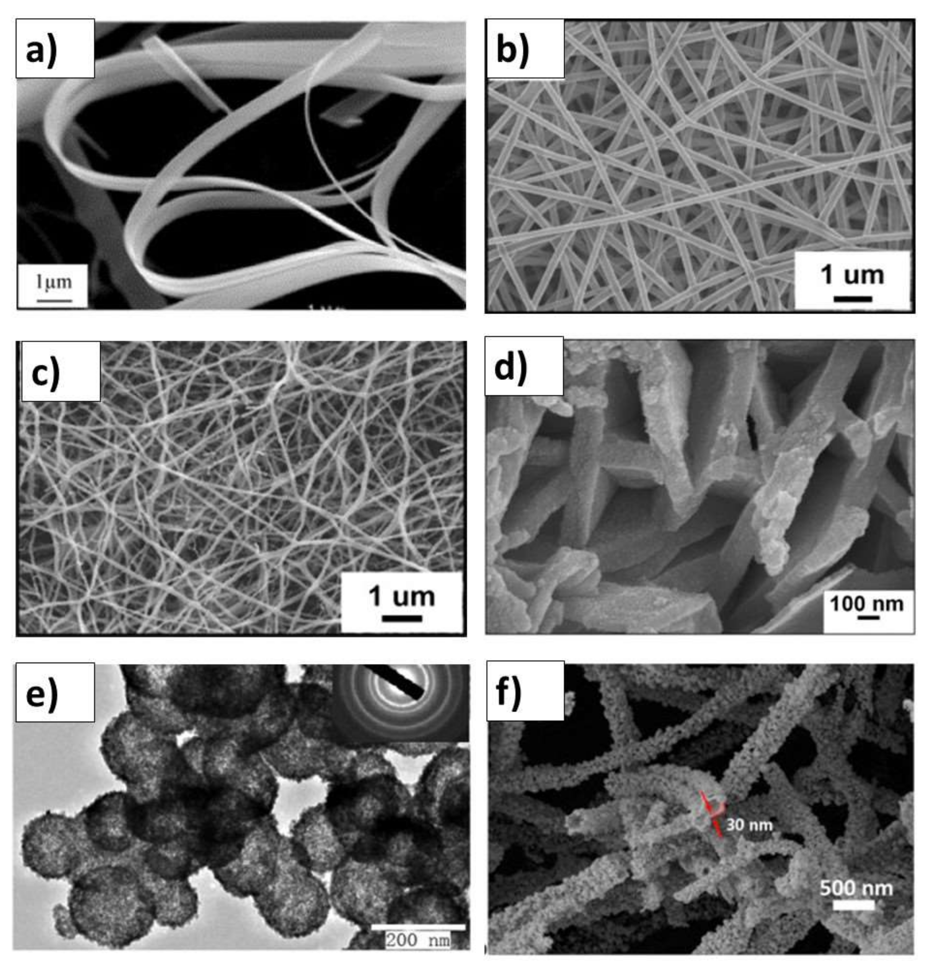
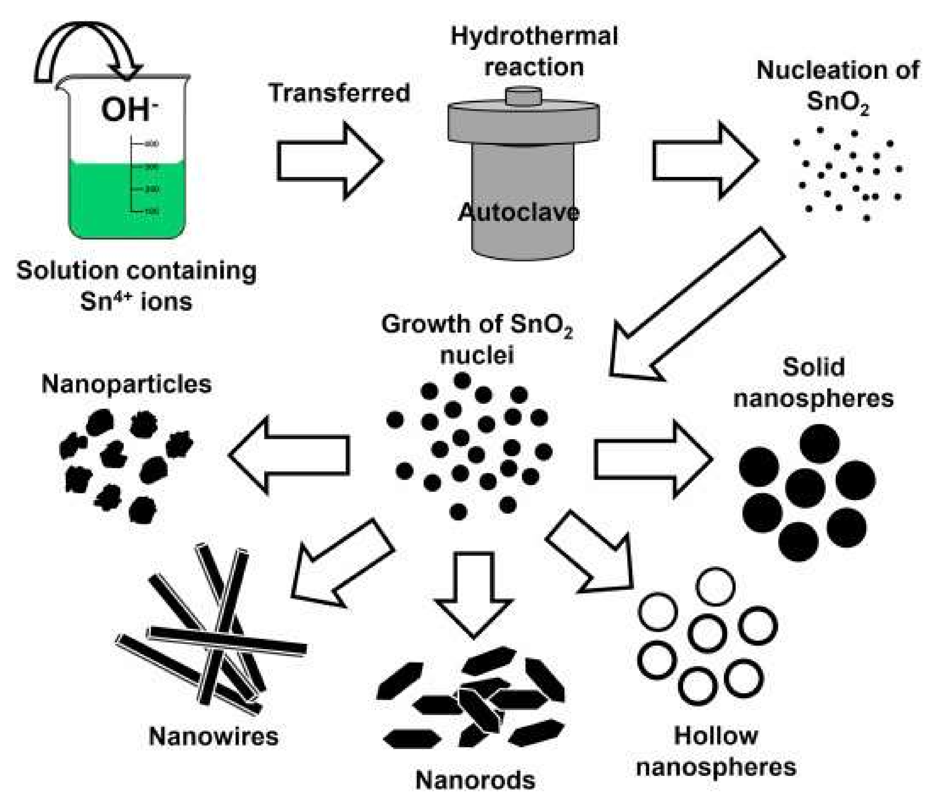
SnO2 and d-SnO2 Morphological Control and Its Impact on the Photocatalysis

For nanostructures, the control of the size and specific surface area is essential to enhance some properties that depend on the electron-hole pair transport to the nanostructure surface. In this sense, the SnO2 morphological control is very necessary to be carefully managed during the synthesis of the nanostructures. This control is significantly associated with the method used as it depends on the precursors, solvents, surfactants, templates, and other factors, such as temperature, atmosphere, time, and pH. The synthesis of the undoped and d-SnO2 is obtained in several nanomorphologies (Figure 8), such as particles, wires, belts, rods, and sheets [85]. It can be assembled in hierarchical structures to obtain, for example, hollow spheres [86], fibres [87], and tubes [88].
The precipitation through the solvothermal treatment is a simple and appropriate method to obtain several of those structures, as presented in Figure 9.
Several papers report the synthesis of nanoparticles and hierarchical structures for d-SnO2, as indicated in Table 1. For SnO2 based materials, the 0D can be prepared, in one way, by simple tin chloride hydrolysis. For tin dioxide, quantum regime will be strong when the nanoparticle diameter is less than the exciton Bohr radius of SnO2 (2.7 nm). Ahmaruzzaman et al. prepared pure tin dioxide quantum dots using a green chemistry approach. The average crystallite size of 1.6 and 2.6 nm were obtained. These quantum dots show superior efficiency in photocatalytic experiments when compared with commercial SnO2 [94].
One-dimensional SnO2 nanostructures, specially nanobelts and nanowires, are synthesized mainly using chemical vapour approaches for undoped and doped, as reported for Sb doped SnO2 [113]. Nanorod structures were obtained for copper doped SnO2 synthesized by a novel one-step microwave irradiation method in solution [114]. The doping effect can be observed in comparison with undoped SnO2 final morphology. Despite the doped structures presenting higher dimensional length, as nanorods, it is composed of nanocrystals. As observed, the crystallite size was reduced when copper was present in the structure. The photocatalytic activity was monitored via the degradation of organic dyes. It was found that doped materials showed better photocatalytic activity than the undoped SnO2. The superior activity for doped nanostructure was attributed to the effective electron–hole separation by surface modification.
Hierarchical nanostructures can be prepared using a hydrothermal method, as demonstrated by Xu et al. The authors obtained nanospheres of undoped and Zn doped SnO2. In this method, the undoped SnO2 nanospheres were converted into nanorods, while in the doped sample, the nanospheres resulted in ultrathin nanosheets. Nanorods and nanosheets self-assembled in hierarchical architectures as final material, which were applied in photocatalytic degradation. The photocatalytic degradation rate of Zn-doped SnO2 catalyst was much higher than that of undoped SnO2 at the same UV irradiation time [115].
Three-dimensional hierarchical structures can be obtained from self-assembly of several nanocrystals. This kind of structure increases porosity and roughness. Consequently, it can improve photocatalytic activity compared to aggregates and sedimented nanoparticles. The SnO2 -based hierarchical structures, such as flower-like and urchin-like ones, could present superior performance, as demonstrated in some studies for Zn-doped SnO2. The photocatalytic performances of Zn-doped SnO2 hierarchical structures were systematically more efficient than the photocatalytic activity of undoped SnO2. The higher activity could be mainly attributed to both the oxygen vacancies and doped Zn2+ centres. More specifically, the development of oxygen vacancies and doped Zn2+ centres are consequences of the preferential growth of certain planes at the expense of the others. For instance, Wulff’s rule predicts that the distance between a crystal facet and an arbitrary origin point in the crystal is proportional to the surface energy of the respective crystallographic plane [116]. Thus, the authors could identify the crystallographic planes with the following increasing surface energy (001) > (101) > (100) > (110). As different planes have different atomic densities, this preferred growth could be a dimensional reason to explain the higher photocatalytic activity of the Zn-doped SnO2 hierarchical structures, in conjunction with the oxygen vacancies and doped Zn2+ centres [117].
Co-precipitation methods were used to prepare undoped and copper doped SnO2. The obtained powder characterized by XRD presented a tetragonal rutile structure. Moreover, it was observed with XRD that increasing the copper amount led to the crystallite size reductions, as observed by the XRD peaks widening. In fact, doping incorporation in the SnO2 matrix lattice will create additional strain in the crystal because, in order to support the doping ions, the crystallite size is reduced, leading to an increase in the specific surface area. Regarding optical properties, when the dopant amount was increased, the energy bandgap was reduced from 3.60 to 3.20 eV. The photocatalytic activity for rhodamine B degradation, the doped material presented higher efficiency than pure SnO2 [99].
Cerium doped SnO2 was prepared by the hydrothermal synthesis method, and the materials were applied in the photocatalytic decomposition of methyl orange dye. The typical rutile phase was obtained in different compositions. Doping the SnO2 reduced the crystallite size from 29.3, for the undoped SnO2, to 24.7 nm in the doped SnO2 materials. Furthermore, a decrease in band gap was identified and associated with superior performance in photocatalytic activity. The authors proposed that the doping prevented the recombination and allowed the effective separation/migration of photoexcited charges [83].
Titanium as the doping agent in tin dioxide led to highly efficient photocatalysts being obtained by an eco-friendly co-precipitation method. It was found that both undoped and Ti doped SnO2 NPs were crystallized as rutile structure, and crystallite sizes were reduced from 19.9 nm for undoped SnO2 nanoparticles to 13.1 nm when Ti 4% was used as a dopant. Morphological aspects in all samples were observed as aggregated nanospheres. The titanium incorporation decreased the optical bandgap from 3.31 in undoped SnO2 to 2.87 eV for 4% Ti. Consequently, this bandgap change widely improved the photocatalytic performance towards RhB photodegradation under UV and visible light irradiations [107].
The influence of W-doping on SnO2 towards photocatalytic detoxification was investigated to obtain nanosphere structures using pluronic123 as a size and morphology controlling template, changing concentrations from 0 to 6% (mol) of tungsten. In the literature, the rutile phase of SnO2 is usually identified in the X-ray diffraction. However, deviations in the oxide crystallinity can be observed due to the large radius of the W6+ (74 pm). The article showed the broadening of the diffraction peaks and a corresponding decrease in crystallite size from 40 to 30 nm with increasing tungsten content. The optical band gap of SnO2 was reduced with the increasing tungsten doping content, in the range evaluated. Photocatalytic performance under visible light irradiation, 6 mol% W at SnO2 demonstrated better efficiency when compared to the commercial TiO2 Degussa P25 standard [109]. Additional examples about photocatalysis can be founded in the recent review for SnO2-based photocatalysts published by Liang et al. [118].

4. SnO2 and d-SnO2 Applied to CO2 Reduction

An interesting example is regarding CO2 reduction, as the annual emission of CO2 into the atmosphere increased by about 40% in the period between 1990 (22,637.134 Mt CO2/yr) and 2018 (37,887.224 Mt CO2/yr), causing serious environmental problems [119].
A sustainable alternative for CO2 abatement would be its use to produce fuels or basic chemicals. The reduction of CO2 makes it possible to obtain a wide variety of products such as: carbon monoxide [120], methane [121], methanol [122], ethylene [123], formate [124], among others.
The conversion of CO2 into fuels can be achieved through several processes that fall into the field of thermochemistry, photochemistry, electrochemistry [125], or photo-electrochemistry [126], depending on the process used to provide the energy needed to force the reduction of CO2. This energy must be obtained through renewable and non-CO2-emitting sources. In the CO2 photoreduction process, the photogenerated electrons in the reaction system can only be consumed if the potential of the semiconductor CB is more negative than the reduction potentials, for example, from the conversion of CO2 to CH4 (Eº (CO2/CH4) = −0.24 V) or from CO2 to CO (E (CO2/CO)= −0.53 V); redox potential versus NHE at pH = 7) [62]. Thus, due to the high theoretical reduction potential of CB from SnO2, it would not be possible to convert CO2 in the photoreduction process. However, Chowdhury et al. experimentally verified the ability to convert CO2 to HCOOH using mesoporous SnO2 nanoparticles in the photocatalysis process in an aqueous medium, both under UV and visible radiation [127]. Torres et al. also observed the activity of SnO2 nanoparticles in the CO2 photoreduction process, converting CO2 into CH4, CO, and ethylene, showing that the surface hydroxyl groups played a key role in the photocatalytic activity of the semiconductor, in which these groups increased CO2 affinity and decreased its reduction potential [128].
Baruch et al. obtained similar results in the process of electroreduction of CO2 with SnO2 and Sn6O4(OH)4 electrocatalysts, where they related the production of formate to a new intermediate, which is formed only superficially, the carbonate bound to tin (Sn-CO3-bidentate and Sn-CO3-monodentate), Figure 10. Furthermore, it suggests that the electrocatalytic activity in reducing CO2 from SnO2 is linked to the formation of tin(II) oxyhydroxide species on the surface [129].
As previously mentioned, the doping of SnO2 has been used effectively to improve the semiconductor properties for application in the CO2 photoreduction process, as well as in the process of generating H2 and in the photodegradation of dyes [4,13,86,96,130,131]. Matussin et al. demonstrated that the doping of SnO2 with Ni caused effects both on the structural properties of SnO2 nanoparticles (NPs) and on the optical properties. Figure 11 presents the schematic diagram of the density of electronic states (DOS), which was proposed according to the combination of UV-visible diffuse reflectance (DRS) spectra results and valence band XPS results, showing that the change in value in the bandgap energy of materials can occur by changing the states of the mid-gap band above the valence band or below the conduction band. The bandgap energy value of the materials SnO2, 1 at% Ni–SnO2, and 5 at% Ni–SnO2 obtained from DRS are 2.81 eV, 2.79 eV, and 2.73 eV, respectively [130].
In addition to changing the electronic structure, the doping process can generate oxygen vacancies that can act as basic Lewis sites, thus increasing the affinity of the semiconductor with the CO2 molecule, facilitating electronic transport in the CO2 photoreduction process [132].
As reported, another strategy used to change the photocatalytic properties of SnO2 for application in the CO2 photoreduction process, is the design of nanostructured materials, which are those systems formed by the presence of two or more distinct phases in the same material, forming heterojunctions. Heterojunctions allow effectively transferring the photogenerated charges between the constituents of the photocatalysts, resulting in unique chemical, optical, and electronic properties that are difficult or impossible to acquire with their individual constituents.
Thus, He et al. [133] studied the formation of a composite formed between SnO2 and g-C3N4 and verified its activity in the CO2 photoreduction process and in the photodegradation of organic pollutants. The authors observed that the formation of the junction between the two materials leads to the obtaining of a photocatalyst with high activity when compared with pure materials and with P25, a classic photocatalyst. The results indicated the formation of a Z-type heterojunction between SnO2 and g-C3N4, which hindered the recombination of electron-hole pairs, consequently increasing their photoactivity (Figure 12a).
In the presence of pristine SnO2, no product from the CO2 photoreduction was observed, which was expected due to its CB potential. However, the heterostructures showed good activity in the CO2 photoreduction process, especially in the conversion of CO2 into CO. The 42.2 wt% SnO2 and g-C3N4 heterostructure showed the best CO2 conversion rate of approximately 22.7 μmol/h gcat, being 4.3 and 5 more active than g-C3N4 and P25, respectively (Figure 12b).
As mentioned previously for CO2 photoreduction, the CB and VB potentials must correspond to the CO2 reduction potentials of different products (Figure 5). Ye et al. investigated the photoexcited electron transfer mechanism in a heterostructure formed by SnO2 and rhenium-modified TiO2 (Re-IO-TiO2–x/SnO2) [134]. The high activity of the heterostructure was attributed to the presence of oxygen vacancies in the material’s structure, which allowed the photocatalysts to have high radiation absorption efficiency and to the excellent electron migration rate of SnO2, which allowed an efficient separation of the electron/hole pair photogenerated. The results showed that the conversion rate of CO2 to CO using the heterostructures was 16.59 μmol/h gcat and 13.75 μmol/h gcat for the materials Re-IO-TiO2−x/SnO2 and IO-TiO2−x/SnO2 respectively, which is approximately six times greater than IO-SnO2.
According to the results obtained by the UV-vis diffuse reflectance spectra, the valence spectra of the XPS, and the Mott–Schottky analysis, the CB potentials of OI-TiO2 and IO-SnO2 are −0.81 eV and −0.72 eV (vs. SHE, pH = 7) and the VB potentials are +2.27 and +2.90 eV, respectively. Therefore, the CB values of IO-TiO2 and IO-SnO2 are more negative than the reduction potentials of CO2 to CO and CO2 to CH4, which indicates that it is theoretically feasible to generate CO and CH4 from CO2. In addition, they performed density functional theory (DFT) calculations, and the results obtained indicated that SnO2 with a higher work function is an oxidation-type photocatalyst and TiO2−x with a lower work function is a reduction-type photocatalyst [134].
As the potential of the CB of IO-TiO2−x is more negative than that of IO-SnO2, electrons from the BC of IO-TiO2−x can be easily transferred to the CB of IO-SnO2. On the other hand, the potential of the VB of IO-SnO2 is more positive than that of IO-TiO2−x, and the holes can be transferred from the VB of IO-SnO2 to the VB of IO-TiO2−x. Therefore, due to heterojunction formation, electrons spontaneously migrate from TiO2−x to SnO2 through its interface. The authors’ first hypothesis was that because of the separation of photogenerated electron/hole pairs, excited electrons could be transferred from the CB of IO-TiO2−x to the CB of IO-SnO2, and holes could be transferred from the VB of IO-SnO2 to the VB of IO-TiO2−x, in which this mechanism belongs to the class of Type II heterojunctions. However, the authors performed radical trapping experiments and confirmed the generation of the •OH radical, which could not happen because the IO-TiO2 is more negative than the potential for the (•OH/OH) radical formation (+2.4 eV). This way, the authors concluded that the photogenerated charge carriers transferring and separation could not be explained by the type II heterojunction system [134].
With these results, they verified that the type of heterostructure formed was of the S-scheme type. As the VB potential of IO-SnO2 (+2.9 eV) is more positive than that of the formation of •OH, these species could be formed in the process, as was verified experimentally. In this way, the photogenerated electrons in the CB of IO-TiO2−x can be transferred to Re7+, and the resulting Re4+ electrons will then be captured by CO2 in the photoreduction process (Figure 13b). Meanwhile, the CB electrons of SnO2 recombine with the holes in the VB of TiO2−x, increasing the separation efficiency of photogenerated charge carriers. Consequently, the charge transfer process at the heterojunction (Re-IO TiO2−x/SnO2) significantly improves the CO2-reducing photocatalytic activity compared to pristine semiconductors [134].
In addition to oxide semiconductors and carbon compounds (g-C3N4 and graphene), nanoparticles of noble metals such as copper (Cu), gold (Au), platinum (Pt), silver (Ag), etc. present themselves as a viable option for the construction of heterojunctions with oxide semiconductors that have a high bandgap value, that is, low absorption in the visible spectrum region, such as SnO2 [135,136]. Ansari et al. reported that, in addition to the Ag nanoparticles on the surface of SnO2 helping in the greater absorption of visible radiation caused by the Localized Surface Plasmon Resonance (SPR) observed in nanoparticles of noble metals, the photocatalytic efficiency of the heterostructure is also improved due to a better separation of the photogenerated charges, in which the lifetime of the e/h+ pair is extended [137].
The strategies presented in this section improved the photocatalytic properties of SnO2, which integrate the effects of doping and heterojunction formation. Thus, they offer a new way to prepare SnO2-based photocatalysts with high activity in the CO2 photoreduction process.

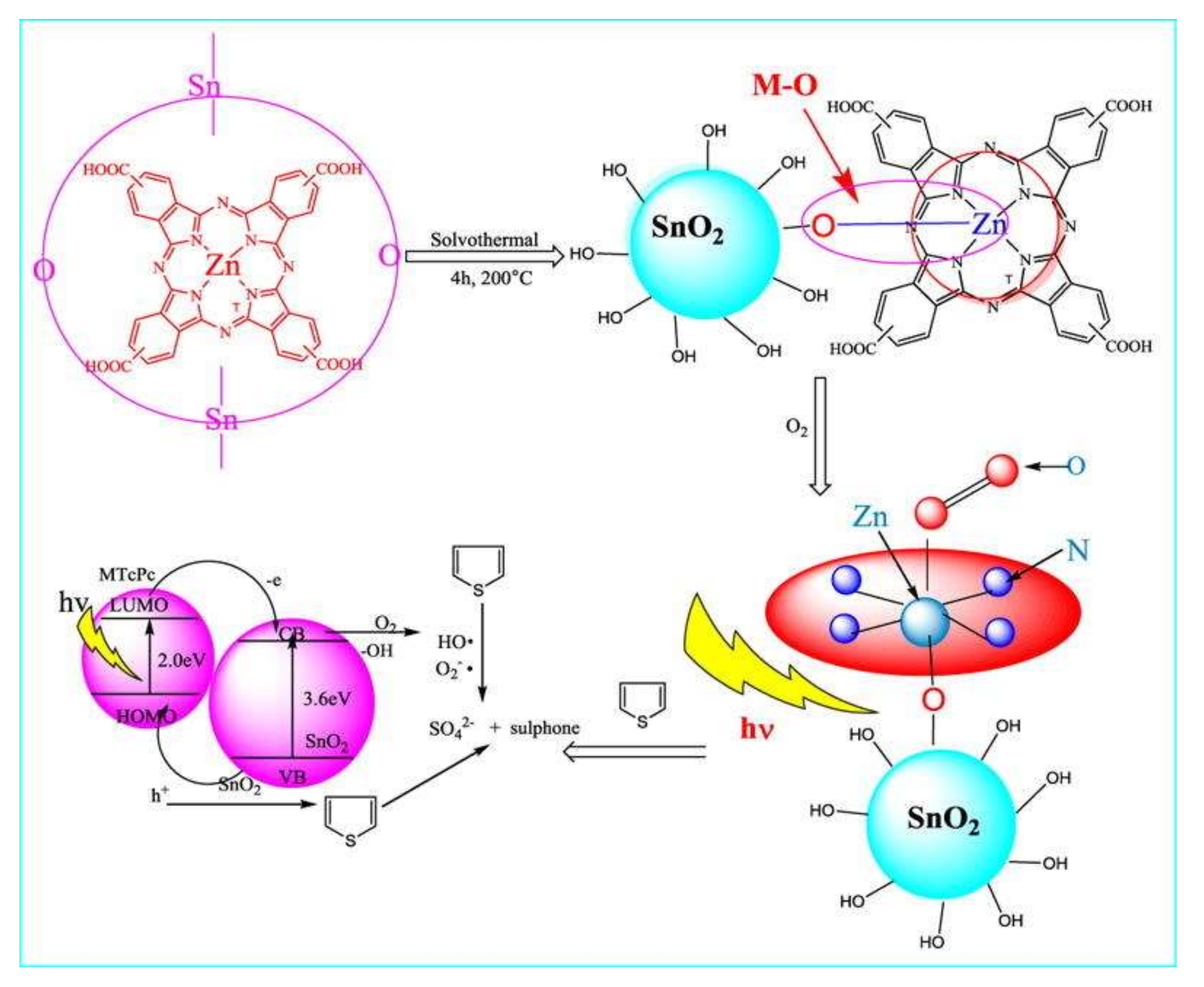
5. SnO2 and Doped-SnO2 Nanostructures Applied to Fuels Desulfurization

In fossil fuels, the presence of sulfur compounds, such as thiophene, benzothiophene, and their derivatives, represents an environmental concern. During fuel combustion, sulfur dioxides (SOx) can be emitted and react with water vapor from the atmosphere producing acid rain, which, consequently, can be harmful to the ecosystems [138].
Additionally, the SOx emission from fossil fuel combustions represents an operational concern for the automotive and petrochemical industries. For example, the SOx in exhaust fumes decreases the efficiency of catalytic converters used in cars to reduce CO and NOx [139]. It also tends to decrease the efficiency of the catalysts used in hydrocarbons downstream and updating in the refineries [140].
These concerns in different fields led the regulatory agencies to establish strict limits to sulfur content in transportation fuels. The USA Environmental Protection Agency (EPA) and the European Union limited the sulfur content in gasoline to 10 ppm [141,142].
The desulfurization technique mostly used in the petrochemical industry is the hydrodesulfurization, which is the conversion of thiophene compounds in H2S and smaller alkenes, by applying high H2 pressure [138].
The hydrodesulfurization presents many cost, safety, and efficiency concerns. For instance, it requires high H2 pressures (between 20 and 100 atm), temperatures as high as 400 °C, and expensive catalysts, such as Co–Mo/Al2O3, Ni–Mo/Al2O3 or Ni–W/Al2O3 [139]. Additionally, hydrodesulfurization is inefficient in removing sterically hindered thiophenes, such as benzothiophene, dibenzothiophene, and alkene derivatives. Consequently, these limitations make hydrodesulfurization hard to comply with the 10 ppm of sulfur content established by EPA and European Union.
One promising alternative to hydrodesulfurization is oxidative desulfurization (ODS). The ODS is subdivided into two steps: oxidation and extraction. During the oxidation, the sulphur-containing compound is oxidized to a sulphone in the presence of an oxidizing agent and a catalyst. Then, after the oxidation, the sulphones are extracted from the fossil fuel mixture, usually by techniques like adsorption, solvent extraction, or thermal distillation. Consequently, the fossil fuel presents a lower sulphur content after the extraction [143].
The SnO2-based nanostructures have been applied in the ODS systems. For instance, Liu et al. prepared SnO2 by SnCl4 co-precipitation with HCl and NH4OH, followed by calcination at temperatures between 200 and 500 °C. Interestingly, the increase in the calcination temperature led to a decrease in the specific surface area of these nanomaterials [144]. The SnO2 powders were used in the ODS of a model fuel made up of dibenzothiophene dissolved in n-octane, using H2O2 as an oxidizing agent. The highest sulphur removal (99.8%) was obtained for the SnO2 sample calcined at 400 °C. Whereas, the lowest sulphur removal (around 65%) was obtained for the SnO2 sample calcined at 200 °C. The authors hypothesized that the SnO2 catalytic activity is based on the partial reduction of the Sn+4 to Sn+2 by the dibenzothiophene-derived species. Then, the Sn+2 is oxidized to Sn+4. The mutual conversion between Sn4+ and Sn2+, facilitated the recycling of the SnO2 catalyst, allowing it to be used for fourteen reaction cycles, practically without loss of catalytic activity [144].
The SnO2 based materials have not been used only as a catalyst, but also as a support for other catalytic materials. For instance, Piao et al. used highly ordered mesoporous SnO2 to support tungsten oxide catalysts (WOx/SnO2) [145]. A possible reason for choosing SnO2 as support is based on its capacity to adsorb sulfur-containing compounds, which could accelerate the interaction between sulfur and the WOx catalyst active sites [146]. The model fuel studied was dibenzothiophene dissolved in n-heptane. The mesoporous SnO2 without the WOx catalyst was able to remove around 50% of the initial sulfur content. Whereas, the WOx/SnO2 system containing the 20% WOx by weight removed 100% of the initial sulphur content in one hour, and it could be recycled for six cycles without losing its catalytic performance. The authors attributed the WOx/SnO2 catalytic activity to the synergistic effect between WOx and SnO2, besides the good WOx dispersion into the SnO2 [145].
Another amenable desulfurization technique is the photocatalytic desulfurization, which relies on the photocatalysis principles explained in Section 3 of this article. Looking at this topic deeper, Zhang et al. prepared composites of SnO2 of metal phthalocyanines (MPcs, with M = Zn+2, Fe+2, Mn+2, or Cu+2) [147]. The MPcs/SnO2 composites were prepared by adding the MPcs dissolved in ethanol, and added to a mixture of dimethylformamide (DMF), water and Sn(OH)4. This mixture underwent hydrothermal treatment at 200 °C for 4 h. After that the product was dried and characterized. Then, the MPcs/SnO2 composites were used in the photocatalytic degradation of a thiophene solution dissolved in n-octane, the oxidizing agent was O2, and the solution was irradiated by visible light. After 3 h, all the MPcs/SnO2 composites present a sulphur removal percentage higher than 80%. The best ones were the ZnPcs/SnO2 and the CuPcs/SnO2, which practically removed 100% of the initial content of sulphur. In comparison, the SnO2 nanoparticles prepared by the same method without MPcs, presented a sulphur removal of only 30%. This result indicates that the synergy between the SnO2 and MPcs enhances the photocatalytic oxidation of the thiophene.
According to the authors, the composites presented higher photocatalytic activity due to the formation between metal-oxygen bond between the metal ion in the phthalocyanine centre and an oxygen in the surface of the SnO2 nanoparticle. This metal-oxygen bond would facilitate the electron transfer from the excited phthalocyanine to the SnO2 CB. Consequently, the charge separation in the SnO2 would be more efficient, reducing the probability of electron-hole recombination, and benefiting the photocatalytic activity in the MPcs/SnO2 composites. A scheme of the proposed charge transfer and photocatalytic mechanism is shown in Figure 14: [147].
Another important product for the petrochemical and energy industries is syngas, which is a gas with high concentration of hydrogen and carbon monoxide. In the coal gasification process, syngas is the main product for electric power generation [148]. As coal can contain a certain amount of sulfur, during the gasification process is very common to form undesired sulfur-containing compounds in the gas phase, such as hydrogen sulphide (H2S) and carbonyl sulphide (COS) [148]. With the goal to remove H2S and COS simultaneously, Yang et al. developed La-doped SnO2 and Y-doped SnO2 nanoparticles that could take advantage of the adsorption desulfurization mechanism [149]. The adsorption desulfurization was based on selective intermolecular interactions between the sulphur-containing compounds (adsorbate) and certain materials able to promote the adsorption of sulphur-containing compounds on their surfaces (adsorbent) [150]. The highest adsorption capacity was obtained by the La-doped SnO2 sample containing 40% La, (La:Sn, atomic ratio), with an total sulfur content adsorption capacity of 148.40 mg/g, whereas the undoped SnO2 had a total sulphur adsorption capacity of 54.86 mg/g. The doping is claimed to provide better sulfur adsorption capacity due to the formation of smaller pores in the surface of the powders, which were not observed in the case of the undoped SnO2 [149].

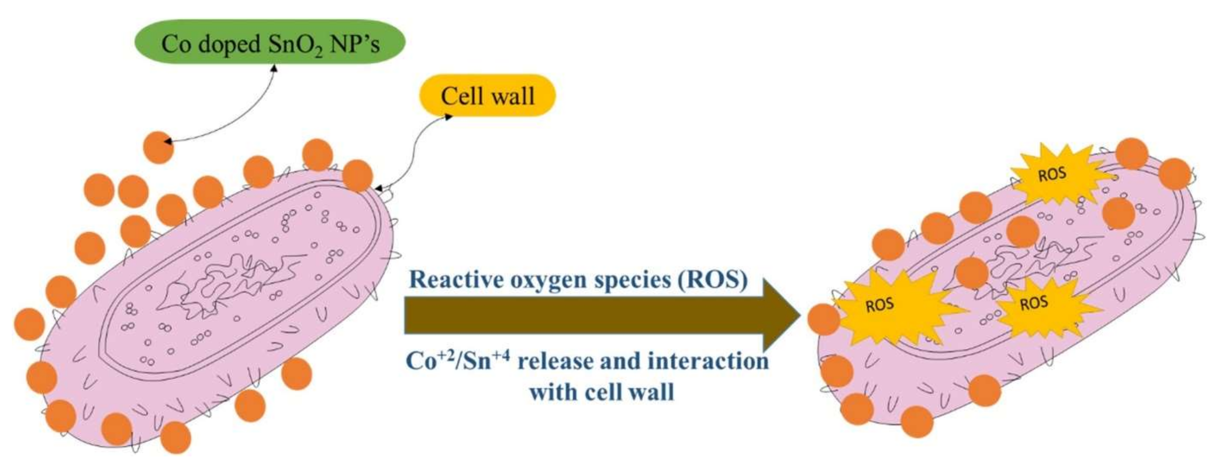
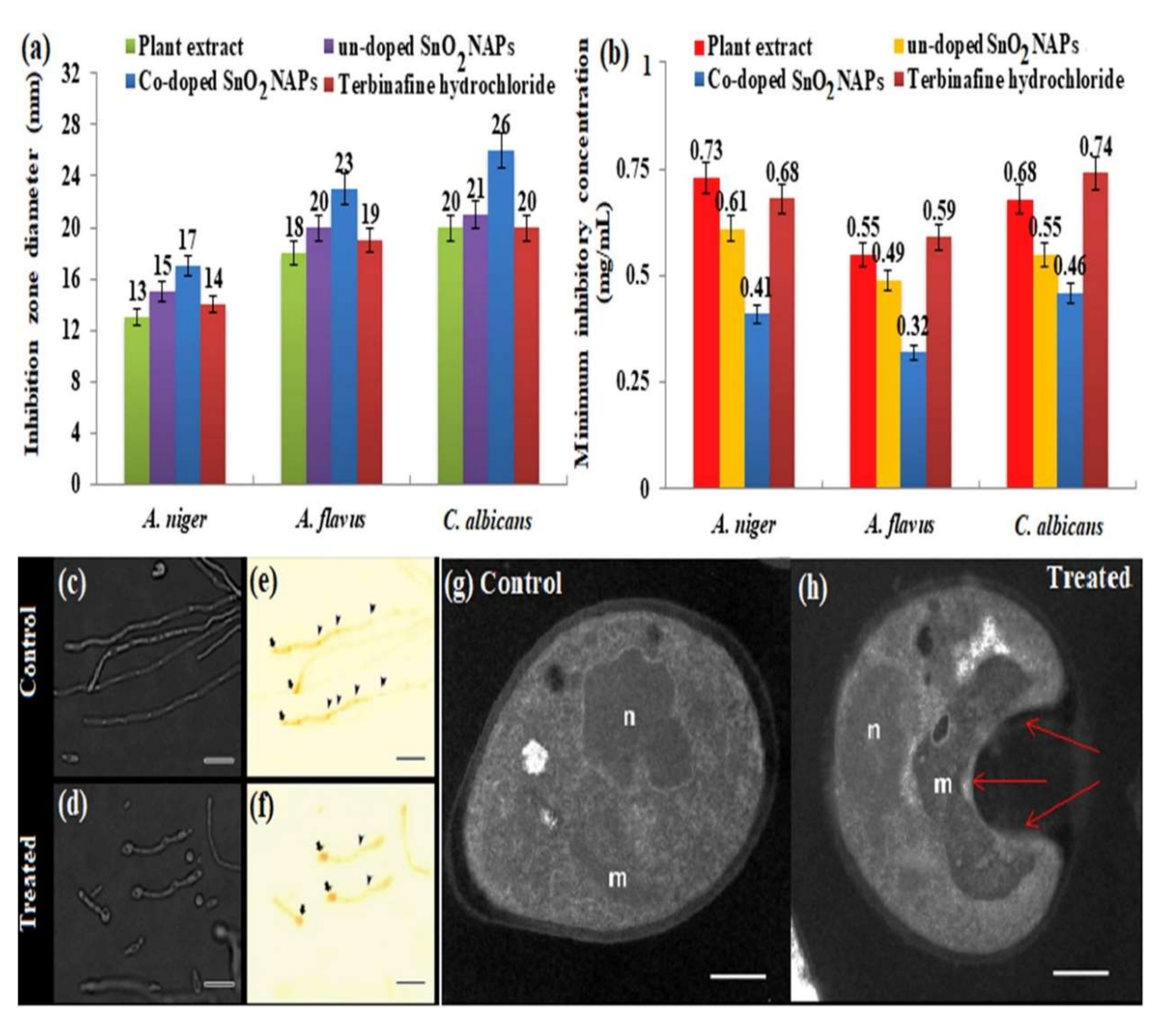
6. Biological Applications

In recent years, nano-sized d-SnO2 nanostructures have gained great attention as promising materials for antimicrobial applications being an important alternative solution to traditional antibiotics against multidrug-resistant microorganisms [151]. In these applications, d-SnO2 is the biologically active material that is responsible for inactivating gram-positive and gram-negative bacteria, fungi, and other microorganisms [87]. In general, the antimicrobial efficiency of metal oxide nanoparticles depends on the surface area, particle size, presence of illumination, and composition of the aqueous medium used in the assay. In this sense, the SnO2-based nanomaterials are preferred for these applications because of their high chemical stability, biocompatibility, and good efficiency in the inactivation of microbes when doped with transition metal ions [19,110,152,153].
Amutha et al. [154] verified the inhibition of Gram-positive (Escherichia coli, Pseudomonas aeruginosa) and Gram-negative (Bacillus subtilis, Staphylococcus aureus) bacterial growth by Fe, Ni, and Fe-Ni co-doped SnO2 NPs. The antibacterial action of d-SnO2 materials was associated with the production of reactive oxygen species (ROS) such as superoxide free radical (O2−), the hydroxyl free radical (OH), and hydrogen peroxide (H2O2). The high reactivity and oxidizing property of ROS are the main responsible for their toxicity. Bacteria inactivation occurs due to the destruction of cell components (lipids, DNA, proteins) by these species. The OH and O2− species have a negative charge and cannot penetrate the cell membrane, so H2O2 ends up being the main responsible in this process, as it can penetrate the cell membrane, causing injury and damage, leading to bacterial cell death [155].
This bacterial inactivation process has been widely reported in the literature. Table 2 presents some of the most recent works in which this mechanism was identified as the main responsible for the antibacterial activity. The materials’ zone of inhibition (ZOI), used to determine the extent of antimicrobial activity, is also indicated in the table for comparison.
Table 2 includes the ZOI of the undoped SnO2 samples used as controls in the antibacterial treatment. In all cases, regardless of the metal used in the doping, an increase in the ZOI of the d-SnO2 samples was observed in relation to the undoped sample, which shows the improvement in the antimicrobial action with doping. The SnO2 doping process favours antibacterial activity because of the reduction it causes in the bandgap energy of the material. The smaller bandgap combined with the high surface area and small crystallite size of the NPs results in more adsorption of bacteria on the surface and greater catalytic reaction capacity even in the dark, leading to the improved antimicrobial action of d-SnO2 NPs [19,66]. Some authors have pointed out that doping also contributes to the extent that it releases Sn2+ ions due to substitution by dopant ions in the lattice [162]. A direct relationship between the increased antimicrobial activity of d-SnO2 NPs and doping content has been reported in the literature [110,152,161,163].
Figure 15 shows how Co-doped SnO2 NPs can induce the ROS production process for bacterial cell destruction [152]. Co-doped SnO2 NPs accumulate on the surface of the bacterial strain by direct or electrostatic forces; the respective metal ions of the NPs (i.e., Co2+ and Sn4+) penetrate the cell oxidizing the cell walls by getting themselves reduced into more stable ones. The oxidation process causes intense oxidative stress giving rise to ROS, the species responsible for attacking the cell and killing it.
Although the production of ROS is the most common argument to explain the antibacterial action of d-SnO2 NPs, different mechanisms have been reported in the literature. Yang et al. [87] showed that the excellent antibacterial activity of Ag-doped SnO2 hollow nanofibres against Staphylococcus aureus and Escherichia coli bacteria could be attributed to the combination of four different processes: (1) Ag+ ions released in the interaction of Ag NPs with the cell membrane can enter the cell and damage the DNA causing mutation or death; (2) neutralization of the negatively charged cell membrane due to interaction with Ag+ ions can result in cell wall destruction and cytoplasm leakage; (3) Ag NPs can destroy mitochondrial cells which results in interruption of energy supply and consequent cell death; and (4) internal destruction of cellular components due to the interaction between Ag+ ions and ROS. Silver is a material with well-known antimicrobial properties; the release of Ag+ ions in the process combined with the production of ROS initiated on the oxide results in an enhancement of the antimicrobial action. Although Ag has a superior antimicrobial action, other metal ions can also cause a disorder in the metabolic functions of bacteria, which can lead to their death [164,165]. Some studies indicate that metal oxides themselves can release metal ions (from adsorption, dissolution, and hydrolysis processes) during antibacterial activity, which indicates that the process described above for Ag+ ions can be quite common and contribute to the performance of d-SnO2 NPs [155,162]. Baig et al. have suggested that the crystallite size and the production of Sn vacancies and/or interstitial oxygen are also relevant factors in this process [83,157].
The antifungal action of d-SnO2 nanostructures has also been successfully reported in the literature. Khan et al. [166] showed that Co-doped SnO2 NPs green synthesized with the aid of Clerodendrum inerme leaf extract have antifungal action against strains of Aspergillus niger, Aspergillus flavus, and Candida albicans. The measurement of zones of inhibition (ZOIs) of samples of Co-SnO2 NPs were higher than those of undoped SnO2 NPs, standard drug, and plant extract for all fungal strains while minimal inhibitory concentrations (MICs) were minimal. For example, the zones of inhibition (ZOIs) of NPs of Co-doped SnO2 were of 17 ± 0.04 mm, 23 ± 0.08 mm and 26 ± 0.06 mm for A. niger, A. flavus, and C. albicans, while for the standard drug were obtained ZOIs of 14 ± 0.08 mm, 19 ± 0.06 mm and 20 ± 0.09 mm, respectively (Figure 16a). Minimum inhibitory concentrations (MICs) of 0.61 ± 0.04 mg mL−1, 0.49 ± 0.07 mg mL−1 and 0.55 ± 0.03 mg mL−1 and 0.68 ± 0.03 mg mL−1 were obtained against the fungi A. niger, A. flavus, and C. albicans when using Co-doped SnO2 NPs while values of 0.59 ± 0.09 mg mL−1 and 0.74 ± 0.08 mg mL−1 respectively were obtained with the standard drug.
Figure 16b presents the MICs for all the analysed samples. Fluorescence microscopy with Calcofluor black of treated cells showed that Co-doped SnO2 NPs are able to reduce the development of hyphae and pseudo-hyphae (Figure 16d,f) in contrast to untreated cells (Figure 16c,e). The TEM images also showed strong morphological changes in treated cells (impaired cell wall and overt swelling of mitochondria as shown in Figure 16h) compared to untreated cells (Figure 16g). The broad-spectrum antifungal activity of this material was associated with (i) doping with Co, (ii) the larger grain size and surface area of the NPs compared to the undoped sample, (iii) the presence of residual plant extract biomolecules (flavonoids and phenolic compounds) in the sample.
Obeizi et al. [167] reported the antifungal action of Fe-doped SnO2 NPs against the Candida albicans strain. The microbial activity was checked by agar well diffusion method and after an incubation period of 24 h at 27 °C a ZOI of 23.59 ± 0.22 mm and an MIC of 1 μg mL−1 were observed for the SnO2 NPs doped with Fe against a ZOI 15.68 ± 0.1 mm for the control sample (gentamicin). The Ag-doped SnO2 nanoparticles modified with curcumin (Cur-Ag-SnO2) were also shown to be an efficient antifungal against Candida albicans (14 mm ZOI, 24 h incubation at 37 °C) showing improved performance compared to undoped SnO2 (11 mm ZOI, 24 h incubation at 37 °C) [168].
Recently, Pandey et al. [165] carried out an extensive study on the antimicrobial properties of the mesoporous composite Ag–Sn/SnO2 in which an excellent antifungal activity of the material against Candida albicans was verified (ZOI of 18 mm, 24 h of incubation at 37 °C) in relation to the activities of composite Sn-SnO2 (7 mm ZOI) and Ag NPs (12 mm ZOI). This work makes clear the importance of metal oxide in bacterial activity; note that although Ag NPs have a good performance, the use of SnO2 as a support increases the activity by 50%. Factors such as smaller particle size, surface porous structure and the elemental composition were found to play an important role in the results. Different processes were identified as possible responsible for killing the fungus: DNA damage caused by Ag/Ag+ entering the cell; cell wall disruption, damage to mitochondria and generation of ROS by Ag NPs; interaction of composite NPs with important cellular components; electronic repulsion between the fungal cell wall and the composite NPs that lead to the generation of H2O2 and others ROS. The authors also showed via in silico molecular docking approaches that the composite has excellent inhibitory properties by targeting different proteins of Candida species followed by several molecular pathways, which indicates that it could be a tool to eliminate resistance to traditional antibiotics.
Ahmaruraman et al. [151] detected both antibacterial and antifungal action of eco-friendly synthesized SnO2-CNT nanohybrid against bacterial strains (Bacillus subtilis, Escherichia coli, Streptococcus pneumonia, Staphylococcus aureus and Pseudomonas aeruginosa), and fungal strains (Aspergillus niger and Candida albicans). Tests performed using the agar well diffusion technique showed SnO2-CNT nanohybrid responses against all microbes are superior to standard drugs. The antimicrobial action was explained by the capture of microbial cells by almost aligned uniform nanotubes and the rupture of the cell wall by SnO2 nanoparticles. Furthermore, it is believed that SnO2 NPs potentiate this effect due to the damage they cause by diffusing into the cell membrane. The heterojunction structure of the nanohybrids was supposed to favour the generation of ROS and consequently increase the damage that leads to the death of the microbes. Recent work has also shown that the ROS generation and consequently the toxicity of the material against microbes can be increased by factors such as crystallite size, defect sites, restructuring, and surface area, among others [155,169,170]. These findings reinforce the role of material morphology in antimicrobial activity.

7. Further Applications and Future Challenges

It is clear that SnO2, d-SnO2 and their nanostructures are promising materials in many fields such as theoretical and computational chemistry, environmental remediation, nanoparticle morphology control, fossil fuels improvement, and biomedical applications. Although many applications have been explored, there are still fields for innovation in this area.
These new advances should benefit from computational chemistry insights, which can offer the theoretical framework for predicting new doping ions, band structure engineering, and physico-chemical properties before the synthetic preparation of these nanomaterials.
One example that has benefited from the computational advancements is the area of transparent conducting oxides (TCOs). Although the tin-doped indium oxide (In2O3:Sn), popularly known as ITO, is the classical material for this application, the indium sources scarcity and high prices have increased the interest for alternative materials for TCOs applications [171]. The d-SnO2 is a potential candidate for fundamental and commercial TCO applications [113]. However, the search for the best dopant ions will not be cost-effective if it can rely only on experimental approaches. So, computational research predicting the physico-chemical, optical, and electronic properties from different types of d-SnO2 is needed. In this sense, Graužinyte et al. used DFT with PBE0 hybrid functionals to screen 63 elements across the periodic table to serve as Sn substitutional dopants in the SnO2 [172]. Two properties of the substitutional dopants were analysed to decide for the best dopants: the density of states (DOS) and the defect energy formation as function of the charge. The calculations revealed that only six elements P, I, Ta, As, Nb, and Sb could n-dope the SnO2 without harming its optoelectronic properties. On the other hand, no elements could satisfactorily p-dope the SnO2.
This study by Graužinyte et al. opened up the field for the development of a couple of other computational and experimental studies about d-SnO2 and TCOs applications [172]. For example, Williamson et al. used DFT to predict and infrared reflectivity and X-ray photoelectron spectroscopy (XPS) to confirm that Ta was a better dopant than more often commercially used Sb and F [173], because the Ta-doped SnO2 presented higher conductivity, mobility, and infrared transparency. The development and publication of the research by Graužinyte et al. and Williamson et al. is a confirmation of the power of the computational techniques to predict properties and provide resources to experimental studies [172,173].
The forensic area is another field where d-SnO2 can do meaningful contributions. For instance crime scene investigations usually need latent fingerprints (LFPs) visualization, in such a way that the LFPs are invisible to naked eyes when illuminated by white light, but visible when illuminated by other less commonly found wavelengths, such as UV radiation [174,175,176]. Aiming to prepare materials for LFPs visualization, Deepthi et al. prepared Eu3+ doped SnO2 using a Green Chemistry route, where aloe vera was used a biotemplate for the hydrothermal [177]. These Eu3+ doped SnO2 nanoparticles were stained onto fingerprint marks on different types of surfaces, such as paper, leaf, granite, and soda can. When these objects were illuminated with UV radiation (λ = 254 nm), the LFPs became clearly visible, as shown in Figure 17A–C, where the LFPs can be observed on the surface of a currency bill.
Early cancer detection is one of the most sought-after applications by science and medicine. For lung cancer, one possible detection mode is associated with the detection of exhaled volatile organic compounds (VOCs). About 250 VOCs are present in the exhaled air, in the ppm to ppb range, and can be used as biomarkers for lung cancer, as their patterns are different in exhaled air between patients with lung cancer and healthy individuals [178]. In this sense, the development of devices able to detect VOCs in the exhaled air could be an effective way to detect lung cancer in its earlier stage. Electronic noses are devices based on electrochemical techniques, i.e., cyclic voltammetry (CV), able to selectively detect specific analytes, such as VOCs, with high sensitivity. Thus, electronic noses can be a cost-effective, non-invasive, point-of-care device to detect VOCs related to lung cancers in exhaled air. Related to this application, Khatoon et al. prepared Ni or Co-doped SnO2 nanoparticles using a sol-gel method, and used these nanoparticles to prepare screen-printed working electrodes for the electronic nose device [5]. Both CV and electronic impedance spectroscopy (EIS) were used as electrochemical techniques to assess the selectivity and sensitivity for isopropanol and 1-propanol solutions. The Ni-doped SnO2 was the most sensitive electrode both for isopropanol and 1-propanol, being 1.5 and 3.0 times more sensitive than the undoped-SnO2 for the isopropanol and 1-propanol detection, respectively [5].

8. Conclusions

This review presented the structural features of SnO2, d-SnO2 and SnO2-based composites. Then, recent advances were presented in different applications, such as photocatalytic degradation of pollutants, CO2 reduction, desulfurization of fossil fuels, and antimicrobial activity and discussed. Some other areas where SnO2-related materials have been poorly explored were presented, by demonstrating some successful studies in these areas, like computational methods to screen possible dopants, forensic applications, and earlier cancer detection.
Although recent advances were presented in these areas, there is still space for development and improvement in all these applications related to SnO2. For instance, by engineering the bandgap through doping or making heterostructures, to delay the charge carrier’s recombination, can boost many applications depending on photocatalysis, such as environmental remediation and CO2 photoreduction. Novel synthetic methods are welcome to improve the insertion of the doping ions in the SnO2 matrix without secondary phases, and to improve the morphological control. The combination of computational and experimental techniques can lead to more efficient materials preparation. For example, computational techniques can screen potential dopants for SnO2 according to the desired property to be obtained. This type of study may reduce the number of experiments to be performed, consequently reducing the overall cost of the project.
In summary, although SnO2 is a well-established and versatile material, the advances in nanotechnology, material preparation, and computational sciences are essential resources to provide tools for novel applications.

Author Contributions

All the authors contributed to the conceptualization, investigation, data curation, writing the original draft. Additionally, the tasks like supervision, writing-review and editing were performed by the corresponding author A.H.P. All authors have read and agreed to the published version of the manuscript.

Funding

This study was financed in part by the Coordenação de Aperfeiçoamento de Pessoal de Nivel Superior—Brasil (CAPES)—Finance Code 001. AEN acknowledges financial support from the Minas Gerais State Agency for Research and Development—FAPEMIG, Project number (APQ-02075-21). The authors gratefully acknowledge the financial support provided by the Brazilian research funding agencies CNPq, FAPES, FAPESP and FAPERGS. ANP Human Resources Program for the Oil and Gas Sector - PRHANP/MCTI, in particular PRH-ANP 53.1 UFES. This study was financed in part by the Coordenação de Aperfeiçoamento de Pessoal de Nível Superior - Brasil (CAPES) - Finance Code 001.(J.C.d. A.) AHP acknowledges the Donors of the American Chemical Society Petroleum Research Fund for support of this research, ACS-PRF Project Number ACS PRF 62421-0UNI10.

Institutional Review Board Statement

Institutional Review Board approval is not applicable for this article.

Informed Consent Statement

Informed Consent is not applicable for this article.

Data Availability Statement

Data Availability is not applicable for this article.

Conflicts of Interest

The authors declare no conflict of interest.

References

  1. Baranowska-Korczyc, A.; Mackiewicz, E.; Ranoszek-Soliwoda, K.; Grobelny, J.; Celichowski, G. Core/shell ag/SnO2 nanowires for visible light photocatalysis. Catalysts 2022, 12, 30. [Google Scholar] [CrossRef]
  2. Dohčević-Mitrovic, Z.D.; Araújo, V.D.; Radović, M.; Aškrabić, S.; Costa, G.R.; Bernardi, M.I.B.; Djokić, D.M.; Stojadinović, B.; Nikolić, M.G. Influence of oxygen vacancy defects and cobalt doping on optical, electronic and photocatalytic properties of ultrafine SnO2-δ nanocrystals. Process. Appl. Ceram. 2020, 14, 102–112. [Google Scholar] [CrossRef]
  3. Manjula, N.; Selvan, G.; Balu, A.R. Photocatalytic Performance of SnO2:Mo Nanopowders Against the Degradation of Methyl Orange and Rhodamine B Dyes Under Visible Light Irradiation. J. Electron. Mater. 2019, 48, 401–408. [Google Scholar] [CrossRef]
  4. Chu, L.; Duo, F.; Zhang, M.; Wu, Z.; Sun, Y.; Wang, C.; Dong, S.; Sun, J. Doping induced enhanced photocatalytic performance of SnO2:Bi3+ quantum dots toward organic pollutants. Colloids Surfaces A Physicochem. Eng. Asp. 2020, 589, 124416. [Google Scholar] [CrossRef]
  5. Khatoon, Z.; Fouad, H.; Alothman, O.Y.; Hashem, M.; Ansari, Z.A.; Ansari, S.A. Doped SnO2 nanomaterials for e-nose based electrochemical sensing of biomarkers of lung cancer. ACS Omega 2020, 5, 27645–27654. [Google Scholar] [CrossRef] [PubMed]
  6. Lv, Z.; Li, J.; Yang, F.; Cao, K.; Bao, Q.; Sun, Y.; Yuan, J. Antimony-Doped Tin Oxide Nanocrystals for Enhanced Photothermal Theragnosis Therapy of Cancers. Front. Bioeng. Biotechnol. 2020, 8, 673. [Google Scholar] [CrossRef] [PubMed]
  7. Choudhari, U.; Jagtap, S. Electrochemical studies on doped SnO2 nanocomposite for selective detection of lung cancer biomarkers. AIP Adv. 2021, 11, 125327. [Google Scholar] [CrossRef]
  8. Kwak, C.H.; Kim, T.H.; Jeong, S.Y.; Yoon, J.W.; Kim, J.S.; Lee, J.H. Humidity-Independent Oxide Semiconductor Chemiresistors Using Terbium-Doped SnO2 Yolk-Shell Spheres for Real-Time Breath Analysis. ACS Appl. Mater. Interfaces 2018, 10, 18886–18894. [Google Scholar] [CrossRef]
  9. Shin, J.; Choi, S.J.; Lee, I.; Youn, D.Y.; Park, C.O.; Lee, J.H.; Tuller, H.L.; Kim, I.D. Thin-wall assembled SnO2 fibers functionalized by catalytic Pt nanoparticles and their superior exhaled-breath-sensing properties for the diagnosis of diabetes. Adv. Funct. Mater. 2013, 23, 2357–2367. [Google Scholar] [CrossRef]
  10. Shariati, M. The field effect transistor DNA biosensor based on ITO nanowires in label-free hepatitis B virus detecting compatible with CMOS technology. Biosens. Bioelectron. 2018, 105, 58–64. [Google Scholar] [CrossRef]
  11. Nguyen, M.H.; Nguyen, H.H.; Mai, A.T. On-chip selective growth of SnO2 nanowires for DNA sensor development. Sens. Actuators A Phys. 2020, 312, 112171. [Google Scholar] [CrossRef]
  12. Kandasamy, M.; Seetharaman, A.; Sivasubramanian, D.; Nithya, A.; Jothivenkatachalam, K.; Maheswari, N.; Gopalan, M.; Dillibabu, S.; Eftekhari, A. Ni-Doped SnO2 Nanoparticles for Sensing and Photocatalysis. ACS Appl. Nano Mater. 2018, 1, 5823–5836. [Google Scholar] [CrossRef]
  13. Babu, B.; Neelakanta Reddy, I.; Yoo, K.; Kim, D.; Shim, J. Bandgap tuning and XPS study of SnO2 quantum dots. Mater. Lett. 2018, 221, 211–215. [Google Scholar] [CrossRef]
  14. Carolin, L.R.; Samuel, S.S.A. Hierarchical nanostructures of In–SnO2 with enhanced photo catalytic activity for the degradation of RR 120 dye. J. Mater. Sci. Mater. Electron. 2020, 31, 12796–12806. [Google Scholar] [CrossRef]
  15. Jain, S.K.; Fazil, M.; Naaz, F.; Pandit, N.A.; Ahmed, J.; Alshehri, S.M.; Mao, Y.; Ahmad, T. Silver-doped SnO2nanostructures for photocatalytic water splitting and catalytic nitrophenol reduction. New J. Chem. 2022, 46, 2846–2857. [Google Scholar] [CrossRef]
  16. Babu, B.; Kadam, A.N.; Ravikumar, R.V.S.S.N.; Byon, C. Enhanced visible light photocatalytic activity of Cu-doped SnO2 quantum dots by solution combustion synthesis. J. Alloys Compd. 2017, 703, 330–336. [Google Scholar] [CrossRef]
  17. Ragupathy, S.; Manikandan, V.; Devanesan, S.; Ahmed, M.; Ramamoorthy, M.; Priyadharsan, A. Enhanced sun light driven photocatalytic activity of Co doped SnO2 loaded corn cob activated carbon for methylene blue dye degradation. Chemosphere 2022, 295, 133848. [Google Scholar] [CrossRef]
  18. Kumar, S.; Bhawna; Yadav, S.K.; Gupta, A.; Kumar, R.; Ahmed, J.; Chaudhary, M.; Suhas; Kumar, V. B-doped SnO2 nanoparticles: A new insight into the photocatalytic hydrogen generation by water splitting and degradation of dyes. Environ. Sci. Pollut. Res. 2022. [Google Scholar] [CrossRef]
  19. Chandran, D.; Nair, L.S.; Balachandran, S.; Rajendra Babu, K.; Deepa, M. Structural, optical, photocatalytic, and antimicrobial activities of cobalt-doped tin oxide nanoparticles. J. Sol-Gel Sci. Technol. 2015, 76, 582–591. [Google Scholar] [CrossRef]
  20. Zhang, J.; Gao, L. Synthesis and characterization of antimony-doped tin oxide (ATO) nanoparticles by a new hydrothermal method. Mater. Chem. Phys. 2004, 87, 10–13. [Google Scholar] [CrossRef]
  21. Wang, J.; Pang, Z.; Jin, F.; Ge, M. Precipitation method to prepare conductive F-doped SnO2-coated rutile TiO2 whisker and its application in coating. J. Mater. Sci. Mater. Electron. 2021, 32, 20583–20597. [Google Scholar] [CrossRef]
  22. Pan, S.; Li, G. Recent Progress in p-Type Doping and Optical Properties of SnO2 Nanostructures for Optoelectronic Device Applications. Recent Pat. Nanotechnol. 2011, 5, 138–161. [Google Scholar] [CrossRef] [PubMed]
  23. Chaparadza, A.; Rananavare, S.B. Towards p-type conductivity in SnO2 nanocrystals through Li doping. Nanotechnology 2010, 21, 035708. [Google Scholar] [CrossRef] [PubMed]
  24. Pan, S.S.; Li, G.H.; Wang, L.B.; Shen, Y.D.; Wang, Y.; Mei, T.; Hu, X. Atomic nitrogen doping and p-type conduction in SnO2. Appl. Phys. Lett. 2009, 95, 10–13. [Google Scholar] [CrossRef]
  25. Callister, W.D.J.; Rethwisch, D.G. Materials Science and Engineering: An Introduction, 8th ed.; John Wiley & Sons, Inc.: Hoboken, NJ, USA, 2010; ISBN 9780470448649. [Google Scholar]
  26. White, M.A. Properties of Materials; Oxford University Press: Oxford, UK, 1999; 334p. [Google Scholar]
  27. Wang, L.; Wang, Y.; Su, D.; Zhao, Y. Enhancement of visible light photocatalytic activity over bistructural SnO2 nanobelts. Superlattices Microstruct. 2018, 114, 416–420. [Google Scholar] [CrossRef]
  28. Umar, A.; Ammar, H.Y.; Kumar, R.; Almas, T.; Ibrahim, A.A.; AlAssiri, M.S.; Abaker, M.; Baskoutas, S. Efficient H2 gas sensor based on 2D SnO2 disks: Experimental and theoretical studies. Int. J. Hydrogen Energy 2020, 45, 26388–26401. [Google Scholar] [CrossRef]
  29. Zhang, F.; Teng, X.; Shi, W.; Song, Y.; Zhang, J.; Wang, X.; Li, H.; Li, Q.; Li, S.; Hu, H. SnO2 nanoflower arrays on an amorphous buffer layer as binder-free electrodes for flexible lithium-ion batteries. Appl. Surf. Sci. 2020, 527, 146910. [Google Scholar] [CrossRef]
  30. Yu, H.; Zhang, Y.; Dong, L.; Wang, J. Fabricating pod-like SnO2 hierarchical micro-nanostructures for enhanced acetone gas detection. Mater. Sci. Semicond. Process. 2021, 121, 105451. [Google Scholar] [CrossRef]
  31. Geraldo, V.; Briois, V.; Scalvi, L.V.A.; Santilli, C.V. EXAFS investigation on Sb incorporation effects to electrical transport in SnO2 thin films deposited by sol-gel. J. Eur. Ceram. Soc. 2007, 27, 4265–4268. [Google Scholar] [CrossRef] [Green Version]
  32. Das, S.; Jayaraman, V. SnO2: A comprehensive review on structures and gas sensors. Prog. Mater. Sci. 2014, 66, 112–255. [Google Scholar] [CrossRef]
  33. Vermeire, F.H.; Green, W.H. Transfer learning for solvation free energies: From quantum chemistry to experiments. Chem. Eng. J. 2021, 418, 129307. [Google Scholar] [CrossRef]
  34. Filippatos, P.-P.; Kelaidis, N.; Vasilopoulou, M.; Davazoglou, D.; Chroneos, A. Impact of boron and indium doping on the structural, electronic and optical properties of SnO2. Sci. Rep. 2021, 11, 13031. [Google Scholar] [CrossRef] [PubMed]
  35. Hohenberg, P.; Kohn, W. Inhomogeneous electron gas. Phys. Rev. 1964, 136, B864. [Google Scholar] [CrossRef] [Green Version]
  36. Kohn, W.; Sham, L.J. Self-consistent equations including exchange and correlation effects. Phys. Rev. 1965, 140, A1133–A1138. [Google Scholar] [CrossRef] [Green Version]
  37. Bloch, F. Über die Quantenmechanik der Elektronen in Kristallgittern. Zeitschrift für Phys. 1929, 52, 555–600. [Google Scholar] [CrossRef]
  38. Kratzer, P.; Neugebauer, J. The Basics of Electronic Structure Theory for Periodic Systems. Front. Chem. 2019, 7, 106. [Google Scholar] [CrossRef] [Green Version]
  39. Fritsch, D.; Morgan, B.J.; Walsh, A. Self-Consistent Hybrid Functional Calculations: Implications for Structural, Electronic, and Optical Properties of Oxide Semiconductors. Nanoscale Res. Lett. 2017, 12, 19. [Google Scholar] [CrossRef] [Green Version]
  40. Gilani, R.; Rehman, S.U.; Butt, F.K.; Ul Haq, B.; Aleem, F. Elucidating the First-Principles Calculations of SnO2 Within DFT Framework and Beyond: A Library for Optimization of Various Pseudopotentials. Silicon 2018, 10, 2317–2328. [Google Scholar] [CrossRef]
  41. Gracia, L.; Beltrán, A.; Andrés, J. Characterization of the High-Pressure Structures and Phase Transformations in SnO2. A Density Functional Theory Study. J. Phys. Chem. B 2007, 111, 6479–6485. [Google Scholar] [CrossRef]
  42. Borges, P.D.; Scolfaro, L.M.R.; Leite Alves, H.W.; da Silva, E.F. DFT study of the electronic, vibrational, and optical properties of SnO2. Theor. Chem. Acc. 2010, 126, 39–44. [Google Scholar] [CrossRef]
  43. Beltrán, A.; Andrés, J.; Longo, E.; Leite, E.R. Thermodynamic argument about SnO2 nanoribbon growth. Appl. Phys. Lett. 2003, 83, 635–637. [Google Scholar] [CrossRef]
  44. Xu, C.; Jiang, Y.; Yi, D.; Sun, S.; Yu, Z. Environment-dependent surface structures and stabilities of SnO2 from the first principles. J. Appl. Phys. 2012, 111, 63504. [Google Scholar] [CrossRef] [Green Version]
  45. Oviedo, J.; Gillan, M.J. Energetics and structure of stoichiometric SnO2 surfaces studied by first-principles calculations. Surf. Sci. 2000, 463, 93–101. [Google Scholar] [CrossRef]
  46. Laranjeira, J.A.S.; Fabris, G.S.L.; Ferrer, M.M.; Albuquerque, A.R.; Sambrano, J.R. Morphological Transformation Network of Nanoparticles via DFT Simulations. Cryst. Growth Des. 2020, 20, 4600–4611. [Google Scholar] [CrossRef]
  47. Chen, Y.; Wang, X.; Shi, C.; Li, L.; Qin, H.; Hu, J. Sensing mechanism of SnO2(1 1 0) surface to H2: Density functional theory calculations. Sens. Actuators B Chem. 2015, 220, 279–287. [Google Scholar] [CrossRef]
  48. Maleki, F.; Pacchioni, G. A DFT study of formic acid decomposition on the stoichiometric SnO2 surface as a function of iso-valent doping. Surf. Sci. 2022, 718, 122009. [Google Scholar] [CrossRef]
  49. García, E.V.; Carbajal-Franco, G.; López-Galán, O.A. DFT transition state study of the catalyzed oxidation of methane on SnO2 surfaces. Catal. Today 2021, 392–393, 41–48. [Google Scholar] [CrossRef]
  50. Ganose, A.M.; Scanlon, D.O. Band gap and work function tailoring of SnO2 for improved transparent conducting ability in photovoltaics. J. Mater. Chem. C 2016, 4, 1467–1475. [Google Scholar] [CrossRef] [Green Version]
  51. Awa, K.; Akashi, R.; Akita, A.; Naya, S.; Kobayashi, H.; Tada, H. Highly Efficient and Selective Oxidation of Ethanol to Acetaldehyde by a Hybrid Photocatalyst Consisting of SnO2 Nanorod and Rutile TiO2 with Heteroepitaxial Junction. ChemPhysChem 2019, 20, 2155–2161. [Google Scholar] [CrossRef]
  52. Beloufa, N.; Cherchab, Y.; Louhibi-Fasla, S.; Daoud, S.; Rekab-Djabri, H.; Chahed, A. Theoretical investigation of structural, electronic and optical properties of Sc-doped SnO2. Comput. Condens. Matter 2022, 30, e00642. [Google Scholar] [CrossRef]
  53. Medina Chanduvi, H.H.; Mudarra Navarro, A.M.; Bilovol, V.; Errico, L.A.; Gil Rebaza, A.V. Structural, Electronic, Magnetic, and Hyperfine Properties of V-doped SnO2 (Sn1–xVxO2, x: 0, 0.042, 0.084, and 0.125). A DFT-Based Study. J. Phys. Chem. C 2021, 125, 11702–11713. [Google Scholar] [CrossRef]
  54. Albanese, E.; Di Valentin, C.; Pacchioni, G.; Sauvage, F.; Livraghi, S.; Giamello, E. Nature of Paramagnetic Species in Nitrogen-Doped SnO2: A Combined Electron Paramagnetic Resonance and Density Functional Theory Study. J. Phys. Chem. C 2015, 119, 26895–26903. [Google Scholar] [CrossRef] [Green Version]
  55. Nath, P.; Chakraborti, A.; Sanyal, D. Ab initio calculation of magnetic properties of p-block element doped ZnO. RSC Adv. 2014, 4, 45598–45602. [Google Scholar] [CrossRef]
  56. Luitel, H.; Roy, S.; Sanyal, D. Ab-initio calculation of the magnetic properties of P and As doped SnO2. Comput. Condens. Matter 2018, 14, 36–39. [Google Scholar] [CrossRef]
  57. Zhang, Y.; Liu, J.; Wei, Z.; Liu, Q.; Wang, C.; Ma, J. Electrochemical CO2 reduction over nitrogen-doped SnO2 crystal surfaces. J. Energy Chem. 2019, 33, 22–30. [Google Scholar] [CrossRef] [Green Version]
  58. Li, W.; Ding, C.; Li, J.; Ren, Q.; Bai, G.; Xu, J. Sensing mechanism of Sb, S doped SnO2 (1 1 0) surface for CO. Appl. Surf. Sci. 2020, 502, 144140. [Google Scholar] [CrossRef]
  59. Náfrádi, M.; Alapi, T.; Bencsik, G.; Janáky, C. Impact of reaction parameters and water matrices on the removal of organic pollutants by TiO2 /led and ZnO/led heterogeneous photocatalysis using 365 and 398 nm radiation. Nanomaterials 2022, 12, 5. [Google Scholar] [CrossRef]
  60. De Almeida, J.C.; Correâ, M.T.; Koga, R.H.; Del Duque, D.M.S.; Lopes, O.F.; Da Silva, G.T.S.T.; Ribeiro, C.; De Mendoncą, V.R. Crystallization time in ZnO: The role of surface OH groups in its photoactivity. New J. Chem. 2020, 44, 18216–18224. [Google Scholar] [CrossRef]
  61. de Mendonça, V.R.; Avansi, W.; Arenal, R.; Ribeiro, C. A building blocks strategy for preparing photocatalytically active anatase TiO2/rutile SnO2 heterostructures by hydrothermal annealing. J. Colloid Interface Sci. 2017, 505, 454–459. [Google Scholar] [CrossRef] [Green Version]
  62. Silva, G.; Lopes, O.; Dias, E.; Torres, J.; Nogueira, A.; Faustino, L.; Prado, F.; Patrocínio, A.; Ribeiro, C. ReduÇÃo de CO2 em hidrocarbonetos e oxigenados: Fundamentos, estrategias e desafios. Quim. Nova 2021, 44, 963–981. [Google Scholar] [CrossRef]
  63. Liang, B.; Zhang, L.; Zhang, W. Preparation of Cl doped SnO powder with excellent photocatalytic property by mechanical alloying. Ceram. Int. 2019, 45, 8908–8913. [Google Scholar] [CrossRef]
  64. Muñoz-Batista, M.J.; Luque, R. Heterogeneous photocatalysis. ChemEngineering 2021, 5, 26. [Google Scholar] [CrossRef]
  65. Mubeen, S.; Singh, N.; Lee, J.; Stucky, G.D.; Moskovits, M.; McFarland, E.W. Synthesis of chemicals using solar energy with stable photoelectrochemically active heterostructures. Nano Lett. 2013, 13, 2110–2115. [Google Scholar] [CrossRef] [PubMed]
  66. Ali Baig, A.B.; Rathinam, V.; Ramya, V. Facile fabrication of Zn-doped SnO2 nanoparticles for enhanced photocatalytic dye degradation performance under visible light exposure. Adv. Compos. Hybrid Mater. 2021, 4, 114–126. [Google Scholar] [CrossRef]
  67. Castro, S.; Albo, J.; Irabien, A. Photoelectrochemical Reactors for CO2 Utilization. ACS Sustain. Chem. Eng. 2018, 6, 15877–15894. [Google Scholar] [CrossRef] [Green Version]
  68. Ola, O.; Maroto-Valer, M.M. Review of material design and reactor engineering on TiO2 photocatalysis for CO2 reduction. J. Photochem. Photobiol. C Photochem. Rev. 2015, 24, 16–42. [Google Scholar] [CrossRef] [Green Version]
  69. Han, Y.; Wu, X.; Ma, Y.; Gong, L.; Qu, F.; Fan, H. Porous SnO2 nanowire bundles for photocatalyst and Li ion battery applications. CrystEngComm 2011, 13, 3506–3510. [Google Scholar] [CrossRef]
  70. Vadivel, S.; Rajarajan, G. Effect of W doping on structural, optical and photocatalytic activity of SnO2 nanostructure thin films. J. Mater. Sci. Mater. Electron. 2015, 26, 7127–7133. [Google Scholar] [CrossRef]
  71. Parthibavarman, M.; Sathishkumar, S.; Jayashree, M.; BoopathiRaja, R. Microwave Assisted Synthesis of Pure and Ag Doped SnO2 Quantum Dots as Novel Platform for High Photocatalytic Activity Performance. J. Clust. Sci. 2019, 30, 351–363. [Google Scholar] [CrossRef]
  72. Sery, A.A.; Mohamed, W.A.A.; Hammad, F.F.; Khalil, M.M.H.; Farag, H.K. Synthesis of pure and doped SnO2 and NiO nanoparticles and evaluation of their photocatalytic activity. Mater. Chem. Phys. 2022, 275, 125190. [Google Scholar] [CrossRef]
  73. Ahmed, A.; Naseem Siddique, M.; Alam, U.; Ali, T.; Tripathi, P. Improved photocatalytic activity of Sr doped SnO2 nanoparticles: A role of oxygen vacancy. Appl. Surf. Sci. 2019, 463, 976–985. [Google Scholar] [CrossRef]
  74. Wrighton, M.S.; Morse, D.L.; Ellis, A.B.; Ginley, D.S.; Abrahamson, H.B. Photoassisted Electrolysis of Water by Ultraviolet Irradiation of an Antimony Doped Stannic Oxide Electrode. J. Am. Chem. Soc. 1976, 98, 44–48. [Google Scholar] [CrossRef]
  75. Asaithambi, S.; Sakthivel, P.; Karuppaiah, M.; Murugan, R.; Yuvakkumar, R.; Ravi, G. Preparation of SnO2 Nanoparticles with Addition of Co Ions for Photocatalytic Activity of Brilliant Green Dye Degradation. J. Electron. Mater. 2019, 48, 2183–2194. [Google Scholar] [CrossRef]
  76. Cao, Y.; Huang, L.; Bai, Y.; Jermsittiparsert, K.; Hosseinzadeh, R.; Rasoulnezhad, H.; Hosseinzadeh, G. Synergic effect of oxygen vacancy defect and shape on the photocatalytic performance of nanostructured TiO2 coating. Polyhedron 2020, 175, 114214. [Google Scholar] [CrossRef]
  77. Matović, B.; Luković, J.; Stojadinović, B.; Aškrabić, S.; Zarubica, A.; Babić, B.; Dohčević-Mitrović, Z. Influence of Mg doping on structural, optical and photocatalytic performances of ceria nanopowders. Process. Appl. Ceram. 2017, 11, 304–310. [Google Scholar] [CrossRef] [Green Version]
  78. Zhang, Q.; Zhao, X.; Duan, L.; Shen, H.; Liu, R. Controlling oxygen vacancies and enhanced visible light photocatalysis of CeO2/ZnO nanocomposites. J. Photochem. Photobiol. A Chem. 2020, 392, 112156. [Google Scholar] [CrossRef]
  79. Liu, J.; Ji, X.; Shi, J.; Wang, L.; Jian, P.; Yan, X.; Wang, D. Experimental and theoretical investigation of the tuning of electronic structure in SnO2 via Co doping for enhanced styrene epoxidation catalysis. Catal. Sci. Technol. 2022, 12, 1499–1511. [Google Scholar] [CrossRef]
  80. Xu, X.; Ding, X.; Yang, X.; Wang, P.; Li, S.; Lu, Z.; Chen, H. Oxygen vacancy boosted photocatalytic decomposition of ciprofloxacin over Bi2MoO6: Oxygen vacancy engineering, biotoxicity evaluation and mechanism study. J. Hazard. Mater. 2019, 364, 691–699. [Google Scholar] [CrossRef]
  81. Karmakar, K.; Sarkar, A.; Mandal, K.; Khan, G.G. Investigating the Role of Oxygen Vacancies and Lattice Strain Defects on the Enhanced Photoelectrochemical Property of Alkali Metal (Li, Na, and K) Doped ZnO Nanorod Photoanodes. ChemElectroChem 2018, 5, 1147–1152. [Google Scholar] [CrossRef]
  82. Putri, N.A.; Fauzia, V.; Iwan, S.; Roza, L.; Umar, A.A.; Budi, S. Mn-doping-induced photocatalytic activity enhancement of ZnO nanorods prepared on glass substrates. Appl. Surf. Sci. 2018, 439, 285–297. [Google Scholar] [CrossRef]
  83. Ali Baig, A.B.; Rathinam, V.; Palaninathan, J. Facile synthesis of Ce-doped SnO2 nanoparticles with enhanced performance for photocatalytic degradation of organic dye. J. Iran. Chem. Soc. 2021, 18, 13–27. [Google Scholar] [CrossRef]
  84. Song, X.; Qin, G.; Cheng, G.; Jiang, W.; Chen, X.; Dai, W.; Fu, X. Oxygen defect-induced NO− intermediates promoting NO deep oxidation over Ce doped SnO2 under visible light. Appl. Catal. B Environ. 2021, 284, 119761. [Google Scholar] [CrossRef]
  85. Nascimento, E.P.; Firmino, H.C.T.; Neves, G.A.; Menezes, R.R. A review of recent developments in tin dioxide nanostructured materials for gas sensors. Ceram. Int. 2022, 48, 7405–7440. [Google Scholar] [CrossRef]
  86. Chen, N.; Liu, B.; Zhang, P.; Wang, C.; Du, Y.; Chang, W.; Hong, W. Enhanced photocatalytic performance of Ce-doped SnO2 hollow spheres by a one-pot hydrothermal method. Inorg. Chem. Commun. 2021, 132, 108848. [Google Scholar] [CrossRef]
  87. Li, Y.; Gao, S.; Zhang, B.; Mao, H.; Tang, X. Electrospun Ag-Doped SnO2 Hollow Nanofibers with High Antibacterial Activity. Electron. Mater. Lett. 2020, 16, 195–206. [Google Scholar] [CrossRef]
  88. Liu, Y.; Zhang, P.; Xue, Y.; Zhou, M.; Cao, R.; Chen, P.; Zeng, X. Hierarchical SnO2 hollow nanotubes as anodes for high performance lithium-ion battery. J. Mater. Sci. Mater. Electron. 2021, 32, 22944–22952. [Google Scholar] [CrossRef]
  89. Sun, S.H.; Meng, G.W.; Zhang, G.X.; Gao, T.; Geng, B.Y.; Zhang, L.D.; Zuo, J. Raman scattering study of rutile SnO2 nanobelts synthesized by thermal evaporation of Sn powders. Chem. Phys. Lett. 2003, 376, 103–107. [Google Scholar] [CrossRef]
  90. Park, J.Y.; Asokan, K.; Choi, S.W.; Kim, S.S. Growth kinetics of nanograins in SnO2 fibers and size dependent sensing properties. Sens. Actuators B Chem. 2011, 152, 254–260. [Google Scholar] [CrossRef]
  91. Wang, A.R.; Xiao, H. Controllable preparation of SnO2 nanoplates and nanoparticles via hydrothermal oxidation of SnS2 nanoplates. Mater. Lett. 2009, 63, 1221–1223. [Google Scholar] [CrossRef]
  92. Ma, X.; Song, H.; Guan, C. Interfacial oxidation-dehydration induced formation of porous SnO2 hollow nanospheres and their gas sensing properties. Sens. Actuators B Chem. 2013, 177, 196–204. [Google Scholar] [CrossRef]
  93. Man, J.; Liu, K.; Du, Y.; Sun, J. Self-assemble SnO2 porous nanotubes as high-performance anodes for lithium-ion batteries. Mater. Chem. Phys. 2020, 256, 123669. [Google Scholar] [CrossRef]
  94. Bhattacharjee, A.; Ahmaruzzaman, M. A novel and green process for the production of tin oxide quantum dots and its application as a photocatalyst for the degradation of dyes from aqueous phase. J. Colloid Interface Sci. 2015, 448, 130–139. [Google Scholar] [CrossRef] [PubMed]
  95. Sik Choi, M.; Ahn, J.; Young Kim, M.; Mirzaei, A.; Choi, S.M.; Won Chun, D.; Jin, C.; Hyoung Lee, K. Changes in the crystal structure of SnO2 nanoparticles and improved H2S gas-sensing characteristics by Al doping. Appl. Surf. Sci. 2021, 565, 150493. [Google Scholar] [CrossRef]
  96. Mallikarjuna, K.; Rafiqul Bari, G.A.K.M.; Vattikuti, S.V.P.; Kim, H. Synthesis of carbon-doped SnO2 nanostructures for visible-light-driven photocatalytic hydrogen production from water splitting. Int. J. Hydrogen Energy 2020, 45, 32789–32796. [Google Scholar] [CrossRef]
  97. Entradas, T.; Cabrita, J.F.; Dalui, S.; Nunes, M.R.; Monteiro, O.C.; Silvestre, A.J. Synthesis of sub-5 nm Co-doped SnO2 nanoparticles and their structural, microstructural, optical and photocatalytic properties. Mater. Chem. Phys. 2014, 147, 563–571. [Google Scholar] [CrossRef] [Green Version]
  98. Reddy, C.V.; Babu, B.; Shim, J. Synthesis of Cr-doped SnO2 quantum dots and its enhanced photocatalytic activity. Mater. Sci. Eng. B Solid-State Mater. Adv. Technol. 2017, 223, 131–142. [Google Scholar] [CrossRef]
  99. Sagadevan, S.; Chowdhury, Z.Z.; Bin Johan, M.R.; Aziz, F.A.; Roselin, L.S.; Podder, J.; Lett, J.A.; Selvin, R. Cu-Doped SnO2 Nanoparticles: Synthesis and Properties. J. Nanosci. Nanotechnol. 2019, 19, 7139–7148. [Google Scholar] [CrossRef]
  100. Kaur, H.; Bhatti, H.S.; Singh, K. Europium doping effect on 3D flower-like SnO2 nanostructures: Morphological changes, photocatalytic performance and fluorescence detection of heavy metal ion contamination in drinking water. RSC Adv. 2019, 9, 37450–37466. [Google Scholar] [CrossRef] [Green Version]
  101. Zhang, J.; Ye, J.; Chen, H.; Qu, Y.; Deng, Q.; Lin, Z. One-pot synthesis of echinus-like Fe-doped SnO2 with enhanced photocatalytic activity under simulated sunlight. J. Alloys Compd. 2017, 695, 3318–3323. [Google Scholar] [CrossRef]
  102. Arif, H.S.; Murtaza, G.; Hanif, H.; Ali, H.S.; Yaseen, M.; Khalid, N.R. Effect of La on structural and photocatalytic activity of SnO2 nanoparticles under UV irradiation. J. Environ. Chem. Eng. 2017, 5, 3844–3851. [Google Scholar] [CrossRef]
  103. Song, J.; Xu, X.; Wu, J.; Lan, Z. Low-temperature solution-processing high quality Nb-doped SnO2 nanocrystals-based electron transport layers for efficient planar perovskite solar cells. Funct. Mater. Lett. 2019, 12, 1850091. [Google Scholar] [CrossRef] [Green Version]
  104. Chen, H.; Ding, L.; Sun, W.; Jiang, Q.; Hu, J.; Li, J. Synthesis and characterization of Ni doped SnO2 microspheres with enhanced visible-light photocatalytic activity. RSC Adv. 2015, 5, 56401–56409. [Google Scholar] [CrossRef]
  105. Liu, Y.; Li, B.; Yao, Y.W.; Gao, J.J.; Liu, Z.D.; Zhou, J. Photocatalytic Activity of Pd Doped Tin Dioxide Inverse Opal Films. Adv. Mater. Res. 2012, 534, 135–140. [Google Scholar] [CrossRef]
  106. Ma, L.; Xu, L.; Xu, X.; Zhou, X.; Zhang, L. One-Pot Hydrothermal Synthesis of Sulfur-Doped SnO2 Nanoparticles and their Enhanced Photocatalytic Properties. Nano 2016, 11, 1650035. [Google Scholar] [CrossRef] [Green Version]
  107. Letifi, H.; Dridi, D.; Litaiem, Y.; Ammar, S.; Dimassi, W.; Chtourou, R. High efficient and cost effective titanium doped tin dioxide based photocatalysts synthesized via co-precipitation approach. Catalysts 2021, 11, 803. [Google Scholar] [CrossRef]
  108. Reddy, C.V.; Babu, B.; Vattikuti, S.V.P.; Ravikumar, R.V.S.S.N.; Shim, J. Structural and optical properties of vanadium doped SnO2 nanoparticles with high photocatalytic activities. J. Lumin. 2016, 179, 26–34. [Google Scholar] [CrossRef]
  109. Ullah, I.; Munir, A.; Muhammad, S.; Ali, S.; Khalid, N.; Zubair, M.; Sirajuddin, M.; Hussain, S.Z.; Ahmed, S.; Khan, Y.; et al. Influence of W-doping on the optical and electrical properties of SnO2 towards photocatalytic detoxification and electrocatalytic water splitting. J. Alloys Compd. 2020, 827, 154247. [Google Scholar] [CrossRef]
  110. Ali Baig, A.B.; Rathinam, V.; Palaninathan, J. Photodegradation activity of yttrium-doped SnO2 nanoparticles against methylene blue dye and antibacterial effects. Appl. Water Sci. 2020, 10, 76. [Google Scholar] [CrossRef] [Green Version]
  111. Ben Soltan, W.; Ammar, S.; Olivier, C.; Toupance, T. Influence of zinc doping on the photocatalytic activity of nanocrystalline SnO2 particles synthesized by the polyol method for enhanced degradation of organic dyes. J. Alloys Compd. 2017, 729, 638–647. [Google Scholar] [CrossRef]
  112. Suthakaran, S.; Dhanapandian, S.; Krishnakumar, N.; Ponpandian, N.; Dhamodharan, P.; Anandan, M. Surfactant-assisted hydrothermal synthesis of Zr doped SnO2 nanoparticles with photocatalytic and supercapacitor applications. Mater. Sci. Semicond. Process. 2020, 111, 104982. [Google Scholar] [CrossRef]
  113. Costa, I.M.; Colmenares, Y.N.; Pizani, P.S.; Leite, E.R.; Chiquito, A.J. Sb doping of VLS synthesized SnO2 nanowires probed by Raman and XPS spectroscopy. Chem. Phys. Lett. 2018, 695, 125–130. [Google Scholar] [CrossRef]
  114. Parthibavarman, M.; Sathishkumar, S.; Prabhakaran, S.; Jayashree, M.; BoopathiRaja, R. High visible light-driven photocatalytic activity of large surface area Cu doped SnO2 nanorods synthesized by novel one-step microwave irradiation method. J. Iran. Chem. Soc. 2018, 15, 2789–2801. [Google Scholar] [CrossRef]
  115. Zhao, Q.; Ju, D.; Deng, X.; Huang, J.; Cao, B.; Xu, X. Morphology-modulation of SnO2 hierarchical architectures by Zn doping for glycol gas sensing and photocatalytic applications. Sci. Rep. 2015, 5, 2–10. [Google Scholar] [CrossRef] [Green Version]
  116. Stroppa, D.G.; Montoro, L.A.; Beltrán, A.; Conti, T.G.; Da Silva, R.O.; Andrés, J.; Longo, E.; Leite, E.R.; Ramirez, A.J. Unveiling the chemical and morphological features of Sb-SnO2 nanocrystals by the combined use of high-resolution transmission electron microscopy and ab initio surface energy calculations. J. Am. Chem. Soc. 2009, 131, 14544–14548. [Google Scholar] [CrossRef] [PubMed]
  117. Lu, C.; Wang, J.; Xu, F.; Wang, A.; Meng, D. Zn-doped SnO2 hierarchical structures formed by a hydrothermal route with remarkably enhanced photocatalytic performance. Ceram. Int. 2018, 44, 15145–15152. [Google Scholar] [CrossRef]
  118. Sun, C.; Yang, J.; Xu, M.; Cui, Y.; Ren, W.; Zhang, J.; Zhao, H.; Liang, B. Recent intensification strategies of SnO2-based photocatalysts: A review. Chem. Eng. J. 2022, 427, 131564. [Google Scholar] [CrossRef]
  119. Crippa, M.; Oreggioni, G.D.G.; Muntean, M.; Schaaf, E.; Lo Vullo, E.; Solazzo, E.; Monforti-Ferrario, F.; Olivier, J.G.; Vignati, E. Fossil CO2 and GHG Emissions of all World Countries—2019 Report Publications Office of the EU. JRC Sci. Policy Rep. 2019, 251. [Google Scholar] [CrossRef]
  120. Júnior, R.C.d.S.; Nogueira, A.E.; Giroto, A.S.; Torres, J.A.; Ribeiro, C.; Siqueira, K.P.F. Microwave-assisted synthesis of Ca1-xMnxMoO4 (x = 0, 0.2, 0.7, and 1) and its application in artificial photosynthesis. Ceram. Int. 2021, 47, 5388–5398. [Google Scholar] [CrossRef]
  121. Nogueira, A.E.; da Silva, G.T.S.T.; Oliveira, J.A.; Torres, J.A.; da Silva, M.G.S.; Carmo, M.; Ribeiro, C. Unveiling CuO role in CO2 photoreduction process—Catalyst or reactant? Catal. Commun. 2020, 137, 105929. [Google Scholar] [CrossRef]
  122. Studt, F.; Sharafutdinov, I.; Abild-Pedersen, F.; Elkjær, C.F.; Hummelshøj, J.S.; Dahl, S.; Chorkendorff, I.; Nørskov, J.K. Discovery of a Ni-Ga catalyst for carbon dioxide reduction to methanol. Nat. Chem. 2014, 6, 320–324. [Google Scholar] [CrossRef]
  123. Li, N.; Jiang, R.; Li, Y.; Zhou, J.; Ma, Q.; Shen, S.; Liu, M. Plasma-Assisted Photocatalysis of CH4 and CO2 into Ethylene. ACS Sustain. Chem. Eng. 2019, 7, 11455–11463. [Google Scholar] [CrossRef]
  124. Hu, X.; Yang, H.; Guo, M.; Gao, M.; Zhang, E.; Tian, H.; Liang, Z.; Liu, X. Synthesis and Characterization of (Cu, S) Co-doped SnO2 for Electrocatalytic Reduction of CO2 to Formate at Low Overpotential. ChemElectroChem 2018, 5, 1330–1335. [Google Scholar] [CrossRef]
  125. Hu, B.; Guild, C.; Suib, S.L. Thermal, electrochemical, and photochemical conversion of CO2 to fuels and value-added products. J. CO2 Util. 2013, 1, 18–27. [Google Scholar] [CrossRef]
  126. Ferreira de Brito, J.; Corradini, P.G.; Silva, A.B.; Mascaro, L.H. Reduction of CO2 by Photoelectrochemical Process Using Non-Oxide Two-Dimensional Nanomaterials—A Review. ChemElectroChem 2021, 8, 4305–4320. [Google Scholar] [CrossRef]
  127. Chowdhury, A.H.; Das, A.; Riyajuddin, S.; Ghosh, K.; Islam, S.M. Reduction of carbon dioxide with mesoporous SnO2 nanoparticles as active photocatalysts under visible light in water. Catal. Sci. Technol. 2019, 9, 6566–6569. [Google Scholar] [CrossRef]
  128. Torres, J.A.; Da Silva, G.T.S.T.; Barbosa de Freitas Silva, F.; Ribeiro, C. Experimental Evidence of CO2 Photoreduction Activity of SnO2 Nanoparticles. ChemPhysChem 2020, 21, 2392–2396. [Google Scholar] [CrossRef] [PubMed]
  129. Baruch, M.F.; Pander, J.E.; White, J.L.; Bocarsly, A.B. Mechanistic Insights into the Reduction of CO2 on Tin Electrodes using in Situ ATR-IR Spectroscopy. ACS Catal. 2015, 5, 3148–3156. [Google Scholar] [CrossRef]
  130. Matussin, S.N.; Tan, A.L.; Harunsani, M.H.; Mohammad, A.; Cho, M.H.; Khan, M.M. Effect of Ni-doping on properties of the SnO2 synthesized using Tradescantia spathacea for photoantioxidant studies. Mater. Chem. Phys. 2020, 252, 123293. [Google Scholar] [CrossRef]
  131. Bhawna; Gupta, A.; Kumar, P.; Tyagi, A.; Kumar, R.; Kumar, A.; Singh, P.; Singh, R.P.; Kumar, V. Facile Synthesis of N-Doped SnO2 Nanoparticles: A Cocatalyst-Free Promising Photocatalyst for Hydrogen Generation. ChemistrySelect 2020, 5, 7775–7782. [Google Scholar] [CrossRef]
  132. Aziz, M.A.A.; Jalil, A.A.; Wongsakulphasatch, S.; Vo, D.V.N. Understanding the role of surface basic sites of catalysts in CO2 activation in dry reforming of methane: A short review. Catal. Sci. Technol. 2020, 10, 35–45. [Google Scholar] [CrossRef]
  133. He, Y.; Zhang, L.; Fan, M.; Wang, X.; Walbridge, M.L.; Nong, Q.; Wu, Y.; Zhao, L. Z-scheme SnO2-x/g-C3N4 composite as an efficient photocatalyst for dye degradation and photocatalytic CO2 reduction. Sol. Energy Mater. Sol. Cells 2015, 137, 175–184. [Google Scholar] [CrossRef]
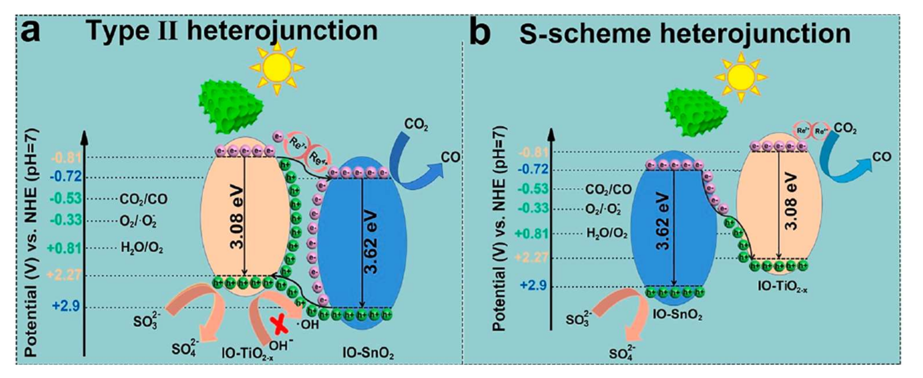
  134. Ye, J.; Xu, J.; Tian, D.; Zhao, X.; Wang, Q.; Wang, J.; Li, Y.; Zhao, C.; Liu, Z.; Fu, Y. Efficient photocatalytic reduction of CO2 by a rhenium-doped TiO2-x/SnO2 inverse opal S-scheme heterostructure assisted by the slow-phonon effect. Sep. Purif. Technol. 2021, 277, 119431. [Google Scholar] [CrossRef]
  135. Lei, M.; Wu, W.; Sun, L.; Tian, Q.; Jiang, C.; Xiao, X. Controlled preparation of hollow SnO2@M (M = Au, Ag) heterostructures through template-assist method for enhanced photocatalysis. Colloids Surfaces A Physicochem. Eng. Asp. 2015, 482, 276–282. [Google Scholar] [CrossRef]
  136. Li, Q.; Fu, J.; Zhu, W.; Chen, Z.; Shen, B.; Wu, L.; Xi, Z.; Wang, T.; Lu, G.; Zhu, J.J.; et al. Tuning Sn-Catalysis for Electrochemical Reduction of CO2 to CO via the Core/Shell Cu/SnO2 Structure. J. Am. Chem. Soc. 2017, 139, 4290–4293. [Google Scholar] [CrossRef] [PubMed]
  137. Ansari, S.A.; Khan, M.M.; Ansari, M.O.; Lee, J.; Cho, M.H. Visible light-driven photocatalytic and photoelectrochemical studies of Ag-SnO2 nanocomposites synthesized using an electrochemically active biofilm. RSC Adv. 2014, 4, 26013–26021. [Google Scholar] [CrossRef]
  138. Betiha, M.A.; Rabie, A.M.; Ahmed, H.S.; Abdelrahman, A.A.; El-Shahat, M.F. Oxidative desulfurization using graphene and its composites for fuel containing thiophene and its derivatives: An update review. Egypt. J. Pet. 2018, 27, 715–730. [Google Scholar] [CrossRef]
  139. Muzic, M.; Sertic-Bionda, K.; Gomzi, Z.; Podolski, S.; Telen, S. Study of diesel fuel desulfurization by adsorption. Chem. Eng. Res. Des. 2010, 88, 487–495. [Google Scholar] [CrossRef]
  140. Chandra Srivastava, V. An evaluation of desulfurization technologies for sulfur removal from liquid fuels. RSC Adv. 2012, 2, 759–783. [Google Scholar] [CrossRef]
  141. Environmental Protection Agency Control of Air Pollution From Motor Vehicles: Tier 3 Motor Vehicle Emission and Fuel Standards. Fed. Regist. 2014, 79, 23414–23886.
  142. World Health Organization. Health Effects of Transport-Related Air Pollution [Internet]; Krzyzanowski, M., Kuna-Dibbert, B., Schneider, J., Eds.; World Health Organization: Copenhagen Ø, Denmark, 2005; pp. 1–190. Available online: https://www.euro.who.int/__data/assets/pdf_file/0006/74715/E86650.pdf (accessed on 1 June 2022).
  143. Rajendran, A.; Cui, T.Y.; Fan, H.X.; Yang, Z.F.; Feng, J.; Li, W.Y. A comprehensive review on oxidative desulfurization catalysts targeting clean energy and environment. J. Mater. Chem. A 2020, 8, 2246–2285. [Google Scholar] [CrossRef]
  144. Liu, A.; Zhu, M.; Dai, B. A novel high-performance SnO2 catalyst for oxidative desulfurization under mild conditions. Appl. Catal. A Gen. 2019, 583, 117134. [Google Scholar] [CrossRef]
  145. Piao, W.; Li, Z.; Li, C.; Park, J.S.; Lee, J.H.; Li, Z.; Kim, K.Y.; Jin, L.Y.; Kim, J.M.; Jin, M. Efficient and reusable ordered mesoporous WOx/SnO2catalyst for oxidative desulfurization of dibenzothiophene. RSC Adv. 2021, 11, 27453–27460. [Google Scholar] [CrossRef] [PubMed]
  146. Rajendran, A.; Fan, H.X.; Cui, T.Y.; Feng, J.; Li, W.Y. Enrichment of polymeric WOx species in WOx@SnO2 catalysts for ultra-deep oxidative desulfurization of liquid fuels. Fuel 2021, 290, 120036. [Google Scholar] [CrossRef]
  147. Zhang, G.; Ren, J.; Liu, B.; Tian, M.; Zhou, H.; Zhao, J. In situ hydrothermal preparation and photocatalytic desulfurization performance of metallophthalocyanine sensitized SnO2. Inorganica Chim. Acta 2018, 471, 782–787. [Google Scholar] [CrossRef]
  148. Ko, T.H.; Chu, H.; Chaung, L.K. The sorption of hydrogen sulfide from hot syngas by metal oxides over supports. Chemosphere 2005, 58, 467–474. [Google Scholar] [CrossRef]
  149. Yang, Y.; Shi, Y.; Cai, N. Simultaneous removal of COS and H2S from hot syngas by rare earth metal-doped SnO2 sorbents. Fuel 2016, 181, 1020–1026. [Google Scholar] [CrossRef]
  150. Babich, I.V.; Moulijn, J.A. Science and technology of novel processes for deep desulfurization of oil refinery streams: A review. Fuel 2003, 82, 607–631. [Google Scholar] [CrossRef]
  151. Ahmaruzzaman, M.; Mohanta, D.; Nath, A. Environmentally benign fabrication of SnO2-CNT nanohybrids and their multifunctional efficiency as an adsorbent, catalyst and antimicrobial agent for water decontamination. Sci. Rep. 2019, 9, 12935. [Google Scholar] [CrossRef]
  152. Nasir, Z.; Shakir, M.L.; Wahab, R.; Shoeb, M.; Alam, P.; Khan, R.H.; Mobin, M. Co-precipitation synthesis and characterization of Co doped SnO2 NPs, HSA interaction via various spectroscopic techniques and their antimicrobial and photocatalytic activities. Int. J. Biol. Macromol. 2017, 94, 554–565. [Google Scholar] [CrossRef]
  153. Qamar, M.A.; Shahid, S.; Khan, S.A.; Zaman, S.; Sarwar, M.N. Synthesis characterization, optical and antibacterial studies of Co-Doped SnO2 nanoparticles. Dig. J. Nanomater. Biostruct. 2017, 12, 1127–1135. [Google Scholar]
  154. Amutha, T.; Rameshbabu, M.; Sasi Florence, S.; Senthilkumar, N.; Vetha Potheher, I.; Prabha, K. Studies on structural and optical properties of pure and transition metals (Ni, Fe and co-doped Ni–Fe) doped tin oxide (SnO2) nanoparticles for anti-microbial activity. Res. Chem. Intermed. 2019, 45, 1929–1941. [Google Scholar] [CrossRef]
  155. Wang, L.; Hu, C.; Shao, L. The antimicrobial activity of nanoparticles: Present situation and prospects for the future. Int. J. Nanomed. 2017, 12, 1227–1249. [Google Scholar] [CrossRef] [PubMed] [Green Version]
  156. Bhawna; Choudhary, A.K.; Gupta, A.; Kumar, S.; Kumar, P.; Singh, R.P.; Singh, P.; Kumar, V. Synthesis, Antimicrobial Activity, and Photocatalytic Performance of Ce Doped SnO2 Nanoparticles. Front. Nanotechnol. 2020, 2, 595352. [Google Scholar] [CrossRef]
  157. Ali Baig, A.B.; Rathinam, V.; Palaninathan, J. Fabrication of Zr-doped SnO2 nanoparticles with synergistic influence for improved visible-light photocatalytic action and antibacterial performance. Appl. Water Sci. 2020, 10, 54. [Google Scholar] [CrossRef] [Green Version]
  158. Roychaudhury, A.M.; Debnath, U.; Munshi, S.; Basak, G.K.; Dutta, A.; Masanta, S.; Singha, A.; Banerjee, A.; Ghosh, D.; Chatterjee, P.; et al. Synthesis Structural and Anti-microbial Characterization of Nanostructured Doped Tin Oxide. J. Theor. Appl. Phys. 2022, 16, 1–8. [Google Scholar]
  159. Thamiz Selvi, R.; Nallaiyan, M.; Nisha, S.; Sagadevan, S.; Mohammad, F.; Sadaiyandi, K.; Pradeev Raj, K.; Ashokkumar, L.; Latha, M.B.; Lett, J.A. Comparative studies on structural, optical, and biological properties of SnO2 and Ni-doped SnO2 nanocrystals. Mater. Res. Express 2019, 6, 125099. [Google Scholar] [CrossRef]
  160. John, N.; Somaraj, M.; Tharayil, N.J. Synthesis, Characterization and Anti—Bacterial Activities of Pure and Ag—Doped SnO2Nanoparticles. Mater. Today Proc. 2017, 4, 4351–4357. [Google Scholar] [CrossRef]
  161. Sathishkumar, M.; Geethalakshmi, S. Enhanced photocatalytic and antibacterial activity of Cu:SnO2 nanoparticles synthesized by microwave assisted method. Mater. Today Proc. 2020, 20, 54–63. [Google Scholar] [CrossRef]
  162. Mala, N.; Ravichandran, K.; Pandiarajan, S.; Srinivasan, N.; Ravikumar, B.; Nithiyadevi, K. Enhanced antibacterial and photocatalytic activity of (Mg+Co) doped tin oxide nanopowders synthesised using wet chemical route. Mater. Technol. 2017, 32, 686–694. [Google Scholar] [CrossRef]
  163. Ali Baig, A.B.; Rathinam, V.; Ramya, V. Synthesis and Investigation of Fe doped SnO2 Nanoparticles for Improved Photocatalytic Activity under Visible Light and Antibacterial performances. Mater. Technol. 2021, 36, 623–635. [Google Scholar] [CrossRef]
  164. Vega-Jiménez, A.L.; Vázquez-Olmos, A.R.; Acosta-Gío, E.; Antonio Álvarez-Pérez, M. In vitro Antimicrobial Activity Evaluation of Metal Oxide Nanoparticles. In Nanoemulsions—Properties, Fabrications and Applications; IntechOpen: London, UK, 2019; p. 13. [Google Scholar]
  165. Pandey, M.; Wasnik, K.; Gupta, S.; Singh, M.; Patra, S.; Gupta, P.; Pareek, D.; Maity, S.; Tilak, R.; Paik, P. Targeted specific inhibition of bacterial and: Candida species by mesoporous Ag/Sn-SnO2composite nanoparticles: In silico and in vitro investigation. RSC Adv. 2022, 12, 1105–1120. [Google Scholar] [CrossRef]
  166. Khan, S.A.; Kanwal, S.; Rizwan, K.; Shahid, S. Enhanced antimicrobial, antioxidant, in vivo antitumor and in vitro anticancer effects against breast cancer cell line by green synthesized un-doped SnO2 and Co-doped SnO2 nanoparticles from Clerodendrum inerme. Microb. Pathog. 2018, 125, 366–384. [Google Scholar] [CrossRef]
  167. Obeizi, Z.; Benbouzid, H.; Bouarroudj, T. Excellent antimicrobial and anti-bio fi lm activities of Fe—SnO2 nanoparticles as promising antiseptics and disinfectants. Adv. Nat. Sci. Nanosci. Nanotechnol. 2021, 12, 1. [Google Scholar]
  168. Vignesh, K.; Hariharan, R.; Rajarajan, M.; Suganthi, A. Photocatalytic performance of Ag doped SnO2 nanoparticles modified with curcumin. Solid State Sci. 2013, 21, 91–99. [Google Scholar] [CrossRef]
  169. Bhattacharya, P.; Neogi, S. Antibacterial properties of doped nanoparticles. Rev. Chem. Eng. 2019, 35, 861–876. [Google Scholar] [CrossRef]
  170. Sirelkhatim, A.; Mahmud, S.; Seeni, A.; Kaus, N.H.M.; Ann, L.C.; Bakhori, S.K.M.; Hasan, H.; Mohamad, D. Review on zinc oxide nanoparticles: Antibacterial activity and toxicity mechanism. Nano-Micro Lett. 2015, 7, 219–242. [Google Scholar] [CrossRef] [Green Version]
  171. Gökçeli, G.; Karatepe, N. Improving the properties of indium tin oxide thin films by the incorporation of carbon nanotubes with solution-based techniques. Thin Solid Films 2020, 697, 137844. [Google Scholar] [CrossRef]
  172. Graužinyte, M.; Goedecker, S.; Flores-Livas, J.A. Computational Screening of Useful Hole-Electron Dopants in SnO2. Chem. Mater. 2017, 29, 10095–10103. [Google Scholar] [CrossRef]
  173. Williamson, B.A.D.; Featherstone, T.J.; Sathasivam, S.S.; Swallow, J.E.N.; Shiel, H.; Jones, L.A.H.; Smiles, M.J.; Regoutz, A.; Lee, T.L.; Xia, X.; et al. Resonant Ta Doping for Enhanced Mobility in Transparent Conducting SnO2. Chem. Mater. 2020, 32, 1964–1973. [Google Scholar] [CrossRef] [Green Version]
  174. Bose, P.K.; Kabir, M.J. Fingerprint: A Unique and Reliable Method for Identification. J. Enam Med. Coll. 2017, 7, 29–34. [Google Scholar] [CrossRef] [Green Version]
  175. Li, Y.; Xu, C.; Shu, C.; Hou, X.; Wu, P. Simultaneous extraction of level 2 and level 3 characteristics from latent fingerprints imaged with quantum dots for improved fingerprint analysis. Chin. Chem. Lett. 2017, 28, 1961–1964. [Google Scholar] [CrossRef]
  176. Figueroa, B.; Chen, Y.; Berry, K.; Francis, A.; Fu, D. Label-Free Chemical Imaging of Latent Fingerprints with Stimulated Raman Scattering Microscopy. Anal. Chem. 2017, 89, 4468–4473. [Google Scholar] [CrossRef] [PubMed]
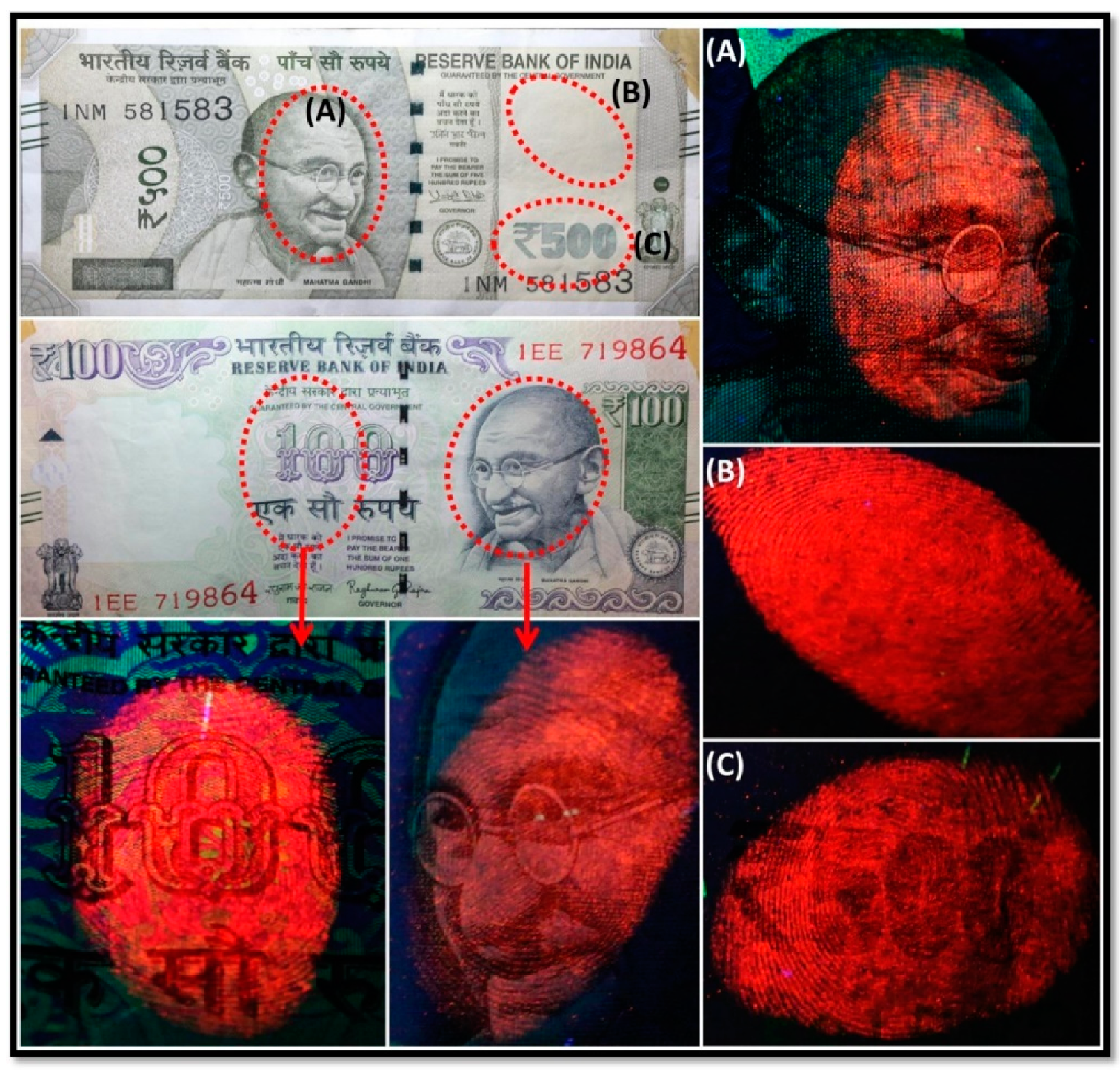
  177. Deepthi, N.H.; Darshan, G.P.; Basavaraj, R.B.; Prasad, B.D.; Sharma, S.C.; Kavyashree, D.; Nagabhushana, H. Nanostructured Stannic Oxides for White Light Emitting Diodes Provides Authentication for Latent Fingerprints Visualization under Diverse Environmental Conditions. ACS Sustain. Chem. Eng. 2019, 7, 578–591. [Google Scholar] [CrossRef]
  178. Hakim, M.; Broza, Y.Y.; Barash, O.; Peled, N.; Phillips, M.; Amann, A.; Haick, H. Volatile organic compounds of lung cancer and possible biochemical pathways. Chem. Rev. 2012, 112, 5949–5966. [Google Scholar] [CrossRef]
Figure 1. VB and CB edge potentials with bandgaps of SnO2 quantum dots annealing with different temperatures. In this figure, the acronym SQD relates to the annealing temperature at °C. For instance, the sample SQD-200 was annealed at 200 °C. Reproduced with permission from Ref. [13]. Copyright 2018 Elsevier.
Solids 03 00024 g001
Figure 2. Representation of the Valence Band (VB) and Conduction Band (CB) for the undoped (left), n-type (centre), and p-type (right) doped semiconductors. Notice the relative position between VB, CB, and the Fermi energy level (EF) in all three cases.
Solids 03 00024 g002
Figure 3. Unit cell of the tetragonal cassiterite structure of the SnO2. The Sn is represented by the orange spheres and O is represented by the blue spheres.
Solids 03 00024 g003
Figure 4. Band alignment scheme for Sn1−xPbxO2, where x = 0.00, 0.06, and 0.12. All energies are given relative to an O 1 s state. Adapted with permission from Ref. [50]. Copyright 2016 the Royal Society of Chemistry.
Solids 03 00024 g004
Figure 5. The bandgap of some photocatalysts concerning the redox potential of different chemical species, measurements conducted at a pH of 7. Adapted with permission from Ref. [68]. Copyright 2015 Elsevier.
Solids 03 00024 g005
Figure 6. Illustration of the influence of oxygen vacancies in the energy levels and the excitation of the electron during the photocatalytic activity of doped SnO2. Adapted from reference [2]. Copyright 2020 Faculty of Technology, University of Novi Sad.
Solids 03 00024 g006
Figure 7. Crystal structure of SnO2 and optimized structures of Ce-SnO2. (R1, R2) represent different replace positions. The Ce atom easily replaces the Sn atom in the R1 position and the O atom connected to the Ce atom is easily lost. Reproduced with permission from Ref. [84]. Copyright 2021 Elsevier.
Solids 03 00024 g007
Figure 8. (a) SnO2 nanobelts, (b) SnO2 nanofibres, (c) SnO2 nanowires, (d) SnO2 nanoplates, (e) SnO2 hollow nanospheres, (f) SnO2 porous nanotubes. Adapted from references [89,90,91,92,93]. Copyright 2003, 2009, 2011, 2013, 2020 Elsevier.
Solids 03 00024 g008
Figure 9. Synthesis routes of various SnO2 nanostructures using hydrothermal treatment. Reproduced with permission from Ref. [85]. Copyright 2022 Elsevier.
Solids 03 00024 g009
Figure 10. Proposed mechanism for the reduction of CO2 to formate on Sn/SnOx cathodes. Reproduced with permission from Ref. [129]. Copyright 2015 American Chemical Society.
Solids 03 00024 g010
Figure 11. Proposed density of electronic states (DOS) for (i) SnO2, (ii) 1 at% Ni–SnO2, and (iii) 5 at% Ni–SnO2 NPs. Reproduced with permission from Ref. [130]. Copyright 2020 Elsevier.
Solids 03 00024 g011
Figure 12. (a) Possible schemes for electron–hole separation and transport at composite interface and (b) photocatalytic activities of materials on CO2 photoreduction under simulated sunlight irradiation. Reproduced with permission from Ref. [133]. Copyright 2015 Elsevier.
Solids 03 00024 g012
Figure 13. A representative scheme of electron transfer in the heterojunction (Re-IO-TiO2–x/SnO2) in the CO2 photoreduction process. Adapted with permission from Reference [134]. Copyright 2021 Elsevier.
Solids 03 00024 g013
Figure 14. Mechanism of the formation of ZnTcPc/SnO2 composites. Reproduced with permission from Ref. [147]. Copyright 2018 Elsevier.
Solids 03 00024 g014
Figure 15. Schematic diagram of the antibacterial activity from generation of ROS induced by Co-doped SnO2 NPs. Figure reprinted from Reference [152]. Copyright 2017 Elsevier.
Solids 03 00024 g015
Figure 16. (a) ZOIs in antifungal test and (b) MIC test of plant extract, un-doped SnO2 NAPs, Co-doped SnO2 NPs and standard drug against fungal strains. Fluorescence microscopy with Calcofluor black shows the development of hyphae and pseudo-hyphae in controls (c,e) and Co-doped SnO2 NPs treated cells (d,f). TEM image of untreated Candida albicans cells (g) and treated with Co-doped SnO2 NPs (h) showing the changes detected in the cellular nucleus (n) and mitochondria (m). Figure reprinted from Reference [166]. Copyright 2018 Elsevier.
Solids 03 00024 g016
Figure 17. LFPs observed in different locations of a currency bill stained with Eu3+ doped SnO2 (7% Eu3+ in mol), when illuminated by UV radiation with λ = 254 nm. (AC) show different regions of the bill when illuminated by the UV radiation. The parts stained, illuminated with the UV light, and analysed are shown within red dashed circles on the bill pictures in the upper left and the central left panels of the figure. Reprinted with oermission from Reference [177]. Copyright 2019 American Chemical Society.
Solids 03 00024 g017
Table 1. Some references for doped SnO2, their synthesis approach and morphological aspects.
DopingSynthesis MethodMorphology—Primary Structures to Hierarchical Reference
AgSol-gel/electrospinning and calcinationNanoparticles to Hollow nanofibres[87]
AlHydrothermalNanoparticles [95]
BiHydrothermalNanoparticles[4]
CeHydrothermalHollow spheres[86]
CCombustionfaceted nanocrystals[96]
CoPrecipitation and hydrothermalNanoparticles[97]
CrCombustionNanoparticles[98]
CuPrecipitation and calcinationFaceted nanocrystals[99]
EuHydrothermalNanorods to flower-like[100]
FeHydrolyses and hydrothermalNanoparticles to echinus-like[101]
LaMicroemulsion and calcinationNanoparticles[102]
NbSolvothermalFaceted nanocrystals[103]
NiSolvothermalParticles to microspheres[104]
PdSol-gel and calcinationParticles to microspheres[105]
SThermal oxidation in airNanosheets to flower-like[106]
SrSol-gel precipitation and calcinationNanoparticles[73]
TiPrecipitation and calcinationNanoparticles[107]
VCombustionNanoparticles[108]
WSol-gel and calcinationNanoparticles[109]
YHydrothermalNanoparticles[110]
ZnPolyol and calcinationNanoparticles[111]
ZrHydrothermalNanoparticles[112]
Table 2. d-SnO2 samples for which antibacterial activity through ROS production has been reported.
d-SnO2 SampleBacteria SpeciesZOI (mm)ZOI of Undoped Sample (mm)Reference
Co-doped SnO2 NPsEnterococcus faecalis
Staphylococcus aureus
Escherichia coli
Enterobacter spp.
Pseudomonas aeruginosa
26
19
28
24
20
inactive
inactive
10
inactive
inactive
[19]
Ce-doped SnO2 NPsEscherichia coli-unanalysed[156]
Zr-doped SnO2 NPsStaphylococcus aureus
Escherichia coli
5 ± 0.5
7 ± 0.5
3 ± 1
4 ± 0.5
[157]
Zn-doped SnO2 NPsStaphylococcus aureus
Escherichia coli
3 ± 0.5
8 ± 0.3
1 ± 1
4 ± 0.5
[66]
Ce-doped SnO2 NPsStaphylococcus aureus
Escherichia coli
9 ± 1
5 ± 1
7 ± 0.5
4 ± 0.5
[83]
B-doped and B-Ag co-doped SnO2 NPsStaphylococcus aureus
Escherichia coli
6–13
13–15
unanalysed
unanalysed
[158]
Ni-doped SnO2 NPsStaphylococcus aureus
Escherichia coli
14–18
16–20
13
14
[159]
Co-doped SnO2 NPsEscherichia coli
Bacillus subtilis
16 ± 0.8
22 ± 1.6
unanalysed
unanalysed
[153]
Ag-doped SnO2 NPsStaphylococcus aureus
Escherichia coli
Aeronomous hydrophila
Shigella flexineri
8
12–13
6–8
10–15
unanalysed
unanalysed
unanalysed
unanalysed
[160]
Cu-doped SnO2 NPsPseudomonas aeruginosa
Staphylococcus aureus
12–19
10–18
unanalysed
unanalysed
[161]
Publisher’s Note: MDPI stays neutral with regard to jurisdictional claims in published maps and institutional affiliations.

