Article

Antimicrobial Activity of Oregano, Thyme, and Lavender Oils Against Oral Pathogens: Perspectives for AI-Supported Research

by
Casandra-Maria Radu
1,*,
Carmen Corina Radu
2,3,4,
Ionut-Daniel Venter
1,
Mihaela Alexandra Bogdan
5 and
Dana Carmen Zaha
1,6
1
Doctoral School of Biological and Biomedical Sciences, University of Oradea, 1 University Street, 410087 Oradea, Romania
2
Department of Forensic Medicine, George Emil Palade University of Medicine, Pharmacy, Sciences and Technology of Targu Mures, 38 Gheorghe Marinescu Street, 540139 Targu Mures, Romania
3
Institute of Forensic Medicine, 540141 Targu Mures, Romania
4
Doctoral School of Medicine and Pharmacy, George Emil Palade University of Medicine, Pharmacy, Sciences and Technology of Targu Mures, 540142 Targu Mures, Romania
5
Department of Pharmacy, Faculty of Medicine and Pharmacy, University of Oradea, 1 December Sq., 410028 Oradea, Romania
6
Department of Preclinical Disciplines, Faculty of Medicine and Pharmacy, University of Oradea, 1 December Sq., 410028 Oradea, Romania
*
Author to whom correspondence should be addressed.
Submission received: 19 July 2025 / Revised: 29 September 2025 / Accepted: 21 October 2025 / Published: 4 November 2025

Abstract

Background: Antimicrobial resistance in oral pathogens drives interest in natural alternatives such as essential oils (EOs). Methods: The chemical composition and in vitro antimicrobial activity of Origanum vulgare, Thymus vulgaris, and Lavandula angustifolia EOs were investigated. Oils were profiled by gas chromatography–mass spectrometry (GC-MS) and tested against Staphylococcus aureus ATCC 29213, Enterococcus faecalis ATCC 29212, Escherichia coli ATCC 25922, and Candida albicans ATCC 10231 using the disc diffusion method (triplicate, 1 µL/disc, ~850–950 µg). Results: O. vulgare oil produced the strongest inhibition against C. albicans (18.4 ± 0.5 mm), T. vulgaris was most active E. coli (13.0 ± 0.5 mm), while L. angustifolia showed negligible activity (6–7 mm). All EO inhibition zones were smaller than those of antibiotics. Conclusions: At clinically relevant doses, Oregano and Thyme oils showed modest antimicrobial effects, whereas Lavender was inactive. However, these findings are limited by the use of ATCC strains, small sample size, and reliance on the disc diffusion method, which provides only qualitative data and does not capture biofilm or host interactions. Future studies should include minimum inhibitory concentrations (MIC)/minimum bactericidal concentrations (MBC) assays, biofilm models, and cytotoxicity testing. AI-assisted GC-MS analysis and automated inhibition zone measurement should be considered as future perspectives to improve reproducibility and translational potential.

1. Introduction

Oral infections such as dental caries, periodontal disease, and candidiasis remain among the most prevalent conditions worldwide and are associated with systemic complications [1,2,3]. These diseases are driven by microbial dysbiosis within the oral cavity, which harbours over 700 bacterial species, along with fungi, viruses, and protozoa [4,5]. Conventional antimicrobials are often limited by their inability to penetrate biofilms and by the rising prevalence of antimicrobial resistance (AMR) [6,7,8].
EOs—volatile plant extracts rich in bioactive secondary metabolites—have emerged as promising antimicrobial agents with applications in both oral hygiene products and therapeutic interventions [9]. EOs possess a complex composition of terpenes, alcohols, phenolics, aldehydes, and ketones, which act synergistically to exert bactericidal, fungicidal, antioxidant, and anti-inflammatory effects [10,11,12]. Their lipophilic nature enables them to penetrate microbial membranes, disrupt lipid bilayers, denature proteins, and inhibit ATP production [13]. Notably, EOs do not typically induce bacterial resistance, making them ideal candidates for long-term oral use [14].
Among the wide variety of medicinal plants studied for their antimicrobial properties, three species stand out for their historical and scientific relevance in oral health:
  • Origanum vulgare (oregano): Rich in carvacrol, this EO is well-documented for its potent activity against Gram-positive and Gram-negative bacteria. Carvacrol has been shown to inhibit biofilm formation and increase membrane permeability in resistant bacterial strains [15,16].
  • Thymus vulgaris (thyme): Known for its high thymol and p-cymene content, thyme oil exerts strong antimicrobial and anti-inflammatory actions, particularly against oral streptococci and Candida species [17].
  • Lavandula angustifolia (lavender): Traditionally used for its calming and anti-inflammatory properties, lavender EO contains linalool and linalyl acetate, which possess mild antibacterial and antifungal effects. While not as potent as oregano or thyme, its use in oral formulations is often linked to its soothing effects on mucosa [1].
Although numerous EOs have shown promise in antimicrobial research, most studies have focused on individual EOs without direct comparative analysis or correlation with chemical profiles. This lack of standardization hinders the translation of EO-based therapies into clinical practice [18]. Furthermore, variability in plant chemotypes, extraction methods, and compound stability can significantly influence bioactivity. Comparative studies that combine GC-MS profiling with functional microbiological assays are essential for identifying the most potent and consistent candidates [16,19]. Evaluating multiple EOs side by side against relevant microorganisms provides a more rational basis for selecting active ingredients for oral formulations, especially as interest in botanical and complementary therapies continues to grow among patients and practitioners [20].
Several in vitro studies have highlighted the antimicrobial potential of these oils individually, but comparative data linking their chemical composition to biological activity against oral pathogens remain limited. Moreover, standardization of EO components and their relative efficacy versus synthetic antibiotics is still underexplored in the dental field [19,21,22,23].
The panel was selected to cover representative Gram-positive, Gram-negative, and fungal organisms. E. faecalis and C. albicans are recognized as direct oral pathogens, while S. aureus and E. coli were included as opportunistic organisms occasionally isolated from oral abscesses, peri-implant infections, and secondary colonizers in immunocompromised patients. Their inclusion allows for an assessment of the broader antimicrobial spectrum of EOs beyond strictly oral pathogens, which is relevant for mixed oral infections where non-oral species may play a secondary role. We acknowledge the absence of classical cariogenic and periodontal pathogens such as S. mutans and Porphyromonas spp., which will be targeted in future studies [24].
In this context, the present study aimed to (i) characterize the chemical composition of O. vulgare, T. vulgaris, and L. angustifolia EOs by GC-MS and (ii) evaluate their in vitro antimicrobial activity against representative Gram-positive, Gram-negative, and fungal strains using the disc diffusion method.

2. Materials and Methods

2.1. Study Design and Bacterial Strains

This in vitro experimental study was conducted in July 2025 and focused on evaluating the antimicrobial activity of EOs against representative members of the oral microbiota. All microorganisms were standard ATCC reference strains (Staphylococcus aureus ATCC 29213, Enterococcus faecalis ATCC 29212, Escherichia coli ATCC 25922, and Candida albicans ATCC 10231), purchased from a certified third-party culture collection. However, these strains do not reflect the genetic and phenotypic variability of clinical isolates, which limits direct clinical extrapolation.
The inclusion of these strains was intended to cover different microbial categories: E. faecalis and C. albicans as common oral inhabitants and opportunistic pathogens, S. aureus as a frequent cause of oral abscesses and implant-associated infections, and E. coli as a Gram-negative representative occasionally isolated in mixed oral infections. This selection ensured coverage of both bacterial and fungal organisms of clinical relevance.
Antimicrobial activity was assessed using the standardized agar disc diffusion (Kirby-Bauer) method, following the protocol described by Hudzicki (American Society for Microbiology©, 2016). The Kirby-Bauer method was selected for its simplicity and reproducibility as an initial screening tool. However, this assay provides only qualitative data on growth inhibition and does not establish MICs or MBCs. Future studies should employ broth microdilution and time—kill assays to more precisely quantify antimicrobial potency and establish dose—response relationships [25,26].
Microbial suspensions were adjusted to a 0.5 McFarland (~1.5 × 10⁸ CFU/mL) and spread uniformly on agar plates [9,27].
Sterile paper discs (6 mm) were loaded with 1 µL of pure EO (≈850–950 µg/disc, based on density), which our pilot trials identified as the lowest load yielding clear, measurable halos without disc saturation. This volume was selected after preliminary trials as the lowest amount that still produced clear, measurable inhibition zones without disc saturation [28,29]. Additional dilutions (0.5, 1, and 2 µL/disc) were tested to approximate the range of concentrations found in topical oral formulations such as mouthwashes or gels. This approach was intended not only to optimize diffusion clarity but also to approximate concentrations encountered in oral care formulations, thereby improving ecological validity compared with studies that employ much higher, less clinically relevant doses.
Positive controls included clindamycin (2 µg/disc), gentamicin (120 µg/disc), and nystatin (100 IU/disc). All plates were incubated at 4 °C for 2 h to allow for EO pre-diffusion, then transferred to 37 °C for 24 h for microbial growth. Inhibition zones were measured in millimetres using a calibrated digital calliper, including the diameter of the paper disc. See Figure 1 for a schematic overview of the experimental setup.

2.2. EOs Used

Pure EOs of Origanum vulgare, Thymus vulgaris, and Lavandula angustifolia were obtained from Laboratoarele Fares Bio vital (Orăștie, Hunedoara, Romania), a certified manufacturer of plant-based EOs and aromatic extracts. The oils were selected based on their traditional medicinal use and documented antimicrobial properties. According to the supplier, all oils were 100% pure (Lot No. 7/24), stored in dark bottles at room temperature, and accompanied by quality assurance documentation and chemical composition certificates. The EOs were handled aseptically throughout the experiments. No additional sterilization was applied, since EOs are intrinsically antimicrobial and sterilization by heat or filtration could alter their volatile constituents. To avoid contamination, all manipulations were carried out under sterile laminar flow conditions using autoclaved pipette tips and sterile paper discs.

2.3. GC-MS

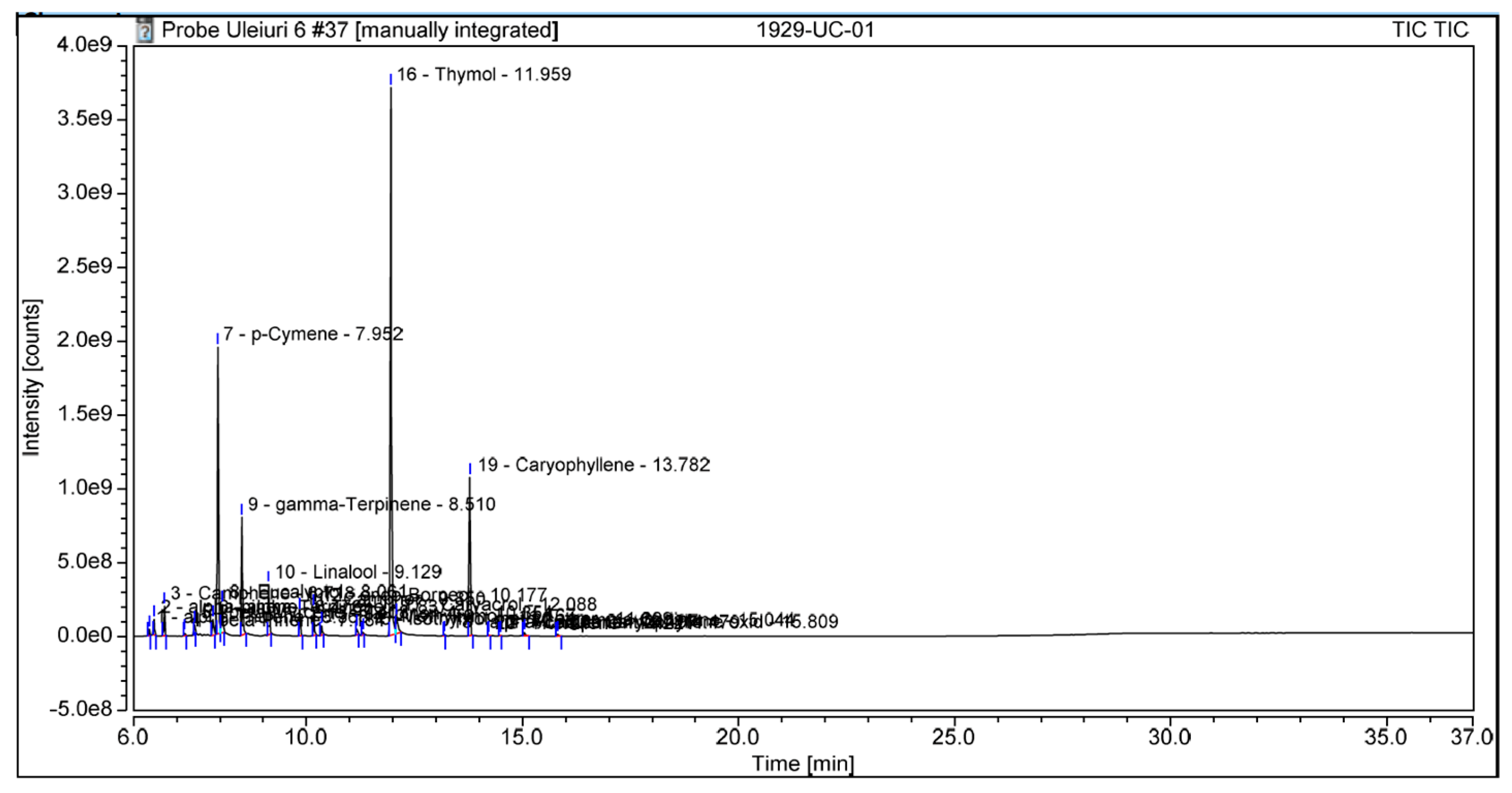
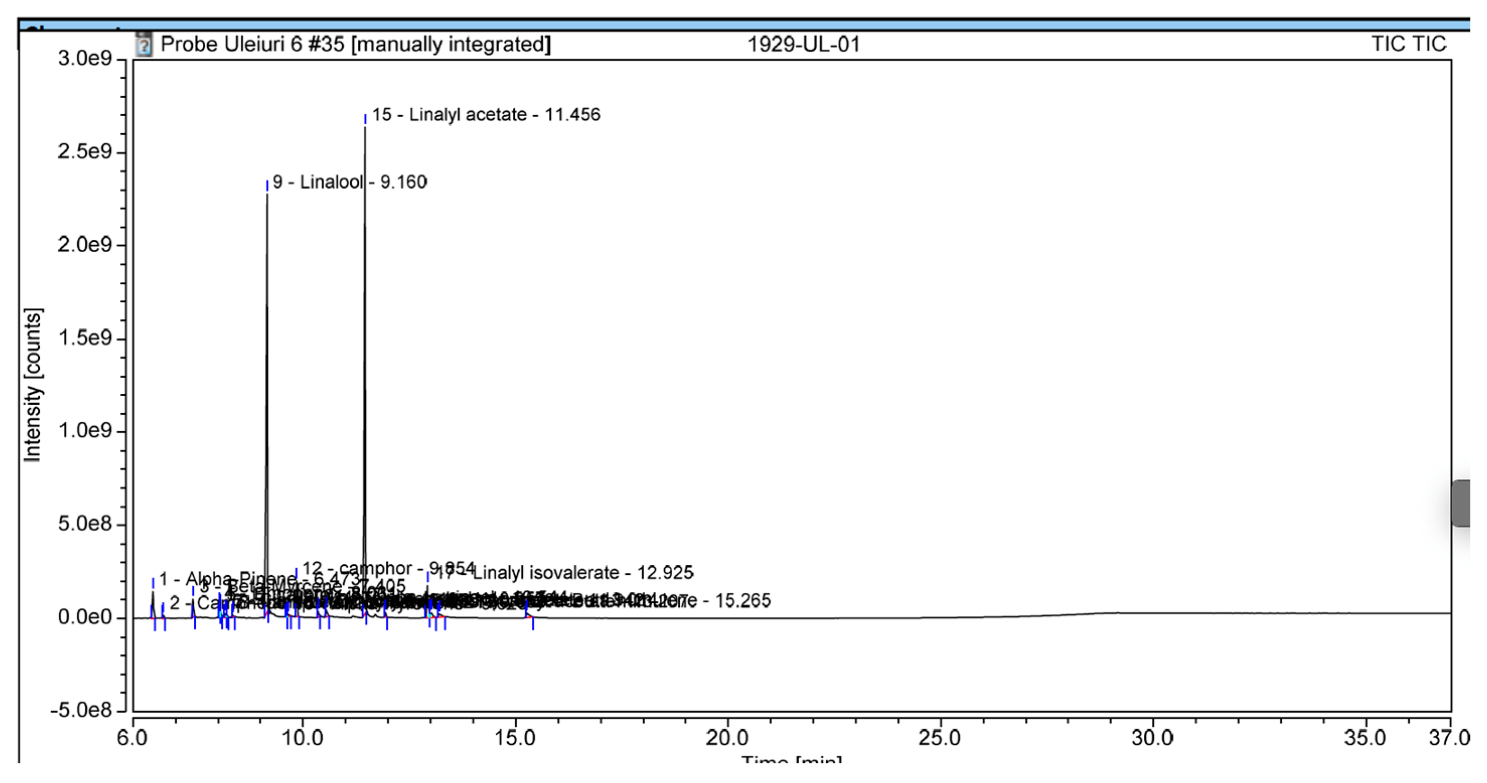
GC-MS analyses was carried out on a Trace GC Ultra gas chromatograph (Thermo Scientific, Bremen, Germany) coupled to a Thermo Electron Polaris Q mass spectrometer. EOs were injected onto a DB-5MS capillary column (30 m × 0.25 mm ID, 0.25 μm film thickness) with helium as the carrier gas at 1.5 mL/min. The oven was programmed from 40 °C (3 min hold), increased at 10 °C/min to 300 °C, and held for 10 min. The injector and transfer line were set to 250 °C and 300 °C, respectively. Mass spectra were obtained at 70 eV, and compounds were identified by comparison with the NIST spectral library (latest version) [30]. The chromatographic results are presented in Figure 2, Figure 3 and Figure 4.
Based on GC-MS analysis, Thymus vulgaris oil was primarily composed in p-cymene, γ-terpinene, linalool, and thymol, Origanum vulgare oil was rich in caryophyllene, linalool, and carvacrol; and Lavandula angustifolia oil was predominantly composed of linalool and camphor [1,31].
GC-MS analysis confirmed the expected dominant compounds for each EO: thymol (44.8%) and p-cymene (19.9%) in T. vulgaris; carvacrol (72.2%) and caryophyllene (5.4%) in O. vulgare; and linalool (42.1%) and camphor (2.7%) in L. angustifolia. Minor components were also detected.

2.4. Statistical Analysis

All results are expressed as mean ± standard deviation (SD) based on triplicate measurements. One-way analysis of variance (ANOVA) was used to evaluate the differences in antimicrobial activity (inhibition zone diameters) among the three EOs (Thymus vulgaris, Origanum vulgare, and Lavandula angustifolia) for each tested microorganism. When ANOVA results were statistically significant (p < 0.05), Tukey’s Honestly Significant Difference (HSD) post hoc test was performed to determine pairwise comparisons between groups. Statistical analysis was conducted using Python (version 3.11), with SciPy and Statsmodels packages. Data visualization was carried out using Matplotlib version 3.10.7 and Seaborn libraries version 3.8. A p-value < 0.05 was considered statistically significant [32,33,34]. Normality (Shapiro–Wilk test) and homogeneity of variances (Levene’s test) were assessed prior to ANOVA. Although triplicate experiments (E = 3) were performed, this sample size is relatively small and may reduce statistical power, potentially increasing the risk of type I errors. Larger replicate numbers are required in future studies for stronger statistical reliability [35]. Data are presented as mean ± standard deviation (SD).

3. Results

The antimicrobial activity of the tested EOs was assessed using the Kirby–Bauer disc diffusion method. Each experiment was performed in triplicate, and the results are presented as mean ± SD, as summarized in Table 1.
Both Thymus vulgaris (P1) and Origanum vulgare (P2) EOs demonstrated pronounced antimicrobial effects across all tested strains, although the inhibition zones were smaller compared to antibiotic controls.
For example, Oregano EO produced 18.4 mm inhibition against Candida albicans, while Thyme EO inhibited Escherichia coli at 13 mm. In contrast, Lavandula angustifolia (P3) exhibited little to no activity beyond the disc margin, with inhibition diameters of 6–7 mm. Antibiotic controls consistently produced larger inhibition zones (Clindamycin: 27 mm vs. S. aureus; Nystatin: 20 mm vs. C. albicans). Post hoc Tukey analysis confirmed statistically significant differences between Oregano and Thyme oils compared with lavender across all pathogens (p < 0.0001). Tukey’s post hoc analysis indicated that Oregano and Thyme oils did not significantly differ from each other, while both showed consistently greater inhibition than lavender, highlighting that the main distinction was between the active oils and lavender. Differences between Oregano and Thyme were small (<1 mm) and borderline significant for S. aureus, E. faecalis, and E. coli, but Oregano demonstrated significantly greater inhibition against E (p < 0.0001).
These findings suggest that while Oregano and Thyme EOs retain antimicrobial activity at clinically relevant doses, their potency is more modest than when tested at artificially high concentrations.
These findings support the significant antimicrobial potential of T. vulgaris and O. vulgare EOs, particularly against both Gram-positive and Gram-negative bacteria, as well as fungal pathogens to oral infections.

4. Discussion

This study demonstrates that Origanum vulgare and Thymus vulgaris EOs exhibit measurable in vitro antimicrobial activity against microorganisms relevant to oral infections, including oral opportunists (S. aureus, E. coli), an endodontic pathogen (E. faecalis), and a major oral opportunist (C. albicans). In contrast, Lavandula angustifolia exhibited more limited antimicrobial activity, particularly against Gram-negative bacteria. These findings align with previous research emphasizing the high efficacy of O. vulgare and T. vulgaris, which is primarily attributed to their high concentrations of carvacrol and thymol, respectively [17,19,23,36].
Carvacrol, the principal component in O. vulgare, is known for its ability to disrupt microbial membrane integrity, leading to ion leakage, ATP depletion, and microbial cell death [37]. Thymol, abundant in T. vulgaris, shares similar bactericidal mechanisms and has been shown to interfere with enzymatic pathways and oxidative stress responses. These mechanisms are especially effective against Gram-positive organisms due to their simpler cell wall structure, although our data suggest that these EOs also exhibit strong activity against Gram-negative species [38].
On the other hand, L. angustifolia oil, rich in linalool and linalyl acetate, demonstrated weaker antimicrobial effects. Although linalool has mild antifungal and anti-inflammatory properties, its lower membrane affinity may account for the smaller inhibition zones observed. This supports its use more as a complementary anti-inflammatory agent in oral care rather than as a primary antimicrobial compound [17].
Although Oregano and Thyme oils produces measurable inhibition zones, their activity remained consistently lower than that of conventional antibiotics. Their role may be best considered as complementary rather than substitutive. This highlights their potential as natural alternatives or adjuncts in oral care formulations such as mouthwashes, gels, or rinses, especially in light of increasing antibiotic resistance.
In this study, an optimized EO dose of 1 µL/disc (~850–950 µg) was used, alongside lower and higher dilutions, to balance diffusion clarity with clinical relevance. While still above systemic MIC thresholds, these values approximate the surface-level concentrations used in oral care formulations and thus offer a more realistic assessment of antimicrobial potential than higher doses. At this dose, Oregano and Thyme oils still demonstrated measurable inhibitory activity, particularly against Candida albicans and E. coli, although their halos were smaller than those of conventional antibiotics. Lavender oil showed negligible antimicrobial activity under these conditions, consistent with its role as an adjunctive soothing agent rather than a primary antimicrobial. Our GC-MS analysis confirmed that Oregano oil contained a high proportion of carvacrol (72%), Thyme oil was dominated by thymol (45%), and Lavender oil contained mainly linalool (42%). These chemical profiles closely align with the antimicrobial outcomes observed. Oregano oil, rich in carvacrol, showed the strongest antifungal activity, and Thyme oil, dominated by thymol, demonstrated measurable inhibition of both bacterial and fungal strains. In contrast, Lavender oil, with linalool as its main constituent, exhibited weak antimicrobial effects, consistent with its limited activity in our assays.
Antibiotic discs were included as positive controls to validate the assay. However, direct comparisons between EOs and antibiotics should be interpreted cautiously, as no standardized clinical breakpoints exist for EOs, and the concentrations tested differ substantially from those of conventional antimicrobials.
However, this study has limitations. The agar disc diffusion method, while useful for preliminary screening, does not provide quantitative data on the minimal concentration needed to inhibit or kill pathogens. Future research should include determination of MIC and MBC using standardized broth microdilution methods. These assays would allow for precise comparisons with conventional antimicrobials and help define dosing strategies for therapeutic applications. Our team plans to implement these experiments in Q4 2025, focusing initially on T. vulgaris and O. vulgare due to their strong performance in this study. Additionally, future work will involve time-kill kinetics, biofilm disruption assays, and cytotoxicity testing on oral mucosa models. These efforts will help confirm the safety and efficacy of EO-based products in real-world oral healthcare settings.
Despite current methodological limitations, the findings clearly support the continued investigation of O. vulgare and T. vulgaris as promising natural agents for oral antimicrobial therapy.

4.1. Study Limitations

This study has several limitations that should be considered when interpreting the findings. First, only four ATCC reference strains were included. While these strains provide reproducibility, they do not fully capture the diversity of the oral microbiome. Of the four, E. coli and S. aureus are not primary oral pathogens but opportunistic organisms occasionally isolated from oral abscesses and peri-implant infections. E. faecalis was tested as a model of endodontic infection and C. albicans as a major fungal opportunist. However, key cariogenic and periodontal organisms such as Streptococcus mutans, Porphyromonas spp., and Prevotella spp. were not assessed due to methodological constraints.
Likewise, beneficial commensals including Streptococcus mitis and Streptococcus salivarius were not tested, even though their selective preservation is critical in oral ecology. Future studies will include these species to provide a more representative view of EO activity within the oral microbiome [2,39].
Second, the disc diffusion method was employed as an initial screening tool. While simple and standardized, this approach is inherently qualitative and does not yield quantitative parameters such as MIC or MBC. Moreover, it does not account for dynamic oral environment, where factors such as salivary flow, enzymatic activity, and host tissues influence antimicrobial activity [40].
Third, the present study examined only planktonic organisms. This represents a significant limitation because most oral pathogens persist within structured biofilms that confer enhanced antimicrobial tolerance. Future investigations should therefore evaluate EOs using biofilm models to more accurately simulate oral conditions.
Fourth, although GC-MS analysis successfully characterized the dominant constituents of each EOs, the profiling was limited to relative peak quantification (% peak area). Reproducibility across different batches was not systematically validated, and the potential influence of minor compounds, including possible synergistic or antagonist interactions, was not explored. These chemical factors may significantly modulate biological activity and deserve attention in future studies.
Fifth, no cytotoxicity and tissue compatibility assays were performed. Since EOs are intended for potential incorporation into oral care products, their effects on oral epithelial cells, fibroblasts, and mucosal integrity must be assessed alongside antimicrobial efficacy. Without such safety data, translation to clinical application remains incomplete.
Finally, the relatively small number of experimental replicates (n = 3) limits statistical power and increases the possibility of type I error. Future work should expand sample size, include clinical isolates in addition to ATCC strains, perform standardized MIC/MBC determinations, and incorporate biofilm disruption and cytotoxicity assays. Addressing these limitations will be essential to confirm the therapeutic potential of EOs in oral healthcare.

4.2. Clinical Implications

Despite these limitations, Oregano and Thyme oils could be considered as adjunctive components in oral healthcare products, such as mouth rinses, gels, or varnishes [1,14]. Their notable antifungal activity, especially against C. albicans, highlights potential applications in the management of recurrent oral candidiasis, including cases resistant to azole antifungals. However, since inhibition zones were consistently smaller than those of conventional antibiotics, these oils should be regarded as complementary agents rather than substitutes for standard antimicrobial therapy. Clinical translation will further require formulation studies, cytotoxicity testing, and validation in biofilm and in vivo models.

4.3. Future Directions: AI in EO-Based Oral Medicine

AI hold potential to strengthen both chemical and microbiological analyses in future studies. GC-MS generates complex data that could benefit from AI-assisted peak identification and chemotype comparison. Likewise, automated image analysis could improve reproducibility in inhibition zone measurement. By linking chemical signatures with microbial outcomes, predictive AI models may help identify the most effective EO chemotypes for oral applications. Importantly, these applications should be regarded as future perspectives rather than current outcomes.

4.4. Limitations and Future Directions

This study provides preliminary screening data on the antimicrobial potential of essential oils. However, it has several limitations, including the lack of quantitative MIC/MBC determinations, absence of biofilm and cytotoxicity testing, and no evaluation using clinical isolates. Future studies should address these methodological gaps using standardized essential oil formulations and clinically relevant models to validate the present findings.

5. Conclusions

At 1 µL/disc (~850–950 µg), Origanum vulgare and Thymus vulgaris EOs demonstrated modest but measurable antimicrobial activity against oral pathogens, particularly Candida albicans and Escherichia coli. Lavandula angustifolia exhibited negligible effects. While inhibition zones were consistently smaller than those of antibiotic controls, these findings support further exploration of Oregano and Thyme oils as complementary agents in oral healthcare. Future studies should quantify MIC/MBC values, evaluate effects on biofilms and clinical isolates, and assess cytotoxicity.
The application of AI to GC-MS analysis and antimicrobial profiling should be considered as a promising avenue for improving reproducibility and supporting translational development of EO-based oral formulations, rather than as a current outcome of this study.

Author Contributions

Conceptualization, C.-M.R. and C.C.R.; methodology, D.C.Z.; software, I.-D.V.; validation, C.-M.R.; formal analysis, M.A.B.; investigation, C.C.R.; resources, C.C.R. and D.C.Z.; data curation, D.C.Z.; writing—original draft preparation, C.-M.R.; writing—review and editing, M.A.B.; visualization, C.-M.R.; supervision, C.C.R.; project administration, D.C.Z. All authors have read and agreed to the published version of the manuscript.

Funding

This research was funded by the University of Oradea, Oradea, Romania.

Institutional Review Board Statement

Not applicable.

Informed Consent Statement

Not applicable.

Data Availability Statement

Data are available from the corresponding author upon reasonable request.

Conflicts of Interest

The authors declare no conflicts of interest.

References

  1. Rosner, O.; Livne, S.; Bsharat, M.; Dviker, S.; Jeffet, U.; Matalon, S.; Sterer, N. Lavandula angustifolia Essential Oil Inhibits the Ability of Fusobacterium nucleatum to Produce Volatile Sulfide Compounds, a Key Components in Oral Malodor. Molecules 2024, 29, 2982. [Google Scholar] [CrossRef]
  2. Rajasekaran, J.J.; Krishnamurthy, H.K.; Bosco, J.; Jayaraman, V.; Krishna, K.; Wang, T.; Bei, K. Oral Microbiome: A Review of Its Impact on Oral and Systemic Health. Microorganisms 2024, 12, 1797. [Google Scholar] [CrossRef]
  3. Quintana Soares Lopes, L.; Fortes Guerim, P.H.; Maldonado, M.E.; Wagner, R.; Hadlich Xavier, A.C.; Gutknecht da Silva, J.L.; Bittencourt da Rosa Leal, D.; de Freitas Daudt, N.; Christ Vianna Santos, R.; Kolling Marquezan, P. Chemical Composition, Cytotoxicity, Antimicrobial, Antibiofilm, and Anti-Quorum Sensing Potential of Mentha piperita Essential Oil against the Oral Pathogen Streptococcus mutans. J. Toxicol. Environ. Health A 2024, 87, 824–835. [Google Scholar] [CrossRef]
  4. Dosoky, N.S.; Setzer, W.N. Chemical Composition and Biological Activities of Essential Oils of Curcuma Species. Nutrients 2018, 10, 1196. [Google Scholar] [CrossRef] [PubMed]
  5. Al-Nasser, L.; Lamster, I.B. Prevention and Management of Periodontal Diseases and Dental Caries in the Older Adults. Periodontol. 2000 2020, 84, 69–83. [Google Scholar] [CrossRef]
  6. Chapple, I.L.C.; Bouchard, P.; Cagetti, M.G.; Campus, G.; Carra, M.C.; Cocco, F.; Nibali, L.; Hujoel, P.; Laine, M.L.; Lingstrom, P.; et al. Interaction of Lifestyle, Behaviour or Systemic Diseases with Dental Caries and Periodontal Diseases: Consensus Report of Group 2 of the Joint EFP/ORCA Workshop on the Boundaries between Caries and Periodontal Diseases. J. Clin. Periodontol. 2017, 44, S39–S51. [Google Scholar] [CrossRef]
  7. Thosar, N.; Basak, S.; Bahadure, R.N.; Rajurkar, M. Antimicrobial Efficacy of Five Essential Oils against Oral Pathogens: An In Vitro Study. Eur. J. Dent. 2013, 7, S071–S077. [Google Scholar] [CrossRef]
  8. Lynch, M.C.; Cortelli, S.C.; McGuire, J.A.; Zhang, J.; Ricci-Nittel, D.; Mordas, C.J.; Aquino, D.R.; Cortelli, J.R. The Effects of Essential Oil Mouthrinses with or without Alcohol on Plaque and Gingivitis: A Randomized Controlled Clinical Study. BMC Oral Health 2018, 18, 6. [Google Scholar] [CrossRef]
  9. Yuan, Y.; Hui, X.; Liu, Z.; Sun, J.; Raka, R.N.; Xiao, J.; Zhang, Z.; Wu, H. Investigation of Differential Multi-Mode Antibacterial Mechanisms of Essential Oils of Satureja montana L. and Leptospermum scoparium J.R.Forst. & G.Forst. Against Porphyromonas gingivalis. BMC Complement. Med. Ther. 2025, 25, 283. [Google Scholar] [CrossRef]
  10. Dosoky, N.S.; Setzer, W.N. Maternal Reproductive Toxicity of Some Essential Oils and Their Constituents. Int. J. Mol. Sci. 2021, 22, 2380. [Google Scholar] [CrossRef] [PubMed]
  11. Alrashdan, M.S.; Al-Shorman, H.; Al-Dwairi, A.; Qutieshat, A.; Al-Omiri, M.K. Salivary Oxidative Stress Biomarkers in Periodontitis-Free Smokers: A Cross Sectional Study. Minerva Dent. Oral Sci. 2024, 73, 209–216. [Google Scholar] [CrossRef]
  12. Aziz, M.I.; Hasan, M.M.; Ullah, R.; Bari, A.; Khan, M.A.; Hasnain, S.Z.U.; Baloch, R.; Akram, M.; Obaid, A.; Ullah, A.; et al. Potential Role of Citrus bergamia Flower Essential Oil against Oral Pathogens. BMC Complement. Med. Ther. 2024, 24, 157. [Google Scholar] [CrossRef]
  13. Leino, V.; Airaksinen, R.; Viluksela, M.; Vähäkangas, K. Toxicity of Colloidal Silver Products and Their Marketing Claims in Finland. Toxicol. Rep. 2021, 8, 106–113. [Google Scholar] [CrossRef] [PubMed]
  14. Oliveira, M.S.; Paula, M.S.A.; Cardoso, M.M.; Silva, N.P.; Tavares, L.C.D.; Gomes, T.V.; Porto, D.L.; Aragão, C.F.S.; Fabri, R.L.; Tavares, G.D.; et al. Exploring the Antimicrobial Efficacy of Tea Tree Essential Oil and Chitosan against Oral Pathogens to Overcome Antimicrobial Resistance. Microb. Pathog. 2024, 196, 107006. [Google Scholar] [CrossRef] [PubMed]
  15. Yuan, Y.; Sun, J.; Song, Y.; Raka, R.N.; Xiang, J.; Wu, H.; Xiao, J.; Jin, J.; Hui, X. Antibacterial Activity of Oregano Essential Oils against Streptococcus mutans In Vitro and Analysis of Active Components. BMC Complement. Med. Ther. 2023, 23, 61. [Google Scholar] [CrossRef] [PubMed]
  16. Muresan, S.M.C.; Dreanca, A.; Repciuc, C.; Dejescu, C.; Rotar, O.; Pop, R.A.; Pantea, S.; Pall, E.; Ciotlaus, I.; Sarosi, C.; et al. Dental Hydrogels with Essential Oils with Potential Activity in Periodontitis. Appl. Sci. 2023, 13, 1787. [Google Scholar] [CrossRef]
  17. Vassiliou, E.; Awoleye, O.; Davis, A.; Mishra, S. Anti-Inflammatory and Antimicrobial Properties of Thyme Oil and Its Main Constituents. Int. J. Mol. Sci. 2023, 24, 6936. [Google Scholar] [CrossRef]
  18. Cuevas-Gonzalez, M.V.; Cuevas-Gonzalez, J.C.; Espinosa-Cristóbal, L.F.; Donohue-Cornejo, A.; Reyes López, S.Y.; Saucedo Acuña, R.A.; García Calderón, A.G.; Guzmán Gastelum, D.A. Use or Abuse of Antibiotics as Prophylactic Therapy in Oral Surgery: A Systematic Review. Medicine 2023, 102, e35011. [Google Scholar] [CrossRef]
  19. Park, S.-Y.; Raka, R.N.; Hui, X.-L.; Song, Y.; Sun, J.-L.; Xiang, J.; Wang, J.; Jin, J.-M.; Li, X.-K.; Xiao, J.-S.; et al. Six Spain Thymus Essential Oils Composition Analysis and Their In Vitro and in Silico Study against Streptococcus mutans. BMC Complement. Med. Ther. 2023, 23, 106. [Google Scholar] [CrossRef]
  20. Moynihan, P.; Petersen, P.E. Diet, Nutrition and the Prevention of Dental Diseases. Public Health Nutr. 2004, 7, 201–226. [Google Scholar] [CrossRef]
  21. Ikeda, N.Y.; Ambrosio, C.M.S.; Miano, A.C.; Rosalen, P.L.; Gloria, E.M.; Alencar, S.M. Essential Oils Extracted from Organic Propolis Residues: An Exploratory Analysis of Their Antibacterial and Antioxidant Properties and Volatile Profile. Molecules 2021, 26, 4694. [Google Scholar] [CrossRef]
  22. Benzaid, C.; Belmadani, A.; Tichati, L.; Djeribi, R.; Rouabhia, M. Effect of Citrus aurantium L. Essential Oil on Streptococcus mutans Growth, Biofilm Formation and Virulent Genes Expression. Antibiotics 2021, 10, 54. [Google Scholar] [CrossRef] [PubMed]
  23. Castagliuolo, G.; Di Napoli, M.; Vaglica, A.; Badalamenti, N.; Antonini, D.; Varcamonti, M.; Bruno, M.; Zanfardino, A.; Bazan, G. Thymus richardii Subsp. nitidus (Guss.) Jalas Essential Oil: An Ally against Oral Pathogens and Mouth Health. Molecules 2023, 28, 4803. [Google Scholar] [CrossRef]
  24. Hammer, K.A.; Carson, C.F.; Riley, T. V Antimicrobial Activity of Essential Oils and Other Plant Extracts. J. Appl. Microbiol. 1999, 86, 985–990. [Google Scholar] [CrossRef]
  25. Salam, M.A.; Al-Amin, M.Y.; Pawar, J.S.; Akhter, N.; Lucy, I.B. Conventional Methods and Future Trends in Antimicrobial Susceptibility Testing. Saudi J. Biol. Sci. 2023, 30, 103582. [Google Scholar] [CrossRef]
  26. Maynard, R.; Manuel, C.; Simpkins, S.; Haro, M.; Ledeboer, N.; Simner, P.J.; Fisher, M.; Humphries, R. Development of Disk Diffusion Susceptibility Test Methods for Aerococcus spp. and Updates to Clinical and Laboratory Standards Institute MIC Breakpoints. J. Clin. Microbiol. 2025, 63, e0011525. [Google Scholar] [CrossRef]
  27. Mendoza, R.; Alvitez-Temoche, D.; Chiong, L.; Silva, H.; Mauricio, F.; Mayta-Tovalino, F. Antibacterial Efficacy of Matricaria recutita Essential Oil against Porphyromonas gingivalis and Prevotella intermedia: In Vitro Study. J. Contemp. Dent. Pract. 2023, 24, 551–555. [Google Scholar] [CrossRef]
  28. Nagy-Bota, M.C.; Man, A.; Santacroce, L.; Brinzaniuc, K.; Pap, Z.; Pacurar, M.; Pribac, M.; Ciurea, C.N.; Pintea-Simon, I.A.; Kovacs, M. Essential Oils as Alternatives for Root-Canal Treatment and Infection Control against Enterococcus faecalis—A Preliminary Study. Appl. Sci. 2021, 11, 1422. [Google Scholar] [CrossRef]
  29. Hans, V.M.; Grover, H.S.; Deswal, H.; Agarwal, P. Antimicrobial Efficacy of Various Essential Oils at Varying Concentrations against Periopathogen Porphyromonas Gingivalis. J. Clin. Diagn. Res. 2016, 10, ZC16–ZC19. [Google Scholar] [CrossRef] [PubMed]
  30. Trindade, L.A.; de Araújo Oliveira, J.; de Castro, R.D.; de Oliveira Lima, E. Inhibition of Adherence of C. Albicans to Dental Implants and Cover Screws by Cymbopogon nardus Essential Oil and Citronellal. Clin. Oral Investig. 2015, 19, 2223–2231. [Google Scholar] [CrossRef]
  31. Bogdan, M.A.; Bungau, S.; Tit, D.M.; Zaha, D.C.; Nechifor, A.C.; Behl, T.; Chambre, D.; Lupitu, A.I.; Copolovici, L.; Copolovici, D.M. Chemical Profile, Antioxidant Capacity, and Antimicrobial Activity of Essential Oils Extracted from Three Different Varieties (Moldoveanca 4, Vis Magic 10, and Alba 7) of Lavandula angustifolia. Molecules 2021, 26, 4381. [Google Scholar] [CrossRef]
  32. Seabold, S.; Perktold, J. Statsmodels: Econometric and Statistical Modeling with Python. In Proceedings of the the 9th Python in Science Conference, Austin, TX, USA, 28 June–3 July 2010; pp. 92–96. [Google Scholar]
  33. Rodríguez, J.A.L.; Casana, S.T.V.; Gómez, P.A.M. Effectiveness of Chlorhexidine and Essential Oils Associated with Scaling and Root Planing in the Treatment of Chronic Periodontitis. Rev. Cienc. Salud 2020, 18, 1–11. [Google Scholar] [CrossRef]
  34. Marinković, J.; Ćulafić, D.M.; Nikolić, B.; Đukanović, S.; Marković, T.; Tasić, G.; Ćirić, A.; Marković, D. Antimicrobial Potential of Irrigants Based on Essential Oils of Cymbopogon Martinii and Thymus Zygis towards In Vitro Multispecies Biofilm Cultured in Ex Vivo Root Canals. Arch. Oral Biol. 2020, 117, 104842. [Google Scholar] [CrossRef]
  35. Sherief, D.I.; Fathi, M.S.; Abou El Fadl, R.K. Antimicrobial Properties, Compressive Strength and Fluoride Release Capacity of Essential Oil-Modified Glass Ionomer Cements—An In Vitro Study. Clin. Oral Investig. 2021, 25, 1879–1888. [Google Scholar] [CrossRef] [PubMed]
  36. Aebisher, D.; Cichonski, J.; Szpyrka, E.; Masjonis, S.; Chrzanowski, G. Essential Oils of Seven Lamiaceae Plants and Their Antioxidant Capacity. Molecules 2021, 26, 3793. [Google Scholar] [CrossRef]
  37. Karami, F.; Dastan, D.; Fallah, M.; Matini, M. In Vitro Antitrichomonal Activity of Satureja Khuzestanica and Main Essential Oil Components Carvacrol, Thymol, and Eugenol. J. Infect. Dev. Ctries. 2023, 17, 80–85. [Google Scholar] [CrossRef]
  38. Bevilacqua, A.; Speranza, B.; Perricone, M.; Sinigaglia, M.; Corbo, M.R. Bioactivity of Essential Oils Towards Fungi and Bacteria. In Essential Oils in Food Processing; John Wiley & Sons, Ltd.: Hoboken, NJ, USA, 2017; pp. 231–246. ISBN 9781119149392. [Google Scholar]
  39. Hashim, N.T.; Babiker, R.; Priya, S.P.; Mohammed, R.; Chaitanya, N.C.; Padmanabhan, V.; El Bahra, S.; Rahman, M.M.; Gismalla, B.G. Microbial Dynamics in Periodontal Regeneration: Understanding Microbiome Shifts and the Role of Antifouling and Bactericidal Materials: A Narrative Review. Curr. Issues Mol. Biol. 2024, 46, 12196–12213. [Google Scholar] [CrossRef] [PubMed]
  40. El-Demerdash, F.M.; El-Sayed, R.A.; Abdel-Daim, M.M. Rosmarinus officinalis Essential Oil Modulates Renal Toxicity and Oxidative Stress Induced by Potassium Dichromate in Rats. J. Trace Elem. Med. Biol. 2021, 67, 126791. [Google Scholar] [CrossRef] [PubMed]
Figure 1. Schematic representation of the standardized agar disc diffusion method (Kirby-Bauer) used in this study (Created with AI-assisted design software—ChatGPT version 5.0).
Oral 05 00087 g001
Figure 2. GC-MS chromatogram of Thymus vulgaris oil (x-axis: retention time in minutes; y-axis: relative abundance, arbitrary units).
Oral 05 00087 g002
Figure 3. GC-MS chromatogram of Origanum vulgare oil (x-axis: retention time in minutes; y-axis: relative abundance, arbitrary units).
Oral 05 00087 g003
Figure 4. GC-MS chromatogram of Lavandula angustifolia oil (x-axis: retention time in minutes; y-axis: relative abundance, arbitrary units).
Oral 05 00087 g004
Table 1. Mean inhibition zone diameters (mm) of Thymus vulgaris, Origanum vulgare, and Lavandula angustifolia EOs compared with standard antimicrobial agents against selected oral pathogens. Data shown as mean ± SD from three independent replicates.
Samples 1 µL/discStaphylococcus aureus (mm)Enterococcus faecalis (mm)Escherichia coli (mm)Candida albicans (mm)
P1 (Thymus vulgaris oil)12.8 ± 0.68.5 ± 0.413.0 ± 0.517.2 ± 0.7
P2 (Origanum vulgare oil)12.4 ± 0.77.8 ± 0.312.3 ± 0.618.4 ± 0.5
P3 (Lavandula angustifolia oil)6.0 6.0 6.0 7.2 ± 0.4
Clindamycin27.0 ± 0.5---
Gentamicin-12.0 ± 0.518.0 ± 0.60.0
Nystatin---20.0 ± 0.8
6.0 mm = disc diameter (no inhibition beyond the paper disc).
Disclaimer/Publisher’s Note: The statements, opinions and data contained in all publications are solely those of the individual author(s) and contributor(s) and not of MDPI and/or the editor(s). MDPI and/or the editor(s) disclaim responsibility for any injury to people or property resulting from any ideas, methods, instructions or products referred to in the content.