Review

The Role of Digital Innovations in Shaping Contemporary Fixed Prosthodontics: A Narrative Review

Department of Prosthetic Dentistry, Faculty of Dental Medicine, Medical University of Plovdiv, 4000 Plovdiv, Bulgaria
Submission received: 4 June 2025 / Revised: 28 July 2025 / Accepted: 16 October 2025 / Published: 20 October 2025
(This article belongs to the Collection Digital Dentistry: State of the Art and Future Perspectives)

Abstract

The rapid digitization of dentistry is significantly transforming fixed prosthodontics, a discipline highly dependent on technical precision. This narrative review, incorporating a structured literature search, provides a critical overview of how digital tools—including computer-aided design and manufacturing (CAD/CAM), intraoral scanners (IOS), and additive manufacturing—are influencing clinical protocols and production methods. A database-guided selection process was employed to identify relevant studies published between 2000 and 2024, spanning in vitro research, observational studies, and clinical trials. While digital workflows offer promising benefits, such as increased accuracy, efficiency, and patient comfort, supporting evidence remains preclinical or short-term in nature. The review highlights areas of innovation as well as ongoing limitations in clinical validation, standardization, and adoption. A more cautious interpretation of the current evidence is warranted, especially regarding long-term clinical outcomes and cost-effectiveness. This review aims to inform clinicians, researchers, and educators about both the potential and the present limitations of digital fixed prosthodontics.

1. Introduction

The digital impression system, which involves CAD/CAM technology, includes both extraoral and intraoral scanners (IOSs) [1]. Initially developed in the 1950s for industrial applications such as modeling, designing, and manufacturing objects, CAD/CAM technology was introduced into dentistry in the 1980s to create a variety of prosthetic restorations [2]. This technology, originally intended for esthetic ceramic restorations, has evolved significantly over the years. It now facilitates the fabrication of inlay and onlay fillings, metal-ceramic crowns, ceramic crowns, and veneers [3].
CAD/CAM technology for prosthesis fabrication employs two main methods: subtractive manufacturing (milling) and additive manufacturing (3D printing or rapid prototyping) [4]. The prosthodontic procedure for creating fixed partial denture (FPD) involves a complex integration of sequential steps, beginning with the clinical preparation of the abutment tooth and impression making, followed by laboratory processes [5].
Modern CAD/CAM systems consist of three key units:
  • A data acquisition unit, which collects data from the prepared area and surrounding structures to create virtual impressions.
  • A designing unit, which uses software to design virtual restorations.
  • A manufacturing unit, which fabricates the prosthesis through milling or 3D printing.
The advancement of digital scanners, along with digital designing and manufacturing technologies, has revolutionized the production of tooth-borne and implant-supported fixed dental prostheses [6]. These technologies enable the creation of prostheses in a fully virtual environment, eliminating the need for physical models and significantly enhancing the efficiency and precision of dental restorations [7].
Complete digital protocols in dentistry encompass three primary steps: the 3D acquisition of the patient’s oral situation using intraoral scanners (IOSs); the digital design of dental restorations using specialized software (CAD, Software Version 2024), followed by rapid prototyping techniques like milling or 3D printing (CAM, Software Version 2024) within a completely virtual environment, eliminating the need for physical dental models such as plaster casts; and the clinical delivery of the dental restoration [8,9]. Key stages include the generation, transfer, and further processing of the IOS data, typically in Standard Tessellation Language (STL) format. Digital workflows have demonstrated potential for producing high-quality monolithic restorations in controlled settings [10]. Additionally, this approach enhances accuracy, improves efficiency, and provides a more comfortable experience for patients, marking a significant advancement in dental practice [11].
Historically, dental research has predominantly concentrated on individual steps within the three-step digital workflow process. The primary focus was on in vitro analyses, assessing precision and accuracy by comparing various intraoral scanner (IOS) systems or different rapid prototyping methods for producing final restorations [12]. Aside from a few isolated case reports, there has been a noticeable scarcity of clinical studies in dental literature, particularly randomized controlled trials (RCTs) that examine the entire digital workflow comprehensively [13].
Understanding the implications of the ongoing digitization trend is crucial, particularly regarding how it alters well-established protocols in terms of clinical and technical feasibility, long-term outcomes, and economic impacts [14]. In 2017, a systematic review undertook the first comprehensive screening of scientific literature to gather evidence on the use of complete digital workflows in fixed prosthodontics for treatments involving tooth-borne or implant-supported fixed restorations [15]. This review revealed that the evidence level for complete digital workflows was low: only three publications focused on single-unit restorations, and no studies were found that investigated multi-unit restorations at that time.
The limited scope of earlier research underscores the need for more extensive clinical studies and RCTs to validate the effectiveness and reliability of complete digital workflows in prosthodontics [16]. Such research is vital to confirm the long-term benefits, address potential challenges, and provide a robust evidence base for adopting these advanced digital techniques in routine dental practice. By thoroughly investigating these aspects, the dental community can better understand the transformative potential of digital workflows and their impact on patient care and clinical outcomes [17,18].
The integration of digital technologies into dentistry has introduced substantial changes to clinical workflows and laboratory procedures, particularly in the field of fixed prosthodontics. Computer-aided design and manufacturing (CAD/CAM), intraoral scanning (IOS), and additive manufacturing (3D printing) have collectively enabled the development of entirely digital treatment protocols [19,20]. These innovations offer the potential for increased accuracy, reduced treatment time, and enhanced patient comfort. However, the clinical validation of these technologies, especially in terms of long-term outcomes and broad adoption, remains incomplete [21].
Historically, research has focused predominantly on individual components of the digital workflow, often limited to in vitro assessments of accuracy or fit. Comprehensive studies evaluating entire digital workflows—particularly randomized controlled trials (RCTs)—are still relatively scarce. While early findings are promising, many cited advantages are extrapolated from preclinical or observational studies, necessitating a more critical interpretation [22].
This narrative review with a structured search strategy aims to examine current digital technologies in fixed prosthodontics, synthesizing key findings across various domains, including digital impression techniques, design software, additive and subtractive manufacturing, and hybrid production systems. The review also addresses practical challenges and areas where evidence is either limited or conflicting, providing a balanced perspective for clinicians and researchers navigating evolving digital protocols.

2. Materials and Methods

This review is structured as a narrative review with systematic search elements, aiming to provide a comprehensive overview of recent digital innovations in fixed prosthodontics. While not a formal systematic review, the literature search was guided by methodological rigor to enhance transparency and reproducibility.

2.1. Search Strategy

A structured search was conducted across PubMed, Web of Science, and Embase databases to identify relevant publications. The search terms included “Digital Prostheses” [MeSH], “3D Printing”, “CAD/CAM Fabrication”, “Digital Dentistry”, and “Fixed Prosthodontics”. Boolean operators were applied to refine results, focusing on the intersection of digital technologies and prosthodontic practice. This review was conducted following the PRISMA guidelines, ensuring the comprehensive retrieval of relevant studies through a rigorous and systematic methodology. By applying predetermined inclusion criteria and documenting the search strategy in detail, the review process maintained transparency, reproducibility, and methodological rigor. This approach enhanced the reliability and validity of the findings, allowing for a robust and meaningful exploration of the topic (Figure 1).
The review process involved several key stages to ensure a comprehensive evaluation of relevant literature, focusing on studies published between 2000 and 2024. The methodology included the following:
  • Database Search:
Studies were retrieved from PubMed, Web of Science, and Embase databases using specific search terms such as “Digital Prostheses” [Mesh], “3D-printing” “CAD/CAM Fabrication,” “Digital Dentistry,” and “Fixed Prosthodontics” AND “CAD/CAM” [Mesh].

2.2. Inclusion and Exclusion Criteria

Studies were considered eligible if they met the following criteria:
  • Published in English between January 2000 and May 2024
  • Addressed fixed prosthetic restorations using additive or subtractive manufacturing
  • Included peer-reviewed in vitro studies, observational clinical studies, case reports, systematic reviews, or RCTs
  • Reported outcomes related to accuracy, fit, clinical performance, or workflow efficiency
  • Articles were excluded if they:
  • They were unrelated to fixed prosthodontics
  • Did not involve digital manufacturing methods
  • Duplicated previously included data

2.3. Data Categorization and Evaluation

To improve interpretability, the included studies were categorized by design type (e.g., RCT, in vitro, case report) and study focus (e.g., intraoral scanning, milling vs. Three-dimensional printing, hybrid workflows). While no formal critical appraisal tool was applied, emphasis was placed on the level of evidence when discussing clinical relevance.
Table 1 summarizes key studies deemed most influential based on study design, methodological clarity, and relevance to the review’s objectives.
This structured approach facilitated a thorough examination of the advancements in post and core restorations, emphasizing the comparison between traditional techniques and modern CAD/CAM methods. The review aims to provide valuable insights into the evolution of these techniques and their implications for clinical practice.
A formal quality appraisal was not performed due to the high heterogeneity of the included study types, which limited the applicability of standard appraisal tools. Additionally, the focus on a narrative synthesis rather than a quantitative meta-analysis reduced the need for a formal quality assessment.

3. Digital Protocol for Fixed Prosthetic Restorations

The digital workflow for fixed dental prostheses entails transforming numerous 2D scanner images into precise 3D data compiled from various perspectives (Figure 2).

3.1. Digital Scanning and Impression Techniques

Digital scanning can be executed using either extraoral scanners (indirect digitalization) or intraoral scanners (direct digitalization). To circumvent the limited accuracy of direct digitalization observed in the early 1980s, indirect digitalization was introduced [23]. This approach involves scanning casts with extraoral scanners and storing the acquired data digitally in Standard Tessellation Language (STL) files. Subsequently, the working cast is crafted using conventional impression-making techniques [24].
Intraoral scanners (IOSs) offer a non-invasive alternative to traditional impression methods. These handheld devices use light to capture digital images of the dental arches and surrounding tissues [25]. Specialized cameras within the scanner record the projected light patterns, and software translates this data into a 3D point cloud representing the scanned surfaces [26]. This point cloud is further refined to generate a highly accurate digital replica of the intraoral anatomy, replacing the need for physical models made from plaster or stone. IOS technology leverages the distinct optical properties of teeth, soft tissues, and saliva to ensure consistent and standardized scanning [27].
The resulting digital impressions are stored in a universal STL file format, facilitating easy transfer and global accessibility [28]. This digital approach offers a pivotal advantage by eliminating errors commonly associated with traditional impression techniques. These digital scans can be used for treatment planning, creating virtual setups, and fabricating customized orthodontic appliances [29]. Moreover, digital impression-making streamlines processes, saving time and reducing clinical and laboratory steps, such as tray selection, material dispensing, and model fabrication.
While studies have shown that the actual scanning time for full arches with IOSs (3–5 min) can be similar or even slightly longer compared to conventional alginate or rubber-based impressions, the overall workflow offers significant time-saving benefits [30]. IOS eliminates the need for post-impression laboratory procedures associated with traditional methods. These procedures, including cast pouring, cleaning, and maintenance of physical models, require dedicated laboratory space and resources [31]. With IOS, digital files are directly transmitted to the lab via email, streamlining the process and eliminating the need for physical models. This translates to considerable time savings and reduced costs associated with consumables over a year [32].

3.2. Digital Smile Design

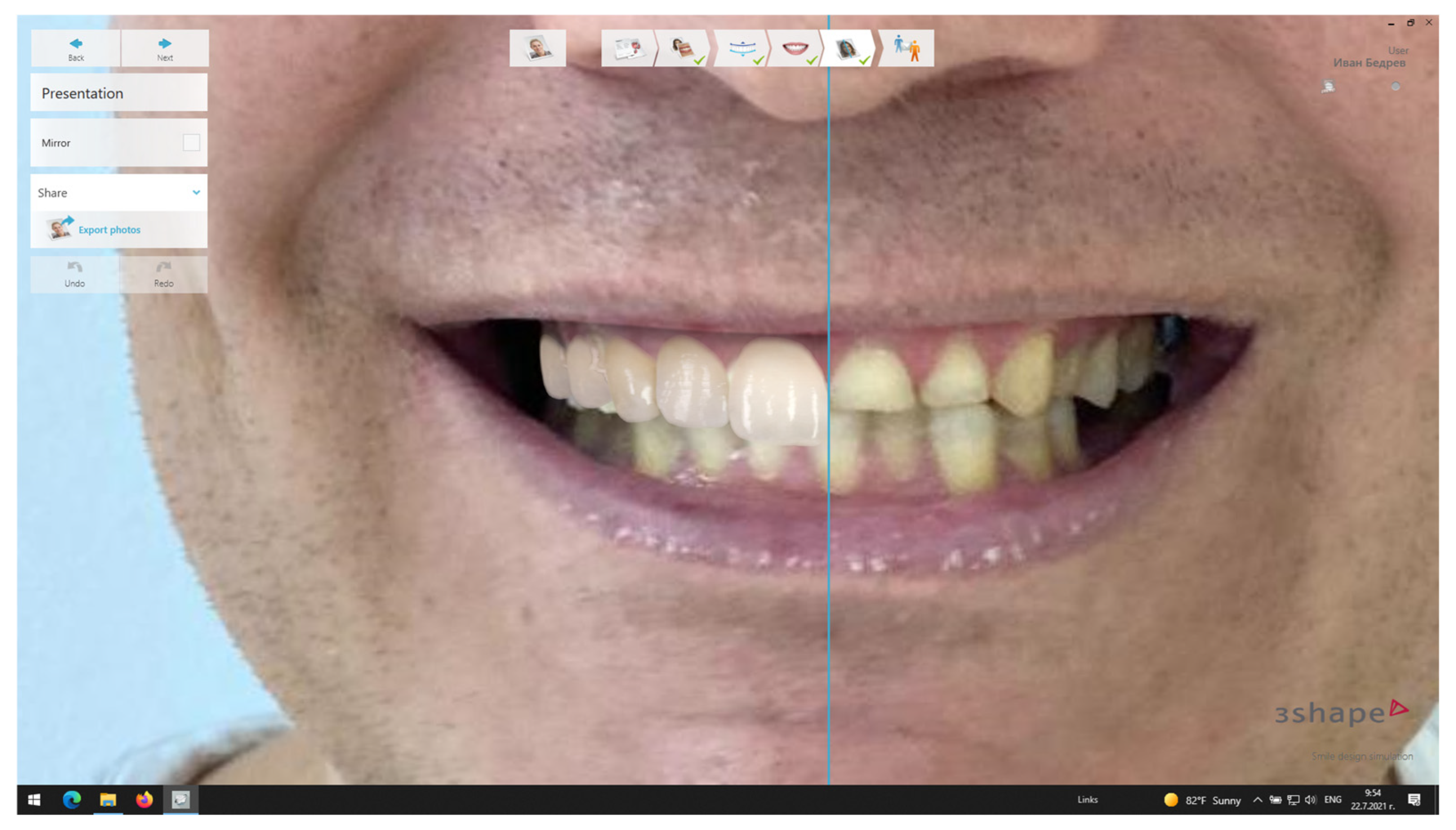
Over the past two decades, smile design has undergone a dramatic shift, transitioning from physical, analog methods to sophisticated digital tools [33]. Initially, dentists relied on hand-drawn sketches on printed patient photographs to communicate treatment plans. Today, however, smile design software (DSD, Software Version 2024) allows for entirely digital visualization on computers. This software offers significant advantages, enabling real-time edits and revisions to achieve a final design that perfectly balances the patient’s esthetic desires with their anatomy [34,35].
Digital Smile Design (DSD) stands for an innovative dental treatment planning tool, leveraging digital technology to craft a patient’s smile based on pre- and post-digital smile design photographs [36]. DSD software empowers dental practitioners to educate patients about potential smile enhancements and to tailor treatment plans to meet their preferences and requirements [37]. This holistic digital approach heralds a revolution in traditional methods of dental treatment planning and smile design, promising enhanced precision and heightened patient satisfaction (Figure 3).
Utilizing an intraoral scanner, digital impressions of both dental arches are obtained, facilitating the transition to CAD/CAM processing for 3D printing [38]. To capture comprehensive data for smile design, high-resolution full-profile photographs and videos are crucial. These media document the dynamic changes in lip, tooth, and facial muscle movements during smiling and talking. Key photographs essential for smile design include: (1) a full facial view capturing a natural smile, (2) an image of the resting face, and (3) a representation of the maxillary and mandibular arches without occlusion [39].
As patient esthetic demands continue to rise, tools for shade matching undergo continual development. Among the most utilized shade-determination tools are Vitapan Classical and Vita 3D-Master (VITA Zahnfabrik, Bad Säckingen, Germany) [40]. These instruments play a pivotal role in achieving accurate shade matching, ensuring optimal esthetic outcomes for dental restorations. The evolution of these tools reflects the ongoing commitment of dental professionals to meet and exceed patient expectations in achieving natural and pleasing esthetic results [41]. This innovative tool facilitates the process of capturing 2D images from various angles by sliding the scanner tip toward the abutment tooth [42]. These digital image files are then exported by the intraoral scanner (IOS), allowing for precise shade analysis and selection. By leveraging technology, the Vita Easyshade guide (VITA Zahnfabrik, Bad Säckingen, Germany) overcomes the limitations associated with traditional visual shade-matching methods, offering dental practitioners a more reliable and standardized approach to achieving optimal shade-matching outcomes for dental restorations [43].

3.3. Digital Design and Fabrication of the Provisional Restorations

Following the acquisition of digital patient data through intraoral scanning, specialized dental CAD software leverages this information (STL files) to design the optimal prosthetic restoration [44]. While initially focused on generating esthetically pleasing ceramic restorations like crowns, advancements in technology have expanded the capabilities of some CAD software. This allows for the design of a wider range of restorations, including full anatomic crowns, inlays (for intracoronal repair), and even inlay-retained fixed partial dentures [45].
To ensure patient comfort during the interim period before receiving the final prosthesis, a provisional restoration is created (Figure 4).
Two primary methodologies exist for fabricating provisional crowns: direct intraoral techniques and indirect CAD/CAM methods. The CEREC3 CAD/CAM system (Sirona, Charlotte, NC, USA) offers a range of high-strength provisional material blocks specifically engineered to withstand the milling process, enabling efficient and precise crown fabrication [46]. In contrast, conventional provisional materials typically require manual fabrication techniques, which are subject to limitations such as inconsistent surface texture and variable mechanical properties, particularly in terms of flexural strength [47]. Some studies have demonstrated that CAD/CAM-fabricated provisional crowns exhibit superior mechanical strength and marginal accuracy compared to directly fabricated bis-acryl composite crowns, especially after simulated aging through thermal cycling [48]. While these findings suggest that CAD/CAM technology may address certain shortcomings of traditional manual fabrication, it is important to note that clinical evidence on long-term performance remains limited, and further validation in vivo is needed.

3.4. Fabrication Techniques in Digital Fixed Prosthodontics

Within CAD/CAM dentistry, two primary fabrication methodologies are widely used: subtractive manufacturing (milling) and additive manufacturing (3D printing) [49]. Subtractive Manufacturing (Milling): Following the design phase in CAD software, construction data is translated into toolpaths for computer-controlled milling machines [50]. These machines precisely remove material from solid blocks to produce the final restoration. Available materials for milling include metals, resins, and silica-based ceramics, with selection dependent on restoration type and milling system capabilities [51].
Continuous advancements in CAD/CAM equipment have driven the development of novel dental materials. Subtractive manufacturing has been the predominant method for fabricating tooth-supported ceramic restorations for over three decades and implant-supported restorations for approximately two decades [52]. However, limitations exist, including challenges in milling certain metals (such as gold) due to esthetic constraints and significant material waste generation inherent to the subtractive process [53].
Subtractive manufacturing workflows primarily utilize disk-shaped materials as feedstock [54]. Milling machines in this category are typically classified as either dry or wet, based on the use of coolant during the milling process. Zirconia blocks require careful drying prior to coloration and sintering because of their high sensitivity to water exposure [55]. Consequently, dry milling has become the preferred technique for processing zirconia, particularly for partially sintered blanks (Figure 5). Despite its widespread use, subtractive manufacturing may face limitations related to tool wear, processing time, and material waste, all of which warrant consideration when selecting fabrication methods.
Other commonly used dental materials, including feldspathic porcelain, lithium disilicate glass, zirconia-resin composites, and various metals, typically require the application of lubricants during the milling process to optimize tool performance and material integrity [56]. The selection of appropriate milling tools depends largely on the specific material being processed. For example, softer metals are often milled effectively with carbide burs, whereas harder metals, ceramics, and resilient resins necessitate more durable electroplated diamond burs to ensure precision and reduce tool wear [57]. Careful matching of equipment and tooling to the material characteristics is critical to achieving optimal restoration quality and manufacturing efficiency.
In clinical settings where same-day restoration delivery is prioritized, subtractive manufacturing (SM) systems have adopted innovative approaches to expedite milling. Some machines utilize burs that simultaneously engage the material from both the left and right sides, significantly reducing machining time [58]. Additionally, there has been a growing trend toward the use of bar-shaped material blocks in in-office SM workflows, which further accelerates the fabrication process [59]. This approach is commonly applied in the production of inlays, frequently made from zirconia-resin composites such as Lava Ultimate (3M Company, Saint Paul, MN, United States) [60,61]. More recently, lithium disilicate glass-ceramics, exemplified by IPS e.max CAD (Ivoclar Vivadent, Schaan, Liechtenstein), have been introduced in bar-shaped formats aimed at single implant-supported restorations, reflecting ongoing material innovation in this domain [62].
For more complex prosthodontic cases, such as three-unit fixed dental prostheses, in-office SM systems demonstrate versatility by accommodating a broader spectrum of materials [63]. These include polymer blanks suitable for provisional restorations, like VITA CAD-Temp (VITA Zahnfabrik, Bad Säckingen, Germany), and zirconia blanks for definitive restorations, such as IPS e.max ZirCAD (Ivoclar Vivadent, Schaan, Liechtenstein). The future trajectory of in-office subtractive manufacturing appears promising, with the development of new materials including cobalt-chromium (Co-Cr) alloys and ceramic-polymer composites currently underway [64].
  • Additive Manufacturing (3D Printing)
The commercialization of additive manufacturing (AM), commonly known as 3D printing, began in the early 1980s with the advent of industrial-level machines [65]. Pioneers such as Charles W. Hull (founder of 3D Systems), S. Scott Crump (founder of Stratasys), and Hans J. Langer and Hans Steinbichler (founders of EOS) played pivotal roles in this development, with Hull securing the first 3D printer patent in 1986 [66]. Initially, these technologies primarily served rapid prototyping needs within design workflows, but significant growth followed the expiration of key patents around 2009, which facilitated broader accessibility and affordability [67,68].
Technological advancements have not only miniaturized and reduced the cost of 3D printers but also greatly diversified printable materials, ranging from plastics to metals, ceramics, and even biological tissues [69]. Rapid prototyping itself can be classified based on the material used—plastics, metals, or powders—which influences both process and application [70].
In additive manufacturing, objects are created sequentially by depositing material layer-by-layer guided by digital 3D models, contrasting with subtractive methods that remove material to shape restorations [71,72]. According to the EN ISO/ASTM 52900 standard, AM is defined as “the process of joining materials to make objects from 3D model data, typically layer by layer” [73]. Common AM technologies include stereolithography (SLA), selective laser sintering (SLS), powder binder printing (PBP), and fused deposition modeling (FDM) [74].
Metal 3D printing has particularly impacted dentistry since the early 2000s. Laser sintering introduced an innovative means of fabricating non-precious metal alloy restorations, especially cobalt-chromium (CoCr), offering new possibilities for crowns and bridges [75,76]. Improvements in post-processing techniques have enabled the production of highly accurate, stress-relieved frameworks suitable for extended-span fixed dental prostheses. Efficient platform utilization allows multiple units to be fabricated simultaneously, reducing individual unit production times to minutes [77]. Consequently, laser sintering has become a well-established and cost-effective method for producing non-precious metal fixed restorations [78].
To ensure stress-free restorations, thermal post-treatment is routinely performed on the build platform during dedicated processing stages, often automated in modern production environments. Following this, support structures are manually removed and finishing procedures are applied [79].
Mechanically, laser-sintered CoCr frameworks exhibit physical properties comparable to those produced via traditional casting methods [80]. Additionally, the inherent surface roughness of laser-sintered frameworks can enhance the adhesion of dental cements [81]. Although minor ridges aligned with the build platform’s z-axis may be present on internal surfaces, these dimensional variations generally fall within clinically acceptable limits [82]. Some studies even suggest superior marginal adaptation for laser-sintered CoCr crowns compared to cast counterparts. Furthermore, the roughened surfaces improve the wettability of opaquer layers, facilitating ceramic veneering [83]. Advances in ceramic build-up techniques have expanded the incorporation of ceramics into restorations through both direct and indirect methods.
  • Hybrid Manufacturing
In digital dentistry, hybrid manufacturing has emerged as a transformative approach by strategically integrating additive and subtractive manufacturing techniques to leverage the strengths of both [36]. This combined methodology harnesses the speed and design flexibility of 3D printing alongside the exceptional precision of CNC milling, resulting in dental restorations with superior surface quality, enhanced fit accuracy, and reduced production costs. For over eight years, Datron (Mühltal, Germany) has been at the forefront of implementing this innovative technology within dental manufacturing [84]. Currently, a collaborative initiative involving Datron, Concept Laser (Lichtenfels, Germany), and the Follow Me Technology Group (Munich, Germany) aims to standardize hybrid workflows by enabling seamless integration with existing milling systems through advanced networking solutions [85].
A critical aspect of hybrid manufacturing is the precise transfer of the “zero point” or origin from the additive manufacturing stage to the CNC milling unit. To achieve this, three measuring pins are strategically embedded into the build platform during the sintering process [86]. These pins are detected by the Datron D5 milling unit using a specialized infrared touch probe—a key innovation unique to hybrid manufacturing. This process allows the milling machine to accurately locate the laser-sintered objects and autonomously calculate necessary correction values, eliminating the need for additional CAM recalculations. Importantly, the restoration remains fixed to the platform throughout post-processing, avoiding transfer errors and ensuring maximal positioning accuracy [12,43].
Hybrid manufacturing also offers significant cost-saving potential, particularly for implant superstructures. By utilizing specialized form cutters and accessing screw holes through the basal screw access canal, manufacturing costs can be reduced by an estimated 30% to 50%, depending on production volume [87].

3.5. Quality Control and Dimensional Evaluation

Upon completion, both milled and 3D-printed crowns undergo comprehensive digital inspection using industrial 3D scanners. This evaluation employs validated quantitative methods to assess dimensional accuracy, focusing on discrepancies between the internal surface of the crown and the corresponding abutment tooth to ensure optimal fit [24,35].
Advanced metrology software, such as GOM Inspect, (Software Version 2024) facilitates this process by enabling precise manual and automated alignment of provisional and definitive restorations [88]. These alignment tools are critical for verifying that restorations meet rigorous standards of fit and function, thereby improving the clinical performance and longevity of the prostheses [89].

3.6. Advantages of Digital Workflow

Digital workflows offer several distinct advantages over conventional fixed prosthodontic methods. A primary benefit is the significant reduction in human error through enhanced digital precision [34]. By automating and streamlining fabrication steps, digital processes reduce labor-intensive tasks, resulting in faster turnaround times and improved efficiency for both clinicians and patients [65].
Emerging evidence suggests that restorations produced via 3D printing and milling exhibit superior mechanical and physical properties compared to those fabricated through traditional techniques [58]. These advanced manufacturing methods allow for restorations with finer detail, more accurate fits, and improved durability and biocompatibility, ultimately enhancing clinical outcomes and patient satisfaction [39].
CAD/CAM technology has revolutionized fixed prosthodontics by enabling the design and fabrication of complex, highly precise restorations that were previously difficult or impossible to achieve manually [26]. The efficiency gains from CAD/CAM significantly shorten both design and production phases, facilitating streamlined workflows in dental offices and laboratories while ensuring consistent restoration quality and minimizing variability associated with manual fabrication [75].

4. Disadvantages and Limitations of CAD/CAM Technology in Fixed Prosthodontics

Despite its many advantages, CAD/CAM technology presents several notable limitations and challenges. One of the primary barriers to adoption is the high initial investment required for purchasing essential equipment such as scanners, software, and milling machines, which can be prohibitive for smaller dental practices and laboratories. Additionally, ongoing maintenance costs and potential repair expenses contribute to the overall financial burden [90].
Another significant challenge is the steep learning curve associated with CAD/CAM systems. Mastering the complex software interfaces and hardware components demands extensive training and experience, which can be time-consuming and may discourage some practitioners from fully embracing the technology [18].
Material compatibility remains a concern as well. Not all conventional materials used in fixed prosthodontics are suitable for CAD/CAM processing. Certain ceramics, for example, may exhibit increased brittleness after milling, thereby elevating the risk of fractures [53]. Furthermore, despite the precision capabilities of CAD/CAM, minor discrepancies in restoration fit can still arise. Such imperfections can lead to clinical complications, including secondary caries or periodontal disease, often necessitating additional manual adjustments to optimize the fit [17].
Technological dependence also represents a critical limitation. The reliability of CAD/CAM workflows hinges on software accuracy and stability, uninterrupted power supply, and, in some cases, stable internet connectivity for system updates and cloud-based services [41]. Disruptions in any of these elements can interrupt clinical workflows and cause significant delays. Moreover, although CAD/CAM standardizes many fabrication steps, it may restrict the degree of artistic customization that experienced technicians can achieve through traditional handcrafted methods [90].
The design phase, particularly for complex prosthodontic cases, can be labor-intensive and time-consuming, potentially offsetting time savings gained during manufacturing. Post-milling adjustments also frequently require substantial manual refinement to ensure optimal fit and function [13]. Finally, an over-reliance on CAD/CAM technology risks erosion of fundamental manual skills among dentists and technicians, which could be problematic in situations where the technology is inaccessible or malfunctions [29].
An important ethical consideration emerging from the adoption of digital workflows in dentistry is the question of professional responsibility in software-based operations. As tasks such as digital design, treatment planning, and data manipulation are increasingly performed using advanced software tools, it becomes crucial to clarify whether these functions are carried out directly by dental professionals or delegated to laboratory technicians or third-party operators. This delegation raises potential concerns regarding accountability, diagnostic accuracy, and adherence to professional standards. From an ethical standpoint, dental practitioners must ensure that any delegated digital tasks are performed under their direct supervision and align with regulatory frameworks governing clinical responsibility. Maintaining transparency in the digital workflow and safeguarding patient welfare must remain central, even as technology continues to streamline and redefine traditional roles within the dental team.
Despite their potential, these methods currently lack long-term clinical outcome data. Most available studies are limited by short follow-up periods, small sample sizes, or observational designs, highlighting the urgent need for more randomized controlled trials (RCTs) and long-term clinical investigations to robustly assess the durability, safety, and clinical outcomes of 3D-printed restorations. Furthermore, the practical implementation of fully digital workflows in everyday dental practice faces several challenges, including the high initial investment costs, the steep learning curve associated with new technologies, integration difficulties across different hardware and software platforms, and variability in operator expertise. These factors can hinder widespread adoption and may impact the consistency and quality of patient care. Addressing these evidence gaps and practical obstacles is essential to optimizing the clinical utility and reliability of digital dentistry solutions moving forward.

5. Critical Appraisal and Future Directions

The rapid integration of digital technologies in fixed prosthodontics—particularly through CAD/CAM systems and additive manufacturing—has transformed traditional workflows and material utilization. Innovations such as high-precision milling machines and metal 3D printing have significantly expanded clinical possibilities. However, despite these technological advances, several critical limitations and gaps remain in the current evidence base that warrant deeper examination.
Firstly, a significant proportion of the existing literature comprises in vitro studies, observational designs, or case reports, with relatively few randomized controlled trials (RCTs) or long-term clinical studies. While laboratory data often demonstrates promising accuracy and mechanical properties of digitally fabricated restorations, the translation of these findings to long-term intraoral performance remains insufficiently documented. For instance, studies comparing subtractive and additive fabrication methods frequently report similar fit and strength in laboratory settings; however, these outcomes may not reliably predict real-world longevity or patient-reported satisfaction over extended periods.
Moreover, notable heterogeneity exists among findings related to clinical outcomes and material behavior. Differences in study protocols, evaluation methods, and materials—such as various zirconia formulations or polymer-ceramic composites—contribute to variability that complicates direct comparison. Some studies suggest superior marginal fit with laser-sintered frameworks, while others highlight potential limitations including increased internal surface roughness or extensive post-processing requirements. These contradictions underscore the need for standardized testing protocols and multicenter clinical validation to enhance reproducibility and generalizability.
A further limitation is the gap in clinical translation. Although in-office CAD/CAM and 3D-printing technologies are increasingly accessible, their adoption in routine clinical practice remains limited by economic, technical, and logistical factors. High equipment costs, steep learning curves, and maintenance demands pose barriers to general dental practitioners. Additionally, current digital workflows, particularly those involving additive manufacturing—often require extensive post-processing, which can offset the purported efficiency gains.
To strengthen the evidence base and better guide clinical decision-making, there is a pressing need for high-quality RCTs and longitudinal studies evaluating long-term success, complication rates, and patient-centered outcomes associated with digitally fabricated fixed prostheses. Practical challenges, including cost-effectiveness and workflow integration across diverse clinical settings, should also be systematically examined.
In this review, a formal quality appraisal was not performed due to the heterogeneity of included study designs and the focus on providing a narrative synthesis rather than a quantitative meta-analysis. This approach, while limiting stratified evaluation of evidence quality, allowed for a broader overview of emerging trends and technologies.
While digital fabrication techniques are revolutionizing fixed prosthodontics, critical gaps remain in understanding their long-term performance and practical implementation. Addressing these limitations through robust clinical research, standardized methodologies, and consensus-driven protocols is essential to fully realize the potential of digital dentistry in routine care.

6. Conclusions

The dental industry is undergoing a notable transformation driven by the integration of advanced 3D printing and computer-aided design/manufacturing (CAD/CAM) technologies. These innovations enable the production of customized dental components that are biocompatible, durable, and esthetically pleasing, addressing the profession’s ongoing demand for precision and personalized patient care. While additive manufacturing has rapidly gained adoption in fabricating fixed prosthetics—including crowns, bridges, study models, temporary restorations, and surgical guides—important questions remain regarding the long-term clinical performance of 3D-printed restorations, the reproducibility of digital workflows, and the regulation of materials and processes. Current research priorities focus on validating the longevity and safety of emerging printable materials through rigorous clinical trials, developing standardized protocols to ensure accuracy and consistency across different platforms and providers, and assessing the cost-effectiveness of these technologies compared to conventional manufacturing methods in diverse practice settings. Although preliminary data suggest promising outcomes for 3D-printed dental applications, comprehensive clinical evidence is still evolving. Therefore, clinicians are advised to approach the adoption of 3D printing with measured optimism, integrating these technologies cautiously while adhering to evidence-based practice principles.

Funding

This research received no external funding.

Institutional Review Board Statement

Not applicable.

Informed Consent Statement

Written informed consent was obtained from the patients whose photos were included in the study.

Data Availability Statement

The original contributions presented in the study are included in the article; further inquiries can be directed to the corresponding author.

Conflicts of Interest

The authors declare no conflicts of interest.

Abbreviations

AMAdditive manufacturing
CAD/CAMComputer-aided design/computer-aided manufacturing
CNC MillingComputer Numerical Control Milling
DSDDigital smile design
IOSIntraoral scanner
FPDFixed partial denture
SMSubtractive manufacturing
STLStereolithography, standard triangle language, standard tessellation language
3DThree-dimensional

References

  1. Pan, S.; Guo, D.; Zhou, Y.; Jung, R.E.; Hämmerle, C.H.F.; Mühlemann, S. Time efficiency and quality of outcomes in a model-free digital workflow using digital impression immediately after implant placement: A double-blind self-controlled clinical trial. Clin. Oral Implants Res. 2019, 30, 617–626. [Google Scholar] [CrossRef]
  2. Ren, S.; Jiang, X.; Lin, Y.; Di, P. Crown Accuracy and Time Efficiency of Cement-Retained Implant-Supported Restorations in a Complete Digital Workflow: A Randomized Control Trial. J. Prosthodont. 2022, 31, 405–411. [Google Scholar] [CrossRef] [PubMed]
  3. Zhang, Y.; Tian, J.; Wei, D.; Di, P.; Lin, Y. Quantitative clinical adjustment analysis of posterior single implant crown in a chairside digital workflow: A randomized controlled trial. Clin. Oral Implants Res. 2019, 30, 1059–1066. [Google Scholar] [CrossRef] [PubMed]
  4. Gintaute, A.; Weber, K.; Zitzmann, N.U.; Brägger, U.; Ferrari, M.; Joda, T. A Double-Blind Crossover RCT Analyzing Technical and Clinical Performance of Monolithic ZrO2 Implant Fixed Dental Prostheses (iFDP) in Three Different Digital Workflows. J. Clin. Med. 2021, 10, 2661. [Google Scholar] [CrossRef] [PubMed]
  5. Gintaute, A.; Zitzmann, N.U.; Brägger, U.; Weber, K.; Joda, T. Patient-reported outcome measures compared to professional dental assessments of monolithic ZrO2 implant fixed dental prostheses in complete digital workflows: A double-blind crossover randomized controlled trial. J. Prosthodont. 2023, 32, 18–25. [Google Scholar] [CrossRef]
  6. Hashemi, A.M.; Hashemi, H.M.; Siadat, H.; Shamshiri, A.; Afrashtehfar, K.I.; Alikhasi, M. Fully Digital versus Conventional Workflows for Fabricating Posterior Three-Unit Implant-Supported Reconstructions: A Prospective Crossover Clinical Trial. Int. J. Environ. Res. Public Health 2022, 19, 11456. [Google Scholar] [CrossRef]
  7. Joda, T.; Gintaute, A.; Brägger, U.; Ferrari, M.; Weber, K.; Zitzmann, N.U. Time-efficiency and cost-analysis comparing three digital workflows for treatment with monolithic zirconia implant fixed dental prostheses: A double-blinded RCT. J. Dent. 2021, 113, 103779. [Google Scholar] [CrossRef]
  8. Cheng, C.W.; Ye, S.Y.; Chien, C.H.; Chen, C.J.; Papaspyridakos, P.; Ko, C.C. Randomized clinical trial of a conventional and a digital workflow for the fabrication of interim crowns: An evaluation of treatment efficiency, fit, and the effect of clinician experience. J. Prosthet. Dent. 2021, 125, 73–81. [Google Scholar] [CrossRef] [PubMed]
  9. Haddadi, Y.; Bahrami, G.; Isidor, F. Accuracy of crowns based on digital intraoral scanning compared to conventional impression-a split-mouth randomised clinical study. Clin. Oral Investig. 2019, 23, 4043–4050. [Google Scholar] [CrossRef]
  10. Mühlemann, S.; Benic, G.I.; Fehmer, V.; Hämmerle, C.H.F.; Sailer, I. Clinical quality and efficiency of monolithic glass ceramic crowns in the posterior area: Digital compared with conventional workflows. Int. J. Comput. Dent. 2018, 21, 215–223. [Google Scholar]
  11. Park, J.S.; Lim, Y.J.; Kim, B.; Kim, M.J.; Kwon, H.B. Clinical Evaluation of Time Efficiency and Fit Accuracy of Lithium Disilicate Single Crowns between Conventional and Digital Impression. Materials 2020, 13, 5467. [Google Scholar] [CrossRef] [PubMed]
  12. Sailer, I.; Benic, G.I.; Fehmer, V.; Hämmerle, C.H.F.; Mühlemann, S. Randomized controlled within-subject evaluation of digital and conventional workflows for the fabrication of lithium disilicate single crowns. Part II: CAD-CAM versus conventional laboratory procedures. J. Prosthet. Dent. 2017, 118, 43–48. [Google Scholar] [CrossRef] [PubMed]
  13. Sakornwimon, N.; Leevailoj, C. Clinical marginal fit of zirconia crowns and patients’ preferences for impression techniques using intraoral digital scanner versus polyvinyl siloxane material. J. Prosthet. Dent. 2017, 118, 386–391. [Google Scholar] [CrossRef]
  14. Zeltner, M.; Sailer, I.; Mühlemann, S.; Özcan, M.; Hämmerle, C.H.; Benic, G.I. Randomized controlled within-subject evaluation of digital and conventional workflows for the fabrication of lithium disilicate single crowns. Part III: Marginal and internal fit. J. Prosthet. Dent. 2017, 117, 354–362. [Google Scholar] [CrossRef]
  15. Capparé, P.; Ferrini, F.; Ruscica, C.; Pantaleo, G.; Tetè, G.; Gherlone, E.F. Digital versus Traditional Workflow for Immediate Loading in Single-Implant Restoration: A Randomized Clinical Trial. Biology 2021, 10, 1281. [Google Scholar] [CrossRef]
  16. Hanozin, B.; Li Manni, L.; Lecloux, G.; Bacevic, M.; Lambert, F. Digital vs. conventional workflow for one-abutment one-time immediate restoration in the esthetic zone: A randomized controlled trial. Int. J. Implant Dent. 2022, 8, 7. [Google Scholar] [CrossRef]
  17. Alauddin, M.S.; Baharuddin, A.S.; Mohd Ghazali, M.I. The Modern and Digital Transformation of Oral Health Care: A Mini Review. Healthcare 2021, 9, 118. [Google Scholar] [CrossRef] [PubMed]
  18. Joda, T.; Yeung, A.W.K.; Hung, K.; Zitzmann, N.U.; Bornstein, M.M. Disruptive Innovation in Dentistry: What It Is and What Could Be Next. J. Dent. Res. 2021, 100, 448–453. [Google Scholar] [CrossRef]
  19. van Noort, R. The future of dental devices is digital. Dent. Mater. 2012, 28, 3–12. [Google Scholar] [CrossRef]
  20. Morsy, N.; El Kateb, M. Accuracy of intraoral scanners for static virtual articulation: A systematic review and meta-analysis of multiple outcomes. J. Prosthet. Dent. 2022, 132, 546–552. [Google Scholar] [CrossRef]
  21. Joda, T.; Brägger, U. Digital vs. conventional implant prosthetic workflows: A cost/time analysis. Clin. Oral Implants Res. 2015, 26, 1430–1435. [Google Scholar] [CrossRef] [PubMed]
  22. Joda, T.; Zarone, F.; Ferrari, M. The complete digital workflow in fixed prosthodontics: A systematic review. BMC Oral Health 2017, 17, 124. [Google Scholar] [CrossRef]
  23. Bernauer, S.A.; Zitzmann, N.U.; Joda, T. The Use and Performance of Artificial Intelligence in Prosthodontics: A Systematic Review. Sensors 2021, 21, 6628. [Google Scholar] [CrossRef] [PubMed]
  24. Ahmed, N.; Abbasi, M.S.; Zuberi, F.; Qamar, W.; Halim, M.S.B.; Maqsood, A.; Alam, M.K. Artificial Intelligence Techniques: Analysis, Application, and Outcome in Dentistry-A Systematic Review. BioMed Res. Int. 2021, 2021, 9751564. [Google Scholar] [CrossRef]
  25. Revilla-León, M.; Gómez-Polo, M.; Vyas, S.; Barmak, B.A.; Gallucci, G.O.; Att, W.; Özcan, M.; Krishnamurthy, V.R. Artificial intelligence models for tooth-supported fixed and removable prosthodontics: A systematic review. J. Prosthet. Dent. 2023, 129, 276–292. [Google Scholar] [CrossRef]
  26. Siqueira, R.; Galli, M.; Chen, Z.; Mendonca, G.; Meirelles, L.; Wang, H.L.; Chan, H.L. Intraoral scanning reduces procedure time and improves patient comfort in fixed prosthodontics and implant dentistry: A systematic review. Clin. Oral Investig. 2021, 25, 6517–6531. [Google Scholar] [CrossRef]
  27. DeSimone, J.M.; Ermoshkin, A.; Ermoshkin, N.; Samulski, E.T. Continuous Liquid Interphase Printing. U.S. Patent 9,205,601 B2, 8 December 2015. [Google Scholar]
  28. Alghazzawi, T.F. Advancements in CAD/CAM Technology: Applications of Recent Developments for CAD/CAM Generated Restorations. Br. Dent. J. 2008, 204, 505–511. [Google Scholar]
  29. Bernauer, S.A.; Zitzmann, N.U.; Joda, T. The Complete Digital Workflow in Fixed Prosthodontics Updated: A Systematic Review. Healthcare 2023, 11, 679. [Google Scholar] [CrossRef]
  30. Ahlholm, P.; Sipilä, K.; Vallittu, P.; Jakonen, M.; Kotiranta, U. Digital Versus Conventional Impressions in Fixed Prosthodontics: A Review. J. Prosthodont. 2018, 27, 35–41. [Google Scholar] [CrossRef] [PubMed]
  31. Ting-Shu, S.; Jian, S. Intraoral Digital Impression Technique: A Review. J. Prosthodont. 2015, 24, 313–321. [Google Scholar] [CrossRef]
  32. Thomas, P.A.; Krishnamoorthi, D.; Mohan, J.; Raju, R.; Rajajayam, S.; Venkatesan, S. Digital Smile Design. J. Pharm. Bioallied Sci. 2022, 14, S43–S49. [Google Scholar] [CrossRef]
  33. Cervino, G.; Fiorillo, L.; Arzukanyan, A.V.; Spagnuolo, G.; Cicciù, M. Dental Restorative Digital Workflow: Digital Smile Design from Aesthetic to Function. Dent. J. 2019, 7, 30. [Google Scholar] [CrossRef]
  34. Diwan, K.; Rathee, M.; Tomar, S.S.; Singh, S.; Divakar, S.; Chahal, S. Applications of 3D Printing in Prosthodontics: A Review. J. Prosthodont. Res. 2016, 60, 72–84. [Google Scholar]
  35. Jain, S.; Sayed, M.E.; Shetty, M.; Alqahtani, S.M.; Al Wadei, M.H.D.; Gupta, S.G.; Othman, A.A.; Alshehri, A.H.; Alqarni, H.; Mobarki, A.H.; et al. Physical and Mechanical Properties of 3D-Printed Provisional Crowns and Fixed Dental Prosthesis Resins Compared to CAD/CAM Milled and Conventional Provisional Resins: A Systematic Review and Meta-Analysis. Polymers 2022, 14, 2691. [Google Scholar] [CrossRef]
  36. Kakinuma, H.; Izumita, K.; Yoda, N.; Egusa, H.; Sasaki, K. Comparison of the Accuracy of Resin-Composite Crowns Fabricated by Three-Dimensional Printing and Milling Methods. Dent. Mater. J. 2022, 41, 808–815. [Google Scholar] [CrossRef] [PubMed]
  37. Baghani, M.T.; Shayegh, S.S.; Johnston, W.M.; Shidfar, S.; Hakimaneh, S.M.R. In vitro evaluation of the accuracy and precision of intraoral and extraoral complete-arch scans. J. Prosthet. Dent. 2021, 126, 665–670. [Google Scholar] [CrossRef] [PubMed]
  38. Gintaute, A.; Keeling, A.J.; Osnes, C.A.; Zitzmann, N.U.; Ferrari, M.; Joda, T. Precision of maxillo-mandibular registration with intraoral scanners in vitro. J. Prosthodont. Res. 2020, 64, 114–119. [Google Scholar] [CrossRef]
  39. Morton, D.; Gallucci, G.; Lin, W.S.; Pjetursson, B.; Polido, W.; Roehling, S.; Sailer, I.; Aghaloo, T.; Albera, H.; Bohner, L.; et al. Group 2 ITI Consensus Report: Prosthodontics and implant dentistry. Clin. Oral. Implants Res. 2018, 29 (Suppl. 16), 215–223. [Google Scholar] [CrossRef] [PubMed]
  40. Sailer, I.; Mühlemann, S.; Kohal, R.J.; Spies, B.C.; Pjetursson, B.E.; Lang, N.P.; Gotfredsen, K.L.; Ellingsen, J.E.; Francisco, H.; Özcan, M.; et al. Reconstructive aspects: Summary and consensus statements of group 3. The 5th EAO Consensus Conference 2018. Clin. Oral Implants Res. 2018, 29 (Suppl. 18), 237–242. [Google Scholar] [CrossRef]
  41. Bhambhani, R.; Bhattacharya, J.; Sen, S.K. Digitization and Its Futuristic Approach in Prosthodontics. J. Indian Prosthodont. Soc. 2013, 13, 165–174. [Google Scholar] [CrossRef]
  42. Kihara, H.; Hatakeyama, W.; Komine, F.; Takafuji, K.; Takahashi, T.; Yokota, J.; Oriso, K.; Kondo, H. Accuracy and practicality of intraoral scanner in dentistry: A literature review. J. Prosthodont. Res. 2020, 64, 109–113. [Google Scholar] [CrossRef]
  43. Alam, M.; Chugh, A.; Kumar, A.; Rathee, M.; Jain, P. Comparative Evaluation of Fracture Resistance of Anterior Provisional Restorations Fabricated Using Conventional and Digital Techniques—An In Vitro Study. J. Indian Prosthodont. Soc. 2022, 22, 361–367. [Google Scholar]
  44. Czigola, A.; Róth, I.; Vitai, V.; Fehér, D.; Hermann, P.; Borbély, J. Comparing the Effectiveness of Shade Measurement by Intraoral Scanner, Digital Spectrophotometer, and Visual Shade Assessment. J. Esthet. Restor. Dent. 2021, 33, 1166–1174. [Google Scholar] [CrossRef]
  45. Schweiger, J.; Edelhoff, D.; Güth, J.F. Update digitale Zahnheilkunde 2020—Aktuelle Möglichkeiten und Limitationen. Bayer. Zahnärzte Bl. 2020, 57, 42–52. [Google Scholar]
  46. Schweiger, J.; Güth, J.F. Neue Entwicklungen in der additiven und subtraktiven Fertigung Teamwork. J. Cont. Dent. Educ. 2020, 23, 82–90. [Google Scholar]
  47. Betriebe—Beschäftigte—Auszubildende im Zahntechniker-Handwerk. Available online: https://www.vdzi.de/Ausbildungsverg%C3%BCtungen-2025 (accessed on 20 May 2024).
  48. Campbell, S.D.; Cooper, L.; Craddock, H.; Hyde, T.P.; Nattress, B.; Pavitt, S.H.; Seymour, D.W. Removable Partial Dentures: The Clinical Need for Innovation. J. Prosthet. Dent. 2017, 118, 273–280. [Google Scholar] [CrossRef]
  49. Jordan, R.A.; Micheelis, W. Fünfte Deutsche Mundgesundheitsstudie (DMS V); Deutscher Zahnärzte: Köln, Germany, 2016. [Google Scholar]
  50. Horn, T.J.; Harryson, O.L.A. Overview of Current Additive Manufacturing Technologies and Selected Applications. Sci. Prog. 2012, 95, 255–282. [Google Scholar] [CrossRef] [PubMed]
  51. Dawood, A.; Marti, B.; Sauret Jackson, V.; Darwood, A. 3D Printing in Dentistry. Br. Dent. J. 2015, 219, 521–529. [Google Scholar] [CrossRef] [PubMed]
  52. Caviezel, C.; Grünwald, R.; Ehrenberg-Silies, S.; Kind, S.; Jetzke, T.; Bovenschulte, M. Additive Fertigungsverfahren (3D-Druck)—Innovationsanalyse; TAB Arbeitsbereicht: Berlin, Germany, 2017. [Google Scholar]
  53. Kessler, A.; Hickel, R.; Reymus, M. 3D Printing in Dentistry—State of the Art. Oper. Dent. 2020, 45, 30–40. [Google Scholar] [CrossRef] [PubMed]
  54. Takeuchi, Y.; Koizumi, H.; Furuchi, M.; Sato, Y.; Ohkubo, C.; Matsumura, H. Use of Digital Impression Systems with Intraoral Scanners for Fabricating Restorations and Fixed Dental Prostheses. J. Oral Sci. 2018, 60, 1–7. [Google Scholar] [CrossRef]
  55. Rathee, M.; Alam, M.; Divakar, S.; Malik, S. Resin-Bonded Bridge as a Simplified Approach to Restore Missing Teeth in Esthetic Zone by Conventional and Digital Techniques. Dent. Res. J. 2022, 19, 92. [Google Scholar] [CrossRef]
  56. Beuer, F.; Schweiger, J.; Edelhoff, D. Digital Dentistry: An Overview of Recent Developments for CAD/CAM Generated Restorations. Br. Dent. J. 2008, 204, 505–511. [Google Scholar] [CrossRef]
  57. Quante, K.; Ludwig, K.; Kern, M. Marginal and internal fit of metal-ceramic crowns fabricated with a new laser melting technology. Dent. Mater. 2008, 24, 1311–1355. [Google Scholar] [CrossRef]
  58. Xu, D.; Xiang, N.; Wie, B. The marginal fit of selective laser melting-fabricated metal crowns: An in vitro study. J. Prosthet. Dent. 2014, 112, 1437–1440. [Google Scholar] [CrossRef]
  59. Huang, Z.; Zhang, L.; Zhu, J.; Zhang, X. Clinical marginal and internal fit of metal ceramic crowns fabricated with a selective laser melting technology. J. Prosthet. Dent. 2015, 113, 623–627. [Google Scholar] [CrossRef]
  60. Lövgren, N.; Roxner, R.; Klemendz, S.; Larsson, C. Effect of production method on surface roughness, marginal and internal fit, and retention of cobalt-chromium single crowns. J. Prosthet. Dent. 2017, 118, 95–101. [Google Scholar] [CrossRef] [PubMed]
  61. Lehmann, K.M.; Hellwig, E.; Wenz, H.J. Zahnärztliche Propädeutik; Deutscher Zahnärzte: Köln, Germany, 2015. [Google Scholar]
  62. Stark, H. Ist die Modellgussprothese adäquater Zahnersatz für den älteren Menschen? Quintessenz 2005, 56, 367–373. [Google Scholar]
  63. Tumbletone, J.R.; Shirvanyants, D.; Ermoshkin, N.; Janusziewicz, R.; Johnson, A.R.; Kelly, D.; Chen, K.; Pinschmidt, R.; Rolland, J.P.; Ermoshkin, A.; et al. Additive Manufacturing: Continuous Liquid Interface Production of 3D Objects. Science 2015, 347, 1349–1352. [Google Scholar] [CrossRef]
  64. Schweiger, J.; Kieschnick, A. CAD/CAM in der Digitalen Zahnheilkunde; Teamwork Media: Fuchstal, Germany, 2017. [Google Scholar]
  65. Alifui-Segbaya, F.; Williams, R.J.; George, R. Additive manufacturing: A novel method for fabricating cobalt-chromium removable partial denture frameworks. Eur. J. Prosthodont. Restor. Dent. 2017, 25, 73–78. [Google Scholar]
  66. Laverty, D.P.; Thomas, M.B.M.; Clark, P.; Addy, L.D. The use of 3D metal printing (direct metal laser sintering) in removable prosthodontics. Dent. Update 2016, 43, 826–835. [Google Scholar] [CrossRef] [PubMed]
  67. Lima, J.M.; Anami, L.C.; Araujo, R.M.; Pavanelli, C.A. Removable partial dentures: Use of rapid prototyping. J. Prosthodont. 2014, 23, 588–591. [Google Scholar] [CrossRef] [PubMed]
  68. Kieschnick, A.; Schweiger, J.; Edelhoff, D.; Güth, J.F. Status Präsens 2020: Additive CAD/CAM-Gestützte Fertigungstechnologien im Zahntechnischen Labor. Available online: https://www.researchgate.net/publication/341990852_Status_Prasens_2020_Additive_CADCAM-gestutzte_Fertigungstechnologien_im_zahntechnischen_Labor#fullTextFileContent (accessed on 24 April 2024).
  69. Joda, T.; Bragger, U. Time-Efficiency Analysis Comparing Digital and Conventional Workflows for Implant Crowns: A Prospective Clinical Crossover Trial. Int. J. Oral Maxillofac. Implants 2015, 30, 1047–1053. [Google Scholar]
  70. Kollenberg, W. Keramik und Multimaterial 3D-Druck. Keram. Z. 2014, 66, 233–236. [Google Scholar] [CrossRef]
  71. ISO/ASTM 52900; Additive Manufacturing—General Principles—Terminology. Beuth: Berlin, Germany, 2017.
  72. ISO/ASTM 17296–2; Additive Manufacturing—General Principles—Part 2: Overview of Process Categories and Feedstock. Beuth: Berlin, Germany, 2016.
  73. Gartner Hype Cycle. Available online: https://www.gartner.com/en/research/methodologies/gartner-hype-cycle (accessed on 28 April 2024).
  74. Dolabdjian, H.; Strietzel, R. Verfahren zur Herstellung von Zahnersatz und dentalen Hilfsteilen. EP 1 021 997 B2, 26 July 2000. [Google Scholar]
  75. Revilla-León, M.; Meyer, M.J.; Özcan, M. Metal additive manufacturing technologies. Int. J. Comput. Dent. 2019, 22, 55–67. [Google Scholar]
  76. Fischer, J.; Stawarczyk, B.; Trottmann, A.; Hämmerle, C.H.F. Festigkeit lasergesinterter Brückengerüste aus einer CoCr-legierung. Quintessenz Zahntech. 2008, 34, 140–149. [Google Scholar]
  77. Rudolph, M.; Setz, J. Ein CAD/CAM-System mit aufbauender Lasertechnologie. Quintessenz Zahntech. 2007, 33, 582–587. [Google Scholar]
  78. Tregermann, I.; Renne, W.; Kelly, A.; Wilson, D. Evaluation of removable partial denture frameworks fabricated using 3 different techniques. J. Prosthet. Dent. 2019, 122, 390–395. [Google Scholar] [CrossRef]
  79. Van Zeghbroeck, L.; Boons, E. Evaluation of technicians working time in the fabrication of removable partial dentures: Cad/Cam versus tradition. In Proceedings of the 14th Biennial Meeting of the International College of Prosthodontics, Waikoloa Village, HI, USA, 7–12 September 2011; p. 63. [Google Scholar]
  80. Schweiger, J.; Güth, J.F.; Erdelt, K.J.; Edelhoff, D.; Schubert, O. Internal porosities, retentive force, and survival of cobalt-chromium alloy clasps fabricated by selective laser sintering. J. Prosthodont. Res. 2019, 64, 210–216. [Google Scholar] [CrossRef] [PubMed]
  81. Torii, M.; Nakata, T.; Takahashi, K.; Kawamura, N.; Shimpo, H.; Ohkubo, C. Fitness and retentive force of cobalt-chromium alloy clasps fabricated with repeated laser sintering and milling. J. Prosthodont. Res. 2018, 62, 342–346. [Google Scholar] [CrossRef] [PubMed]
  82. Nakata, T.; Shimpo, H.; Ohkubo, C. Clasp fabrication using one-process molding by repeated laser sintering and high-speed milling. J. Prosthodont. Res. 2017, 61, 276–282. [Google Scholar] [CrossRef]
  83. Revilla-León, M.; Özcan, M. Additive manufacturing technologies used for processing polymers: Current status and potential application in prosthetic dentistry. J. Prosthodont. 2019, 28, 146–158. [Google Scholar] [CrossRef] [PubMed]
  84. Jockusch, J.; Özcan, M. Additive manufacturing of dental polymers: An overview on processes, materials, and applications. Dent. Mater. J. 2020, 39, 345–354. [Google Scholar] [CrossRef]
  85. Quan, H.; Zhang, T.; Xu, H.; Luo, S.; Nie, J.; Zhu, X. Photo-curing 3D-printing technique and its challenges. Bioact. Mater. 2020, 22, 110–115. [Google Scholar] [CrossRef]
  86. Dietrich, C.A.; Ender, A.; Baumgartner, S.; Mehl, A. A validation study of reconstructed rapid prototyping models produced by two technologies. Angle Orthod. 2017, 87, 782–787. [Google Scholar] [CrossRef]
  87. Brown, G.B.; Currier, G.F.; Kadioglu, O.; Kierl, J.P. Accuracy of 3-dimensional printed dental models reconstructed from digital intraoral impressions. Am. J. Orthod. Dentofac. Orthop. 2018, 154, 733–739. [Google Scholar] [CrossRef] [PubMed]
  88. Kim, S.Y.; Shin, Y.S.; Jund, H.D.; Hwang, C.J.; Baik, H.S.; Cha, J.Y. Precision and trueness of dental models manufactured with different 3-dimensional printing technologies. Am. J. Orthod. Dentofac. Orthop. 2018, 153, 144–153. [Google Scholar] [CrossRef]
  89. Kallweit, D.; Mönch, W.; Zappe, H. Kontrolliert kippen: Silizium-Mikrospiegel mit integriertem optischen Feedback. Photonik 2006, 4, 62–65. [Google Scholar]
  90. Viereck, V.; Li, Q.; Jäkel, A.; Hillmer, H. Großflächige Anwendung von optischen MEMS: Mikrospiegel-Arrays zur Tageslichtlenkung. Photonik 2009, 2, 28–29. [Google Scholar]
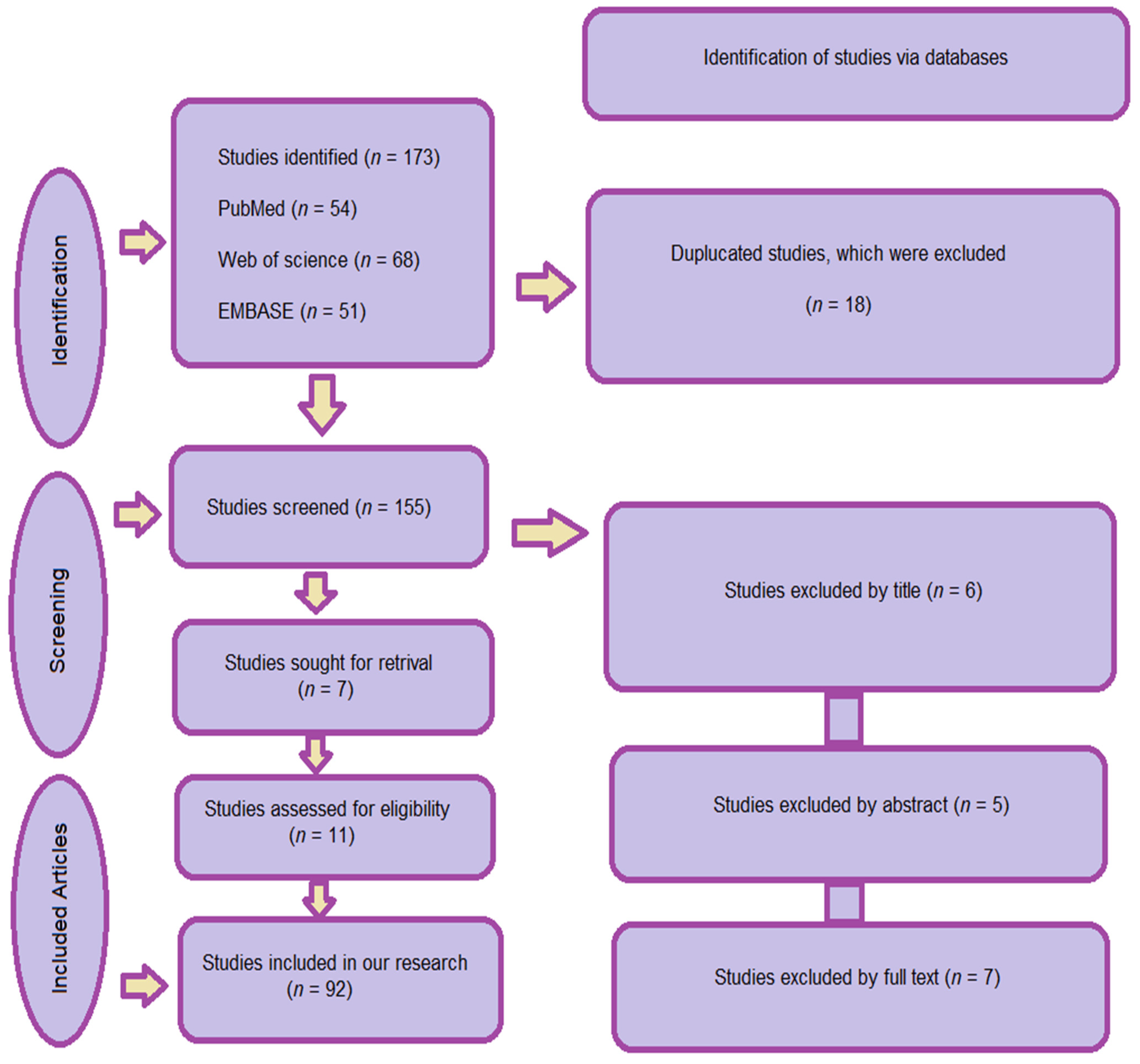
Figure 1. A flow chart of the search strategy of the study.
Oral 05 00084 g001
Figure 2. Digital Fabrication Process for Fixed Dental Restorations.
Oral 05 00084 g002
Figure 3. Digital Smile Design—a comparison between natural and selected teeth, (origin of the Figure: author’s clinical case, no copyright issue), (3Shape, Copenhagen, Denmark).
Oral 05 00084 g003
Figure 4. Provisional restorations, fabricated by CAD/CAM technology (origin of the Figure: author’s clinical case, no copyright issue) (Temporary CB Resin, Formlabs Dental, Somerville, MA, USA).
Oral 05 00084 g004
Figure 5. Permanent zirconia restorations, manufactured by CAD/CAM technology (milling) (origin of the Figure: author’s clinical case, no copyright issue) (Dentsply Sirona, Charlotte, NC, USA).
Oral 05 00084 g005
Table 1. The most esteemed articles included in our study.
Author’s NameTopicArticle TypeYear of PublicationReference Number
Morsy, N.; El Kateb, M.Intraoral scannersSystematic review and meta-analysis2022[21]
Joda, T.; Brägger, U.Digital vs. conventional implant prosthetic workflowsCost/time analysis2015[22]
Joda, T.; Zarone, F.; Ferrari, M.The complete digital workflow in fixed prosthodonticsSystematic review2017[23]
Bernauer, S.A.; Zitzmann, N.U.; Joda, T.Artificial intelligence in prosthodonticsSystematic review2021[24]
Ahmed, N.; et al.Artificial intelligence techniques in dentistrySystematic review2021[25]
Revilla-León, M.; et al.Artificial intelligence models for fixed and removable prosthodonticsSystematic review2023[26]
Siqueira, R.; et al.Intraoral scanning in fixed prosthodontics and implant dentistrySystematic review2021[27]
DeSimone, J.M.; et al.Continuous Liquid Interface PrintingUS Patent2015[28]
Alghazzawi, T.F.Advancements in CAD/CAM TechnologyLiterature review2008[29]
Bernauer, S.A.; et al.The complete digital workflow in fixed prosthodontics (updated)Systematic review2023[30]
Disclaimer/Publisher’s Note: The statements, opinions and data contained in all publications are solely those of the individual author(s) and contributor(s) and not of MDPI and/or the editor(s). MDPI and/or the editor(s) disclaim responsibility for any injury to people or property resulting from any ideas, methods, instructions or products referred to in the content.