Review

Interim Fixed Dental Prostheses Fabrication Techniques and Factors Affecting Their Success: A Narrative Review

by
Nour Abdelmohsen
1,2,
Christoph Bourauel
1 and
Tarek M. Elshazly
1,*
1
Oral Technology, Dental School, University Hospital Bonn, 53111 Bonn, Germany
2
Department of Fixed Prosthodontics, Faculty of Dentistry, Ain Shams University, Cairo 11435, Egypt
*
Author to whom correspondence should be addressed.
Submission received: 2 April 2025 / Revised: 17 May 2025 / Accepted: 6 June 2025 / Published: 10 June 2025
(This article belongs to the Collection Digital Dentistry: State of the Art and Future Perspectives)

Abstract

:
Interim fixed dental prostheses (FDPs) play a crucial role in maintaining oral stability during the construction of final FDPs. Traditionally, interim FDPs were fabricated using conventional methods. However, advancements in digital dentistry have introduced computer-aided manufacturing (CAM) techniques, including milling from prefabricated blanks and three-dimensional (3D) printing using light-sensitive resins, as common production methods. The aim of this review was to accumulate data on various fabrication techniques for interim FDPs, the materials used in their production, and the impact of each technique on key factors influencing the success of interim FDPs. We concluded that each technique for fabricating interim FDPs has its own advantages and limitations, and all can be effectively utilized for FDP production. However, digital techniques provide superior quality compared to conventional methods, particularly for long-term use.

1. Introduction

Interim fixed dental prostheses (FDPs) play a significant role in maintaining stable oral conditions during final FDP construction. Their role is achieved through the prevention of tilting and super-eruption of reduced abutments or opposing and neighboring teeth, protection of reduced abutments from sensitivity and pulpal inflammation if exposed directly to the oral environment, and allowing healthy soft tissue healing around the dental prosthesis. In addition, interim FDPs have an important functional and esthetic role by mimicking the final dental prosthesis form and dimensions. This way the prosthodontist can check the esthetics and function before delivery of the final FDP [1,2,3,4,5].
While the typical duration for a temporary FDP is one to two weeks, it can be used for an extended period (ranging from 6 months to up to 1 year) in certain situations, such as during a two-stage implant treatment protocol, adjusting the patient’s vertical dimension (raising the vertical dimension of occlusion), or restoring abutments with an uncertain prognosis. In the last case, more affordable materials (interim FDP materials) can be utilized for FDP fabrication during the abutment monitoring phase before the final prosthesis is constructed, or in emergency palliative care for medically compromised patients, such as cancer patients. In such instances, the term “long-term interim FDP” is applied [6].
Interim FDPs are conventionally fabricated directly (chairside) or indirectly (laboratory) using traditional techniques, such as powder–liquid systems and resin paste. However, with the advent of digital dentistry and the integration of technology into routine dental practice, additional fabrication methods have become available. This involves computer-aided manufacturing (CAM), which includes milling from prefabricated blanks and three-dimensional (3D) printing of light-sensitive resins. These contemporary techniques have significantly enhanced the accuracy, stability, esthetics, and fabrication speed of interim FDPs [2,7].
In this review article, we provided a comprehensive evidence-based overview of current fabrication techniques and the various available materials for interim FDPs. The focus was on the advantages and disadvantages of each technique, with special emphasis on accuracy, esthetics, and mechanical stability (Figure 1).

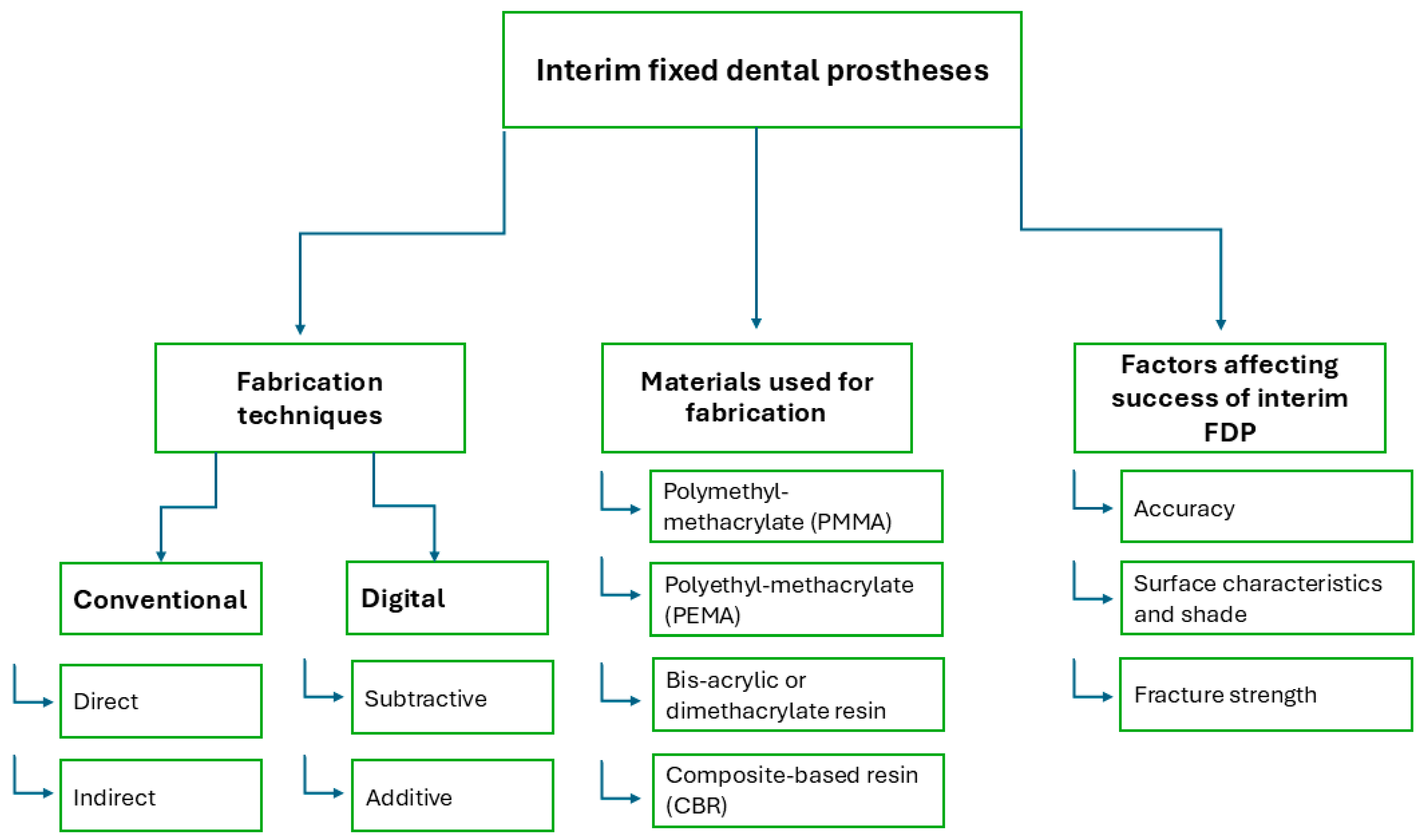
2. Methods of Fabrication of Interim FDPs

The methods of fabrication of interim FDPs could be classified according to fabrication settings into direct chairside and indirect laboratory, and according to fabrication technique into conventional and digital methods. The digital method is further subclassified according to fabrication process into subtractive and additive techniques [8].

2.1. Conventional Fabrication

2.1.1. Direct (Chairside)

In this technique, an index is created using polymeric impression material on intact teeth prior to preparation. Self-curing acrylic resin (temporary crown and bridge material) is then mixed and placed into the mold, which is seated directly on the prepared abutments in the patient’s mouth until the material hardens. Afterward, the interim prosthesis is checked for defects, followed by finishing and polishing [7]. In the case of a bridge, a silicone rubber index is made over the waxed-up primary cast. After abutment preparation, the temporary crown and bridge materials are loaded into the index and seated over the prepared abutments intraorally [9] (Figure 2). This technique is straightforward, low-cost and time efficient as it is direct and eliminates the need for laboratory steps [5]. However, its potential drawbacks include diminished mechanical properties, a rough surface finish, and an improper fit of the FDP, which may be exacerbated by inadequate mixing or the incorporation of air voids [10].
Another approach to direct bridge construction is a fiber-reinforced composite bridge. In this technique, the clinician utilizes fiberglass pins or bands. A fiber pin is positioned horizontally in the edentulous space, parallel to the occlusal or incisal plane. After adjusting the pin length, silane is applied to its surface, and the adjacent abutments undergo surface treatment with etching and bonding agents. The pin is then secured using flowable composite on the abutments, and a vertical reinforcing pin is placed perpendicular to the horizontal pin within the edentulous space. Composite is incrementally applied over the pins to form the pontic, or alternatively, packable composite is shaped into the pontic using a pre-formed index. The bridge is completed with finishing and polishing to refine the fiber-reinforced composite structure [11,12] (Figure 3). When specific case selection criteria are met, this technique can serve as a permanent treatment option for restoring a single missing tooth as reported by Martínez et al. [11]. In the study a success rate of 95.2% over a 9-year follow-up period is documented, suggesting it is an effective alternative to a conventional bridge for restoring a non-load-bearing tooth. In other cases, this method can function as a transitional phase during implant treatment [11,12].

2.1.2. Indirect (Laboratory) Fabrication

In the case of a longer service period, a laboratory interim FDP is recommended over a direct one since it has the advantage of an enhanced surface finish, improved surface contours and contacts, and reduced intraoral adjustments. Indirect laboratory procedures include fabricating an index and an interim FDP. A laboratory index can be prepared by using a vacuum-formed clear sheet pressed over the designated segment of the primary cast. Then, an auto-polymerizing resin is injected into the vacuum-formed index and placed on the master cast to construct the interim FDP [13] (Figure 4). Meanwhile, a conventional direct interim FDP is applied immediately following tooth preparation and acts as a provisional solution until the laboratory-fabricated interim FDP is ready for long-term application.
In esthetic cases, such as those involving veneers, an intraoral mockup is a crucial step prior to initiating tooth preparation. The mockup is created using temporary crown material, either through conventional methods or digital techniques. Conventional mockups are produced using indices crafted over manually waxed primary casts, prepared by an experienced technician. Meanwhile, digital mockups are milled or 3D–printed from digital wax-ups designed with 3D modeling software. Compared to conventional mockups, digitally fabricated mockups offer greater accuracy and closer alignment with the original design [14].

2.2. Digital Fabrication

2.2.1. Subtractive Technique (Milling)

Computer-aided design and manufacturing (CAD/CAM) technology, initially developed for the automotive and aerospace industries, has been successfully adapted for dental applications. A typical in-office dental CAD/CAM system includes an optical scanner, a desktop computer, and a milling unit. The digital workflow begins with an intraoral scanner capturing the prepared tooth, generating a digital impression that is displayed on the computer monitor for restoration design. Once the design of the restoration is finalized, it is transmitted to the milling unit, which fabricates the restoration [15].
CAD/CAM technology has been implemented in the fabrication of interim FDPs due to its ability to produce restorations of superior quality, regarding physical, esthetic, and mechanical properties, such as fracture strength, internal fit, and marginal adaptation [2,7]. Huettig et al. [6] conducted a clinical trial and evaluated the longevity of milled fixed–fixed polymethyl methacrylate (PMMA) bridges used as long-term temporary FDPs, reporting a success rate of 90.4%, with 88.3% of cases free from complications over 16 months. It was concluded that milled PMMA bridges could be a viable option for long-term temporary FDPs.
While CAD/CAM systems produce high-quality interim restorations, certain limitations can arise with the milling technology. Challenges may include restrictions in 3D designing software, difficulties with scanner cameras in accessing distal intraoral areas, image artifacts caused by uncontrolled moisture and saliva, and inaccuracies in milling machine production of fine restoration details [2]. Additionally, the expertise of the CAD/CAM operator plays a significant role in determining the quality of the final restoration [2,16].

2.2.2. Additive Technique (3D Printing)

The 3D–printing technique, an additive manufacturing process, begins with a digital file created using CAD modeling software, based again on intraoral scans or scanned plaster casts. This data enables the production of thin cross-sectional layers, allowing for the creation of complex shapes unattainable with traditional methods. The process involves designing the part in compatible software, generating a file format for the printer, and constructing the product by layering thin sections sequentially. Most 3D–printing methods use this layer-by-layer approach, interpreting the design as a series of two-dimensional layers [17].
With the development of 3D–printing production technology and its recent widespread use in dentistry, a direct comparison between milling and 3D printing in terms of reduced production time, raw material saving, and the ability to produce complex structures has tipped the balance in favor of 3D printing [18,19]. Photopolymerization 3D printing is the most widely used technique in dentistry due to its ability to fabricate highly detailed objects with excellent resolution, a fine surface finish, and reduced production time. Both, stereolithography (SLA) and digital light processing (DLP) utilize photopolymerization technology. SLA employs a laser to cure the resin layer by layer, while DLP uses a wider light reflector that solidifies the entire layer of resin on the platform at once (Figure 5). DLP is faster than SLA. After each layer is polymerized, the platform moves downward to allow the next layer to be cured, continuing this process until the entire object is complete. In both systems, the operator can control the layer thickness [19,20,21].
Several studies [4,22,23,24,25,26,27,28] employed 3D–printing technology in fabricating interim FDP, comparing the 3D–printed restorations to traditionally produced restorations. Furthermore, Cuschieri et al. [16] assessed patient satisfaction with conventional temporary restorations compared to digitally designed and fabricated restorations. Their findings revealed that digital restorations significantly improved satisfaction with the esthetic outcome compared to conventional restorations, while the designer’s expertise impacted satisfaction with both esthetic and functional outcomes.

3. Materials Available for Interim FDPs

The most common materials used for direct chairside and indirect interim restorations are PMMA and polyethyl-methacrylate (PEMA), bis-acrylic or dimethacrylate resin, and composite-based resin (CBR) [10,18,29]. Interim FDP composites are methacrylate-based materials composed of resin, such as triethyleneglycol dimethacrylate (TEGDMA), 2,2-bis(4-(2-methacryloxyethoxy) phenyl) propane (Bis-EMA), bisphenol A glycidyl dimethacrylate (Bis-GMA), or urethane dimethacrylate (UDMA) [30]. With the evolution of digital dentistry, milling restorations out of polymerized blanks—industrially prepared under optimum conditions—or using 3D printing and creating an object layer by layer by sequential polymerization of light-sensitive liquid resin have influenced the overall properties of final restorations, even though the same materials are being used [18,29]. Berghaus et al. [30] investigated the leaching of residual unpolymerized monomers from interim restoration materials fabricated using milling, 3D printing, and the direct technique. Regarding the effect of fabrication techniques, the direct technique exhibited the highest percentage of residual monomer leaching, which negatively affects the biological, physical, and mechanical properties of the material. In contrast, both 3D printing and milling showed a higher degree of conversion, with no detectable free monomers observed in the case of milled restorations.
One of the major drawbacks of direct interim restoration fabrication is polymerization shrinkage that can affect the accuracy and mechanical properties of an interim prosthesis. Modifications such as filler incorporation into direct interim materials have been made to enhance the mechanical properties. The most common fillers used were metal, glass fiber, polyethylene, or carbon [10]. Peñate et al. [10] compared the marginal fit and fracture strength of glass fiber-reinforced and non-reinforced direct interim FDPs to milled PMMA bridges. In their study they found that glass fiber-reinforced bridges had the least marginal discrepancy, and that fiber reinforcement improved mechanical properties and influenced the failure mode of the interim bridges.

4. Factors Affecting the Success of Interim FDPs

4.1. Accuracy of Interim FDPs

The accuracy of dental restoration is defined by its internal fit, marginal adaptation, and occlusal accuracy; these play a pivotal role in its clinical performance [31]. Several factors can influence the accuracy of FDPs, including the fabrication technique, span length, and impression methods utilized [32]. While in numerous studies [33,34,35,36] the accuracy of permanent FDPs concerning these factors has been investigated, there is a notable lack of data on the accuracy of interim FDPs.

4.1.1. Marginal Adaptation

Marginal discrepancies in interim fixed dental prostheses (FDPs) can lead to sensitivity in the reduced abutment and periodontal inflammation around the prosthesis. Such discrepancies may also compromise subsequent steps in final FDP fabrication, including margin assessment during the try-in stage and final cementation [5,37]. Aldahian et al. [25] and Alharbi et al. [4] reported that 3D–printed full-coverage restorations exhibited smaller marginal gaps compared to those produced using milling or conventional methods.

4.1.2. Internal Fit

An inaccurately fitted interim FDP can lead to loss of retention and impaired function [38,39]. In several studies [4,22,23,25] it was concluded that 3D–printed single crowns exhibited superior internal fit and trueness compared to their milled counterparts. Shalaby et al. [26] stated that even with varying span length bridges, 3D–printed interim FDPs demonstrated higher internal fit accuracy. SLA 3D printing can produce the smallest internal and marginal gaps compared to DLP printing, milling, and conventional techniques [4,40].

4.1.3. Occlusal Surface Accuracy

Giannetti et al. [24] performed a direct comparison between milled and 3D–printed interim crowns regarding trueness of occlusal surface compared to original CAD design. In the study it was found that 3D–printed crowns had higher occlusal surface accuracy and required fewer intraoral occlusal adjustments.

4.2. Surface Characteristics and Shade

For cases requiring interim FDPs over extended durations, such as in rehabilitation or implant-supported prostheses, the esthetic properties of interim materials are primarily influenced by factors like shade availability, color stability, surface finish, and micro-roughness [25,28,41]. According to a recent study, the current interim resin materials available for additive manufacturing techniques failed to match their conventional composite and acrylic resin counterparts of the same shade regarding the three parameters of color. This makes shade selection and shade matching with adjacent natural teeth a challenging process for additive interim resins [42,43]. However, shade matching with natural teeth could be improved for 3D–printed interim restorations by using external add-on stains. According to Ellakany et al. [44], the color stability of interim FDPs is highest in milled interim resin materials, followed by those produced through SLA 3D printing, then DLP 3D printing, with conventional interim resins exhibiting the lowest color stability.
In a study by Yao et al. [45], it was highlighted that there is an impact of surface treatments on the color stability of digitally fabricated interim FDPs. The findings revealed that applying a nano-filled light-polymerizing protective agent significantly reduces color change in milled restorations during service. For additively manufactured restorations, both surface polishing and protective coating application were found to effectively minimize color changes.
Surface roughness is another critical consideration. Interim FDPs fabricated using additive techniques exhibit the highest surface roughness, followed by those manufactured using the conventional direct technique, with the subtractive technique producing the smoothest surfaces [3,25,28]. Expanding on these findings, Al-Wahadni et al. [46] highlighted that within additive techniques, DLP–printed restorations show higher surface roughness compared to SLA–printed restorations. Surface finish is further influenced by filler content and particle size in the resin. Gantz et al. [41] observed that only materials with nano-sized fillers achieve the smoothest surfaces both before and after polishing.
Along with micro-roughness, porosity and fungal adherence also play a significant role in the performance of interim FDPs regarding pink esthetics (gingival display) and gingival health as plaque retention can further affect soft tissue health in patients with underlying periodontal conditions and bad oral hygiene. Ribeiro et al. [47] found that milled restorations exhibit the lowest porosity and fungal adherence, followed by 3D–printed restorations, while conventional materials, especially composite resin materials have the highest porosity and susceptibility to fungal adherence. Additionally, Radwan et al. [48] concluded that 3D–printed interim restorations are unsuitable for extended use due to persistent issues with surface roughness and staining.

4.3. Fracture Strength

Limited data exist on the fracture resistance of interim fixed dental prostheses (FDPs), particularly long-span interim prostheses. Milled interim crowns demonstrate higher fracture resistance compared to 3D–printed crowns. Consequently, Alsarani et al. [49] recommend avoiding the use of 3D–printed interim crowns with occlusal thicknesses under 1 mm. Efforts to improve the fracture resistance of 3D–printed restorations, such as incorporating micro-fillers like zirconia glass and glass silica, were unsuccessful. Instead, these fillers increased surface roughness, which compromised the esthetics of the crowns as noted by Alshamarani et al. [50]. Studies comparing the fracture resistance of interim crowns fabricated manually, by milling, and through 3D printing consistently found that milled crowns outperformed 3D–printed crowns, with both surpassing manually fabricated crowns in fracture resistance [8,51]. However, Abdullah et al. [52] highlighted that not all milled interim materials are superior to direct materials in terms of fracture resistance.
Three-unit interim FDPs produced using additive and subtractive techniques have also been evaluated. Abad-Coronel et al. [53] reported that milled three-unit bridges exhibited higher fracture resistance. Contrarily, Falahchai et al. [54] found that 3D–printed three-unit interim bridges achieved the highest fracture resistance values, which were statistically significant compared to those produced using direct and indirect techniques. This variation could stem from differences in methodologies applied, materials used, 3D–printing systems, or the models employed for testing. The rigidity of the models used for fracture testing can significantly influence the results; the more rigid the model, the higher the fracture resistance values obtained [55,56].
Among 3D–printed interim FDPs, Al-Wahadni et al. [46] reported that SLA crowns exhibit higher fracture resistance compared to DLP crowns, while DLP–printed crowns demonstrated more consistent fracture patterns.

4.4. Clinical Performance

Patient satisfaction and a dentist-friendly workflow are critical factors for successful treatment. Studies indicate that interim crown fabrication techniques, whether conventional or digital, have minimal impact on patient satisfaction. However, digital fabrication techniques significantly reduce processing time compared to conventional methods. Unlike conventional techniques, digital workflows are unaffected by the operator’s expertise and consistently produce high-quality restorations within a short time frame [57,58].
In a cohort study by Hougne et al. [59], the survival rate of 3D–printed interim crowns was reported as 100% after a 6-month follow-up, comparable to the performance of milled temporary crowns. Patients expressed satisfaction with the esthetics and functionality of the 3D–printed crowns.
Limited data exist regarding the cost-effectiveness of digital fabrication techniques for interim FDPs. Further research is warranted to address this topic.

5. Conclusions

Each technique for fabricating interim FDPs has its advantages and limitations, and all can be effectively utilized for FDP production. However, digital techniques provide superior quality compared to conventional methods, particularly for long-term use. Additive manufacturing has the potential to completely replace subtractive techniques for interim FDP fabrication in the future, owing to its numerous advantages. Nonetheless, further studies are necessary to evaluate the accuracy and fracture strength of 3D–printed interim FDPs with varying span lengths. Additionally, more research is needed to enhance the esthetic appearance of 3D–printed interim FDPs.

6. Clinical Recommendations

  • The conventional fabrication technique for interim FDPs is recommended for cases with short span lengths and when the prosthesis is intended for short-term use.
  • Digital techniques are recommended for fabricating interim FDPs intended for long-term use.
  • Milled interim FDPs are preferred in cases with high esthetic demands, expected heavy occlusal forces, periodontal diseases, or poor oral hygiene.
  • 3D–printed FDPs are best suited for cases involving longer span lengths or full-arch rehabilitation as try-in restorations due to their precise accuracy compared to milled long-span bridges. However, reduced mechanical properties may limit their long-term use as interim FDPs.
  • 3D–printed interim crowns demonstrated reliable clinical durability and efficiency. However, longer span FDPs might not meet the required clinical standards in terms of fracture resistance.

Author Contributions

N.A.: writing—review and editing, writing—original draft, visualization, validation, data curation. C.B.: writing—review and editing. T.M.E.: writing—review and editing, visualization, Validation. All authors have read and agreed to the published version of the manuscript.

Funding

This scientific work was supported by a fund from Forschungsgemeinschaft Dental e.V. (Project No. 02/2023).

Institutional Review Board Statement

This article does not contain any studies on human or animal subjects.

Data Availability Statement

No new data were created or analyzed in this study. Data sharing is not applicable to this article.

Conflicts of Interest

The authors declare no conflicts of interest.

References

  1. Peng, C.C.; Chung, K.H.; Yau, H.T.; Ramos, V. Assessment of the internal fit and marginal integrity of interim crowns made by different manufacturing methods. J. Prosthet. Dent. 2020, 123, 514–522. [Google Scholar] [CrossRef]
  2. Sadighpour, L.; Geramipanah, F.; Falahchai, M.; Tadbiri, H. Marginal adaptation of three-unit interim restorations fabricated by the CAD-CAM systems and the direct method before and after thermocycling. J. Clin. Exp. Dent. 2021, 13, e572–e579. [Google Scholar] [CrossRef]
  3. Mârțu, I.; Murariu, A.; Baciu, E.R.; Savin, C.N.; Foia, I.; Tatarciuc, M.; Diaconu-Popa, D. An Interdisciplinary Study Regarding the Characteristics of Dental Resins Used for Temporary Bridges. Medicina 2022, 58, 811. [Google Scholar] [CrossRef]
  4. Alharbi, N.; Alharbi, S.; Cuijpers, V.M.J.I.; Osman, R.B.; Wismeijer, D. Three-dimensional evaluation of marginal and internal fit of 3D-printed interim restorations fabricated on different finish line designs. J. Prosthodont. Res. 2018, 62, 218–226. [Google Scholar] [CrossRef]
  5. Emetwaly el shazly, M.; Abdelhamid, T.; Atta, M.O. Marginal Integrity of Temporary Bridges Constructed by CAD/CAM, Three Dimentional Printer and Conventional Method. Dent. Sci. Updates 2023, 4, 357–365. [Google Scholar] [CrossRef]
  6. Huettig, F.; Prutscher, A.; Goldammer, C.; Kreutzer, C.A.; Weber, H. First clinical experiences with CAD/CAM-fabricated PMMA-based fixed dental prostheses as long-term temporaries. Clin. Oral Investig. 2016, 20, 161–168. [Google Scholar] [CrossRef]
  7. Abdullah, A.O.; Pollington, S.; Liu, Y. Comparison between direct chairside and digitally fabricated temporary crowns. Dent. Mater. J. 2018, 37, 957–963. [Google Scholar] [CrossRef]
  8. Sakr, O.A.; Mandour, M.H.; Gabal, Z.A. Fracture Resistance of Provisional Crowns Fabricated by Conventional, CAD/CAM, and 3D Printing Techniques. Al-Azhar Dent. J. Girls 2022, 9, 623–630. [Google Scholar] [CrossRef]
  9. Al Twal, E.Q.H.; Chadwick, R.G. Fibre reinforcement of two temporary composite bridge materials—Effect upon flexural properties. J. Dent. 2012, 40, 1044–1051. [Google Scholar] [CrossRef]
  10. Peñate, L.; Basilio, J.; Roig, M.; Mercadé, M. Comparative study of interim materials for direct fixed dental prostheses and their fabrication with CAD/CAM technique. J. Prosthet. Dent. 2015, 114, 248–253. [Google Scholar] [CrossRef]
  11. Martínez, M.F.E.; López, S.R.; Fontela, J.V.; García, S.O.; Quevedo, M.M. A new technique for direct fabrication of fiber-reinforced composite bridge: A long-term clinical observation. Dent. J. 2020, 8, 48. [Google Scholar] [CrossRef]
  12. Karabekmez, D.; Aktas, G. Single anterior tooth replacement with direct fiber-reinforced composite bridges: A report of three cases. Niger J. Clin. Pract. 2020, 23, 434–436. [Google Scholar] [CrossRef]
  13. Lee, J.; Clark, S.R.; Tantbirojn, D.; Korioth, T.V.P.; Hill, A.E.; Versluis, A. Strength and stiffness of interim materials and interim fixed dental prostheses when tested at different loading rates. J. Prosthet. Dent. 2022, 127, 161–167. [Google Scholar] [CrossRef]
  14. Cattoni, F.; Teté, G.; Calloni, A.M.; Manazza, F.; Gastaldi, G.; Capparè, P. Milled versus moulded mock-ups based on the superimposition of 3D meshes from digital oral impressions: A comparative in vitro study in the aesthetic area. BMC Oral Health 2019, 29, 230. [Google Scholar] [CrossRef]
  15. Davidowitz, G.; Kotick, P.G. The Use of CAD/CAM in Dentistry. Dent. Clin. N. Am. 2011, 55, 559–570. [Google Scholar] [CrossRef]
  16. Cuschieri, L.A.; Casha, A.; No-Cortes, J.; Ferreira Lima, J.; Cortes, A.R.G. Patient Satisfaction with Anterior Interim CAD-CAM Rehabilitations Designed by CAD Technician versus Trained Dentist—A Clinical Preliminary Study. Appl. Sci. 2023, 13, 8243. [Google Scholar] [CrossRef]
  17. Jandyal, A.; Chaturvedi, I.; Wazir, I.; Raina, A.; Ul Haq, M.I. 3D printing—A review of processes, materials and applications in industry 4.0. Sustain. Oper. Comput. 2022, 3, 33–42. [Google Scholar] [CrossRef]
  18. Jain, S.; Sayed, M.E.; Shetty, M.; Alqahtani, S.M.; Al Wadei, M.H.D.; Gupta, S.G.; Othman, A.A.A.; Alshehri, A.H.; Alqarni, H.; Mobarki, A.H.; et al. Physical and Mechanical Properties of 3D-Printed Provisional Crowns and Fixed Dental Prosthesis Resins Compared to CAD/CAM Milled and Conventional Provisional Resins: A Systematic Review and Meta-Analysis. Polymers 2022, 14, 2691. [Google Scholar] [CrossRef]
  19. Nestler, N.; Wesemann, C.; Spies, B.C.; Beuer, F.; Bumann, A. Dimensional accuracy of extrusion- and photopolymerization-based 3D printers: In vitro study comparing printed casts. J. Prosthet. Dent. 2021, 125, 103–110. [Google Scholar] [CrossRef]
  20. Resende, C.C.D.; Barbosa, T.A.Q.; Moura, G.F.; Rizzante, F.A.P.; Mendonça, G.; Zancopé, K.; das Neves, F.D. Cost and effectiveness of 3-dimensionally printed model using three different printing layer parameters and two resins. J. Prosthet. Dent. 2023, 129, 350–353. [Google Scholar] [CrossRef]
  21. Tian, Y.; Chen, C.; Xu, X.; Wang, J.; Hou, X.; Li, K.; Lu, X.; Shi, H.; Lee, E.S.; Jiang, H.B. A Review of 3D Printing in Dentistry: Technologies, Affecting Factors, and Applications. Scanning 2021, 2021, 9950131. [Google Scholar] [CrossRef]
  22. Kakinuma, H.; Izumita, K.; Yoda, N.; Egusa, H.; Sasaki, K. Comparison of the accuracy of resin-composite crowns fabricated by three-dimensional printing and milling methods. Dent. Mater. J. 2022, 41, 808–815. [Google Scholar] [CrossRef]
  23. Son, K.; Lee, J.H.; Lee, K.B. Comparison of intaglio surface trueness of interim dental crowns fabricated with sla 3d printing, dlp 3d printing, and milling technologies. Healthcare 2021, 9, 983. [Google Scholar] [CrossRef]
  24. Giannetti, L.; Apponi, R.; Mordini, L.; Presti, S.; Breschi, L.; Mintrone, F. The occlusal precision of milled versus printed provisional crowns. J. Dent. 2022, 1, 117. [Google Scholar] [CrossRef]
  25. Aldahian, N.; Khan, R.; Mustafa, M.; Vohra, F.; Alrahlah, A. Influence of Conventional, CAD-CAM, and 3D Printing Fabrication Techniques on the Marginal Integrity and Surface Roughness and Wear of Interim Crowns. Appl. Sci. 2021, 11, 8964. [Google Scholar] [CrossRef]
  26. Shalaby, M.; Wahba, M.; Mohamed, A. Assessment of the Effect of two Different Digital Fabrication Techniques on Marginal and Internal Fit of Interim Fixed Dental Prothesis. Future Dent. J. 2023, 9, 65–72. [Google Scholar] [CrossRef]
  27. Kessler, A.; Reymus, M.; Hickel, R.; Kunzelmann, K.H. Three-body wear of 3D printed temporary materials. Dent. Mater. 2019, 35, 1805–1812. [Google Scholar] [CrossRef]
  28. Al-Qahtani, A.S.; Tulbah, H.I.; Binhasan, M.; Abbasi, M.S.; Ahmed, N.; Shabib, S.; Farooq, I.; Aldahian, N.; Nisar, S.S.; Tanveer, S.A.; et al. Surface properties of polymer resins fabricated with subtractive and additive manufacturing techniques. Polymers 2021, 13, 4077. [Google Scholar] [CrossRef]
  29. Karaokutan, I.; Sayin, G.; Kara, O. In vitro study of fracture strength of provisional crown materials. J. Adv. Prosthodont. 2015, 7, 27–31. [Google Scholar] [CrossRef]
  30. Berghaus, E.; Klocke, T.; Maletz, R.; Petersen, S. Degree of conversion and residual monomer elution of 3D-printed, milled and self-cured resin-based composite materials for temporary dental crowns and bridges. J. Mater. Sci. Mater. Med. 2023, 34, 23. [Google Scholar] [CrossRef]
  31. Ghavami-Lahiji, M.; Falahchai, M.; Habibi Arbastan, A.; Arbastan, H. Review Paper: Different ways to measure marginal fit and internal adaptation of restorations in dentistry Citation. J. Dentomaxillofac. Radiol. Pathol. Surg. 2023, 12, 16–23. [Google Scholar]
  32. Bousnaki, M.; Chatziparaskeva, M.; Bakopoulou, A.; Pissiotis, A.; Koidis, P. Variables affecting the fit of zirconia fixed partial dentures: A systematic review. J. Prosthet. Dent. 2020, 123, 686–692. [Google Scholar] [CrossRef]
  33. Sachs, C.; Groesser, J.; Stadelmann, M.; Schweiger, J.; Erdelt, K.; Beuer, F. Full-arch prostheses from translucent zirconia: Accuracy of fit. Dent. Mater. 2014, 30, 817–823. [Google Scholar] [CrossRef]
  34. Akçin, E.T.; Güncü, M.B.; Aktaş, G.; Aslan, Y. Effect of manufacturing techniques on the marginal and internal fit of cobalt-chromium implant-supported multiunit frameworks. J. Prosthet. Dent. 2018, 120, 715–720. [Google Scholar] [CrossRef]
  35. Kim, W.K.; Kim, S. Effect of number of pontics and impression technique on the accuracy of four-unit monolithic zirconia fixed dental prostheses. J. Prosthet. Dent. 2018, 119, 860.e1–860.e7. [Google Scholar] [CrossRef]
  36. Kim, M.W.; Kim, J.Y.; Shim, J.S.; Kim, S. Effect of the number of splinted abutments on the accuracy of zirconia copings. J. Prosthet. Dent. 2018, 120, 790.e1–790.e7. [Google Scholar] [CrossRef]
  37. Borba, M.; Cesar, P.F.; Griggs, J.A.; Della Bona, Á. Adaptation of all-ceramic fixed partial dentures. Dent. Mater. 2011, 27, 1119–1126. [Google Scholar] [CrossRef]
  38. Son, K.; Lee, S.; Kang, S.H.; Park, J.; Lee, K.B.; Jeon, M.; Yun, B.J. A comparison study of marginal and internal fit assessment methods for fixed dental prostheses. J. Clin. Med. 2019, 8, 785. [Google Scholar] [CrossRef]
  39. Attia, M.A.; Blunt, L.; Bills, P.; Tawfik, A.; Radawn, M. Micro-CT analysis of marginal and internal fit of milled and pressed polyetheretherketone single crowns. J. Prosthet. Dent. 2023, 129, 906.e1–906.e10. [Google Scholar] [CrossRef]
  40. Wadhwani, V.; Sivaswamy, V.; Rajaraman, V. Surface roughness and marginal adaptation of stereolithography versus digital light processing three-dimensional printed resins: An in-vitro study. J. Indian Prosthodont. Soc. 2022, 22, 377. [Google Scholar]
  41. Gantz, L.; Fauxpoint, G.; Arntz, Y.; Pelletier, H.; Etienne, O. In vitro comparison of the surface roughness of polymethyl methacrylate and bis-acrylic resins for interim restorations before and after polishing. J. Prosthet. Dent. 2021, 125, 833.e1–833.e10. [Google Scholar] [CrossRef]
  42. Alikhasi, M.; Jafarian, Z. Additional Manufactured Interim Restorations: A Review on the Literature. J. Dent. 2022, 23, 424–437. [Google Scholar]
  43. Revilla-León, M.; Umorin, M.; Özcan, M.; Piedra-Cascón, W. Color dimensions of additive manufactured interim restorative dental material. J. Prosthet. Dent. 2021, 123, 754–760. [Google Scholar] [CrossRef]
  44. Ellakany, P.; Fouda, S.M.; AlGhamdi, M.A.; Aly, N.M. Comparison of the color stability and surface roughness of 3-unit provisional fixed partial dentures fabricated by milling, conventional and different 3D printing fabrication techniques. J. Dent. 2023, 131, 104458. [Google Scholar] [CrossRef]
  45. Yao, Q.; Morton, D.; Eckert, G.J.; Lin, W.S. The effect of surface treatments on the color stability of CAD-CAM interim fixed dental prostheses. J. Prosthet. Dent. 2021, 126, 248–253. [Google Scholar] [CrossRef]
  46. Al-Wahadni, A.; Abu Rashed, B.O.; Al-Fodeh, R.; Tabanjah, A.; Hatamleh, M. Marginal and Internal Gaps, Surface Roughness and Fracture Resistance of Provisional Crowns Fabricated With 3D Printing and Milling Systems. Oper. Dent. 2023, 48, 464–471. [Google Scholar] [CrossRef]
  47. Ribeiro, A.K.C.; de Freitas, R.F.C.P.; de Carvalho, I.H.G.; de Miranda, L.M.; da Silva, N.R.; de Fátima Dantas de Almeida, L.; Zhang, Y.; da Fonte Porto Carreiro, A.; de Assunção e Souza, R.O. Flexural strength, surface roughness, micro-CT analysis, and microbiological adhesion of a 3D-printed temporary crown material. Clin. Oral Investig. 2023, 27, 2207–2220. [Google Scholar] [CrossRef]
  48. Radwan, H.; Elnaggar, G.; El Deen, I.S. Surface roughness and color stability of 3D printed temporary crown material in different oral media (In vitro study). Int. J. Appl. Dent. Sci. 2021, 7, 327–334. [Google Scholar] [CrossRef]
  49. Alsarani, M.M. Influence of aging process and restoration thickness on the fracture resistance of provisional crowns: A comparative study. Saudi Dent. J. 2023, 35, 939–945. [Google Scholar] [CrossRef]
  50. Alshamrani, A.; Alhotan, A.; Owais, A.; Ellakwa, A. The Clinical Potential of 3D-Printed Crowns Reinforced with Zirconia and Glass Silica Microfillers. J. Funct. Biomater. 2023, 14, 267. [Google Scholar] [CrossRef]
  51. Othman, A.; Sandmair, M.; Alevizakos, V.; von See, C. The fracture resistance of 3D-printed versus milled provisional crowns: An in vitro study. PLoS ONE 2023, 18, e0285760. [Google Scholar] [CrossRef]
  52. Abdullah, A.O.; Tsitrou, E.A.; Pollington, S. Comparative in vitro evaluation of CAD/CAM vs conventional provisional crowns. J. Appl. Oral Sci. 2016, 24, 258–263. [Google Scholar] [CrossRef]
  53. Abad-Coronel, C.; Carrera, E.; Córdova, N.M.; Fajardo, J.I.; Aliaga, P. Comparative analysis of fracture resistance between cad/cam materials for interim fixed prosthesis. Materials 2021, 14, 7791. [Google Scholar] [CrossRef]
  54. Falahchai, M.; Rahimabadi, S.; Khabazkar, G.; Babaee Hemmati, Y.; Neshandar Asli, H. Marginal and internal fit and fracture resistance of three-unit provisional restorations fabricated by additive, subtractive, and conventional methods. Clin. Exp. Dent. Res. 2022, 8, 1404–1412. [Google Scholar] [CrossRef]
  55. Greuling, A.; Matthies, A.; Eisenburger, M. Fracture load of 4-unit interim fixed partial dentures using 3D-printed and traditionally manufactured materials. J. Prosthet. Dent. 2023, 129, 607.e1–607.e8. [Google Scholar] [CrossRef]
  56. Rand, A.; Kohorst, P.; Greuling, A.; Borchers, L.; Stiesch, M. Stress distribution in all-ceramic posterior 4-unit fixed dental prostheses supported in different ways: Finite element analysis. Implant. Dent. 2016, 25, 485–491. [Google Scholar] [CrossRef]
  57. Cheng, C.W.; Ye, S.Y.; Chien, C.H.; Chen, C.J.; Papaspyridakos, P.; Ko, C.C. Randomized clinical trial of a conventional and a digital workflow for the fabrication of interim crowns: An evaluation of treatment efficiency, fit, and the effect of clinician experience. J. Prosthet. Dent. 2021, 125, 73–81. [Google Scholar] [CrossRef]
  58. de Souza, F.A.; Blois, M.C.; Collares, K.; dos Santos, M.B.F. 3D-printed and conventional provisional single crown fabrication on anterior implants: A randomized clinical trial. Dent. Mater. 2024, 40, 340–347. [Google Scholar] [CrossRef]
  59. del Hougne, M.; Di Lorenzo, I.; Höhne, C.; Schmitter, M. A retrospective cohort study on 3D printed temporary crowns. Sci. Rep. 2024, 14, 17295. [Google Scholar] [CrossRef]
Figure 1. Schematic map of the narrative review content.
Oral 05 00044 g001
Figure 2. Silicone rubber index fabrication steps and temporary crown and bridge material loading.
Oral 05 00044 g002
Figure 3. Fiber-reinforced composite bridge fabrication steps. (A) Fiber pins positioning and fixation, (B) pontic shaping using polymer index, (C) embrasures checking using micro-brushes [12].
Oral 05 00044 g003
Figure 4. Steps of interim FDP fabrication using vacuum-formed clear sheet index.
Oral 05 00044 g004
Figure 5. SLA and DLP 3D–printing operating technology.
Oral 05 00044 g005
Disclaimer/Publisher’s Note: The statements, opinions and data contained in all publications are solely those of the individual author(s) and contributor(s) and not of MDPI and/or the editor(s). MDPI and/or the editor(s) disclaim responsibility for any injury to people or property resulting from any ideas, methods, instructions or products referred to in the content.

