Abstract
Forensic dentistry is still an emerging field in Pakistan. The lack of scientific literature on the topic may lead to difficulties in situations where age estimation has a significant part such as in criminal and civil litigation. In mass disasters such as earthquakes and accidents, the correct investigation of the chronological age can be less troublesome if population-specific evidence is available. This is the rationale that justifies dedicated dental age estimation studies. This cross-sectional study aimed to test the time efficiency, validity and applicability of four dental age estimation methods: two invasive (Bang and Ramm and Lamendin) and two non-invasive (Kvaal and Cameriere) in an adult Pakistani population. A total of 37 teeth collected from a dental hospital in Islamabad, Pakistan, were used. Teeth included the central and lateral incisors, canines, and first and second premolars of males and females. Results were calculated using a Microsoft Office 2007 excel spreadsheet. Overall, Kvaal’s method mean absolute error between chronological and estimated ages (MAE: 12.33) showed the highest variation and Bang and Ramm showed more accurate results in comparison with other methods (MAE: 4.80). It was both time-efficient and practical to use. It can be stated that these were preliminary cross-sectional outcomes and that studies with larger samples are necessary.
1. Introduction
Forensic age estimation is a scientific process which can estimate an individual’s true chronological age by assessing their skeletal and dental development and maturation. Age estimation plays a significant role in forensic science, and dental age estimation is the most widely used method. It is not surprising that human growth and maturation are unique for each individual but dental techniques for estimating age are currently considered as one of the accurate ways to determine chronological age. They involve a series of chronological changes in tooth development as well as various other methods in order to estimate age in children, adolescents and adults [1,2,3]. The estimation of age is an important part of the human identification process performed in a medico-legal scenario [1]. An assessment of age is often required in the civil and criminal courts [2,3], for instance, in the cases of asylum seekers [4], adoption, clandestine migration, human trafficking, the judgment of alleged minor offenders [5], sports practice, the identification of unknown bodies [6] as well as requested retirement and pensions.
Dental age estimation methods have been widely reported on [7,8,9,10,11]. Some methods are relatively accurate, conservative and preserve the teeth’s structure, while other methods require tooth extraction as well as some preparation. The various methods are divided into three categories which include morphohistological methods, radiological/developmental methods, and biochemical methods. The applicability of these methods differs in living and dead individuals as they can be invasive in nature and according to the population under investigation [12].
Tooth eruption and development are dental parameters related to age which are analyzed via non-invasive methods, such as visual and imaginological (radiographic or not) examination [13]. Because of the non-invasive aspect of these parameters, the methods for age assessment are usually applicable to the living—especially among children and adolescents [14]. For adults, age estimation methods rely on (regressive) parameters, namely attrition, deposition of secondary dentin, cementum apposition, root resorption, translucency, as well as the level of periodontosis [15,16]. A relatively new method uses the eruption of the third molar in the maxilla and mandible, which was proposed by Gambier [17].
The invasive aspect of most dental age estimation methods for adults makes research more difficult because ex-vivo specimens and destructive laboratory procedures are required. Therefore, for dental age estimation in living individuals radiological methods are preferred.
Remarkable dental age estimation methods for adults include non-invasive and invasive methods. The invasive methods include Gustafson’s, Johansson, Bang and Ramm, Lamendin, Dalit and Maples methods. The non-invasive methods include Kvaal, Cameriere and micro-CT (computed tomography) [11].
One of the famous scientific techniques for age estimation in adults was presented by Gustafson. It was based on longitudinal sections of teeth cut through the central area.
The technique consisted in attributing scores from 0 to 3 for the presence and number of age-related changes such as attrition, periodontal ligament retractions, secondary dentin formations, root translucency, and root resorption [18]. Johansson tested Gustafson’s method on a larger, independent sample. His method evaluated the same six age-related changes as Gustafson but scored them by an enlarged seven-point criteria system. He also used multiple regressions to calculate a regression line from which ages of unknown individuals could be estimated [19].
Among the six variables suggested by Gustafson, dentinal translucency showed accurate results for age prediction. The literature has shown that dentine transparency is directly proportional to an increase in age. Later, Bang and Ramm worked on a detailed method to measure the translucency length and developed tooth-specific formulae for age estimation on Norwegian samples [20]. These formulae provided accurate results for the European and American population [18,19,21]. Bang and Ramm’s method uses the length of the root dentine translucency in sectioned or un-sectioned teeth. It can be used to estimate chronological age in human remains [20].
Modifications to Gustafson’s method were also proposed by Lamendin, who considered two factors for age estimation: translucency of the tooth root and gingival recession (periodontosis). This method can be used on dead individuals and can be further used to analyze a single-rooted tooth which needs no preparation [16].
Dental radiography is a simple technique which is used regularly in dental practice. It has been used for age estimation, as it is a non-destructive method [22]. Intraoral radiographs and full-mouth dental radiographs have been used to estimate age. The use of radiographs involves techniques that observe morphologically distinct stages of mineralization. They can be based on the degree of formation of the crown and root structures and their eruption stages [23]. Kvaal and co-authors have shown radiographic methods for age estimation to be reliable but possible variations in different ethnicities have affected their accuracy [24]. Kvaal developed a method based on the pulp size using periapical dental radiographs [25]. Cameriere published a method of age estimation based on the measurement of the ratio between the length of the projection of the open apices and the length of the tooth axis major, known as the open apices method [26].
Among the biochemical methods, aspartic acid racemization, advanced glycation-end products collagen crosslinks, and mitochondrial DNA (mtDNA) mutations are some of the most established methods of biochemical age estimation [25]. One disadvantage of these methods is the technique-sensitivity and requiring longer periods of time to achieve results [27,28].
In recent studies using root dentin translucency in extracted teeth from living individuals, it was proven to be a reliable indicator for age estimation [29]. Studies were also carried out using Kvaal and Cameriere’s method in Saudi Arabian and African populations showing the accuracy of these methods [30,31].
Several studies have been conducted in the past to devise an age estimation method for their representative population but currently limited data are available for methods which are applicable to the Pakistani population. It is important to recognize that results from previous studies cannot be extrapolated onto the Pakistani population as they are a distinct ethnic group. Very few studies have been carried out in the past to identify age estimation methods in the Pakistani population. It is mainly due to this reason that many difficulties are faced by asylum seekers as well as in criminal, civil litigation, and pension lawsuits. The studies conducted on the Pakistani population include methods such as the tooth coronal index. This method has shown a weak correlation between chronological age and the tooth coronal index [32]. Our study aims to apply and compare two different invasive and non-invasive age estimation methods on teeth samples from the living Pakistani population in order to suggest which method gives optimal age estimation. It will also highlight the role of forensic odontology as this is not a field much explored in Pakistan.
Aim
To apply and compare two invasive and non-invasive age estimation methods in adult living Pakistani subjects in order to assess their validity and accuracy.
2. Material and Methods
2.1. Study Design
This is a preliminary observational cross-sectional study, structured according to the Strengthening the Reporting of Observational Studies in Epidemiology (STROBE) Initiative [33].
2.2. Settings and Participants
The teeth were collected from the existing database of the patients (male and female) who visited the Oral and Maxillofacial department of the Islamic International Dental Hospital, Islamabad, Pakistan, from January to March 2022.
No patient underwent tooth extraction merely for research purposes and all the extractions performed in the above-mentioned time period were necessary for therapeutic reasons. A total of 37 extracted teeth were collected as shown in Table 1 and stored in a solution of phosphate buffered saline as shown in Figure 1.

Table 1.
Sample of extracted teeth distribution according to sex.
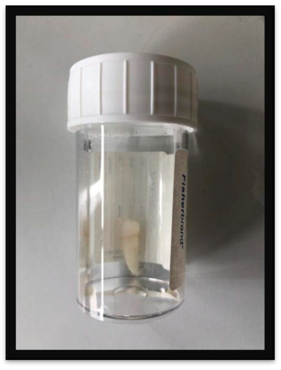
Figure 1.
Each tooth was stored in a separate bottle containing a solution of phosphate buffered saline. Every bottle was labeled according to the Fédération Dentaire Internationale (FDI) tooth numbering system.
Ethical approval was obtained from Riphah International University’s Ethical Committee. Only age and sex were used anonymously to ensure the data confidentiality.
2.3. Inclusion and Exclusion Criteria
Participants involved in the study had a lower age limit of 15 years because below this age there is mixed dentition and the tooth is still in its developmental stages. Therefore, only the regressive morphological changes related to age were used in the present study. The teeth selected for the study were extracted for mild carious lesions (the level of decay should not affect the crown length and should not involve the pulp chamber), periodontal involvement and orthodontic treatment, and broken teeth.
Teeth involving crown restorations, root caries or abnormal dental anatomy were not included for study purposes.
2.4. Variables
In this study, teeth from the upper and lower jaw (maxilla and mandible) of males and females were evaluated by two invasive (Bang and Ramm and Lamendin) and two non-invasive (Kvaal and Cameriere) methods. Their methods are explained as below:
2.5. Bang and Ramm Method
Measurements were carried out by using an American Board of Forensic Odontology (ABFO) no.2 scale and illuminator box (Dentarum 40-Watt, 50 Hz) as shown in Figure 2 and Figure 3. Soft tissues were removed from the root surface and the teeth and kept in 10% neutral formaldehyde solution (formalin). The total length of the root (RL) was measured “buccally and in the midline from the cementoenamel junction (CEJ) to the apex”. The transparent root dentin was “measured from the apex of the root in coronal direction to the borderline between transparent and opaque dentin”. According to a Bang and Ramm paper (1970), TM was calculated (TL1 + TL2)/2. The estimated age was assessed using the formula A = B0 + B1 X + B2 X2 (transparent length less than or equal to 9.0 mm) and the formula A = B0 + B1 X (transparent length more than 9.0 mm) employing the coefficients for intact roots as presented in the original paper. For each measurement, triple recordings were performed. The mean value of the three measurements was subjected to evaluation [20]. This method took around 5 to 6 min to perform on each tooth.
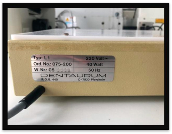
Figure 2.
An Illuminator Box (Dentarum 40-Watt, 50 Hz) was used for the Bang and Ramm and Lamendin methods. The measurements of the root translucency (Bang and Ramm) and Root height and periodontosis (Lamendin) were taken in this box.
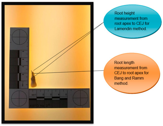
Figure 3.
American Board of Forensic Odontology (ABFO) no.2 scale is placed on the illuminator box showing an anterior tooth (central incisor) measurement. Measurements such as the root length, root height, transparent root dentin, and periodontosis (gingival regression) were taken by using this scale.
2.6. Lamendin Method
This included the assessment of the root height using the American Board of Forensic Odontology (ABFO) no.2 scale and illuminator box (Dentarum 40-Watt, 50 Hz) as shown in Figure 3. The root height is defined according to Lamendin et al. (1992) as ”the distance between the apex of the root and the cementoenamel junction (CEJ)”, measured on the labial surface of the tooth. Periodontosis (gingival regression), which is described as ”the maximum distance between the cementoenamel junction and the line of soft tissue attachment”, was measured on the labial surface of the tooth. The estimated age was obtained using the equation A = 0.18 P + 0.42 T + 25.53 as proposed by Lamendin et al., where A indicates the estimated age, P periodontosis (periodontosis height 100/root height) and T transparency (transparency height 100/root height) [16]. For each tooth, approximately 10 min were required to perform this method.
2.7. Cameriere Method
Digital X-rays (peri-apical) were taken using a hand-held dental X-Ray device NOMAD (Aribex, Orem, UT, USA) combined with a digital sensor (DSX, Anthos, Italy) linked to a portable pc. These radiographs were taken with a Rinn-type digital sensor holder with 0.05 s exposure time and 60 kV. The X-ray images were photo-edited using Image J (NIH, Bethesda, MD, USA). A total of 10 points from each pulp outline were identified and 20 points from each tooth outline were identified and used to assess these areas [26]. It took around 5 to 6 min to perform this method on each tooth.
2.8. Kvaal Method
This radiographic method, which allows age estimation based on radiographic features of the individual tooth by taking their morphological measurements, was reported in 1995. The teeth included were the maxillary central incisor, the maxillary lateral incisor, the maxillary second premolar, the mandibular lateral incisor, the mandibular canine, and the first premolar. Digital X-rays (peri-apical) were taken using a hand-held dental X-ray device NOMAD (Aribex, Orem, UT, USA). The X-ray images were photo-edited using Image J (NIH, Bethesda, MD, USA). The measurements included length and width ratios such as the pulp-root length (P), pulp-tooth length (R), tooth-root length (T), pulp-root width at the ECJ (A), pulp-root width at the mid-root level (C), pulp-root width at the midpoint between level C and A(B), mean value of all the ratios excluding T(M), the mean value of width ratios B and C(W), and the mean value of length ratios P and R(L). These ratios are used in order to compensate for magnification and angulations errors in teeth and the radiograph [25]. It took 10 to 15 min to perform this method on each tooth.
2.9. Statistical Method
The age and sex of participants were anonymously used for data analysis. The difference between actual and estimated age was tabulated using a Microsoft Office 2007 Excel spreadsheet. The mean absolute error (MAE) was calculated, which is the average of the absolute values of the error.
3. Results
The mean absolute error for the Kvaal method (MAE: 12.33) showed the highest variation and Bang and Ramm showed more accurate results in comparison to other methods (MAE: 4.80) regardless of sex.
Sample
The sample teeth which were used for the Bang and Ramm and Lamendin methods can be seen in Table 2. These teeth are divided according to sex and their location in the dental arch (upper or lower jaw).

Table 2.
Sample distribution used for Bang and Ramm and Lamendin methods.
In Table 3, the calculations for Bang and Ramm samples are explained where each tooth is tabulated by sex, together with their date of extraction, chronological age, and estimated age. A total of 8 male and 11 female teeth ranging from 20 to 90 years of age were assessed for this method.

Table 3.
Estimated age for Bang and Ramm method.
The calculations for Lamendin samples are explained in Table 4 where each tooth is tabulated by sex, mentioning their date of extraction, chronological age, and estimated age. Participants ranging from 40 to 90 years of age consisting of 7 male and 10 female teeth were assessed for this method.

Table 4.
Estimated age for Lamendin method.
The mean absolute error (MAE) was calculated for the Bang and Ramm and Lamendin methods regardless of sex. The mean absolute error for the Bang and Ramm method was 4.80 and 10.72 for the Lamendin method as mentioned in Table 5. The standard deviation was 9.44 for Lamendin, whereas it came out to be 2.2 in the original paper [20].

Table 5.
Variables for Bang and Ramm and Lamendin method regardless of sex.
Histograms were plotted for each method. In Figure 4, it can be seen that for the Bang and Ramm method the average absolute error was higher in females compared to males. In females, the maximum absolute error was below 20 years whereas in males it was found below 14 years.
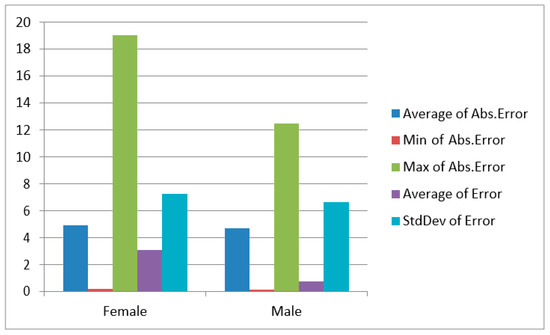
Figure 4.
Histogram for Bang and Ramm method.
The average absolute error was higher in males compared to females as represented in Figure 5 for the Lamendin method. Males and females were analyzed separately. In females, the maximum error value was below 20 years and it was below 40 years in males.
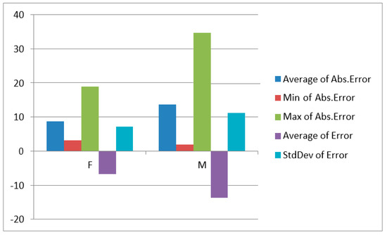
Figure 5.
Histogram for Lamendin method.
In Table 6, sample teeth are shown which were used for the Kvaal and Cameriere method. These teeth are divided according to gender and their location in the dental arch (upper or lower jaw).

Table 6.
Sample distribution used for Kvaal and Cameriere method.
The calculation for the Kvaal method is shown in Table 7 where each tooth is tabulated by gender, together with their date of extraction, chronological age, and estimated age. A total of six female and eight male teeth ranging from 17 to 70 years of age were assessed.

Table 7.
Estimated age for Kvaal method.
In the case of the Cameriere method as seen in Table 8, four canines, one female tooth, and three male teeth ranging from 40 to 90 years of age were tabulated according to their gender together with their date of extraction, chronological age, and estimated age.

Table 8.
Estimated Age for Cameriere method.
The Mean absolute error for the Kvaal method was 12.33 and 7.145 for the Cameriere method as shown below in Table 9. The standard deviation in years was 15.65 years, which in the original study was 8.6 years for Kvaal’s method [25]. The standard deviation for the Cameriere method was 10.10 whereas it was found to be 4.33 years in the original paper [26].

Table 9.
Variables for Kvaal and Cameriere method regardless of sex.
The average absolute error was higher in females in comparison to males for the Kvaal method as shown in Figure 6. It can be seen that the maximum absolute error was below 35 years for males and below 30 years for females.
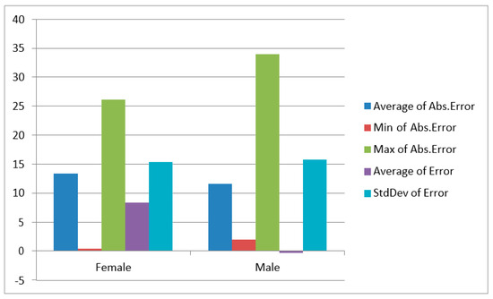
Figure 6.
Histogram for Kvaal method.
The average absolute error was higher in males in comparison to females for the Cameriere method as represented in Figure 7. In the case of females, the maximum absolute error was below 10 years and below 20 years for males.
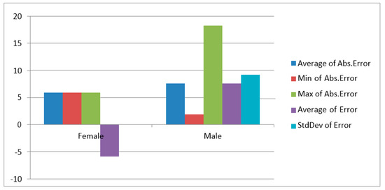
Figure 7.
Histogram for Cameriere method.
The non-homogeneity and limited sample size for the Cameriere method can be seen in Figure 5 as the mean error for females is presented on the negative side and for males it is on the positive side.
4. Discussion
Dental age estimation has been widely used worldwide since human dental hard tissues do not undergo any remodeling processes just like bone. For this reason, teeth are less susceptible to impaired growth or pathological conditions. They can withstand extremes of temperature and often act as useful evidence for dental age estimation analysis [11]. A number of methods have been employed for dental age estimation in both the living and the dead, but most of them have not been tested on the Pakistani population. It would seem clear that the field of forensic odontology needs to be looked into in order to highlight its importance. This preliminary study aimed to evaluate and compare different age estimation methods on the living Pakistani population in order to check their validity and accuracy. The study focused on the practical use of these invasive and non-invasive methods and which method can be used more efficiently.
Age estimation is a significant key for the identification process in different cases such as asylum seekers and civil litigation. It is also significant in post-mortem profiling leading to confirmative identification. The methods involved in this study consisted of invasive, non-destructive (Bang and Ramm and Lamendin), and non-invasive (Kvaal and Cameriere). The accuracy of each method was assessed by using the mean absolute error (chronological age-estimated age). MAE is the average magnitude of error in a set of predictions and has been used as a measure of accuracy of age estimation methods [34]. The error rates <± 10 years are considered as acceptable in forensic age estimation [21]. Schmeling et al. suggested that age estimation methods that produce mean errors of six to eight years are moderately good [35].
In the case of the invasive non-destructive methods such as Bang and Ramm and Lamendin, mean absolute errors were calculated. According to the results of this study, the MAE was found to be 4.80 for Bang and Ramm, which was less in comparison with a study carried out on the Norwegian population in which the MAE was 6.47 [34]. In a study conducted on the Indian population using a modified Bang and Ramm method, the MAE came out to be 5.6, which was greater in comparison with our study [36]. Few studies suggested that population specification formulae such as the Indian population for the Bang and Ramm method produced better results as compared to the original methodology [20].
The Bang and Ramm methodology was carried out on intact teeth in this study; however, original methodology also involves assessing age on the longitudinal sections of the teeth. Further studies can be performed by sectioning teeth and assessing mean estimated errors in the Pakistani population.
In our study, the results of the MAE for the Lamendin method came out to be 10.72, which was more than in a study where the MAE found to be 8.9 [16]. Root translucency increases with age but periodontosis has no correlation with chronological age in cases where periodontal disease is present [16]. Periodontal disease is due to several factors such as exposure to oral bacteria, tissue structure, and physiological response to these bacteria [37]. This pathology constitutes a major bias with age assessment with Lamendin’s method but periodontal disease is not taken into account in this method.
In the sample for the Lamendin method, age was best estimated in females as compared to males. A study on a larger sample was required to establish if this difference has statistical significance. The literature has shown diverse results in correlation of this technique with gender. Some authors found the age estimation for males to be better in comparison to females and vice versa [19,38,39]. Some studies reported no sex-specific differences [40].
Non-invasive methods such as the Kvaal method and the Cameriere method can be simply used for both the living and the dead [41,42]. The radiographic methods are advantageous when compared to other methods because of their collectability in data and simplicity in age estimation [43]. The phenomenon of aging leads to a decrease in pulp cavity due to apposition of the secondary dentine, which is used to estimate age in dental radiographs such as described by the Kvaal method [42].
In the case of the Kvaal method, mostly available teeth from the sample were included as was mentioned in the original study carried out by Kvaal. This study was conducted on full mouth radiographs where a certain amount of image distortion was reported such as an overshadowed apical third of the tooth, but in our study peri-apical radiographs were used [25]. The mean estimated error for the Kvaal method was 12.33, which was in corroboration with a study carried out on the Indian population where it came out to be 12.3. There is a possibility that if the formula for Kvaal is modified for the Pakistani population, the results can come out more accurate as mentioned in a study conducted by Patil. et al., which was done on the Indian population [44].
The difference between chronological and dental age was calculated for all the available teeth with these four methods; however, for the Cameriere method only canines were assessed as it is conducted with original Cameriere methodology. According to studies that use the Cameriere methodology, one of the advantages of using canines for age estimation is that the survival rate of these teeth is higher as compared to other anterior teeth [45]. They are single-rooted teeth with a large pulp area, making them easy to analyze in comparison with multi-rooted teeth [26]. The measurements of pulp and tooth on the digitized peri-apical images of canines have produced more reliable, reproducible data and clear-cut lateral modifications can be made.
The extracted teeth examination also revealed that peri-apical images of single canines make the data analysis precise [46].
The non-homogeneity of the sample for the Kvaal and Cameriere method is due to the fact that Kvaal allows more teeth to be assessed according to its methodology and the latter was originally done on canines which in our sample were only four since this tooth has the highest survival rate and so could not be collected.
These conventional dental age assessment methods take less time and are not technique-sensitive in comparison to the new ones such as Aspartic acid racemization [47]. The time required to assess the age of each tooth according to Bang and Ramm was less in comparison to all other methods; it took only 5 min, which was the same as Cameriere, while Lamendin took 5 to 8 min and Kvaal took 10 to 15 min to assess the age of a single tooth. Methods such as Bang and Ramm and Lamendin do not require elaborate equipment to assess the age; for example, the Lamendin method involves measurements on the labial surface of the entire tooth without sectioning and do not require special equipment or training. This factor is of immense importance for countries like Pakistan where equipment is not available. It is also practical to perform these procedures in mass casualties and disasters where the time span is limited. The methods involving specific equipment such as the hand-held dental X-ray device NOMAD, image editing software such as ”Image J” reduce the practical use of these methods. It is therefore important that forensic dentistry should be given importance by the authorities and that they train individuals in this field in order to enhance the scope of forensic dentistry in Pakistan.
Few studies conducted recently to check the accuracy of the forensic age estimation in the dead also involved the use of root dentin translucency in Peruvian adults and showed accurate results [48,49]. The use of the Kvaal method in living Malay adults also showed that the modified Bang and Ramm method has revealed more accuracy [50]. These studies were conducted on a large sample size in comparison with our study, which had a limited sample.
Our study consisted of a cross-sectional sample over a large age span; any possible influences of chronological or sexual differences cannot be ruled out. Further studies are suggested over a large homogenous sample employing different methods for age estimation in the Pakistani population in order to assess the validity and accuracy of these methods.
5. Conclusions
To our knowledge, this is the first study to test the four dental age estimation methods in adult living Pakistanis regardless of sex. Given the arbitrary and limited sample collection, the results obtained can be considered preliminary until further representative samples are collected according to the present data. The lower mean absolute error rates between chronological and estimated ages were obtained through the Bang and Ramm method. This method also figured as the most time-efficient and practical to use. Despite being invasive, the Bang and Ramm method does not require the destruction of tooth specimens enabling the addition of additional age estimation methods. This can enable the use of this method in mass disasters. In addition to this, radiological (non-invasive) methods such as Kvaal and Cameriere can be used for dental age estimation in immigrant cases. This study can be made the basis and further explored for future studies related to dental age estimation over a large sample.
Author Contributions
Conceptualization: A.K. and S.M. Methodology: A.K. and A.F. Software: A.K. and A.F. Validation: S.M. Formal Analysis: A.K. Investigation: A.K. Resources: A.K. Data Curation: A.K. Original Draft Preparation: A.K. Writing—Review and Editing: A.K., S.M. and A.F. Visualization: A.K. Supervision: S.M. and A.F. All authors have read and agreed to the published version of the manuscript.
Funding
This research received no external funding.
Institutional Review Board Statement
The study was conducted according to the guidelines of the Declaration of Helsinki and approved by the Institutional Review Board of RIPHAH International University, Pakistan. Reference number: IIDC/IRC/2022/002/008, Date of Approval: 28 February 2022.
Informed Consent Statement
Informed consent was obtained from all subjects involved in the study.
Data Availability Statement
The data are not publicly available because of data protection rules established by the Ethics committee that approved the study. In case of reasonable request to the authors, data might be provided in form of anonymized .xls format files.
Acknowledgments
Special acknowledgement to Bakhtawar Yaqoob for her contribution to managing the sample collection of teeth in Pakistan.
Conflicts of Interest
The authors declare no conflict of interest.
References
- Chidambaram, R. Forensic Odontology: A Boon to Community in Medico-legal Affairs. JNMA J. Nepal. Med. Assoc. 2016, 54, 46–54. [Google Scholar] [CrossRef] [PubMed]
- Aggrawal, A. Estimation of age in the living: In matters civil and criminal. J. Anat. 2009, 11, 1–17. [Google Scholar] [CrossRef] [PubMed]
- Schmeling, A.; Olze, A.; Reisinger, W.; Rösing, F.W.; Geserick, G. Forensic age diagnostics of living individuals in criminal pro-ceedings. Homo Int. Z. Vgl. Forsch. Am. Menschen 2003, 54, 162–169. [Google Scholar]
- Nuzzolese, E.; Di Vella, G. Forensic dental investigations and age assessment of asylum seekers. Int. Dent. J. 2008, 58, 122–126. [Google Scholar] [CrossRef] [PubMed]
- Marsh, R.; Romero, S.; Patrick, S. estimating age: College males versus convicted male child sex offenders. J. Child Sex. Abus. 2013, 22, 968–986. [Google Scholar] [CrossRef] [PubMed]
- Alkass, K.; Buchholz, B.A.; Ohtani, S.; Yamamoto, T.; Druid, H.; Spalding, K.L. Age estimation in forensic sciences: Application of combined aspartic acid racemization and radiocarbon analysis. Mol. Cell Proteom. 2010, 9, 1022–1030. [Google Scholar] [CrossRef]
- Kurniawan, A.; Chusida, A.; Atika, N.; Gianosa, T.K.; Solikhin, M.D.; Margaretha, M.S.; Utomo, H.; Marini, M.I.; Rizky, B.N.; Prakoeswa, B.F.W.R.; et al. The Applicable Dental Age Estimation Methods for Children and Adolescents in Indonesia. Int. J. Dent. 2022, 2022, 6761476. [Google Scholar] [CrossRef] [PubMed]
- Kurniawan, A.; Yodokawa, K.; Kosaka, M.; Ito, K.; Sasaki, K.; Aoki, T.; Suzuki, T. Determining the effective number and surfaces of teeth for forensic dental identification through the 3D point cloud data analysis. Egypt. J. Forensic Sci. 2020, 10, 3. [Google Scholar] [CrossRef]
- Mohammed, R.B.; Krishnamraju, P.; Prasanth, P.; Sanghvi, P.; Reddy, M.A.L.; Jyotsna, S. Dental age estimation using Willems method: A digital orthopantomographic study. Contemp. Clin. Dent. 2014, 5, 371–376. [Google Scholar] [CrossRef]
- Phulari, R.G.S.; Dave, E.J. Evolution of dental age estimation methods in adults over the years from occlusal wear to more so-phisticated recent techniques. Egypt. J. Forensic Sci. 2021, 11, 36. [Google Scholar] [CrossRef]
- Verma, M.; Verma, N.; Sharma, R.; Sharma, A. Dental age estimation methods in adult dentitions: An overview. J. Forensic Dent. Sci. 2019, 11, 57–63. [Google Scholar] [CrossRef] [PubMed]
- Stavrianos, C.; Mastagas, D.; Stavrianou, I.; Karaiskou, O. Dental age estimation of adults: A review of methods and principals. Res. J. Med. Sci. 2008, 2, 258–268. [Google Scholar]
- Sharma, N.; Dhillon, S. Identification through dental age estimation in skeletal remains of a child. J. Forensic Dent. Sci. 2019, 11, 48–50. [Google Scholar] [CrossRef] [PubMed]
- Kvaal, S.; Solheim, T. A non-destructive dental method for age estimation. J. Forensic Odonto-Stomatol. 1994, 12, 6–11. [Google Scholar]
- Solheim, T. A new method for dental age estimation in adults. Forensic Sci. Int. 1993, 59, 137–147. [Google Scholar] [CrossRef]
- Lamendin, H.; Baccino, E.; Humbert, J.F.; Tavernier, J.C.; Nossintchouk, R.M.; Zerilli, A. A simple technique for age estimation in adult corpses: The two criteria dental method. J. Forensic Sci. 1992, 37, 1373–1379. [Google Scholar] [CrossRef]
- Uhrová, P.N.; Beňuš, R.; Kondeková, M.C.N.; Vojtušová, A.; Novotný, M.; Thurzo, A. Use of third molar eruption based on Gambier’s criteria in assessing dental age. Int. J. Leg. Med. 2023, 137, 691–699. [Google Scholar] [CrossRef]
- Olze, A.; Geserick, G.; Schmeling, A. Age estimation of unidentified corpses by measurement of root translucency. J. Forensic Odonto-Stomatol. 2004, 22, 28–33. [Google Scholar]
- Ubelaker, D.H.; Parra, R.C. Application of three dental methods of adult age estimation from intact single rooted teeth to a peruvian sample. J. Forensic Sci. 2008, 53, 608–611. [Google Scholar] [CrossRef]
- Bang, G.; Ramm, E. Determination of age in humans from root dentin transparency. Acta Odontol. Scand. 1970, 28, 3–35. [Google Scholar] [CrossRef]
- Solheim, T.; Sundnes, P.K. Dental age estimation of Norwegian adults—A comparison of different methods. Forensic Sci. Int. 1980, 16, 7–17. [Google Scholar] [CrossRef] [PubMed]
- Vandevoort, F.M.; Bergmans, L.; Van Cleynenbreugel, J.; Bielen, D.J.; Lambrechts, P.; Wevers, M.; Peirs, A.; Willems, G. Age calculation using X-ray microfocus computed tomographical scanning of teeth: A pilot study. J. Forensic Sci. 2004, 49, 787–790. [Google Scholar] [CrossRef] [PubMed]
- Avon, S.L. Forensic odontology: The roles and responsibilities of the dentist. J. Can. Dent. Assoc. 2004, 70, 453–458. [Google Scholar] [PubMed]
- Babshet, M.; Acharya, A.B.; Naikmasur, V.G. Age estimation in Indians from pulp/tooth area ratio of mandibular canines. Forensic Sci. Int. 2010, 197, 125.e1–125.e4. [Google Scholar] [CrossRef]
- Kvaal, S.I.; Kolltveit, K.M.; Thomsen, I.O.; Solheim, T. Age estimation of adults from dental radiographs. Forensic Sci. Int. 1995, 74, 175–185. [Google Scholar] [CrossRef]
- Cameriere, R.; Ferrante, L.; Belcastro, M.G.; Bonfiglioli, B.; Rastelli, E.; Cingolani, M. Age estimation by pulp/tooth ratio in canines by peri-apical X-rays. J. Forensic Sci. 2007, 52, 166–170. [Google Scholar] [CrossRef]
- Helfman, P.M.; Bada, J.L. Aspartic acid racemisation in dentine as a measure of ageing. Nature 1976, 262, 279–281. [Google Scholar] [CrossRef]
- Spalding, K.L.; Buchholz, B.A.; Bergman, L.-E.; Druid, H.; Frisén, J. Age written in teeth by nuclear tests. Nature 2005, 437, 333–334. [Google Scholar] [CrossRef]
- Rinaldo, N.; Saguto, I.; De Luca, F.; Neri, M.; Frisoni, P.; Gualdi-Russo, E. Estimation of Age in Humans Using Dental Translucency of Permanent Teeth: An Experimental Study. Appl. Sci. 2023, 13, 6289. [Google Scholar] [CrossRef]
- Alharbi, H.S., Sr.; Alharbi, A.M.; Alenazi, A.O.; Kolarkodi, S.H.; Elmoazen, R. Age Estimation by Kvaal’s Method Using Digital Panoramic Radiographs in the Saudi Population. Cureus 2022, 14, e23768. [Google Scholar] [CrossRef]
- Ishwarkumar, S.; Pillay, P.; Chetty, M.; Satyapal, K.S. The Application of the Cameriere’s Methodologies for Dental Age Estimation in a Select KwaZulu-Natal Population of South Africa. Dent. J. 2022, 10, 130. [Google Scholar] [CrossRef] [PubMed]
- Badar, S.B.; Ghafoor, R.; Khan, F.R.; Hameed, M.H. Age estimation of a sample of Pakistani population using Coronal Pulp Cavity Index in molars and premolars on Orthopantomogram. JPMA J. Pak. Med. Assoc. 2016, 66, S39–S41. [Google Scholar]
- Von Elm, E.; Altman, D.G.; Egger, M.; Pocock, S.J.; Gøtzsche, P.C.; Vandenbroucke, J.P. The Strengthening the Reporting of Ob-servational Studies in Epidemiology (STROBE) statement: Guidelines for reporting observational studies. Ann. Intern. Med. 2007, 147, 573–577. [Google Scholar] [CrossRef]
- Cameriere, R.; Cunha, E.; Sassaroli, E.; Nuzzolese, E.; Ferrante, L. Age estimation by pulp/tooth area ratio in canines: Study of a Portuguese sample to test Cameriere’s method. Forensic Sci. Int. 2009, 193, 128.e1–128.e6. [Google Scholar] [CrossRef] [PubMed]
- Schmeling, A.; Geserick, G.; Reisinger, W.; Olze, A. Age estimation. Forensic Sci. Int. 2007, 165, 178–181. [Google Scholar] [CrossRef]
- Acharya, A.B.; Vimi, S. Effectiveness of Bang and Ramm’s formulae in age assessment of Indians from dentin translucency length. Int. J. Leg. Med. 2009, 123, 483–488. [Google Scholar] [CrossRef]
- González-Colmenares, G.; Botella-López, M.C.; Moreno-Rueda, G.; Fernández-Cardenete, J.R. Age estimation by a dental method: A comparison of Lamendin’s and Prince & Ubelaker’s technique. J. Forensic Sci. 2007, 52, 1156–1160. [Google Scholar]
- Foti, B.; Adalian, P.; Signoli, M.; Ardagna, Y.; Dutour, O.; Leonetti, G. Limits of the Lamendin method in age determination. Forensic Sci. Int. 2001, 122, 101–106. [Google Scholar] [CrossRef]
- Martrille, L.; Ubelaker, D.H.; Cattaneo, C.; Seguret, F.; Tremblay, M.; Baccino, E. Comparison of four skeletal methods for the esti-mation of age at death on white and black adults. J. Forensic Sci. 2007, 52, 302–307. [Google Scholar] [CrossRef]
- Prince, D.A.; Ubelaker, D.H. Application of Lamendin’s adult dental aging technique to a diverse skeletal sample. J. Forensic Sci. 2002, 47, 107–116. [Google Scholar] [CrossRef]
- Cameriere, R.; Ferrante, L.; Liversidge, H.; Prieto, J.; Brkic, H. Accuracy of age estimation in children using radiograph of devel-oping teeth. Forensic Sci. Int. 2008, 176, 173–177. [Google Scholar] [PubMed]
- Roh, B.-y.; Lee, W.-J.; Ryu, J.-W.; Ahn, J.-M.; Yoon, C.-L.; Lee, S.-S. The application of the Kvaal method to estimate the age of live Korean subjects using digital panoramic radiographs. Int. J. Leg. Med. 2018, 132, 1161–1166. [Google Scholar]
- Bosmans, N.; Ann, P.; Aly, M.; Willems, G. The application of Kvaal’s dental age calculation technique on panoramic dental ra-diographs. Forensic Sci. Int. 2005, 153, 208–212. [Google Scholar] [CrossRef]
- Patil, S.K.; Mohankumar, K.P.; Donoghue, M. Estimation of age by Kvaal’s technique in sample Indian population to establish the need for local Indian-based formulae. J. Forensic Dent. Sci. 2014, 6, 166–170. [Google Scholar] [PubMed]
- Brothwell, D.R. The relationship of tooth wear to aging. In Age Markers in the Human Skeleton; Charles C Thomas Publisher: Springfield, IL, USA, 1989; pp. 303–318. [Google Scholar]
- Cameriere, R.; Brogi, G.; Ferrante, L.; Mirtella, D.; Vultaggio, C.; Cingolani, M.; Fornaciari, G. Reliability in age determination by pulp/tooth ratio in upper canines in skeletal remains. J. Forensic Sci. 2006, 51, 861–864. [Google Scholar] [CrossRef] [PubMed]
- Ohtani, S. Age estimation by aspartic acid racemization in dentin of deciduous teeth. Forensic Sci. Int. 1994, 68, 77–82. [Google Scholar] [CrossRef]
- Alvarado Muñoz, E.; Requena Calla, S.R. Accuracy of age estimation using root dentin translucency in Peruvian adults: A pilot study. J. Forensic Odonto-Stomatol. 2023, 41, 19–26. [Google Scholar]
- Suciyanie, I.M.; Gultom, F.P.; Hidayat, A.N.; Suhartono, A.W.; Yuniastuti, M.; Auerkari, E.I. Accuracy of forensic age estimation using cementum annulation and dentin translucency in adult: A systematic review and meta-analysis. Int. J. Leg. Med. 2022, 136, 1443–1455. [Google Scholar]
- Ramli, I.S.; Muhd, U.S.; Yusof, M.Y.P.M. Accuracy of Kvaal’s radiographic and translucent dentinal root techniques of extracted teeth in Malay adults for dental age estimation. J. Forensic Odonto-Stomatol. 2021, 39, 38–44. [Google Scholar]
Disclaimer/Publisher’s Note: The statements, opinions and data contained in all publications are solely those of the individual author(s) and contributor(s) and not of MDPI and/or the editor(s). MDPI and/or the editor(s) disclaim responsibility for any injury to people or property resulting from any ideas, methods, instructions or products referred to in the content. |
© 2023 by the authors. Licensee MDPI, Basel, Switzerland. This article is an open access article distributed under the terms and conditions of the Creative Commons Attribution (CC BY) license (https://creativecommons.org/licenses/by/4.0/).