Review

Genome-Based Medicine for Acute Myeloid Leukemia: Study and Targeting of Molecular Alterations and Use of Minimal Residual Disease as a Biomarker

Department of Oncology, Istituto Superiore di Sanità, 00161 Rome, Italy
*
Author to whom correspondence should be addressed.
Hemato 2022, 3(3), 543-568; https://doi.org/10.3390/hemato3030038
Submission received: 27 June 2022 / Revised: 31 August 2022 / Accepted: 1 September 2022 / Published: 6 September 2022
(This article belongs to the Special Issue Current Topics in Acute Myeloid Leukemia)

Abstract

:
Acute myeloid leukemia (AML) is a highly heterogeneous hematologic malignancy characterized by the clonal proliferation of hematopoietic stem and progenitor cells (HSPCs) and blockade of differentiation and proliferation of immature myeloid cells that accumulate in bone marrow at the expense of normal hematopoiesis. AMLs originate from the expansion of HSPCs progressively acquiring somatic mutations. The development of high-throughput sequencing techniques has helped to discover the genetic heterogeneity and complexity of AMLs, revise diagnostic and prognostic criteria, and to identify new therapeutic targets. These studies have allowed the identification of several recurrent driver mutations and the definition of a rational molecular classification of these tumors. In parallel, the development of techniques for the determination of single-cell mutational profiling has considerably contributed to understanding the clonal heterogeneity and evolution of AMLs. The acquisition of these genetic data coupled with the identification of molecular therapeutic targets has determined a considerable expansion of the therapeutic armamentarium, with the development of several new drugs highly active against specific AML subtypes. These developments have increased the interest and the need for sensitive techniques for the identification of minimal residual disease, the population of leukemia cells that survives despite morphological remission and causes disease relapse.

1. Introduction

Acute myeloid leukemia (AML) is a heterogeneous malignant disease initiated through the clonal expansion of hematopoietic stem/progenitor cells due to the progressive acquisition of somatic genetic alterations. AML development is induced by the acquisition of somatic gene mutations and/or chromosomal abnormalities that induce impaired myeloid differentiation and abnormal proliferation at the expense of of the normal hematopoietic system. A careful assessment of cytogenetic abnormalities and gene mutation analysis is required for the diagnosis, classification, prognosis, and treatment of AMLs.
The incidence of AML is age-dependent, rising markedly at the age of ≥60 years, with a median age at diagnosis of about 68–70 years [1,2]. The incidence of AML in Europe increased from 3.48 in 1976 to 5.06 cases per 100,000 people in 2013, a phenomenon at least in part related to the aging of the population [3].
The identification and classification of cellular and molecular abnormalities occurring in AML are of fundamental importance for understanding their pathogenesis and for the development of a more rational approach to their treatment. Thus, the initial classification of AML, the French–American–British (FAB) classification, was based on the evaluation of the hematopoietic cell lineage of leukemic cells and of their differentiation stages based on cytological and cytochemical techniques. The development of techniques in the study of cytogenetic abnormalities introduced new fundamental criteria in the classification of AMLs, reflected in the World Health Classifications of AML proposed in 2001 and 2008 [4,5].
The aim of the present paper is to review the recent developments in the definition of the mutational landscape of AMLs and of the genetic drivers present in these AMLs. These studies have contributed to a better understanding of the pathogenesis of AMLs, defining their molecular heterogeneity and identifying targetable pathways. The development of sensitive molecular techniques also permits defining the amount and the molecular features of leukemic cells eventually surviving the various antileukemic treatments (defined as minimal residual disease, MRD) and thus monitoring the efficacy of these treatments and providing an important marker for prognosis and rapid evaluation of the efficacy of new drugs or for guiding optimal treatment. Therefore, the molecular characterization of AMLs may provide three essential parameters: definition of molecular alterations and molecular classification of AML; definition of its clonal heterogeneity; evaluation of the presence of residual leukemic cells after treatment.

2. Molecular Abnormalities of AMLs

2.1. De Novo, Secondary and Therapy-Related AMLs

AMLs can be classified into three different groups depending on their origin: de novo, secondary (sAML), and therapy-related AML (tAML). sAML and tAML are recognized as AML clinical subtypes. Following the WHO classification of myeloid neoplasms, sAMLs are defined as AMLs occurring after an antecedent myeloid neoplasia, such as a myelodysplastic syndrome (MDS) or a myeloproliferative neoplasm (MPN), independently of the therapy used for the treatment of these disorders. tAMLs are defined as AMLs occurring as a late complication related to the mutagenic potential of cytotoxic chemotherapy and/or radiotherapy for a neoplastic or non-neoplastic disease [6].
A Danish population-based study carried out on 3055 AML patients diagnosed over 13 years from 2000 to 2013 showed that 73.6% of cases correspond to de novo AMLs. Of these, 19.8% to sAMLs and 8.3% to tAMLs [7]. tAMLs were mostly related to solid tumors or lymphoproliferative disorders [7]. An antecedent myeloid disorder (sAML) or prior cytotoxic exposure (tAML) was associated with a reduced rate of complete remission and decreased overall survival compared to de novo AMLs [7].
Molecular profiling studies have shown remarkable differences in the frequency of several molecular abnormalities between sAMLs, tAMLs, and de novo AMLs, as well as between sAMLs and tAML. The presence of mutations in SRSF2, SF3B1, U2AF1, ZRSR2, ASXL1, EZH2, BCOR, or STAG2 was specific for sAMLs; tAMLs frequently displayed TP53 mutations (23% of cases), and in 33% of cases harbored secondary-type mutations in SRSF2, SF3B1, U2AF1, ZRS2, ASXL1, AZH2, BCOER, or STAG2 (Figure 1) [8].
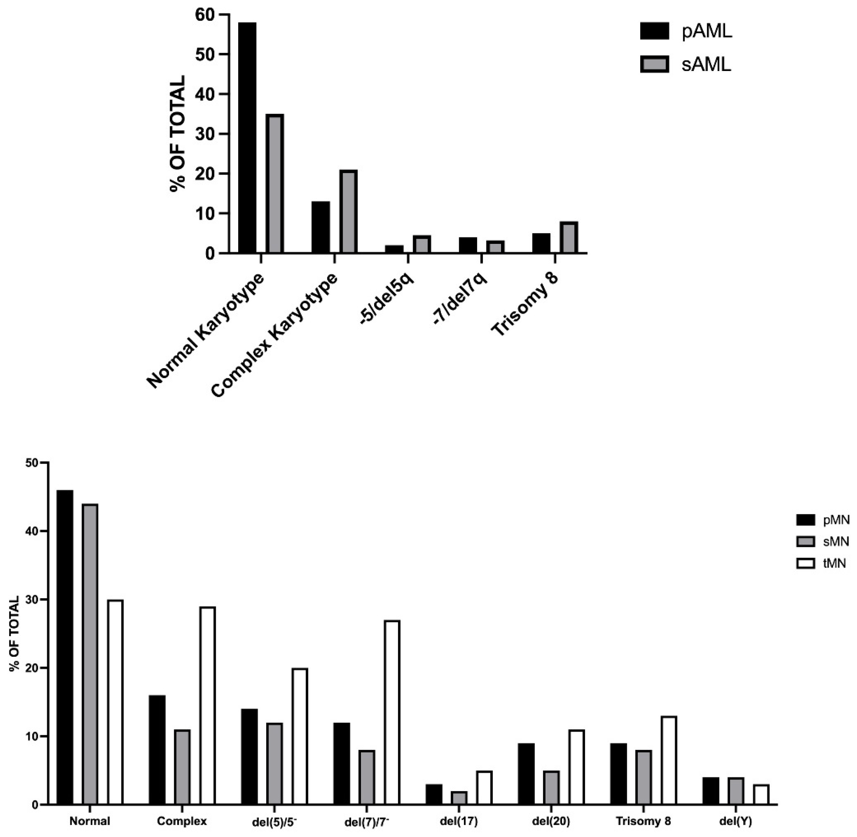
Nazha et al., through the analysis of a large set of primary and secondary AMLs, confirmed that mutations of the genes DHX29, ASXL1, SF3B1, BCOR, PRPF8, CBL, BCORL1, EZH2, STAGF2, JAK2, U2AF1, TET2 are more specific for sAML, whereas CEBPA, IDH2, DNMT3A, NPM1, and FLT3 mutations are more specific for primary de novo AMLs (Figure 1) [9]. The cytogenetic profile showed that sAMLs were more frequently classified as pertaining to an unfavorable risk category than pAMLs (Figure 2) [9]. Patients with tAML are older and display more frequently than patients with pAML cytogenetic abnormalities, including monosomal (−7, −5 or 5q, 7q) and complex karyotypes, events associated with a poor outcome [10]. More recent studies on a large set of tAML patients confirmed the decrease in the frequency of normal karyotype (30% vs. 46%) and the increase of complex karyotype (29% vs. 16% in sAML) compared to pAMLs (Figure 2) [11]. Targeted mutational analysis in strictly defined sAML showed that the genes most frequently mutated are those involved in RNA splicing (55%), DNA methylation (44%), chromatin modification (42%), RAS signaling (42%), transcriptional regulation (34%), and cohesion complex (22%).
Metzler et al. explored the association of driver gene mutations with clinical characteristics and cytogenetic alterations. The major findings of this analysis showed that DNMT3A and NPM1 mutations were more common in women than in men; RUNX1, SRSF2, ASXL1, STAG2, and BCOR were less common in women than in men; FLT3-ITD mutations were associated with high blast cell counts; mutations in SRSF2, ASXL1, STAG2, U2AF1, RUNX1, and PTPN11 were more frequent in secondary AMLs (sAMLs, AMLs developing from a pre-existing myelodysplastic syndrome or a myeloproliferative disorder) than in de novo-occurring AMLs; TP53 mutations were more frequent in therapy-related AMLs (tAMLs); mutations at the level of DNMT3A, FLT3, NPM1, IDH1, IDH2, and CEBPA are present predominantly at the level of patients with normal karyotype [12].
tAMLs represent the most aggressive and chemo-resistant malignancies with a 5-year survival of <10% [13]. The 2016 and 2022 WHO classifications of myeloid neoplasms classified the myeloid neoplasms occurring after therapy, including tMDS (therapy-related myelodysplastic syndrome), tMDS/MPN, and tAML, as unique clinical entities called tMN (therapy-related myeloid neoplasm) [14,15,16]. As a consequence, several studies have considered tMDS and tAML together. As for tAMLs, tMDSs are observed in patients treated for solid tumors (54%) or hematological disease (43%), and tMDSs are observed in patients treated with chemotherapy alone or combined chemo-radiotherapy [17]. Two patterns of tAML development have been reported, one characterized by tAML onset 5–7 years after alkylating chemotherapy or radiation therapy and a second one occurring 2–3 years after exposure to topoisomerase II inhibitors, such as etoposide and anthracyclines. tMDSs, compared to pMDSs, display a higher proportion of cases pertaining to high/very high-risk scoring, a higher proportion of cases with multiple cytogenetic aberrations, and shorter overall survival [17]. tAMLs are characterized by a higher frequency of TP53, DNMT3A, FLT3, NPM1, and NRAS mutations and fewer secondary-type mutations, such as SRSF2, ASXL1, BCOR, and RUNX1, as compared to sAML.
De novo AMLs are characterized by recurrent mutations of some genes, including NPM1, FLT3 (including both FLT3-ITD and FLT3-TKD mutations), DNMT3A, NRAS, CEBPA, IDH1, IDH2, TET2, KIT, RUNX1, and PTPN11. In de novo AMLs, mutations of the genes involved in spliceosome machinery (SF3B1, SRSF2, U2AF1, ZRSR2) are rare. A recent study provided a detailed genetic characterization of 863 de novo AML patients within an age range between 17 and 59 years [18]. The most frequently observed mutations in the ELN favorable-risk group of patients consisted of biallelic CEBPA mutations and NPM1 mutations with no FLT3-ITD or a low FLT3-ITD allelic ratio. Regarding NRAS and KIT mutations, the most recurrent mutations in the intermediate-risk group were NPM1 mutations in association with FLT3-ITD with high alleleic ration (FLT3-ITDhigh) and IDH1 and IDH2 mutations, in large part not associated with NPM1 mutations. In the adverse-risk group, the most frequent mutations were TP53, FLT3-ITDhigh, and NPM1 wild-type, RUNX1, and ASXL1 mutations [18]. The gene mutations distributed according to pathways showed some remarkable differences between the three ELN risk groups. RAS pathway mutations were enriched in the favorable-risk group, mutations in kinase and methylation-related genes were preferentially observed in the intermediate-risk group, and mutations in genes encoding for spliceosomes, transcription factors, and tumor suppressors were more frequent in the adverse-risk group [18]. Importantly, in multivariate analysis, the presence of some gene mutations conferred a change in prognosis compared to that provided by the risk ELN evaluation; thus, the presence of BCOR, WT1, or SETB1 mutations in AML otherwise classified as favorable-risk AMLs was associated with an overall survival comparable to that of an intermediate-risk and not of a favorable-risk AML. In the intermediate-risk group, the presence of WT1 mutations was associated with a prognostic profile more compatible with an adverse-risk AML. The presence of FTL3_ITDhigh was associated with short DFS and OS independently of co-occurring NPM1 mutations; the co-occurrence of WT1 and NPM1 mutations was associated with a particularly poor outcome. The presence of DNMT3A mutations in intermediate-risk AMLs was associated with poorer DFS and OS [18]. These observations strongly support the consistent utility of extensive characterization of the genetic abnormalities of de novo AML patients for an accurate evaluation of risk and for the definition of the optimal therapeutic strategy.
The mutational pattern of AMLs in elderly patients was compared to that observed in younger patients. There are remarkable differences in the frequency of some gene mutations in older AML patients compared to younger AML patients. TET2, SRSF2, ASXL1, RUNX1, TP53, and BCOR mutations are more frequent in older (>60 years) than in younger (<60 years) AML patients, FLT3-ITD and WT1 mutations are less frequent in older than younger AML patients, and DNMT3A and NPM1 mutations have similar frequencies in older and younger AML patients [19,20]. The complete remission rate in these patients was low (about 40% of patients), and their overall survival was low (about 20% at 3 years) [21]. NPM1 and FLT3-ITD mutations do not seem to have a significant impact on overall survival [22].

2.2. Molecular Classification of AML

AMLs are a heterogeneous group of hematological malignancies characterized by a complexity of molecular alterations and clonal development. Considerable progress has been made in the characterization of the molecular abnormalities underlying AMLs, with the identification of recurrent chromosomal alterations and gene mutations, allowing the classification of these leukemias into various subgroups characterized by different genetic alterations and responses to current treatments [23,24,25,26]. This molecular classification identified some major molecular subtypes: (i) AMLs characterized by peculiar translocation events (balanced rearrangements) leading to the formation of fusion genes and correspondent fusion proteins, including inv(6), t(15;17), t(8;21), inv(3), MLL fusions, and t(6;9); (ii) AMLs with chromatin-spliceosome gene abnormalities, including mutations of genes involved in RNA splicing (SRSF2, SF3B1, U2AF1, ZRSR2), chromatin, and transcription; (iii) AMLs characterized by TP53 mutations, complex karyotype alterations, and copy-number chromosome alterations; (iv) AMLs displaying mutations of the nucleophosmin 1 (NPM1) gene; (v) AMLs characterized by double CEBPA mutation; (vi) AMLs with IDH2R172 mutation, defined as a distinct subgroup for the mutual exclusivity with NPM1 mutation and other class-defining lesions [26]. AMLs with mutated RUNX1 have been included in the WHO classification as a provisional entity in the category of AMLs with recurrent genetic abnormality [26].
Recently, the 5th WHO classification of hematolymphoid tumors proposed a classification of AMLs based on all available molecular and histopathological criteria [14,15]. This classification includes a group of AMLs with defining molecular abnormalities and a group of AMLs without defining genetic abnormalities but defined by differentiation [15]. The large group of AMLs with defining genetic abnormalities includes three subgroups: a subgroup including well-defined genetic alterations (PML-RARA, RUNX1-RUNXT1, CBFB-MYH11, DEK-NUP214, RBM15-MRTFA, BCR-ABL1 fusions, KMT2A, MECOM, NUP98 rearrangements, and NPM1 and CEBPA mutations); a subgroup with other defined genetic alterations, representing a landing spot for new, rare, emerging entities, such as rare genetic fusions; (iii) a subgroup with myelodysplasia-related AMLs, including AMLs with rare genetic fusions; a subgroup of dysplasia-related AMLs, characterized by the presence of cytogenetic abnormalities typically observed in myelodysplasia and some gene mutations (ASXL1, BCOR, EZH2, SF3B1, SESF2, STAG2, U2AF1, ZRSR2). The group of AMLs with AMLs without defining mutations but defined by differentiation includes 8 subtypes (AML with minimal differentiation, AML without maturation, AML with maturation, acute basophilic leukemia, acute myelomonocytic leukemia, acute monocytic leukemia, acute erythroid leukemia, and acute megakaryoblastic leukemia). Some of these leukemias are associated with frequent genetic non-defining alterations, such as frequent biallelic TP53 mutations in acute erythroid leukemia [15].
Compared to other tumors, AMLs are characterized by a relatively low number of mutations in coding genes [27].
According to various molecular criteria, the European Leukemia Net stratified AMLs into 3 risk subgroups, those with favorable prognosis (comprising t(15;17), t(8;21), inv(6), biallelic mutated CEBPA and NPM1 mutant (without FLT3-ITD), intermediate prognosis (encompassing NPM1 mutant with FLT3-ITDlow, t(9;21) and various cytogenetic abnormalities not classified as favorable or adverse), and adverse prognosis (comprising monosomy 7 and 5, deletion of long arm (q) chromosome 7, abnormalities of 3q, 17p and 11q, multiple cytogenetic abnormalities, NPM1 wt and FLT3-ITDhigh, TP53 mutations associated with complex karyotype, ASXL1 mutations, t(6;9) and t(3;3) groups [28].
At the moment, conventional cytogenetic and FISH analysis remains an essential component of the diagnostic machinery required for patients with AML. However, a recent study showed that a single technique, whole-genome sequencing, is able to provide rapid, accurate, and complete genomic profiling in AML patients. In fact, whole-genome sequencing detected all 49 recurrent translocations and 91 copy-number alterations that had been identified by cytogenetic analysis. In addition, this technique identified new, clinically reportable genomic events in 17% of patients [29]. Prospective sequencing of 117 consecutive AML samples showed that whole-genome sequencing could be performed within 5 days, a lapse of time compatible with clinical activity, provide new genetic information in about 25% of patients, and change the risk category in about 16% of cases [29].

2.3. Genetic Alterations in Relapsed AMLs

A total of 40 to 80% of adults and about 35% of children relapse within 3 years. Relapse is associated with a dismal outcome and is the main cause of death for these patients. Higher frequencies of relapse have been observed in AML patients with TP53 alterations, adverse-risk cytogenetics, and FLT3-ITD mutations.
To understand the most frequent mutational changes at relapse in AML, it is of fundamental importance to consider the consistent variability of the variant allele frequencies of the most recurrent driver gene mutations. TP53, IDH2, MLL rearrangements, DNMT3A, CEBPA, TET2, EZH2, NPM1, inv(16), U2AF1, and t(8;21) displayed a VAF just below 0.5, implying that these mutant genes were present at a heterozygous state and were present in most cells in the AML samples. Mutations in genes involved in growth factor signaling (FLT3, KIT, NRAS, KRAS, PTPN11) were present at a lower allelic ratio, suggesting that the acquisition of the mutations of these genes often occurs late in leukemia development [15].
Greif et al. explored the genetic alterations in 50 AML patients with normal karyotype at diagnosis, complete remission, and relapse [30]. At the level of mutational burden, at relapse, a small but significant increase in mutational burden was observed. At relapse, 67 mutations were lost and 104 were acquired compared to AML samples at diagnosis [30]. Two genes (CBL and PTPN11) were recurrently altered only at diagnosis, whereas three genes (KDM6A, DRD1, and NFE2) were recurrently altered only at relapse [30]. Most recurrently mutated genes displayed similar frequencies of mutation at diagnosis and at relapse; however, mutations of WT1, IDH1, KDM6A, and KPNB1 were recurrently gained at relapse, while FLT3 point mutations were lost in a number of patients at relapse. Some patients acquired FLT3-ITD mutations at relapse [30], and 10% of patients acquired chromosomal alterations at relapse, trisomy 8 being the only recurrent chromosomal alteration gained in a few patients at relapse. Taking into account all types of genetic alterations (sequence variants, cytogenetic alterations, and copy number alterations), the relapsed patients were subdivided into four different groups: patients with stable genetic alterations; patients with a gain in genetic alterations at relapse (“stable + gain”, 24%); patients with loss of genetic alterations at relapse (“stable + loss”, 16%); patients with both gain and loss of genetic alterations at relapse (“mixed”, 30%) [30]. Interestingly, patients with a stable + loss evolutionary profile relapsed earlier (all within the first year after complete remission) compared to those with mixed or stable + gain profiles. Finally, a number of relevant findings concerned the mutations in genes linked to epigenetic regulation. Mutations in DNMT3A, IDH1, and IDH2 showed similar variant allele frequencies at diagnosis and relapse in the large majority of cases, no patients acquired DNMT3A, TET2, or ASXL1 mutations during disease progression, none of the mutations affecting the various genes involved in epigenetic regulations were lost at relapse, and in 20% of patients, mutations in epigenetic regulators were acquired at relapse [30]. In contrast to epigenetic regulators, mutations in genes involved in signaling pathways, such as RTK genes or RAS signaling pathway genes, were consistently unstable, with frequent losses or gains from diagnosis to relapse [30].
Rapaport et al. recently explored the genomic and evolutionary portraits of disease relapse in 120 AML patients who received standard-of-care combination chemotherapy, achieved complete remission, and experienced disease relapse [31]. Of these, 43% of patients displayed at least one mutation loss and 47% at least one mutation gain at relapse. The analysis of the gene mutation profile showed that in more than 50% of the patients possessing mutations at the level of TP53, WT1, or FLT3-ITD, the mutant subclone exhibited an expansion from diagnosis to relapse. More than 50% of subclones bearing NRAS, PTPN11, and FLT3 point mutations were contracted at relapse; mutations at the level of DNMT3A, NPM1, and CEBPA were frequently associated with a clonal fraction and were stable from diagnosis to relapse [31]. The analysis of the VAF of the main genetic alterations also allowed defining the pattern of clonal evolution of these relapsing tumors. About 52% of cases displayed significant changes in subclonal composition, 32% of cases exhibited a conversion of at least one subclonal fraction at diagnosis into a clonal event at relapse or displayed a novel clonal event at relapse, and about 16% of patients showed a stable clonal/subclonal pattern [31].
It is of fundamental importance to understand the leukemia relapse process at the clonal level. In terms of clonal evolution, the reappearance of leukemic disease after relapse can involve four different mechanisms: the founding leukemic clone may acquire new mutations, mediating chemoresistance and determining its expansion and, consequently, its emergence as the predominant clone at disease relapse; a subclone or a non-founding clone is resistant to chemotherapy, develops new mutations, undergoes an expansion process and emerges as the predominant clone at relapse; the chemotherapy treatments induce the development of a new leukemic clone, not present at diagnosis, responsible for disease relapse; an ancestral, very minority pre-diagnostic clone, develops a consistent oncogenic potential through new mutations and emerges as the predominant clone at relapse [32].

2.4. Clonal Hematopoiesis of Undetermined Potential (CHIP) and tAML Development

The current pathogenetic interpretation of tAML development implies the origin of the expansion of clonal hematopoiesis clones due to the mutagenic activity of cytotoxic chemotherapy or radiotherapy. Alternatively, new mutations occur in the normal HSC compartment and progressively drive the leukemic process. The first mechanism seems to play a major role in the development of tAMLs. Clonal hematopoiesis of undetermined potential (CHIP) is a biological event associated with age observed in healthy individuals and corresponding to the presence in their blood/bone marrow of clonal mutations at the level of DNMT3A, TET2, and ASXL1 genes; a fraction of the individuals with CHIP develop a hematological neoplasm later [33]. In addition to the three genes mentioned above, mutations of the epigenetic modifiers IDH1 and IDH2 and the splicing factor genes SF3B1, SRSF2, and U2AF1, of TP53 and JAK2 genes are also observed at the level of CHIP. Pre-AML cases of clonal hematopoiesis are characterized by more mutations per sample, higher mutant allele frequencies, and enrichment of mutations in specific genes (such as TP53, IDH1, IDH2, DNMT3A, TET2, and spliceosome genes) [34,35]. Detection of clonal mutations ≥0.01 VAF identifies subjects at increased risk for developing AML [36]. The considerable variation observed in variant allele frequencies among individuals is mainly driven by chance differences in the timing of mutation acquisition combined with differences in the cell-intrinsic fitness of variants; thus, CHIP development reflects a stochastic process of acquisition of mutations by hematopoietic stem cells and possible clonal expansion driven by some mutations with increased fitness conferring a selective advantage to mutant hematopoietic stem cells [37].
CHIP is a risk factor for blood malignancies and particularly for developing AML. However, it is still unclear why some individuals who harbor CHIP driver mutations progress to AML while others do not. A recent study evidenced the existence of purifying selection operating more or less in all individuals and preventing disease-predisposing clones from rising to dominance and from inducing a pre-leukemic process [38]. The balance between evolutionary pressures ultimately drives mutation dynamics and health outcomes in aging blood elements [38].
Two large studies by Gillis et al. [39] and Takahashi et al. [40] provided evidence that patients with CHIP in pre-treatment PB samples have a significantly increased probability of developing tAML after treatment. CHIP can be detected in 70% of patients with cancer who subsequently develop tMN [41]. Not only gene mutations but also chromosome arm-level copy-number alterations are detectable as CHIP and preexist before exposure of patients to chemotherapy or radiotherapy [41].
Some mutations recurrently observed in tAMLs are related to the previous therapy to which these patients were exposed. Thus, Coombs et al. assessed the occurrence of CHIP in 8810 cancer patients with solid tumors. CHIP was identified in 25% of these patients, 4.5% with presumptive leukemic driver mutations (CH-PD) [42]. PPM1D and TP53 mutations were associated with prior exposure to chemotherapy [42]. Another study confirmed that mutations in the DNA damage response regulator PPM1D (protein phosphatase Mn2+/Mg2+-dependent 1D) present in CHIP are observed in about 1/5 of tAML patients and are correlated with cisplatin exposure [43]. A recent study explored a very large set of cancer patients (24,439 individuals) and observed CHIP in 30% of these patients; 68% of these patients had one mutation in CHIP, and 32% had two or more mutations. The most frequently mutated genes were the epigenetic regulators DNMT3A and TET2 and the genes involved in DNA Damage Response (DDR) pathway, including PPM1D, TP53, and CHEK2; 90% of the mutations observed in CHIP were classified as driver myeloid mutations [44]. The presence of specific gene mutations was associated with some pathogenic events. Mutations of the spliceosome genes SRSF2 and SF3B1 were less frequent than other CH mutations and were clearly associated with age. CHIP mutations in the DDR genes TP53, PPM1D, and CHEK2 were strongly associated with prior oncologic therapy, and CHIP mutations in the ASXL1 gene were strongly associated with smoking [44]. The characterization of the clonal dynamics of evolution of CHIP mutations in 525 cancer patients in a median lapse time of 23 months provided evidence that 62% remained stable, 28% increased, and 10% decreased in clonal size; the growth rate was most pronounced for CHIP mutations in DDR genes [44]. The risk of a CHIP transforming into therapy-related myeloid neoplasia is related to the type of CHIP mutations (mostly TP53 and spliceosome genes SRSF2, U2AF1, and SF3B1 mutations), the number of CHIP mutations, and clonal size [44].

3. Genetic Heterogeneity and Clonal Evolution of AML

AML is frequently an oligoclonal disease at its origin due to the development of multiple leukemic clones present in various proportions in the bone marrow of each individual patient. The evaluation of clonal heterogeneity is based on the assumption that all mutations present in leukemic cells occur heterozygously. Mutant allele frequencies around 0.5 correspond to clonal mutational events, while mutations that occur at lower frequencies are assumed to have occurred later during the leukemic process and constitute a subclone. The clonal architecture of an AML at presentation may be driven by a single predominant clone or, alternatively, by multiple leukemic clones participating in the leukemic development and, in some instances, also by genetically distinct or combining clones. These processes greatly contribute to generating leukemia heterogeneity, leukemia progression, and drug resistance.
An initial study by Ding et al. on eight AML patients allowed defining clonality and clonal evolution patterns at relapse [45]. Two main clonal evolution patterns during AML relapse were observed: (i) the founding clone in the primary leukemia acquired new mutations and evolved into a chemoresistant clone at relapse; (ii) a subclone of the founding clone survived chemotherapy and acquired new mutations, increasing its oncogenic potential, and expanded at leukemia relapse [45].
Hirsch et al. performed an analysis of the clonal composition of 72 AMLs through evaluation of mutational profiling (at diagnosis and at relapse), including analysis of VAF analysis and single leukemic colonies generated in vitro. According to the results observed, some genetic alterations, such as DNMT3A, TET2, ASXL1 mutations, CBF and MLL translocation, and del(20q), fulfill the criteria for initiating genetic events [46]. Reconstruction of genetic alterations and deduced clonal architectures allowed distinguishing four groups of AMLs. Of these, 37.5% of AMLs displayed a genetic hierarchy similar to CHIP, with DNMT3A, TET2, or ASXL1 mutations co-occurring with either NPM1 or some transcription factors (RUNX1, CEBPA, GATA2), 14% of AMLs with mutations in NPM1 or in transcription factors, in the absence of mutations in epigenetic regulators were observed, 19.5% of AMLs displayed MLL, CBF, or chromosome 20q rearrangements, (iv) 29% of AMLs did not display these mutations [46].
The development of a suitable methodology of multiplexed PCR adapted from assays used clinically on bulk material allowed exploring genetic alterations at the level of single leukemic cells. A first pivotal study by Paguirigan et al. exploring single leukemic cells in patients harboring NPM1 and FLT3-ITD mutations showed that mutations of these two genes could occur in both homozygous and heterozygous states, distributed among at least nine distinct clonal populations in all samples analyzed [47]. A second pivotal study was performed by van Galen and coworkers using a modified nanowell-based technology to analyze the transcriptional and mutational profile of single cells from 16 AML patients and normal healthy bone marrow cells. The developed technology allowed detecting gene mutations, insertions, and fusions to distinguish subclones [48]. The single-cell transcriptomic analysis allowed identifying single cells according to differentiation stages, and the single-cell genotyping allowed determining the genetic alterations present in individual cells. Through the use of a machine learning approach, it was possible to distinguish leukemic cells from normal hematopoietic cells [48]. The analysis of transcriptomic data showed that the presence of undifferentiated and differentiated leukemic cells is highly variable in various AML samples and the whole AML tumor cellular ecosystem (including the presence of different subpopulations of T cells) is equally highly variable in different AMLs [48]. The combined genomic and transcriptomic analyses showed that AMLs harbor subclones with distinct cell-type compositions, reflecting underlying genetic alterations [48].
Morita et al. analyzed the clonal architecture and mutational histories of 123 AML patients. The single cells analysis allowed a better definition of the patterns of mutation co-occurrence and exclusivity at the clonal level. FLT3, NRAS, KRAS, PTPN11, KIT, and MYC mutations were detected in the same patients and often present in mutually exclusive clones at the cellular level; IDH1 and IDH2 and TET2 and IDH1/2 mutations were mutually exclusive at the clonal level [49]. DNMT3A, WT1, and TET2 often carry two different mutations, co-occurring in the same cells [50]. The reconstruction of evolutionary histories supported a linear evolution model in 55% of cases and a branching evolutionary model in 45% of cases. Samples with branching evolution displayed a significantly higher clonal diversity compared to those with linear evolution. In some samples with branching evolution, an evolutionary history compatible with a model of convergent evolution was observed [49]. Experiments of xenotransplantation of AML samples into immunodeficient mice suggested that clonal expansion in patient-derived xenotransplants reflects the fitness of the various subclones of each AML sample [49].
Miles and coworkers reported the results of single-cell mutational profiling on patients with clonal hematopoiesis, myeloproliferative neoplasms, and, mostly, AMLs [50]. A significant increase in clone number in AML compared to MPN or CH was observed, with the highest number of clones observed in AMLs harboring FLT3 mutations. The majority of AML patients had one or two clones accounting for ≥30% of leukemic cells [50]. There is some specificity in the clonal distribution of several gene mutations. IDH1, NPM1, and JAK2 mutations are nearly always distributed at the level of the dominant clone, while FLT3 or KRAS mutations are more frequently observed at the level of minoritarian subclones and, more rarely, are present in dominant clones. Additionally, 52% of the AMLs harboring epigenetic mutations displayed multiple mutations of epigenetic modifiers, usually located in the same clone, frequently being the dominant clone. RAS and FLT3 mutations usually do not co-occur at the level of the same clones; variable cooperativity between DNMT3A and IDH1 or IDH2 mutations at the clonal levels is observed [50]. Reconstruction of clonal evolutionary trajectories supported that single-mutant clones with an epigenetic mutation are likely clonal initiating events. In contrast, it is more difficult to provide a reconstruction of clonal trajectories when the first mutation occurs in signaling genes such as FLT3 or NRAS [50]. The analysis of clone size provided evidence of the cooperative interaction between some mutations. In AMLs with co-occurring DNMT3A/IDH1 or DNMT3A/IDH2 mutations, clones co-expressing the two mutations are larger than single mutant clones; the same applies to AML co-expressing NPM1 and FLT3 mutations [50].

4. MRD Evaluation in AML

4.1. Methodology

Minimal residual disease (MRD), also known as measurable residual disease, can be defined as a measure of the number of residual tumor cells surviving in the body after the end of a given treatment. The objective of all the current treatments is the complete eradication of all leukemic cells, thus achieving a curative effect; however, many AML patients display a residual number of resistant cells that constitute MRD and are responsible for disease relapse.
Historically, various techniques have been used to detect and quantify MRD. The cytological examination of bone marrow and peripheral blood, a technique currently used in diagnostics and clinical monitoring of AML patients, possesses a detection limit of 1–5 × 10⁻². Given this intrinsic limitation in its sensitivity threshold, cytologic examination, although a fundamental technique at the clinical level, is not suitable for sensitive detection of MRD. The cytological examination remains of fundamental importance for the assessment of complete remission, defined by the presence of less than 5% of bone marrow leukemic cells on cytologic examination. Standard cytogenetic techniques can be used to detect leukemic cells in AMLs bearing cytogenetic abnormalities (about 50%), with a sensitivity similar to that reported for cytological analysis. In complementation with standard cytogenetics, fluorescence in situ hybridization (FISH) provided the opportunity to detect leukemic cells bearing a specific cytogenetic abnormality with a sensitivity of about 1 × 10⁻².
The three techniques most currently used for the evaluation of MRD in AML patients are represented by multiparametric flow cytometry (MFC), real-time quantitative polymerase chain reaction (qPCR), and next-generation sequencing (NGS). MFC techniques evaluate the immunophenotypic properties of leukemic cells based on the detection of cell surface marker combinations selectively expressed on leukemic cells but not on the normal hematopoietic counterpart [51]. Two different techniques of MFC are currently used for the evaluation of MRD in AML, MFC-LAIP (leukemia-associated immunophenotype), which defines individual-specific surface markers at diagnosis and evaluates these markers at later times after treatment [52], and MFC-DfN (different from normal), based on the identification of aberrant surface marker expression at follow-up [53]. The sensitivity of MFC techniques is in the order of 10−3–10−5 [51] (Table 1).
Two techniques based on PCR are commonly used for evaluation of MRD, RT-qPCR (reverse transcriptase-quantitative) and ddPCR (digital droplet). RT-qPCR, compared to traditional nested-PCR, has the advantages of high sensitivity and specificity, better evaluation of the quality of the RNA sample, and the possibility of monitoring real-time MRD levels [54]. DdPCR is a technique that, at variance with RT-qPCR, provides an absolute quantification through the amplification of target genes without a reference standard curve [54]. Suitable targets of MRD detection by RT-qPCR and ddPCR are found in about 50% of AMLs; the sensitivity of RT-qPCR and ddPCR is included in the 10−4–10−6 range [52].
Next-generation sequencing (NGS) is a technique for the wide characterization of genetic alterations that can be used at diagnosis and for MRD in virtually all AML patients. This technique may allow the characterization of clonal and subclonal genetic alterations occurring in individual AMLs. Basically, three different NGS technologies are currently used, Whole Exome Sequencing (WES), Whole Genome Sequencing (WGS), and Targeted-Gene Sequencing (TGS). The detection of small leukemic subclones is fundamental to evaluating both clonal evolution and MRD during disease evolution. However, short read sequencers used for NGS assay are inherently prone to base calling errors at 3–5%, thus limiting variant detection at 3–5%, a value too high in the context of MRD evaluation. In order to bypass this important limitation, error-corrected sequencing techniques have been developed based on the physical incorporation of random oligonucleotides or unique molecular identifiers (UMI) during the library preparation stage before amplification of DNA, thus generating DNA molecules with a unique molecular fingerprint [55]. Using UMIs, the intrinsic errors of the NGS technique have been markedly reduced to detect mutations below 0.1% VAF [56]. Recent advances in Duplex Seq [57], Nano Seq [58], and Safer SeqS [59] have provided an additional reduction in sequencing errors by grouping both strands of a DNA molecule into a duplex family to distinguish DNA damage with real mutation, thus achieving confident variant calling at 0.01% VAF. The development of the Quantitative Blocker Displacement Amplification (QBDA), integrating sequence-selective variant enrichment into UMI quantitation, allowed the detection of mutations below 0.01% VAF [60]. This technique allows sensitive MRD detection in AML patients in complete remission. In one patient, a residual NPM1 mutation at 0.005% during remission was detected [60].
Two recent studies have reported two different approaches to error-corrected MRD evaluation in AML patients using error-corrected NGS. Thol et al. used a sensitive patient-specific mutation tracking approach using UMI-based MRD detection [61], and Patkar et al. used an error-corrected NGS assay developed to detect MRD in AML using single molecule molecular inversion probes (smMIPS) in which each smMIP contains an 8bp UMI and binds to a single molecule of DNA [62]. Using this approach, a limit detection of 0.018% of VAF was achieved [62].
The advantages and the limits of these different techniques used for MRD evaluation in AML have been recently analyzed in detail [63,64,65].
All these issues were carefully considered by a team of experts, The Europe and Leukemia Net (ELN) MRD working party, who, at the end of 2021, evaluated the standardization and harmonization of MRD and updated the 2018 ELN MRD recommendations according to recent developments in the field [66]. The term minimal residual disease was changed to measurable residual disease; a positive or negative MRD evaluation refers to the detection, or not, of measurable disease above a specific threshold that is related to the type of assay and to the single laboratory [66]. This implies the need for a consistent interchange of information between the clinical and the laboratory teams to carefully consider whether a negative MRD test does not necessarily correspond to disease eradication and just represents a disease level below the assay’s threshold [66]. These guidelines, reporting also technical recommendations for specific AML subtypes, represent a fundamental tool to facilitate the development of clinically relevant and standardized approaches to MDR detection.

4.2. MRD in AML Patients after Induction Chemotherapy and in Pre-Transplantation

The only curative approach for treating most AML patients consists of first inducing a clinical remission with induction chemotherapy and consolidation chemotherapy and then performing an HSCT after a conditioning regimen. The evaluation of MRD in AML patients after induction chemotherapy, pre-HSCT, consistently contributes to defining the residual tumor burden in these patients.

Some Studies Were Based on the MRD Assessment by MFC

Current clinical algorithms typically define patients in morphologic remission as patients having <5% bone marrow blasts and patients with active disease as patients with ≥5% blasts in the bone marrow. Araki et al. reported the evaluation in terms of morphologic remission and MRD as assessed by MFC in 359 consecutive adult AML patients who underwent myeloablative allogeneic HSCT. The 3-year relapse estimates were 67% for patients with MDR-positive morphologic remission, 65% for patients with active AML, and 22% for patients with MRD-negative remission [67]. After multivariate adjustments, MRD negative status remains a prognostic indicator of better overall survival and longer PFS (progression-free survival) [67].
A retrospective analysis explored the link between MRD as assessed by 10-color MFC and the clinical response observed in 245 adult AML patients who achieved a clinical response characterized by a condition genetically defined as complete response and subclassified as complete remission with full hematological recovery (CR), complete remission with incomplete platelet recovery (CRp), and complete remission with incomplete blood count recovery (CRi). MRD positivity was observed in 19%, 54.2%, and 60.9% of AML patients with CR, CRp, and CRi, respectively [68]. MRD status and clinical response were two main independent prognostic factors for the outcome, and their evaluation, thus, seemed of key importance in planning post-induction therapy [68].
Freeman et al. evaluated the CR and MRD status as assessed by MFC in a large set of 2450 adult AML patients undergoing standard induction chemotherapy. The patients were explored after one (C1) or two cycles (C2) of induction chemotherapy [69]. After cycle 1, partial remission (PR) and MRD-positive patients displayed similar outcomes. CRi/MRD positivity was associated with reduced OS, and MRD positivity appeared to be less discriminatory in poor-risk patients. For NPM1-WT patients, MRD positivity at C2 was associated with poorer outcomes, and the transplant-related benefit was more evident in MRD-positive patients than in those who were MRD-negative [69].
A recent study explored 549 younger Chinese AML patients with intermediate risk. Of these, 154 received chemotherapy, 116 autologous-SCT (auto-SCT), and 279 allogeneic SCT (allo-SCT), and the MRD status of these patients was evaluated by MFC after 1, 2, or 3 courses of chemotherapy (a 0.1% cutoff was used to distinguish MRD-positive from MRD-negative patients) [70]. Patients who were MRD-negative after 1, 2, and 3 courses of chemotherapy displayed comparable incidences of leukemia relapse, leukemia-free survival, and overall survival. Among patients with MRD-positivity after 1 course and MRD-negativity after 2–3 courses, patients who underwent auto-SCT and allo-SCT showed a lower incidence of relapse and better overall survival compared to chemotherapy. Among patients with MRD-positivity after 1 or 2 courses and MRD-negativity after 3 courses, allo-SCT had improved relapse and leukemia-free survival compared to chemotherapy, but not to auto-SCT. Among patients with MRD-positivity after all three courses of chemotherapy, allo-SCT had a better incidence of relapse, leukemia-free survival, and overall survival compared to chemotherapy or auto-SCT [70].
Other studies have used molecular tools to assess the presence of MRD in peripheral blood or bone marrow cells; most of these studies used RT-qPCR or NGS to determine the mutational profile in blood samples at diagnosis and after disease remission.
Klco et al. made a pivotal study, reporting the analysis of 50 AML patients by whole-genome or exome sequencing both at disease presentation and after clinical remission following induction chemotherapy [71]. They found that 48% of these patients had persistent leukemia-associated mutations, whereas 52% of the patients cleared all mutations [71]. The presence of persistent-associated mutations was associated with a significantly increased risk of relapse and reduced overall survival [71].
A similar study was performed by Morita et al., who analyzed 131 AML patients undergoing intensive chemotherapy treatment by targeted capture deep sequencing. They found that 30% achieved a complete molecular response, 32% achieved a mutation clearance with a VAF <1%, and 38% with a mutation clearance with VAF <2.5% [72]. Patients who achieved a complete molecular response or a mutation clearance with VAF<1% had better OS and RFS than those who achieved a molecular remission if the VAF was <2.5% [72].
Thol et al. developed an error-corrected NGS MRD approach for the evaluation of 116 AML patients undergoing allogeneic SCT in complete morphologic remission; a suitable mutation for NGS detection was observed in 93% of these patients [70]. MRD evaluation was performed before allo-SCT and showed MRD positivity in 45% of cases; cumulative incidence of relapse was higher in MRD-positive than in MRD-negative patients [61]. In multivariate analysis, MRD positivity was an independent predictor of relapse incidence, in addition to FLT3-ITD and NPM1 mutation status at diagnosis, conditioning regimen, TP53, and KRAS mutation status [61].
A study by Hourigan et al. provided evidence that modulation of the intensity of the allo-SCT conditioning regimen in MRD-positive AML patients can prevent relapse and improve survival [73]. Ultra-deep, error-corrected sequencing for 13 commonly mutated genes in AML was performed in pre-transplantation AML patients in complete morphologic remission randomly assigned to a non-myeloablative (RIC) or to a myeloablative conditioning (MAC) regimen. A total of 32% of MAC and 37% of RIC patients were MRD-negative, and these patients displayed a similar survival [73]. In MRD-positive patients, relapse and survival were less favorable in the RIC arm compared to the MAC arm [73].
Tsai et al. addressed the important problem of the most appropriate timing of MRD evaluation after induction chemotherapy [74] by exploring the mutational profile using targeted NGS in 335 de novo AML patients at diagnosis, first complete remission (MRDist), and after the first consolidation chemotherapy (MRD2nd). They found that 46.4% of these patients were MRD-positive after MRDist and 28.9% after MRD2nd [74]. Patients who are MRD-positive at either time point have shorter relapse-free survival and overall survival, and patients who are MRD-positive at MRDist and MRD-negative at MRD2nd have a prognosis similar to patients negative at both MRD evaluations [74].
A pivotal study carried out in 346 NPM1-mutant AML patients enrolled in the AML17 trial allowed defining the role of MRD in predicting the risk of disease relapse in these patients. In this study, the analysis of mutant NPM1 transcripts by RT-PCR was used to track MRD in PB. The persistence of mutant NPM1 transcripts was detected in 15% of patients after the second chemotherapy cycle and was associated with an increased risk of relapse after 3 years of follow-up and a lower rate of survival compared to patients with undetectable abnormal NPM1 transcript (MRD-negativity) [75]. In multivariate analysis, MRD-positivity was the only independent prognostic factor for death in these patients [75]. Importantly, NPM1 mutations were detectable in 69/70 patients at the time of disease relapse, thus supporting NPM1 mutations as a suitable, leukemic-specific marker to monitor disease evolution and response to therapy [75].
Using ddPCR, Bill et al. evaluated in 51 NPM1-mutant AML patients whether the level of NPM1 mutation burden predicts relapse following SCT. MRD-positive patients displayed a higher incidence of relapse and shorter overall survival, and this finding was observed for patients receiving either myeloablative or non-myeloablative conditioning regimens [76]. Dillon et al. evaluated pretransplant NPM1-mutant levels by RT-qPCR in bone marrow and blood samples and, according to these levels, they subdivided the patients into negative, low, and high. Following transplantation, MDR negative, low, and high patients displayed a 2-year overall survival of 83%, 63%, and 13%, respectively [77]. Among patients with low MDR values, those with FLT3-ITD mutations had significantly poorer outcomes [77].
A recent study explored the impact of the MRD status in AML patients undergoing allo-SCT in first versus second remission and showed that the MRD status at SCT was an independent prognostic factor irrespective of the number of remissions at allo-SCT. MRD-positive patients transplanted in first remission and MRD-negative patients in the second remission had similar outcomes. In the ELN2017 AML-intermediate risk group, the assessment of MRD status provided the highest predictive value, with a very negative prognosis for patients with MRD positivity in second remission [78].
Three recent studies performed a comparative analysis of MRD detection by MFC and NGS.
Getta and coworkers compared the MRD assessment in AML patients by MFC-DfN and by targeted 28-gene NGS, the concordance between the two techniques was observed in 71% of evaluable cases, and the discordance between the two techniques was related to the presence of residual mutations in DNMT3A and TET2 and presence of residual leukemia mutations with VAF below the thresholds for mutation calling [79]. Residual disease detected by concurrent MFC and NGS positivity was associated with the highest relapse risk [79].
Patkar et al. performed the analysis of 201 adult AML patients treated with conventional induction chemotherapy [62]. Patients with post-induction NGS MRD positivity displayed inferior outcomes; furthermore, patients with an NGS MDR negative status post-induction therapy displayed a significantly improved survival compared to patients who became NGS MRD-negative at later stages of treatment [62]. The comparative analysis of post-induction MRD evaluation by MFC and NGS assays showed that NGS identified more than 80% of the cases identified by MFC, while MFC identified only 49% of the cases identified by NGS [62]. Most NGS MDR-negative and MFC MDR-positive cases did not relapse and were therefore considered false positives [62]. Interestingly, AML cases displaying double MFC and NGS MDR positivity showed a higher prevalence of RUNX1 mutations [62].
In conclusion, the studies carried out in AML patients after induction chemotherapy, and pre-transplantation have shown a good predictivity capacity of MDR assessment and that molecular MRD detection possesses a higher sensitivity compared to MDR assay by MFC.

4.3. Clonal Hematopoiesis and MRD Evaluation Post-Chemotherapy

The evaluation of MRD in AML patients is consistently complicated by the fact that many treated patients have persistent clonal hematopoiesis after chemotherapy, and its presence may not reflect residual leukemic disease [80]. The presence of clonal hemtopoiesis in AML patients in complete remission may imply the existence of different conditions, from the presence of CH ancestral to the AML condition to the presence of true residual or early recurrent AML [80].
Murphy et al. explored a group of 283 AML patients undergoing chemotherapy treatment and screened 54 myeloid neoplasia-associated mutations, including CH-associated mutations DNMT3A, TET2, ASXL1, or SRSF2 (DTAS), present at diagnosis in 30%, 17%, 8%, and 10%, respectively, of these patients [81]. Times to platelet and neutrophil recovery post-chemotherapy were significantly delayed in AMLs bearing DNMT3AR822 mutations compared to those observed in patients without DTAS. Similarly, platelet and neutrophil recovery was significantly delayed also in AML patients bearing ASXL1, TET2, or SRSF2 mutations. In contrast, AMLs bearing non-DNMT3AR822 mutations did not display a delayed time to platelet recovery [81]. In contrast, patients not displaying DTAS-like mutations, such as NPM1, NRAS, KRAS, or FLT3 mutations, did not show delayed platelet or neutrophil recovery [81]. In 86 of these patients explored at CR, it was observed that the most common persistent mutations were found in TET2 and DNMT3A. The large majority of persistent mutations with VAF >20% were in the genes TET2, DNMT3A, and ASXL1, and these patients displayed significantly longer times of platelet and neutrophil recovery compared to patients with no detectable mutations in remission [81]. These observations support a model in which the persistence of mutations associated with clonal hematopoiesis and preleukemic conditions in stem/progenitor cells compromise their hematopoietic capacity not under steady-state conditions but under stress conditions related to hematopoietic reconstitution [81].
Two recent studies have shown that there is a consistent difference in MRD post-chemotherapy involving CHIP-related and CHIP-unrelated mutations. Cappelli et al. explored 150 NPM1-mutated AML patients undergoing standard induction and consolidation therapy and achieving a condition of complete remission, as assessed by the absence of NPM1 transcripts after treatment [82]. These patients were explored by targeted NGS with the aim of distinguishing between CHIP-like mutations and mutations with oncogenic potential (defined as clonal hematopoiesis of oncogenic potential, CHOP). At complete molecular remission, 27% of these patients displayed CHIP-associated DTA (DNMT3A, TET2, ASXL1) mutations and 15% had persisting non-DTA gene mutations. Patients with either persistent or acquired non-DTA mutations at complete molecular remission displayed a worse prognosis than those who had only persistent/acquired DTA mutations [82]. Tanaka et al. explored the prevalence, dynamics, and clinical implications of post-remission clonal hematopoiesis in 164 AML patients who achieved complete remission after induction chemotherapy [83]. Post-remission CHIP was identified in 48% of these patients. In 91% of these patients, CHIP-associated mutations remained positive after various types of consolidation and maintenance therapies; post-remission CHIP-associated mutations were eradicated in 20/21 patients undergoing allo-SCT [83]. The post-remission clonal hematopoiesis had no significant impact on hematopoiesis compared to patients with no-CHIP, with the exception of significant neutropenia observed in patients with persistent TET2 mutations; persistent post-remission CHIP had no impact on relapse risk and non-relapse mortality [83].
Onate et al. analyzed the impact of co-mutational status in NPM1-mutant AMLs on the clearance of MRD following induction chemotherapy. DNMT3A-mutant patients (either in the presence or in the absence of concomitant FLT3-ITD mutations) showed a higher number of mutated NPM1 transcripts following induction and first consolidation; furthermore, these patients presented a trend of a greater risk of molecular relapse [84]. However, the presence of DNMT3A mutations did not impact overall survival and relapse rate; furthermore, the presence of DNMT3A mutations does not seem to have any significant impact on the prognosis exerted by FLT3-ITD in these AMLs [84].

4.4. MRD in AML Patients in Post-Transplantation

Allogeneic SCT exerts a curative effect on a high number of patients with AML and other myeloid malignancies; however, relapse is frequent following allo-SCT and is the most frequent cause of death in these patients [85]. The longitudinal post-transplantation evaluation of MRD represents an important strategy for monitoring transplanted patients and the early detection of relapse [86].
Several studies have explored the prognostic relevance of MRD evaluation in posttransplant AML patients.
Zhou et al. evaluated MRD in the pre- and post-transplant period in 279 adult AML patients undergoing allogeneic HSCT after myeloablative conditioning. MRD was evaluated at +28 after transplantation by MFC [87]. They found that 49/63 patients cleared MRD positivity with HSCT conditioning. The MRD-negative patients both in pre- and post-transplant displayed good outcomes; patients with an MRD-positive status pre-HSCT had poor outcomes regardless of post-HSCT status. Survival beyond 3 years was only observed in patients exhibiting a decreasing MRD expression level [87].
Other studies have explored post-HSCT MRD using the MFC assay. Shah et al. explored MRD by MFC in 269 AML patients at various time points: pre-HSCT, at day +30, +100, and +180 after HSCT. In pre-HSCT, 30.8% of patients were MRD-positive; at day +30, +100, and +180 post-HSCT MRD-positive patients were 3.7%, 3.1%, and 3%, respectively [88]. MRD positivity at day +30 post-HSCT predicted the highest risk of leukemia relapse within one year, and MRD positivity beyond day +30 predicted impending relapse [88].
Heuser et al. reported the study of MRD evaluation by NGS in 138 AML patients with at least one mutation not related to clonal hematopoiesis. The patients were explored at day 90 and/or 180 post-allogeneic HSCT; in parallel, the patients were also explored for clonal hematopoiesis-related mutations (DTA: DNMT3A, TET2, ASXL1) [89]. In this study, a low VAF (0.01%) was applied to define MRD positivity/negativity by NGS, and using this approach, 25% of patients were classified as MRD-positive after allo-HSCT [89]. NGS-MRD monitoring using non-DTA mutations after HSCT was prognostic for PFS and OS, in that MRD-positive patients had a higher incidence of relapse and a shorter PFS and OS compared to MRD-negative patients post-transplant. MRD status had the strongest prognostic impact for patients MRD-positive before transplantation [89]. In contrast, there was no prognostic impact of DTA mutations on RFS and OS [89]. Almost all (90%) patients who were MRD-positive before allo-HSCT and who remained MRD-positive on day 90 post HSCT relapsed within 5 years [90]. Importantly, post-transplantation MRD monitoring on day 90 and 180 after allo-HSCT in these patients is highly predictive of relapse and OS after HSCT [89]. The predictive value of this post-HSCT MRD evaluation is further enhanced by the inclusion in the analysis of 2–4 molecular markers instead of a limited number (1–2 markers) [90].
A recent study by Martin-Rojas and coworkers reported the analysis of the impact of MRD and chimerism monitoring at different time points after HSCT for AML. MRD was evaluated by both MFC and quantitative RT-PCR and chimerism by STR pre-HSCT and at day +30 and +90 post-HSCT [91]. A total of 115 patients were analyzed, and pre-HSCT MRD was positive in 49.6% and negative in 50.4%. Patients with negative MRD at day +30 showed a 2-year survival of 83% compared to 58% of those who were MRD-positive; EFS was 79% vs. 48%. Patients with mixed chimerism at day +30 showed a significantly lower 3-year OS and EFS than those with complete chimerism [91].
In conclusion, the monitoring of MRD after allo-SCT remains problematic, particularly concerning the optimal timing of MRD assay post-transplant and its clinical interpretation, due to the slow mechanisms of anti-leukemia effects exerted by allo-SCT through graft-versus-leukemia effects.

4.5. MRD in Refractory/Relapsing AML (R/R AML)

About 10–40% of newly diagnosed AML patients do not achieve complete remission following induction chemotherapy (primary refractory AMLs) or relapse within ≤6 months after CR1 (early relapsing AMLs).
Few studies have explored MRD evaluation in R/R AML patients. A retrospective study evaluated the prognostic role of MRD assessment in R/R AMLs [92]. The evaluation of some key prognostic factors is of key importance for risk stratification of patients in first relapse: length of relapse-free survival time after first relapse, including cytogenetic features at diagnosis, age at relapse, and prior allogeneic HSCT. A total of 141 AML patients with R/R disease were evaluated for MRD status by MFC analysis: 67% of these patients achieved CR, 18% achieved CR with incomplete hematologic recovery (CRi), and 14% achieved a morphologic leukemia-free status (MLFS); 61% of patients achieved MRD negativity at the time of best response [92]. Patients who achieved MRD negativity vs. those who remained MRD-positive had a lower rate of relapse and better relapse-free survival, but not overall survival [92]. In spite of the lack of a statistically significant impact of MRD status on OS, there was a strong trend toward better OS in patients who achieved MRD negativity [92]. The lack of a significant difference in OS related to MRD status may be dependent on the increased availability of effective salvage regimens for many relapsing AML patients [92]. AML patients who achieved both CR and MRD negativity displayed the lowest rates of relapse and best overall survival, a phenomenon seemingly due to the lower rates of early relapse and an increased ability to undergo HSCT [92].
Hao et al. retrospectively analyzed 197 AML R/R AML patients who underwent allogeneic HSCT for MRD status by MFC. They found that 86 of these patients achieved a morphological CR before HSCT, and 32 of these patients displayed an MRD-positive status [93]. The 3-year OS was 59.5%, 34.5%, and 14.5% for MRD-negative, and MRD-positive 1+ and 2+, respectively [93]. Another retrospective study of 56 R/R AML patients treated with allo-HSCT evaluated the prognostic impact of MRD evaluation by MFC on day 100 post-transplantation; 71% of these patients were MRD-negative and 29% MRD-positive. A median follow-up of 16 months suggested that MRD status was predictive of OS [94].
The treatment of R/R AML patients remains a very challenging medical problem, and there is an absolute need to develop more efficacious drug regimens. In this context, recent studies support the promising clinical efficacy in R/R AML patients of a BCL2 inhibitor, Venetoclax, in association with intensive chemotherapy. Di Nardo et al. reported that of 68 AML patients, 29 with newly diagnosed AML and 39 with R/R AML, all treated with Venetoclax plus FAG-IDA (fludarabine, cytabarine, G-CSF, and idarubicin), 69% of R/R AML patients achieved composite CR/MRD-negativity and 46% of R/R AML patients proceeded to HSCT. The 1-year survival in all R/R patients was 78%, and in those undergoing HSCT, it was 87% [95]. Another recent study presented at the annual congress of the European Hematology Association confirmed these findings. The addition of venetoclax to the intensive FLAG-IDA chemotherapy regimen elicited a high rate of overall responses (73%) and MRD negativity (43%) in R/R AML patients [96]. Survival outcomes were better for patients who achieved MRD negativity. The median event-free survival of 9.5 months in MRD-positive compared to not reached in MRD-negative patients; median OS of 11.3 months in MRD-positive vs. not reached in MRD-negative patients [96].
In conclusion, additional studies on larger numbers of R/R AML patients are required to assess the predictive value of MRD detection in these AML patients; however, in spite of this limitation, the studies carried out support the predictive role of MRD evaluation in R/R AML patients and suggest that MRD assessment may help to define possible candidates for early post-transplantation interventions aiming to decrease the relapse risk and improve survival.

4.6. MRD Evaluation in Elderly AML Patients Undergoing Reduced-Intensity Treatments

While younger AML patients can be treated with induction chemotherapy, often achieving remission, older (≥65–70 years) AML patients are poor candidates for induction chemotherapy and need to be treated with low-intensity therapies. These low-intensity therapies involve either low-dose chemotherapy or hypomethylating agents, such as azacitidine or decitabine. The mechanisms of action and the dynamics of response of hypomethylating agents considerably differ from conventional chemotherapy approaches; therefore, it is important to evaluate whether the clinical response to these agents can be measured in terms of MRD clearance.
Boddu et al. examined the prognostic value of MRD detection by MFC in a group of older AML patients treated with hypomethylating agents [97]. Patients with an MRD-negative status at CR had an inferior incidence of relapse compared to those with an MRD-positive condition; however, the difference in relapse rate did not translate into significant differences at the level of OS [97].
Simoes et al. reported the results of MRD evaluation in elderly AML patients enrolled in the phase III PETHEMA-FLUGAZA clinical trial. In this study, the patients were randomized to induction and consolidation therapy with fludarabine plus cytarabine vs. 5-azacitidine. After consolidation, patients continued treatment if MRD was ≥0.01% or stopped the treatment if MRD was ≤0.01%, as evaluated by MFC assay [98]. In patients achieving CR, MRD status was the only independent prognostic factor for relapse-free survival; undetectable MRD improved the RFS of AML patients with adverse genetics. Longer OS was observed in patients with negative MRD after induction chemotherapy but not after consolidation [98]. Therefore, the achievement of an MRD-negative status after reduced-intensity chemotherapy or hypomethylating agents is prognostically relevant in older AML patients [98].
Maiti et al. reported the evaluation of the prognostic impact of MRD after venetoclax and decitabine treatment in a group of 97 older AML patients [99]. In this study, MRD was evaluated in bone marrow by MFC. A total of 85% of these patients achieved a CR and 54% MRD negativity; higher levels of MRD negativity were observed among patients with intermediate-risk cytogenetics (67%) compared to those with high-risk cytogenetics (33%) [99]. Patients achieving an MRD-negative CR within 1 month displayed an improved overall survival compared with MRD-positive patients (median OS 25.1 vs. 3,4 months); this trend was confirmed at 2 and 4 months after starting therapy [99].
In the phase III VIALE-A trial, the rates of complete responses and MRD negativity (MRD ≤ 10−3) were higher in patients treated with venetoclax + azacitidine compared to those treated with azacytidine alone [100]. Patients who achieved a complete response and an MRD negative status exhibited a longer duration of response, overall survival, and event-free survival [100].
Vazquez et al. reported the study of 19 AML patients ineligible for standard induction chemotherapy treated with venetoclax combination therapy and found that 84% of these patients achieved complete remission. MFC MRD evaluation was negative in 9/11 patients achieving complete remission and remained negative in 4/6 patients achieving a durable remission condition [101]. Interestingly, in some responding patients, venetoclax treatment induced eradication of the leukemic stem cell fraction [101].
A small clinical study reported the effects of venetoclax-based treatments on MRD status in a small group of mutant NPM1 AML patients either in CR with MRD positivity or with molecular relapse/progression [102]. These patients were treated with venetoclax either in association with low-dose AraC or with azacitidine. Five patients with persistent NPM1mut -MRD at the end of chemotherapy received venetoclax-based treatment, and all five patients achieved CR-MRD negativity in response to 1, 2, or 4 cycles of this treatment [102]. In patients with NPM1mut progression after the end of chemotherapy, a CR-MRD-negative condition was achieved in 6/7 cases; none of these responding patients displayed molecular or hematological relapse after a median of 10.8 months follow-up [102].
An interesting area of ongoing clinical studies explores the possible clinical efficacy of hypomethylated agents in the maintenance therapy of MRD-positive AML patients in CR. The aim of these studies consists in evaluating the capacity of hypomethylating agents to induce the clearance of MRD, reduce the rate of relapse events, and delay the occurrence of relapse.
Ragon et al. reported the results of a study carried out on 23 CBF-AML exhibiting low levels of MRD after consolidation therapy and then treated with hypomethylating agents. All MRD-negative patients (6/6) remained negative at a median follow-up of 11.3 months, 12/17 AML patients MRD-positive before therapy with hypomethylating agents remained in remission at follow-up, and 11/12 displayed a decrease in MRD levels [103].
Platzbecker et al. reported the results of a multicenter phase II clinical study based on MRD-guided treatment with azacitidine to prevent relapse in patients with MDS and AML in CR with MRD positivity, as assessed by molecular studies or by MFC [104]. After azacitidine treatment, 19/53 patients converted from an MRD-positive to an MRD-negative condition; in the whole population, RFS and OS after the start of azacitidine treatment were 46% and 75%, respectively. Patients converted to an MRD-negative status exhibited RFS and OS rates of 88% and 91%, respectively [104].
A randomized clinical trial further supported the clinical benefit deriving from azacitidine administration to older AML patients. Thus, in the randomized QUAZAR AML-001 study, older AML patients who achieved remission after intensive chemotherapy, but were evaluated as ineligible for HSCT, were randomized to be treated either with CC-486 (oral formulation of azacitidine) or placebo as maintenance therapy [105]. Both patients with MRD-positive and MRD-negative status received a benefit from CC-486 administration: (i) patients with MRD-negative condition at the start of the study showed an OS of 30.1 months in the group of CC-486-treated patients compared to 24.3 for the corresponding placebo group; (ii) patients with an MRD-positive status displayed an OS of 14.6 months in the CC-486-treated group, compared to 10.4 months in the corresponding placebo group [106]. Importantly, about one-fourth of MRD responders treated with CC-486 achieved MRD negativity >6 months after study entry [106]. CC-486 was approved by the FDA for use as maintenance therapy in AML patients who cannot complete a curative therapeutic approach.
In conclusion, the studies on MDR evaluation in elderly AML patients undergoing reduced-intensity treatment support their predictivity and their potential clinical utility.

5. Conclusions

The studies carried out in the last two decades have shown that AML is a highly heterogeneous hematologic malignancy characterized by the progressive acquisition of a variable set of gene mutations or chromosomal aberrations that induce the proliferation (clonal expansion) and differentiation block of hematopoietic stem/progenitor cells. Several genetic abnormalities have emerged as markers of disease and therapeutic targets for the development of new treatments. The understanding of this molecular heterogeneity has permitted a more accurate classification of AMLs. A combination of clinical parameters and cytogenetic and gene alteration profiles are required for an appropriate diagnosis classification, risk stratification, and treatment strategy of AMLs.
In spite of the consistent progress in the understanding of the molecular basis of AMLs and the development of therapeutic approaches, the current treatments are unable in most cases to completely eradicate the leukemic disease, and it is essential in these patients to define minimal/measurable residual disease remaining after the various treatments. Thus, MRD monitoring was introduced into clinical practice as a tool for early prediction of subsequent relapse and as a tool for improving outcomes through the planning of the therapeutic strategy, including transplantation. However, several methodological limitations complicate MRD testing in AML patients, such as not having a well-defined cutoff between positivity and negativity and testing timing and intervals, the lack of leukemia-specific markers in some cases, the use of not fully standardized techniques, and possible loss of MRD-targets as a consequence of clonal/molecular evolution.
The most challenging issue in future studies concerning the clinical application of MRD evaluation is related to its use as a surrogate endpoint in clinical trials and its incorporation in tailored medicine. This advancement in the clinical use of MRD assays is made difficult by several limitations. For example, (i) MRD predictivity is not sufficiently accurate at the level of some individual AML patients who relapse in spite of an apparently negative MRD condition; (ii) the technique of optimal MRD detection for the various AML patients remains, in many instances, to be determined; (iii) the optimal timing for MRD evaluation during the clinical course remains, in many instances, to be determined; (iv) the usage of bone marrow or peripheral blood for MRD evaluation remains to be elucidated. Ongoing clinical trials incorporating MRD evaluation at the level of primary or secondary endpoints should clarify some of these issues. Molecular monitoring of MRD seems to be more sensitive than MFC; however, at the level of molecular techniques, q-PCR has limited applicability for MRD detection, while NGS has a much more wide extension to virtually all AML subtypes. However, the experience with NGS is still limited, and additional studies are absolutely required to validate the use of NGS. Particularly, future studies are required to validate the value of rare variants as targets for MRD analyses.
In conclusion, a better definition of molecular alterations occurring in AML, associated with the identification of molecular targets and their therapeutic targeting, represents the main way to improve the outcomes of AML patients. In parallel, careful and real-time monitoring of MRD in these patients may offer the unique opportunity to try to prevent or treat disease relapse better.

Author Contributions

Conceptualization, U.T., G.C. and E.P.; writing—original draft preparation, U.T., G.C. and E.P.; writing—review and editing, U.T., G.C. and E.P. All authors have read and agreed to the published version of the manuscript.

Funding

This research received no external funding.

Conflicts of Interest

The authors declare no conflict of interest.

References

  1. Shallis, R.M.; Wang, R.; Davidoff, A.; Ma, X.; Zeidan, A.M. Epidemiology of acute myeloid leukemia: Recent progress and enduring challenges. Blood Rev. 2019, 36, 70–87. [Google Scholar] [CrossRef] [PubMed]
  2. Juliusson, G.; Abrahamsson, J.; Lazarevic, V.; Antonovic, P.; Derolf, A.; Gorelius, H.; Lehmanns, S.; Myhr-Eriksson, K.; Mollgord, L.; Uggla, B.; et al. Prevalence and characteristics of survivors from acute myeloid leukemia in Sweden. Leukemia 2017, 31, 728–731. [Google Scholar] [CrossRef] [PubMed]
  3. Roman, E.; Smith, A.; Appleton, S.; Crouch, S.; Kelly, R.; Kinsey, S.; Corgo, C.; Patmore, R. Myeloid malignancies in the real-world: Occurrence, progression and survival in the UK’s population-based Haematological Malignancy Research Network 20094-15. Cancer Epidemiol. 2016, 42, 186–198. [Google Scholar] [CrossRef]
  4. Vardiman, J.W.; Harris, N.L.; Brunning, R.D. The World Health Organization (WHO) classification of the myeloid neoplasms. Blood 2002, 100, 2292–2302. [Google Scholar] [CrossRef] [PubMed]
  5. Vardiman, J.W.; Thiele, J.; Arber, D.A.; Brunning, R.D.; Borowitrz, M.T.; Porwit, A.; Harris, N.L.; LeBeau, M.M.; Hallstrom-Lindberg, E.; Tefferi, A.; et al. The 2008 revision of the World Health Organization (WHO) classification of myeloid neoplasms and acute leukemia: Rationale and important changes. Blood 2009, 114, 937–951. [Google Scholar]
  6. Swerdlow, S.H.; Campo, E.; Harris, N.L. WHO Classification of Tumours of Haematopoietic and Lymphoid Tissues; WHO: Geneva, Switzerland, 2008. [Google Scholar]
  7. Ostgard, L.S.; Medeiros, B.C.; Sengelov, H.; Nargaard, M.; Andersen, M.K.; Dufva, I.K.; Friis, L.S.; Kjeldsen, E.; Werenbeg Marcher, C.; Preiss, B.; et al. Epidemiology and clinical significance of secondary and therapy-related acute myeloid leukemia: A national population-based cohort study. J. Clin. Oncol. 2015, 33, 3641–3649. [Google Scholar] [CrossRef]
  8. Lindsley, R.C.; Mar, B.G.; Mazzola, E.; Grauman, P.V.; Shareef, S.; Allen, S.L.; Pigneux, A.; Wetzler, M.; Stuart, R.K.; Erba, H.P.; et al. Acute myeloid leukemia ontogeny is defined by distinct somatic mutations. Blood 2015, 125, 1367–1376. [Google Scholar] [CrossRef]
  9. Nazha, A.S.; Zarzour, A.; Al-Issa, K.; Radivoyevitch, T.; Carraway, H.E.; Hirsch, C.M.; Przychodzen, B.; Patel, B.J.; Clemente, M.; Sanikommu, S.R.; et al. The complexity of interpreting genomic data in patients with acute myeloid leukemia. Blood Cancer J. 2016, 6, e510. [Google Scholar] [CrossRef]
  10. Kayser, S.; Dohner, K.; Krauter, J.; Kohne, C.H.; Horst, H.A.; Held, G.; von Lillienfeld-Tost, M.; Wilhelm, S.; Kundgen, A.; Gotze, K.; et al. The impact of therapy-related acute myeloid leukemia (AML) on outcome in 2853 adult patients with newly diagnosed AML. Blood 2011, 117, 2137–2145. [Google Scholar] [CrossRef]
  11. Kuzmanovic, T.; Patel, B.J.; Sankommu, S.R.; Nagata, Y.; Awada, H.; Kerr, C.M.; Przychodzen, B.P.; Iha, B.K.; Hiwasa, D.; Singhal, D.; et al. Genomics of therapy-related myeloid neoplasms. Haematologica 2020, 105, e98–e101. [Google Scholar] [CrossRef]
  12. Metzler, K.H.; Herold, T.; Rothenberg-Thurley, M.; Amler, S.; Sauerland, M.C.; Gorlich, D.; Schneider, S.; Kostandin, N.P.; Dufour, A.; Braundl, K.; et al. Spectrum and prognostic relevance of driver gene mutations in acute myeloid leukemia. Blood 2016, 128, 686–698. [Google Scholar]
  13. McNermey, M.E.; Godley, L.A.; LeBeau, M.M. Therapy-related neoplasms: When genetics and environment collide. Nat. Rev. Cancer 2017, 17, 503–527. [Google Scholar]
  14. Arber, D.A.; Orazi, A.; Hasserjian, R.; Thiele, J.; Borowitz, M.J.; Le Beau, M.M.; Blumfield, C.D.; Cazzola, M.; Vardiman, J.W. The 2016 revision of the World Health Organization (WHO) classification of myeloid neoplasms and acute leukemia. Blood 2016, 127, 2391–2405. [Google Scholar] [CrossRef] [PubMed]
  15. Cree, I.A. The WHO classification of hematolymphoid tumors. Leukemia 2022, in press. [Google Scholar] [CrossRef]
  16. Khoury, J.D.; Solary, E.; Abla, O.; Akkari, Y.; Alaggio, R.; Apperley, J.F.; Bejar, R.; Berti, E.; Busque, L.; Chan, J.; et al. The 5th edition of the world health organization classification of hematolymphoid tumors: Myeloid and histiocytic/dendritic neoplasms. Leukemia 2022, in press. [Google Scholar]
  17. Kuendgen, A.; Nomdedeu, M.; Tuechler, H.; Garcia-Manero, G.; Korokji, R.S.; Sekeres, M.A.; Della Porta, M.G.; Cazzola, M.; DeZern, A.E.; Roboz, G.J.; et al. Therapy-related myelodysplastic syndromes deserve specific diagnostic sub-classification and risk-stratification—An approach to classification of patients with t-MDS. Leukemia 2021, 35, 835–849. [Google Scholar]
  18. Eisfeld, A.K.; Kohlschidt, J.; Mims, A.; Nicolet, D.; Walker, C.J.; Blachly, J.S.; Carroll, A.J.; Papaioannou, D.; Kolitz, J.E.; Powell, B.E.; et al. Additional gene mutations may refine the 2017 European Leukemia Net classification in adult patients with de novo acute myeloid leukemia aged <60 years. Leukemia 2020, 34, 3215–3227. [Google Scholar] [CrossRef]
  19. Tsai, C.H.; Hou, H.A.; Tang, J.L.; Liu, C.Y.; Lin, C.C.; Chou, W.C.; Tseng, M.H.; Chiang, Y.C.; Kuo, Y.Y.; Liu, M.C.; et al. Genetic alterations and their clinical implications in older patients with acute myeloid leukemia. Leukemia 2016, 30, 1485–1492. [Google Scholar] [CrossRef]
  20. Silva, P.; Neumann, M.; Schroeder, M.P.; Vosberg, S.; Schlee, C.; Isaakidis, K.; Ortiz-Tranchez, J.; Fransecky, L.R.; Hartung, T.; Turmen, S.; et al. Acute myeloid leukemia in elderly is characterized by a distinct genetic and epigenetic landscape. Leukemia 2017, 31, 1640–1644. [Google Scholar] [CrossRef]
  21. Eisfeld, A.K.; Kohlschmidts, J.; Mrozek, K.; Blanchly, J.S.; Walker, C.J.; Nicolet, D.; Orwick, S.; Maharry, S.E.; Carroll, A.J.; Stone, R.M.; et al. Mutation patterns identify adult patients with de novo acute myeloid leukemia aged 60 years or older who respond favorably to standard chemotherapy: An analysis of Alliance studies. Leukemia 2018, 3260, 1338–1348. [Google Scholar] [CrossRef]
  22. Prassek, V.V.; Rothenberg-Thurley, M.; Sauerland, M.C.; Herold, T.; Janke, H.; Kaslezyh, B.; Kostandin, N.P.; Goerlich, D.; Krug, U.; Faldum, A.; et al. Genetics of acute myeloid leukemia in the elderly: Mutation spectrum and clinical impact in intensively treated patients aged 75 years or older. Haeamtologica 2018, 103, 1853–1861. [Google Scholar] [CrossRef] [PubMed]
  23. Cancer Genome Atlas Research; Ley, T.J.; Miller, C.; Ding, L.; Raphael, B.J.; Mingall, A.J.; Robertson, A.G.; Hoadley, A.S.K.; Triche, T.J.; Laird, P.W.; et al. Genomic and epigenomic landscapes of adult de novo acute myeloid leukemia. N. Engl. J. Med. 2013, 368, 20959–22074. [Google Scholar]
  24. Hou, H.A.; Lin, C.C.; Chou, W.C.; Liu, C.Y.; Chen, C.Y.; Tang, J.L.; Lai, Y.J.; Tseng, M.H.; Huang, C.F.; Chiang, Y.C.; et al. Integration of cytogenetic and molecular alterations in risk stratification of 318 patients with de novo non-M3 acute myeloid leukemia. Leukemia 2014, 28, 50–58. [Google Scholar] [PubMed]
  25. Papaemmanuil, E.; Gerstung, M.; Bullinger, L.; Gaidzik, K.; Paschka, P.; Roberts, N.D.; Potter, N.E.; Hauser, M.; Thol, F.; Bolli, N.; et al. Genomic classification and prognosis in acute myeloid leukemia. N. Engl. J. Med. 2016, 374, 2209–2221. [Google Scholar] [CrossRef]
  26. Bullinger, L.; Dohner, K.; Dohner, H. Genomics of acute myeloid leukemia diagnosis and pathways. J. Clin. Oncol. 2017, 35, 934–946. [Google Scholar] [CrossRef] [PubMed]
  27. Kishtagari, A.; Levine, R.L.; Viny, A.D. Driver mutations in acute myeloid leukemia. Curr. Opin. Hematol. 2020, 27, 49–57. [Google Scholar]
  28. Dohner, H.; Estey, E.; Grimwade, D.; Amadori, S.; Appelbaum, F.R.; Borthakur, G. Diagnosis and management of AML in adults: 2017 ELN recommendations from an international expert panel. Blood 2017, 129, 424–447. [Google Scholar]
  29. Duncavage, E.J.; Schroeder, M.C.; O’Loughlin, M.; Wilson, R.; MacMillan, S.; Bohannon, A.; Kruchowski, S.; Garza, J.; Du, F.; Highes, A.E.O.; et al. Genome sequencing as an alternative to cytogenetic analysis in myeloid cancers. N. Engl. J. Med. 2021, 384, 925–935. [Google Scholar] [CrossRef]
  30. Greif, P.A.; Hartmann, L.; Vosberg, S.; Stief, S.M.; Mattes, R.; Helmann, I.; Metzler, K.H.; Herold, T.; Bamopoulos, S.A.; Kerbs, P.; et al. Evolution of cytogenetically normal acute myeloid leukemia during therapy and relapse: An exome sequencing study of 50 patients. Clin. Cancer Res. 2018, 24, 1716–1726. [Google Scholar]
  31. Rapaport, F.; Neelaraju, Y.; Baslan, T.; Hassane, D.; Gruszczynska, A.; de Massy, M.R.; Farnoud, N.; Haddox, S.; Lee, T.; Medina-Martinez, J.; et al. Genomic and evolutionary portraits of disease relapse in acute myeloid leukemia. Leukemia 2021, 35, 2688–2692. [Google Scholar] [CrossRef]
  32. Vosberg, S.; Greif, P.A. Clonal evolution of acute myeloid leukemia from diagnosis to relapse. Genes Chrom. Cancer 2020, 58, 839–849. [Google Scholar] [CrossRef] [PubMed]
  33. Jaiswal, S.; Fontanillas, P.; Flannick, J. Age-related clonal hematopoiesis associated with diverse outcomes. N. Engl. J. Med. 2014, 371, 2488–2498. [Google Scholar] [CrossRef] [PubMed]
  34. Abelson, S.; Collord, G.; Ng, S.; Weissbrod, O.; Cohen, M.N.; Niemer, E.; Barda, N.; Zuzarte, P.C.; Heisler, R.; Sundravadanam, Y.; et al. Prediction of acute myeloid leukemia risk in healthy individuals. Nature 2018, 559, 400–404. [Google Scholar] [PubMed]
  35. Desai, P.; Mencia-Trinchant, N.; Savenkov, O.; Simon, M.S.; Cheang, G.; Lee, S.; Samuel, M.; Ritchie, E.K.; Guzman, M.L.; Baliman, K.V.; et al. Somatic mutations precede acute myeloid leukemia years before diagnosis. Nat. Med. 2018, 24, 1015–1023. [Google Scholar] [CrossRef] [PubMed]
  36. Young, A.L.; Tong, R.S.; Birmann, B.M.; Druley, T.E. Clonal hematopoiesis and risk of acute myeloid leukemia. Haematologica 2019, 104, 2010–2017. [Google Scholar] [CrossRef] [PubMed]
  37. Watson, C.J.; Papula, A.; Poon, Y.; Wong, W.H.; Young, A.L.; Druley, T.E.; Fisher, D.S.; Blundell, J.R. The evolutionary dynamics and fitness landscape of clonal hematopoiesis. Science 2020, 367, 1449–1454. [Google Scholar] [CrossRef]
  38. Skead, K.; Houle, A.A.; Abelson, S.; Agbessi, M.; Bruat, V.; Lin, B.; Soave, D.; Shlush, L.; Wright, S.; Dick, J.; et al. Interacting evolutionary pressures drive mutation dynamics and health outcomes in aging blood. Nat. Commun. 2021, 2, 4921. [Google Scholar] [CrossRef]
  39. Gillis, N.K.; Ball, M.; Zhang, Q.; Ma, Z.; Zhao, Y.; Yoder, S.J.; Balasis, M.E.; Masa, T.E.; Sallman, D.A.; Lancet, J.E.; et al. Clonal haemopoiesis and therapy-related myeloid malignancies in elderly patients: A proof-of-concept, case-control study. Lancet Oncol. 2017, 18, 112–121. [Google Scholar] [CrossRef]
  40. Takahashi, K.; Wang, F.; Kantarjian, H.; Doss, D.; Khanna, K.; Thompson, E.; Zhao, L.; Patel, K.; Neelapu, S.; Gumbs, C.; et al. Pre-leukemic clonal hematopoiesis and the risk of therapy-related myeloid neoplasms: A case-control study. Lancet Oncol. 2017, 18, 100–111. [Google Scholar] [CrossRef] [Green Version]
  41. Takahashi, K.; Wang, F.; Kantarjian, H.; Song, X.; Patel, K.; Neelapu, S.; Gumbs, C.; Little, L.; Tippen, S.; Thornton, R.; et al. Copy number alterations detected as clonal hematopoiesis of indeterminate potential. Blood Adv. 2017, 1, 1031–1036. [Google Scholar]
  42. Coombs, C.C.; Zehir, A.; Devlin, S.M.; Kishtagari, A.; Syed, A.; Jonsson, P.; Hyman, D.M.; Solit, D.B.; Robson, M.E.; Baselga, J.; et al. Therapy-related clonal hematopoiesis in patients with non-hematologic cancers is common and associated with adverse clinical outcomes. Cell Stem. Cell 2017, 21, 374–382. [Google Scholar] [CrossRef] [PubMed]
  43. Hsu, J.I.; Dayaram, T.; Tovy, A.; De Braekeleer, E.; Jeong, M.; Wang, F.; Zhang, J.; Heffernan, T.; Gera, S.; Kovacs, J.J.; et al. PPM1D mutations drive clonal hematopoiesis in response to cytotoxic chemotherapy. Cell Stem. Cell 2018, 23, 700–713. [Google Scholar] [CrossRef] [PubMed]
  44. Bolton, K.L.; Ptaskin, R.N.; Gao, T.; Braunstein, L.; Devlin, S.M.; Kelly, D.; Patel, M.; Barthon, A.; Syed, A.; Yabe, M.; et al. Cancer therapy shapes the fitness landscape of clonal hematopoiesis. Nat. Genet. 2020, 52, 1219–1226. [Google Scholar] [CrossRef] [PubMed]
  45. Ding, L.; Ley, T.J.; Larson, D.E.; Miller, C.A.; Koboldt, D.C.; Welch, J.S.; Ritchey, J.K.; Young, M.A.; Lamprecht, T.; McLellan, M.D.; et al. Clonal evolution in relapsed acute myeloid leukemia revealed by whole-genome sequencing. Nature 2012, 481, 506–511. [Google Scholar] [CrossRef]
  46. Hirsch, P.; Zhang, Y.; Tang, R.; Joulin, V.; Boutroux, H.; Pronier, E.; Moatti, H.; Flandrin, P.; Marzac, C.; Bories, D.; et al. Genetic hierarchy and temporal variegation in the clonal history of acute myeloid leukemia. Nat. Commun. 2016, 7, 12475. [Google Scholar] [CrossRef]
  47. Paguirigan, A.L.; Smith, J.; Meshinchi, S.; Carroll, M.; Maley, C.; Radich, J.P. Single-cell genotyping demonstrates complex clonal diversity in acute myeloid leukemia. Sci. Transl. Med. 2015, 7, 281re2. [Google Scholar] [CrossRef] [PubMed]
  48. Van Galen, P.; Hovestadt, V.; Wadsworth, M.H.; Hugehs, T.K.; Griffin, G.K.; Battaglia, S.; Verga, G.A.; Stephansky, J.; Pastika, T.J.; Story, J.L.; et al. Single-cell RNA-seq reevals AML hierarchies relevant to disease progression and immunity. Cell 2019, 176, 1255–1281. [Google Scholar] [CrossRef] [PubMed]
  49. Morita, K.; Wang, F.; Jahn, K.; Hu, T.; Tanaka, T.; Sasaki, Y.; Kuipers, J.; Loghavi, S.; Wang, S.A.; Yan, Y.; et al. Clonal evolution of acute myeloid leukemia revealed by high-throughput single-cell genomics. Nat. Commun. 2020, 11, 5327. [Google Scholar] [CrossRef]
  50. Miles, L.A.; Bowman, R.L.; Merlinsky, T.R.; Csete, I.S.; Ooi, A.T.; Durruthy-Durruthy, R.; Bowman, M.; Famulare, C.; Patel, M.A.; Mendez, P.; et al. Single cell mutation analysis of clonal evolution in myeloid malignancies. Nature 2020, 587, 477–482. [Google Scholar] [CrossRef]
  51. Dix, C.; Lo, T.H.; Clark, G.; Abadir, E. Measurable residual disease in acute myeloid leukemia using flow cytometry: A review of where we are and where we are going. J. Clin. Med. 2020, 9, 1714. [Google Scholar] [CrossRef]
  52. Zeijlemaker, W.; Kelder, A.; Cloos, J.; Schuurhuis, G.J. Immunophenotypic detection of measurable residual (stem cell) disease using LAIP approach in acute myeloid leukemia. Curr. Prot. Cytom. 2019, 91, e66. [Google Scholar] [CrossRef] [PubMed]
  53. Baer, M.; Stewart, C.; Dodge, R.; Laget, G.; Sule, N.; Mrozek, K.; Schiffer, C.; Powell, B.; Kolitz, J.; Moore, J.; et al. High frequency of immunophenotype changes in acute myeloid leukemia at relapse: Implications for residual disease detection (Cancer and Leukemia Group B Study B361). Blood 2001, 97, 3574–3580. [Google Scholar] [CrossRef] [PubMed]
  54. Voso, M.T.; Ottone, T.; Lavorgna, S.; Venditti, A.; Maurillo, L.; Lo-Coco, F.; Buccisano, F. MRD in AML: The role of new techniques. Front. Oncol. 2019, 9, 655. [Google Scholar] [CrossRef]
  55. Hiatt, J.B.; Pritchard, C.D.; Salipante, S.J.; O’Roak, B.J.; Shendure, J. Single molecule molecular inversion probes for targeted, high-accuracy detection of low-frequency variation. Genome Res. 2013, 23, 843–854. [Google Scholar] [CrossRef] [PubMed]
  56. Newmann, A.M.; Lovejoy, A.F.; Klass, D.M.; Kurtz, D.M.; Chabon, J.J.; Scherer, F.; Stehr, H.; Liu, C.L.; Bratman, S.V.; Say, C.; et al. Integrated digital error suppression for improved decision of circulating tumor DNA. Nat. Biotechnol. 2016, 34, 547–555. [Google Scholar] [CrossRef]
  57. Schmitt, M.W.; Kennedy, S.R.; Salk, J.J.; Fox, E.J.; Hiatt, J.B.; Loeb, L.A. Detection of ultra-rare mutations by next-generation sequencing. Proc. Natl. Acad. Sci. USA 2012, 109, 14508–14513. [Google Scholar] [CrossRef] [PubMed]
  58. Abascal, F.; Harvey, L.; Martincorena, I. Somatic mutation landscapes at single-molecule resolution. Nature 2021, 593, 405–410. [Google Scholar] [CrossRef]
  59. Cohen, J.D.; Douville, C.; Dudley, J.C.; Mog, B.J.; Popoli, M.; Ptak, M.; Dobbyn, L.; Silliman, N.; Schaefer, J.; Tie, J.; et al. Detection of low-frequency DNA variants by targeted sequencing of the Watson and Crick strands. Nat. Biotechnol. 2021, 39, 1220–1227. [Google Scholar] [CrossRef]
  60. Dai, P.; Wu, L.R.; Chen, S.X.; Wang, M.X.; Cheng, L.Y.; Zhang, J.X.; Hao, P.; Yao, W.; Zarka, J.; Issa, G.C.; et al. Calibration-free NGS quantitation of mutations below 0.01% VAF. Nat. Commun. 2021, 12, 6123. [Google Scholar] [CrossRef]
  61. Thol, F.; Gabdoulline, R.; Liebich, A.; Klement, P.; Schiller, J.; Kandziora, C. Measurable residual disease monitoring by NGS before allogeneic hematopoietic cell transplantation in AML. Blood 2018, 132, 1703–1713. [Google Scholar] [CrossRef] [Green Version]
  62. Patkar, N.; Kakirde, C.; Shaikh, A.F.; Salve, R.; Bhanshe, P.; Chatterjee, G.; Rajpal, S.; Joshi, S.; Chaudhary, S.; Kodgule, R.; et al. Clinical impact of panel-based error-corrected next generation sequencing versus flow cytometry to detect measurable residual disease (MRD) in acute myeloid leukemia (AML). Leukemia 2021, 35, 1392–1404. [Google Scholar] [CrossRef] [PubMed]
  63. Walter, R.B.; Ofran, Y.; Wiezbowska, A.; Ravandi, F.; Hourigan, C.S.; Ngai, L.L.; Venditti, A.; Buccisano, F.; Ossenkoppele, G.J.; Roboz, G.J. Measurable residual disease as a biomarker in acute myeloid leukemia: Theoretical and practical considerations. Leukemia 2021, 35, 1529–1538. [Google Scholar] [CrossRef] [PubMed]
  64. Aitken, M.; Ravandi, F.; Patel, K.P.; Short, N.J. Prognostic and therapeutic implications of measurable residual disease in acute myeloid leukemia. J. Hematol. Oncol. 2021, 14, 137. [Google Scholar] [CrossRef]
  65. Ngai, L.L.; Kelder, A.; Janssen, J.; Ossenkoppele, G.J. MRD tailored therapy in AML: What we have learned so far. Front. Oncol. 2021, 10, 603636. [Google Scholar] [CrossRef]
  66. Heuser, M.; Freeman, S.D.; Ossenkoppele, G.J.; Bussisano, F.; Hourigan, C.S.; Ngai, L.L.; Tettero, J.M.; Bachas, C.; Baer, C.; Béné, M.C.; et al. 2021 update on MDR in acute myeloid leukemia: A consensus document from the European LeukemiaNet MRD Working Party. Blood 2021, 138, 2753–2773. [Google Scholar] [CrossRef] [PubMed]
  67. Araki, D.; Wood, B.L.; Othus, M.; Radich, J.P.; Halpern, A.B.; Zhou, Y.; Mielcarek, M.; Estey, E.H.; Appelbaum, F.R.; Walter, R.B. Allogeneic hematopoietic cell transplantation for acute myeloid leukemia: Time to move toward a minimal residual disease-based definition of complete remission? J. Clin. Oncol. 2016, 34, 329–336. [Google Scholar] [CrossRef] [PubMed]
  68. Chen, X.; Xie, H.; Wood, B.L.; Walter, R.B.; Pagel, J.M.; Becker, P.S.; Sandhu, V.K.; Abkowitz, J.L.; Appelbaum, F.R. Relation of clinical response and minimal residual disease and their prognostic impact on outcome in acute myeloid leukemia. J. Clin. Oncol. 2015, 33, 1258–1264. [Google Scholar] [CrossRef]
  69. Freeman, S.D.; Hills, R.K.; Virgo, P.; Khan, N.; Couzens, S.; Dillon, R.; Gilkes, A.; Gilkes, A.; Upton, L.; Nielsen, O.J.; et al. Measurable residual disease at induction redefines partial response in acute myeloid leukemia and stratifies outcomes in patients at standard risk without NPM1 mutations. J. Clin. Oncol. 2018, 38, 1486–1497. [Google Scholar] [CrossRef]
  70. Yu, S.; Fan, Z.; Ma, L.; Wang, Y.; Huang, F.; Zhang, Q.; Huang, J.; Wang, S.; Wang, S.; Xu, N.; et al. Association between measurable residual disease in patients with intermediate-risk acute myeloid leukemia and first remission, treatment, and outcomes. JAMA Netw. Open 2021, 4, e2115991. [Google Scholar] [CrossRef]
  71. Klco, J.M.; Miller, C.A.; Griffith, M.; Petti, A.; Spencer, D.H.; Ketkar-Kulkarni, S.; Wartman, L.D.; Christopher, M.; Lamprecht, T.L.; Helton, N.M.; et al. Association between mutation clearance after induction therapy and outcomes in acute myeloid leukemia. JAMA 2015, 314, 811–822. [Google Scholar] [CrossRef] [Green Version]
  72. Morita, K.; Kantarjian, H.M.; Wang, F.; Yan, Y.; Bueso-Ramos, C.; Sasaki, K.; Issa, G.C.; Wang, S.; Jorgensen, J.; Song, X.; et al. Clearance of somatic mutations at remission and the risk of relapse in acute myeloid leukemia. J. Clin. Oncol. 2018, 36, 1788–1797. [Google Scholar] [CrossRef] [PubMed]
  73. Hourigan, C.S.; Dillon, L.W.; Gui, G.; Logan, B.R.; Fei, M.; Ghannam, J.; Li, Y.; Licon, A.; Alyea, E.P.; Bashey, A.; et al. Impact of conditioning intensity of allogeneic transplantation for acute myeloid leukemia with genomic evidence of residual disease. J. Clin. Oncol. 2019, 38, 1273–1283. [Google Scholar] [CrossRef] [PubMed]
  74. Tsai, C.H.; Tang, J.L.; Tien, F.M.; Kuo, T.Y.; Lin, C.C.; Tseng, M.H.; Peng, Y.L.; Hou, M.F.; Chuang, Y.K.; Liu, M.C.; et al. Clinical implications of sequential MRD monitoring by NGS at 2 time points after chemotherapy in patients with AML. Blood Adv. 2021, 5, 2456–2464. [Google Scholar] [CrossRef]
  75. Ivey, A.; Hills, R.K.; Simpson, M.A.; Jovanovic, J.V.; Gilkes, A.; Grech, A.; Patel, Y.; Bhudia, N.; Farah, H.; Mason, J.; et al. Assessment of minimal residual disease in standard-risk AML. N. Engl. J. Med. 2016, 374, 422–433. [Google Scholar] [CrossRef] [PubMed]
  76. Bill, M.; Grimm, J.; Jentzsch, M.; Kloss, L.; Goldmann, K.; Schutz, J.; Beinicke, S.; Hantschel, J.; Cross, M.; Vucinic, V.; et al. Digital droplet PCR-based absolute quantification of pre-transplant NPM1 mutation burden predicts relapse in acute myeloid leukemia patients. Ann. Hematol. 2018, 97, 1757–1765. [Google Scholar] [CrossRef]
  77. Dillon, R.; Hills, R.; Freeman, S.; Potter, N.; Jovanovic, J.; Ivey, A.; Kanda, A.S.; Runglall, M.; Foot, N.; Valganon, M.; et al. Molecular MRD status and outcome after transplantation in NPM1-mutated AML. Blood 2020, 135, 680–688. [Google Scholar] [CrossRef] [PubMed]
  78. Jentzsch, M.; Bischof, L.; Backaus, D.; Brauer, D.; Schilz, J.; Georg-Nikolaus, F.; Vucinic, V.; Niedeweiser, D.; Platzbecker, U.; Schwind, S. Impact of the MDR status in AML patients undergoing allogeneis stem cell transplantation in first vs. second remission. Blood Adv. 2022, in press. [Google Scholar] [CrossRef] [PubMed]
  79. Getta, B.M.; Devlin, S.M.; Levine, R.L.; Arcila, M.E.; Mohanty, A.S.; Zehir, A.; Tallman, M.S.; Giralt, S.A.; Roshal, M. Multicolor flow cytometry and multigene next-generation sequencing are complementary and highly predictive for relapse in acute myeloid leukemia after allogeneic transplantation. Biol. Blood Marrow Transplant. 2017, 23, 1064–1071. [Google Scholar] [CrossRef]
  80. Hasserjian, R.P.; Steensma, D.P.; Graubert, T.A.; Ebert, B.L. Clonal hematopoiesis and measurable residual disease assessment in AML. Blood 2020, 135, 1729–1738. [Google Scholar] [CrossRef]
  81. Murphy, T.; Zou, J.; Daher-Reyes, G.S.; Arruda, A.; Gupta, V.; McNamara, C.J.; Mindeu, M.D.; Schimmer, A.D.; Sibai, H.; Yee, K.W.; et al. Impact of preleukemic mutations and their persistence on hematologic recovery after induction chemotherapy for AML. Blood Adv. 2019, 3, 2307–2311. [Google Scholar] [CrossRef]
  82. Cappelli, L.V.; Meggendorfer, M.; Baer, C.; Nadarajah, N.; Hutter, S.; Jeromin, S.; Dicker, F.; Kern, W.; Haferlach, T.; Haferlach, C.; et al. Indeterminate and oncogenic potential: CHIP vs. CHOP mutations in AML with NPM1 alteration. Leukemia 2021, in press. [Google Scholar] [CrossRef] [PubMed]
  83. Tanaka, T.; Morita, K.; Loghavi, S.; Wang, F.; Furudate, K.; Sasaki, Y.; Little, L.; Gumbs, C.; Matthws, J.; Daver, N.; et al. Clonal dynamics and clinical implications of postremission clonal hematopoiesis in acute myeloid leukemia. Blood 2021, 138, 1733–1739. [Google Scholar] [CrossRef] [PubMed]
  84. Onate, G.; Bataller, A.; Garrido, A.; Hoyos, M.; Arnan, M.; Vives, S.; Coll, R.; Tormo, M.; Sampol, M.A.; Escoda, L.; et al. Prognostic impact of DNMT3A mutation in acute myeloid leukemia with mutated NPM1. Blood Adv. 2021, in press. [Google Scholar]
  85. Assi, R.; Masri, N.; Dalli, I.A.; El-Cheikh, J.; Zarbachi, A. Post-transplant maintenance therapy for patients with acute myeloid leukemia: Current approaches and the need for more trials. J. Blood Med. 2021, 12, 21–32. [Google Scholar] [CrossRef]
  86. Schuler, E.; Boughoufala, S.; Rautenberg, C.; Nachtkamp, K.; Dienst, A.; Fenk, R.; Haas, R.; Kondakci, M.; Germing, U.; Schroeder, T.R.; et al. Relapse patterns and treatment strategies in patients receiving allogeneic hematopoietic stem cell transplantation for myeloid malignancies. Ann. Hematol. 2019, 98, 1225–1235. [Google Scholar] [CrossRef]
  87. Zhou, Y.; Othus, M.; Araki, D.; Wood, B.L.; Radich, J.P.; Halpern, A.B.; Mielcarek, M.; Estey, E.H.; Appelbaum, F.R.; Walter, R.B. Pre- and post-transplant quantification of measurable (“minimal”) residual disease via multiparameter flow cytometry in adult acute myeloid leukemia. Leukemia 2016, 30, 1456–1464. [Google Scholar] [CrossRef]
  88. Shah, M.V.; Jorgensen, J.L.; Saliba, R.M.; Wang, S.A.; Alousi, A.M.; Andersson, B.S.; Bashir, Q.; Ciurea, S.O.; Kebriaei, P.; Marin, D.; et al. Early post-transplant minimal residual disease assessment improves risk stratification in acute myeloid leukemia. Biol. Blood Marrow Transpl. 2018, 24, 1514–1520. [Google Scholar] [CrossRef]
  89. Heuser, M.; Heida, B.; Buttner, K.; Wienecke, C.P.; Teich, K.; Franke, C.; Brandes, M.; Klement, P.; Liebich, A.; Wichmann, M.; et al. Posttransplantation MRD monitoring in patients with AML by next-generation sequencing using DTA and non-DTA mutations. Blood Adv. 2021, 5, 2294–2304. [Google Scholar]
  90. Thol, F.; Heida, B.; Buettner, K.; Wienecke, C.; Teich, K.; Funke, C.; Maximilian, B.; Klement, P.; Liebich, A.; Schiller, J.; et al. Post transplantation measurable residual disease (MRD) monitoring using next-generation sequencing is highly predictive for relapse after allogeneic stem cell transplantation. Blood 2019, 134 (Suppl. S1), 184. [Google Scholar] [CrossRef]
  91. Martin-Rojas, R.; Oarbeascoa, G.; Bailén, R.; Gomez-Centuriòn, I.; Juarez, L.M.; Dorado, N.; Martinez-Lapierche, C.; Pérez-Corral, A.; Buno, I.; Anguita, J.; et al. Impact of minimal residual disease and chimerism monitoring at different timepoints after allogeneic stem cell transplantation for acute myeloid leukemia. Blood 2020, 136 (Suppl. S1), abst.3353. [Google Scholar] [CrossRef]
  92. Short, N.J.; Rafei, H.; Dever, N.; Hwang, H.; Ninh, J.; Jorgensen, J.L.; Kadia, T.M.; Di Nardo, C.D.; Wang, S.A.; Jabbour, E.; et al. Prognostic impact of complete remission with MRD negativity in patients with relapsed or refractory AML. Blood Adv. 2020, 4, 6117–6126. [Google Scholar] [CrossRef] [PubMed]
  93. Hao, Q.; Liu, X.; Zhang, Y.; Zhang, D.; Li, B.; Wang, J. MRD abnormal expression predict poor outcomes for refractory or relapsed acute myeloid leukemia after allogeneic hematopoietic stem cell transplantation. J. Clin. Lab. Anal. 2021, e23974, in press. [Google Scholar] [CrossRef] [PubMed]
  94. Klyuchnov, E.; Badbaran, A.; Massoud, R.; Fritsche-Friedland, U.; Janson, D.; Ayuk, F.; Christopeit, M.; Woslchke, C.; Backer, U.; Kroger, N. Post-transplantation multicolored flow cytometry-minimal residual disease status on day 100 predicts outcomes for patients with refractory acute myeloid leukemia. Transplant. Cell Ther. 2022, 28, 267.e1–267.e7. [Google Scholar]
  95. Di Nardo, C.D.; Lachowiez, C.A.; Takahashi, K.; Loghavi, S.; Xiao, L.; Kadia, T.; Daver, N.; Adeoti, M.; Short, N.J.; Sasaki, K.; et al. Venetoclax combined with FLAG-IDA induction and consolidation in newly diagnosed and relapsed or refractory acute myeloid leukemia. J. Clin. Oncol. 2021, 39, 2768–2778. [Google Scholar] [CrossRef] [PubMed]
  96. Shahswar, R.; Beutel, G.; Gabdoulline, R.; Konecke, K.; Stadler, M.; Gohring, G.; Schlegelerger, B.; Li, Z.; Dallmann, L.K.; Wienecke, C.P.; et al. FLAVIDA chemotherapy induces MRD-negative remission in patients with relapsed/refractory acute myeloid leukemia. In Proceedings of the EHA 2021 Virtual Congress, Virtual, 9–17 June 2021. Abst. #S139. [Google Scholar]
  97. Boddu, P.; Jorgensen, J.; Kantarjian, H.; Borthakur, G.; Kadia, T.; Daver, N.; Alvarado, Y.; Pemmaraju, N.; Bose, P.; Naqvi, K.; et al. Achievement of a negative minimal residual disease state after hypomethylating agent therapy in older patients with AML reduces the risk of relapse. Leukemia 2018, 32, 241–244. [Google Scholar] [CrossRef]
  98. Simoes, C.; Paiva, B.; Martinez-Cudròn, D.; Bargna, J.M.; Vives, S.; Algarra, L.; Tormo, M.; Martinez, P.; Serrano, J.; Herrera, P.; et al. Measurable residual disease in elderly acute myeloid leukemia: Results from the PETHEMA-FLUGAZA phase 3 clinical trial. Blood Adv. 2021, 5, 760–770. [Google Scholar] [CrossRef] [PubMed]
  99. Maiti, A.; DiNardo, C.D.; Wang, S.A.; Jorgensen, J.; Kadia, T.M.; Daver, N.G.; Short, N.J.; Yilmaz, M.; Pemmaraju, N.; Borthakur, G.; et al. Prognostic value of measurable residual disease after venetoclax and decitabine in acute myeloid leukemia. Blood Adv. 2021, 5, 1875–1884. [Google Scholar] [CrossRef]
  100. Pratz, K.; Jonas, B.A.; Pullarkat, V.; Recher, C.; Schuh, A.C.; Thirman, M.J.; Suen Garcia, J.; Dinardo, C.D.; Vorobyev, V.; Fracchiolla, N.; et al. Measurable residual disease response in acute myeloid leukemia treated with venetoclax and azacitidine. J. Clin. Oncol. 2021, 39 (Suppl. S15), 7018. [Google Scholar] [CrossRef]
  101. Vazquez, R.; Breal, C.; Zalmai, L.; Friedrich, C.; Almire, C.; Contejean, A.; Barreau, S.; Grignano, E.; Willems, L.; Deau-Fischer, B.; et al. Venetoclax combination therapy induces deep AML remission with eradication of leukemic stem cells and remodeling of clonal hematopoiesis. Blood Cancer J. 2021, 11, 62. [Google Scholar] [CrossRef]
  102. Tiong, I.S.; Dillon, R.; Ivey, A.; Teh, T.C.; Nguyen, P.; Cummings, N.; Taussig, D.C.; Latif, A.L.; Potter, N.E.; Runglall, M.; et al. Venetoclax induces rapid elimination of NPM1 mutant measurable residual disease in combination with low-intensity chemotherapy in acute myeloid leukemia. Brit. J. Haematol. 2021, 192, 1026–1030. [Google Scholar] [CrossRef]
  103. Ragon, B.K.; Daver, N.; Garcia-Manero, G.; Ravandi, F.; Cortes, J.; Kadia, T.; Oran, B.; Ohanian, M.; Ferrajoli, A.; Pemmaraju, N.; et al. Minimal residual disease eradication with epigenetic therapy in core binding factor acute myeloid leukemia. Am. J. Hematol. 2017, 92, 845–850. [Google Scholar] [CrossRef] [PubMed]
  104. Platzbecker, U.; Middeke, J.M.; Sockel, K.; Herbst, R.; Wolf, D.; Baldus, C.D.; Oelschlagel, U.; Mutherig, A.; Fransecky, L.; Noppeney, R.; et al. Measurable residual disease-guided treatment with azacitidine to prevent haematological relapse in patients with myelodysplastic syndrome and acute myeloid leukemia (RELAZA2): An open-label, multicentre, phase 2 trial. Lancet Oncol. 2018, 19, 1668–1679. [Google Scholar] [CrossRef]
  105. Wei, A.H.; Dohner, H.; Pocock, C.; Montesinos, P.; Afananyev, B.; Dombret, H.; Ravandi, F.; Sayar, H.M.; Jang, H.; Porka, K.; et al. Oral azacitidine maintenance therapy for acute myeloid leukemia in first remission. N. Engl. J. Med. 2020, 383, 2526–2537. [Google Scholar] [CrossRef] [PubMed]
  106. Roboz, G.J.; Ravandi, F.; Wei, A.H.; Dombret, H.; Dohner, H.; Thol, F.; Voso, M.T.; Schuh, A.C.; Porkka, K.; La Torre, I.; et al. Oral azacitidine prolongs survival of patients with AML in remission independently of measurable residual disease status. Blood 2022, 139, 2145–2154. [Google Scholar] [CrossRef] [PubMed]
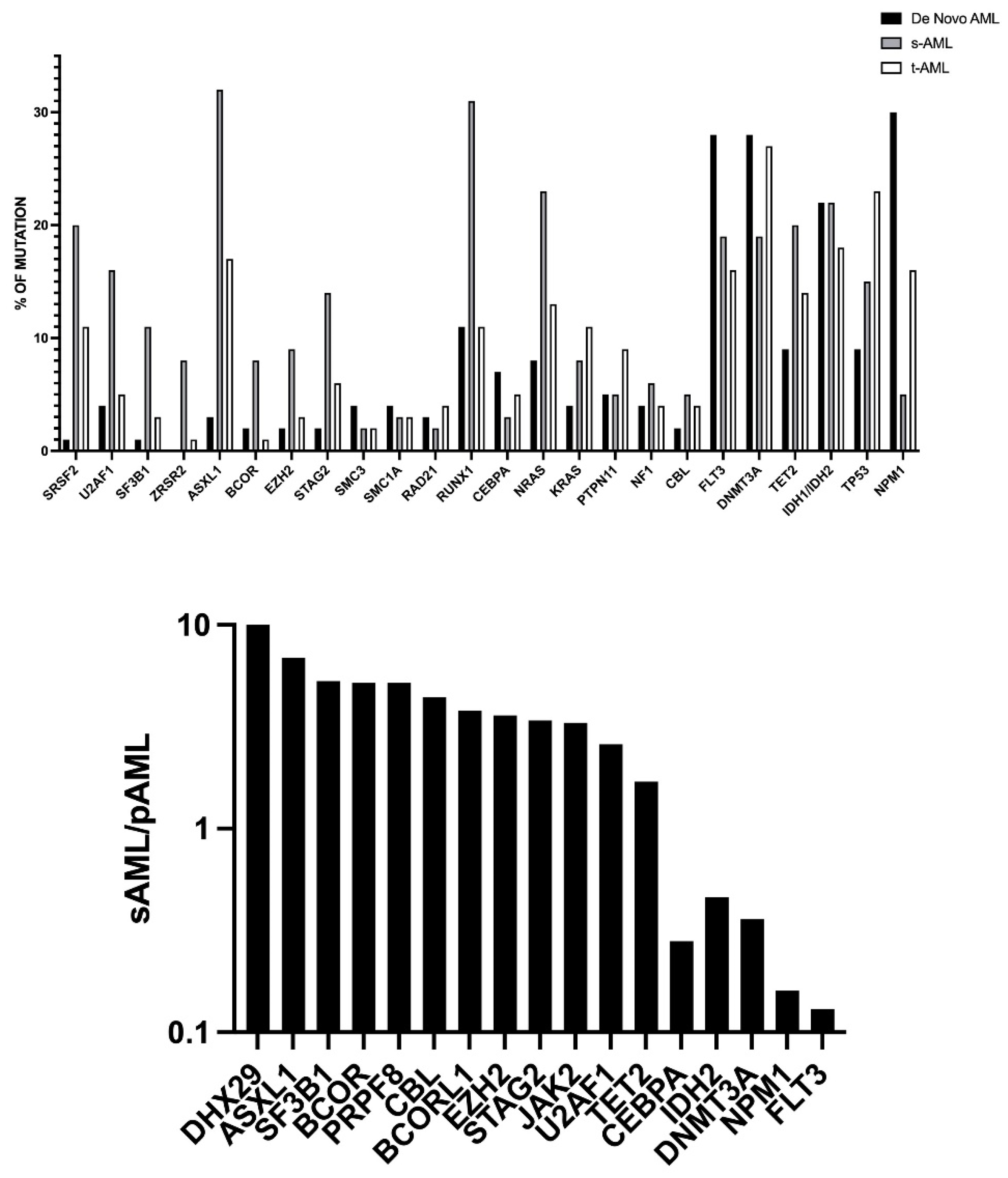
Figure 1. Frequency of main driver mutations in pAML, sAML, and tAML.Top Panel: frequency of recurrent gene mutations in adult pAML, sAML, and tAML. The Figure is based on the data reported by Lindsley et al., 2015 [8]. Bottom Panel: ratio between mutation frequency of some relevant driver gene mutations observed in sAML and in pAML. This graph reports the data relative to a set of genes whose mutations were clearly more frequent or less frequent in sAML compared to pAML. The Figure is based on the data reported by Nazha et al., 2016 [9].
Hemato 03 00038 g001
Figure 2. Cytogenetic abnormalities in pAML, sAML and tAML.Top Panel: main cytogenetic abnormalities observed in pAML and sAML. The Figure is based on the data reported by Nazha et al., 2016 [9]. Bottom Panel: main cytogenetic abnormalities observed in pAML, sAML, and tAML. The Figure is based on the data reported by Kuzmanovic et al., 2016. [11].
Hemato 03 00038 g002
Table 1. Techologies for MRD detection in AML.
NameMFC-LAIPMFC-DfNRT-qPCRddPCRNGS
Sensitivity10−3–10−510−3–10−510−4–10−610−4–10−610−4–10−7
Applicability>90%>90%50–60%50–60%80–90%
PrincipleFlow cytometry evaluation of membrane immuno-phenotype
Leukemia Associated Immunophenotype (LAIP).
The technique defines individual-specific surface markers at diagnosis and evaluates these markers at various times during and after the end of treatment.
Flow cytometry evaluation of membrane immuno-phenotype
Different from Normal (DfN).
The technique is based on the detection of aberrant surface marker expression at follow-up.
Reverse transcription-quantitative polymerase chain reaction (RT-qPCR) measures the amount of a specific mRNA.Digital droplet polymerase chain reaction (RT-qPCR) measures the amount of a specific mRNA.Next-generation sequencing (NGS), a DNA sequencing technology that rapidly sequences the whole genome.
Error-corrected NGS involves the physical incorporation of random oligonucleotides or unique molecular identifiers (UMI) at the library preparation prior to amplification of DNA, reduces the errors of standard NGS, and thus increases the capacity to detect gene mutation at low–very low VAF.
Main CharacteristicsMajor advantages:
It is widely available given the diffusion of flow cytometry;
It is widely applicable to >90% of AMLs;
Its evaluation is relatively fast.
Main limitations:
It requires standardization and harmonization between laboratories;
It requires a relatively high number of cells;
It requires technical expertise for the analysis and interpretation of the results.
Major advantages:
It is widely available given the diffusion of flow cytometry;
It is widely applicable to >90% of AMLs;
Its evaluation is relatively fast.
Main limitations:
It requires standardization and harmonization between laboratories;
It requires a relatively high number of cells;
It requires technical expertise for the analysis and interpretation of the results.
It is used for the detection of the following gene alterations:
NPM1 mutations;
PML-RARA fusion;
RUNX1-RUNXT1 fusion;
CBFB-MYH11 fusion.
Major advantages:
It is a sensitive technique;
Well-standardized;
It is a semi-quantitative technique;
It allows an easy interpretation of the results.
Major limitations:
It requires a standard curve;
Single gene assessed per assay;
The capacity to detect a gene alteration is limited to the primer-spanning regions.
It is used for the detection of the following gene alterations:
NPM1 mutations;
PML-RARA fusion;
RUNX1-RUNXT1 fusion;
CBFB-MYH11 fusion.
Major advantages:
It is a very sensitive technique;
Higher sensitivity than RT-qPCR;
No requirement for a standard curve;
It provides an absolute quantitation.
Major limitations:
The capacity to detect a gene alteration is limited to the primer-spanning regions;
It requires technical experience.
Major advantages:
It is widely applicable to about 80–90% of AMLs;
It can simultaneously examine multiple genes;
Sensitivity very high with error-corrected NGS.
Main limitations:
Availability only in state-of-the-art and well-funded centers;
Bioinformatics required for the interpretation of the results;
Relatively expensive;
It requires considerable technical expertise in the analysis and interpretation of data.
Publisher’s Note: MDPI stays neutral with regard to jurisdictional claims in published maps and institutional affiliations.