Article

A Novel Method for Preparation of Silk Fibroin Hydrogels with Significantly Reduced Gelation Time


1
Faculty of Technology Novi Sad, University of Novi Sad, Bulevar cara Lazara 1, 21000 Novi Sad, Serbia
2
Mining and Metallurgical Institute, Satbayev University, Satbayev str. 22a, 050013 Almaty, Kazakhstan
3
Petroleum Engineering Institute “One Belt, One Road”, Kazakh-British Technical University, Tole bi str. 59, 050000 Almaty, Kazakhstan
*
Author to whom correspondence should be addressed.
Macromol 2026, 6(3), 59; https://doi.org/10.3390/macromol6030059
Submission received: 1 July 2026 / Revised: 4 August 2026 / Accepted: 6 August 2026 / Published: 7 August 2026

Abstract

Hydrogels are considered to be a versatile and desirable material, due to their great ability of water retention, flexibility, viscoelasticity, and good mechanical properties. Silk fibroin (SF) is one of the most abundant biopolymers used for preparation of hydrogels. SF hydrogels can be prepared by self-assembly or induced by physical or chemical cross-linking. Self-assembly of SF hydrogels was shown to be favorable due to its simplicity and the potential harmfulness of chemical cross-linking agents. However, it is often very slow and time-consuming process, which is the main drawback and limitation of SF industrial application. In this study, a novel method of preparing SF hydrogels from aqueous dispersions of SF powder is presented. The results showed that the gelation time is significantly reduced when SF hydrogels are prepared from SF dispersions instead of SF solutions, yet both types of hydrogels had similar rheological and viscoelastic properties, within the investigated concentration range. All prepared hydrogels showed thixotropic behavior and the prevailing of storage modulus in comparison to loss modulus.

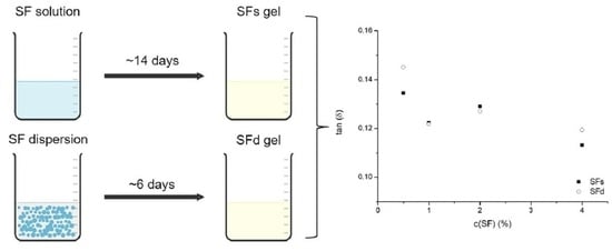
Graphical Abstract

1. Introduction

Hydrogels are three-dimensional colloidal structures, consisting of cross-linked polymer networks, with very high water content compared to polymer dry matter. They are prepared by polymers’ cross-linking in aqueous media, resulting in soft and rubbery or viscous material. Due to their many advantages, hydrogels have a wide range of applications in various scientific and industrial fields. Some of the potential applications of hydrogels are drug delivery, biosensors, soft and wearable electronics, biodegradable healing band aids, etc. Also, they can be easily modified and functionalized for use in tissue engineering and cell culture growth. Owing to their great ability of water retention, flexibility, viscoelasticity, and good mechanical properties, hydrogels are considered to be a versatile and desirable material. Hydrogels are usually prepared using synthetic or natural polymers, and can be shaped as films, membranes, coating or particles. While synthetic polymers exhibit better chemical stability and reproducibility, natural polymers have advantages in terms of biocompatibility and biodegradability. Collagen, fibrin, elastin, gelatin, chitosan, alginate, and silk fibroin are among the most commonly used biopolymers for hydrogel preparation [1,2,3,4].
Silk fibroin (SF) is the fibrillar structural protein of silkworm cocoons, produced by the B. mori silkworm. The silk fiber of Bombyx mori has a core–shell structure, with the core comprising a fibroin complex formed by two polypeptide chains, heavy-chain fibroin (H-chain, ~350 kg/mol), light-chain fibroin (L-chain, ~25 kg/mol), and the glycoprotein P25 (~30 kg/mol). The H- and L-chains are connected by disulfide bonds, while P25 is associated with the complex through non-covalent hydrophobic interactions. In its native form it is present in complex with sericin, a smaller protein that has a binding role in silk fiber formation, assembling a shell. Usually, silk fiber consists of 75% fibroin and 25% sericin. It is important to note that the main difference between these two proteins is their solubility, and while sericin is soluble in hot water, fibroin is water-insoluble owing to the presence of around 76% of non-polar amino acids in its structure [5]. This characteristic allows easy removing of sericin and extraction of fibroin from silk fibers. Further, SF heavy chain consists of repetitive hydrophilic and hydrophobic sequences, and hydrophobic sequences enable folding of the SF protein chain into a β-sheet conformation and formation of SF crystalline structure. β-sheet conformation of the SF protein chain is responsible for formation of SF colloidal structures, such as films or hydrogels [5,6].
Among the other biopolymers, silk fibroin is highly attractive for preparation of hydrogels for medical and pharmaceutical applications, due to its biocompatibility, mechanical stability, and biodegradability [7]. SF hydrogels can be prepared by self-assembly or induced by physical (ultrasonication, temperature change, etc.) or chemical (photopolymerization, chemical cross-linking agents, irradiation) cross-linking [2]. Self-assembly of SF hydrogels was shown to be favorable due to its simplicity and the potential harmfulness of chemical cross-linking agents. However, it is often very slow and is a time-consuming process [6,8], which is the main drawback and limitation of SF industrial application. For that reason, many different approaches were used to enhance gelation rate of SF hydrogels. Nagarkar et al. [9] used vortex mixing to induce gelation, while adjusting pH to 2–4 and varying temperature from 5 to 70 °C. Authors showed that, at low concentrations (0.05–1% wt.), change in temperature and pH value had very little effect on gelation time. Another study by Kim et al. [10] also used the combination of physical (dialysis, temperature and pH change) and chemical (addition of ions and poly(ethylene oxide)) methods to induce and expedite gelation. Dialysis against polyethylene-glycol was used to increase the concentration of SF solutions, while different temperatures and pH alongside addition of Ca2+ ions and PEO were studied for promotion of gelation of concentrated SF solutions. The study showed that gelation time decreased with decrease in pH, increase in temperature, addition of Ca2+, and addition of PEO; however, chemically cross-linked hydrogels showed worse rheological properties than the ones without cross-linkers. Pudkon et al. [8] also showed that addition of chemical cross-linkers deteriorates rheological properties of SF hydrogels, despite enhancing the gelation rate. Ultrasonication was also often used for gelation time decrease [11,12], as well as the addition of alcohols [13], genipin, glutaraldehyde or cellulose derivates [14]. Despite the efficiency of different physical and chemical cross-linking methods, most of them yet come with a certain drawback. Hence, it is indicated that more investigations need to be conducted in order to obtain simple, versatile, low-cost methods for preparation of SF hydrogels without potential harmful additives.
The main goal of this research was to develop and evaluate a novel preparation strategy for silk fibroin (SF) hydrogels based on the direct dispersion of dry SF powder in water. This approach was designed to simplify fibroin concentration adjustment and accelerate hydrogel formation while maintaining obtained properties comparable to those of conventional SF hydrogels prepared from fibroin solutions. SF hydrogels were prepared by gelation of SF powder dispersions, and their structural and rheological properties were characterized by FTIR spectroscopy and rheological measurements, respectively. The properties of powder-based SF hydrogels were further compared with those of hydrogels obtained using conventional SF solutions to evaluate the influence of the preparation approach on the resulting hydrogel characteristics.

2. Materials and Methods

2.1. Materials

Bombyx mori silkworm cocoons were obtained from a farm cooperative in Wuhan, China. Sodium carbonate (Na2CO3, anhydrous, 99.5%) was purchased from Loba Chemie Pvt. Ltd. (Mumbai, India), and lithium bromide (LiBr, anhydrous) from Sigma-Aldrich Co. (St. Louis, MO, USA).

2.2. Silk Fibroin Extraction

Firstly, SF solution was prepared as described in [15]. Namely, 5 g of silkworm cocoons were cut into small pieces and placed in a boiling 0.5% w/v solution of Na2CO3 for 30 min. Thereafter, obtained fibers were rinsed in distilled water at 40 °C for 30 min. This procedure of boiling and rinsing was repeated two more times, in order to fully remove sericin from silk fibers. Resulting SF fibers were left to dry overnight. Dry SF fibers were then dissolved in 9.3 M solution of LiBr at 60 °C for 90 min, yielding a 10% w/v SF solution. The obtained SF solution was dialyzed against distilled water for 3 days, using dialysis tubing cellulose membrane (molecular weight cut off 14 kg/mol; Sigma-Aldrich, Co., USA), in order to remove LiBr and other residual ions. After dialysis, the SF solution was centrifuged for 20 min at 5000 rpm and subsequently filtered through 0.45 μm regenerated cellulose filter. The concentration of the final purified SF solution was determined gravimetrically and found to be around 4.5% wt. The solution prepared as such was refrigerated until further use.

2.3. Drying of SF Solution

The purified SF solution was dried using Buchi Mini Spray Drier (Büchi, Flawil, Switzerland) under the following conditions: inlet temperature 170 °C, air flow rate 700 mL/min, and sample flow rate 7.4 mL/min. The obtained SF powder was kept in a desiccator until further use.

2.4. Preparation of SF Gels

Two different forms of SF were used for preparation of SF gels in this study—SF dry powder and SF water solution. Namely, previously dried SF powder was dispersed in distilled water in order to obtain dispersions with 0.5%, 1%, 2%, and 4% wt. of SF. Prepared dispersions were kept at room temperature until gelation took place and gels from SF dispersions (SFd gels) were obtained. Furthermore, previously prepared purified SF water solutions were diluted with distilled water, in order to obtain 0.5%, 1%, 2%, and 4% SF solutions. Diluted solutions were also kept at room temperature until gels (SFs) were formed. Gelation time (GT), in days, was determined empirically by tilting the beakers with gelling samples. Gelation time was considered to be the day when solution and dispersion samples transitioned from the liquid to solid state [16].

2.5. Rheological Measurements

Rheological properties of SFd and SFs gels were determined using a rheometer HAAKE RheoStress RS600 (Thermo Electron GmbH, Karlsruhe, Germany) at 24 ± 0.5 °C. The cone-and-plate geometry (d = 60 mm, h = 1°) was used for all measurements. Flow curves of all gel samples were obtained by continual linear increase in shear rates, γ (s−1), from zero (γ = 0) to γmax = 100 s−1 within 3 min, and reversely (after 3 min shearing at γmax), also within 3 min. Based on the obtained flow curves, fluid type and the thixotropic loop area were determined for each gel.
Additionally, oscillatory measurements were carried out to investigate viscoelastic properties of SFd and SFs gels. The amplitude-sweep method, with shear stress in range 0.01–500 Pa, was used for determination of the gel samples’ viscoelastic properties, according to [17]. Oscillating frequency was 1 Hz. Plateau values of storage, G′, and loss modulus, G″, were determined from the linear viscoelastic region (LVR). Also, tan (δ) was calculated based on G′ and G″ values within LVR, as the G″/G′ ratio.
All measurements were conducted on the day of gelation for each gel sample.

2.6. FTIR Spectroscopy

FTIR spectra of SF powder, SF solution, and SFd and SFs gels were obtained by IRAffinity-1S FTIR spectrophotometer (Shimadzu, Tokyo, Japan). The spectra were recorded from 4000 to 500 cm−1, in a transmittance mode.

2.7. Statistical Analysis

One-way ANOVA and Tukey test were used for statistical analysis. Experimental data were analyzed by single-factor analysis of variance (ANOVA) with the confidence interval of 95%. When pertinent, means were compared through the Tukey Honest Significant Difference (HDS) test.

3. Results and Discussion

3.1. Gelation Time

Gelation time is one of the main aspects when it comes to gel preparation. Often, this part of the process can be the slowest and most time-consuming, which further leads to poor time efficiency of the gel preparation process [8]. Therefore, it is important to improve feasibility and tuneability of gelation process. When SF gels are prepared from SF solutions, concentration of dissolved SF determines the gelation process. Consequently, gelation time and hydrogel properties are determined and limited by the concentration of SF solution, which, in most cases, reaches only up to 5% wt. of dry mass [8,9].
In this study, gelation time of SFd and SFs samples was determined empirically. Table 1 shows the influence of SF concentration on gelation time of SF dispersions and SF solutions. It can be observed that it took 11–16 days for SF solutions’ gelation, and 4–7 days for SFd gels formation. Statistical analysis showed that GT was mostly independent of SF concentration, within one gel type, except for 4% SFs and 1% SFd gels. Nevertheless, no specific trend was observed. The non-monotonic dependence of gelation time on fibroin concentration may be attributed to the competing effects of intermolecular interactions and molecular mobility. Increasing fibroin concentration initially accelerates gelation by promoting intermolecular contacts and β-sheet nucleation. However, beyond a certain concentration, electrostatic repulsion between negatively charged RSF molecular chains slows this process and molecular crowding can restrict chain diffusion and conformational rearrangements necessary for β-sheet assembly, thereby slowing network formation [18]. Also, it was previously suggested that the time of gelation depends as well on SF source, isolation and extraction conditions and duration, which is why different authors may report different trends in gelation time dependence on concentration [19]. More importantly, one can observe that formation of SFd gels was significantly faster compared to SFs gels. Namely, SFs hydrogels are formed by association of dissolved SF molecules by hydrogen bonding creating an interconnected skeleton structure [20]. On the other hand, SFd hydrogels can be considered as particulate gels. These gels form through physical aggregation and association of the initial colloidal particles, due to surface forces and attractive interactions mediated by the solvent [21,22,23]. Given that the hydrogels were prepared directly from dry fibroin powder, the initial stages of gelation may involve processes characteristic of particulate gelation, including particle hydration, aggregation, and percolation, before the formation of a continuous β-sheet-rich fibroin network. This may contribute to the distinct gelation time difference observed in the present study compared with conventional regenerated fibroin solution-based systems. SFd gels showed two-times-faster gelation, compared to SFs samples of the same concentration, which suggests that association of colloidal particles is a significantly faster process than hydrogen bonding of SF molecules. Consequently, preparation of SF hydrogels from SF dispersions was shown to be the more efficient and less time-consuming preparation method.
The accelerated gelation observed for SFd hydrogels may be related to differences in the initial organization of fibroin molecules. In solution-based systems, network formation relies on intermolecular association of individually dispersed fibroin chains, whereas powder-based systems start from hydrated fibroin particles that may provide pre-organized regions or interfaces favorable for further molecular rearrangement and network growth (Figure 1).
Compared with conventional fibroin solution-based approaches, the proposed method enables approximately two-fold-faster gelation at each investigated fibroin concentration, while the use of dry fibroin powder as the starting material enables easier adjustment of fibroin concentration and allows preparation of hydrogels over a broader concentration range, providing additional flexibility for tailoring hydrogel properties for specific applications.

3.2. FTIR Analysis

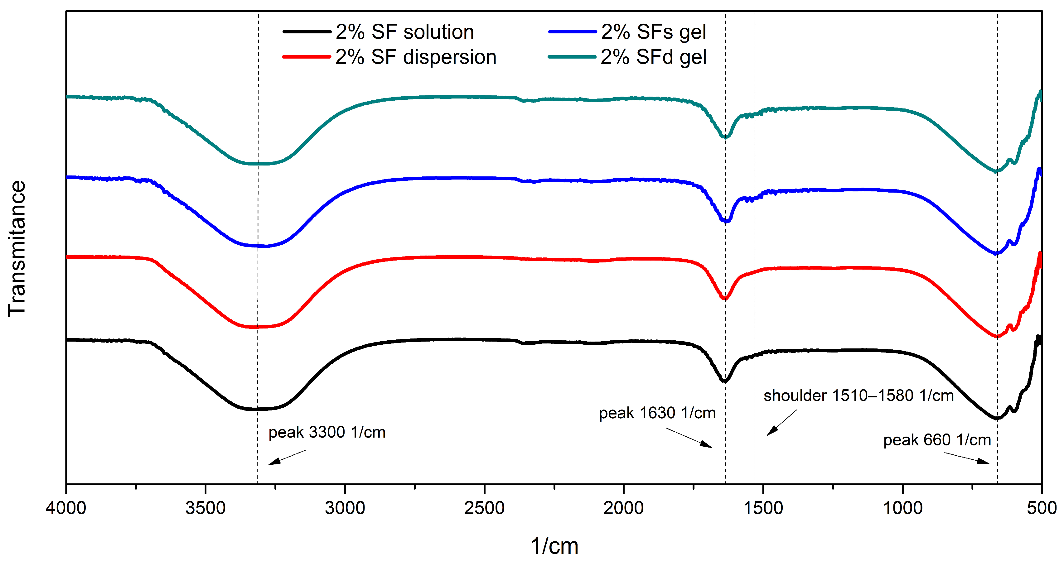
Figure 2 shows the FTIR spectra of the SF solution, SF dispersion, SFs gel, and SFd gel. One can see that three distinct peaks were observed, around 3300 cm−1, 1630 cm−1, and 660 cm−1, in each spectrum. The three peaks represent N-H stretching, and amide I and amide V bands of silk fibroin, respectively [24,25,26]. Besides those peaks, a shoulder consisting of two peaks was observed in SFs and SFd gel spectra around 1510–1580 cm−1, which was less pronounced in the spectra of SF solution and SF dispersion. This shoulder indicates the presence of the amide II band [26,27]. The amide I band represents primarily the C=O stretching vibration of the amide group. The frequency of this vibration depends on the strength of hydrogen bonding between the C=O and N–H groups, which in turn is determined by the particular conformational structure of the protein backbone. Generally, amide I band located around 1630 cm−1 is assigned to parallel β-sheet conformation [28,29,30,31], while amide I around 1650 cm−1 is usually assigned to random coil structure [8] which explains its presence in all three presented systems—SF water solution, SF water dispersion, and SF hydrogels. On the other hand, the amide II band around 1520 cm−1 is assigned to β-sheet conformation while the amide II band around 1540 cm−1 is assigned to random coil conformation [29,30]. While the amide III band around 1230 cm−1 is also characteristic for silk fibroin and represents its helical structure [8], and is found to be present in the spectrum of SF powder (Supplementary Figure S1), that peak was not observed in any of the analyzed samples. It can be seen from Figure 2 that formation of both SFs and SFd gels brings about an increase in the amide II band around 1500–1580 cm−1, indicating an increased β-sheet contribution when compared to both SF solution and SF dispersion. The obtained FTIR results suggest that there is no significant difference in β-sheet conformation between SFs and SFd gels.

3.3. Rheological Properties of SF Hydrogels

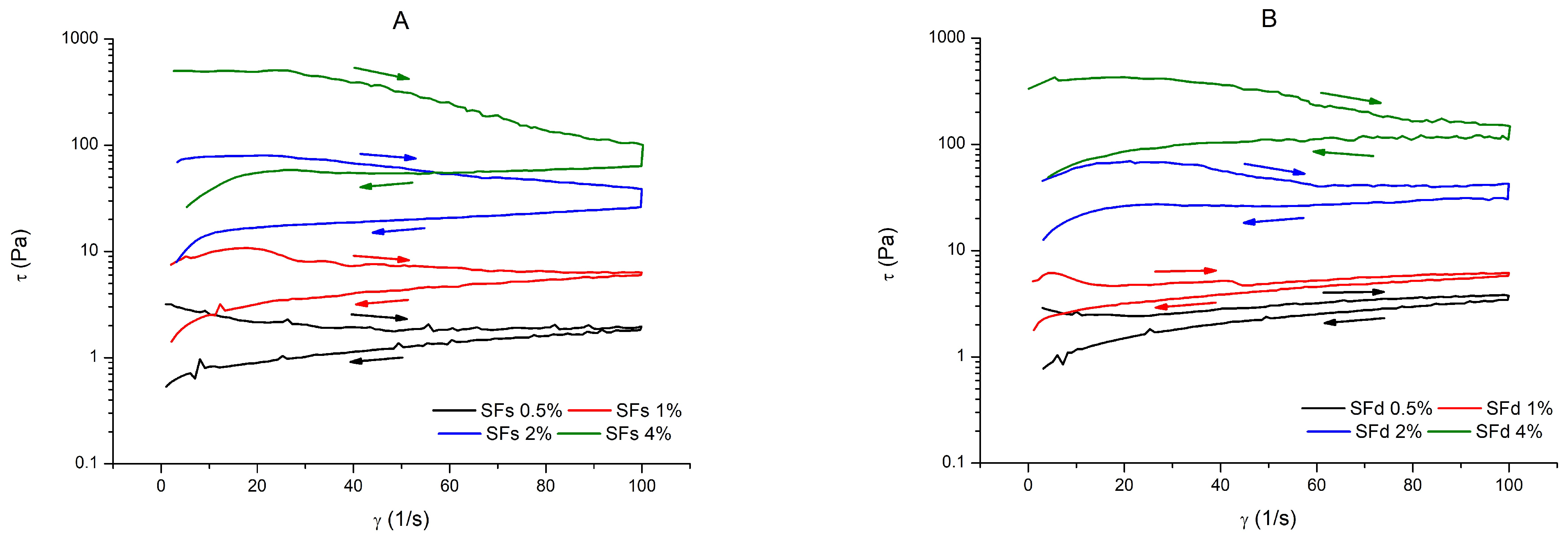
Rheological properties of prepared SF hydrogels were primarily characterized by obtaining flow curves of SFd and SFs gels with different SF concentrations. Figure 3 shows the recorded flow curves. It can be observed that all samples of SFs and SFd gels showed thixotropic behavior, regardless of the SF concentration, where thixotropy was more expressed in SF gels with higher SF concentrations, i.e., gels with 2% and 4% wt. Furthermore, it can be seen from Figure 3 that the resulting shear stress at a corresponding sheer rate increases almost by an order of magnitude when SF concentration was doubled, in both SFs and SFd gels, which indicates the increase in gel viscosity with increasing SF concentration. Shear-thinning behavior was observed in all samples.
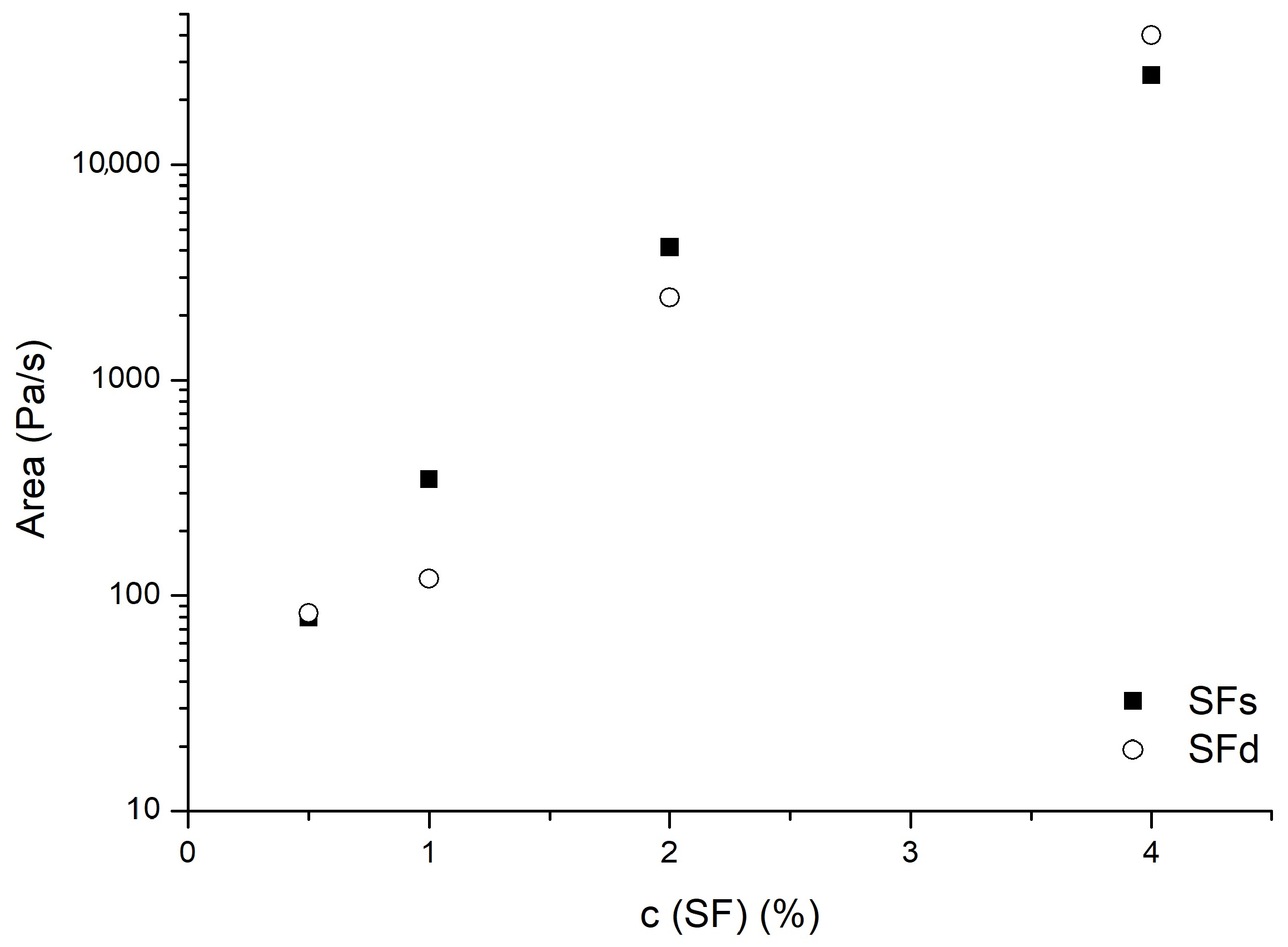
Influence of SF concentration on thixotropic loop area of SFs and SFd gels is presented in Figure 4. Results showed that thixotropic loop area increases from 80 Pa/s to around 20,000 Pa/s with SF concentration increasing from 0.5% to 4%. The size of loop area was independent of gel type at SF concentration 0.5%, while in 1% and 2% SF gels the loop area of SFs samples was higher compared to SFd. At SF concentration of 4%, the loop area of the SFd gel was higher than that of the SFs gel. The size of the thixotropic loop area corresponds to the energy required for the breakup of three-dimensional gel structure in the sample volume [32], where increase in the loop area indicates an increase in the gel structure strength.

3.4. Viscoelastic Properties of SF Hydrogels

The linear viscoelastic region (LVR) is of particular importance because it represents the range of deformation in which the hydrogel network remains intact and the measured storage (G′) and loss (G″) moduli reflect the intrinsic viscoelastic properties of the material, whereas outside the LVR the progressive disruption of the network leads to strain-dependent behavior.
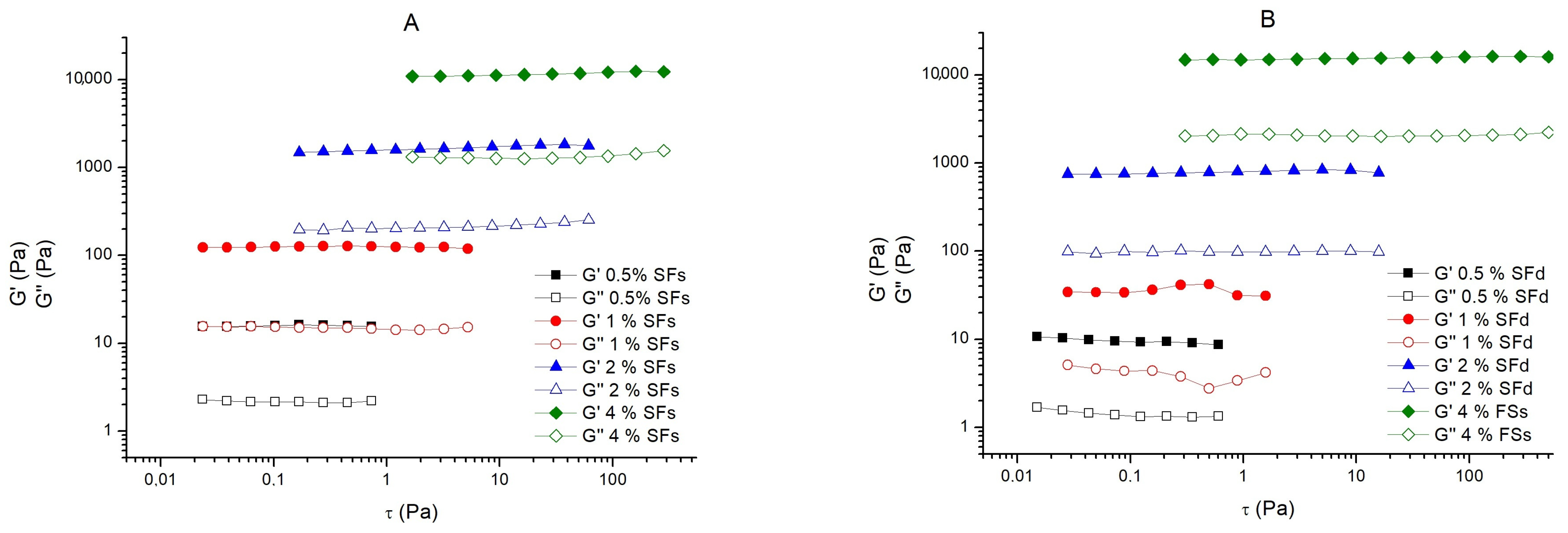
Figure 5 shows the linear viscoelastic region of the storage modulus (G′) and loss modulus (G″) of SFs and SFd gels obtained in the amplitude-sweep experiments. It can be observed that in both gel types, LVR increased and shifted to higher amplitude values, with increasing SF concentration. Broader LVR, where measured moduli are independent of the applied strain or stress, indicates higher resilience of gel samples to applied stress [33]. One can see that both G′ and G″ increased with SF concentration increasing from 0.5% to 4%. Furthermore, it can be observed that both G′ and G″ of SFs gels are higher compared to those of SFd gels, at each concentration. Also, in each sample, the storage modulus was higher than corresponding loss modulus which shows that all samples dominantly behave as elastic solids, under the stress applied [7]. The storage modulus (G′) is one of the most important rheological parameters for hydrogel characterization because it reflects the elastic behavior and mechanical integrity of the three-dimensional network. Consequently, G′ is widely used to assess the suitability of hydrogels for applications such as tissue engineering, drug delivery, injectable biomaterials, food structuring, and 3D bioprinting, where adequate mechanical stability and shape retention are essential [7,8,19,33,34,35]. Moreover, the difference between G′ and G″ increased with increasing silk fibroin concentration, indicating the formation of progressively stronger and more elastic networks. This behavior suggests that higher fibroin concentrations promote the development of a more robust three-dimensional structure, resulting in improved mechanical properties [36]. Although the storage modulus of the 4% SFd hydrogel (9.6 kPa) was slightly lower than that of the corresponding SFs hydrogel (12.5 kPa), the values were comparable, indicating that the proposed powder-based preparation method yields hydrogels with mechanical properties similar to those obtained by the conventional solution-based approach.
Figure 4. Influence of SF concentration on thixotropic loop area of SFs and SFd gels.
Macromol 06 00059 g004
Figure 5. Linear viscoelastic region of storage modulus (G′) and loss modulus (G″) obtained in amplitude-sweep experiments for (A) SFs gels and (B) SFd gels.
Macromol 06 00059 g005
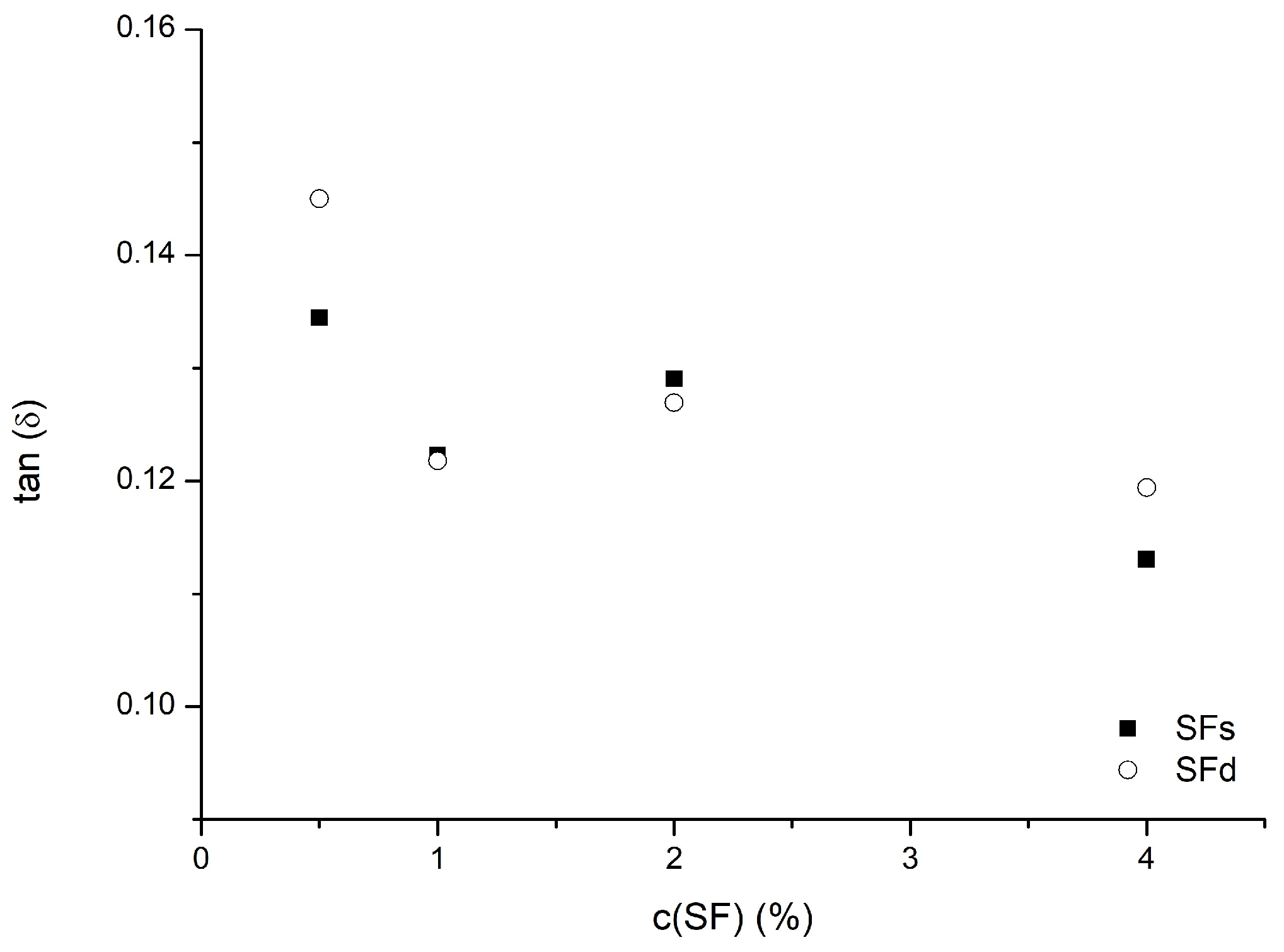
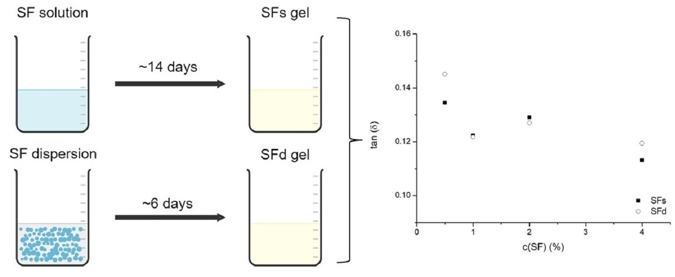
The ratio of the loss modulus (G″) to the storage modulus (G′) within the linear viscoelastic region (LVR), expressed as tan (δ), provides information on the relative contributions of the viscous and elastic behavior of the hydrogels. Lower tan (δ) values indicate a more elastic response, whereas values below 1 are characteristic of solid-like materials. As shown in Figure 6, all hydrogels exhibited tan (δ) values well below 1, confirming the predominance of elastic behavior and the successful formation of stable hydrogel networks. Furthermore, tan (δ) generally decreased with increasing silk fibroin concentration for both SFs and SFd hydrogels, indicating that increasing fibroin concentration promoted the formation of more elastic and mechanically stable networks. This trend is consistent with the increase in storage modulus observed with increasing fibroin concentration and suggests enhanced intermolecular interactions and network connectivity. Although some differences in tan (δ) were observed between SFs and SFd hydrogels, no systematic dependence on the preparation method was evident, indicating that the powder-based preparation approach produced hydrogels with viscoelastic characteristics comparable to those prepared from conventional fibroin solutions.
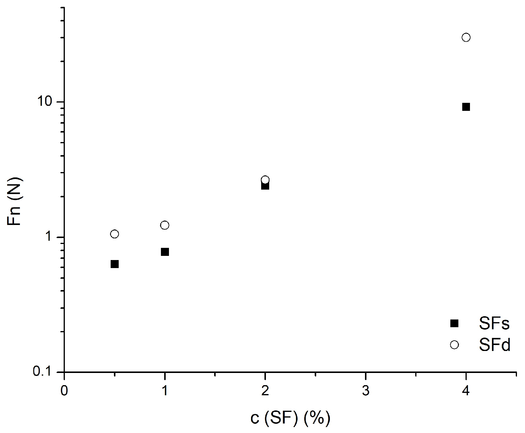
Influence of SF concentration on normal force (Fn) area of SFs and SFd gels is shown in Figure 7. The normal force recorded during rheological measurements increased with increasing SF concentration, indicating enhanced resistance of the hydrogel network to deformation. Interestingly, SFd hydrogels exhibited higher normal force values compared with SFs hydrogels at corresponding concentrations, suggesting that the powder-based preparation approach may promote the formation of networks with increased mechanical resistance.
The observed increase in normal force with increasing silk fibroin concentration was consistent with the increase in storage modulus (G′), suggesting enhanced elastic resistance and strengthening of the hydrogel network. Similarly, the higher normal force values observed for SFd hydrogels compared with SFs hydrogels support the rheological findings, indicating that the powder-based preparation approach resulted in hydrogels with increased resistance to deformation.

4. Conclusions

Silk fibroin hydrogels have already been extensively studied for biomedical and pharmaceutical applications, as controlled drug delivery systems, injectable biomaterials, wound healing scaffolds, and tissue engineering matrices for cartilage, bone, skin, and neural regeneration. SF hydrogels can be prepared by physical or chemical cross-linking. However, physical cross-linking, preferably self-assembly, was shown to be favorable, but often a very slow and time-consuming process. In this work, we proposed an alternative method for preparation of SF hydrogels with significantly reduced gelation time, where SF gels were prepared by dispersing SF particles, obtained by spray drying, in water. The proposed method enabled faster hydrogel formation, while maintaining comparable structural and rheological properties to conventionally prepared gels. The increase in fibroin concentration promoted the formation of stronger and more elastic hydrogel networks, as confirmed by rheological analysis, including increased storage modulus and reduced tan (δ) values. The developed powder-based approach provides a versatile platform for tailoring silk fibroin hydrogel properties and concentration, and may simplify future preparation of fibroin-based materials for various applications. Further optimization of the gelation process through adjustment of physical parameters represents an important direction for achieving additional reductions in gelation time. Future studies should also focus on application-specific evaluation of mechanical, biological, and functional properties.

Supplementary Materials

The following supporting information can be downloaded at https://www.mdpi.com/article/10.3390/macromol6030059/s1, Figure S1: FTIR spectrum of SF dry powder obtained via spray drying, Figure S2: Photographs of (a) SFs gels and (b) SFd gels.

Author Contributions

Conceptualization, L.S.; methodology, L.S., J.O. and S.R.; validation, S.B., J.F., J.M.B. and S.A.; formal analysis, L.S. and S.R.; investigation, L.S. and J.O.; resources, J.K., L.P. and S.A.; data curation, L.S., A.B. and A.I.; writing—original draft preparation, L.S.; writing—review and editing, L.S. and J.K.; visualization, L.S.; supervision, J.K., L.P. and A.S.; project administration, A.B. and A.I.; funding acquisition, J.K. and A.S. All authors have read and agreed to the published version of the manuscript.

Funding

This research was funded by the Ministry of Science and Higher Education of the Republic of Kazakhstan, Grant No. 14869304 and the Ministry of Science, Technological Development and Innovation of the Republic of Serbia, Grant No. 451-03-66/2024-03/200134, Grant No. 451-03-65/2024-03/200134, Grant no. 451-03-136/2026-03/200134, and Grant no. 451-03-137/2026-03/200134.

Institutional Review Board Statement

Not applicable.

Data Availability Statement

The raw data supporting the conclusions of this article will be made available by the authors on request.

Conflicts of Interest

The authors declare no conflicts of interest.

Abbreviations

The following abbreviations are used in this manuscript:
SFSilk fibroin
SFd gelsGels from SF dispersions
SFs gelsGels from SF solutions
GTGelation time
FTIRFourier transform infrared spectroscopy
LVRLinear viscoelastic region

References

  1. Elango, J.; Lijnev, A.; Zamora-Ledezma, C.; Alexis, F.; Wu, W.; Marín, J.M.G.; Sanchez de Val, J.E.M. The Relationship of Rheological Properties and the Performance of Silk Fibroin Hydrogels in Tissue Engineering Application. Process Biochem. 2023, 125, 198–211. [Google Scholar] [CrossRef]
  2. Zheng, H.; Zuo, B. Functional Silk Fibroin Hydrogels: Preparation, Properties and Applications. J. Mater. Chem. B 2021, 9, 1238–1258. [Google Scholar] [CrossRef] [PubMed]
  3. Grabska-Zielińska, S.; Sionkowska, A. How to Improve Physico-chemical Properties of Silk Fibroin Materials for Biomedical Applications?—Blending and Cross-linking of Silk Fibroin—A Review. Materials 2021, 14, 1510. [Google Scholar] [CrossRef] [PubMed]
  4. Montaseri, Z.; Abolmaali, S.S.; Tamaddon, A.M.; Farvadi, F. Composite Silk Fibroin Hydrogel Scaffolds for Cartilage Tissue Regeneration. J. Drug Deliv. Sci. Technol. 2023, 79, 104018. [Google Scholar] [CrossRef]
  5. Mottaghitalab, F.; Hosseinkhani, H.; Shokrgozar, M.A.; Mao, C.; Yang, M.; Farokhi, M. Silk as a Potential Candidate for Bone Tissue Engineering. J. Control. Release 2015, 215, 112–128. [Google Scholar] [CrossRef] [PubMed]
  6. Hardy, J.G.; Römer, L.M.; Scheibel, T.R. Polymeric Materials Based on Silk Proteins. Polymer 2008, 49, 4309–4327. [Google Scholar] [CrossRef]
  7. Rojas, J.E.U.; Gerbelli, B.B.; Ribeiro, A.O.; Nantes-Cardoso, I.L.; Giuntini, F.; Alves, W.A. Silk Fibroin Hydrogels for Potential Applications in Photodynamic Therapy. Biopolymers 2019, 110, 23245. [Google Scholar] [CrossRef] [PubMed]
  8. Pudkon, W.; Laomeephol, C.; Damrongsakkul, S.; Kanokpanont, S.; Ratanavaraporn, J. Comparative Study of Silk Fibroin-Based Hydrogels and Their Potential as Material for 3-Dimensional (3D) Printing. Molecules 2021, 26, 3887. [Google Scholar] [CrossRef] [PubMed]
  9. Nagarkar, S.; Nicolai, T.; Chassenieux, C.; Lele, A. Structure and Gelation Mechanism of Silk Hydrogels. Phys. Chem. Chem. Phys. 2010, 12, 3834–3844. [Google Scholar] [CrossRef] [PubMed]
  10. Kim, U.J.; Park, J.; Li, C.; Jin, H.J.; Valluzzi, R.; Kaplan, D.L. Structure and Properties of Silk Hydrogels. Biomacromolecules 2004, 5, 786–792. [Google Scholar] [CrossRef] [PubMed]
  11. Na, K.; Shin, S.; Lee, H.; Shin, D.; Baek, J.; Kwak, H.; Park, M.; Shin, J.; Hyun, J. Effect of Solution Viscosity on Retardation of Cell Sedimentation in DLP 3D Printing of Gelatin Methacrylate/Silk Fibroin Bioink. J. Ind. Eng. Chem. 2018, 61, 340–347. [Google Scholar] [CrossRef]
  12. Wang, X.; Kluge, J.A.; Leisk, G.G.; Kaplan, D.L. Sonication-Induced Gelation of Silk Fibroin for Cell Encapsulation. Biomaterials 2008, 29, 1054–1064. [Google Scholar] [CrossRef] [PubMed]
  13. Kaewprasit, K.; Kobayashi, T.; Damrongsakkul, S. Thai Silk Fibroin Gelation Process Enhancing by Monohydric and Polyhydric Alcohols. Int. J. Biol. Macromol. 2018, 118, 1726–1735. [Google Scholar] [CrossRef] [PubMed]
  14. Johari, N.; Moroni, L.; Samadikuchaksaraei, A. Tuning the Conformation and Mechanical Properties of Silk Fibroin Hydrogels. Eur. Polym. J. 2020, 134, 109842. [Google Scholar] [CrossRef]
  15. Škrbić, J.; Spasojević, L.; Sharipova, A.; Aidarova, S.; Babayev, A.; Sarsembekova, R.; Popović, L.; Bučko, S.; Milinković Budinčić, J.; Fraj, J.; et al. Investigation of Silk Fibroin/Poly(Acrylic Acid) Interactions in Aqueous Solution. Polymers 2024, 16, 936. [Google Scholar] [CrossRef] [PubMed]
  16. Kasoju, N.; Hawkins, N.; Pop-Georgievski, O.; Kubies, D.; Vollrath, F. Silk Fibroin Gelation via Non-Solvent Induced Phase Separation. Biomater. Sci. 2016, 4, 460–473. [Google Scholar] [CrossRef] [PubMed]
  17. Milinkovic-Budincic, J.; Petrovic, L.; Fraj, J.; Bucko, S.; Katona, J.; Spasojevic, L. Rheological Characterisation of Chitosan/Sodium Lauryl Ether Sulfate Complexes. Acta Period. Technol. 2019, 50, 152–162. [Google Scholar] [CrossRef]
  18. Peng, L.; Zhou, P.; Liao, F.; Liu, G.; Bao, S.; Yang, X.; Xiao, B.; Duan, L. Silk Fibroin Hydrogels: Gelation Mechanisms, Fabrication Techniques, and Biomedical Applications. Int. J. Biol. Macromol. 2025, 322, 146699. [Google Scholar] [CrossRef] [PubMed]
  19. Fan, L.; Cai, Z.; Zhao, J.; Mahmoudi, N.; Wang, Y.; Cheeseman, S.; Aguilar, L.C.; Reis, R.L.; Kundu, S.C.; Kaplan, D.L.; et al. Gelation Dynamics, Formation Mechanism, Functionalization, and 3D Bioprinting of Silk Fibroin Hydrogel Materials for Biomedical Applications. ACS Nano 2025, 19, 17979–18002. [Google Scholar] [CrossRef] [PubMed]
  20. Le, T.T.; Park, Y.; Chirila, T.V.; Halley, P.J.; Whittaker, A.K. The Behavior of Aged Regenerated Bombyx Mori Silk Fibroin Solutions Studied by 1H NMR and Rheology. Biomaterials 2008, 29, 4268–4274. [Google Scholar] [CrossRef] [PubMed]
  21. Bantawa, M.; Keshavarz, B.; Geri, M.; Bouzid, M.; Divoux, T.; McKinley, G.H.; Del Gado, E. The Hidden Hierarchical Nature of Soft Particulate Gels. Nat. Phys. 2023, 19, 1178–1184. [Google Scholar] [CrossRef]
  22. Ravanagh, M. Characterisation Polymer Gels. Science 1998, 23, 533–562. [Google Scholar] [CrossRef]
  23. Brinker, C.J.; Scherer, G.W. Sol-Gel Science: The Physics and Chemistry of Sol-Gel Processing; Academic Press: Cambridge, MA, USA, 2013; pp. 1–908. [Google Scholar]
  24. Hino, T.; Tanimoto, M.; Shimabayashi, S. Change in Secondary Structure of Silk Fibroin during Preparation of Its Microspheres by Spray-Drying and Exposure to Humid Atmosphere. J. Colloid Interface Sci. 2003, 266, 68–73. [Google Scholar] [CrossRef] [PubMed]
  25. Asakura, T.; Kuzuhara, A.; Tabeta, R.; Saito, H. Conformation Characterization of Bombyx Mori Silk Fibroin in the Solid State by High-Frequency 13C Cross Polarization-Magic Angle Spinning NMR, X-Ray Diffraction, and Infrared Spectroscopy. Macromolecules 1985, 18, 1841–1845. [Google Scholar] [CrossRef]
  26. Huang, Y.; Bailey, K.; Wang, S.; Feng, X. Silk Fibroin Films for Potential Applications in Controlled Release. React. Funct. Polym. 2017, 116, 57–68. [Google Scholar] [CrossRef]
  27. Indrakumar, S.; Panicker, A.T.; Parasuram, S.; Joshi, A.; Kumar Dash, T.; Mishra, V.; Tandon, B.; Chatterjee, K. 3D-Printed Ultra-Stretchable Silk Fibroin-Based Biocompatible Hydrogels. Bioprinting 2023, 36, e00315. [Google Scholar] [CrossRef]
  28. Tretinnikov, O.N.; Tamada, Y. Influence of Casting Temperature on the Near-Surface Structure and Wettability of Cast Silk Fibroin Films. Langmuir 2001, 17, 7406–7413. [Google Scholar] [CrossRef]
  29. Zhong, J.; Liu, X.; Wei, D.; Yan, J.; Wang, P.; Sun, G.; He, D. Effect of Incubation Temperature on the Self-Assembly of Regenerated Silk Fibroin: A Study Using AFM. Int. J. Biol. Macromol. 2015, 76, 195–202. [Google Scholar] [CrossRef] [PubMed]
  30. Cai, Y.; Guo, J.; Chen, C.; Yao, C.; Chung, S.M.; Yao, J.; Lee, I.S.; Kong, X. Silk Fibroin Membrane Used for Guided Bone Tissue Regeneration. Mater. Sci. Eng. C 2017, 70, 148–154. [Google Scholar] [CrossRef] [PubMed]
  31. Dorishetty, P.; Balu, R.; Athukoralalage, S.S.; Greaves, T.L.; Mata, J.; De Campo, L.; Saha, N.; Zannettino, A.C.W.; Dutta, N.K.; Choudhury, N.R. Tunable Biomimetic Hydrogels from Silk Fibroin and Nanocellulose. ACS Sustain. Chem. Eng. 2020, 8, 2375–2389. [Google Scholar] [CrossRef]
  32. Schramm, G. A Practical Approach to Rheology and Rheometry, 2nd ed.; Gebrüder Haake: Karlruhe, Germany, 2000. [Google Scholar]
  33. Grillet, A.M.; Wyatt, N.B.; Gloe, L.M. Polymer Gel Rheology and Adhesion. In Rheology; IntechOpen: London, UK, 2012. [Google Scholar]
  34. Malkin, A.Y.; Derkach, S.R.; Kulichikhin, V.G. Rheology of Gels and Yielding Liquids. Gels 2023, 9, 715. [Google Scholar] [CrossRef] [PubMed]
  35. Zhang, H.; Xu, D.; Zhang, Y.; Li, M.; Chai, R. Silk Fibroin Hydrogels for Biomedical Applications. Smart Med. 2022, 1, e20220011. [Google Scholar] [CrossRef] [PubMed]
  36. Liu, J.; Sun, H.; Peng, Y.; Chen, L.; Xu, W.; Shao, R. Preparation and Characterization of Natural Silk Fibroin Hydrogel for Protein Drug Delivery. Molecules 2022, 27, 3418. [Google Scholar] [CrossRef] [PubMed]
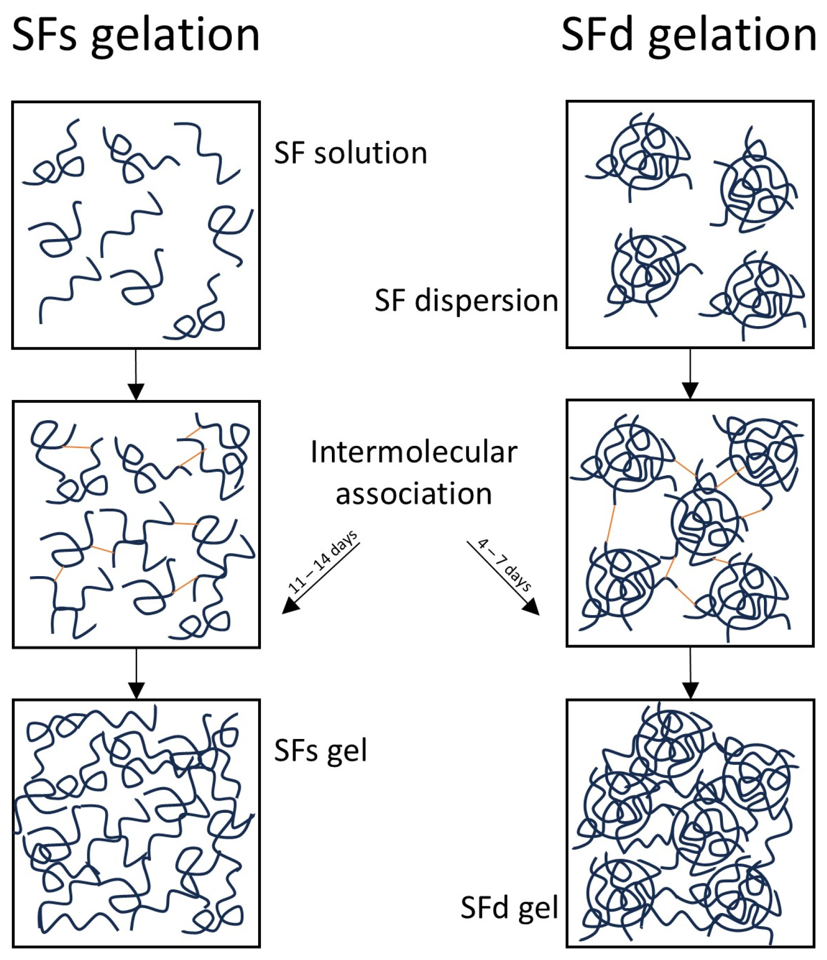
Figure 1. Schematic illustration of proposed gelation mechanism.
Macromol 06 00059 g001
Figure 2. FTIR spectra of SF solution, SF dispersion, SFs gel, and SFd gel.
Macromol 06 00059 g002
Figure 3. Flow curves of (A) SFs gels and (B) SFd gels. The arrows indicate the direction of the increasing and decreasing shear rate during the measurement.
Macromol 06 00059 g003
Figure 6. Influence of SF concentration on tan (δ) of SFs and SFd gels.
Macromol 06 00059 g006
Figure 7. Influence of SF concentration on normal force (Fn) area of SFs and SFd gels.
Macromol 06 00059 g007
Table 1. Influence of SF concentration in SFs and SFd gels on gelation time. Different superscript letters (within one column) represent significant differences (p < 0.05), as obtained by ANOVA and Tukey’s HSD test.
SF Concentration (%)Gelation Time SFs (Days)Gelation Time SFd (Days)
0.512 a5 c
111 a4 d
213 a6 c
416 b7 c
Disclaimer/Publisher’s Note: The statements, opinions and data contained in all publications are solely those of the individual author(s) and contributor(s) and not of MDPI and/or the editor(s). MDPI and/or the editor(s) disclaim responsibility for any injury to people or property resulting from any ideas, methods, instructions or products referred to in the content.