Article

Silver-Functionalized Silk Fibroin Films: Development and Characterization for Antibacterial Wound Dressings


1
Department of Engineering for Innovation, University of Salento, 73100 Lecce, Italy
2
Department of Experimental Medicine, University of Salento, 73100 Lecce, Italy
*
Author to whom correspondence should be addressed.
These authors equally contributed as senior authors.
Macromol 2026, 6(2), 29; https://doi.org/10.3390/macromol6020029
Submission received: 10 March 2026 / Revised: 2 April 2026 / Accepted: 7 May 2026 / Published: 12 May 2026

Abstract

In this work, an in situ UV-assisted photoreduction was used to functionalize silk fibroin films with silver nanoparticles in order to develop antibacterial devices for wound healing applications. The process showed high efficiency (~80%) in terms of reacted silver precursor. The effects of the silver treatment on fibroin macromolecule were evaluated in function of the process parameters in terms of chemical structure, thermal and mechanical properties, swelling behavior, resistance to degradation and antibacterial activity. Silver functionalization significantly improved the mechanical properties of the films, with Young’s modulus increasing from 0.23 ± 0.04 MPa (methanol-treated samples) to 7.26 ± 0.46 MPa (silver-functionalized samples). In parallel, a marked reduction in swelling degree was observed (from ~360–420% to ~60%), indicating enhanced structural stability. The treated films also exhibited improved resistance to degradation over 7 days under physiological conditions. From a functional perspective, the materials showed strong antibacterial activity, with up to 97–98% reduction in bacterial proliferation for Pseudomonas aeruginosa and Escherichia coli, and up to 93% for methicillin-resistant Staphylococcus aureus. Overall, the results demonstrate that silver functionalization process improves the structural stability of silk fibroin while conferring sustained antibacterial activity, thus supporting their potential application as antimicrobial dressings for the treatment of superficial and low-exudate wounds.

1. Introduction

The skin is the biggest organ of the human body and serves as the major physical barrier against the external environment, preventing microbial invasion and contributing to the maintenance of internal homeostasis, including fluid balance and thermoregulation [1,2,3].
When the skin is damaged or compromised, the wound bed and the underlying tissues can be colonized by pathogenic organisms, which may induce inflammatory responses and lead to localized or systemic infections [4]. Moreover, epidermal injuries result in the loss of tissue fluids, a condition that becomes significant when wounds encompass more than 10% of the total body surface area, with a consequent risk of life-threatening extracellular fluid loss [5]. According to the World Health Organization (WHO), over 4.4 million fatalities worldwide were related to injuries in 2022, accounting for nearly 8% of total global mortality [6].
Wound healing is a complex and dynamic physiological process involving a series of coordinated cellular events, including hemostasis, inflammation, proliferation, migration, and tissue remodeling, which typically lead to wound closure within 3–14 days, while tissue remodeling may continue for several months following the initial injury [7].
Wound dressings have evolved from passive covering devices to advanced systems designed to actively support tissue regeneration. In this context, natural biomaterials such as collagen, chitosan, hyaluronic acid, and silk fibroin, have attracted significant attention due to their biocompatibility and ability to mimic the extracellular matrix [8,9]. These materials enable the development of multifunctional dressings capable of maintaining a moist environment, preventing infections and promoting cell proliferation and tissue regeneration [10,11,12]. Depending on the application, they can be processed into hydrogels, films, and nanofibers, thus adapting to different wound conditions [13,14].
Among natural biomaterials, silk fibroin produced from Bombyx mori has emerged as a viable candidate for wound healing applications mainly due to its biocompatibility and tunable degradation profile [15,16]. In addition to providing structural support, fibroin has been shown to promote cell proliferation and migration, stimulate growth factors production and modulate the inflammatory response, contributing to improved healing outcomes [17,18,19,20,21]. In particular, fibroin films provide conformability, transparency, and the ability to maintain a moist wound environment, making them especially suitable for superficial wound treatment [22,23,24,25]. Furthermore, the intrinsic ability of silk fibroin to undergo β-sheet formation upon post processing crystallization treatments, such as methanol exposure, enables a significant enhancement of mechanical properties and stability under physiological conditions [26,27,28,29].
Fibroin functionalization represents a widely explored strategy to enhance its biomedical performances [30]. In particular, silk fibroin has been widely explored as a platform for the localized delivery of antibiotics to prevent and treat wound infections [31,32]. However, the growing issue of antibiotic resistance has prompted the investigation of alternative antimicrobial approaches [33,34]. Among them, metal and metal oxide nanoparticles (NPs), such as zinc oxide NPs, copper and copper oxide NPs, and silver NPs have demonstrated antimicrobial activity [35] due to multiple mechanisms of actions, contrasting the development of bacterial resistance [36,37,38,39,40,41,42]. Silver NPs (AgNPs), in particular, have shown antibacterial activity against numerous pathogens, including Gram positive and Gram-negative bacteria, fungi and viruses, through cell membrane destruction, protein denaturation, interference with bacterial DNA replication, the release of Ag+ ions and the production of ROS [43,44,45,46,47,48].
Combined with silk proteins, the potential of silver NPs has been explored for the development of advanced antimicrobial wound-healing systems. Previous studies have also demonstrated that incorporating AgNPs into fibroin scaffolds greatly increases antibacterial efficacy while maintaining cytocompatibility, assessing the potential of these systems for wounds that are infected or prone to infection [49,50,51].
Among the various incorporation techniques described in the literature, in situ photochemical synthesis and deposition of AgNPs represent a controllable method to develop antimicrobial coatings with strong adhesion to the substrate and long-term efficacy [52]. The process, successfully applied in previous works on different natural and synthetic surfaces [53], has never been tested on silk fibroin films, thus requiring a preliminary evaluation of the process parameters on the specific structure and functions of the silk protein, particularly in relation to silk fibroin crystallization, which can influence physicochemical and mechanical properties, and degradation behavior.
Moreover, aiming at the development of antimicrobial wound dressings for superficial injuries, the antibacterial efficacy of the silver-treated fibroin films was investigated against specific microorganisms involved in wound infections, including antibiotic-resistant strains.

2. Materials and Methods

2.1. Materials

The aqueous silk fibroin solution (5% w/v) from Bombyx mori cocoons was kindly provided by Caresilk S.r.l.s. (Lecce, Italy). Silver nitrate (AgNO3, 99+%) was purchased from Alfa Aesar (Fisher Scientific, Waltham, MA, USA); Trypticasein soy agar (TSA) was purchased from Lab Logistics Group GmbH (Meckenheim, Germany); methanol (≥99.9%, MW 32.04), sodium chloride (NaCl, ≥99%, MW 58.44), phosphate-buffered saline (PBS) tablets, and Tryptic Soy Broth (TSB) were purchased from Sigma-Aldrich (Saint Louis, MO, USA). All aqueous solutions were prepared using distilled water.

2.2. Preparation of Silk Fibroin Films and Surface Treatment

Non-porous substrates were prepared in the form of thin films by air drying. Briefly, 20 mL of 5% w/v aqueous silk fibroin solution were poured into polystyrene dishes (10 cm diameter) and allowed to dry in a laminar flow hood for 72 h at room temperature (RT), obtaining silk fibroin films (SFs).
Films were functionalized with a silver-based coating deposited via a photoreduction process, enabling the in situ synthesis of AgNPs directly on the material surface without the use of chemical binders [52].
The silver-based solution was prepared by dissolving 0.5% w/w AgNO3 in methanol, which represents both the solvent and the photo-reducing agent in the process. Fibroin films were immersed in the silver solution for 5 min and subsequently exposed to UV irradiation (1000 W, 20 cm distance; Jelosil, Milano, Italy) for 10 min on each side to promote the reduction of silver ions (Ag+) and the formation of AgNPs on the surface of the material. The resulting samples were washed three times with distilled water to remove any non-reacted reagent and then labeled as SF_Ag.
In order to investigate the effect of any stage of the silver treatment on fibroin, (i) films immersed in methanol for 5 min without subsequent UV exposure (SF_M) and (ii) films immersed in methanol for 5 min and exposed to UV irradiation for 10 min on each side (SF_M/UV) were also prepared and tested. Silk fibroin films (SFs) were used as the control.

2.3. Films Characterization

2.3.1. Quantification of Reacted Ag+

To evaluate the effectiveness of the treatment, it is important to determine the amount of silver ions (Ag+) in the precursor solution that participates in the functionalization of the resulting silk fibroin films. This amount was determined following the method described by Pellegrino et al. [54]. Briefly, after generating a calibration curve, the precursor solution (before and after impregnation) and the distilled water collected after the first washing step of one film were mixed 1:1 v/v with a 0.1 M NaCl solution. After 10 min, the resulting silvery-white colloidal silver chloride suspensions were measured for their absorbance at 450 nm using a Jasco V-660 UV–visible spectrophotometer (Jasco, Palo Alto, CA, USA).
The amount of Ag+ that reacted and contributed to the functionalization of silk fibroin films was calculated using Equation (1):
A g r e a c t e d   p p m = A g b e f o r e A g a f t e r A g u n r e a c t e d ,
where Agunreacted is the concentration of Ag+ in the distilled water collected after the first washing step and Agbefore and Agafter are the concentrations of Ag+ in the precursor solution before and after film impregnation, respectively.
In this way, it was possible to evaluate the efficiency (E) of the process using Equation (2):
E   ( % ) = A g r e a c t e d A g b e f o r e A g a f t e r × 100 ,
The experiments were performed in triplicate, and results were reported as the mean ± standard deviation (SD).

2.3.2. Fourier-Transform Infrared Spectroscopy (FT-IR)

FT-IR spectra were recorded to investigate the chemical structure of the untreated (SF) and treated films (SF_M, SF_M/UV and SF_Ag). Measurements were performed in Attenuated Total Reflectance (ATR) mode using a Jasco FT/IR-6300 spectrometer (Thermo Fisher Scientific, Waltham, MA, USA). Spectra were collected over a wavenumber range of 800–4000 cm−1, with a resolution of 4 cm−1.

2.3.3. Thermal Analysis

Differential scanning calorimetry (DSC) measurements were performed using a Q2000 DSC instrument (TA Instruments, New Castle, DE, USA). Approximately 5 mg of silk fibroin films were sealed in hermetic aluminum pans and subjected to a heating ramp from 25 °C to 350 °C under an inert nitrogen atmosphere (flow rate 50 mL min−1) at a heating rate of 10 °C min−1. An empty pan was used as a reference. DSC data were analyzed using OriginPro software (OriginLab, version 8, Northampton, MA, USA). Each sample (SF, SF_M, SF_M/UV and SF_Ag) was measured in duplicate.

2.3.4. Mechanical Properties

The tensile mechanical properties of fibroin films (SF_M, SF_M/UV, and SF_Ag) were evaluated using a universal testing machine (Zwick Roell, Ulm, Germany) equipped with a 100 N load cell. The films (10 × 50 mm2) were conditioned in 0.01 M PBS for 10 min at RT; then the tests were performed at a constant displacement rate of 10 mm/min, with a preload of 0.1 N applied to ensure consistent initial tension. The thickness and width of each sample in the wet state were measured using a digital microscope (Dino-Lite digital microscope, New Taipei City, Taiwan) equipped with DinoCapture 2.0 image analysis software. Young’s modulus (E) was calculated from the slope of the linear elastic region of the stress–strain curve, specifically in the low-strain interval (2–5%), to characterize the elastic response of the material in physiological-like conditions. Each sample was tested duplicate and the results were reported as the mean ± SD; SF films without methanol-based treatment were not included due to the fast dissolution in aqueous environment.

2.3.5. Swelling Capacity Assessment

The ability of the films to absorb liquids was evaluated by calculating the swelling ratio. Dried samples were cut into pieces (1 × 3 cm2), and their initial dry weight was recorded. The samples were then incubated in PBS solution at 37 °C, and at predetermined time points (30 min, 1, 2, 3, 5 and 24 h) they were removed. The excess liquid was gently removed using filter paper, and the weight of the swollen films was measured. The swelling ratio was determined using Equation (3):
S w e l l i n g   d e g r e e   ( % ) = w w w d w d × 100 ,
where ww is the weight of the swollen sample and wd is its initial dry weight.
The experiments were performed in triplicate, and results were reported as the mean ± SD.

2.3.6. Evaluation of In Vitro Degradation Resistance

The stability of SF, SF_M, SF_M/UV, and SF_Ag films was evaluated to assess their resistance to degradation in physiological conditions. Air-dried substrates, measuring 1 × 3 cm2, were initially weighed and incubated in 5 mL of 1× PBS at 37 °C. At predetermined time points (0, 1, 3, and 7 days), aliquots of the solution were collected to quantify the degraded protein using the bicinchoninic acid (BCA) colorimetric assay (QuantiPro™ BCA kit, Sigma-Aldrich, Saint Louis, MO, USA). Three measurements were performed at each time point for each sample type.

2.4. Assessment of the Antibacterial Activity

The antibacterial activity of SF_Ag films was assessed against three bacterial strains, namely Escherichia coli (E. coli, ATCC 25922), Pseudomonas aeruginosa (P. aeruginosa, ATCC 27853), and methicillin-resistant Staphylococcus aureus (S. aureus, ATCC 43300).
SF_Ag samples and SF_M/UV as control (1 cm2, average weight 25 mg) were incubated in 5 mL of sterile PBS at 37 °C for the time points corresponding to the previously defined degradation points (0, 1, 3, and 7 days), in order to simulate the potential application of the device. At each time point, samples were collected and characterized through both qualitative and quantitative analyses to evaluate the residual efficacy of the silver-based coating during film degradation.
Qualitative evaluation of antibacterial activity was performed using an agar inclusion test. In this method, TSA was inoculated with bacteria to achieve an optical density at 600 nm (OD600) of 0.1. SF_Ag and SF_M/UV films at different time points were placed within the agar and incubated for 12 h at 37 °C (Incubator IGS100, Thermo Scientific Heratherm, Thermo Fisher Scientific, Waltham, MA, USA). After incubation, the width of bacterial growth inhibition zones around each sample was measured and analyzed according to the levels of antibacterial activity defined by Standard SNV 195920-1992 [53]. Measurements were performed using ImageJ software (version 1.54c14, National Institute of Health, Bethesda, MD, USA), and reported as mean ± SD.
Bacterial growth inhibition was then quantified by spectrophotometric analysis, measuring the OD600 using a UV–Vis spectrophotometer (V-1200, VWR, Radnor, PA, USA). The quantitative test was performed in triplicate for SF_Ag and SF_M/UV control samples at each degradation time point. Samples were incubated at 37 °C in 8 mL of bacterial suspension for each strain, with an initial OD600 of 0.0005. At 6 h, the OD600 was measured and converted to CFU/mL, and the percentage of antibacterial efficacy (ABE) of the treated samples was calculated according to Equation (4):
A B E   ( % ) = D c D t D t × 100 ,
where Dc and Dt are the number of CFU/mL calculated in the presence of the control (SF_M/UV) and treated (SF_Ag) samples, respectively [52].

2.5. Statistical Analysis

The data are presented as the mean ± SD for the indicated number of experiments. The statistical analysis was conducted by using One- and Two-way ANOVA. In all comparisons, p < 0.05 was considered statistically significant, and the p-values are reported for statistically significant results. All ANOVA post hoc analyses were performed using Tukey’s test.

3. Results

3.1. Functionalization and Characterization of Silk Fibroin Films

The technology adopted for the deposition of the silver coatings onto the substrate of the fibroin films involves the use of methanol as photo reducing agent, in order to promote the photo-assisted synthesis of AgNPs from silver precursor. As methanol is also known to promote the crystallization of silk fibroin, this study aimed at investigating the effect of the silver treatment, and in particular the effect of methanol, on the properties of the fibroin film.
As first step of the study, the efficiency of the process was investigated to evaluate the amount of Ag+ ions that actually participated in the functionalization of the films. To this purpose, the absorbance of the precursor solution, before and after the silver treatment, as well as the absorbance of the distilled water after the first washing step, was measured at 450 nm. The amount of reacted Ag+ was calculated using the calibration curve obtained from AgNO3 standards (A = 0.0023 C + 0.0017, R2 = 0.992; where A is the absorbance and C is the Ag+ concentration in ppm). These data, reported in Table 1, were used to evaluate the efficiency of the process, which was found to be 80%.
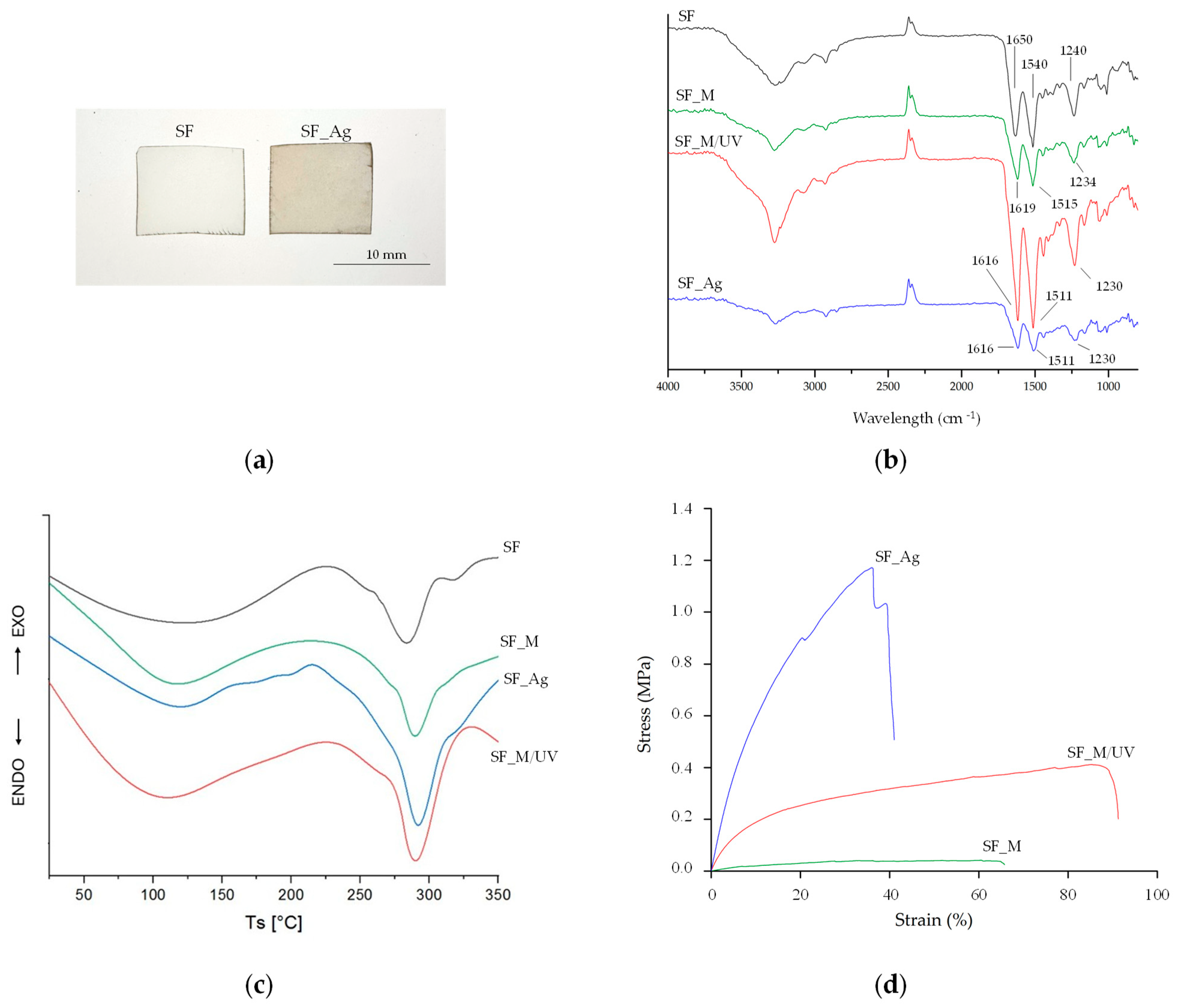
Functionalization was also confirmed by visually observing a color change in the SF_Ag films from white to brown after treatment, compared to the SF control (Figure 1a). The different samples categories were then characterized through FT-IR, DSC and mechanical tests.
SF is a protein that can assume random coil and α-helix conformation (silk I structure), with characteristic absorption peaks of Amide I, Amide II and Amide III at 1650–1660 cm−1 (-CO- and -CN- stretching), 1531–1542 cm−1 (-NH bending), and 1230 cm−1 (-CN- stretching), respectively. When immersed in methanol, SF can rearrange into a β-sheet structure (silk II) due to hydrogen bonding, with characteristic peaks of amides at 1620–1630, 1515–1530, and 1240 cm−1 [55,56]. In this case, the characteristic peaks of Amide I and Amide II shifted from 1650 to 1619 cm−1 and from 1540 to 1515 cm−1, respectively, after the treatment with methanol (from SF to SF_M films), confirming a rearrangement of the silk fibroin structure into β-sheets after treatment with methanol, according to previous studies [57,58]. Furthermore, additional exposure to UV irradiation led to an additional slight band shift, resulting in shifts from 1619 to 1616 and from 1515 to 1511 from SF_M to SF_M/UV films, which do not indicate a significant additional increase in β-sheet content. The peaks remained unchanged for the SF_Ag films (Figure 1b).
Thermal properties were also influenced by the process. As shown in the graph (Figure 1c), DSC curve of SF films showed two endothermic peaks. The first and broader peak occurred between 100 and 120 °C and is related to the loss of adsorbed water; the second sharper peak occurred below 300 °C and is related to the degradation of the SF chains. The exothermic peak around 210 °C is attributed to the formation of β-sheet domains. The intensity of the exothermic peak was markedly reduced for the SF_M sample, indicating that methanol had already induced structural reorganization into β-sheets. Additionally, a decrease in the enthalpy of the first endothermic peak was observed, which is consistent with a lower water content. There was also a slight shift in the degradation peak, which is indicative of increased thermal stability due to enhanced crystallinity. The sample subjected to combined methanol and UV irradiation (SF_M/UV) exhibited further attenuation of the exothermic event and a more defined second endothermic peak. This suggests that the combined methanol and UV irradiation may influence the thermal stability of the films. In the SF_Ag film, the exothermic peak was less pronounced than in SF, suggesting an interaction between the AgNPs and the fibroin functional groups. The degradation peak was also more defined and shifted compared to the other samples, suggesting a possible contribution of silver nanoparticles to the overall structural stabilization of the system. Overall, the applied treatments promoted an increase in the structural order of fibroin, resulting in a reduction in thermal reorganization events and an increase in degradation stability.
The tensile mechanical behavior was tested in the wet state for SF_M, SF_M/UV and SF_Ag films, and the results are reported in Table 2. A stress–strain plot obtained during these tests is shown as an example in Figure 1d. Due to the high solubility of the untreated fibroin films, SF samples were not included in this test. On the other hand, as shown in Table 2, both UV irradiation and AgNPs influenced the mechanical properties of the films compared to SF_M samples. In particular, the average Young’s Modulus of SF_M films was 0.23 ± 0.04 MPa, which increased when the treatment also included exposure to UV irradiation, probably due to photochemical processes and increased intermolecular interaction. SF films functionalized with AgNPs presented the highest Young’s Modulus value and maximum strength at failure, indicating improved mechanical properties due the presence of NPs.
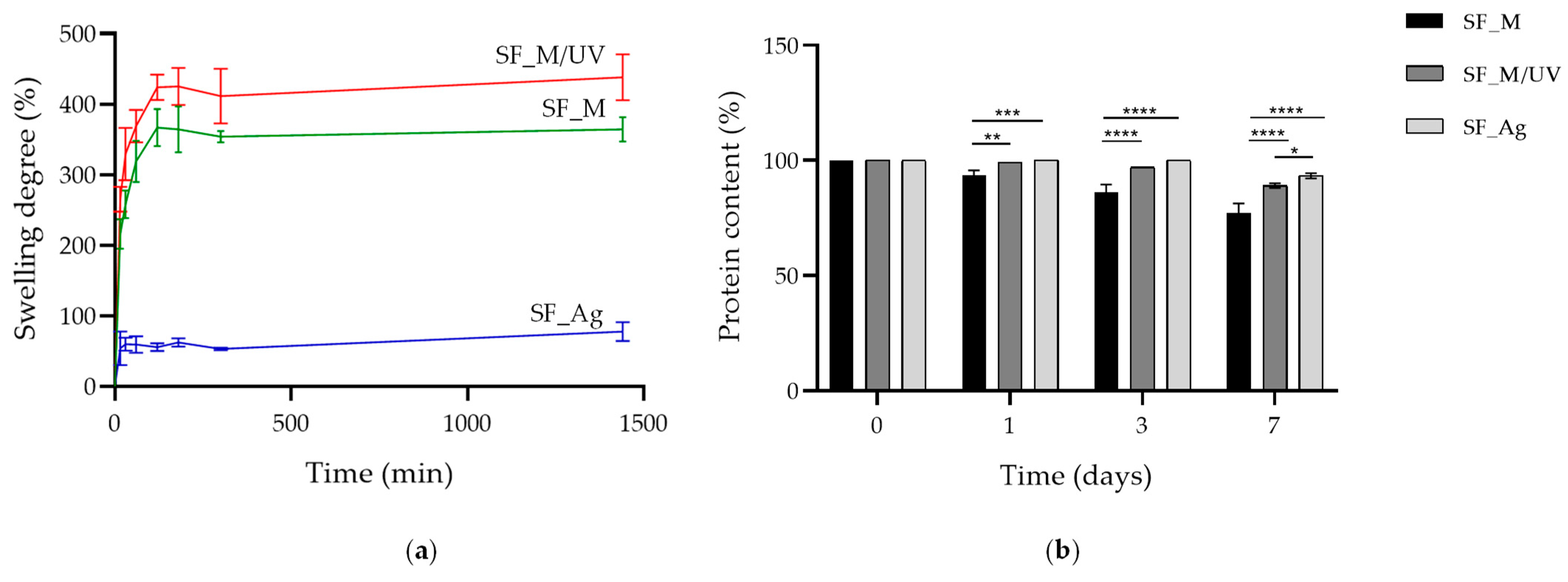
To assess the behavior of films in vitro under physiological conditions, their capacity to absorb liquids (in terms of swelling) and their ability to resist degradation over a period of 7 days were evaluated. Swelling tests revealed differences in absorption properties among the various samples. The SF_M/UV and SF_M films exhibited the highest degree of swelling at the initial time points, of approximately 420% and 360%, respectively, and maintained these values over time. This behavior suggests the presence of a hydrated polymeric network, despite the formation of β-sheet structures induced by methanol treatment, which provided greater structural stabilization to the film. In contrast, the SF_Ag film showed a lower swelling degree of approximately 60% (Figure 2a). This is likely due to intermolecular interactions induced by the incorporation of AgNPs, which limit polymer chain mobility and reduce water diffusion within the matrix. The films’ resistance to degradation was instead evaluated as the percentage of residual fibroin at different time points. On day 1, 100% residual fibroin was observed for the SF_Ag film, whereas minimal reduction in protein content was detected for the SF_M/UV and SF_M samples. By day 3, a more pronounced degradation was observed for SF_M (approximately 80% residual fibroin), whereas SF_Ag and SF_M/UV exhibited greater stability (approximately 99.95% and 95.90%, respectively). After 7 days, the differences among the films became more evident, with residual fibroin values of approximately 93% for SF_Ag, 85% for SF_M/UV, and about 56% for SF_M (Figure 2b). These results suggest that the silver-based deposition treatment combined with methanol and UV exposure provides enhanced resistance to degradation, thereby improving the structural stability of the SF_Ag sample.

3.2. Antibacterial Properties

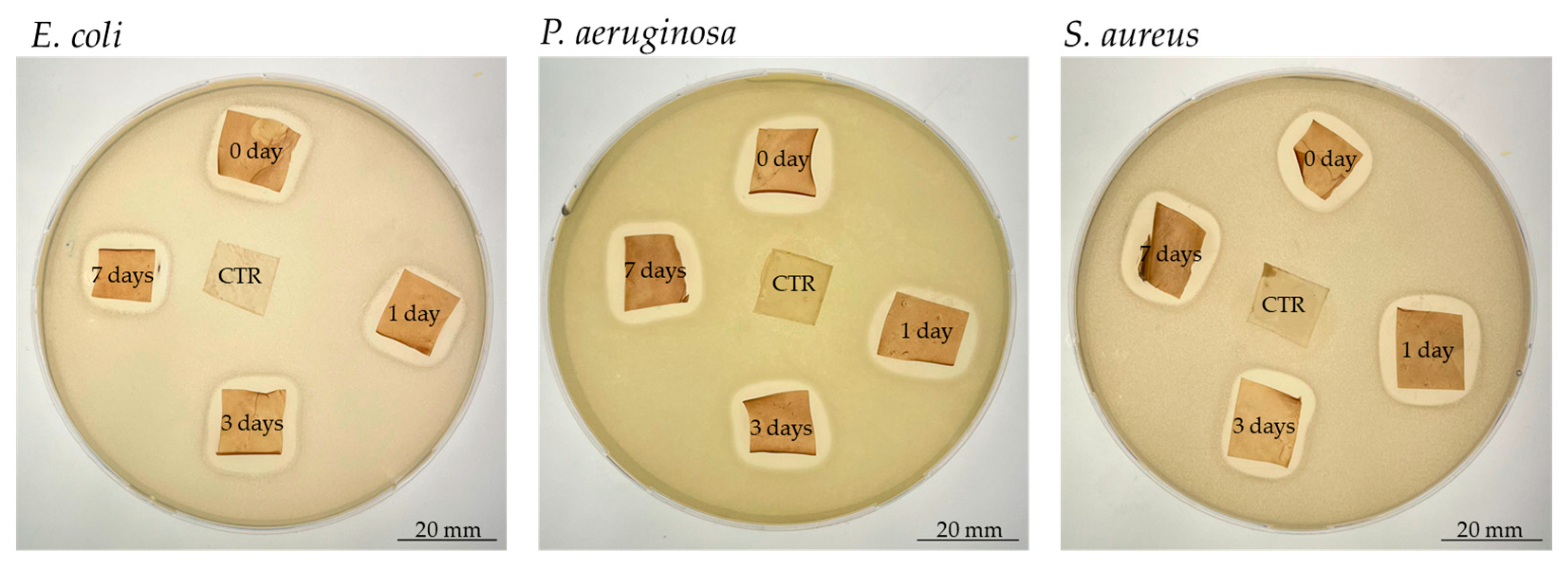
The main focus of this study was to produce SF films with antibacterial properties to prevent infection in superficial wounds. To this end, the films were functionalized with AgNPs, which are known to prevent bacterial colonization. The effect of the treatment was first qualitatively evaluated using the agar inclusion method, whereby the samples were fully immersed in liquid agar prior to solidification to prevent them losing their shape upon drying. As shown in Figure 3, the treated samples exhibited antibacterial properties against both Gram-positive and Gram-negative bacteria, while the control was fully covered by bacterial growth. Even after 7 days of degradation, a clearly visible zone of bacterial growth inhibition was evident around the films, with a width value greater than 1 mm in all cases. This confirms ‘good’ antibacterial efficacy according to the standard over time (Table 3).
The results of the quantitative tests, reported in Table 4, confirm these results and allow their quantification in terms of reducing bacterial proliferation. At 6 h, 92% decrease was observed for E. coli, 89% for P. aeruginosa, and 70% for S. aureus, the latter antibiotic-resistant strain. At the first degradation time point (t = 0 days), antibacterial activity was lower than subsequent time points, where an increase in antibacterial capacity was observed instead, indicating that silver treatment retains its efficacy over time.

4. Discussion

Bacterial colonization can compromise wound healing, resulting in an increased risk of localized or systemic infections [5]. In this context, functionalized biopolymer-based dressings are widely proposed to promote wound healing and prevent microbial contamination [59,60,61]. Silk fibroin, in particular, has been confirmed as a promising biomaterial due to its biocompatibility, conformability, and ability to maintain a moist environment favorable for tissue regeneration [22,62,63]. Integrating AgNPs into fibroin significantly enhances antibacterial activity, rendering such systems suitable for treating infected or potentially infected wounds [52,54,64,65].
In this work, antibacterial silk fibroin films were developed through the in situ synthesis of AgNPs via UV-assisted photoreduction, based on a technology previously applied to a variety of natural and synthetic substrates, including medical devices, cotton and linen gauze, surgical sutures, and catheters [52,66,67]. The technology relies on a photo-assisted deposition process in which a silver precursor is converted into metallic silver under UV irradiation, leading to the simultaneous in situ synthesis and deposition of metallic silver clusters onto the substrate surface, resulting in a stable coating characterized by homogeneous distribution and strong adhesion of the metallic phase [68]. In the specific case of fibroin, never explored before, this strategy combines two processes occurring during the treatment, namely the structural stabilization of fibroin induced by methanol and the antimicrobial functionalization through silver deposition. At this purpose, the first part of the study was addressed to the investigation of the effect of methanol on the protein structure, which is related to chemical-physical and mechanical properties. Then, the antibacterial capability of the silver treated fibroin films was evaluated against relevant strains in wound infections, thus assessing the potential of the developed devices for the specific application.
The structural characterization confirmed that methanol treatment played a key role in stabilizing the fibroin matrix. FT-IR analysis revealed the typical shift of the amide I and amide II bands associated with the transition from random coil/α- helix conformation (silk I) to the β-sheet-rich silk II structure, which is widely recognized as the main mechanism responsible for the water stability of regenerated fibroin-derived materials. Alcohol-induced β-sheet formation has been extensively reported in literature and represents one of the most common strategies to convert soluble fibroin films into insoluble and mechanically stable matrices [34,35,36]. DSC analysis showed a slight shift in the degradation peak of SF_Ag, SF_M, and SF_M/UV compared to the SF control, demonstrating the influence of the treatments on the crystallization [27,69,70,71,72].
Consistently with this structural transition, untreated fibroin films did not maintain integrity in aqueous environment, whereas methanol treated samples exhibited sufficient stability to allow further testing under hydrated conditions. Although UV irradiation was not applied as an independent treatment, the comparison between methanol-treated samples with and without irradiation suggests that the irradiation step may further influence the mechanical behavior of the films. Importantly, this effect cannot be directly attributed to an additional increase in β-sheet crystallinity, as FT-IR spectra did not show substantial changes in the characteristic amide bands after UV exposure. Indeed, previous studies have reported that UV irradiation of silk fibroin can induce modifications in network organization rather than in the secondary structure of the protein [69]. Such processes may increase the density of intermolecular interaction within the amorphous regions of the fibroin matrix, potentially contributing to the observed increase in stiffness without significant changes in β-sheet content, also explaining the fast dissolution of the UV-treated only samples in aqueous environment. The incorporation of AgNPs further enhanced the mechanical performance of the films. The increase in Young’s modulus observed in the silver-functionalized samples may be associated with the reinforcing effect commonly reported for inorganic NPs dispersed within the polymer matrices [14,41]. In fibroin-based systems, metallic NPs can interact with functional groups of the protein backbone and restrict the mobility of the polymer chain, resulting in a stiffer network. Similar reinforcement effects have been described in other silk–nanoparticle composites, where the presence of nanofillers contributes to improving mechanical stability while maintaining the intrinsic biocompatibility of the fibroin matrix [73].
The swelling behavior and degradation resistance of the films also reflected the structural modifications induced by the treatment. Methanol-treated samples displayed a relatively high swelling capacity, indicating the presence of hydrated amorphous domains within the fibroin network. On the other hand, silver functionalized films exhibited a lower swelling degree, suggesting that the presence of the NPs and the associated intermolecular interaction may reduce the chain mobility in the macromolecule and limit the water diffusion into the matrix [42,43]. At the same time, the treated films showed improved resistance to degradation under physiological conditions, confirming the stabilizing effect of β-sheet formation combined with NPs [44].
From an application perspective, the reduced swelling observed for the silver-treated films may be advantageous for specific wound types. While highly absorbent materials are typically preferred for heavily exuding wounds, films with moderate swelling and improved structural stability may be more suitable for superficial or low-exudating wounds, where maintaining structural integrity and preventing microbial colonization are key requirements.
The antibacterial tests confirmed that the incorporation of AgNPs provided strong antimicrobial activity against both Gram-positive and Gram-negative bacterial strains relevant for wound infections. Silver is known to exert its antimicrobial activity through multiple mechanisms, including membrane disruption, interaction with intracellular components, interference with DNA replication and generation of reactive oxygen species [43,54,74,75]. Importantly, beyond antimicrobial activity, previous studies on this technology have demonstrated that silver functionalized materials exhibit good biocompatibility and positively contributed to the wound healing process, promoting tissue regeneration and supporting a favorable biological response [76,77,78,79]. The inhibition zones observed for E. coli, P. aeruginosa and methicillin-resistant S. aureus in the qualitative assays, together with the marked reduction in bacterial proliferation detected in the quantitative assays confirmed the broad-spectrum antibacterial potential of silver-based materials in this study. Importantly, the antibacterial activity was maintained even after several days of incubation in physiological-like conditions, thus suggesting that the silver functionalization process leads simultaneously to a stable and an antibacterial surface and supporting the effectiveness of the technology in producing devices suitable for wound dressing applications. Future in-depth studies will be conducted to better elucidate the underlaying mechanisms and long-term performance of these systems.

5. Conclusions

In this work, silk fibroin films functionalized with silver nanoparticles were successfully developed through an in situ UV-assisted photoreduction process, demonstrating the effectiveness of this approach in producing structurally stable and antibacterial biomaterials for wound dressing applications. The combined effect of methanol-induced β-sheet formation and silver incorporation resulted in a significant enhancement of mechanical properties and resistance to degradation, while maintaining a controlled swelling behavior. At the same time, the films exhibited sustained and broad-spectrum antibacterial activity against clinically relevant bacterial strains.
From an application perspective, these results highlight the potential of this approach to produce advanced dressing materials specifically suited for superficial and low-exudate wounds, where structural integrity and long-lasting antimicrobial efficacy with demonstrated biocompatibility further supports its relevance for biomedical applications.
However, some aspects deserve further investigation. Although previously investigated on different substrates treated through the same deposition technology, a detailed analysis of silver release kinetics, along with a direct correlation of these data with antibacterial performances and biocompatibility will be addressed in future studies.

Author Contributions

Conceptualization, A.M., R.P., F.P. and M.P.; methodology, A.M. and R.P.; software, R.P.; validation, F.P. and M.P.; formal analysis, A.M. and R.P.; investigation, A.M. and R.P.; resources, F.P. and M.P.; data curation, A.M. and R.P.; writing—original draft preparation, A.M. and R.P.; writing—review and editing, A.M., R.P., F.P. and M.P.; visualization, A.M., R.P., F.P. and M.P.; supervision, F.P. and M.P.; project administration, F.P. and M.P.; funding acquisition, F.P. and M.P. All authors have read and agreed to the published version of the manuscript.

Funding

This research received no external funding.

Institutional Review Board Statement

Not applicable.

Data Availability Statement

The raw data supporting the conclusions of this article will be made available by the authors on request.

Acknowledgments

The authors would like to acknowledge Angelica Panico (Caresilk S.r.l.s., Italy) for her kind support in this research.

Conflicts of Interest

The fibroin solution used in this study was provided by Caresilk S.r.l.s. The company was not involved in the study design, collection, analysis, interpretation of data, the writing of this article or the decision to submit it for publication.

Abbreviations

The following abbreviations are used in this manuscript:
WHOWorld Health Organization
NPsNanoparticles
AuNPsGold Nanoparticles
ZnONPsZinc Oxide Nanoparticles
CuNPsCopper Nanoparticles
CuONPsCopper Oxide Nanoparticles
AgNPsSilver nanoparticles
UVUltraviolet
TSATrypticasein soy agar
PBSPhosphate-buffered saline
TSBTryptic Soy Broth
RTRoom Temperature
SDStandard Deviation
FT-IRFourier-transform infrared spectroscopy
ATRAttenuated Total Reflectance
DSCDifferential scanning calorimetry
BCABicinchoninic acid
ATCCAmerican Type Culture Collection
ODOptical Density
CFUColony Forming Unit
ABEAntibacterial efficacy
PpmParts per million
ROSReactive Oxygen Species

References

  1. Lee, H.-J.; Kim, M. Skin Barrier Function and the Microbiome. Int. J. Mol. Sci. 2022, 23, 13071. [Google Scholar] [CrossRef]
  2. Rajkumar, J.; Chandan, N.; Lio, P.; Shi, V. The Skin Barrier and Moisturization: Function, Disruption, and Mechanisms of Repair. Ski. Pharmacol. Physiol. 2023, 36, 174–185. [Google Scholar] [CrossRef] [PubMed]
  3. Panico, A.; Paladini, F.; Sannino, A.; Pollini, M. Antibacterial silver treatments on polymeric membranes for fouling control and disinfection in water filtration. J. Appl. Polym. Sci. 2016, 133. [Google Scholar] [CrossRef]
  4. Souto-Silva, M.V.; Bispo, E.C.I.; de Oliveira, N.N.; Pogue, R.; Zonari, A.; Saldanha-Araujo, F.; Carvalho, J.L. The cytokine–skin barrier axis in health and disease. Cytokine Growth Factor Rev. 2026, 87, 113–123. [Google Scholar] [CrossRef]
  5. Percival, N.J. Classification of Wounds and their Management. Surg.-Oxf. Int. Ed. 2002, 20, 114–117. [Google Scholar] [CrossRef]
  6. Yadav, R.; Kumar, R.; Kathpalia, M.; Ahmed, B.; Dua, K.; Gulati, M.; Singh, S.; Singh, P.J.; Kumar, S.; Shah, R.M.; et al. Innovative approaches to wound healing: Insights into interactive dressings and future directions. J. Mater. Chem. B 2024, 12, 7977–8006. [Google Scholar] [CrossRef] [PubMed]
  7. Uberoi, A.; McCready-Vangi, A.; Grice, E.A. The wound microbiota: Microbial mechanisms of impaired wound healing and infection. Nat. Rev. Microbiol. 2024, 22, 507–521. [Google Scholar] [CrossRef]
  8. Gaspar-Pintiliescu, A.; Stanciuc, A.M.; Craciunescu, O. Natural composite dressings based on collagen, gelatin and plant bioactive compounds for wound healing: A review. Int. J. Biol. Macromol. 2019, 138, 854–865. [Google Scholar] [CrossRef]
  9. Bianchera, A.; Catanzano, O.; Boateng, J.; Elviri, L. The Place of Biomaterials in Wound Healing. In Therapeutic Dressings and Wound Healing Applications; John Wiley & Sons: Hoboken, NJ, USA, 2020; pp. 337–366. [Google Scholar]
  10. Singh, B.; Sharma, S.; Dhiman, A. Design of antibiotic containing hydrogel wound dressings: Biomedical properties and histological study of wound healing. Int. J. Pharm. 2013, 457, 82–91. [Google Scholar] [CrossRef]
  11. Costa, N.N.; de Faria Lopes, L.; Ferreira, D.F.; de Prado, E.M.L.; Severi, J.A.; Resende, J.A.; de Paula Careta, F.; Ferreira, M.C.P.; Carreira, L.G.; de Souza, S.O.L.; et al. Polymeric films containing pomegranate peel extract based on PVA/starch/PAA blends for use as wound dressing: In vitro analysis and physicochemical evaluation. Mater. Sci. Eng. C Mater. Biol. Appl. 2020, 109, 110643. [Google Scholar] [CrossRef] [PubMed]
  12. Dong, R.; Guo, B. Smart wound dressings for wound healing. Nano Today 2021, 41, 101290. [Google Scholar] [CrossRef]
  13. Deng, Z.H.; Yang, Z.L.; Yi, S.; Liu, Z.Q. Hydrogel-exosome systems in the treatment of refractory diabetic ulcers: Mechanisms, progress, and prospects. Eur. Cells Mater. 2025, 53, 82–97. [Google Scholar] [CrossRef]
  14. Li, H.; Wen, H.; Zhang, H.; Cao, X.; Li, L.; Hu, X.; Zhang, Y.; Shen, X.; Shubhra, Q.T.H.; Yang, H.; et al. A multifunctional dihydromyricetin-loaded hydrogel for the sequential modulation of diabetic wound healing and glycemic control. Burns Trauma 2025, 13, tkaf024. [Google Scholar] [CrossRef]
  15. Mazurek, Ł.; Szudzik, M.; Rybka, M.; Konop, M. Silk Fibroin Biomaterials and Their Beneficial Role in Skin Wound Healing. Biomolecules 2022, 12, 1852. [Google Scholar] [CrossRef]
  16. Lehmann, T.; Vaughn, A.E.; Seal, S.; Liechty, K.W.; Zgheib, C. Silk Fibroin-Based Therapeutics for Impaired Wound Healing. Pharmaceutics 2022, 14, 651. [Google Scholar] [CrossRef]
  17. Ju, H.W.; Lee, O.J.; Lee, J.M.; Moon, B.M.; Park, H.J.; Park, Y.R.; Lee, M.C.; Kim, S.H.; Chao, J.R.; Ki, C.S.; et al. Wound healing effect of electrospun silk fibroin nanomatrix in burn-model. Int. J. Biol. Macromol. 2016, 85, 29–39. [Google Scholar] [CrossRef]
  18. Park, Y.R.; Sultan, M.T.; Park, H.J.; Lee, J.M.; Ju, H.W.; Lee, O.J.; Lee, D.J.; Kaplan, D.L.; Park, C.H. NF-κB signaling is key in the wound healing processes of silk fibroin. Acta Biomater. 2018, 67, 183–195. [Google Scholar] [CrossRef]
  19. Martínez-Mora, C.; Mrowiec, A.; García-Vizcaíno, E.M.; Alcaraz, A.; Cenis, J.L.; Nicolás, F.J. Fibroin and sericin from Bombyx mori silk stimulate cell migration through upregulation and phosphorylation of c-Jun. PLoS ONE 2012, 7, e42271. [Google Scholar] [CrossRef] [PubMed]
  20. Chen, Z.; Cheng, J.; Wang, M.; Jiang, Y.; Jiang, D.; Yang, Y.; Bai, Y.; Wang, L.; Ma, C.; Ye, J.; et al. In situ forming silk fibroin hydrogel dressing accelerates acute wound healing via immunomodulation and extracellular matrix regeneration. J. Control. Release 2025, 387, 114228. [Google Scholar] [CrossRef] [PubMed]
  21. You, C.; Wang, C.; Ma, Z.; Yu, Q.; Liu, S. Review on application of silk fibroin hydrogels in the management of wound healing. Int. J. Biol. Macromol. 2025, 298, 140082. [Google Scholar] [CrossRef]
  22. Panico, A.; Paladini, F.; Pollini, M. Development of regenerative and flexible fibroin-based wound dressings. J. Biomed. Mater. Res. Part B Appl. Biomater. 2019, 107, 7–18. [Google Scholar] [CrossRef] [PubMed]
  23. Vidya, M.; Rajagopal, S. Silk Fibroin: A Promising Tool for Wound Healing and Skin Regeneration. Int. J. Polym. Sci. 2021, 2021, 9069924. [Google Scholar] [CrossRef]
  24. De Giorgio, G.; Matera, B.; Vurro, D.; Manfredi, E.; Galstyan, V.; Tarabella, G.; Ghezzi, B.; D’Angelo, P. Silk Fibroin Materials: Biomedical Applications and Perspectives. Bioengineering 2024, 11, 167. [Google Scholar] [CrossRef]
  25. Liu, T.L.; Miao, J.C.; Sheng, W.H.; Xie, Y.F.; Huang, Q.; Shan, Y.B.; Yang, J.C. Cytocompatibility of regenerated silk fibroin film: A medical biomaterial applicable to wound healing. J. Zhejiang Univ. Sci. B 2010, 11, 10–16. [Google Scholar] [CrossRef]
  26. Johari, N.; Moroni, L.; Samadikuchaksaraei, A. Tuning the conformation and mechanical properties of silk fibroin hydrogels. Eur. Polym. J. 2020, 134, 109842. [Google Scholar] [CrossRef]
  27. Kaewpirom, S.; Boonsang, S. Influence of alcohol treatments on properties of silk-fibroin-based films for highly optically transparent coating applications. RSC Adv. 2020, 10, 15913–15923. [Google Scholar] [CrossRef]
  28. Hofmann, S.; Foo, C.T.; Rossetti, F.; Textor, M.; Vunjak-Novakovic, G.; Kaplan, D.L.; Merkle, H.P.; Meinel, L. Silk fibroin as an organic polymer for controlled drug delivery. J. Control. Release 2006, 111, 219–227. [Google Scholar] [CrossRef]
  29. Pritchard, E.; Kaplan, D. Silk Fibroin Biomaterials for Controlled Release Drug Delivery. Expert Opin. Drug Deliv. 2011, 8, 797–811. [Google Scholar] [CrossRef]
  30. Ghalei, S.; Handa, H. A review on antibacterial silk fibroin-based biomaterials: Current state and prospects. Mater. Today Chem. 2022, 23, 100673. [Google Scholar] [CrossRef] [PubMed]
  31. Bidossi, A.; Bottagisio, M.; Logoluso, N.; De Vecchi, E. In Vitro Evaluation of Gentamicin or Vancomycin Containing Bone Graft Substitute in the Prevention of Orthopedic Implant-Related Infections. Int. J. Mol. Sci. 2020, 21, 9250. [Google Scholar] [CrossRef] [PubMed]
  32. Nair, M.B.; Kretlow, J.D.; Mikos, A.G.; Kasper, F.K. Infection and tissue engineering in segmental bone defects—A mini review. Curr. Opin. Biotechnol. 2011, 22, 721–725. [Google Scholar] [CrossRef] [PubMed]
  33. Khatoon, Z.; McTiernan, C.D.; Suuronen, E.J.; Mah, T.-F.; Alarcon, E.I. Bacterial biofilm formation on implantable devices and approaches to its treatment and prevention. Heliyon 2018, 4, e01067. [Google Scholar] [CrossRef]
  34. Gebreyohannes, G.; Nyerere, A.; Bii, C.; Sbhatu, D.B. Challenges of intervention, treatment, and antibiotic resistance of biofilm-forming microorganisms. Heliyon 2019, 5, e02192. [Google Scholar] [CrossRef]
  35. Sánchez-López, E.; Gomes, D.; Esteruelas, G.; Bonilla, L.; Lopez-Machado, A.L.; Galindo, R.; Cano, A.; Espina, M.; Ettcheto, M.; Camins, A.; et al. Metal-Based Nanoparticles as Antimicrobial Agents: An Overview. Nanomaterials 2020, 10, 292. [Google Scholar] [CrossRef]
  36. Aguilar-Marcelino, L.; Mendoza-de-Gives, P.; Al-Ani, L.K.T.; López-Arellano, M.E.; Gómez-Rodríguez, O.; Villar-Luna, E.; Reyes-Guerrero, D.E. Chapter 26—Using molecular techniques applied to beneficial microorganisms as biotechnological tools for controlling agricultural plant pathogens and pest. In Molecular Aspects of Plant Beneficial Microbes in Agriculture; Sharma, V., Salwan, R., Al-Ani, L.K.T., Eds.; Academic Press: Cambridge, MA, USA, 2020; pp. 333–349. [Google Scholar]
  37. Stensberg, M.C.; Wei, Q.; McLamore, E.S.; Porterfield, D.M.; Wei, A.; Sepúlveda, M.S. Toxicological studies on silver nanoparticles: Challenges and opportunities in assessment, monitoring and imaging. Nanomedicine 2011, 6, 879–898. [Google Scholar] [CrossRef]
  38. Yuan, P.; Ding, X.; Yang, Y.Y.; Xu, Q.-H. Metal Nanoparticles for Diagnosis and Therapy of Bacterial Infection. Adv. Healthc. Mater. 2018, 7, 1701392. [Google Scholar] [CrossRef]
  39. Abdel-Kareem, M.M.; Zohri, A.A. Extracellular mycosynthesis of gold nanoparticles using Trichoderma hamatum: Optimization, characterization and antimicrobial activity. Lett. Appl. Microbiol. 2018, 67, 465–475. [Google Scholar] [CrossRef] [PubMed]
  40. Zhang, Y.; Shareena Dasari, T.P.; Deng, H.; Yu, H. Antimicrobial Activity of Gold Nanoparticles and Ionic Gold. J. Environ. Sci. Health C Environ. Carcinog. Ecotoxicol. Rev. 2015, 33, 286–327. [Google Scholar] [CrossRef] [PubMed]
  41. Lara, H.H.; Ayala-Nuñez, N.V.; Ixtepan-Turrent, L.; Rodriguez-Padilla, C. Mode of antiviral action of silver nanoparticles against HIV-1. J. Nanobiotechnol. 2010, 8, 1. [Google Scholar] [CrossRef]
  42. Kumar, S.V.; Bafana, A.P.; Pawar, P.; Faltane, M.; Rahman, A.; Dahoumane, S.A.; Kucknoor, A.; Jeffryes, C.S. Optimized production of antibacterial copper oxide nanoparticles in a microwave-assisted synthesis reaction using response surface methodology. Colloids Surf. A Physicochem. Eng. Asp. 2019, 573, 170–178. [Google Scholar] [CrossRef]
  43. Pollini, M.; D’Urso, F.; Broccolo, F.; Paladini, F. Silver Nanoparticle–Silk Protein Nanocomposites: A Synergistic Biomimetic Approach for Advanced Antimicrobial Applications. Biomimetics 2025, 10, 669. [Google Scholar] [CrossRef]
  44. Das, C.G.A.; Kumar, V.G.; Dhas, T.S.; Karthick, V.; Govindaraju, K.; Joselin, J.M.; Baalamurugan, J. Antibacterial activity of silver nanoparticles (biosynthesis): A short review on recent advances. Biocatal. Agric. Biotechnol. 2020, 27, 101593. [Google Scholar] [CrossRef]
  45. Tripathi, N.; Goshisht, M.K. Recent Advances and Mechanistic Insights into Antibacterial Activity, Antibiofilm Activity, and Cytotoxicity of Silver Nanoparticles. ACS Appl. Bio Mater. 2022, 5, 1391–1463. [Google Scholar] [CrossRef]
  46. Dakal, T.C.; Kumar, A.; Majumdar, R.S.; Yadav, V. Mechanistic Basis of Antimicrobial Actions of Silver Nanoparticles. Front. Microbiol. 2016, 7, 1831. [Google Scholar] [CrossRef]
  47. Losasso, C.; Belluco, S.; Cibin, V.; Zavagnin, P.; Mičetić, I.; Gallocchio, F.; Zanella, M.; Bregoli, L.; Biancotto, G.; Ricci, A. Antibacterial activity of silver nanoparticles: Sensitivity of different Salmonella serovars. Front. Microbiol. 2014, 5, 227. [Google Scholar] [CrossRef] [PubMed]
  48. Gordon, O.; Vig Slenters, T.; Brunetto Priscilla, S.; Villaruz Amer, E.; Sturdevant Daniel, E.; Otto, M.; Landmann, R.; Fromm Katharina, M. Silver Coordination Polymers for Prevention of Implant Infection: Thiol Interaction, Impact on Respiratory Chain Enzymes, and Hydroxyl Radical Induction. Antimicrob. Agents Chemother. 2010, 54, 4208–4218. [Google Scholar] [CrossRef]
  49. Li, Y.; Zha, X.; Xiong, X.; Zhang, Y.; Feng, Y.; Xie, H.; Zhang, L.; Jiang, Q. A Promising Wound Dressing from Regenerated Silk Fibroin Sponge with Sustain-ed Release of Silver Nanoparticles. J. Renew. Mater. 2021, 9, 295–310. [Google Scholar] [CrossRef]
  50. Li, Y.; Yong, D.; Shen, J.; Bian, R.; Wang, Y. Silver nanoparticle-loaded konjac glucomannan/silk fibroin composite hydrogels for enhanced wound healing. Int. J. Biol. Macromol. 2025, 300, 140199. [Google Scholar] [CrossRef] [PubMed]
  51. Pollini, M.; Paladini, F. The Emerging Role of Silk Fibroin for the Development of Novel Drug Delivery Systems. Biomimetics 2024, 9, 295. [Google Scholar] [CrossRef] [PubMed]
  52. Paladini, F.; Russo, F.; Masi, A.; Lanzillotti, C.; Sannino, A.; Pollini, M. Silver-Treated Silk Fibroin Scaffolds for Prevention of Critical Wound Infections. Biomimetics 2024, 9, 659. [Google Scholar] [CrossRef]
  53. Paladini, F.; Sannino, A.; Pollini, M. In vivo testing of silver treated fibers for the evaluation of skin irritation effect and hypoallergenicity. J. Biomed. Mater. Res. B Appl. Biomater. 2014, 102, 1031–1037. [Google Scholar] [CrossRef]
  54. Pellegrino, R.; Lanzillotti, C.; Pollini, M.; Paladini, F. UV-Assisted Silver Functionalization of Cotton Gauze for Antimicrobial and Biocompatible Wound Healing Applications. Microorganisms 2026, 14, 213. [Google Scholar] [CrossRef]
  55. Wang, H.-Y.; Zhang, Y.-Q. Effect of regeneration of liquid silk fibroin on its structure and characterization. Soft Matter 2013, 9, 138–145. [Google Scholar] [CrossRef]
  56. Chen, X.; Shao, Z.; Marinkovic, N.S.; Miller, L.M.; Zhou, P.; Chance, M.R. Conformation transition kinetics of regenerated Bombyx mori silk fibroin membrane monitored by time-resolved FTIR spectroscopy. Biophys. Chem. 2001, 89, 25–34. [Google Scholar] [CrossRef]
  57. Wang, R.; Li, X.; Yu, G.; Ren, L.; Liu, J.; Jiao, T. Construction of silk fibroin-based composite nanofiber films with effective antibacterial and filtration properties. Chem. Eng. J. 2026, 527, 171596. [Google Scholar] [CrossRef]
  58. Pallod, S.; DeCambra, W.; Chowdhury, T.; Ristich, E.; Cherry, B.; Zhang, R.; Arturo Ruiz Pardo, D.; Daggett-Vondras, J.; Yaron, J.R.; Yarger, J.L.; et al. Modulation of Secondary Structure, Bioavailability, Immunomodulation, and Tissue Repair Outcomes Using Differential Processing of Silk Biomaterials. ACS Biomater. Sci. Eng. 2025, 11, 4881–4897. [Google Scholar] [CrossRef]
  59. Su, X.; Zhang, J.; Gong, P.; Zou, H. Multifunctional Wound Dressings Based on Biopolymers for Chronic Wound Healing. ACS Appl. Polym. Mater. 2026, 8, 706–724. [Google Scholar] [CrossRef]
  60. Sadati, S.; Swann, M.J.; Percival, S.L.; Charmet, J.; Unnikrishnan, M.; Isakov, D. Microbial-Responsive Wound Dressings Based on Biopolymer Degradation Strategy for Detecting Bacterial Infections. ACS Appl. Mater. Interfaces 2026, 18, 7884–7899. [Google Scholar] [CrossRef] [PubMed]
  61. Ghosh, A.; Das, A. Underpinning the Therapeutic Potential of Biopolymer-Based Nano Medications in Wound Healing. In Functionalized Nanoparticles Hydrogels for Wound Healing; Mandal, A.K., Sarkar, H., Vega-Baudrit, J.R., Eds.; Springer Nature: Singapore, 2025; pp. 123–141. [Google Scholar]
  62. Mashhadi, R.; Nourmohammadi, J.; Tavakol, M. Enzymatically crosslinked pectin–silk fibroin hydrogel with embedded ZnO@Gallic acid nanoparticles: An injectable, self-healing, and antibacterial wound dressing. Int. J. Biol. Macromol. 2026, 349, 150823. [Google Scholar] [CrossRef]
  63. Lyu, H.; Deng, N.; Wang, J.; Dai, D.; Sun, Z.; Guo, C. Bioactive Chemically Modified Silk Fibroin Accelerates Wound Healing via the Regulation of Macrophage Polarization. ACS Appl. Bio Mater. 2026, 9, 2167–2181. [Google Scholar] [CrossRef] [PubMed]
  64. Sneka, A.; Subbarao, R.B.; Selvamuthukumaran, T.; Muneer, S. Development of Antimicrobial Films Using Eri Silk Fibroin Blended With Green-Synthesized Silver Nanoparticles Against Common Human Pathogens. Biopolymers 2026, 117, e70078. [Google Scholar] [CrossRef]
  65. Wang, D.-L.; Wu, D.-N.; Chen, Y.-C.; Kuo, W.-K.; Wei, J.-B.; Lin, C.-H. Eco-friendly synthesis of genipin-crosslinked silk fibroin–silver nanoparticle films with improved mechanical strength and long-term antibacterial stability. Appl. Nanosci. 2026, 16, 13. [Google Scholar] [CrossRef]
  66. Paladini, F.; Panico, A.; Masi, A.; Russo, F.; Sannino, A.; Pollini, M. Silver-Treated Sutures for the Prevention of Biofilm-Associated Surgical Site Infections. Antibiotics 2025, 14, 49. [Google Scholar] [CrossRef]
  67. Cooper, I.R.; Pollini, M.; Paladini, F. The potential of photo-deposited silver coatings on Foley catheters to prevent urinary tract infections. Mater. Sci. Eng. C Mater. Biol. Appl. 2016, 69, 414–420. [Google Scholar] [CrossRef]
  68. Raho, R.; Paladini, F.; Lombardi, F.A.; Boccarella, S.; Zunino, B.; Pollini, M. In-situ photo-assisted deposition of silver particles on hydrogel fibers for antibacterial applications. Mater. Sci. Eng. C 2015, 55, 42–49. [Google Scholar] [CrossRef]
  69. Lee, S.; Kim, S.H.; Jo, Y.-Y.; Ju, W.-T.; Kim, H.-B.; Kweon, H. Effects of Ultraviolet Light Irradiation on Silk Fibroin Films Prepared under Different Conditions. Biomolecules 2021, 11, 70. [Google Scholar] [CrossRef]
  70. Motta, A.; Fambri, L.; Migliaresi, C. Regenerated silk fibroin films: Thermal and dynamic mechanical analysis. Macromol. Chem. Phys. 2002, 203, 1658–1665. [Google Scholar] [CrossRef]
  71. Li, Y.; Zhao, L.; Wang, H.; Li, B. Fabrication and characterization of microencapsulated n-octadecane with silk fibroin–silver nanoparticles shell for thermal regulation. J. Mater. Res. 2019, 34, 2047–2056. [Google Scholar] [CrossRef]
  72. Dhas, S.P.; Anbarasan, S.; Mukherjee, A.; Chandrasekaran, N. Biobased silver nanocolloid coating on silk fibers for prevention of post-surgical wound infections. Int. J. Nanomed. 2015, 10, 159–170. [Google Scholar] [CrossRef]
  73. Xu, Z.; Shi, L.; Yang, M.; Zhu, L. Preparation and biomedical applications of silk fibroin-nanoparticles composites with enhanced properties—A review. Mater. Sci. Eng. C 2019, 95, 302–311. [Google Scholar] [CrossRef] [PubMed]
  74. Pollini, M.; Paladini, F.; Licciulli, A.; Maffezzoli, A.; Sannino, A. Engineering Nanostructured Silver Coatings for Antimicrobial Applications. In Nano-Antimicrobials: Progress and Prospects; Cioffi, N., Rai, M., Eds.; Springer: Berlin/Heidelberg, Germany, 2012; pp. 313–336. [Google Scholar]
  75. Fan, M.; Si, J.; Xu, X.; Chen, L.; Chen, J.; Yang, C.; Zhu, J.; Wu, L.; Tian, J.; Chen, X.; et al. A versatile chitosan nanogel capable of generating AgNPs in-situ and long-acting slow-release of Ag+ for highly efficient antibacterial. Carbohydr. Polym. 2021, 257, 117636. [Google Scholar] [CrossRef]
  76. Paladini, F.; Di Franco, C.; Panico, A.; Scamarcio, G.; Sannino, A.; Pollini, M. In Vitro Assessment of the Antibacterial Potential of Silver Nano-Coatings on Cotton Gauzes for Prevention of Wound Infections. Materials 2016, 9, 411. [Google Scholar] [CrossRef] [PubMed]
  77. Picca, R.A.; Paladini, F.; Sportelli, M.C.; Pollini, M.; Giannossa, L.C.; Di Franco, C.; Panico, A.; Mangone, A.; Valentini, A.; Cioffi, N. Combined Approach for the Development of Efficient and Safe Nanoantimicrobials: The Case of Nanosilver-Modified Polyurethane Foams. ACS Biomater. Sci. Eng. 2017, 3, 1417–1425. [Google Scholar] [CrossRef]
  78. Gallo, A.L.; Pollini, M.; Paladini, F. A combined approach for the development of novel sutures with antibacterial and regenerative properties: The role of silver and silk sericin functionalization. J. Mater. Sci. Mater. Med. 2018, 29, 133. [Google Scholar] [CrossRef] [PubMed]
  79. Paladini, F.; Lanzillotti, C.; Panico, A.; Pollini, M. Biological Evaluation of Silver-Treated Silk Fibroin Scaffolds for Application as Antibacterial and Regenerative Wound Dressings. Nanomaterials 2025, 15, 919. [Google Scholar] [CrossRef] [PubMed]
Figure 1. Film characterization: (a) Visual observation of SF and SF_Ag films; (b) FT-IR spectra of SF (black), SF_M (green), SF_M/UV (red), and SF_Ag (blue) films; (c) DSC curves of SF (black), SF_M (green), SF_M/UV (red), and SF_Ag (blue) films; (d) representative graph of stress–strain plot obtained in tensile tests of SF_M (green), SF_M/UV (red), and SF_Ag (blue) films.
Macromol 06 00029 g001
Figure 2. Films characterization: (a) swelling degree of SF_M (green), SF_M/UV (red) and SF_Ag (blue); (b) protein content in SF_M, SF_M/UV and SF_Ag films over time. * p < 0.05; ** p < 0.01; *** p < 0.001; **** p < 0.0001.
Macromol 06 00029 g002
Figure 3. Qualitative antibacterial test on E. coli ATCC 25922, P. aeruginosa ATCC 27853, S. aureus ATCC 43300.
Macromol 06 00029 g003
Table 1. Unreacted and reacted [Ag+] during the process.
Agbefore − Agafter (ppm)Agunreacted (ppm)Agreacted (ppm)
73.91 ± 18.450.20 ± 0.0159.30 ± 15.37
Table 2. Young’s Modulus and maximum strength at failure of SF_M, SF_M/UV, and SF_Ag films.
SampleE (MPa)σmax
SF_M0.23 ± 0.040.03 ± 0.01
SF_M/UV1.48 ± 0.730.28 ± 0.18
SF_Ag7.26 ± 0.461.15 ± 0.03
Table 3. Width of the inhibition zone.
Dimension of the Inhibition Zone (mm)
Degradation DaysE. coliP. aeruginosaS. aureus
0 days2.22 ± 0.522.70 ± 1.10 2.90 ± 0.51
1 days2.04 ± 0.682.03 ± 0.672.50 ± 0.87
3 days2.76 ± 0.97 2.60 ± 0.87 2.09 ± 0.90
7 days 2.58 ± 0.762.66 ± 0.512.71 ± 1.28
Table 4. Antibacterial efficacy (ABE) calculated through quantitative tests.
Antibacterial Efficacy
BacteriaDegradation DaysCFU After 6 hABE (%) 1
E. coli0 days4.89 × 107 92
1 day1.23 × 107 98
3 days1.08 × 107 98
7 days1.65 × 107 97
P. aeruginosa0 days4.10 × 107 89
1 day9.81 × 106 97
3 days1.20 × 107 97
7 days1.40 × 107 96
S. aureus0 days4.60 × 107 70
1 day3.67 × 107 76
3 days2.10 × 107 86
7 days1.10 × 107 93
1 ABE was calculated relative to the control SF_M/UV that presented 6.49 × 10⁸ CFU/mL for E. coli; 3.60 × 10⁸ CFU/mL for P. aeruginosa; and 1.50 × 10⁸ CFU/mL for S. aureus.
Disclaimer/Publisher’s Note: The statements, opinions and data contained in all publications are solely those of the individual author(s) and contributor(s) and not of MDPI and/or the editor(s). MDPI and/or the editor(s) disclaim responsibility for any injury to people or property resulting from any ideas, methods, instructions or products referred to in the content.