Review

Advances in Collagen-/Gelatin-Based Hydrogels: Rheological Properties and Applications

1
Food Science Department, Purdue University, 745 Agriculture Mall Dr., West Lafayette, IN 47907, USA
2
Department of Food Engineering, Faculty of Engineering, Ege University, 35100 Izmir, Türkiye
3
Department of Animal, Veterinary and Food Sciences, University of Idaho, 875 Perimeter Dr. MS 2312, Moscow, ID 83844, USA
*
Authors to whom correspondence should be addressed.
Macromol 2025, 5(4), 55; https://doi.org/10.3390/macromol5040055
Submission received: 30 June 2025 / Revised: 27 August 2025 / Accepted: 8 November 2025 / Published: 11 November 2025

Abstract

Owing to their tunable and biocompatible characteristics, collagen- and gelatin-based hydrogels have gained attention in numerous applications, including biomedical, food, pharmaceutical, and environmental. The gelation mechanisms and resulting network structures of collagen and gelatin differ significantly depending on the presence of intra- and intermolecular crosslinks. These differences enable the tailoring of mechanical properties to achieve desired characteristics in the final product. Mechanical gel strength and elasticity determine how effectively hydrogels can mimic natural tissues and respond to deformations. Probing the rheological properties of these gels enables a deeper understanding of their structure, physical attributes, stability, and release profiles. This review provides an in-depth evaluation of the factors affecting the mechanical strength of collagen- and gelatin-based hydrogels, highlighting the influence of co-molecules and the application of physical, chemical, and mechanical treatments. Herewith, it brings insights into how to manipulate the mechanical properties of these gels to improve their end-use functionality.

1. Introduction

There is a great demand for collagen and gelatin in the food industry because of their high protein content and various functional properties such as water absorption capacity, gel formation, and emulsion stabilization. Collagen and gelatin not only offer nutritional benefits but also serve as versatile biopolymers in the design of advanced material systems. In recent years, gelatin-based hydrogels have attracted considerable interest for use in drug delivery systems, tissue engineering, and food applications due to their biodegradability, biocompatibility, elasticity, flexibility, and non-toxic properties [1]. These hydrogels have emerged as promising materials due to their tunable physical properties and compatibility with bioactive compounds and are now being actively investigated for applications in food packaging, delivery systems, and functional foods [2,3,4].
Collagen (Col) is a fibrillar protein characterized by a unique triple helix structure that provides both high mechanical strength and biological stability [5]. Col structure is stabilized by hydrogen bonds and intermolecular interactions, and due to its hydrophobic nature, collagen is insoluble in water [6]. On the other hand, gelatin, which is derived from the partial hydrolysis of collagen, is water-soluble due to its disrupted triple-helix structure and increased presence of hydrophilic groups. As a result of distinct molecular structures, collagen and gelatin exhibit different gelation behaviors and capabilities. Hydrogels made from collagen offer significant advantages as three-dimensional substrates for cell culture, making them widely applicable in tissue engineering [7].
One of the primary challenges in using Col hydrogels is their insufficient mechanical strength, which limits their ability to provide adequate structural support [8,9]. Moreover, gelatin hydrogels derived from aquatic animals exhibit lower mechanical gel strength compared to those from terrestrial animals, primarily due to differences in amino acid composition and molecular weight associated with their origin [10,11,12].
To overcome these limitations, various treatments to induce physical, chemical, and enzymatic crosslinking have been employed. As a physical crosslinking method, UV irradiation induces the formation of covalent bonds between aromatic amino acid residues, such as tyrosine and phenylalanine, through photochemical reactions. This process enhances the structural integrity of collagen- and gelatin-based gels, although prolonged exposure may lead to photodegradation [13]. Chemical crosslinking of collagen- and gelatin-based gels can be performed using glutaraldehyde, genipin, ethyl-3-(3-dimethylaminopropyl) carbodiimide (EDC), N-hydroxysuccinimide (NHS) active esters [14,15]. These agents primarily interact with the amino groups of collagen and gelatin, leading to the formation of crosslinks between collagen/gelatin molecules [16]. Transglutaminase (TGase), microbial transglutaminase (MTGase), tyrosinase, and laccases are some of the enzymatic crosslinkers used to promote collagen and/or gelatin crosslinking, and therefore to improve the resulting gel strength [17,18,19,20,21]. In addition to crosslinking applications, the rheological properties of collagen- and gelatin-based hydrogels can also be improved through the addition of biomacromolecules [22,23] or through charge modification with treatments including phosphorylation [24,25] and salt treatment [26]. In the food industry, gels are desired to be strong and elastic for various applications, like stabilizing emulsions, providing a desirable mouthfeel, and maintaining structural integrity in processed foods. On the other hand, for biomedical applications such as drug delivery, tissue engineering, or injectable hydrogels, gels do not always need to be highly strong and elastic [27,28,29,30], whereas for scaffold applications, an improved mechanical strength of the gel is desired. For instance, in encapsulation and drug delivery applications, collagen- and gelatin-based hydrogels are expected to remain stable for a certain period of time by withstanding the applied deformations before dissolving [31].
All this information highlights the importance of regulating the mechanical strength of collagen and gelatin hydrogels based on the intended application. Therefore, the aim of this review is to bring an in-depth understanding of how to manipulate the rheological properties of collagen and gelatin hydrogels based on their end-use.
Structural properties of collagen and its derivatives and their application in the food industry have been discussed by Tang et al. [10]. The methods to modify the functional properties of fish gelatin have been reviewed to bring alternatives to mammalian gelatin used in food processing [11,17,32]. Other studies evaluated the impact of source and extraction protocols on the structural and functional properties of gelatin [33,34]. In addition, collagen- and gelatin-based hydrogels have been predominantly studied for tissue engineering, wound healing, and other biomedical applications [35,36,37,38]. According to current literature, the rheological properties of collagen- and gelatin-based hydrogels have not been thoroughly addressed. This review is novel as it provides a comprehensive evaluation of the factors influencing the stability of both collagen- and gelatin-based hydrogels, across various applications, from a rheological perspective, and highlighting the role of co-molecules as well as the effects of diverse chemical and mechanical treatments.

2. Properties of Collagen and Gelatin

2.1. Collagen

Collagen is a fibrous protein that constitutes the major component of the extracellular matrix, a structural network supporting cells, and accounts for about 30% of the animal protein content. Col is characterized by a distinctive structural motif, where three parallel polypeptide chains, each in a left-handed polyproline type II helix conformation, coil around one another with a one-residue stagger to create a right-handed triple helix (Figure 1) [5,10]. Each α-helix features a distinctive repetitive amino acid sequence, Gly-X-Y, where X and Y are typically proline and hydroxyproline, respectively. Every third amino acid residue in each α-chain is located at the center of the collagen triple helix, with the smallest amino acid, glycine, occupying every third position in the primary structure [39].
The repetitive nature of this sequence plays a crucial role in the unique formation of the triple helix structure of Col. Based on their structure and three-dimensional organization, collagens are classified into several types: fibril-forming collagens, hexagonal network-forming collagens, fibril-associated collagens, anchoring fibrils, transmembrane collagens, basement membrane collagens, microfibrillar collagens, and multiplexins [41]. The fibril-forming collagens, comprising approximately 90% of the total Col, represent the most abundant and widespread Col family. Based on the structure and supramolecular organization of the 28 collagen types identified in vertebrates so far [42]. Among these collagens, types I, II, and III are the most abundant Col types that are classified within the group of fibril-forming collagens. Type I Col, [α1(I)]2 α2(I), is the predominant Col type in the human body. It consists of two identical α1 chains and one α2 chain. It is primarily found in the skin, tendons, bones, ligaments, and other connective tissues [43]. Type II Col, [α1(II)]3, is the essential collagen of cartilage tissue. It comprises three identical α1(II) chains and accounts for 90–95% of the protein content in the cartilage [44]. It is utilized in the treatment of joint diseases, including osteoarthritis and rheumatoid arthritis. Type III collagen, [α1(III)]3, comprises of network-structured fibers and is a major component of the extracellular matrix. It consists of three identical α1(III) chains and is commonly co-located with type I Col. It plays a crucial role in providing elasticity and firmness to the skin and is predominantly found in blood vessels, and wound healing sites [45]. Other Col types are found in minimal amounts, primarily in specific organs such as lungs, heart muscle, etc. [46].
Col is extracted from animal by-products. The functional and structural properties of Col vary depending on the source and extraction technique. Table 1 shows the various sources of Col and the extraction techniques. By-products are promising sources for collagen extraction. Col sources can be categorized based on their animal origin, such as bovine, porcine, marine, and poultry. Bovine collagen is cost-effective and abundant in Type I and III collagen, but ethical and religious concerns, as well as infection risks such as bovine spongiform encephalopathy (BSE), transmissible spongiform encephalopathy (TSE), and foot-and-mouth disease (FMD), can limit its use [47,48]. Porcine collagen is similar to human collagen and a biocompatible surgical material that can be fabricated for use in soft tissues, serving as an alternative to autogenous transplants, but it faces religious constraints for some ethnic groups [49]. Recently, marine collagen has become more popular thanks to its universal acceptability across religions and the absence of reports on potential transmissible diseases [50]. However, Kobayashi et al. [51] reported that fish collagen may have significant allergens resulting in IgE-mediated food hypersensitivity. The by-products of chicken (keel cartilage and feet) are also promising sources for Col. Chicken keel bone collagen is rich in type II Col and utilized as a therapeutic agent to alleviate pain associated with degenerative arthritis in humans [6]. Other Col sources coming from bullfrog skin, snakeskin, and deer tendon are also reported in the literature (Table 1). However, these sources are not generally available commercially.
Chemical and enzymatic hydrolysis are commonly utilized for Col extraction. Pepsin hydrolysis results in higher collagen yield, and the pepsin-acetic acid combination is generally preferred as a conventional method for Col extraction [46]. Additionally, alternative techniques listed in Table 1 are also employed to enhance both the yield and techno-functional properties of Col.

2.2. Gelatin

Gelatin is produced by partially hydrolyzing collagen. Two types of gelatins, known as type A (pI ≈ 8–9) and type B (pI ≈ 4–5), can be produced depending on acid, alkaline, or enzymatic pre-treatments. During the processing of gelatin, the collagen structure is broken down, and collagen loses its native configuration [12]. Although collagen is denatured and loses its native structure during gelatin production, the resulting fragments can still partially reassemble into collagen-like triple helical structures during the cooling phase. The amino acid composition of type A gelatin closely resembles that of native collagen, whereas in type B gelatin, asparagine and glutamine are almost entirely deamidated to aspartic acid and glutamic acid, respectively [65].
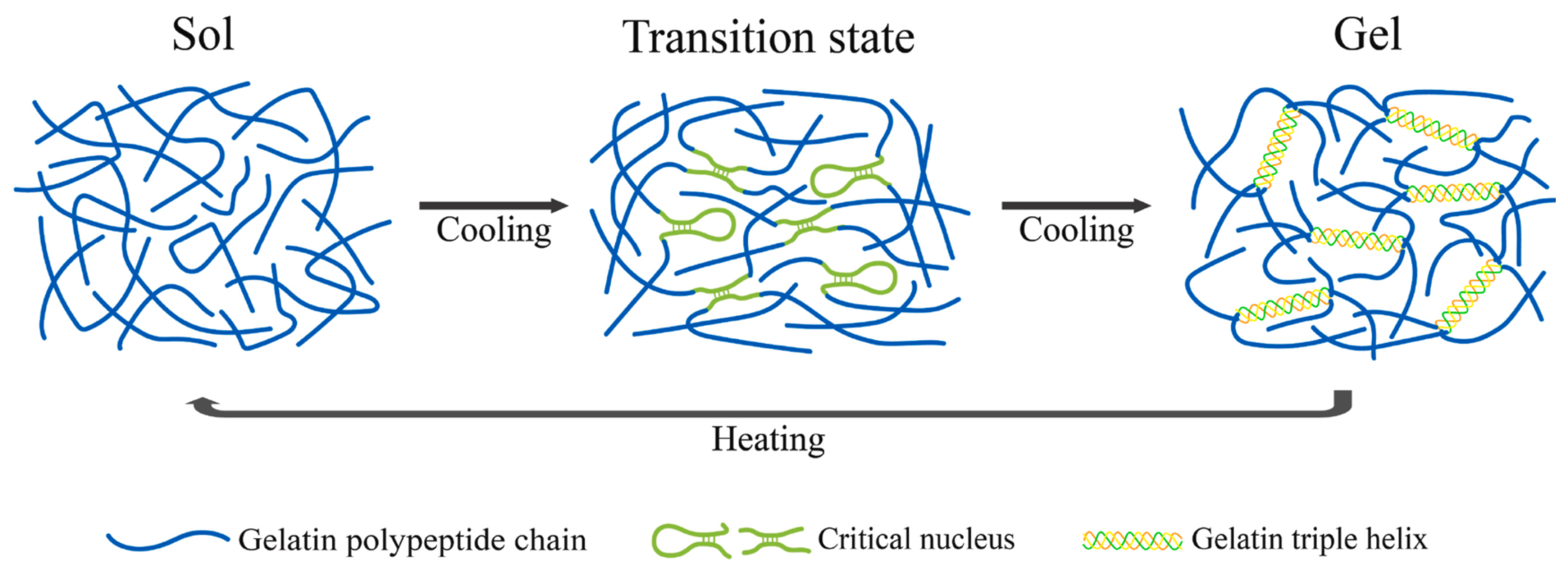
Gelatin exhibits thermo-reversible properties, which means it can transition between gel and sol states depending on temperature changes. Above gelling temperature, polypeptide chains adopt random coil configurations whereas upon cooling, some of the chains intertwine to create partially ordered, collagen-like triple helical structures, leading to the formation of a gel network (Figure 2) [10]. This process is thermally reversible, as the hydrogen and van der Waals bonds participating in the gelation process are non-covalent.
The gelling temperature and gel strength are directly affected by the molecular weight, as well as the complex interactions arising from the amino acid composition and the proportion of α to β chains in gelatin [48]. Higher amounts of α-chains result in greater gel strength. Table 2 shows the molecular weight determined by SDS-PAGE, gelling temperature, and Bloom strength of gelatins from various sources. The term ‘Bloom strength’ is typically expressed in grams (g) and is used to indicate the gel strength or firmness of the gelatin. Commercial gelatins typically have Bloom values ranging from 50 to 300 g. [66]. The gelation temperatures of gelatin range from 7 °C to 28 °C, with corresponding gel strengths varying from 206 g to 717 g (Table 2).
The amount of hydrophobic amino acids is another important factor in gel strength [67]. Depending on variations in the source, the composition and distribution of hydrophobic amino acids can also vary, significantly impacting the gelling temperature and Bloom strength [12].
Table 2. Molecular weight, gelling temperature and gel strength of gelatins from various sources.
SourceMolecular Weight
Distribution
(kDa)
Gelling Temperature (°C)Bloom Strength (g)Reference
Bovine bone gelatinα1- 130
α2- 120
25.05221[68]
Duck feet gelatinβ- ∼180
α1- ∼115
α2- ∼100
20.50209.63[69]
Sheep hoof gelatinβ- 245
α1- 63–75
α2- 100–135
25.38378.55[70]
Porcine skin gelatinβ > 180
α1- 135
α2 < 135
28.06581.61[71]
Frog skin gelatinβ- ∼200
α1- ∼120
α2- ∼120
28363[72]
Camel skin gelatinβ- ∼225
α1- ∼120
α2- ∼116
20.9–25.8365.5[73]
Chicken head gelatinβ- ∼202
α- ∼113
27°–28>309[74]
Cold-water fish gelatinβ > 200
α1- 130
α2- 110
NR253[75]
Dog shark skin gelatinβ-∼200
α1–116
α2–97
20.8206[76]
Bigeye snapper skin gelatinβ-∼205
α-97
10108[77]
NR: not reported.

3. Collagen-/Gelatin-Based Hydrogels

Understanding the gelation mechanisms of collagen and gelatin hydrogels is essential for tailoring their functional properties to meet the end-use needs in various applications. The gelation mechanisms and resulting network structures of gelatin and collagen differ significantly. During collagen gelation, collagen molecules undergo aggregation and fibril formation, initiated by changes in ionic strength, pH, and temperature. This process begins with a lag phase, during which primary aggregates such as dimers and trimers are nucleated. This is followed by microfibrillar assembly through lateral aggregation of subunits until the system reaches equilibrium. In collagen obtained from vertebrates, self-assembly occurs when the temperature is increased from 20 °C to 28 °C [12]. This structural organization is primarily stabilized by intra-molecular interactions, including hydrogen bonding, electrostatic interactions, and hydrophobic interactions, which play a key role in aligning triple-helical domains and enabling orderly fibril formation through self-assembly [78]. In contrast, gelatin gelation is driven by a reverse coil-to-helix transition that takes place upon cooling below 30 °C. The resulting helices resemble the collagen triple-helix structure, although the assembly is less ordered, and equilibrium is not achieved. The gelatin network continues to evolve over time, but never reaches the ordered D-banding structure (a ~67 nm periodic pattern characteristic of native collagen fibrils)observed in native collagen [79]. In this case, both intra- and inter-molecular crosslinking contribute to network formation: intra-molecular interactions promote partial reformation of helix-like segments, while inter-molecular associations between gelatin chains help establish a three-dimensional gel matrix. Intra-molecular interactions primarily influence properties such as gel strength, whereas inter-molecular interactions tend to extend the effective molecular chain length, which may have a greater impact on properties like viscosity [18]. While both collagen and gelatin exhibit thermoreversible gelation behavior, the temperature dependence of the transition is opposite: collagen molecules self-assemble into fibrillar structures upon heating, [80], whereas gelatin solutions form a gel upon cooling, which becomes progressively stiffer and more elastic as the temperature decreases.
Table 3 summarizes the various applications of collagen and gelatin-based hydrogels along with the co-agents used. While the use of these hydrogels has been widely explored in biomedical fields, their application in the food industry is also emerging as a promising area of research. On the other hand, studies in literature generally lack comprehensive analyses regarding scalability, allergenicity, and toxicity concerns. Future studies should address these gaps to enable the wider application of collagen- and gelatin-based hydrogels.

4. Methods to Modify the Rheological Properties of Collagen/Gelatin Gels for Improved End-Use Applications

Collagen and gelatin have been widely used in the food industry to improve the elasticity, consistency, and stability of foods. These applications have been mostly conducted on an empirical basis due to the lack of in-depth information on the rheological behaviors of collagen and gelatin in complex food systems [113]. In the last decade, studies have mostly focused on manipulating the rheological responses of collagen and gelatin hydrogels to meet the requirements of certain processing applications. The use of co-agent biomacromolecules and/or the application of physical, chemical, or mechanical treatments are among the most common ways to alter the rheological properties of collagen and gelatin hydrogels [10,22]. Therefore, the following sections of this review focus on the effect of these applications on the rheological responses of collagen and gelatin hydrogels. Detailed descriptions of the rheological methods mentioned throughout the next sections have been provided by Duvarci et al. [114]. Also, please see the Nomenclature for the definitions of the rheological parameters mentioned in this review.

4.1. Addition of Co-Agent Molecules

4.1.1. Rheological Properties of Collagen Gels with Added Co-Agents

Pure collagen gels lack sufficient strength and elasticity to meet the requirements of most processing applications [10]. The rheological properties of collagen- hydroxypropyl methylcellulose (HMPC) gels were governed by the hydrogen bond interactions formed between collagen and HPMC [115]. The elastic modulus as a function of frequency [G(ω)] of collagen gels with HPMC were all larger than that of pure collagen gel. Coupling rheological tests with AFM suggested that the decrease in G(ω) at high percentages of HPMC was caused by the interruption of the nucleation of collagen. This affected the lateral growth of the fibrils in the hydrogel. Therefore, Ding et al. [115] showed that both hydrogen bond interactions and the size of fibrils in the gel alter the rheological properties.
The addition of sodium alginate (SA), hyaluronic acid (HA), and chondroitin sulfate (CS) increased the maximum mechanical strength of the pure collagen gel considerably. The mechanical gel strength in the presence of CS was lower, which was attributed to the lower molecular weight of CS. The increase in the maximum strength of the fibrillar collagen gels with the addition of polysaccharides was the result of a higher degree of crosslinking, which led to denser fibril networks. FTIR spectra showed that the N-H group was involved in more hydrogen bonds in polysaccharide-collagen fibrillar gels in comparison to pure collagen gels. Except for SA, reductions in the mechanical strength of the collagen gels with added HA and CS above 40% (polysaccharide:collagen ratio, w/w) were found [22], indicating a decrease in the synergistic effect between collagen self-assembly and the crosslinking of these polysaccharides with collagen [81,116]. Zhang et al. [81] suggested that the collagen self-assembly is responsible for forming the main structure in collagen-oxidized CS hydrogels. The amide bonds formed between CS and collagen acted as a supporting mechanism to enhance the crosslinking density of the hydrogel fibril network, leading to enhanced mechanical strength. On the other hand, the reduction in the mechanical gel strength at high levels of CS was caused by the formation of excess covalent bonds that disrupted the physical self-assembled structure of the collagen-based gels [81]. These results were consistent with the decreasing trend found for the G(ω) of collagen-HPMC gels above a certain amount of added HPMC [115]. G(ω) of collagen type II (Col II)- chondroitin sulfate (CS-sNHS) hydrogels displayed a constantly developing increase as the amount of CS-sNHS increased. This was attributed to the higher degree of chemical crosslinking of Col II and CS-sNHS [14].
Linear viscoelastic properties of collagen-chitosan gels with different ratios of collagen to chitosan (0:100, 25:72, 50:50, 75:25, 100:0 on a weight basis) increased progressively. The frequency sweeps showed an increase in G(ω) values of the gels as the percentage of chitosan increased [116], consistent with the data obtained for collagen-CS gels by Gao et al. [14]. Gels formed by collagen from different species display different mechanical properties [10]. For example, tilapia collagen gels had higher mechanical strength and elasticity than porcine collagen gels. Higher levels of hydroxyproline and cysteine in collagen caused more crosslinking in the resulting gels, leading to improved elasticity and mechanical strength [117]. There are various protocols to prepare the collagen-chitosan gels. For example, one of the methods enabled the gelation of collagen first, while the other method involved the gelation of chitosan first. The earlier gelation of collagen resulted in more cohesive gel networks. The gaps in the 3D gel network formed by collagen were then interconnected by the gelling chitosan. This prevented the self-assembly of collagen from being interrupted by the polysaccharide networks [116].
The synergistic effect of collagen and chitosan on imparting elasticity to gels started to disrupt the ability of the gels to withstand the increasing deformations beyond the collagen:chitosan ratio of 50:50 (w/w). This was evidenced by the sharp decrease in critical strain (γcri). Sánchez-Cid et al. [116] showed that chitosan improved the elasticity of the collagen gels but made them less deformable as the ratio increased above 50:50 (w/w).
These studies revealed the possibility to manipulate the rheological properties of the self-assembled collagen gels depending on the processing needs by using polysaccharides in the appropriate ratios and through the application of an accurately designed gelation protocol.
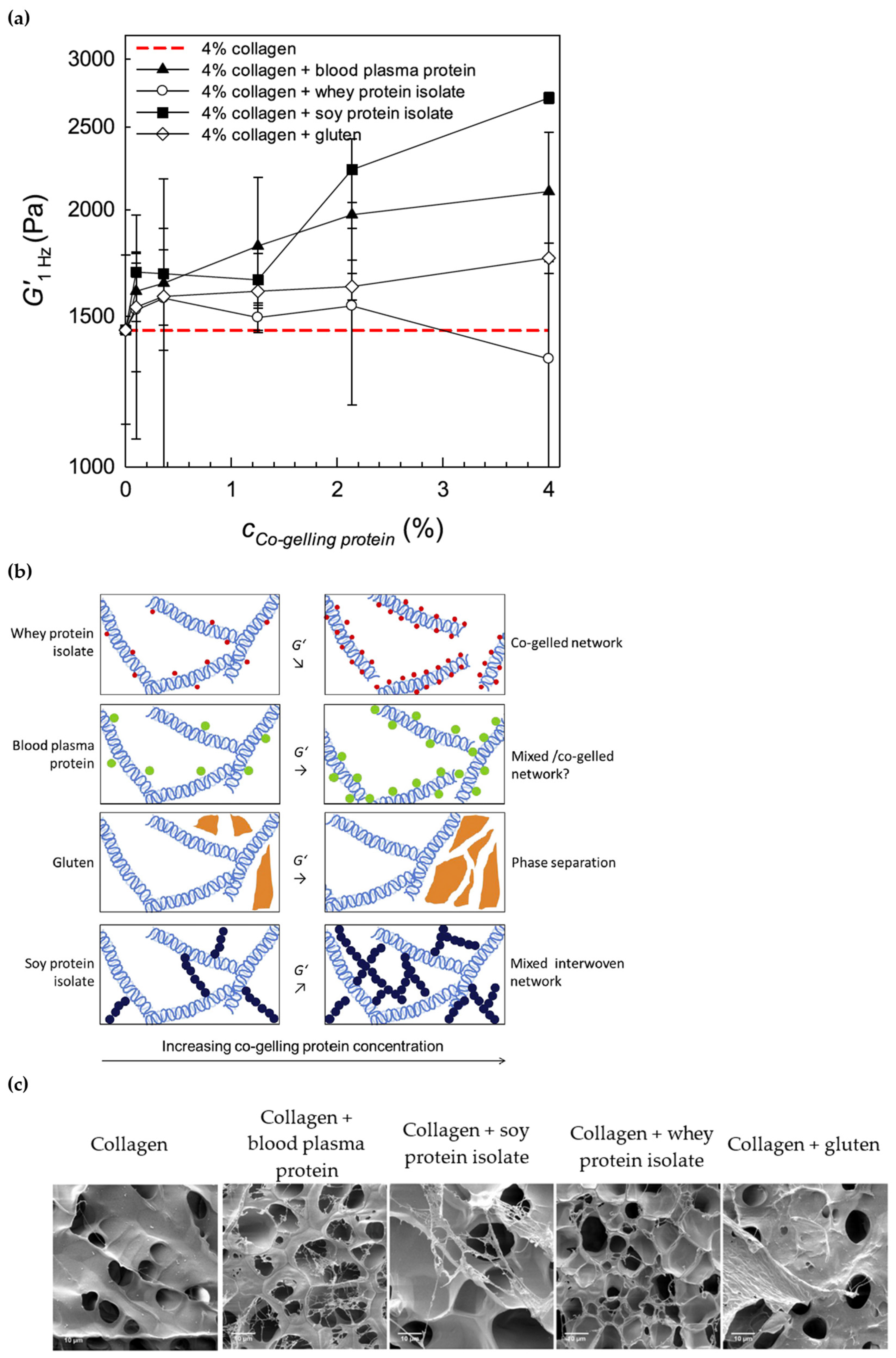
Besides polysaccharides, proteins are also used to improve the rheological properties of collagen gels. Oechsle et al. [118] used proteins, including soy protein isolate, whey protein isolate, and gluten as co-gelling agents in collagen-based gels. They compared the G(ω) of 4% (w/w) collagen as a function of protein concentration to evaluate the impact of proteins on the mechanical properties of the gel. Increasing levels of added soy protein isolate increased the G(ω) of the control (Figure 3a). The mechanism behind the increase in G(ω) was attributed to the formation of mixed interwoven networks between soy protein isolate and collagen, as shown in the predictive model in Figure 3b, based on the SEM images (Figure 3c).
Increasing the blood plasma protein content in the gel also increased the G(ω), but the large standard deviations suggested an inhomogeneous network for these gels (Figure 3a). SEM images indicated the formation of additional strands within the collagen pores with added blood plasma protein (Figure 3c), but these structures were not strong enough to affect the molecular organization in the network (Figure 3b) and the elasticity of the gels (Figure 3a). Gluten did not significantly affect the elasticity of the gels throughout the whole concentration range (Figure 3a), which was due to the phase separation (Figure 3b). SEM images indicated a separate gluten layer on top of the gelatin matrix (Figure 3c). Whey protein isolate caused a decrease in G(ω) at high concentrations (Figure 3a). No interconnections between gelatin and whey protein isolate or additional networks were visible on the SEM images (Figure 3c). Whey protein isolate might have occupied the binding sites of collagen-collagen interactions by covering the collagen molecules. And therefore, the weakening effect shown through the rheology data was associated with the interruption of the collagen gel network by the whey protein isolate (Figure 3b). The results by Oechsle et al. [118] showed that proteins with different molecular weights and structures could modify collagen gel strength differently to develop matrices with new functionalities.

4.1.2. Rheological Properties of Gelatin Gels with Added Co-Agents

The interactions of charged macro-ions in polypeptides and polysaccharides lead to the formation of (bio)polyelectrolyte complexes. And therefore, the combination of gelatin with ionic polysaccharides enables to modify the mechanical properties of gelatin-based gels or films [119]. Several studies investigated the impact of κ-carrageenan on the mechanical strength of gelatin [120,121,122,123,124]. de Alcântara et al. [122] reported a decrease in the creep compliance [J(t)] of gelatin-carrageenan gels as the ratio of carrageenan increased, indicating the contribution of carrageenan to the formation of a strong gel network. On the other hand, increasing the gelatin amount in the gel resulted in a soft structure that could be easily deformed, as evidenced by the higher J(t) values. Similarly, Warner et al. [121] reported an increase in the G(ω) of gelatin-carrageenan gels as the percentage of carrageenan increased. The hydrogen bonds formed between gelatin and carrageenan were stronger than those formed among the gelatin molecules, leading to a stronger gel in the presence of carrageenan [121]. The elasticity-promoting effect of carrageenan is due to the self-association of carrageenan [120]. On the other hand, Sow et al. [120] found a decrease in the G(ω) of gelatin-carrageenan gels when the amount of carrageenan was as low as 2:98 (carrageenan: gelatin, w/w). Carrageenan disturbs the protein gel network when added at very low concentrations, while improving the gel strength when added at higher concentrations. Sow et al. [120] explained this mechanism through the illustration shown in Figure 4. Besides the hydrogen bonds, gelatin and carrageenan also interact via electrostatic interactions formed between the positively charged amide groups of lysine, hydroxylysine, histidine, and arginine residues in gelatin and the negatively charged sulphate groups of carrageenan. The shift of the amide I band in native gelatin to lower frequencies in the FTIR spectra upon the addition of carrageenan indicates the presence of electrostatic interactions between gelatin and carrageenan [125]. At a critical mixing ratio of 4:96 (carrageenan:gelatin, w/w), large complex coacervates are formed, contributing to the gelatin network strength. Below this critical ratio, the unbound gelatin dominates the network, but the presence of carrageenan disrupts the network, leading to a decay in both G(ω) and G(ω). And above the critical mixing ratio, excess carrageenan self-association contributes to additional strength of the gel, while causing the formation of a bi-continuous gel consisting of separate gelatin and carrageenan network regions [120] as shown in Figure 4.
Chitosan also displayed a concentration-dependent effect on the viscoelastic properties of gelatin-chitosan gels [119]. Increasing the chitosan content up to 0.6% (gchit/ggel, w/w) resulted in an exponential increase in G′ and an increase in the critical strain. However, the addition of chitosan above 0.6% caused these parameters to decrease, suggesting a reduction in elasticity and resilience. The contribution of chitosan at concentrations below 0.6% (w/w) to gelatin’s strength was associated with the formation of new chitosan double helices in the gel network resulting from the electrostatic interactions and hydrogen bonds between the two biopolymers. The reduction in gel strength at high chitosan concentrations was attributed to the electrostatic repulsion between complexes with positive charges below the isoelectric point of the gel. Besides, chitosan-gelatin interactions suppressed the formation of collagen-like triple spirals, leading to another reason behind the decrease in the gel strength at high levels of added chitosan [119]. Similarly, Ata et al. [126] found G′ values for the gelatin gel that were nearly three times higher than those of the gelatin:chitosan gel within the linear viscoelastic region. Although the elasticity of gel decreased in the presence of chitosan, the crossover point occurred at a strain amplitude approximately twice as high for the gelatin:chitosan gel in comparison with the gelatin gel. This indicated the contribution of added chitosan to the mechanical strength and resilience of the gelatin gels when exposed to moderate deformations beyond the linear viscoelastic region.
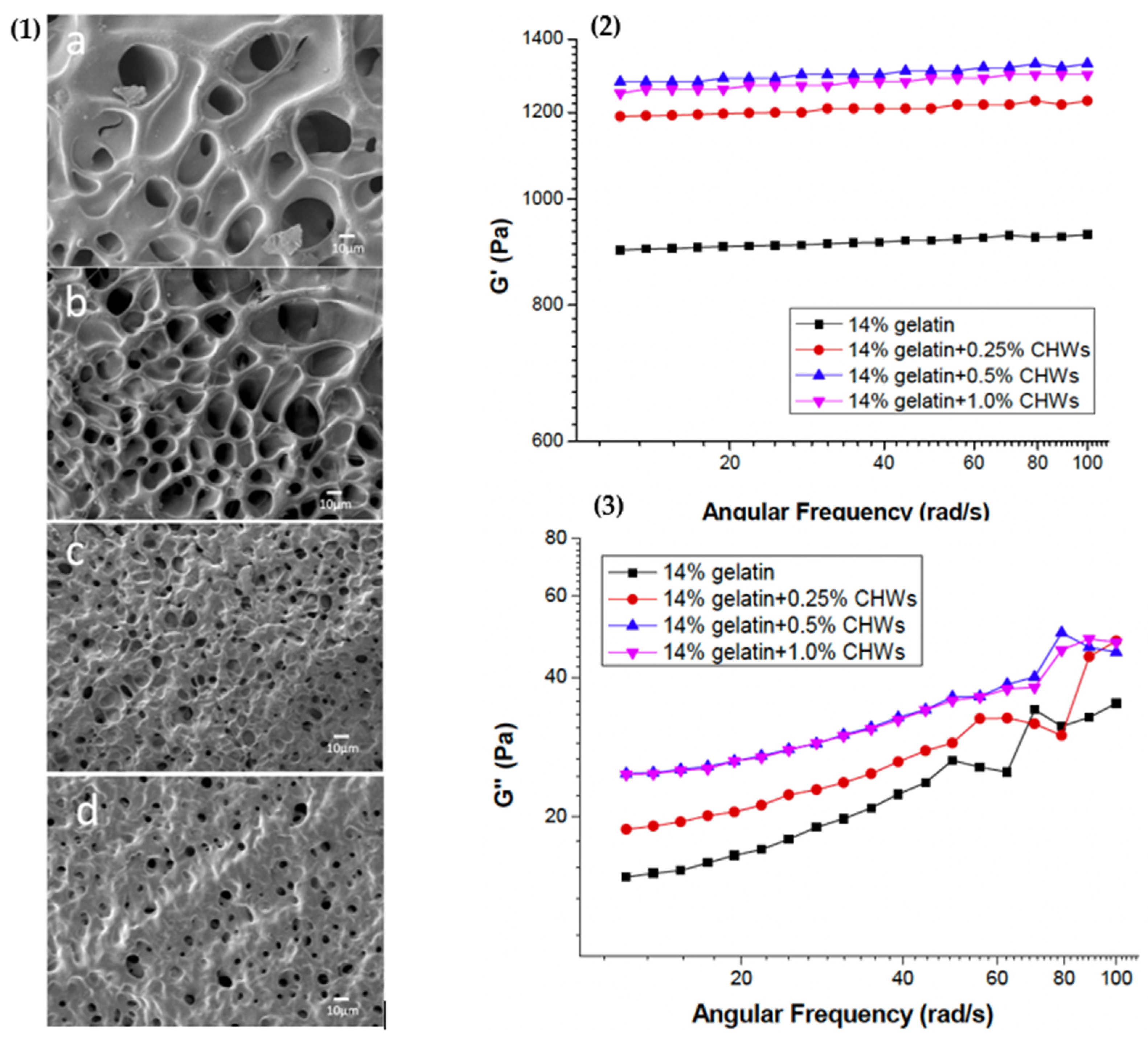
Ge et al. [127] used chitin whiskers, that are obtained from chitin by acid hydrolysis, as a nanofiller in gelatin. Pure gelatin hydrogels have porous microstructures, with pore sizes ranging from 10 to 50 μm. Increasing the concentration of chitin whiskers from 0.25% to 1% (w/v) decreased the pore sizes in the composite hydrogels gradually to 1–10 μm (Figure 5(1a–d)), indicating a denser network in the presence of chitin whiskers. This effect was reflected on frequency sweeps as an increase in both the elastic (G′) (Figure 5(2)) and viscous (G″) moduli (Figure 5(3)) of the composite hydrogels, indicating that chitin whiskers interacted with the gelatin matrix by noncovalent interactions, and as also suggested by others [119,121].
Similarly, the interactions between gelatin and pectin to form a gel are based on the electrostatic attractions between the positive charges on gelatin and the negative charges on pectin [128]. Increasing pectin concentration in gelatin gummies (with 6.47 wt% gelatin) by replacing 0–2.27 wt% of gelatin with pectin increased the gelation temperature from 27 °C to 77.2 °C, as evidenced by the G-G″ crossovers throughout the temperature sweeps. The G′ and G″ values gradually increased as the pectin concentration increased, revealing a synergistic effect of gelatin and pectin leading to a stiffer network [23]. Song et al. [26] also reported an increase in G′ and G″ versus frequency for gelatin (4 wt%) with the addition of 1 wt% pectin. However, tanδ(ω) significantly increased when pectin was added, indicating a decrease in the networking ability of the gel. In the non-linear region, gelatin gel displayed a G″ overshoot [26] that was defined as type III (weak strain overshoot) non-linear behavior [129]. The extent of the G″ overshoot was reduced with added pectin and almost disappeared at high frequencies [26], suggesting a reduction in the resistance of the network against the increasing deformations in the presence of pectin.
The addition of gelatin, even at small concentrations, into 10 wt% egg albumen significantly increased the G′ and G″ of the gel, while causing a decrease in the gelation temperature. These were attributed to the interruption in aggregation of egg albumen in the presence of gelatin. Upon heating, gelatin formed a liquid structure, while the egg albumen proteins formed a less aggregated network. These simultaneously occurring phenomena led to the formation of a uniform, strong gel [67]. In another study, the addition of gelatin increased the G′ and G″ of the gel and decreased the denaturation temperature of myosin. These changes were due to the electrostatic interactions between gelatin and myosin [130].
Pang et al. [131] found that the addition of skim milk powder (SMP) and milk protein concentrate (MPC) significantly increased the G′ of gelatin gels, while the addition of whey protein isolate (WPI) caused a decrease in G′ consistent with the studies we described. The casein particle network formed in SMP and MPC due to depletion flocculation contributed to the gelatin gel strength, while the nano-sized particles in WPI disturbed the gelatin network [131]. On the other hand, Ge et al. [23] found higher G′ and G″ values throughout the temperature sweep for gelatin (4.55 wt%)-WPI (22.72 wt%) mixture when compared to both gelatin (6.47 wt%) and WPI (22.72 wt%). This result shows that the diminishing effect of WPI on the viscoelastic properties of gelatin as reported by Pang et al. [131] could be altered by using higher ratios of WPI to gelatin in the mixture.
Another interesting application of added proteins in gelatin-based composite gels included the use of collagen [132]. The assembly characteristics of collagen, gelatin, and gelatin-collagen mixture were studied through time sweeps. The moduli of gelatin remained constant over the time range, and G″ > G′ indicating fluid-like behavior with no self-assembly characteristics. Both pure collagen and gelatin-collagen had solid-like properties (G′ > G″) and their moduli displayed an initial increase reaching an equilibrium, suggesting self-assembly properties for gelatin-collagen mixture as observed for pure collagen. Steady shear flow tests showed that the viscosity of gelatin-collagen mixture increased at each low temperature shear flow and decreased to around 0 Pa.s at each high temperature shear flow. These viscosity changes were not observed for collagen alone, indicating that the gelatin-collagen mixture had sol-gel transition properties that pure collagen did not. The gelatin-collagen gel preparation method used by He et al. [132] resulted in composite gels with the self-assembly properties of collagen and the sol-gel transition properties of gelatin.
Besides polysaccharides and proteins, a limited number of studies evaluated the effect of lipids on the viscoelastic properties of gelatin. Howe et al. [133] studied the viscoelastic properties of gelatin when anionic and cationic surfactants were added. At concentrations above the critical micelle concentration, anionic surfactants [sodium dodecyl sulfate (SDS), sodium dioctyl sulfosuccinate (AOT)] caused a greater increase in the viscosity of gelatin when compared to cationic surfactants [cetyltrimethylammonium bromide (CTAB)], indicating a stronger interaction between gelatin and anionic surfactants. The head groups in cationic surfactants are larger compared to anionic surfactants. This property has been associated with their weaker ability to interact with polymers [133]. On the other hand, Żamojć et al. [134] suggested that the ability of surfactants to bind to proteins was governed by the hydrophobic interactions of surfactant methylene chains rather than the head group characteristics.
In a system evaluating the gelatin-surfactant interactions [135], typically three gelatin molecules were considered to bind to a single micelle formed due to surfactant aggregation. In this micellar binding mechanism, micelles act as nodes for the aggregation of gelatin chains, leading to the formation of a 3D network and an increase in the elasticity of the gel as reported by Howe et al. [133].
Gravelle and Marangoni [136] studied the impact of solid fat and liquid oil droplets stabilized by whey protein isolate (WPI) on the large deformation mechanical properties of emulsion-filled gelatin gels. By modulating the electrostatic interactions at pH values above and below the isoelectric point of WPI (∼5), the emulsion droplets produced either a homogenous network (pH 6.0) or a heterogeneous network (pH 4.0). In the homogeneous network, interfacial debonding appears to arise from the increasing emulsion filler concentration which interrupts the elastic network. As a result, the strain-stiffening behavior typically observed for gelatin [31,137] shifted to a strain-softening behavior in the emulsion-filled gelatin gels. On the other hand, increasing the filler concentration in the heterogeneous network led to linear elastic behavior, resulting in brittle fracture. Ultimately, this study [136] showed that the deformation properties of composite gelatin gels were caused by the network architecture induced by the fillers.

4.2. Chemical and Mechanical Treatments to Modify the Rheological Properties of Collagen and Gelatin Gels

4.2.1. Treatments to Modify the Rheological Properties of Collagen Gels

The self-assembly kinetics of collagen and the rheological properties of the resulting collagen fibrils are strongly affected by external factors such as temperature, mechanical forces, electrolyte type and concentration, pH, magnetic and electrical fields and gravity [10,25]. Collagen fibrils organize themselves through hydrogen bonding and hydrophobic-electrostatic interactions [138]. Ultrasound treatment has been among the widely used treatments to control the collagen fiber formation process, especially because it is an efficient non-thermal treatment [55]. Table 4 summarizes the use of ultrasound treatment for collagen self-assembly. Jiang et al. [27] found that ultrasonic treatment reduced the diameter size and diameter size distribution of fibrils formed during nucleation. Increasing the ultrasonic power resulted in more heterogeneous pore structures and larger pore sizes in the gel. These microstructural changes led to softening of the gels. Similarly, Liu et al. [139] also reported a decrease in both G′ and G″ of collagen versus frequency as the ultrasonic power and application time increased. X-ray diffraction spectra showed that collagen molecules exposed to ultrasonic treatment maintained their native triple-helical conformations and crystallinity. However, the distance between the collagen chains increased, causing a decrease in G′ and G″. Imparting a softer structure to collagen gels through ultrasonic treatment during nucleation can make them more suitable for drug delivery [139].
Although some applications (i.e., drug delivery carrier) require further lowering the viscosity of collagen gels, some others (i.e., scaffold for tissue engineering) require strengthening of the collagen gels. Considering collagen gels are usually characterized by their low mechanical strength, low thermo-stability and susceptibility to enzymatic degradation [15], crosslinking to improve the gel strength has been widely used. Synthetic crosslinkers such as aldehydes (i.e., glutaraldehyde) and isocyanates (i.e., hexamethylene diisocyanate) have often been used to modify the mechanical properties of collagen gels. Most of these synthetic crosslinkers have potential cytotoxic effects. Recently, N-hydroxysuccinimide (NHS) active esters have gained attention as a non-toxic and biocompatible crosslinking agent due to their ability to form stable amide bonds under mild conditions. NHS active esters are synthesized by the reaction between a carboxylic acid and NHS in the presence of carbodiimide [15,140]. Crosslinking with ethyl-3-(3-dimethylaminopropyl) carbodiimide (EDC)-NHS significantly improved the mechanical stability of gelatin gels compared to unmodified gelatin gels as reported by Skopinska-Wisniewska et al. [141]. The tensile strength and elongation at break for the EDC-NHS crosslinked gelatin gel were approximately twice as high as those of unmodified gels, indicating a considerable increase in stiffness with EDC-NHS crosslinking [141]. As another novel crosslinking agent, N-hydroxysuccinimide activated adipic acid (NHS-AA), was synthesized by Zhang et al. [142] using adipic acid, NHS and carbodiimide. When collagen was treated with NHS-AA, the crosslinking reaction occurred between the two active ester groups in NHS-AA and ε-amino groups in collagen molecules. Both G(ω) and G(ω) of collagen treated with NHS-AA increased, while tanδ(ω) decreased with increasing concentrations of NHS-AA, showing that the gel got stiffer and stronger.
As the temperature gradually increased up to 35 °C, G(ω) increased with increasing concentrations of NHS-AA, while the slope of G(ω) decreased [142]. An increase in the magnitude of G(ω) along with a decrease in its slope versus frequency was shown as the markers for crosslinking [143]. Therefore, the results by Zhang et al. [142] showed that collagen displayed a more solid-like behavior due to the increasing extent of crosslinking as the concentration of NHS-AA increased.
Due to the disadvantages associated with the synthetic crosslinkers, recently, more emphasis has been put on naturally occurring crosslinkers for their biocompatibility [15]. The elasticity of collagen gels increased considerably with the addition of genipin, indicating that genipin could be used as a crosslinker to effectively modulate the mechanical properties of collagen gels [30]. Tang et al. [144] showed that the incorporation of laccase-catalyzed gallic acid (L-GA) solution into collagen hydrogels significantly enhanced the gel strength in a concentration-dependent manner. This increase in the gel strength was attributed to the enzymatic oxidation of gallic acid by laccase, which facilitated additional crosslinking within the collagen network. Eslami et al. [145] reported that the addition of TGase as a crosslinker at the levels of 1.25 and 2.5 U/mL increased the G(ω) and G(ω) of a gel consisting of pea protein isolate (PPI) (50 mg/mL) and collagen (3 mg/mL). The SEM images in this study pointed to a network where the insoluble PPI particles were entrapped by the collagen fibrils that led to the formation of a compact and robust gel, consistent with the rheology data. However, increasing the TGase concentration well beyond 2.5 U/mL up to 18 U/mL resulted in G(ω) and G(ω) values similar to those of the control gel (without TGase treatment), indicating a reduction in the gel strength [145]. Excessive levels of TGase appear to induce individual crosslinking of the proteins in the gel, leading to a phase separation and a reduction in the gel strength [146]. Another reason behind the decrease in G′ with high levels of TGase treatment could be the reduction in protein-water interactions due to increased protein-protein interactions. This would result in higher levels of free water in the gel, leading to a decrease in the viscoelastic moduli [145,147]. These results pointed to the importance of TGase concentration and the ratio of collagen in the gel mixture, which was suggested to be less than 10 wt% of PPI in the case of a collagen-PPI gel [145], to achieve an improved gel strength.
When alginate dialdehyde (ADA) was used as a natural crosslinker in collagen gels [15], the flow curves of ADA-crosslinked collagen solutions indicated an increase in viscosity as the ADA concentration increased. Complex viscosity versus frequency [η*(ω)] also showed a similar trend. With increasing levels of ADA, the number of C=N linkages formed between the aldehyde groups of ADA and the lysine residues in collagen molecules increased. This led to the formation of more aggregates, causing an increase in viscosity.
The creep-recovery data revealed the ability of collagen gels with higher levels of ADA to resist deformation, as evidenced by the decrease in creep compliance and the increase in the recovery percentage. These results showed that ADA-crosslinking accelerated the transformation of collagen gels from a more liquid-like state to a solid gel-like state [15].
Sánchez-Cid et al. [148] used fructose as a crosslinker in collagen aerogels to develop collagen-based scaffolds. Collagen aerogels were selected over collagen hydrogels due to the higher elasticity of aerogels, making them a better scaffold alternative. Low levels of fructose addition resulted in less rigid scaffolds. Increasing the fructose concentration from 10 wt% to 40 wt% caused a significant increase in elasticity. A higher increase in elasticity was obtained by increasing the collagen concentration. This points to the weaker effect of fructose crosslinking on elasticity in comparison to collagen.
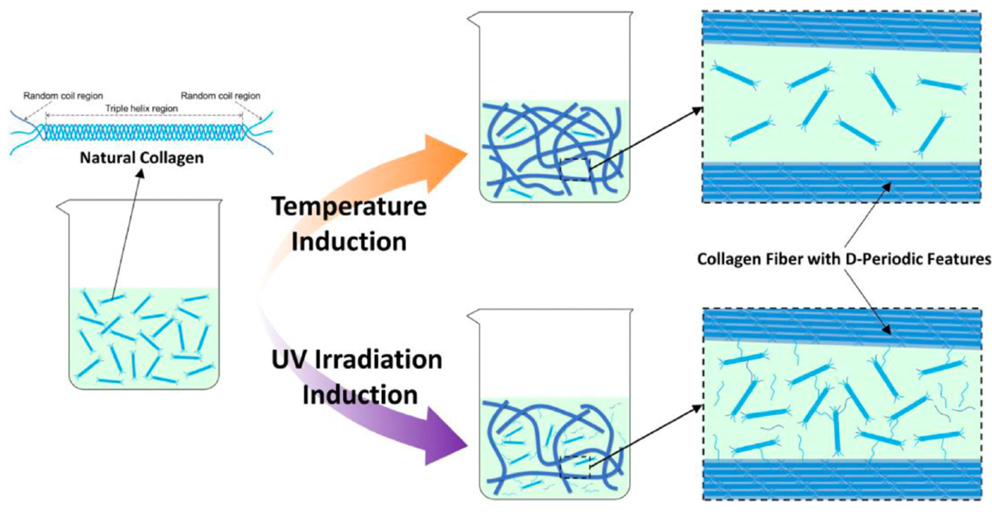
Instead of using crosslinkers, Xu et al. [13] used UV irradiation to induce crosslinking of collagen. UV irradiation leads to a strong connection between the polypeptide chains by stimulating the formation of bonds between certain amino acid residues, such as tyrosine and phenylalanine [149]. Xu et al. [13] compared the strengths of collagen gels induced by temperature and UV irradiation. During the UV irradiation-induced gelation, less collagen molecules participated in the fiber network formation in comparison with the temperature-induced gelation, as shown in Figure 6.
However, the crosslinking reaction caused by UV irradiation led to the formation of collagen fibers with more branches. The presence of branches enhanced the secondary entanglements of the collagen fibers. And therefore, the UV irradiation-induced collagen gel had a greater mechanical gel strength compared to the temperature-induced collagen gel. Similarly, Ishibashi et al. [150] reported an increase in both G′ and G″ of UV irradiation-treated collagen solutions as the treatment time increased. This was attributed to the increase in the molecular weight resulting from photo-crosslinking. The easy application protocol of the UV irradiation and the absence of the toxic effects of most chemical crosslinking agents make the UV irradiation an advantageous treatment for the crosslinking of collagen [149]. The effects of UV irradiation on the mechanical properties of collagen gels and the optimum treatment parameters are summarized in Table 4.
Table 4. Ultrasound and UV irradiation applications on collagen solutions/gels.
TreatmentSamplesParameters AppliedSuggested Optimum
Values for the Treatment
Achieved Traits in the Gel
UV irradiation0.5, 1.0, 1.5% (w/w) collagen solutionsPower: 4.8 W
Wavelength: 366 nm
Time: 10, 20, 30, and 60 min
All treatments were found to be useful.Increase in the viscosity of the collagen solutions led to the flexibility to work with lower collagen concentrations to form gel [150].
Collagen gels with 0.5, 1.0, 2.0, and 3.0 mg/mL concentrationsWavelength: 254–290 nm
Time: 30, 60 and 120 min
0.5 mg/mL collagen concentration and a UV irradiation treatment for 30 and 60 min were suggested.A stabilized collagen gel surface area for cell cultivation [149].
2 mg/mL collagen solutionWavelength: 254 nm
Intensity: 5.0 × 10⁻³ W/cm2
Time: 30 min for 5 times (with intervals of >60 min)
The applied protocol was successful.UV irradiation-induced collagen gels with thermal stability, mechanical properties, and cell growth compatibility comparable to those of temperature-induced collagen gel [13].
UltrasonicationCollagen solution with a concentration of 3 g/LUltrasonic power: 0, 100, 140, 180 W
Time (at 30 °C): 0, 5, 15, 60 min
Manipulating power was suggested to meet the end-use needs.
Increasing the treatment time did not affect the collagen self-assembly dynamics. Therefore, 5-min treatment was enough.
Viscosity of the collagen gel decreased with increasing ultrasonication power [27].
5 mg/mL of collagen solutionUltrasonic power: 0, 50, 100, 200, 400 W
Time (at 30 °C): 0, 5, 10, 15, 30, 60 min
Power value of ≤200 W for a time period of ≤15 min have been suggested as proper ultrasonic treatment parameters.With the proper parameters used, the self-assembly rate of collagen increased, fibril diameters became more homogenous, thermal stability increased; while viscosity decreased, leading to a softer gel [139].
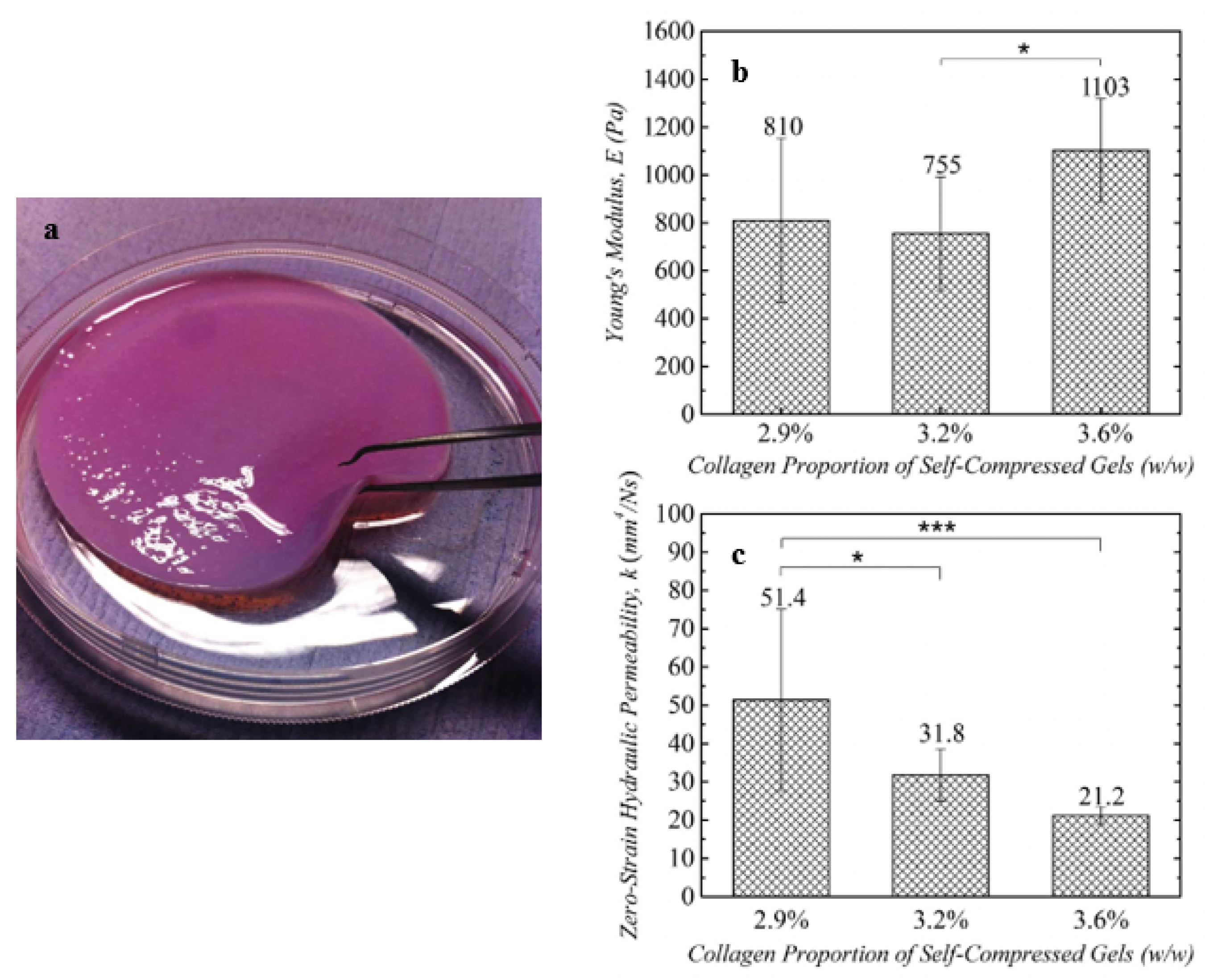
Recently, Andriakopoulou et al. [8] proposed a self-compression technique to increase the density of collagen fibrils and to improve the mechanical properties of collagen gels. The hyper-hydrated collagen hydrogels with an initial collagen concentration of 0.4% (w/v) and 13 mm thickness were compressed into gels with three different thicknesses with a corresponding collagen ratio. The collagen gel that was exposed to the highest degree of compression is shown in Figure 7a. The increase in the collagen fibril density due to compression did not cause a significant change in the Young’s modulus (Figure 7b), indicating that the compression did not affect the elasticity of the gel. However, hydraulic permeability decreased gradually as the collagen content increased in the self-compressed collagen gels (Figure 7c), indicating the flow dynamics were governed by the morphology of the gel network [151].
Cacheux et al. [152] reported an increase in the Young’s modulus along with a decrease in the permeability of collagen gels upon compression. The properties imparted to collagen gels with compression could be useful during tissue remodeling to control tumor growth, drug delivery mechanisms etc.

4.2.2. Treatments to Modify the Rheological Properties of Gelatin Gels

High-pressure processing (HPP) was applied to gelatin-whey protein mixture [153]. Pressurizing the gelatin-whey protein solutions at 600 MPa for 15 min produced less elastic gels in comparison to thermal treatment. During cooling, G′ of HPP treated gelatin-whey protein gels displayed a decreasing trend in the strength of the gel. This suggested that gelatin constituted the continuous phase of the HPP-treated composite gels, in which monodispersed whey protein aggregates were embedded [153]. Increasing the pH above the intermediate values (pH 4–6) during the gelation of high-pressure-treated gelatin-whey protein gels resulted in higher G(ω) and melting temperature values compared to those of heat-treated gels. The continuity of the gels was improved with increasing pressure, concurrent with the image analysis data, indicating fine-stranded gels with a denser network for the pressure-treated gels at high pH [154]. When the impact of pressure on the rheological properties of gelatin was evaluated at a constant and low temperature (10 °C), the gelatin solution gelling under pressure displayed higher G′ in comparison to the gelatin solution gelling under ambient pressure [155]. This was attributed to the shorter distance of the triple helix junction zones for the high-pressure gels. Therefore, Kulisiewicz and Delgado [155] hypothesized that the gelatin gelling under high pressure was interconnected by a higher number of triple helix junction zones when compared to the gelatin setting under ambient pressure.
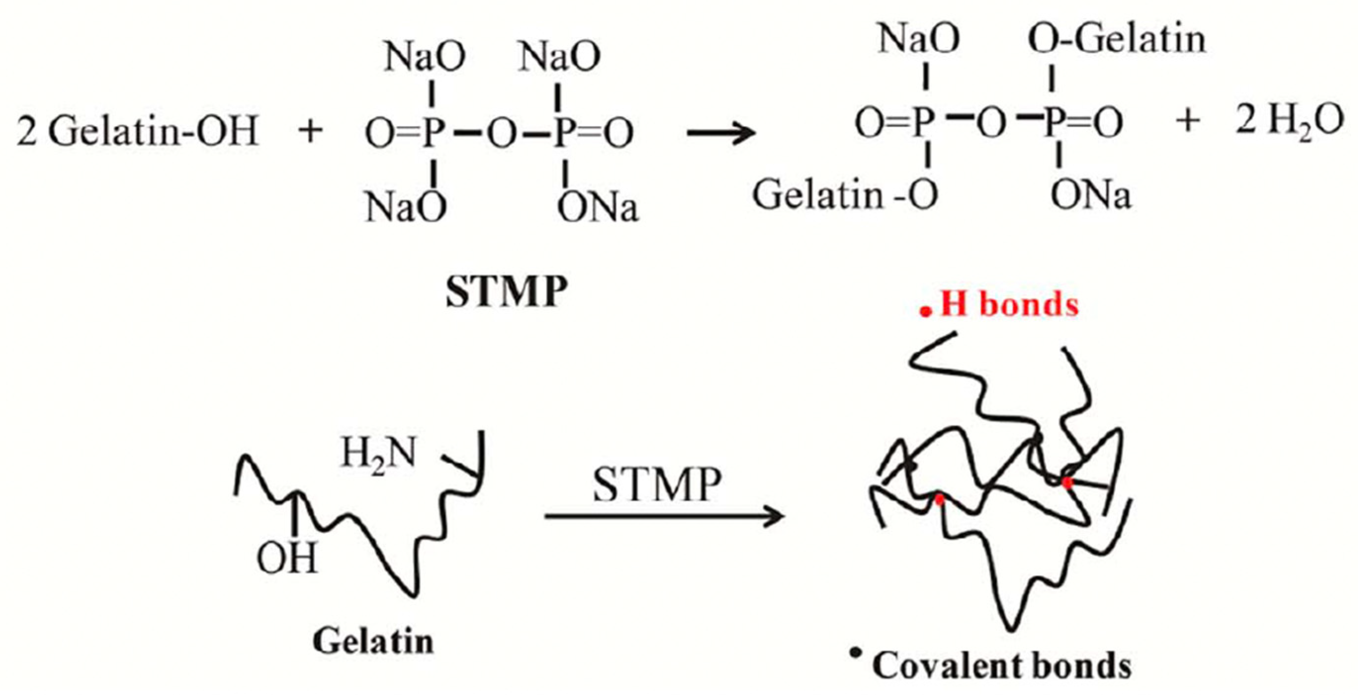
Phosphorylation of gelatin has also been used to alter the rheological properties of gelatin gels through charge modification [24,25]. Phosphate groups react with the -OH on serine and threonine, and the -NH2 on lysine and arginine in gelatin, as shown in Figure 8. As alkaline conditions make these functional groups in gelatin free for interaction, Huang et al. [156] used sodium trimetaphosphate (STMP) to phosphorylate fish gelatin at pH 9.0. Phosphorylation of STMP:fish gelatin solutions (1:20, w/w) at 50 °C for a short period of time (0.5 h) improved the hardness and chewiness of the resulting gels. This was due to the ionic interaction between phosphate groups and NH3+ of amino acids in gelatin that enhanced protein aggregation during gelation process. Increasing the phosphorylation time up to 2 h increased the phosphate content in gelatin. And the excessive levels of phosphate increased the electrostatic repulsion between the gelatin molecules, leading to the formation of a coarser and non-uniform gel network with a lower gel strength [157].
Kaewruang et al. [24] phosphorylated fish gelatin during a pretreatment by soaking the skin in sodium tripolyphosphate (STPP) solution and during the extraction process by mixing the skin with the STPP solution. Both treatments increased the gel strength and reduced the setting time. Phosphorylation imparted a negative charge to gelatin, leading to enhanced aggregation. This was more evident in the gelatin phosphorylated during extraction. As seen in Figure 9, the control gelatin displayed a looser network with large voids. Phosphorylation led to the formation of finer strands and a more compact and denser network with small pores. The smaller pores were more pronounced in the gelatin phosphorylated during extraction (Figure 9), which was in line with the highest gel strength observed for this gelatin gel. These studies [24,157] show that the phosphorylation treatments leading to an appropriate concentration of phosphate in the gelatin solution/gel are the key to an improved gel strength.
Treating phosphorylated fish gelatin with CaCl2 was found to further increase the gel strength. The simultaneous application of phosphorylation and CaCl2 was used to produce fish-origin gelatin with improved rheological properties that could replace bovine gelatin [25].
Treating the gelatin-based gels with salts was also used to manipulate the mechanical gel strength. Chen et al. [123] immersed the carrageenan-gelatin gels in a 10% K2SO4 solution and the G(ω) of the gels increased by around an order of magnitude. K+ and SO4 ions improved the gel strength by enhancing the electrostatic attraction between gelatin and carrageenan, reducing the repulsion forces within carrageenan, and inducing hydrophobic interactions between gelatin chains. Tong et al. [124], on the other hand, found a decrease in the apparent viscosity and gel strength of gelatin-carrageenan gels with the addition of NaCl and Na2SO4. Salts interrupted the hydrogen bonds in gelatin-carrageenan gels and unfolded the gelatin molecules, leading to a decrease in α-helix and β-sheet contents. Although both studies [123,124] used fish gelatin, the treatment with salts affected the rheological and textural properties of the gels in different manners. This could be due to the electrostatic effects exerted by the different salt ions or to the method of salt treatment (i.e., immersing the gel in the salt solution, or addition of the salt in the gel). Song et al. [26] immersed the gelatin-pectin composite gel in a CaCl2 solution to induce the aggregation of pectin. The presence of aggregated pectin led to an order of magnitude increase in the G(ω) of the composite gel, concurring with the findings of Chen et al. [123]. Song et al. [26] also evaluated the non-linear viscoelastic properties of gelatin-pectin gels immersed in CaCl2 solution using the Large Amplitude Oscillatory Shear (LAOS) tests. The stiffening ratio (S) showed a sharp increase for the gelatin-pectin gel after the treatment with CaCl2. This increase in the S value of the gelatin-pectin gel treated with CaCl2 was attributed to the aggregated egg-box bundles in pectin. Therefore, the LAOS parameter S was suggested as a marker for pectin aggregation in hydrogels [26]. The formation of aggregated polymers was considered to inhibit the stretching of the triple helix bundles in the gelatin network, leading to a higher degree of strain-stiffening behavior [31].
Gelatin finds application in emulsion gels due to its amphiphilic nature that enables it to act both as an emulsifier and a gelling agent [157]. However, gelatin-based gels display relatively weak response against the external forces as their stability mainly depends on molecular entanglements and hydrogen bonds [18]. Lin et al. [21] investigated the effect of MTGase crosslinking on the non-linear rheological properties of fish gelatin-based emulsion gel to explore its potential as an encapsulation material for β-carotene. TGase catalyzes the interaction of the γ-carboxyamide groups of glutamine and the ε-amino groups of lysine in gelatin chains to form the isopeptide bond ε-(γ-Glu)-Lys, leading to changes in the protein network structure [18]. Fish gelatin had the lowest crossover strain indicating the lowest stability against the increasing deformations compared to MTGase-crosslinked fish gelatins (Figure 10A,D). The stability of the gelatin-based hydrogels are governed by the molecular entanglements and hydrogen bonds [18]. For improved stability against deformations, the crosslinking time should be decreased as the temperature increases (Figure 10A–D). Elastic Lissajous-Bowditch curves (Figure 10E) of the MTGase-crosslinked fish gelatin for 2 h remained elliptical at large strains, while other gels displayed rectangular trajectories at lower strain amplitudes. Similarly, viscous Lissajous-Bowditch curves were still circular for the MTGase-crosslinked fish gelatin for 2 h, especially up to 46% strain, while other gels started to have distorted elliptical viscous trajectories at the same strain amplitude (Figure 10F). With increased crosslinking time up to 2 h, the covalent bonds formed between gelatin molecules led to a more resilient and interconnected network, which resisted deformation [21]. Although increasing deformations weaken the gelatin network, increased crosslinking simultaneously promotes the formation of weak intermolecular associations and new entanglements within the gel matrix, which temporarily consolidate the network under increasing deformations [158]. Further increasing the crosslinking time resulted in a highly rigid and over-crosslinked network, however, it reduced the ability of gelatin to withstand increasing deformations. This was attributed to the interruption of the intermolecular protein aggregation during gel network formation by the excessive intramolecular covalent bonds catalyzed by TGase. Consequently, the Lissajous-Bowditch curves of the MTGase-crosslinked fish gelatin for 4 h showed a higher degree of distortion from elliptical of circular trajectories at lower strain amplitudes, indicating a transition from linear to nonlinear behavior under smaller deformations. These results showed that excessive crosslinking did not contribute to further improving the stability of gelatin, and moderate crosslinking with MTGase was adequate to improve the deformation resistance of fish gelatin. The stronger deformation stability of the moderately crosslinked fish gelatin enhanced the resistance of the gel to pepsin hydrolysis and provided a controlled release of β-carotene during digestion [21].
When crosslinked gelatin-based hydrogels are used in tissue engineering, the degree of crosslinking should be tuned in a manner to provide sufficient resistance to degradation, while not exceeding the mechanical properties of the native tissue [159]. Kirchmajer et al. [160] developed gelatin gels by crosslinking gelatin with genipin at different gelatin to genipin ratios. The compression tests conducted on these gels suggested an increase in the elasticity of the gels as the concentrations of gelatin and genipin increased, which was evidenced by the increasing compressive tangent modulus (Etan) and compressive stress at failure (σmax) values. On the other hand, increasing both the gelatin and genipin concentrations led to a decrease in the compressive strain at failure (εmax). The compression tests revealed that the increased density of covalent crosslinking in the hydrogels caused by the higher gelatin and genipin concentrations led to more elastic and brittle gels. Slower proteolytic degradation rates were reported for the gels as the concentrations of gelatin and genipin increased, suggesting that the degradation resistance of the gels was proportional to the gelatin and genipin concentrations and thus, to the elasticity of the gels. Ultimately, this study showed the possibility to control the degradability and the mechanical properties of genipin-crosslinked gelatin by manipulating the degree of crosslinking through the adjustment of gelatin and genipin concentrations [160].A previous study also suggested using different concentrations of genipin to control the degradation rate and the degree of crosslinking in genipin-crosslinked gelatin, depending on the end-use. This study reported a genipin concentration of above 0.5% (w/w) of the total weight of the gelatin-based hydrogel in case a complete crosslinking reaction between gelatin and genipin molecules is required [161].
Studies have also shown the possibility to control the crosslinking of gelatin and the resulting rheological properties through the use of thermosensitive polymers as crosslinking agents. Boudet et al. [162] used a reactive co-polymer consisting of N-isopropylacrylamide (NIPAM) and water-soluble carbodiimide (EDC) for the chemical crosslinking of gelatin. NIPAM is a promising thermosensitive polymer due to its low lower critical solution temperature (LCST) of 32–34 °C close to human body temperature. It has both hydrophilic and hydrophobic groups in the monomer structure [163]. Below the LCST, NIPAM is in a hydrophilic state and its amide groups form hydrogen bonds with water, enabling it to solubilize and remain in a swollen conformation. As the temperature approaches the LCST, the hydrophobic interactions formed by the isopropyl groups on the NIPAM chains become more pronounced. This interrupts the equilibrium of intermolecular forces, leading to a decrease in the solubility that causes the polymer to aggregate and go through a transition from a soluble state to an insoluble state. When the temperature increases above the LCST, NIPAM switches into a hydrophobic gel-like state, in which the densely packed polymer chains interlock and crosslink, trapping water in the network [164]. When gelatin is crosslinked with NIPAM, the acrylic acid units in NIPAM formed amide bonds with the amino groups of gelatin in the presence of EDC [162]. Time sweeps showed that the network formation took longer and the plateau values of G′ decreased as the temperature increased from 25 to 45 °C. A significant decay in G′ was observed that was indicative of a weak network development especially when the temperature reached above 35 °C. As the temperature increased above the LCST, the switching of PINAM from coil conformation to a globular confirmation dramatically reduced its ability to interact with gelatin. These data showed that the chemical crosslinking reaction of gelatin with PINAM was off above the LCST (around 34 °C) and switched on as the temperature dropped below the LCST. This switchable reactivity enables to control the gelation of gelatin hydrogels by setting the temperature above or below the LCST depending on the desired end-use [162].
Samimi Gharaie et al. [165] used Poly(N-isopropylacrylamide)-co-Acrylic acid (PNIPAM-co-AA) as a pH-sensitive drug carrier to develop a gelatin-laponite-based smart shear-thinning gel for localized drug delivery purposes. The flow curves (shear stress versus shear rate) obtained at 37 °C revealed no significant effect of added PNIPAM-co-AA on the shear-thinning behavior of the gelatin-laponite gel, which is an important factor for the injectability of the gel. The degradation measured through the remaining weight (%), swelling rate (%) and drug release (μg/mL) values for the gelatin-laponite gel with PNIPAM-co-AA were negligible at pH 5.0 and 7.4, while they significantly increased at pH 9.8. This pH-responsive behavior imparted to the injectable gel by PNIPAM-co-AA has been suggested to be useful for the treatment of wounds with bacterial infections, as local pH changes are observed depending on the type of bacterial infection. For example, bacterial infections caused by Pseudomonas and Escherichia coli lead to a shift in the pH of the wound area to alkaline conditions. The gelatin-based pH-responsive gel developed by Samimi Gharaie et al. [165] could be a potential drug carrier for such treatments that require activated drug delivery in response to local basic pH.
In the food industry applications, the pH of the medium affects the gelation properties and the viscoelastic properties of gelatin hydrogels. The gelation properties of gelatin-based gummies were studied under different pH conditions through the addition of acids, such as citric acid and malic acid [23]. The pH of the gummies dropped from 5.0 to 3.5 with the addition of both acids, which decreased G′, G″ and the gelling temperature of the gummies. This was attributed to the protonation of the amino acids in gelatin at a low pH, preventing the formation of hydrogen bonds during gelation. Although Ge et al. [23] suggested a weakening effect of low pH on the gelatin network based on the gelation properties, Anvari and Joyner [166] reported improved deformation stability for the fish gelatin (FG)-gum arabic (GA) complex emulsion gel at low pH (3.6) when compared to that formed at high pH (9.0). When the pH was high, the FG-GA concentrated emulsion showed a lower critical strain and a greater extent of intracycle strain-stiffening and shear-thinning behaviors. Elastic Lissajous-Bowditch curves pointed to a gradually increasing stress response for FG-GA concentrated emulsion as the pH decreased (Figure 11), indicating an increase in the elastic response at low pH. The emulsion at pH 3.6 also showed the lowest extent of distortion from the elliptical trajectories of the elastic Lissajous-Bowditch curves (Figure 11), which highlighted the resistance of the FG-GA emulsion network against the large deformations when the pH was low. The improved resilience of the FG-GA emulsions at low pH was due to the increased electrostatic interactions between FG and GA and the increased network extension through steric stabilization as the pH decreased from 9.0 to 3.6 [166]. The different results reported by Ge et al. [23] and Anvari and Joyner [166] regarding the effect of pH on the rheological properties of gelatin-based gels could be due to the differences in the origin of gelatin and the gel preparation protocols.

5. Conclusions

Collagen and gelatin obtained from animal by-products represent a promising source of protein. One of the most promising ways to utilize these proteins in various industrial applications is through the development of hydrogels due to their tunable structures and favorable functional properties. This review revealed the possibility to improve the functionality of collagen- and gelatin-based hydrogels depending on their end-use through the manipulation of mechanical gel strength.
The incorporation of co-agent molecules such as hydroxypropyl methylcellulose, sodium alginate, hyaluronic acid, chondroitin sulfate, chitosan, protein isolates, pectin, various lipids, and salts has been shown to influence the mechanical strength of these hydrogels. Therefore, the effects of co-agent molecules on the mechanical properties of collagen-/gelatin- based hydrogels should be considered based on the intended application needs. Among these biomacromolecules, polysaccharides more effectively enhance the gel strength of collagen- and gelatin-based hydrogels due to their ability to form strong and dense network structures via electrostatic and hydrogen bonding interactions. Protein type and concentration play a key role in determining gel stability, and the elasticity of the gel varies accordingly. In contrast, lipids exhibit only a limited effect on collagen-/gelatin-based hydrogel. However, recent applications revealed the fact that lipids could alter the strain-stiffening response of collagen-/gelatin- gelatin-based hydrogels under large deformations, especially when used along with proteins in the form of emulsions. Therefore, the interactions of these co-agent molecules should be considered, and their concentrations carefully optimized to achieve the desired gel strength.
Various treatments, including ultrasound, UV irradiation, high-pressure processing, self-compression techniques, chemical modifications such as EDC/NHS coupling, phosphorylation, genipin crosslinking as well as enzymatic modifications using TGase or laccase, have been employed to alter the rheological properties of collagen and gelatin hydrogels. Although most of the treatments enhance gel stability, the concentration levels and treatment power must be optimized for the desired gel strength. For instance, increasing the ultrasonic power and excessive levels of phosphorylation may reduce the strength of collagen and gelatin hydrogels. The effect of HPP on gel stability is also highly dependent on processing conditions such as pH and gelation temperature, with stronger and more stable gels being obtained, particularly under high pH or low temperature. Chemical and enzymatic modifications usually increase stiffness; however, since excessive crosslinking does not further improve the stability of these gels, the level of crosslinkers should be carefully optimized.
The gelation mechanisms of collagen and gelatin is driven by intra- and inter-molecular crosslinks. The recent advanced methods discussed in this review showed how to alter these network characteristics that would possibly enable the design of hydrogels with end-use specific properties.
Compared to medical applications, research on the use of collagen–gelatin hydrogels in the food industry remains relatively limited, highlighting a significant gap that this study aims to address. Future studies are needed to explore the potential applications of these hydrogels in the food industry, with an emphasis on tailoring their rheological and structural properties to fulfill specific functional requirements.

Author Contributions

Conceptualization, O.A., J.L.K. and G.Y.; investigation, O.A. and G.Y.; writing—original draft preparation, O.A. and G.Y.; writing—review and editing, J.L.K., S.T. and G.Y.; supervision, J.L.K., S.T. and G.Y. All authors have read and agreed to the published version of the manuscript.

Funding

This research received no external funding.

Data Availability Statement

No new data were created.

Conflicts of Interest

The authors declare no conflicts of interest.

Nomenclature

Steady Simple Shear: η: viscosity (Pa.s). Linear Viscoelasticity: Simple shear J(t):creep compliance (Pa−1). Small amplitude oscillatory shear (SAOS). ω: angular frequency (rad/s). γcri: critical strain (−). G′: elastic (storage) modulus (Pa). G″: viscous (loss) modulus (Pa). tanδ: loss tangent (−), where δ is the phase angle (rad). η*:Complex viscosity (Pa.s) Non-linear Viscoelasticity in Extension: E:Young’s modulus of a solid (Pa). k: zero-strain hydraulic permeability (mm4/Ns). Non-linear Viscoelasticity in Compression: εmax: compressive strain at failure (−). σmax: compressive stress at failure (Pa). Etan: compressive tangent modulus (Pa). Non-linear Viscoelasticity in Shear: Large amplitude oscillatory shear (LAOS). S: stiffening ratio.

Abbreviations

The following abbreviations are used in this manuscript:
ColCollagen
UVUltraviolet
EDCEthyl-3-(3-dimethylaminopropyl) carbodiimide
NHSN-hydroxysuccinimide
TGaseTransglutaminase
MTGaseMicrobial transglutaminase
BSEBovine spongiform encephalopathy
TSETransmissible spongiform encephalopathy
FMDFoot-and-mouth disease
HMPCHydroxypropyl methylcellulose
AFMAtomic force microscopy
SASodium alginate
HAHyaluronic acid
CSChondroitin sulfate
SMPSkim milk powder
MPCMilk protein concentrate
WPIWhey protein isolate
SDSSodium dodecyl sulfate
AOTSodium dioctyl sulfosuccinate
CTABCetyltrimethylammonium bromide
L-GALaccase-catalyzed gallic acid
PPIPea protein isolate
ADAAlginate dialdehyde
HPPHigh pressure processing
STPPSodium tripolyphosphate
LAOSLarge amplitude oscillatory shear
FGFish gelatin
GAGum Arabic

References

  1. Mehdi-Sefiani, H.; Chicardi, E.; Romero, A.; Perez-Puyana, V.M. Unveiling the impact of gelation temperature on the rheological and microstructural properties of type A gelatin hydrogels. Polymers 2024, 16, 1842. [Google Scholar] [CrossRef]
  2. Shan, P.; Wang, K.; Sun, F.; Li, Y.; Sun, L.; Li, H.; Peng, L. Humidity-adjustable functional gelatin hydrogel/ethyl cellulose bilayer films for active food packaging application. Food Chem. 2024, 439, 138202. [Google Scholar] [CrossRef]
  3. Yang, J.; Duan, A.; Shen, L.; Liu, Q.; Wang, F.; Liu, Y. Preparation and application of curcumin loaded with citric acid crosslinked chitosan-gelatin hydrogels. Int. J. Biol. Macromol. 2024, 264, 130801. [Google Scholar] [CrossRef]
  4. Nazir, S.; Wani, I.A. Stability and release properties of alginate, modified starch/basil seed gum, and gelatin-based hydrogel beads infused with basil seed (Ocimum basilicum L.) oil. ACS Food Sci. Technol. 2024, 4, 2835–2846. [Google Scholar] [CrossRef]
  5. Shoulders, M.D.; Raines, R.T. Collagen structure and stability. Annu. Rev. Biochem. 2009, 78, 929–958. [Google Scholar] [CrossRef] [PubMed]
  6. Jayaprakash, S.; Razeen, Z.M.A.; Kumar, R.N.; He, J.; Milky, M.G.; Renuka, R.; Sanskrithi, M.V. Enriched Characteristics of Poultry Collagen over Other Sources of Collagen and Its Extraction Methods: A Review. Int. J. Biol. Macromol. 2024, 273, 133004. [Google Scholar] [CrossRef] [PubMed]
  7. Osidak, E.O.; Kalabusheva, E.P.; Alpeeva, E.V.; Belousov, S.I.; Krasheninnikov, S.V.; Grigoriev, T.E.; Sergey, P.D.; Vorotelyak, E.A.; Chermnykh, E.S. Concentrated collagen hydrogels: A new approach for developing artificial tissues. Materialia 2021, 20, 101217. [Google Scholar] [CrossRef]
  8. Andriakopoulou, C.E.; Zadpoor, A.A.; Grant, M.H.; Riches, P.E. Development and mechanical characterisation of self-compressed collagen gels. Mater. Sci. Eng. C 2018, 84, 243–247. [Google Scholar] [CrossRef]
  9. An, X.; Duan, S.; Jiang, Z.; Chen, S.; Sun, W.; Liu, X.; Sun, Z.; Li, Y.; Yan, M. Role of chlorogenic acid and procyanidin in the modification of self-assembled fibrillar gel prepared from tilapia collagen. Polym. Degrad. Stab. 2022, 206, 110177. [Google Scholar] [CrossRef]
  10. Tang, C.; Zhou, K.; Zhu, Y.; Zhang, W.; Xie, Y.; Wang, Z.; Zhou, H.; Yang, T.; Zhang, Q.; Xu, B. Collagen and its derivatives: From structure and properties to their applications in food industry. Food Hydrocoll. 2022, 131, 107748. [Google Scholar] [CrossRef]
  11. Derkach, S.R.; Voron’ko, N.G.; Kuchina, Y.A.; Kolotova, D.S. Modified fish gelatin as an alternative to mammalian gelatin in modern food technologies. Polymers 2020, 12, 3051. [Google Scholar] [CrossRef]
  12. Gómez-Guillén, M.C.; Giménez, B.; López-Caballero, M.A.; Montero, M.P. Functional and bioactive properties of collagen and gelatin from alternative sources: A review. Food Hydrocoll. 2011, 25, 1813–1827. [Google Scholar] [CrossRef]
  13. Xu, C.; Wei, X.; Shu, F.; Li, X.; Wang, W.; Li, P.; Li, Y.; Li, S.; Zhang, J.; Wang, H. Induction of fiber-like aggregation and gelation of collagen by ultraviolet irradiation at low temperature. Int. J. Biol. Macromol. 2020, 153, 232–239. [Google Scholar] [CrossRef]
  14. Gao, Y.; Li, B.; Kong, W.; Yuan, L.; Guo, L.; Li, C.; Fan, H.; Fan, Y.; Zhang, X. Injectable and self-crosslinkable hydrogels based on collagen type II and activated chondroitin sulfate for cell delivery. Int. J. Biol. Macromol. 2018, 118, 2014–2020. [Google Scholar] [CrossRef]
  15. Zhu, S.; Yu, X.; Xiong, S.; Liu, R.; Gu, Z.; You, J.; Yin, T.; Hu, Y. Insights into the rheological behaviors evolution of alginate dialdehyde crosslinked collagen solutions evaluated by numerical models. Mater. Sci. Eng. C 2017, 78, 727–737. [Google Scholar] [CrossRef]
  16. Katarzyna, A.; Alina, S. Current methods of collagen crosslinking: Review. Int. J. Biol. Macromol. 2020, 161, 550–560. [Google Scholar] [CrossRef]
  17. Yang, H.; Wang, H.; Huang, M.; Cao, G.; Tao, F.; Shen, Q.; Zhou, G.; Yang, H. Repurposing fish waste into gelatin as a potential alternative for mammalian sources: A review. Compr. Rev. Food Sci. Food Saf. 2022, 21, 942–963. [Google Scholar] [CrossRef] [PubMed]
  18. Mao, L.; Ma, L.; Fu, Y.; Chen, H.; Dai, H.; Zhu, H.; Wang, H.; Yu, Y.; Zhang, Y. Transglutaminase modified type A gelatin gel: The influence of intra-molecular and inter-molecular crosslinking on structure-properties. Food Chem. 2022, 395, 133578. [Google Scholar] [CrossRef] [PubMed]
  19. Chen, H.; Wu, D.; Ma, W.; Wu, C.; Liu, J.; Du, M. Strong fish gelatin hydrogels double crosslinked by transglutaminase and carrageenan. Food Chem. 2022, 376, 131873. [Google Scholar] [CrossRef] [PubMed]
  20. Chen, Y.; Wang, Y.; Liu, R.; Xiong, S.; Xu, Y.; Hu, Y. Effects of microbial transglutaminase on the gelling property and in vitro digestibility of fish scale gelatin from grass carp. Food Biosci. 2023, 53, 102569. [Google Scholar] [CrossRef]
  21. Lin, Y.; Roos, Y.H.; Miao, S. Transglutaminase crosslinked fish gelatin emulsion gels: Structure, rheology behaviors, and delivery functionality. Food Hydrocoll. 2025, 162, 111001. [Google Scholar] [CrossRef]
  22. Yan, M.; Jiang, X.; Wang, G.; Wang, A.; Wang, X.; Wang, X.; Zhao, X.; Xu, H.; An, X.; Li, Y. Preparation of self-assembled collagen fibrillar gel from tilapia skin and its formation in presence of acidic polysaccharides. Carbohydr. Polym. 2020, 233, 115831. [Google Scholar] [CrossRef]
  23. Ge, H.; Wu, Y.; Woshnak, L.L.; Mitmesser, S.H. Effects of hydrocolloids, acids and nutrients on gelatin network in gummies. Food Hydrocoll. 2021, 113, 106549. [Google Scholar] [CrossRef]
  24. Kaewruang, P.; Benjakul, S.; Prodpran, T. Effect of phosphorylation on gel properties of gelatin from the skin of unicorn leatherjacket. Food Hydrocoll. 2014, 35, 694–699. [Google Scholar] [CrossRef]
  25. Kaewruang, P.; Benjakul, S.; Prodpran, T.; Encarnacion, A.B.; Nalinanon, S. Impact of divalent salts and bovine gelatin on gel properties of phosphorylated gelatin from the skin of unicorn leatherjacket. LWT-Food Sci. Technol. 2014, 55, 477–482. [Google Scholar] [CrossRef]
  26. Song, J.; Hu, S.; Liu, Z.; Wang, Y.; Lei, L.; Zhao, G.; Zhou, Y. Oscillatory rheometry for elucidating the influence of non-network biopolymer aggregation on pectin-gelatin composite gels. Int. J. Biol. Macromol. 2024, 257, 128543. [Google Scholar] [CrossRef]
  27. Jiang, Y.; Wang, H.; Deng, M.; Wang, Z.; Zhang, J.; Wang, H.; Zhang, H. Effect of Ultrasonication on the Fibril-Formation and Gel Properties of Collagen from Grass Carp Skin. Mater. Sci. Eng. C 2016, 59, 1038–1046. [Google Scholar] [CrossRef]
  28. Davidenko, N.; Schuster, C.F.; Bax, D.V.; Farndale, R.W.; Hamaia, S.; Best, S.M.; Cameron, R.E. Evaluation of cell binding to collagen and gelatin: A study of the effect of 2D and 3D architecture and surface chemistry. J. Mater. Sci. Mater. Med. 2016, 27, 148. [Google Scholar] [CrossRef]
  29. Varela, M.S.; Palacio, M.A.; Navarro, A.S.; Yamul, D.K. Structural and functional properties and digital image texture analysis of gelatin, pectin, and carrageenan gels with honey addition. J. Texture Stud. 2023, 54, 646–658. [Google Scholar] [CrossRef]
  30. Ishihara, S.; Kurosawa, H.; Haga, H. Stiffness-Modulation of Collagen Gels by Genipin-Crosslinking for Cell Culture. Gels 2023, 9, 148. [Google Scholar] [CrossRef]
  31. Yang, Z.; Hemar, Y.; Hilliou, L.; Gilbert, E.P.; McGillivray, D.J.; Williams, M.A.; Chaieb, S. Nonlinear behavior of gelatin networks reveals a hierarchical structure. Biomacromolecules 2016, 17, 590–600. [Google Scholar] [CrossRef]
  32. Nitsuwat, S.; Zhang, P.; Ng, K.; Fang, Z. Fish gelatin as an alternative to mammalian gelatin for food industry: A meta-analysis. LWT-Food Sci. Technol. 2021, 141, 110899. [Google Scholar] [CrossRef]
  33. Rather, J.A.; Akhter, N.; Ashraf, Q.S.; Mir, S.A.; Makroo, H.A.; Majid, D.; Barba, F.J.; Khaneghah, A.M.; Dar, B.N. A comprehensive review on gelatin: Understanding impact of the sources, extraction methods, and modifications on potential packaging applications. Food Packag. Shelf Life 2022, 34, 100945. [Google Scholar] [CrossRef]
  34. Samatra, M.Y.; Noor, N.Q.I.M.; Razali, U.H.M.; Bakar, J.; Shaarani, S.M. Bovidae-based gelatin: Extractions method, physicochemical and functional properties, applications, and future trends. Compr. Rev. Food Sci. Food Saf. 2022, 21, 3153–3176. [Google Scholar] [CrossRef] [PubMed]
  35. Umar, S.; Umar, K.; Rahat, R.; Bhawani, S.A. Collagen/gelatin-based hydrogels for tissue engineering. In Protein-Based Nanocomposites for Tissue Engineering; Woodhead Publishing: Cambridge, UK, 2025; pp. 219–252. [Google Scholar]
  36. Kang, J.I.; Park, K.M. Advances in gelatin-based hydrogels for wound management. J. Mater. Chem. B 2021, 9, 1503–1520. [Google Scholar] [CrossRef] [PubMed]
  37. Mohanto, S.; Narayana, S.; Merai, K.P.; Kumar, J.A.; Bhunia, A.; Hani, U.; Fatease, A.A.; Gowda, B.H.J.; Nag, S.; Ahmet, M.G.; et al. Advancements in gelatin-based hydrogel systems for biomedical applications: A state-of-the-art review. Int. J. Biol. Macromol. 2023, 253, 127143. [Google Scholar] [CrossRef] [PubMed]
  38. Rana, D.; Desai, N.; Salave, S.; Karunakaran, B.; Giri, J.; Benival, D.; Gorantla, S.; Kommineni, N. Collagen-based hydrogels for the eye: A comprehensive review. Gels 2023, 9, 643. [Google Scholar] [CrossRef]
  39. Gaar, J.; Naffa, R.; Brimble, M. Enzymatic and non-enzymatic crosslinks found in collagen and elastin and their chemical synthesis. Org. Chem. Front. 2020, 7, 2789–2814. [Google Scholar] [CrossRef]
  40. Kirkness, M.W.; Lehmann, K.; Forde, N.R. Mechanics and structural stability of the collagen triple helix. Curr. Opin. Chem. Biol. 2019, 53, 98–105. [Google Scholar] [CrossRef]
  41. Gelse, K.; Pöschl, E.; Aigner, T. Collagens—Structure, function, and biosynthesis. Adv. Drug Deliv. Rev. 2003, 55, 1531–1546. [Google Scholar] [CrossRef]
  42. Fidler, A.L.; Boudko, S.P.; Rokas, A.; Hudson, B.G. The triple helix of collagens–an ancient protein structure that enabled animal multicellularity and tissue evolution. J. Cell Sci. 2018, 131, jcs203950. [Google Scholar] [CrossRef]
  43. Krishnamoorthi, J.; Ramasamy, P.; Shanmugam, V.; Shanmugam, A. Isolation and partial characterization of collagen from outer skin of Sepia pharaonis (Ehrenberg, 1831) from Puducherry coast. Biochem. Biophys. Rep. 2017, 10, 39–45. [Google Scholar] [CrossRef]
  44. Jeevithan, E.; Jingyi, Z.; Wang, N.; He, L.; Bao, B.; Wu, W. Physico-Chemical, Antioxidant and Intestinal Absorption Properties of Whale Shark Type-II Collagen Based on Its Solubility with Acid and Pepsin. Process Biochem. 2015, 50, 463–472. [Google Scholar] [CrossRef]
  45. Nielsen, M.J.; Karsdal, M.A. Type III collagen. In Biochemistry of Collagens, Laminins and Elastin; Academic Press: New York, NY, USA, 2016; pp. 21–30. [Google Scholar]
  46. Schmidt, M.M.; Dornelles, R.C.P.; Mello, R.O.; Kubota, E.H.; Mazutti, M.A.; Kempka, A.P.; Demiate, I.M. Collagen extraction process. Int. Food Res. J. 2016, 23, 913. [Google Scholar]
  47. Islam, J.; Mis Solval, K.E. Recent Advancements in Marine Collagen: Exploring New Sources, Processing Approaches, and Nutritional Applications. Mar. Drugs 2025, 23, 190. [Google Scholar] [CrossRef] [PubMed]
  48. Abedinia, A.; Nafchi, A.M.; Sharifi, M.; Ghalambor, P.; Oladzadabbasabadi, N.; Ariffin, F.; Huda, N. Poultry gelatin: Characteristics, developments, challenges, and future outlooks as a sustainable alternative for mammalian gelatin. Trends Food Sci. Technol. 2020, 104, 14–26. [Google Scholar] [CrossRef]
  49. Avila Rodríguez, M.I.; Rodríguez Barroso, L.G.; Sánchez, M.L. Collagen: A review on its sources and potential cosmetic applications. J. Cosmet. Dermatol. 2018, 17, 20–26. [Google Scholar] [CrossRef]
  50. Coppola, D.; Oliviero, M.; Vitale, G.A.; Lauritano, C.; D’Ambra, I.; Iannace, S.; de Pascale, D. Marine collagen from alternative and sustainable sources: Extraction, processing and applications. Mar. Drugs 2020, 18, 214. [Google Scholar] [CrossRef]
  51. Kobayashi, Y.; Kuriyama, T.; Nakagawara, R.; Aihara, M.; Hamada-Sato, N. Allergy to fish collagen: Thermostability of collagen and IgE reactivity of patients’ sera with extracts of 11 species of bony and cartilaginous fish. Allergol. Int. 2016, 65, 450–458. [Google Scholar] [CrossRef]
  52. Ata, O.; Bakar, B.; Turkoz, B.K.; Kumcuoglu, S.; Aydogdu, Y.; Gumustas, B.; Doganay, G.D.; Basturk, E.; Tavman, S. Structural and molecular characterization of collagen-type I extracted from lamb feet. J. Food Sci. 2024, 89, 330–341. [Google Scholar] [CrossRef]
  53. Kaewbangkerd, K.; Hamzeh, A.; Yongsawatdigul, J. Ultrasound-assisted extraction of collagen from broiler chicken trachea and its biochemical characterization. Ultrason. Sonochem. 2023, 95, 106372. [Google Scholar] [CrossRef]
  54. Petcharat, T.; Benjakul, S.; Karnjanapratum, S.; Nalinanon, S. Ultrasound-assisted extraction of collagen from clown featherback (Chitala ornata) skin: Yield and molecular characteristics. J. Sci. Food Agric. 2021, 101, 648–658. [Google Scholar] [CrossRef]
  55. Akram, A.N.; Zhang, C. Effect of ultrasonication on the yield, functional and physicochemical characteristics of collagen-II from chicken sternal cartilage. Food Chem. 2020, 307, 125544. [Google Scholar] [CrossRef]
  56. Sousa, R.O.; Martins, E.; Carvalho, D.N.; Alves, A.L.; Oliveira, C.; Duarte, A.R.C.; Silva, T.H.; Reis, R.L. Collagen from Atlantic cod (Gadus morhua) skins extracted using CO2 acidified water with potential application in healthcare. J. Polym. Res. 2020, 27, 73. [Google Scholar] [CrossRef]
  57. Asaduzzaman, A.K.M.; Getachew, A.T.; Cho, Y.J.; Park, J.S.; Haq, M.; Chun, B.S. Characterization of pepsin-solubilised collagen recovered from mackerel (Scomber japonicus) bone and skin using subcritical water hydrolysis. Int. J. Biol. Macromol. 2020, 148, 1290–1297. [Google Scholar] [CrossRef]
  58. Pan, Z.; Ge, B.; Wei, M.; Elango, J.; Wu, W. Isolation and biochemical properties of type II collagen from blue shark (Prionace glauca) cartilage. Mar. Drugs 2023, 21, 260. [Google Scholar] [CrossRef] [PubMed]
  59. Li, Y.; Yang, L.; Wu, S.; Chen, J.; Lin, H. Structural, functional, rheological, and biological properties of the swim bladder collagen extracted from grass carp (Ctenopharyngodon idella). LWT-Food Sci. Technol. 2022, 153, 112518. [Google Scholar] [CrossRef]
  60. Sukkon, P.; Ali, A.M.M.; Nalinanon, S.; Kishimura, H.; Takeungwongtrakul, S. Characterization of acid soluble collagen from the skin of snakeskin gourami (Trichogaster pectoralis). Carpath. J. Food Sci. Technol. 2020, 12, 75–87. [Google Scholar]
  61. Indriani, S.; Benjakul, S.; Kishimura, H.; Karnjanapratum, S.; Nalinanon, S. Impact of extraction condition on the yield and molecular characteristics of collagen from Asian bullfrog (Rana tigerina) skin. LWT-Food Sci. Technol. 2022, 162, 113439. [Google Scholar] [CrossRef]
  62. Xu, X.; Wang, D.; Li, J.; Zeng, X.; Zhang, Z.; Zhu, J.; Liu, G.; Zhang, J.; Liang, L.; Liu, X.; et al. Collagen hydrolysates from deer tendon: Preparation assisted with different ultrasound pretreatment times and promotion in MC3T3-E1 cell proliferation and antioxidant activities. Process Biochem. 2023, 133, 228–240. [Google Scholar] [CrossRef]
  63. Lai, C.S.; Tu, C.W.; Kuo, H.C.; Sun, P.P.; Tsai, M.L. Type II collagen from cartilage of Acipenser baerii promotes wound healing in human dermal fibroblasts and in mouse skin. Mar. Drugs 2020, 18, 511. [Google Scholar] [CrossRef]
  64. Saallah, S.; Roslan, J.; Julius, F.S.; Saallah, S.; Mohamad Razali, U.H.; Pindi, W.; Sulaiman, M.R.; Pa’ee, K.F.; Mustapa Kamal, S.M. Comparative study of the yield and physicochemical properties of collagen from sea cucumber (Holothuria scabra), obtained through dialysis and the ultrafiltration membrane. Molecules 2021, 26, 2564. [Google Scholar] [CrossRef]
  65. Kleinnijenhuis, A.J.; Van Holthoon, F.L.; Herregods, G. Validation and theoretical justification of an LC-MS method for the animal species specific detection of gelatin. Food Chem. 2018, 243, 461–467. [Google Scholar] [CrossRef]
  66. Dille, M.J.; Haug, I.J.; Draget, K.I. Gelatin and collagen. In Handbook of Hydrocolloids; Woodhead Publishing: Cambridge, UK, 2021; pp. 1073–1097. [Google Scholar]
  67. Badii, F.; Howell, N.K. Fish gelatin: Structure, gelling properties and interaction with egg albumen proteins. Food Hydrocoll. 2006, 20, 630–640. [Google Scholar] [CrossRef]
  68. Cao, S.; Wang, Y.; Xing, L.; Zhang, W.; Zhou, G. Structure and physical properties of gelatin from bovine bone collagen influenced by acid pretreatment and pepsin. Food Bioprod. Process. 2020, 121, 213–223. [Google Scholar] [CrossRef]
  69. Kuan, Y.H.; Nafchi, A.M.; Huda, N.; Ariffin, F.; Karim, A.A. Comparison of physicochemical and functional properties of duck feet and bovine gelatins. J. Sci. Food Agric. 2017, 97, 1663–1671. [Google Scholar] [CrossRef] [PubMed]
  70. Li, Y.; Ma, Z.; Yan, Q.; Cao, D.; Yuan, R.; Wang, J.; Lu, S. Effect of low-frequency ultrasound-assisted acid extraction on gel properties and structural characterization of sheep’s hoof gelatin. Int. J. Biol. Macromol. 2024, 271, 132701. [Google Scholar] [CrossRef] [PubMed]
  71. Sha, X.M.; Zhang, L.J.; Chen, W.M.; Wang, G.Y.; Li, J.L.; Hu, Z.Z.; Tu, Z.C. Characteristic tryptic peptides and gelling properties of porcine skin gelatin affected by thermal action. Int. J. Food Sci. Technol. 2022, 57, 1573–1586. [Google Scholar] [CrossRef]
  72. Tümerkan, E.T.A.; Cansu, Ü.; Boran, G.; Mac Regenstein, J.; Özoğul, F. Physiochemical and functional properties of gelatin obtained from tuna, frog and chicken skins. Food Chem. 2019, 287, 273–279. [Google Scholar] [CrossRef]
  73. Fawale, S.O.; Abuibaid, A.; Hamed, F.; Kittiphattanabawon, P.; Maqsood, S. Molecular, structural, and rheological characterization of camel skin gelatin extracted using different pretreatment conditions. Foods 2021, 10, 1563. [Google Scholar] [CrossRef]
  74. Ee, S.C.; Bakar, J.; Saari, N.; Abas, F.; Ismail, A. Rheological and molecular properties of chicken head gelatin as affected by combined temperature and time using warm water rendering. Int. J. Food Prop. 2021, 24, 1495–1509. [Google Scholar] [CrossRef]
  75. Wu, J.; Xiao, J.; Zhu, M.; Yang, H.; Liu, J.; Liu, Y. Study of physicochemical and gelation properties of fish gelatin from different sources. Appl. Sci. 2023, 13, 5337. [Google Scholar] [CrossRef]
  76. Shyni, K.; Hema, G.S.; Ninan, G.; Mathew, S.; Joshy, C.G.; Lakshmanan, P.T. Isolation and characterization of gelatin from the skins of skipjack tuna (Katsuwonus pelamis), dog shark (Scoliodon sorrakowah), and rohu (Labeo rohita). Food Hydrocoll. 2014, 39, 68–76. [Google Scholar] [CrossRef]
  77. Binsi, P.K.; Shamasundar, B.A.; Dileep, A.O.; Badii, F.; Howell, N.K. Rheological and functional properties of gelatin from the skin of Bigeye snapper (Priacanthus hamrur) fish: Influence of gelatin on the gel-forming ability of fish mince. Food Hydrocoll. 2009, 23, 132–145. [Google Scholar] [CrossRef]
  78. Giannetti, G.; Matsumura, F.; Caporaletti, F.; Micha, D.; Koenderink, G.H.; Ilie, I.M.; Bonn, M.; Woutersen, S.; Giubertoni, G. Water and Collagen: A Mystery Yet to Unfold. Biomacromolecules 2025, 26, 2784–2799. [Google Scholar] [CrossRef]
  79. Pawelec, K.M.; Best, S.M.; Cameron, R. Collagen: A network for regenerative medicine. J. Mater. Chem. B 2016, 4, 6484–6496. [Google Scholar] [CrossRef] [PubMed]
  80. Drzewiecki, K.E.; Parmar, A.S.; Gaudet, I.D.; Branch, J.R.; Pike, D.H.; Nanda, V.; Shreiber, D.I. Methacrylation induces rapid, temperature-dependent, reversible self-assembly of type-I collagen. Langmuir 2014, 30, 11204–11211. [Google Scholar] [CrossRef] [PubMed]
  81. Zhang, Y.; Shen, L.; Cheng, Y.; Li, G. Stable and biocompatible fibrillar hydrogels based on the self-crosslinking between collagen and oxidized chondroitin sulfate. Polym. Degrad. Stab. 2021, 193, 109742. [Google Scholar] [CrossRef]
  82. Kilmer, C.E.; Walimbe, T.; Panitch, A.; Liu, J.C. Incorporation of a collagen-binding chondroitin sulfate molecule to a collagen type I and II blend hydrogel for cartilage tissue engineering. ACS Biomater. Sci. Eng. 2022, 8, 1247–1257. [Google Scholar] [CrossRef]
  83. Menezes, M.D.L.L.R.; Ribeiro, H.L.; Flávia de Oliveira, M.; de Andrade Feitosa, J.P. Optimization of the collagen extraction from Nile tilapia skin (Oreochromis niloticus) and its hydrogel with hyaluronic acid. Colloids Surf. B Biointerfaces 2020, 189, 110852. [Google Scholar] [CrossRef]
  84. Yan, M.; An, X.; Duan, S.; Jiang, Z.; Liu, X.; Zhao, X.; Li, Y. A comparative study on crosslinking of fibrillar gel prepared by tilapia collagen and hyaluronic acid with EDC/NHS and genipin. Int. J. Biol. Macromol. 2022, 213, 639–650. [Google Scholar] [CrossRef]
  85. Kong, W.; Gao, Y.; Liu, Q.; Dong, L.; Guo, L.; Fan, H.; Fan, Y.; Zhang, X. The effects of chemical crosslinking manners on the physical properties and biocompatibility of collagen type I/hyaluronic acid composite hydrogels. Int. J. Biol. Macromol. 2020, 160, 1201–1211. [Google Scholar] [CrossRef]
  86. Gao, Y.; Liu, Q.; Kong, W.; Wang, J.; He, L.; Guo, L.; Lin, H.; Fan, H.; Fan, Y.; Zhang, X. Activated hyaluronic acid/collagen composite hydrogel with tunable physical properties and improved biological properties. Int. J. Biol. Macromol. 2020, 164, 2186–2196. [Google Scholar] [CrossRef] [PubMed]
  87. Li, J.; Zhai, Y.N.; Xu, J.P.; Zhu, X.Y.; Yang, H.R.; Che, H.J.; Liu, C.K.; Qu, J.B. An injectable collagen peptide-based hydrogel with desirable antibacterial, self-healing and wound-healing properties based on multiple-dynamic crosslinking. Int. J. Biol. Macromol. 2024, 259, 129006. [Google Scholar] [CrossRef] [PubMed]
  88. Sánchez-Cid, P.; Alonso-González, M.; Jiménez-Rosado, M.; Benhnia, M.R.E.I.; Ruiz-Mateos, E.; Ostos, F.J.; Romero, A.; Perez-Puyana, V.M. Effect of different crosslinking agents on hybrid chitosan/collagen hydrogels for potential tissue engineering applications. Int. J. Biol. Macromol. 2024, 263, 129858. [Google Scholar] [CrossRef] [PubMed]
  89. Vázquez-Portalatin, N.; Alfonso-García, A.; Liu, J.C.; Marcu, L.; Panitch, A. Physical, biomechanical, and optical characterization of collagen and elastin blend hydrogels. Ann. Biomed. Eng. 2020, 48, 2924–2935. [Google Scholar] [CrossRef]
  90. Amaya-Chantaca, N.J.; Caldera-Villalobos, M.; Claudio-Rizo, J.A.; Flores-Guía, T.E.; Becerra-Rodríguez, J.J.; Soriano-Corral, F.; Herrera-Guerrero, A. Semi-IPN hydrogels of collagen and gum arabic with antibacterial capacity and controlled release of drugs for potential application in wound healing. Prog. Biomater. 2023, 12, 25–40. [Google Scholar] [CrossRef]
  91. Chen, K.; Sivaraj, D.; Davitt, M.F.; Leeolou, M.C.; Henn, D.; Steele, S.R.; Huskins, S.L.; Trotsyuk, A.A.; Kussie, H.C.; Greco, A.H.; et al. Pullulan-Collagen hydrogel wound dressing promotes dermal remodelling and wound healing compared to commercially available collagen dressings. Wound Repair Regen. 2022, 30, 397–408. [Google Scholar] [CrossRef]
  92. Li, Y.; Wang, H.; Li, J.; Zhang, N.; Xu, B.; Li, Y.; Ding, N.; Ge, B. Self-assembly and crosslinking preparation of tilapia-skin-derived collagen/alginate hydrogels for efficient wound repairing. Polym. Eng. Sci. 2024, 64, 2146–2156. [Google Scholar] [CrossRef]
  93. Zhang, L.; Li, K.; Xiao, W.; Zheng, L.; Xiao, Y.; Fan, H.; Zhang, X. Preparation of collagen–chondroitin sulfate–hyaluronic acid hybrid hydrogel scaffolds and cell compatibility in vitro. Carbohydr. Polym. 2011, 84, 118–125. [Google Scholar] [CrossRef]
  94. Goodarzi, H.; Jadidi, K.; Pourmotabed, S.; Sharifi, E.; Aghamollaei, H. Preparation and in vitro characterization of crosslinked collagen–gelatin hydrogel using EDC/NHS for corneal tissue engineering applications. Int. J. Biol. Macromol. 2019, 126, 620–632. [Google Scholar] [CrossRef]
  95. Zou, J.; Wang, L.; Sun, G. Sustainable and reusable gelatin-based hydrogel “jelly ice cubes” as food coolant. II: Ideal freeze–thaw conditions. ACS Sustain. Chem. Eng. 2021, 9, 15365–15374. [Google Scholar] [CrossRef]
  96. Munawaroh, H.S.H.; Pratiwi, R.N.; Gumilar, G.G.; Aisyah, S.; Rohilah, S.; Nurjanah, A.; Ningrum, A.; Susanto, E.; Pratiwi, A.; Arindita, N.P.Y.; et al. Synthesis, modification and application of fish skin gelatin-based hydrogel as sustainable and versatile bioresource of antidiabetic peptide. Int. J. Biol. Macromol. 2023, 231, 123248. [Google Scholar] [CrossRef]
  97. Ponsubha, S.; Jaiswal, A.K. Effect of interpolymer complex formation between chondroitin sulfate and chitosan-gelatin hydrogel on physico-chemical and rheological properties. Carbohydr. Polym. 2020, 238, 116179. [Google Scholar]
  98. Moshayedi, S.; Sarpoolaky, H.; Khavandi, A. Fabrication, swelling behavior, and water absorption kinetics of genipin-crosslinked gelatin–chitosan hydrogels. Polym. Eng. Sci. 2021, 61, 3094–3103. [Google Scholar] [CrossRef]
  99. Peng, Z.; Peng, Z.; Shen, Y. Fabrication and properties of gelatin/chitosan composite hydrogel. Polym.-Plast. Technol. Eng. 2011, 50, 1160–1164. [Google Scholar] [CrossRef]
  100. Yang, C.; Xu, L.; Zhou, Y.; Zhang, X.; Huang, X.; Wang, M.; Han, Y.; Zhai, M.; Wei, S.; Li, J. A green fabrication approach of gelatin/CM-chitosan hybrid hydrogel for wound healing. Carbohydr. Polym. 2010, 82, 1297–1305. [Google Scholar] [CrossRef]
  101. Da Silva, M.A.; Bode, F.; Drake, A.F.; Goldoni, S.; Stevens, M.M.; Dreiss, C.A. Enzymatically cross-linked gelatin/chitosan hydrogels: Tuning gel properties and cellular response. Macromol. Biosci. 2014, 14, 817–830. [Google Scholar] [CrossRef]
  102. Liu, Y.; Wang, R.; Wang, D.; Sun, Z.; Liu, F.; Zhang, D.; Wang, D. Development of a food packaging antibacterial hydrogel based on gelatin, chitosan, and 3-phenyllactic acid for the shelf-life extension of chilled chicken. Food Hydrocoll. 2022, 127, 107546. [Google Scholar] [CrossRef]
  103. Li, C.; Yang, Y.; Zhang, R.; Wang, J.; Zhong, S.; Cui, X. Chitosan-gelatin composite hydrogel antibacterial film for food packaging. Int. J. Biol. Macromol. 2025, 285, 138330. [Google Scholar] [CrossRef]
  104. Du, M.; Zhao, Y.; Zhang, Y.; Sun, S.; Fang, Y. Fabrication of agarose/fish gelatin double-network hydrogels with high strength and toughness for the development of artificial beef tendons. Food Funct. 2022, 13, 6975–6986. [Google Scholar] [CrossRef]
  105. Bostancı, N.S.; Büyüksungur, S.; Hasirci, N.; Tezcaner, A. pH responsive release of curcumin from photocrosslinked pectin/gelatin hydrogel wound dressings. Biomater Adv. 2022, 134, 112717. [Google Scholar] [CrossRef] [PubMed]
  106. Makarova, A.O.; Derkach, S.R.; Kadyirov, A.I.; Ziganshina, S.A.; Kazantseva, M.A.; Zueva, O.S.; Gubaidullin, A.T.; Zuev, Y.F. Supramolecular structure and mechanical performance of κ-carrageenan–gelatin gel. Polymers 2022, 14, 4347. [Google Scholar] [CrossRef] [PubMed]
  107. Cheng, Y.; Xu, J.; Zhang, R.; Lin, J.; Zhou, M.; Qin, X.; Wang, K.; Zhou, Y.; Zhu, Q.; Jin, Y.; et al. Development of multi-crosslinking, rapid curing, and easy cleaning, edible hydrogels for meat preservation. Food Hydrocoll. 2024, 155, 110186. [Google Scholar] [CrossRef]
  108. Serafin, A.; Culebras, M.; Collins, M.N. Synthesis and evaluation of alginate, gelatin, and hyaluronic acid hybrid hydrogels for tissue engineering applications. Int. J. Biol. Macromol. 2023, 233, 123438. [Google Scholar] [CrossRef]
  109. Kim, Y.M.; Lee, K.; Lee, Y.; Yang, K.; Choe, D.; Roh, Y.H. Thermoresponsive semi-interpenetrating gelatin-alginate networks for encapsulation and controlled release of scent molecules. Int. J. Biol. Macromol. 2022, 208, 1096–1105. [Google Scholar] [CrossRef]
  110. Van Nieuwenhove, I.; Salamon, A.; Peters, K.; Graulus, G.J.; Martins, J.C.; Frankel, D.; Kersemans, K.; De Vos, F.; Van Vlierberghe, S.; Dubruel, P. Gelatin-and starch-based hydrogels. Part A: Hydrogel development, characterization and coating. Carbohydr. Polym. 2016, 152, 129–139. [Google Scholar] [CrossRef]
  111. Yan, J.; Li, S.; Chen, G.; Ma, C.; McClements, D.J.; Liu, X.; Liu, F. Formation, physicochemical properties, and comparison of heat- and enzyme-induced whey protein-gelatin composite hydrogels. Food Hydrocoll. 2023, 137, 108384. [Google Scholar] [CrossRef]
  112. Cui, T.; Wu, Y.; Ni, C.; Sun, Y.; Cheng, J. Rheology and texture analysis of gelatin/dialdehyde starch hydrogel carriers for curcumin controlled release. Carbohydr. Polym. 2022, 283, 119154. [Google Scholar] [CrossRef]
  113. Nicoleti, J.F.; Telis, V.R.N. Viscoelastic and thermal properties of collagen–xanthan gum and collagen–maltodextrin suspensions during heating and cooling. Food Biophys. 2009, 4, 135–146. [Google Scholar] [CrossRef]
  114. Duvarci, O.C.; Yazar, G.; Dogan, H.; Kokini, J.L. Linear and non-linear rheological properties of foods. In Handbook of Food Engineering; Heldman, D.R., Lund, D.B., Sabliov, C., Eds.; CRC Press: Boca Raton, FL, USA, 2019; pp. 1–152. [Google Scholar]
  115. Ding, C.; Zhang, M.; Tian, H.; Li, G. Effect of hydroxypropyl methylcellulose on collagen fibril formation in vitro. Int. J. Biol. Macromol. 2013, 52, 319–326. [Google Scholar] [CrossRef]
  116. Sánchez-Cid, P.; Jiménez-Rosado, M.; Rubio-Valle, J.F.; Romero, A.; Ostos, F.J.; Rafii-El-Idrissi Benhnia, M.; Perez-Puyana, V. Biocompatible and thermoresistant hydrogels based on collagen and chitosan. Polymers 2022, 14, 272. [Google Scholar] [CrossRef]
  117. Bao, Z.; Sun, Y.; Rai, K.; Peng, X.; Wang, S.; Nian, R.; Xian, M. The promising indicators of the thermal and mechanical properties of collagen from bass and tilapia: Synergistic effects of hydroxyproline and cysteine. Biomater. Sci. 2018, 6, 3042–3052. [Google Scholar] [CrossRef] [PubMed]
  118. Oechsle, A.M.; Häupler, M.; Gibis, M.; Kohlus, R.; Weiss, J. Modulation of the rheological properties and microstructure of collagen by addition of co-gelling proteins. Food Hydrocoll. 2015, 49, 118–126. [Google Scholar] [CrossRef]
  119. Derkach, S.R.; Voron’ko, N.G.; Sokolan, N.I. The rheology of hydrogels based on chitosan–gelatin (bio) polyelectrolyte complexes. J. Dispers. Sci. Technol. 2017, 38, 1427–1434. [Google Scholar] [CrossRef]
  120. Sow, L.C.; Chong, J.M.N.; Liao, Q.X.; Yang, H. Effects of κ-carrageenan on the structure and rheological properties of fish gelatin. J. Food Eng. 2018, 239, 92–103. [Google Scholar] [CrossRef]
  121. Warner, E.L.; Norton, I.T.; Mills, T.B. Comparing the viscoelastic properties of gelatin and different concentrations of kappa-carrageenan mixtures for additive manufacturing applications. J. Food Eng. 2019, 246, 58–66. [Google Scholar] [CrossRef]
  122. de Alcântara, M.G.; de Freitas Ortega, N.; Souza, C.J.F.; Garcia-Rojas, E.E. Electrostatic hydrogels formed by gelatin and carrageenan induced by acidification: Rheological and structural characterization. Food Struct. 2020, 24, 100137. [Google Scholar] [CrossRef]
  123. Chen, H.; Wu, D.; Ma, W.; Wu, C.; Tian, Y.; Wang, S.; Du, M. Strong fish gelatin hydrogels enhanced by carrageenan and potassium sulfate. Food Hydrocoll. 2021, 119, 106841. [Google Scholar] [CrossRef]
  124. Tong, L.; Kang, X.; Fang, Q.; Yang, W.; Cen, S.; Lou, Q.; Huang, T. Rheological properties and interactions of fish gelatin–κ-carrageenan polyelectrolyte hydrogels: The effects of salt. J. Texture Stud. 2022, 53, 122–132. [Google Scholar] [CrossRef]
  125. Derkach, S.R.; Kuchina, Y.A.; Kolotova, D.S.; Voron’ko, N.G. Polyelectrolyte polysaccharide–gelatin complexes: Rheology and structure. Polymers 2020, 12, 266. [Google Scholar] [CrossRef]
  126. Ata, O.; Yazar, G.; Tavman, S.; Kokini, J.L. Linear and nonlinear rheological properties of gelatin-chitosan hydrogels: Evaluation of crosslinker concentration and temperature effects. Food Hydrocoll. 2025, 163, 111130. [Google Scholar] [CrossRef]
  127. Ge, S.; Liu, Q.; Li, M.; Liu, J.; Lu, H.; Li, F.; Zhang, S.; Sun, Q.; Xiong, L. Enhanced mechanical properties and gelling ability of gelatin hydrogels reinforced with chitin whiskers. Food Hydrocoll. 2018, 75, 1–12. [Google Scholar] [CrossRef]
  128. Lara-Espinoza, C.; Carvajal-Millán, E.; Balandrán-Quintana, R.; López-Franco, Y.; Rascón-Chu, A. Pectin and pectin-based composite materials: Beyond food texture. Molecules 2018, 23, 942. [Google Scholar] [CrossRef] [PubMed]
  129. Hyun, K.; Kim, S.H.; Ahn, K.H.; Lee, S.J. Large amplitude oscillatory shear as a way to classify the complex fluids. J. Non-Newton. Fluid Mech. 2002, 107, 51–65. [Google Scholar] [CrossRef]
  130. Yang, Y.L.; Zhou, G.H.; Xu, X.L.; Wang, Y. Rheological properties of myosin–gelatin mixtures. J. Food Sci. 2007, 72, C270–C275. [Google Scholar] [CrossRef]
  131. Pang, Z.; Deeth, H.; Sopade, P.; Sharma, R.; Bansal, N. Rheology, texture and microstructure of gelatin gels with and without milk proteins. Food Hydrocoll. 2014, 35, 484–493. [Google Scholar] [CrossRef]
  132. He, L.; Li, S.; Xu, C.; Wei, B.; Zhang, J.; Xu, Y.; Zhu, B.; Cao, Y.; Wu, X.; Xiong, Z.; et al. A new method of gelatin modified collagen and viscoelastic study of gelatin-collagen composite hydrogel. Macromol. Res. 2020, 28, 861–868. [Google Scholar] [CrossRef]
  133. Howe, A.M.; Wilkins, A.G.; Goodwin, J.W. The interactions between gelatin and surfactants—A rheological study. J. Photogr. Sci. 1992, 40, 234–243. [Google Scholar] [CrossRef]
  134. Żamojć, K.; Wyrzykowski, D.; Chmurzyński, L. On the effect of pH, temperature, and surfactant structure on bovine serum albumin–cationic/anionic/nonionic surfactants interactions in cacodylate buffer–fluorescence quenching studies supported by UV spectrophotometry and CD spectroscopy. Int. J. Mol. Sci. 2021, 23, 41. [Google Scholar] [CrossRef]
  135. Greener, J.; Contestable, B.A.; Bale, M.D. Interaction of anionic surfactants with gelatin: Viscosity effects. Macromolecules 1987, 20, 2490–2498. [Google Scholar] [CrossRef]
  136. Gravelle, A.J.; Marangoni, A.G. The influence of network architecture on the large deformation and fracture behavior of emulsion-filled gelatin gels. Food Struct. 2021, 29, 100193. [Google Scholar] [CrossRef]
  137. Bot, A.; van Amerongen, I.A.; Groot, R.D.; Hoekstra, N.L.; Agterof, W.G.M. Large deformation rheology of gelatin gels. Polym. Gels Networks 1996, 4, 189–227. [Google Scholar] [CrossRef]
  138. Ju, H.Y.; Liu, X.Y.; Zhang, G.; Liu, D.Z.; Yang, Y.S. Comparison of the structural characteristics of native collagen fibrils derived from bovine tendons using two different methods: Modified acid-solubilized and pepsin-aided extraction. Materials 2020, 13, 358. [Google Scholar] [CrossRef]
  139. Liu, H.; Zhang, H.; Wang, K.; Qi, L.; Guo, Y.; Zhang, C.; Xu, Y. Impact of ultrasonication on the self-assembly behavior and gel properties of bovine bone collagen I. Molecules 2023, 28, 3096. [Google Scholar] [CrossRef]
  140. Zhang, M.; Wu, K.; Li, G. Interactions of collagen molecules in the presence of N-hydroxysuccinimide activated adipic acid (NHS-AA) as a crosslinking agent. Int. J. Biol. Macromol. 2011, 49, 847–854. [Google Scholar] [CrossRef]
  141. Skopinska-Wisniewska, J.; Tuszynska, M.; Olewnik-Kruszkowska, E. Comparative study of gelatin hydrogels modified by various crosslinking agents. Materials 2021, 14, 396. [Google Scholar] [CrossRef]
  142. Zhang, M.; Li, J.; Ding, C.; Liu, W.; Li, G. The rheological and structural properties of fish collagen crosslinked by N-hydroxysuccinimide activated adipic acid. Food Hydrocoll. 2013, 30, 504–511. [Google Scholar] [CrossRef]
  143. Ashokan, B.K.; Kokini, J.L. Determination of the WLF constants of cooked soy flour and their dependence on the extent of cooking. Rheol. Acta 2005, 45, 192–201. [Google Scholar] [CrossRef]
  144. Tang, P.; Zheng, T.; Shen, L.; Li, G. Properties of bovine type I collagen hydrogels crosslinked with laccase-catalyzed gallic acid. Polym. Degrad. Stab. 2021, 189, 109614. [Google Scholar] [CrossRef]
  145. Eslami, P.; Haritos, V.; Crawford, S.; van‘t Hag, L. Enhancing rheological and textural properties of pea protein-collagen gels via transglutaminase crosslinking. Food Struct. 2025, 43, 100409. [Google Scholar] [CrossRef]
  146. Schlangen, M.; Ribberink, M.A.; Dinani, S.T.; Sagis, L.M.; van der Goot, A.J. Mechanical and rheological effects of transglutaminase treatment on dense plant protein blends. Food Hydrocoll. 2023, 136, 108261. [Google Scholar] [CrossRef]
  147. Li, Y.; Wang, S.; Liu, X.; Yang, L.; Liu, H.; He, Y. Characterization of enzymatic crosslinking soy protein isolate xerogels and its shape memory effect induced by ethylcellulose. Food Chem. 2023, 412, 135564. [Google Scholar] [CrossRef] [PubMed]
  148. Sánchez-Cid, P.; Jiménez-Rosado, M.; Perez-Puyana, V.; Guerrero, A.; Romero, A. Rheological and microstructural evaluation of collagen-based scaffolds crosslinked with fructose. Polymers 2021, 13, 632. [Google Scholar] [CrossRef] [PubMed]
  149. Nashchekina, Y.A.; Trusova, N.A.; Nikonov, P.O.; Nashchekin, A.V.; Mikhailova, N.A. The stability of a collagen gel after UV irradiation. Biophysics 2023, 68, 190–194. [Google Scholar] [CrossRef]
  150. Ishibashi, Y.; Haraguchi, R.; Aoki, S.; Oishi, Y.; Narita, T. Effect of UV Irradiation of Pre-Gel Solutions on the Formation of Collagen Gel Tubes. Gels 2023, 9, 458. [Google Scholar] [CrossRef]
  151. Mollenkopf, P.; Kochanowski, J.A.; Ren, Y.; Vining, K.H.; Janmey, P.A.; Purohit, P.K. Poroelasticity and permeability of fibrous polymer networks under compression. Soft Matter 2025, 21, 2400–2412. [Google Scholar] [CrossRef]
  152. Cacheux, J.; Ordonez-Miranda, J.; Bancaud, A.; Jalabert, L.; Alcaide, D.; Nomura, M.; Matsunaga, Y.T. Asymmetry of tensile versus compressive elasticity and permeability contributes to the regulation of exchanges in collagen gels. Sci. Adv. 2023, 9, eadf9775. [Google Scholar] [CrossRef]
  153. Devi, A.F.; Buckow, R.; Hemar, Y.; Kasapis, S. Modification of the structural and rheological properties of whey protein/gelatin mixtures through high pressure processing. Food Chem. 2014, 156, 243–249. [Google Scholar] [CrossRef]
  154. Walkenström, P.; Hermansson, A.M. High-pressure treated mixed gels of gelatin and whey proteins. Food Hydrocoll. 1997, 11, 195–208. [Google Scholar] [CrossRef]
  155. Kulisiewicz, L.; Delgado, A. Network structure of gelatin gel at high pressure determined by rheological measurements. High Press. Res. 2009, 29, 67–71. [Google Scholar] [CrossRef]
  156. Huang, T.; Tu, Z.C.; Shangguan, X.; Wang, H.; Sha, X.; Bansal, N. Rheological behavior, emulsifying properties and structural characterization of phosphorylated fish gelatin. Food Chem. 2018, 246, 428–436. [Google Scholar] [CrossRef]
  157. Lin, Y.; Du, H.; Roos, Y.; Miao, S. Binary complexes of whey protein fibers/isolates and fish gelatins for emulsion stabilization. Food Hydrocoll. 2023, 143, 108880. [Google Scholar] [CrossRef]
  158. Xu, J.; Yang, L.; Nie, Y.; Yang, M.; Wu, W.; Wang, Z.; Wang, X.; Zhong, J. Effect of transglutaminase crosslinking on the structural, physicochemical, functional, and emulsion stabilization properties of three types of gelatins. LWT–Food Sci. Technol. 2022, 163, 113543. [Google Scholar] [CrossRef]
  159. Grover, C.N.; Cameron, R.E.; Best, S.M. Investigating the morphological, mechanical and degradation properties of scaffolds comprising collagen, gelatin and elastin for use in soft tissue engineering. J. Mech. Behav. Biomed. Mater. 2012, 10, 62–74. [Google Scholar] [CrossRef]
  160. Kirchmajer, D.M.; Watson, C.A.; Ranson, M.; Panhuis, M.I.H. Gelapin, a degradable genipin cross-linked gelatin hydrogel. RSC Adv. 2013, 3, 1073–1081. [Google Scholar] [CrossRef]
  161. Yao, C.H.; Liu, B.S.; Chang, C.J.; Hsu, S.H.; Chen, Y.S. Preparation of networks of gelatin and genipin as degradable biomaterials. Mater. Chem. Phys. 2004, 83, 204–208. [Google Scholar] [CrossRef]
  162. Boudet, C.; Iliopoulos, I.; Poncelet, O.; Cloitre, M. Control of the chemical cross-linking of gelatin by a thermosensitive polymer: Example of switchable reactivity. Biomacromolecules 2005, 6, 3073–3078. [Google Scholar] [CrossRef] [PubMed]
  163. Gheysoori, P.; Paydayesh, A.; Jafari, M.; Peidayesh, H. Thermoresponsive nanocomposite hydrogels based on Gelatin/poly (N–isopropylacrylamide)(PNIPAM) for controlled drug delivery. Eur. Polym. J. 2023, 186, 111846. [Google Scholar] [CrossRef]
  164. Kopač, T. Mathematical model for characterization of temperature-responsive polymers: A study on the rheological behavior of gelatin and poly (N-isopropylacrylamide). Polym. Test. 2024, 133, 108402. [Google Scholar] [CrossRef]
  165. Samimi Gharaie, S.; Dabiri, S.M.H.; Akbari, M. Smart shear-thinning hydrogels as injectable drug delivery systems. Polymers 2018, 10, 1317. [Google Scholar] [CrossRef]
  166. Anvari, M.; Joyner, H.S. Effect of fish gelatin and gum arabic interactions on concentrated emulsion large amplitude oscillatory shear behavior and tribological properties. Food Hydrocoll. 2018, 79, 518–525. [Google Scholar] [CrossRef]
Figure 1. The triple helix structure of collagen. From top: Full-length type I collagen model (300 nm) predicted using the BuScr collagen-building script, where the three α-chains are shown in dark blue, light blue, and red. Below, a single fibril-repeat unit (67 nm) is displayed, highlighting structural heterogeneity. Reproduced from Kirkness et al. [40] with the permission from the publisher.
Macromol 05 00055 g001
Figure 2. The gel formation process of gelatin. The transition from coil to triple helix could convert gelatin from sol to gel, which depends on the temperature. Reproduced from Tang et al. [10] with the permission from the publisher.
Macromol 05 00055 g002
Figure 3. Effect of different proteins on the elasticity of collagen gels: (a) G′ at 1 Hz of 4% collagen (dashed line) containing co-gelling proteins at different concentrations. (b) Mechanisms showing the influence of the co-gelling proteins on the G′ of collagen gels. When added in collagen gels, whey protein isolate interrupted the collagen gel network; blood plasma protein led to a inhomogeneous network resulting from the formation of additional strands; gluten resulted in phase separation in the collagen gel; mixed interwoven networks were formed between soy protein isolate and collagen. (c) Scanning electron microscopy images of 2.8% (w/w) collagen with 1.25% (w/w) blood plasma protein, soy protein isolate, whey protein isolate, and gluten (scale bar = 100 μm, 10 μm).Reproduced from Oechsle et al. [118] with the permission from the publisher.
Macromol 05 00055 g003
Figure 4. Schematic illustration of the effect of added κ-carrageenan (KC, pink) on fish gelatin (FG, blue) structure below the critical mixing ratio of KC: FG (w/w) at 4:96, at the critical ratio (coacervates formation), and above the critical ratio (bi-continuous network). Reproduced from Sow et al. [120] with the permission from the publisher.
Macromol 05 00055 g004
Figure 5. Effect of chitin whiskers (CWH) on gelatin’s microstructural properties: (1) Scanning electron microscopy images of gelatin (a) and nanocomposite hydrogels reinforced with CWH at concentrations of 0.25% (b), 0.5% (c), and 1% (d); (2) G(ω) of gelatin with different levels of added CWH; (3) G(ω) of gelatin with different levels of added CWH. Reproduced from Ge et al. [127] with the permission from the publisher.
Macromol 05 00055 g005
Figure 6. UV-induced gelation mechanism for collagen as proposed by Xu et al. [13]. Reproduced with the permission from the publisher.
Macromol 05 00055 g006
Figure 7. Self-compressed collagen gel sheet of 3.6% (w/w) collagen and 0.99 mm thickness (a). Young’s modulus (b) and zero-strain hydraulic permeability (c) of self-compressed collagen gels. Error bars indicate the standard deviation for each group. The symbols “*” and “***” stand for p < 0.05 and p < 0.001, respectively. Reproduced from Andriakopoulou et al. [8] with the permission from the publisher.
Macromol 05 00055 g007
Figure 8. Phosphorylation of gelatin using sodium trimetaphosphate (STMP). Reproduced from Huang et al. [157] with the permission from the publisher.
Macromol 05 00055 g008
Figure 9. The effect of phosphorylation on the microstructure of fish gelatin as visualized using the Scanning Electron Microscopy (SEM). Typical process, Process I and Process II stand for unphosphorylated gelatin, phosphorylated gelatin during pretreatment, and phosphorylated gelatin during extraction. Magnification: 3000x. Reproduced from Kaewruang et al. [24] with the permission from the publisher.
Macromol 05 00055 g009
Figure 10. Strain sweeps of the microbial transglutamiase (TGase)-crosslinked fish gelatin emulsion gels at different temperatures: 4 °C (A), 25 °C (B), and 37 °C (C). Impact of MTGase crosslinking on crossover strains of the emulsion gels at different temperatures (D). Elastic (E) and viscous (F) Lissajous-Bowditch curves of the emulsion gels at different temperatures (γ: 0.1–1000%, ω: 1 Hz). Different letters indicate the significant differences among the samples at different temperatures (p < 0.05). Reproduced from Lin et al. [21] with the permission from the publisher.
Macromol 05 00055 g010
Figure 11. Elastic Lissajous-Bowditch curves for fish gelatin-gum arabic based concentrated emulsions at different pH values. Reproduced from Anvari and Joyner [166] with the permission from the publisher.
Macromol 05 00055 g011
Table 1. Overview of collagen sources, types, and extraction procedures.
SourceCollagen TypeExtraction ProcedureReference
Lamb feetIpepsin hydrolysis + ultrasound-assisted extraction
pepsin hydrolysis + ultrasound-assisted extraction
acetic acid hydrolysis + ultrasound-assisted extraction
supercritical fluid extraction

subcritical water hydrolysis

acetic acid- pepsin hydrolysis

acetic acid hydrolysis

acetic acid-pepsin hydrolysis
acetic acid hydrolysis
[52]
Broiler chicken tracheaI[53]
Clown featherback (Chitala ornata) skinI[54]
Chicken sternal cartilageII[55]
Atlantic cod (Gadus morhua) skinI[56]
Mackerel (Scomber japonicus) boneI[57]
Blue Shark (Prionace glauca) cartilageII[58]
Grass carp (Ctenopharyngodon idella) swim bladderI[59]
Snakeskin

Bullfrog skin
I

I
[60]

[61]
Deer tendonI[62]
Sturgeon (Acipenser baerii) cartilageIIHCl-pepsin hydrolysis[63]
Sea Cucumber (Holothuria scabra)Iultrafiltration membrane[64]
Table 3. Overview of applications of collagen/gelatin-based hydrogels.
Collagen/GelatinCo-AgentsApplicationReference
collagen type IIchondroitin sulfatecell delivery[14]
collagen type Ichondroitin sulfatetissue engineering[81,82]
collagen type Ihyaluronic acidcell encapsulation
biomaterial
tissue engineering
[83]
[84]
[85,86]
collagen peptidedextranwound healing[87]
collagen type Ichitosantissue engineering[88]
collagen type Ielastintissue engineering[89]
collagen type Igum arabicbiomedical field[90]
collagen type Ipullulanwound dressing[91]
collagen type Isodium alginatewound healing[92]
collagen type Ichondroitin sulfate/hyaluronic acid

chondroitin sulfate/hyaluronic acid/sodium alginate
tissue engineering


biomaterial
[93]


[22]
collagen-gelatin-corneal tissue engineering[94]
gelatin-food coolant
antidiabetic peptide
[95]
[96]
gelatinethyl cellulosefood packaging[2]
gelatinchitosan/chondroitin sulfatebiomaterial[97]
gelatinchitosandrug delivery
skin tissue engineering
wound healing
tissue repair
[3,98],
[99]
[100]
[101]
gelatinchitosan/3-phenyllactic acid
chitosan/lysine
food packaging
food packaging
[102]
[103]
gelatinagaroseartificial beef tendons[104]
gelatinpectinwound dressings[105]
gelatinκ-carrageenanbiomedicine/biotechnology
meat preservation
jelly foods
[106]
[107]
[19]
gelatinalginatetissue engineering
encapsulation and controlled release of scent molecules
[108]
[109]
gelatinstarchtissue regeneration[110]
gelatinwhey proteinfood industry[111]
gelatindialdehyde starchcurcumin controlled release[112]
Disclaimer/Publisher’s Note: The statements, opinions and data contained in all publications are solely those of the individual author(s) and contributor(s) and not of MDPI and/or the editor(s). MDPI and/or the editor(s) disclaim responsibility for any injury to people or property resulting from any ideas, methods, instructions or products referred to in the content.