Review

Adenovirus Protease: An Overlooked but Druggable Antiviral Target

by
Polina Belova
and
Christos Papaneophytou
*
Department of Life Sciences, School of Life and Health Sciences, University of Nicosia, Nicosia 2417, Cyprus
*
Author to whom correspondence should be addressed.
Macromol 2025, 5(4), 52; https://doi.org/10.3390/macromol5040052
Submission received: 29 August 2025 / Revised: 3 October 2025 / Accepted: 30 October 2025 / Published: 3 November 2025

Abstract

Human adenovirus infections are typically self-limiting but can become life-threatening in pediatric populations and immunocompromised individuals. Despite this clinical importance, efforts to develop antiviral drugs against adenoviruses remain limited. A promising strategy is to target the adenovirus protease (AVP), an enzyme essential for viral maturation and infectivity. Yet, research on AVP has lagged far behind that on other viral proteases. In this work, we aimed to reposition AVP as a viable target for antiviral therapy. We first discuss why AVP research has fallen behind and emphasize the need to redirect attention toward this protease. Building on advances in SARS-CoV-2 drug discovery, we evaluated the potential of repurposing inhibitors of the main protease (Mpro) and papain-like protease (PLpro) as modulators of AVP. Additionally, we examined the untapped potential of phytochemicals as novel scaffolds. These analyses were supported by original molecular docking studies. Our results revealed that previously reported SARS-CoV-2 inhibitors, such as the Mpro inhibitor ensitrelvir and the PLpro inhibitor (compound) 19, engage the catalytic site of AVP and may serve as starting scaffolds for inhibitor design. Screening of phytochemicals further identified promising candidates, including apigenin, camptothecin, kaempferol, and piperine. Together, these findings highlight AVP’s druggability and suggest that both repurposed antivirals and natural products provide complementary avenues for inhibitor development. Finally, we provide some recommendations to facilitate efforts in the discovery of novel AVP inhibitors.

1. Introduction

Human adenoviruses (HAdVs) are non-enveloped, icosahedral, double-stranded DNA viruses of the genus Mastadenovirus, family Adenoviridae [1,2]. The viral genome spans approximately 34–45 kb, and HAdVs are medium-sized particles (70–100 nm in diameter) [3,4]. Based on genomic and biochemical characteristics, over 110 distinct genotypes have been identified, categorized into seven species (A–G) [5]. Members of the Mastadenovirus genus encode both genus-specific proteins—such as protein IX and protein V—and proteins that are conserved across other genera within the Adenoviridae family. These shared proteins include DNA polymerase (Pol), terminal protein (TP), DNA-binding protein (DBP), 52 K, pIVa2, pIIIa, pIII, pVII, pX, pVI, hexon, protease, 100 K, 33 K, pVIII, and fiber proteins (reviewed in [6]).
HAdVs are globally ubiquitous and primarily cause self-limiting infections involving the respiratory tract, gastrointestinal tract, conjunctiva, and urinary system [1,7]. However, in pediatric populations and immunocompromised individuals, including hematopoietic stem cell and solid organ transplant recipients, HAdV infections can lead to severe, life-threatening diseases such as pneumonia, colitis, hepatitis, encephalitis, and disseminated systemic infections [8,9]. Notably, untreated severe HAdV pneumonia or disseminated disease can have mortality rates exceeding 50% [1].
Clinically, HAdVs contribute to 5–10% of pediatric and 1–7% of adult respiratory tract infections (RTIs) [10,11]. Specific serotypes exhibit tissue tropism that underlies diverse clinical manifestations, including pharyngoconjunctival fever [12], keratoconjunctivitis, gastroenteritis, febrile respiratory illness, and hemorrhagic cystitis [13]. Among them, species F genotypes (HAdV-F40/41) are prominent causes of acute gastroenteritis in young children, ranking third globally in diarrheal mortality after rotavirus and Shigella [14]. Epidemics frequently arise in closed or crowded environments, notably among military recruits and institutionalized populations, where adenovirus infections can progress to acute respiratory distress syndrome (ARDS) and pneumonia [15]. Moreover, immunocompromised patients, especially post-transplant recipients and those with HIV, are particularly vulnerable to severe, often fatal complications [16].
Despite the significant morbidity and mortality associated with severe HAdV infections, no HAdV-specific antiviral therapies have been approved to date. Cidofovir remains the primary agent for off-label treatment of severe cases; however, its efficacy is limited, and nephrotoxicity remains a concern [8,17]. Recent clinical advances, particularly the ongoing Phase 2a trials of intravenous brincidofovir [18], demonstrate continued progress in adenoviral therapeutics. However, these developments remain focused on broad-spectrum DNA polymerase inhibitors rather than virus-specific targets. Additionally, clinical trials to establish standardized treatment protocols are lacking, and most data derives from case reports or series. Recent outbreaks and the emergence of novel adenoviral strains underscore the urgent need for effective and targeted antiviral strategies [19].
HAdV protease (AVP), also known as adenain, is a critical cysteine endopeptidase required for the maturation of infectious adenoviral particles [20]. AVP belongs to a class of cysteine proteases characterized by a catalytic triad comprising histidine, cysteine, and glutamate residues (His54–Glu71–Cys122)—reminiscent of the papain-like protease family [20,21]. Approximately 7–50 copies of AVP are incorporated into each mature virion [22,23], where it plays indispensable roles in both viral assembly and the disassembly of incoming virions within host cells [24].
AVP is synthesized in an essentially inactive form and undergoes a tightly regulated activation process within the immature virion [22]. This activation requires two viral cofactors: (1) the viral DNA, and (2) pVIc, an 11-amino acid peptide (GVQSLKRRRCF) [25]. AVP cleaves the C-terminal region of the precursor protein pVI, releasing the pVIc peptide [26,27]. The liberated pVIc peptide binds non-covalently to AVP in the presence of viral DNA, forming a ternary AVP–pVIc–DNA complex, which is essential for achieving the fully active conformation of the protease [27]. A unique feature of AVP activation is the DNA-guided movement of the AVP–pVIc complex within the virion. The pVIc peptide functions as a “molecular sled,” enabling AVP to diffuse along the viral DNA by one-dimensional sliding and thereby efficiently locate its substrates [21,28]. This mechanism enables AVP to process more than 2000 cleavage sites across adenoviral precursor proteins and, when combined with DNA, enhances catalytic efficiency by up to 34,000-fold [21,29]. Remarkably, pVIc can also confer sliding capability to heterologous proteins, suggesting a broader biochemical strategy for DNA-target search processes. In vitro, recombinant AVP exhibits only modest intrinsic activity; however, the kcat increases over 350-fold in the presence of pVIc and over 6000-fold when supplemented with adenovirus serotype 2 (Ad2) DNA [21].
Once activated, AVP processes six virion precursor proteins, namely IIIa, VI, VII, VIII, Mu/X, and TP, to produce mature structural components essential for virion infectivity [30]. Additionally, AVP cleaves the nonstructural protein 52 K/55 K, which is required for viral genome packaging [31]. Interestingly, AVP can also recognize specific sequence motifs, such as (M/I/L)XGX-G and (M/I/L)XGG-X (where X denotes any amino acid), allowing it to cleave both canonical and non-canonical sites within viral and host proteins [32,33]. For example, AVP-mediated cleavage of cytokeratin 18 in the host cytoplasm suggests a role in modulating host cell architecture during infection [34].
Despite AVP’s essential role in viral maturation, disassembly, and host interaction, making it an attractive antiviral target where inhibition could effectively halt infectious progeny production, research on AVP has remained notably limited compared to other viral proteases. The available studies are concentrated around several key foundational investigations from the 1990s and early 2000s, with subsequent research remaining relatively narrow in scope, focusing primarily on entry, uncoating, and maturation rather than exploring diverse aspects of the protease or its therapeutic potential [2,35].
This review aims to identify key research gaps in AVP studies, draw lessons from successful antiviral protease programs in other viruses, and provide strategic recommendations for repositioning AVP as a viable therapeutic target. Through comparative analysis, we outline a roadmap to improve understanding of adenovirus pathogenesis and to support the development of targeted antiviral therapies. In addition, we consolidate essential information on methodologies for screening and characterizing potential AVP inhibitors, creating a resource to guide future research and therapeutic development. This review moves beyond the traditional format of summarizing past efforts to identify AVP inhibitors. Instead, it provides a comprehensive and integrative perspective on the challenges and opportunities in AVP inhibitor discovery, distilling critical insights from the development of protease inhibitors for other viruses such as SARS-CoV-2. Particular emphasis is placed on conceptual and strategic approaches, most notably drug repurposing and the identification of phytochemicals capable of modulating AVP activity. By framing the analysis around technological and translational dimensions, this work seeks to uncover patterns, highlight unmet challenges, and identify novel opportunities, rather than merely catalog existing findings.

2. Methodology

2.1. Search Strategy

Although this is not a traditional review that merely lists and summarizes efforts to identify AVP inhibitors, it follows the Scale for the Assessment of Narrative Review Articles (SANRA) guidelines to ensure methodological rigor and transparency [36]. A targeted literature search was conducted in PubMed, Scopus, and Web of Science. No specific starting date was applied for the literature search; all available publications up to July 2025 were considered. Boolean operators were used to combine search terms relevant to the topic, including: “adenovirus,” “adenovirus protease,” “adenin,” “inhibitors,” “protease,” “SARS-CoV-2,” “main protease,” “papain-like protease,” and “drug repurposing.” Additional terms such as “structure-based drug design” and “computational screening” were included to capture both approved drugs and experimental compounds. The search strategy focused on inhibitors of viral proteases from clinically significant pathogens, including the SARS-CoV-2 main protease (Mpro/3CLpro) and papain-like protease (PLpro). Relevant publications were screened to identify inhibitors with documented or predicted activity against these viral proteases for further consideration.

2.2. Eligibility Criteria

We included peer-reviewed articles published in English that examined AVP in the context of adenovirus maturation and pathogenesis. Eligible publications encompassed original research articles, systematic reviews, meta-analyses, clinical guidelines, and selected high-impact narrative reviews. Exclusion criteria included: (i) articles not published in English, and (ii) abstracts, conference proceedings, and unpublished materials such as theses or dissertations.

2.3. Study Selection Process

Titles and abstracts were first screened for relevance, followed by a full-text review of eligible articles. Both authors independently assessed each study for inclusion, with disagreements resolved by discussion. Reference lists of key articles and recent reviews were manually screened to identify additional relevant studies.

2.4. Quality Appraisal

A brief critical appraisal of the included literature was performed. Narrative and expert reviews were assessed using the SANRA checklist for comprehensiveness, synthesis quality, and critical perspective [36]. Empirical studies were evaluated based on (i) clarity of study design, (ii) appropriateness of selected protease inhibitors, and (iii) strength and validity of conclusions. Studies with significant methodological limitations were excluded unless they provided valuable historical context or conceptual insights.

2.5. Data Synthesis

Key information from the selected studies was extracted and synthesized narratively, with emphasis on concepts directly relevant to AVP. The evidence was organized into thematic categories that align with the aims of this review:
i.
Drug repurposing strategies—highlighting efforts to reposition existing or investigating protease inhibitors for SARS-CoV-2 Mpro or PLpro, as candidate modulators of AVP.
ii.
Lessons from other viral proteases—distilling mechanistic and translational insights from successful protease-targeted antiviral programs, particularly those for HIV protease, human rhinovirus 3C protease (HRV-3Cpro), hepatitis C (HCV) NS3/4A protease, and SARS-CoV-2 Mpro/PLpro, to inform AVP-directed strategies.
iii.
Phytochemicals as novel scaffolds—evaluating natural compounds with reported antiviral activity, screening outcomes, and structural diversity as an untapped resource for AVP inhibition.
This structured synthesis enabled the integration of heterogeneous data into a cohesive overview, with the goal of identifying conceptual gaps, highlighting promising scaffolds, and proposing strategic directions for advancing AVP as a viable antiviral target.

2.6. Molecular Docking

Although this is primarily a narrative review, we also performed molecular docking studies to explore the potential of reported viral protease inhibitors as modulators of AVP. We aimed to assess the repurposing potential of inhibitors originally developed for SARS-CoV-2 Mpro and PLpro, as well as to investigate phytochemicals as unexplored resources.
The crystal structure of AVP (PDB ID: 4PIE) was retrieved from the RCSB Protein Data Bank (https://www.rcsb.org/; accessed on 1 July 2025). This structure is co-crystallized with the tetrapeptide nitrile inhibitor 3FO (PubChem CID: 77232207), which binds directly to the active site, in complex with its peptide cofactor pVIc [37]. Accordingly, all docking studies were performed using the AVP–pVIc complex. It should be noted that before docking studies, the co-crystallized tetrapeptide nitrile inhibitor present in the 4PIE structure was removed to ensure that the binding site was free and available for ligand docking. Protein preparation was carried out in UCSF ChimeraX v1.10 (Resource for Biocomputing, Visualization, and Informatics, University of California, San Francisco, CA, USA) [38,39], using the DockPrep tool, which added polar hydrogens and assigned charges at physiological pH (7.4) [40].
Docking simulations were conducted with PyRx v1.1 [41], using AutoDock Vina with an exhaustiveness level of 8. The grid box was centered on the 3FO binding site coordinates from PDB: 4PIE (x = 0.4695, y = 4.4556, z = −6.5734), with a cubic grid box of 22 Å per side to encompass the active site.
All docking results were analyzed and visualized using BIOVIA Discovery Studio Visualizer 2025 (Dassault Systèmes, San Diego, CA, USA). To enhance clarity of presentation, the pVIc cofactor was omitted from the relevant docking figures, allowing the ligand–protein interactions within the AVP active site to be more clearly illustrated.

3. Why Research on Adenovirus Protease Lags Behind Other Viral Proteases

As noted above, despite its pivotal role in viral maturation, disassembly, and host interactions—rendering AVP a high-value antiviral target whose inhibition could decisively block the generation of infectious progeny—research on AVP has lagged far behind that on other viral proteases. This disparity is not merely an academic oversight but a critical gap that could delay the development of effective anti-adenoviral therapies. Addressing this shortfall requires confronting several practical challenges.
The primary factors contributing to the limited research focus on AVP are outlined in Figure 1. First, adenoviruses typically cause mild, self-limiting infections in healthy individuals, which has led to a low clinical urgency perception and consequently deprioritized investment in antiviral development. Unlike proteases from HIV, HCV, or SARS-CoV-2,where the global disease burden demanded immediate therapeutic advances—adenoviruses have not been viewed as a major public health threat outside of immunocompromised populations. This has also contributed to a limited commercial demand for AVP-targeted treatments, further disincentivizing pharmaceutical interest.
Compounding these issues is the mechanistic complexity of AVP, which requires both viral DNA and the pVIc peptide for activation. This multi-component requirement makes AVP more challenging to study in vitro compared to more straightforward viral proteases. Limited research infrastructure, few optimized biochemical systems, and a lack of suitable animal models have slowed progress further.
Perhaps most critically, AVP suffers from low visibility within the scientific community. It is rarely featured in high-impact grant calls, major virology conferences, or interdisciplinary collaborations. As a result, few labs specialize in AVP research, and the overall momentum in the field remains low. Notably some early-stage inhibitors have shown promise, though none have reached clinical use yet [35,42].
Unlike well-characterized viral proteases such as HIV protease and SARS-CoV-2 main protease, commercially available AVP enzyme preparations and standardized assay kits remain unavailable. The complex activation requirements, including the need for the pVIc cofactor (and viral DNA) and specialized in vitro protease-peptide incubation protocols, have created technical barriers that discourage many research groups from pursuing AVP investigations. Furthermore, recent research trends have increasingly focused on adenovirus core proteins and capsid proteins rather than the protease, likely driven by the current emphasis on adenoviral vectors for gene therapy and vaccine development.
In stark contrast, extensive research on viral proteases from HIV, SARS-CoV-2, and HCV has provided remarkable insights into viral protease function and led to highly successful therapeutic interventions. HIV protease inhibitors form the backbone of modern antiretroviral therapy, while recent SARS-CoV-2 protease research has rapidly produced multiple therapeutics approved by the U.S. Food and Drug Administration (FDA), including nirmatrelvir-ritonavir (Paxlovid) [43]. Concurrently, the renewed emphasis on viral protease research, catalyzed by these remarkable successes, has reinvigorated interest in protease-targeted antiviral strategies across multiple viral families [44]. This confluence of clinical need and methodological advancement creates an unprecedented opportunity to leverage established protease research paradigms—including detailed understanding of substrate specificity, allosteric regulation, resistance mechanisms, and structure-based drug design principles—for AVP development, potentially accelerating the timeline from basic research to clinical application.

4. Adenovirus Protease: A High-Potential Target Deserving Greater Attention

Despite receiving limited scientific attention to date, there are compelling reasons why AVP should be prioritized in future research (Figure 2). AVP is essential for viral maturation, making it a strategically valuable drug target. Inhibiting AVP directly prevents the formation of infectious viral particles—a mechanism with clear antiviral potential [45]. Beyond its therapeutic promise, AVP offers unique scientific value. Its activation depends on both viral DNA and pVIc presenting a rare opportunity to uncover new molecular mechanisms governing protein-DNA interactions and viral assembly regulation.
Furthermore, AVP research also holds important translational implications. HAdVs are widely used in gene therapy and vaccine platforms, including several COVID-19 vaccines [46,47]. A deeper understanding of AVP could enhance vector safety, improve replication control, and increase precision in viral vector engineering. Finally, due to the stability, transmissibility, and adaptability of adenoviruses, AVP also represents a key point of interest for pandemic preparedness and biodefense. Investing in this understudied protease now could provide long-term value across virology, therapeutics, and public health resilience.

4.1. Where AVP Research Stands—And Where It Needs to Go

Although the number of experimental studies on AVP remains limited compared with other viral proteases, the work that has been carried out provides valuable mechanistic and pharmacological insights. In a prior review by our group [35] the main advances in the development of AVP inhibitors have been discussed; these efforts are briefly revisited here. McGrath et al. [48] applied structure-based drug design and virtual screening of compounds from the NCI Developmental Therapeutics Program. Their approach targeted both the catalytic site and a conserved cofactor-binding pocket identified in AVP crystal structures. The lead candidate, NSC 36806, inhibited AVP with an IC50 of 18 μM and bound both the protease alone and the AVP–pVIc complex. However, due to its high molecular weight (829 g/mol) and suboptimal interactions, follow-up screening identified related compounds such as NSC 37248 and NSC 37249 as more effective inhibitors.
A complementary approach was described by Mac Sweeney et al. [37] who used a two-pronged hit-discovery strategy that yielded two covalent inhibitors: a tetrapeptide nitrile and a pyrimidine nitrile. Co-crystal structures confirmed that both inhibitors covalently bound to the catalytic residue Cys122 through their nitrile groups. The tetrapeptide nitrile (3FO) exhibited high potency in biochemical assays but lacked efficacy in viral replication models, likely due to its peptidic nature. To address this limitation, non-peptidic derivatives were developed, achieving picomolar-range IC50 values [49]. Subsequent optimization of tetrapeptide-nitrile scaffolds produced additional analogs with improved activity, though cytotoxicity and the absence of extensive cell-based or in vivo testing remain significant concerns.
Mali and Pandey [50] employed 2D/3D-QSAR and molecular docking to design hydroxybenzamide analogs as potential AVP inhibitors. Screening over 10 million ZINC drug-like molecules, they identified 12 top hits, with ZINC01088642 showing the most favorable docking score. Alongside dataset compound 34, these hits also displayed favorable in silico absorption distribution metabolism excretion-toxicity (ADME/Tox) profiles, including good solubility, intestinal absorption, and non-mutagenicity.
Despite these promising advances, antiviral development for HAdV infections—and in particular strategies targeting AVP—remains underexplored and continues to face significant challenges. Understanding why AVP research has lagged other viral protease programs requires examining the methodological, structural, and practical barriers that have constrained progress. These challenges extend across the drug discovery pipeline, from computational modeling to experimental validation, and differ in fundamental ways from more established viral protease research paradigms.
In our laboratory, we are developing a drug discovery pipeline that integrates in silico screening with in vitro validation to identify potential AVP inhibitors. However, as this project has progressed, it has become increasingly clear that several critical obstacles—from limitations in computational modeling to experimental bottlenecks—must be addressed before systematic AVP inhibitor discovery can be realized. The following sections examine these barriers in detail, offering insights into why AVP research has remained limited compared to other viral proteases and outlining strategies to overcome them. Recognizing and addressing these challenges is essential not only for our own work but also for the broader community seeking to establish AVP as a viable therapeutic target.

4.2. Virtual Screening of Adenovirus Protease: Opportunities and Challenges

The foundation of any structure-based drug discovery program is the availability of a reliable three-dimensional (3D) structure of the target protein. Experimentally determined protein structures can be retrieved from the Protein Data Bank (PDB) [51]. For many proteins—including viral enzymes—experimentally solved structures may be unavailable or limited. In such cases, artificial intelligence (AI)-based tools, particularly AlphaFold2 developed by DeepMind, have transformed the field by accurately predicting protein 3D structures from their primary amino acid sequences [52,53]. These predictive models have significantly expanded the structural coverage of the proteome and serve as a critical resource for drug discovery, especially for targets lacking crystal structures.
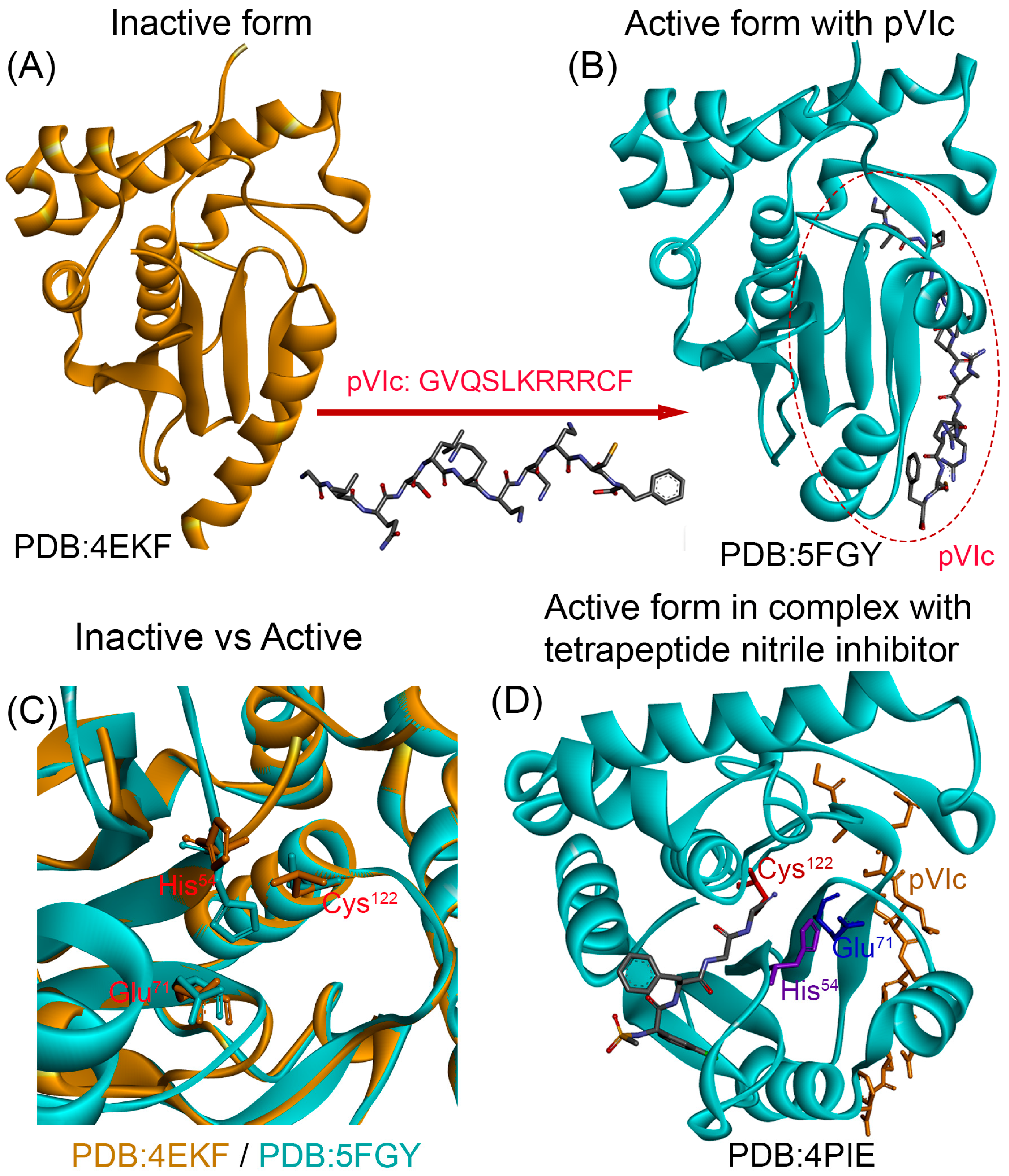
For AVP specifically, several crystal structures representing distinct functional states are available in PDB, as illustrated in Figure 3. The inactive form, which lacks the essential cofactor pVIc, is represented by PDB ID 4EKF (Figure 3A). Binding of the 11-amino-acid pVIc peptide (GVQSLKRRRCF) shifts AVP to an active conformation, exemplified by PDB ID 5FGY (Figure 3B). Comparison of these states (Figure 3C) indicates that pVIc binding is accompanied by a reorientation of catalytic residues His54, Glu71, and Cys122 to assemble a functional Cys–His–Glu triad; notably, His54 rotates toward Cys122, reinforcing the hydrogen-bond/ion-pair network that enhances Cys122 nucleophilicity at the scissile bond. Superposition of the inactive (4EKF) and active (5FGY) structures suggests that these active-site rearrangements occur with minimal changes to surrounding secondary-structure elements (Figure 3C). An active conformation bound to a tetrapeptide nitrile inhibitor is captured in PDB ID 4PIE (Figure 3D), further delineating the architecture of the active site. Collectively, these structural snapshots provide a framework for structure-based drug discovery against AVP; nevertheless, additional high-resolution studies will be valuable to fully elucidate the conformational pathway by which pVIc activates the protease.
Despite the availability of these crystal structures, virtual screening campaigns targeting AVP have remained limited compared to more extensively studied viral proteases. For example, HIV protease has been the subject of thousands of in silico screening studies since the 1990s, whereas AVP remains relatively neglected. This disparity highlights both the untapped opportunities and the methodological challenges that must be addressed to establish AVP as a tractable target for antiviral drug discovery.

4.3. From Virtual Screening to the Bench: Overcoming Barriers in AVP Assay Development

Following the in silico screening and ADME/Tox profiling, the next step is the in vitro evaluation of potential inhibitors, particularly when the target protein exhibits enzymatic activity. This stage involves testing the most promising computational hits to confirm their inhibitory effects under laboratory conditions. Three key components are essential for successful in vitro screening:
i.
The target enzyme in a pure and active form.
ii.
A robust enzymatic assay, ideally adaptable to a high-throughput screening (HTS) format.
iii.
The test compounds in sufficiently pure form.
While these requirements are routine for well-established viral proteases such as HIV protease—where commercial enzyme preparations and standardized assay kits are readily available—they remain major obstacles for AVP research. Unlike most viral proteases, in vitro activation of AVP depends on the presence of the pVIc peptide. This unusual dependency has created experimental bottlenecks and complicates traditional drug discovery approaches, which are typically designed to target isolated enzymes. The main components and drawback in identifying potential inhibitors of AVP as summarized in Figure 4 and discussed below.

4.3.1. Recombinant AVP and Its Cofactor pVIc: Progress and Limitations

To support structural and biochemical studies, several academic groups have successfully expressed and purified recombinant AVP, typically using Escherichia coli expression systems (e.g., BL21(DE3)). These protocols involve optimized folding procedures and, in some cases, the addition of exogenous pVIc and viral DNA in vitro to reconstitute full proteolytic activity [21,27,54,55].
In the commercial space, biotechnology companies such as Biomatik, Cusabio, and Signalway Antibody offer recombinant HAdV C serotype five protease (L3) produced in E. coli, with purities exceeding 90%. However, to our knowledge, these recombinant preparations are generally supplied in their inactive form, without the accompanying pVIc peptide or guidelines for activation, limiting their immediate applicability for inhibitor screening.
Regarding the pVIc peptide, while it is not broadly available as a commercial catalog item, several studies have sourced it via custom peptide synthesis from specialized vendors such as New England Peptide Inc. (Gardner, MA) [56,57] and Invitrogen (Carlsbad, CA) [48]. Furthermore, detailed protocols for forming the AVP–pVIc complex are available in the literature [28,56].
Taken together, while recombinant AVP and synthetic pVIc peptides can be obtained through academic protocols or custom synthesis, the absence of commercially available, recombinant AVP and pVIc as well as standardized activation protocols continues to pose a major bottleneck for systematic inhibitor screening and high-throughput drug discovery efforts.

4.3.2. Measuring AVP Activity: Current Assays and Their Limitations

Once active AVP is available, the next essential step is to develop reliable assays that can accurately monitor its proteolytic activity and quantify the effects of potential inhibitors. Effective drug discovery requires assays that are rapid, sensitive, and reproducible, providing a robust platform for systematic screening.
Several fluorogenic substrates have been developed and validated for this purpose, enabling real-time monitoring of AVP activity and facilitating inhibitor discovery. One of the first and most well-established substrates to monitor AVP activity the fluorogenic peptide substrate (Cbz-Leu-Arg-Gly-Gly-NH)2-Rhodamine [48]. This substrate incorporates the AVP consensus cleavage sequence, Leu-Arg-Gly-Gly, and is conjugated to Rhodamine 110, a highly fluorescent dye known for its sensitivity and detectability. Upon cleavage by AVP, free Rhodamine 110 is released, producing a strong fluorescence signal with excitation and emission maxima at 492 nm and 523 nm, respectively [21]. The synthesis and purification of this substrate have been described in detail, and it has been successfully employed in multiple studies examining AVP activity and enzyme kinetics [48,58].
However, a notable limitation of this substrate is its lack of commercial availability, which necessitates custom synthesis for laboratory use. It is worth noting that Invitrogen and other vendors offer rhodamine 110–based fluorogenic substrates for various proteases, including elastase, trypsin, plasmin, and thrombin. These substrates utilize the same fluorophore, rhodamine 110, which is known for its high fluorescence quantum yield and sensitivity. While such analogs are widely used in enzymatic assays, a specific rhodamine 110-based substrate incorporating the adenovirus protease cleavage motif (e.g., Leu-Arg-Gly-Gly) is not currently available commercially.
An alternative and widely accessible substrate for AVP assays is Z-Leu-Arg-Gly-Gly-AMC (Z-LRGG-AMC), which has been validated as a reliable probe for measuring AVP activity [59]. Importantly this substrate is commercially available from suppliers such as Bachem, Creative Peptides, and AdipoGen Life Sciences In this substrate, the Z group (benzyloxycarbonyl) stabilizes the peptide, while AMC (7-amino-4-methylcoumarin) serves as a fluorophore that is released upon proteolytic cleavage. The liberated AMC fluoresces with excitation at ~360–380 nm and emission at ~440–460 nm. Z-LRGG-AMC mimics the preferred cleavage motif of AVP (Leu/Arg-Gly-Gly), which is present in several adenoviral precursor proteins such as pVI and pVII. Cleavage at the Gly-Gly bond releases AMC, generating a fluorescence signal proportional to enzyme activity.
It should be noted that fluorogenic substrates such as Z-LRGG-AMC or (Cbz-Leu-Arg-Gly-Gly-NH)2-Rhodamine have inherent limitations. Background fluorescence may result from enzyme impurities or spontaneous substrate degradation, while some candidate inhibitors exhibit intrinsic fluorescence or quenching effects that interfere with readouts. Despite that fluorogenic substrates remain a robust and efficient platform for quantifying AVP activity and screening inhibitors, their success depends on careful optimization, appropriate controls, and complementary validation strategies. In addition, ensuring reliability requires optimization of parameters such as substrate concentration and reaction conditions (e.g., pH, ionic strength, and reducing agents like β-mercaptoethanol or DTT).
In summary the relatively limited application AVP assays, compared with the extensive characterization available for other viral proteases, highlights the urgent need for standardized protocols to accelerate AVP research and drug discovery. Moreover, the limited adoption of these assays reflects the broader challenges in AVP research—while HIV protease and SARS-CoV-2 Mpro have been the subject of thousands of enzymatic studies with standardized commercial assay systems, AVP activity assays have been employed in fewer than 50 published studies over the past three decades, highlighting the significant research gap in this field.

4.3.3. Availability and Purity of Test Compounds

Compared with the challenges of producing active AVP and establishing reliable assays, obtaining test compounds in sufficiently pure form is generally less problematic. Most computationally identified hits, whether repurposed antivirals or natural products, are commercially available through established chemical suppliers in high-purity grades suitable for biochemical assays. Custom synthesis services further expand accessibility for compounds not listed in commercial catalogs. For this reason, we do not elaborate further on compound procurement.

5. Learning from Successful Viral Protease Programs: A Roadmap for AVP Development

Proteases constitute roughly 2% of the human genome and orchestrate a wide range of metabolic and homeostatic processes [60]. Because aberrant proteolysis is implicated in cancer, metabolic and inflammatory disorders, and neurodegenerative diseases, proteases have become prominent therapeutic targets. Protease activity is equally vital to pathogens, which depend on proteases for survival and replication [61]. The first clinically approved protease inhibitor, captopril, entered the market in 1981 for the treatment of hypertension [62]. Since then, many protease-directed drugs have been developed for diverse indications, ranging from oncology and diabetes to antiviral therapy against HIV, HCV, and, more recently, SARS-CoV-2 [63,64,65,66].
The story of antiviral drugs that target viral proteases represents one of the most remarkable success stories in modern medicine. In little over forty years, what began as basic biochemical curiosity has grown into therapies that save millions of lives. These milestones offer a roadmap that can be followed as we begin to seriously explore AVP as a drug target.
i.
The HIV protease program is a landmark example of success in antiviral drug development. In the 1980s, scientists identified the viral protease, and by 1995 the first inhibitor—saquinavir—received approval from the FDA, followed by authorization from the European Medicines Agency (EMA) in 1996 under the trade name Invirase (Hoffmann-La Roche) [67]. This achievement was driven by meticulous structure-based drug design, robust assay development, and substantial industry investment. The impact extended far beyond HIV: it not only revolutionized HIV therapy but also established a blueprint for viral protease drug discovery, informing strategies later applied to COVID-19 and even cancer [68].
ii.
The case of HCV offers another pivotal lesson in protease-targeted drug design. Its NS3/4A protease is a serine protease that depends on the NS4A cofactor for proper function [69,70]. NS4A acts as a molecular tether, anchoring NS3 to the cellular membrane and stabilizing its active conformation [71]—an arrangement reminiscent of the activation requirements of AVP. Despite this structural complexity, researchers successfully developed potent NS3/4A inhibitors by (i) mapping the protease–cofactor interface to define how NS4A modulates activity, (ii) leveraging structure-based design to create molecules that bind effectively to the active site, and (iii) advancing pan-genotypic inhibitors such as grazoprevir, glecaprevir, and voxilaprevir, which retained efficacy across diverse HCV strains and resistance-associated substitutions [72]. When combined with NS5A and NS5B inhibitors, these agents produced sustained virological response (SVR) rates above 95%, even in patients with cirrhosis or prior treatment failure [73]. The success of HCV protease inhibitors highlights a critical principle: cofactor dependency is not an insurmountable barrier but a design challenge that can be overcome with molecular insight and strategic targeting,
iii.
The COVID-19 pandemic demonstrated what happens when decades of protease knowledge are combined with global collaboration. The SARS-CoV-Mpro program showed unprecedented speed: in less than two years, nirmatrelvir (part of Paxlovid) moved from concept to approval [74]. That timeline was not achieved by luck—it was the payoff from years of learning how to target viral proteases efficiently and systematically. Since early 2020, proteases like the Mpro and PLpro have been at the center of SARS-CoV-2 antiviral strategies. High-resolution crystal structures of both enzymes were rapidly published [75,76], and their roles in viral polyprotein processing made them ideal therapeutic targets [77]. Among these, Mpro has emerged as especially druggable, owing to its conserved active site and unique substrate specificity—favoring glutamine at the P1 position—a feature not shared with human proteases [78]. This has enabled the design of highly selective inhibitors with minimal off-target effects.
Across successful viral protease programs, several common factors stand out: early investment in structural biology, reliable enzyme assays, robust screening platforms, close collaboration between academia and industry, and systematic strategies to address resistance. By contrast, AVP research is still in the process of building many of these foundations. Learning from both the successes and failures of other protease programs offers a strategic roadmap for positioning AVP as a viable therapeutic target.
Potential sources of AVP inhibitors include virtual compound libraries, such as the ZINC database [79], which provides access to commercially available molecules for structure-based screening. In addition, natural products, including phytochemicals and microbial metabolites, remain a historically rich reservoir for drug discovery [80].
One particularly promising strategy is drug repurposing, which employs computational tools to identify new therapeutic uses for existing drugs by analyzing their chemical structures and biological activity profiles. This approach leverages established pharmacological and safety data, thereby reducing both development costs and timelines. It is especially valuable in urgent contexts such as pandemics or in rare diseases, where traditional drug discovery pathways are often impractical.
In the following sections, we present key strategies for identifying novel AVP inhibitors and demonstrate how these approaches can accelerate the development of promising drug candidates. We highlight the potential for repurposing existing protease inhibitors, with particular emphasis on those targeting SARS-CoV-2 Mpro and PLpro. Additionally, we assess the potential of phytochemicals as AVP inhibitors. For both repurposed drugs and phytochemicals, we provide representative docking examples conducted specifically for this review. Traditional de novo approaches to small-molecule drug discovery are beyond the scope of review and are therefore not considered here.

6. Drug Repurposing: A Strategic Path Toward AVP Inhibition

Drug repurposing—the practice of finding new uses for existing drugs—has become a cornerstone of rapid-response drug development, especially in times of urgent public health need [81]. The COVID-19 pandemic accelerated its adoption, providing clear examples of how repurposing can deliver viable therapeutic options faster and with lower risk. By building on already-established safety and pharmacokinetic profiles, repurposed drugs can often bypass early-phase trials and move more quickly into clinical evaluation [82,83]. Computational tools played a key role in this effort. Molecular docking and dynamics simulations helped prioritize candidates by evaluating how stably and specifically they could bind to the viral protease active site [84]. Large-scale screening studies have identified both approved and preclinical drugs with inhibitory activity against SARS-CoV-2 Mpro [85,86]. For example, the clinically approved anti-HCV drug boceprevir and the preclinical coronavirus protease inhibitor GC376 (developed for feline infectious peritonitis virus, FIPV) were both shown to effectively inhibit SARS-CoV-2 replication in Vero cells by targeting Mpro [87].
For AVP, repurposing is particularly attractive given the extensive arsenal of protease inhibitors already developed for HIV, HCV, and SARS-CoV-2. Structural and mechanistic similarities among these viral proteases suggest that existing inhibitors could be repositioned for AVP, offering a faster path to therapeutic options while building on decades of protease research and clinical experience.
In a previous review by our group the potential of repurposing existing antiviral protease inhibitors to target AVP has been examined [35]. In our computational docking studies, several HCV protease inhibitors approved by FDA—including danoprevir, grazoprevir, paritaprevir, simeprevir, telaprevir, and vaniprevir—exhibited favorable binding to AVP’s catalytic site, with docking energies ranging from −7.5 to −10.1 kcal/mol, suggesting strong inhibitory potential. Similarly, six FDA-approved HIV protease inhibitorsamprenavir, darunavir, indinavir, lopinavir, nelfinavir, and tipranavir—also showed predicted binding interactions with AVP, with docking scores between −6.5 and −7.7 kcal/mol [35]. While these affinities are lower than those observed for the HCV compounds, they still indicate a plausible capacity to engage the AVP active site.
Building on the success and lessons learned from efforts targeting SARS-CoV-2 proteases, particularly the Mpro and papain-like protease PLpro, either though computational approaches or repurposing approaches herein we examine how these emerging proteases inhibitors could be redirect for AVP inhibition. Although AVP is structurally distinct from Mpro and PLpro, all three enzymes belong to the cysteine protease family and employ similar catalytic dyads/triads centered on an essential cysteine and histidine residue. This mechanistic similarity raises the possibility that certain small molecules developed to target Mpro and PLpro may also engage the AVP catalytic pocket. Our evaluation was therefore designed as an exploratory in silico study to assess the feasibility of repurposing existing cysteine protease inhibitors as potential AVP modulators.
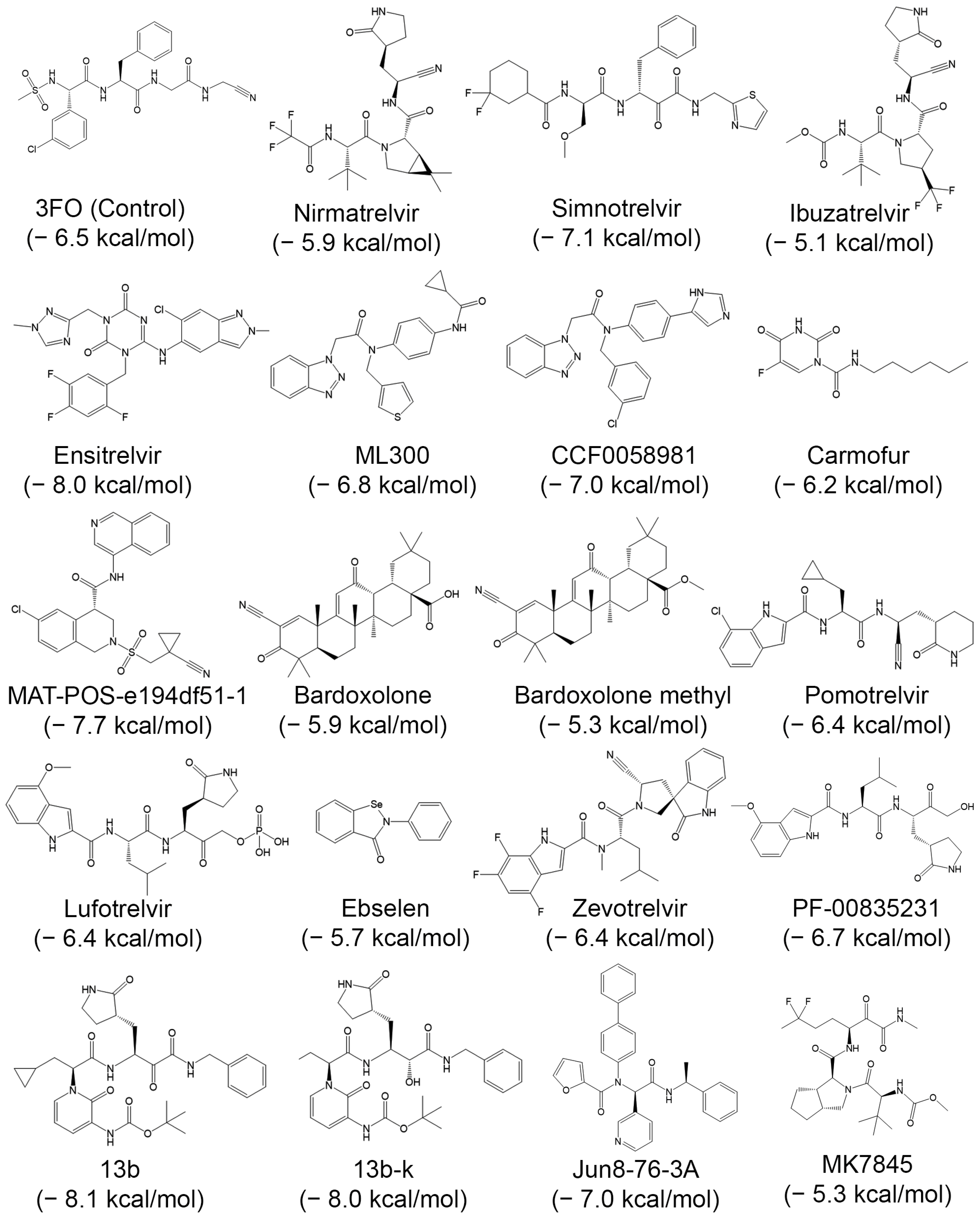
We began with a targeted literature search to identify studies on SARS-CoV-2 Mpro inhibitors. Because our objective was to illustrate the potential of repurposing these inhibitors for AVP rather than to exhaustively catalog every report, we selected representative examples instead of conducting a full systematic review. Likewise, we did not perform extensive computational analyses or molecular-dynamics simulations, focusing instead on the conceptual framework and preliminary evidence supporting this repurposing strategy. Our literature search identified 19 compounds evaluated against the SARS-CoV Mpro (Table 1). These span both molecules originally designed as Mpro inhibitors and those repurposed for activity against this protease.
We subsequently evaluated the binding scores of the 19 selected compounds against AVP using the 4PIE crystal structure of the enzyme. Prior to the docking studies, the co-crystallized tetrapeptide nitrile inhibitor was removed to free the active site. Τhe tetrapeptide nitrile inhibitor (3FO) was used as the control compound for comparative analysis throughout the study. The control compound, 3FO, achieved a docking score of −6.5 kcal/mol. Of the 19 tested compounds 9 scored equal to or better than the control compound (Figure 5).
The highest docking score was observed for compound 13b (−8.1 kcal/mol), followed closely by compound 13b-k and ensitrelvir, both of which achieved binding scores of −8.0 kcal/mol. Other top-performing compounds showed docking energies ranging between −6.5 and −7.7 kcal/mol. Compound 13b is a specialized α-ketoamide inhibitor designed to target the Mpro. It builds upon earlier prototypes, such as compound 11r [106], with structural modifications including a cyclopropyl group at the P2 position to enhance potency and specificity. Compared to its predecessors, 13b demonstrates improved aqueous solubility, an extended plasma half-life, and focused antiviral activity against SARS-related coronaviruses, albeit with reduced broad-spectrum efficacy. Co-crystal structures confirm its tight binding to Mpro, reinforcing its potential as a therapeutic lead. Compound 13b-k is the S,S,S diastereomer of 13b. Ensitrelvir is a non-covalent Mpro inhibitor developed through a collaboration between Hokkaido University and Shionogi & Co., Ltd. It exhibits potent antiviral activity against SARS-CoV-2 variants and has advanced rapidly in clinical development. Marketed as Xocova®, ensitrelvir received emergency use approval in Japan in November 2022 and full regulatory approval in March 2024 for the treatment of COVID-19 [107].
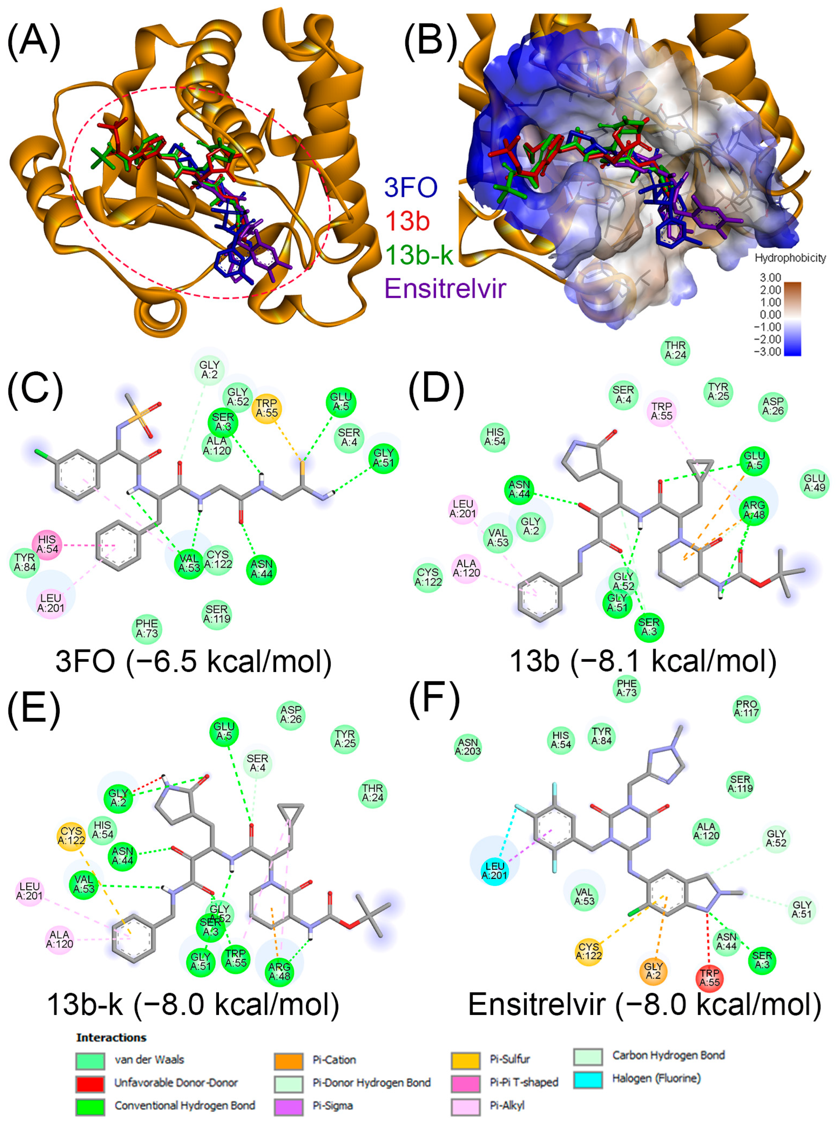
Figure 6 illustrates the predicted binding interactions of the three top-scoring compounds—13b, 13b-k, and ensitrelvir—together with the reference inhibitor 3FO. As shown in Figure 6A, all three test compounds occupy the active site of AVP in a manner comparable to the control compound, fitting well into the catalytic pocket. The hydrophobic surface map (Figure 6B) further confirms that each compound, like 3FO, spans both polar (blue) and nonpolar (brown) regions of the active site, highlighting their ability to engage in complementary interactions across the binding pocket.
The detailed 2D interaction diagrams (Figure 6C–F) illustrate the specific molecular contacts between AVP and the ligands. For the control inhibitor 3FO (Figure 6C), the binding mode involves key residues of the catalytic site, including π–π interactions with His54 and van der Waals interactions with Cys122. These predictions are consistent with the crystallographic evidence from the AVP–3FO complex (PDB ID: 4PIE), supporting the reliability of the docking approach.
Among the test compounds, 13b (Figure 6D) interacts with residues of the catalytic triad, forming van der Waals contacts with His54 and additional stabilizing interactions with Cys122. Similarly, 13b-k (Figure 6E) engages His54 via van der Waals interactions and Cys122 through a combination of van der Waals and π–cation contacts, suggesting a potentially stronger binding mode relative to 13b. Ensitrelvir (Figure 6F) also targets the catalytic site, forming π–sulfur interactions with Cys122 and van der Waals interactions with His54, while extending additional contacts with surrounding residues such as Gly52, Ala120, and Leu201.
Taken together, these results suggest that 13b, 13b-k, and ensitrelvir not only occupy the catalytic pocket in a manner comparable to the control inhibitor 3FO but also establish favorable interactions with the critical catalytic residues His54 and Cys122. This highlights their potential as candidate scaffolds for further optimization in the development of AVP inhibitors. However, further in silico and in vitro experiments are required to assess the inhibitory effect of these compounds against AVP.
In addition to Mpro, the second SARS-CoV-2 protease, PLpro, has attracted significant attention as a drug target due to its dual role in viral polyprotein processing and suppression of host immune responses [108]. Unlike SARS-CoV-2 Mpro, which has been a central focus of drug discovery efforts (e.g., Paxlovid), PLpro presents unique challenges, including broader substrate specificity and a more flexible binding pocket [109]. Among the most studied PLpro inhibitors is GRL0617, a non-covalent compound that exhibits strong in vitro activity (IC50 = 2.1 μM) and potent antiviral effects in cell-based models. Co-crystal structures of the GRL0617–PLpro complex have identified a key ligandable hotspot [110], which has guided the design of improved derivatives, such as Jun9-72-2 [111].
Other notable inhibitors include ebselen and related organoselenium compounds, which have demonstrated PLpro inhibition with broader antiviral implications [112]. Additionally, quinoline-based inhibitors have been rationally developed to enhance pharmacokinetics and oral bioavailability. Among these, Jun13296 has shown robust activity against multiple SARS-CoV-2 variants, including strains resistant to Mpro inhibitors. In mouse models, Jun13296 significantly improves survival, reduces viral load in the lungs, and mitigates tissue damage [113].
Shan et al. [114] reported the development of compound (inhibitor) 19, a potent and selective inhibitor of the SARS-CoV-2 PLpro. Notably, compound 19 not only blocks substrate cleavage and the immunosuppressive functions mediated by PLpro, but also significantly suppresses viral replication in human cells with a submicromolar IC50 of 182 nM.
A recent study by Bader et al. [115] reported the discovery and optimization of a novel PLpro inhibitor series through the screening of over 400,000 compounds. This led to identification of the WEHI-P scaffold, which exploits a previously unrecognized Met208-mediated hydrophobic pocket to achieve low-nanomolar potency, high cellular activity, and no detectable off-target effects on human deubiquitinases. Medicinal chemistry optimization yielded WEHI-P8, an orally bioavailable compound with a 14 h half-life in mice. In preclinical models, WEHI-P8 outperformed Paxlovid-equivalent regimens by reducing viral loads, suppressing lung inflammation and cytokine storms, and—importantly—preventing long-term complications such as lung hemorrhage, fibrosis, and cognitive deficits associated with post-acute COVID-19.
The numerous reported PLpro inhibitors [116,117,118] offer a rich source of chemical scaffolds for potential AVP inhibitor development. A recent review by Yue et al. [119] systematically cataloged all SARS-CoV-2 PLpro crystal structures published between April 2020 and December 2024. The review mapped the enzyme’s four structural domains (ubiquitin-like, thumb, palm, and fingers), identified both well-characterized and cryptic binding sites, and compiled an extensive database of known small-molecule inhibitors for structure-based analysis.
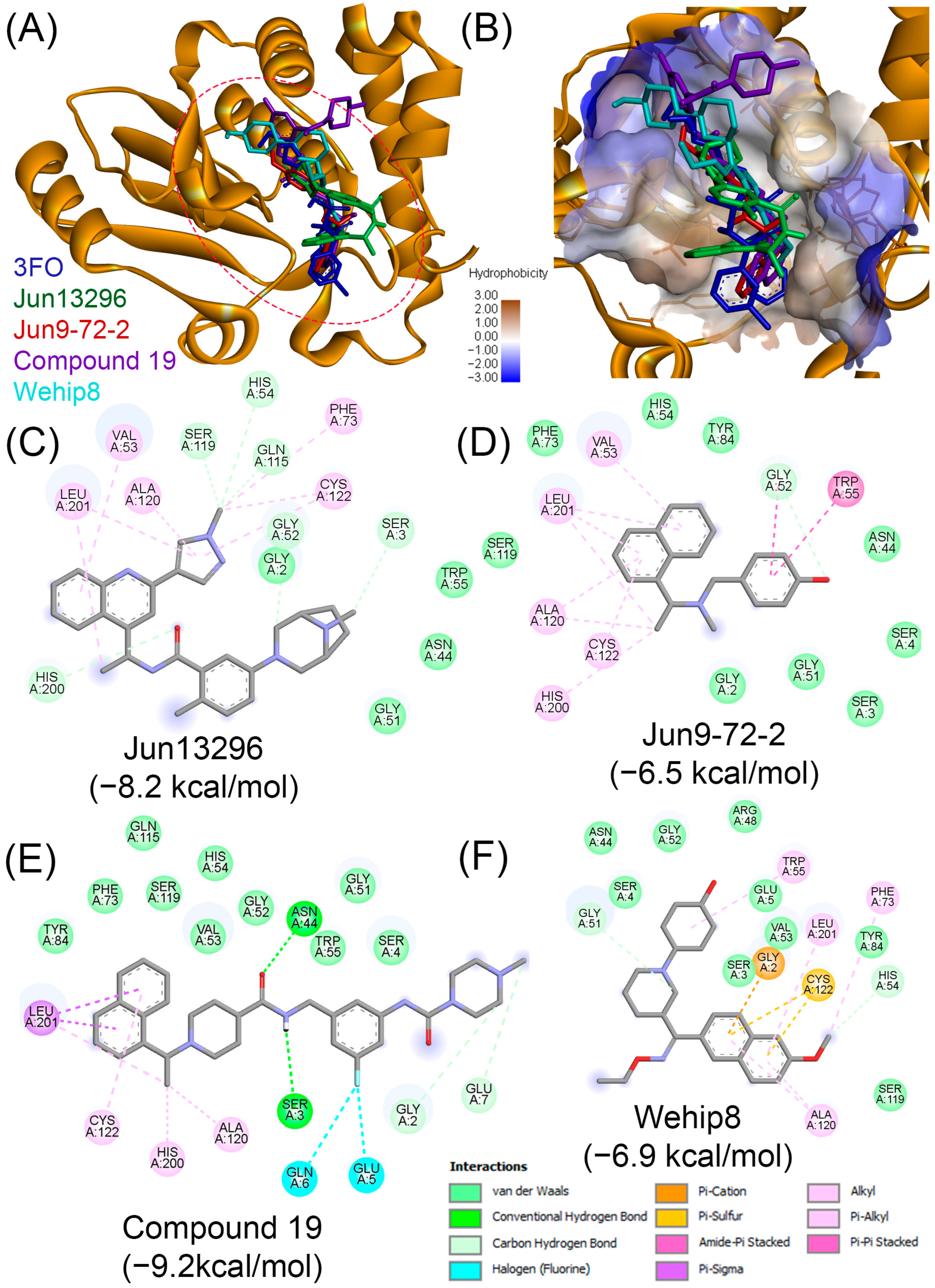
To evaluate the translational potential of PLpro inhibitors against AVP, we selected four representative compounds namely, Jun13296 (Pubchem CID:172643385), Jun9-72-2 (Pubchem CID: 156612931), compound 19 (PubChem CID: 168321860), and WEHI-P8 (PubChem CID: 175670503), on the basis of their structural diversity, potency, and favorable pharmacological profiles. In detail, Jun13296 was chosen for its pan-variant activity, favorable pharmacokinetic profile, and demonstrated efficacy in animal models. Jun9-72-2, a derivative of the well-characterized GRL0617 scaffold, validates the ligandable hotspot adjacent to the PLpro active site. Compound 19 was included for its high potency and selectivity, with submicromolar inhibition of PLpro’s enzymatic and immunomodulatory functions. Finally, WEHI-P8 represents a next-generation scaffold optimized for oral bioavailability and superior preclinical efficacy. Their binding modes and scores were assessed through molecular docking (Figure 7) using the crystal structure of AVP (PDB ID: 4PIE). The tetrapeptine nitrile (3FO) was used as a control for comparative purposes.
As illustrated in Figure 7A, all four compounds bind within the active site of AVP. Interestingly, they fit into the AVP catalytic pocket in orientations similar to 3FO (Figure 7A) and span both polar (blue) and nonpolar (brown) regions of the binding pocket, as confirmed by the hydrophobic surface mapping (Figure 7B). The 2D interaction diagrams (Figure 7C–F) highlight distinct interaction profiles for each compound. Jun13296 engages His54 through van der Waals interactions and establishes additional stabilizing hydrogen bonds with adjacent residues, consistent with its reported in vivo activity (Figure 7C). Jun9-72-2, interacts with Cys122 via π–cation contacts and with His54 through van der Waals forces, anchoring it firmly in the catalytic pocket (Figure 7D). Compound 19 shows the strongest predicted binding (−9.3 kcal/mol), forming multiple stabilizing interactions with both His54 and Cys122, which likely accounts for its superior docking score (Figure 7E). WEHI-P8 not only interacts with the catalytic dyad residues His54 and Cys122 but also extends into adjacent hydrophobic regions.
Given the conserved catalytic architecture among viral cysteine proteases, these advanced PLpro inhibitors represent compelling candidates for repurposing against AVP. However, further computational docking, biochemical validation, and in vivo studies will be essential to assess their cross-reactivity and therapeutic potential in the context of adenovirus infections. While most therapeutic development around PLpro has focused on coronaviruses, its structural and mechanistic similarity to other viral cysteine proteases raises the possibility that established PLpro inhibitors could be repurposed to target AVP.

7. Phytochemicals: An Unexplored Source of Potential AVP Inhibitors

Phytochemicals are bioactive compounds produced by plants and have historically provided a rich source of drug leads [120], with several of approved drugs derived from or inspired by natural sources [121]. Secondary metabolites from medicinal and aromatic plants (MAPs) exhibit diverse structural scaffolds and evolutionarily refined mechanisms for interacting with biological targets, often displaying multi-target (polypharmacological) effects not commonly achieved by synthetic compound libraries [122].
Phytochemicals have been pivotal in antiviral drug discovery. A notable example is oseltamivir (Tamiflu), an influenza treatment whose synthesis relies on shikimic acid—a compound originally extracted from star anise (Illicium verum) [123]. Additionally, numerous plant-derived compounds have demonstrated inhibitory activities against viral enzymes, including proteases [117,124]. Therefore, the structural diversity and biological relevance inherent in phytochemicals make them especially attractive for identifying inhibitors of complex viral enzymes, including AVP.
Notably, to the best of our knowledge, no published studies have investigated the potential of phytochemicals as direct inhibitors of AVP. However, several studies have explored the antiviral potential of natural products against HAdV. For example, Balsera-Manzanero et al. [125] identified a series of isoflavonoid-type compounds, known as rotenoids, with potent antiviral activity against HAdV. Through screening of 1233 natural products, they found that rotenolone and its analogs—rotenone, deguelin, millettone, and tephrosin—exhibited nanomolar IC50 values and high selectivity indices, with deguelin achieving a selectivity index exceeding 2400. Mechanistic studies revealed that while most compounds act during late stages of viral replication, rotenone uniquely disrupts microtubule polymerization, preventing viral particles from reaching the nucleus. A comprehensive review by Musarra-Pizzo et al. [126] highlights several natural antiviral compounds, including shikonin, p-coumaric acid, chlorogenic acid, quercetin, and apigenin. While these compounds exhibit antiviral activity against various human viruses, the review does not provide specific evidence of their efficacy against adenoviruses.
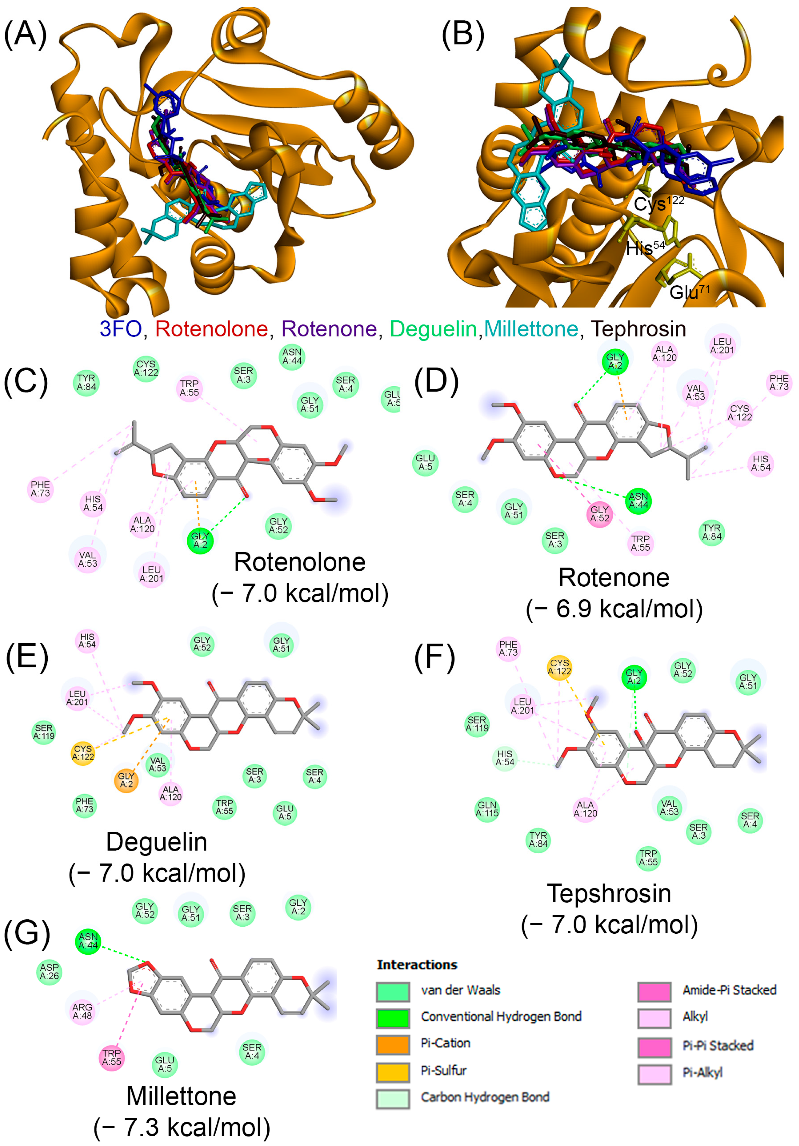
In this study, we investigated the potential of phytochemicals as inhibitors of AVP through virtual screening. As a first step, we evaluated the binding affinities of the recently reported antiviral compound rotenolone (PubChem CID: 68184) and its structural analogs—rotenone (CID: 6758), deguelin (CID: 107935), millettone (CID: 442810), and tephrosin (CID: 114909)—to assess whether their known antiviral activity could be associated with AVP inhibition (Figure 8). Molecular docking analyses were performed using the crystal structure of AVP (PDB ID: 4PIE) while 3FO was included as a reference control.
Docking results revealed that all tested phytochemicals achieved binding scores below −6.5 kcal/mol (ranging from −6.9 to −7.3 kcal/mol). As mentioned above the control compound 3FO exhibited a binding score of −6.5 kcal/mol. Notably, with the exception of millettone, all compounds bound within the catalytic pocket of AVP in conformations closely resembling that of 3FO and positioned in proximity to two key residues of the catalytic triad, His54 and Cys122 (Figure 8A,B). In contrast, millettone, despite achieving the most favorable docking score (–7.3 kcal/mol), was positioned near the catalytic pocket but failed to form close interactions with His54 or Cys122 (Figure 8B).
Two-dimensional interaction diagrams (Figure 8 C–G) further illustrate the binding profiles. Rotenolone interacted with His54 via π–alkyl contacts and with Cys122 through van der Waals interactions, stabilized additionally by polar and nonpolar side chains in the pocket (Figure 8C). Rotenone similarly engaged His54 and Cys122 via alkyl interactions (Figure 8D). Deguelin formed a π–alkyl interaction with His54 and a π–sulfur interaction with Cys122, suggesting stable anchoring in the catalytic site (Figure 8E). Tephrosin established a carbon–hydrogen bond with His54 and both alkyl and π–sulfur interactions with Cys122 (Figure 8F). By contrast, millettone, although yielding the lowest docking energy, failed to engage catalytic residues directly, raising uncertainty about its ability to effectively inhibit AVP activity (Figure 8G).
Taken together, these findings suggest that rotenolone, rotenone, deguelin, and tephrosin may serve as promising AVP inhibitors by engaging key catalytic residues, whereas millettone may act through an alternative mechanism or may not effectively inhibit AVP despite its favorable docking score. Further molecular docking refinements and in vitro validation studies will be essential to confirm the inhibitory potential of these phytochemicals.
We subsequently evaluated the potential of additional phytochemicals as inhibitors of AVP. In a previous study by our group, we constructed a chemical library of approximately 2500 phytochemicals and screened them against the HRV-3Cpro using a combination of in silico and in vitro approaches [124]. From this effort, eight compounds—apigenin, carnosol, chlorogenic acid, kaempferol, luteolin, quercetin, rosmarinic acid, and rutin—emerged as promising inhibitors of HRV-3Cpro. Among them, carnosol and rosmarinic acid demonstrated potent inhibitory activity, reducing HRV-3Cpro enzymatic function by more than 55% in vitro. Mechanistic analyses further indicated that these two compounds act through a competitive mode of inhibition [124].
Given the structural and mechanistic similarities between HRV-3Cpro and AVP—both being cysteine proteases that employ a catalytic triad for substrate cleavage [35]—we hypothesized that inhibitors of HRV-3Cpro may also display cross-reactivity against AVP. Building on this rationale and following our initial analysis of rotenolone and its analogs (Figure 8), we expanded our investigation by compiling a broader panel of 50 phytochemicals (Supplementary Table S1). This expanded dataset increased the chemical diversity available for virtual screening and provided a more comprehensive framework for identifying potential AVP inhibitors. It should be noted that the 50 phytochemicals evaluated in this study represent an exploratory subset the larger library of ~2500 compounds mentioned above. Rather than performing an exhaustive screen, we deliberately selected compounds to capture scaffold diversity across major classes of natural products (e.g., flavonoids, alkaloids, terpenoids, and phenolic acids) while prioritizing well-characterized and readily available molecules with reported bioactivities. This pragmatic strategy is consistent with common practice in in silico drug discovery, where representative subsets are frequently used to identify promising chemical scaffolds for further investigation. Future studies could expand this effort to a systematic, large-scale screening of the full phytochemical library to validate and extend the present findings.
We next performed molecular docking studies to assess the binding affinities and interaction profiles of this expanded phytochemical panel with the AVP active site. Similar to the above efforts the tetrapeptide nitrile 3FO that achieved a binding score of −6.5 kcal/mole was used as a control.
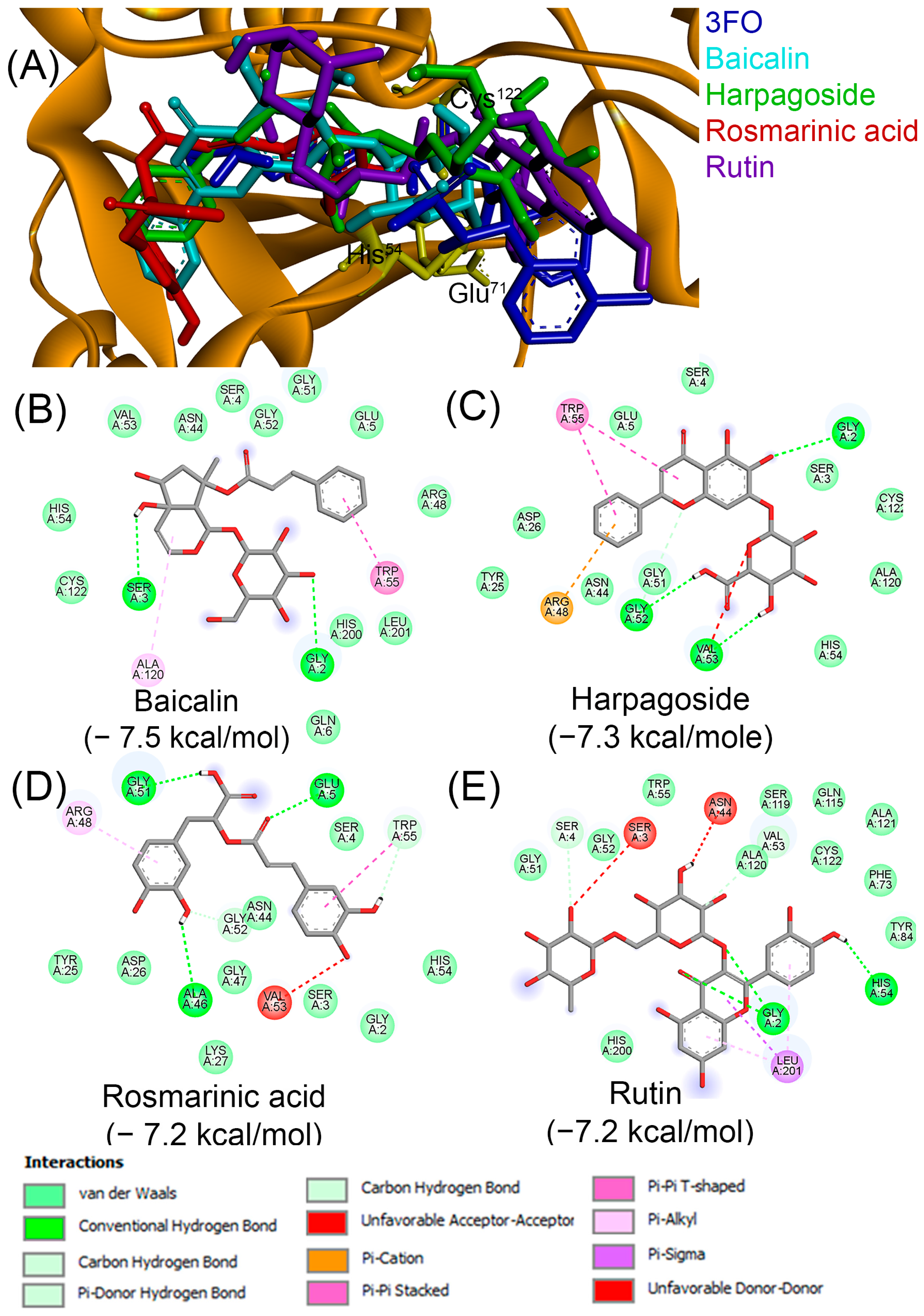
Of the 50 compounds tested, 19 achieved binding scores equal to or better than the control compound 3FO (Figure 9). Baicalin obtained the best score (–7.5 kcal/mol), followed by harpagoside (–7.3 kcal/mol), rosmarinic acid (−7.2 kcal/mol), and rutin (−7.2 kcal/mol).
We next examined the binding profiles of the four top-ranking phytochemicals—baicalin, harpagoside, rosmarinic acid, and rutin—and compared them to the reference compound 3FO (Figure 10). All four compounds bound within the AVP active site in orientations closely resembling 3FO, positioning near the catalytic residues His54 and Cys122 of the catalytic triad (Figure 10A). This alignment supports their favorable docking scores.
The two-dimensional interaction diagrams (Figure 10B–E) further detail the specific binding interactions. Baicalin (Figure 10B) and harpagoside (Figure 10C) established contacts with both His54 and Cys122 primarily through van der Waals interactions, in addition to multiple stabilizing hydrogen bonds with surrounding residues in the binding pocket. Rosmarinic acid (Figure 10D) engaged His54 via van der Waals forces but did not directly contact Cys122; a network of hydrogen bonds and additional van der Waals interactions with nearby residues stabilized its binding. Finally, rutin (Figure 10E) formed a hydrogen bond with His54 and also interacted with Cys122 via van der Waals contacts, while establishing multiple additional interactions across the catalytic pocket.
Collectively, these results suggest that baicalin, harpagoside, and rutin may exert more substantial inhibitory potential through direct engagement with both His54 and Cys122. In contrast, rosmarinic acid may adopt a slightly different binding mode that still supports stable association with the protease active site. Nevertheless, further molecular dynamics simulations and in vitro validation experiments will be required to confirm the stability of these interactions and assess their true inhibitory potential against AVP
Because binding affinity alone does not determine the suitability of a compound as a drug candidate, we evaluated the ADME properties of all 19 phytochemicals using the SwissADME platform (http://www.swissadme.ch/, accessed on 14 August 2025). Notably, the four phytochemicals with the most favorable (i.e., most negative) docking scores—baicalin, harpagoside, rosmarinic acid, and rutin—displayed suboptimal drug-likeness profiles (Table 2). Specifically, each of these compounds exhibited low predicted gastrointestinal (GI) absorption, which could restrict their oral bioavailability. Additionally, multiple violations of Lipinski’s rule of five and elevated topological polar surface area (TPSA) values indicate potential challenges with membrane permeability. Among these, baicalin had the lowest synthetic accessibility score, suggesting it is relatively straightforward to synthesize, whereas rutin demonstrated the least favorable drug-likeness attributes, including the highest molecular weight and the greatest number of rule violations.
We then examined the ADME profiles of the remaining 15 compounds that achieved binding scores equal to or better than 3FO (Figure 9). Among these, apigenin, camptothecin, jaempferol, and piperine stood out as the top four candidates due to their distinctive characteristics, as detailed in Table 2. All four compounds demonstrated perfect Lipinski compliance (zero violations) and high gastrointestinal absorption, which is promising for oral bioavailability. No Pan-Assay INterference Compounds (PAINS) alerts were detected, indicating a lower likelihood of false positives during screening. These compounds are classified as lead-like, meaning they fall within an optimal range of size and polarity for further optimization. Their synthetic accessibility scores are moderate, suggesting they are not overly complex to synthesize. It should be noted that a comprehensive ADME analysis for all 19 compounds that scored equal of better than 3FO is not included in this review, as our primary aim is to demonstrate the potential of phytochemicals as AVP inhibitors. A full ADME evaluation falls outside the scope of this work.
Furthermore, the toxicity profiles of the four aforementioned phytochemicals with favorable ADME profiles were evaluated using the StopTox platform (https://stoptox.mml.unc.edu/; accessed on 14 August 2025). The results are summarized in Table 3 and indicate that all four candidates—apigenin, camptothecin, kaempferol, and piperine—underwent comprehensive toxicity screening across six endpoints: acute oral, dermal, and inhalation toxicity, as well as eye irritation/corrosion, skin irritation/corrosion, and skin sensitization. For acute inhalation toxicity, all compounds were classified as non-toxic, with confidence scores ranging from 63% (piperine) to 74% (camptothecin). In terms of acute oral toxicity, apigenin and Kaempferol were predicted to be non-toxic (58% and 70%, respectively), while camptothecin and piperine were classified as toxic with high confidence (90%). Regarding acute dermal toxicity, camptothecin and piperine were non-toxic (68% and 72%), whereas apigenin and kaempferol showed toxicity (54% and 65%). Eye irritation and corrosion predictions revealed that apigenin, camptothecin, and piperine were classified as toxic (confidence scores: 71%, 71%, and 52%, respectively), while Kaempferol was considered non-toxic (50%). For skin sensitization, camptothecin was a non-sensitizer (70%), but apigenin, kaempferol, and piperine were identified as sensitizers (60%, 70%, and 60%). Importantly, all compounds were predicted negative for skin irritation and corrosion, with confidence scores between 70% and 90%. Overall, most compounds met most of the safety criteria, but notable exceptions were observed in acute oral toxicity (camptothecin and Piperine), acute dermal toxicity (apigenin and kaempferol), eye irritation/corrosion (all except kaempferol), and skin sensitization (all except camptothecin).
We subsequently evaluated the binding profiles of four phytochemicals with acceptable ADME/T properties—apigenin, camptothecin, kaempferol, and piperine—to further assess their potential as competitive inhibitors of AVP. The docking results are summarized in Figure 11A. All four compounds were accommodated within the AVP catalytic pocket in orientations generally comparable to the reference inhibitor 3FO, with the exception of camptothecin, which adopted a distinct orientation but still localized in close proximity to the catalytic triad residues.
The two-dimensional interaction diagrams (Figure 11B–E) provide a more detailed view of the specific binding interactions. Interestingly, none of the tested compounds established simultaneous interactions with both His54 and Cys122, the key residues of the catalytic triad. Apigenin (Figure 11B) and camptothecin (Figure 11C) interacted with His54 through van der Waals forces, in addition to forming multiple stabilizing contacts—including hydrogen bonds and π-type interactions—with surrounding residues in the binding pocket. Kaempferol (Figure 11D), despite being oriented in a manner similar to 3FO, failed to interact directly with either His54 or Cys122, suggesting a weaker potential for catalytic inhibition. In contrast, piperine (Figure 11E) formed a hydrogen bond with Cys122, while lacking direct interaction with His54; its stabilization within the pocket was instead mediated by van der Waals forces and π-type interactions with adjacent residues.
The above results suggest that although the four compounds can be accommodated within the AVP active site, their lack of consistent interactions with both His54 and Cys122 may limit their inhibitory potential compared to other phytochemicals tested. Nevertheless, further molecular dynamics simulations and in vitro validation studies are required to clarify whether these compounds can effectively disrupt AVP catalytic activity.
Taken together, the results presented in this section highlight the potential of phytochemicals as an excellent source or pool of scaffolds for the identification of AVP inhibitors. Further studies using larger libraries are needed to discover promising phytochemical inhibitors of AVP.

8. Perspectives and Recommendations

Viral proteases play essential roles in the viral life cycle by cleaving polyproteins into functional components required for replication complex assembly and the production of infectious virions. Targeting these enzymes has proven to be an effective antiviral strategy, exemplified by the clinical success of protease inhibitors in the treatment of HIV and HCV infections. By contrast, the proteases of viruses that also cause respiratory tract infections, such as AVP and HRV-3Cpro, remain comparatively underexplored [35].
In this review, we highlight the opportunities for discovering novel AVP inhibitors as a strategy to combat HAdV infections. Although HAdV typically causes self-limiting disease in healthy individuals, it can lead to severe outcomes in immunocompromised patients and infants. The evidence summarized here underscores that AVP is a well-defined and druggable target, warranting renewed attention and systematic investigation.
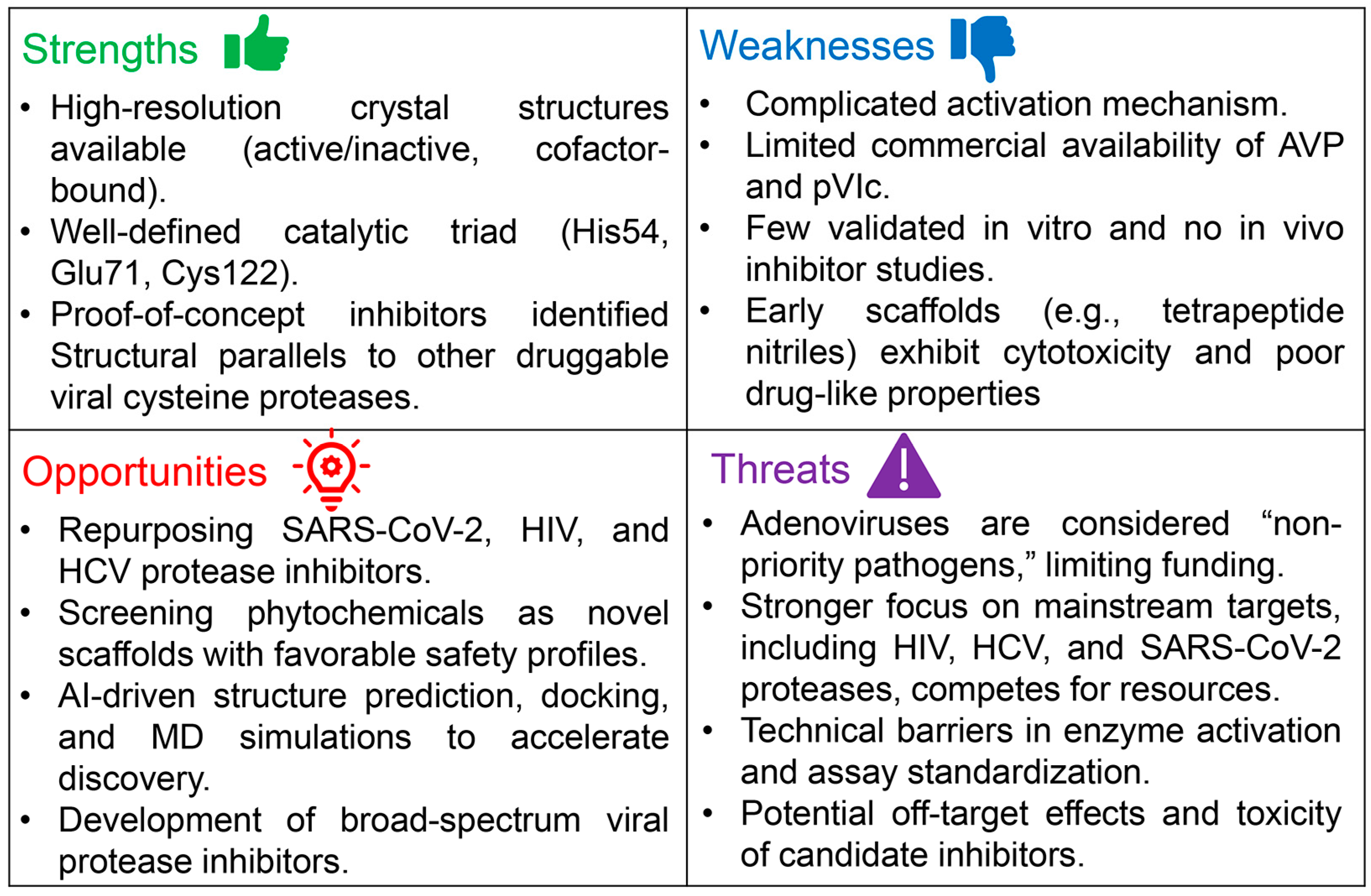
To provide a structured overview of the current landscape, we conducted a SWOT (strengths, weaknesses, opportunities, and threats) analysis of AVP as an antiviral target (Figure 12). This framework highlights the strengths that establish AVP as druggable, the weaknesses that have limited its study, the opportunities for advancing inhibitor discovery, and the threats or challenges that continue to hinder progress. Together, this analysis illustrates both the promise of AVP-targeted drug discovery and the key areas that require investment and innovation.
In previous efforts, our group highlighted opportunities to identify inhibitors of another underexplored viral protease, the HRV-3CproLike AVP, HRV-3Cprohas attracted limited attention, largely because rhinoviruses typically cause mild infections. For HRV-3Cpro we have proposed a comprehensive strategy that included optimization of every step in the inhibitor discovery pipeline—from cloning and soluble expression of the protease [127], to the development and validation of a high-throughput colorimetric assay for inhibitor screening [128]. More recently, we emphasized the potential of phytochemicals as candidate inhibitors of HRV-3Cprothrough in silico and in vitro studies [124], and we also evaluated the repurposing of approved protease inhibitors from other viral strains [35]. Through these efforts, we demonstrated that knowledge gained from well-characterized viral proteases can be successfully leveraged to discover inhibitors for HRV-3Cproand, by extension, other less-studied proteases. Building on this “success story,” we suggest that similar approaches can be applied to the discovery of AVP inhibitors. Taken together, these insights reinforce the need to establish coordinated and systematic discovery pipelines for AVP—integrating cloning, expression, assay development, and both repurposing and phytochemical screening—to accelerate the identification of clinically relevant inhibitors.
In this review, we present evidence that AVP is a highly druggable target. Building on this foundation, several strategic priorities can transform AVP research from a conceptual framework into a structured drug discovery pipeline. At each stage of this pipeline, processes should be carefully optimized using systematic approaches such as design of experiments (DoE), which allows efficient evaluation of multiple variables to maximize protein expression yields and to refine assay conditions for monitoring enzymatic activity. Our group has successfully applied DoE to optimize the soluble expression of the HRV 3Cpro [127] as well as the assay conditions for monitoring its activity [128], and we have extended this approach to optimize assay conditions for lactate dehydrogenase B [129]. More broadly, DoE has been demonstrated as a valuable tool across the drug discovery continuum—from gene cloning to crystallization of enzyme–inhibitor complexes—providing a rational framework for reproducibility and efficiency (Reviewed in [130]).
The following recommendations outline the critical steps required to accelerate the identification, validation, and eventual clinical translation of AVP inhibitors.
i.
Establish standardized protocols for recombinant AVP soluble expression. This step is among the most straightforward in the drug discovery pipeline, as cloning and expression technologies are now routine. Several companies, including GenScript, offer artificial gene synthesis directly into expression vectors, with options for codon optimization to enhance expression in both prokaryotic and eukaryotic systems. For AVP, we strongly recommend the use of pGEX vectors, which enable expression of the protein as a GST-tagged fusion. Importantly, the pGEX-6P series encodes a cleavage site for the HRV 3C pro, and the corresponding GST-HRV-3Cpro (commercially available as PreScission Protease) facilitates on-column tag removal and simultaneous purification of the target protein. This strategy has been successfully employed for the production of numerous proteins, including TNF [131], and RANKL [132]. As discussed earlier, soluble expression conditions can be further optimized using DoE approaches, which allow rapid small-scale optimization of parameters. In our experience, DoE enables identification of conditions that maximize soluble enzyme yield within just two days of experimentation [130,133]. Regarding the second essential component for AVP activation, the pVIc peptide, although it is not broadly available as a catalog item, several studies have obtained it through custom peptide synthesis from specialized vendors. Protocols describing AVP activation with pVIc are available in the literature; however, these methods can be further refined and optimized using DoE to maximize reproducibility and efficiency.
ii.
Establish robust HTS assays to monitor AVP activity. This area requires particular attention, as currently only fluorogenic substrates have been reported for monitoring AVP activity. Fluorescence-based assays can be affected by background signals and by the intrinsic fluorescence or quenching properties of candidate inhibitors. To overcome these limitations, we strongly recommend the development of non-fluorescent substrates, such as those labeled with p-nitroaniline (pNA). Adenovirus protease recognizes consensus sequences (M/I/L)XGX-G and (M/I/L)XGG-X [134]. Accordingly, synthetic substrates such as Leu-Arg-Gly-Gly-pNA can be designed, in which pNA is conjugated to the C-terminus of the peptide and released upon cleavage. The liberated pNA produces a yellow color that can be quantitatively measured at 405 nm using a spectrophotometer or plate reader. Such pNA-based substrates have been successfully used to establish HTS-compatible assays, including in our group’s work on HRV-3Cpro, where the assay was validated and proven effective in distinguishing true inhibitors from false positives [124,128]. In addition to optimizing enzymatic conditions, it is essential to implement rigorous controls. Negative controls without enzyme are required to account for spontaneous substrate hydrolysis, while positive controls with active AVP ensure the assay is functioning correctly. Including unrelated proteases as additional controls can further validate substrate specificity and exclude non-specific cleavage.
iii.
Leverage drug repurposing strategies. The in silico screening results presented in this work, together with our previous studies, highlight the potential of repurposing established viral protease inhibitors as modulators of AVP. In particular, the extensive compound libraries developed over the past five years for the SARS-CoV-2 Mpro and PLpro represent a valuable resource that can be systematically screened for AVP activity. Similarly, inhibitors originally designed for other well-characterized viral proteases, including HIV and HCV proteases, should be evaluated for cross-reactivity with AVP. This strategy offers clear advantages over de novo drug discovery, as it can significantly reduce both the time and cost associated with developing new antivirals. It should be noted that while molecular docking provides a rapid and cost-effective approach to screen large compound libraries and generate testable hypotheses, it also has inherent limitations. Docking can identify plausible binding modes and prioritize candidates, which is particularly valuable when experimental data are limited, as in the case of AVP. However, docking relies on simplified scoring functions that may not fully account for solvation, entropic effects, or protein flexibility, and predictions can vary depending on the structural model used. Therefore, docking results should be viewed as a starting point for hypothesis generation rather than definitive evidence of binding. Integrating docking with biochemical assays, structural biology, and medicinal chemistry will be essential to validate and optimize repurposed inhibitors against AVP.
iv.
Encourage multi-viral protease inhibitor development. Building on the experience gained with protease inhibitors for SARS-CoV-2, HIV, and HCV, as well as findings from our earlier work [35], there is clear potential to develop broad-spectrum protease inhibitors that target conserved catalytic mechanisms across different viruses. Such agents could be particularly valuable in the context of co-infections, for example, AVP and HIV, which are associated with especially severe and life-threatening outcomes. Expanding efforts toward multi-viral inhibitor development would not only enhance the therapeutic relevance of AVP research but also contribute to more versatile antiviral strategies.
v.
Explore phytochemicals as structurally diverse scaffolds. As discussed above, phytochemicals represent an excellent resource for the discovery of inhibitors targeting AVP and other viral proteases. In this work, we provide supporting evidence that compounds such as apigenin, camptothecin, and piperine can bind to the AVP active site while also exhibiting favorable in silico ADME/Tox profiles. However, to fully exploit this potential, larger-scale screening efforts are required. Expanding beyond the limited panel of 50 phytochemicals examined here, systematic exploration of broader phytochemical libraries—supported by virtual screening, ADME/Tox profiling, and subsequent biochemical validation—will be essential to enlarge the chemical space of candidate inhibitors and identify promising leads.
vi.
Integrate AI and molecular modeling approaches. In addition to drug repurposing and phytochemical screening, AI and machine learning (ML) approaches represent powerful tools to accelerate AVP drug discovery. These computational strategies can be applied to identify and prioritize novel scaffolds by mining large chemical libraries, predicting ligand–protein interactions, and optimizing lead compounds with improved pharmacological properties. Moreover, molecular docking combined with MD simulations can provide mechanistic insights into inhibitor binding, reveal conformational flexibility of the AVP active site, and guide the rational design of next-generation inhibitors. Recent advances in generative AI and deep learning frameworks further enable de novo design of small molecules tailored to the structural features of AVP. Integrating these approaches into AVP research pipelines could significantly shorten discovery timelines, reduce costs, and improve the likelihood of identifying inhibitors with both potency and drug-like properties.
vii.
Target the AVP–pVIc -DNA interaction. Because AVP activity strictly depends on the presence of both the pVIc peptide and viral DNA, disrupting the AVP–pVIc-DNA complex represents a promising alternative strategy for inhibitor development. Identifying compounds that interfere with pVIc interaction could provide novel avenues for modulating AVP activity beyond active-site inhibition
viii.
Increase prioritization and funding for AVP research: Greater investment is needed to overcome the perception of adenoviruses as “low-priority pathogens.” Enhanced prioritization and targeted funding would stimulate translational studies, support the development of standardized research tools, and accelerate progress toward clinically relevant AVP inhibitors.
In summary, AVP represents an overlooked yet druggable antiviral target. With high-resolution structural data, proof-of-concept inhibitors, and emerging computational tools, the foundation for systematic inhibitor discovery is already in place. Notably, although AVP possesses a catalytic triad (His54, Glu71, Cys122), our analyses revealed that most tested compounds preferentially interacted with His54 and Cys122, mirroring the binding mode of the reference inhibitor 3FO. This suggests that engaging the His–Cys dyad may be sufficient to stabilize binding and disrupt protease function. Moving forward, a coordinated effort is urgently needed to address the technical bottlenecks in enzyme activation, assay standardization, and translational validation. By integrating drug repurposing, phytochemical screening, and AI-driven design with robust in vitro and in vivo studies, the field can unlock the therapeutic potential of AVP. Prioritizing this protease within the broader antiviral research agenda will not only advance therapies for human adenovirus infections but also facilitate the development of broad-spectrum protease inhibitors with wider clinical relevance.

9. Conclusions

Although AVP plays a crucial role in adenovirus maturation and infectivity, it has remained largely overlooked as a target in antiviral drug discovery. In this review, we synthesize conceptual and strategic insights from well-characterized viral proteases—including those from HIV, HCV, and SARS-CoV-2—which collectively demonstrate the therapeutic viability of targeting viral proteases. Our molecular docking analyses further establish AVP as a druggable target: several SARS-CoV-2 protease inhibitors (such as ensitrelvir and compound 19), along with natural products like baicalin, harpagoside, and rutin, were found to interact stably with the AVP essential for catalysis residues including His54 and Cys122. These findings suggest that these compounds could serve as promising scaffolds for the development of AVP inhibitors. However, the divergent binding behaviors observed with compounds like millettone and kaempferol highlight the necessity for additional molecular dynamics simulations and in vitro validation to conclusively determine inhibitory activity. Collectively, these results reposition AVP as a viable therapeutic target and underscore the need for renewed efforts to advance both repurposed antivirals and phytochemicals in adenovirus drug discovery.
In conclusion, our findings indicate that several clinically approved viral protease inhibitors have the potential to be repurposed for targeting AVP, providing a strategic shortcut in the search for effective therapeutics. Inhibitors developed or optimized through structure-guided approaches offer particularly compelling starting points for AVP-focused repurposing, given their well-characterized binding profiles, favorable safety data, and established antiviral efficacy. These agents could function either as direct antivirals or as scaffolds for further structure-based optimization aimed at enhancing potency and specificity against AVP. It is important to note, however, that computational docking represents only an initial step; advancing these candidates will require rigorous biochemical assays, cellular models, and ultimately in vivo studies to confirm efficacy and define therapeutic windows. Nevertheless, this repurposing strategy holds significant promise. In a field where few AVP-targeted agents exist and de novo drug discovery is slow, leveraging existing inhibitors may represent the most expedient path to developing the first generation of AVP-directed therapies.

Supplementary Materials

The following supporting information can be downloaded at: https://www.mdpi.com/article/10.3390/macromol5040052/s1; Table S1: The 50 phytochemicals tested as potential adenovirus protease inhibitors in this study.

Author Contributions

Conceptualization, P.B. and C.P.; methodology, P.B. and C.P.; software, C.P.; validation, P.B. and C.P.; formal analysis, P.B. and C.P.; investigation, P.B. and C.P.; resources, P.B. and C.P.; data curation, P.B. and C.P.; writing—original draft preparation, C.P.; writing—review and editing, P.B. and C.P.; visualization, C.P.; supervision, C.P.; project administration, C.P.; funding acquisition, C.P. All authors have read and agreed to the published version of the manuscript.

Funding

This research received no external funding.

Data Availability Statement

No new data were created or analyzed in this study. Data sharing is not applicable to this article.

Acknowledgments

During the preparation of this manuscript, the authors used ChatGPT (OpenAI, GPT-4o) for the purposes of improving the clarity, coherence, and grammar of the text. The authors have reviewed and edited the output and take full responsibility for the content of this publication.

Conflicts of Interest

The authors declare no conflicts of interest.

References

  1. Lynch, J.P., 3rd; Kajon, A.E. Adenovirus: Epidemiology, global spread of novel serotypes, and advances in treatment and prevention. Semin. Respir. Crit. Care Med. 2016, 37, 586–602. [Google Scholar] [CrossRef]
  2. Crenshaw, B.J.; Jones, L.B.; Bell, C.R.; Kumar, S.; Matthews, Q.L. Perspective on adenoviruses: Epidemiology, pathogenicity, and gene therapy. Biomedicines 2019, 7, 61. [Google Scholar] [CrossRef]
  3. Lion, T. Adenovirus infections in immunocompetent and immunocompromised patients. Clin. Microbiol. Rev. 2014, 27, 441–462. [Google Scholar] [CrossRef]
  4. Kadhim Jwaziri, A.; Karbalaie Niya, M.H.; Khales, P.; Kachooei, A.; Sabaei, M.; Rahmani Fard, S.; Tavakoli, A. Molecular prevalence and genotype distribution of human adenovirus in Iranian children with gastroenteritis. Fetal Pediatr. Pathol. 2023, 42, 901–913. [Google Scholar] [CrossRef]
  5. Zhang, Z.; Jiang, S.; Jiang, H.; Lyu, X.; Wang, Y.; Dong, T.; Li, Y. Rapid genotype recognition of human adenovirus based on surface-enhanced Raman scattering combined with machine learning. Sens. Actuators B Chem. 2024, 400, 134873. [Google Scholar] [CrossRef]
  6. Kulanayake, S.; Tikoo, S.K. Adenovirus core proteins: Structure and function. Viruses 2021, 13, 388. [Google Scholar] [CrossRef]
  7. Lindemans, C.A.; Leen, A.M.; Boelens, J.J. How I treat adenovirus in hematopoietic stem cell transplant recipients. Blood 2010, 116, 5476–5485. [Google Scholar] [CrossRef] [PubMed]
  8. Matthes-Martin, S.; Boztug, H.; Lion, T. Diagnosis and treatment of adenovirus infection in immunocompromised patients. Expert Rev. Anti-Infect. Ther. 2013, 11, 1017–1028. [Google Scholar] [CrossRef] [PubMed]
  9. Matthes-Martin, S.; Feuchtinger, T.; Shaw, P.J.; Engelhard, D.; Hirsch, H.H.; Cordonnier, C.; Ljungman, P. European guidelines for diagnosis and treatment of adenovirus infection in leukemia and stem cell transplantation: Summary of ECIL-4 (2011). Transpl. Infect Dis. 2012, 14, 555–563. [Google Scholar] [CrossRef]
  10. Zhu, G.; Xu, D.; Zhang, Y.; Wang, T.; Zhang, L.; Gu, W.; Shen, M. Epidemiological characteristics of four common respiratory viral infections in children. Virol. J. 2021, 18, 10. [Google Scholar] [CrossRef]
  11. Ison, M.G. Adenovirus infections in transplant recipients. Clin. Infect. Dis. 2006, 43, 331–339. [Google Scholar] [CrossRef]
  12. D’Angelo, L.J.; Hierholzer, J.C.; Keenlyside, R.A.; Anderson, L.J.; Martone, W.J. Pharyngoconjunctival fever caused by adenovirus type 4: Report of a swimming pool-related outbreak with recovery of virus from pool water. J. Infect. Dis. 1979, 140, 42–47. [Google Scholar] [CrossRef]
  13. Osborne, C.M.; Montano, A.C.; Robinson, C.C.; Schultz-Cherry, S.; Dominguez, S.R. Viral gastroenteritis in children in Colorado 2006–2009. J. Med. Virol. 2015, 87, 931–939. [Google Scholar] [CrossRef]
  14. Wu, X.; Zhang, J.; Lan, W.; Quan, L.; Ou, J.; Zhao, W.; Wu, J.; Woo, P.C.Y.; Seto, D.; Zhang, Q. Molecular typing and rapid identification of human adenoviruses associated with respiratory diseases using universal PCR and sequencing primers for the three major capsid genes: Penton base, hexon, and fiber. Front. Microbiol. 2022, 13, 911694. [Google Scholar] [CrossRef]
  15. Khanal, S.; Ghimire, P.; Dhamoon, A.S. The repertoire of adenovirus in human disease: The innocuous to the deadly. Biomedicines 2018, 6, 30. [Google Scholar] [CrossRef]
  16. Narsana, N.; Ha, D.; Ho, D.Y. Treating adenovirus infection in transplant populations: Therapeutic options beyond Cidofovir? Viruses 2025, 17, 599. [Google Scholar] [CrossRef]
  17. Riggsbee, D.L.; Alali, M.; Kussin, M.L. Cidofovir for viral infections in immunocompromised children: Guidance on dosing, safety, efficacy, and a review of the literature. Ann. Pharmacother. 2024, 58, 286–304. [Google Scholar] [CrossRef] [PubMed]
  18. Grimley, M.; Maron, G.; Gomez, C.A.; Prasad, V.K.; Dara, J.; Papanicolaou, G.A.; Fukushima, K.; Wynn, R.F.; Boeckh, M. Preliminary results of a phase 2a clinical trial to evaluate safety, tolerability and antiviral activity of intravenous Brincidofovir (BCV IV) in immunocompromised patients with adenovirus infection. Blood 2023, 142, 112. [Google Scholar] [CrossRef]
  19. Zang, J.; Xing, Y.; Zhang, H.; Wu, Y.; Ya, X.; Shen, Q.; Dong, Z. Molecular characteristics of human adenovirus isolated from the 2024 influenza-like illness outbreaks in Suzhou City, China. Virol. J. 2025, 22, 243. [Google Scholar] [CrossRef] [PubMed]
  20. Ding, J.; McGrath, W.J.; Sweet, R.M.; Mangel, W.F. Crystal structure of the human adenovirus proteinase with its 11 amino acid cofactor. EMBO J. 1996, 15, 1778–1783. [Google Scholar] [CrossRef]
  21. Mangel, W.F.; Toledo, D.L.; Brown, M.T.; Martin, J.H.; McGrath, W.J. Characterization of three components of human adenovirus proteinase activity in vitro. J. Biol. Chem. 1996, 271, 536–543. [Google Scholar] [CrossRef] [PubMed]
  22. Brown, M.T.; McGrath, W.J.; Toledo, D.L.; Mangel, W.F. Different modes of inhibition of human adenovirus proteinase, probably a cysteine proteinase, by bovine pancreatic trypsin inhibitor. FEBS Lett. 1996, 388, 233–237. [Google Scholar] [CrossRef]
  23. Benevento, M.; Di Palma, S.; Snijder, J.; Moyer, C.L.; Reddy, V.S.; Nemerow, G.R.; Heck, A.J.R. Adenovirus composition, proteolysis, and disassembly studied by in-depth qualitative and quantitative proteomics. J. Biol. Chem. 2014, 289, 11421–11430. [Google Scholar] [CrossRef] [PubMed]
  24. Greber, U.F.; Webster, P.; Weber, J.; Helenius, A. The role of the adenovirus protease on virus entry into cells. EMBO J. 1996, 15, 1766–1777. [Google Scholar] [CrossRef]
  25. McGrath, W.J.; Baniecki, M.L.; Li, C.; McWhirter, S.M.; Brown, M.T.; Toledo, D.L.; Mangel, W.F. Human adenovirus proteinase: DNA binding and stimulation of proteinase activity by DNA. Biochemistry 2001, 40, 13237–13245. [Google Scholar] [CrossRef]
  26. Webster, A.; Hay, R.T.; Kemp, G. The adenovirus protease is activated by a virus-coded disulphide-linked peptide. Cell 1993, 72, 97–104. [Google Scholar] [CrossRef]
  27. Mangel, W.F.; McGrath, W.J.; Toledo, D.L.; Anderson, C.W. Viral DNA and a viral peptide can act as cofactors of adenovirus virion proteinase activity. Nature 1993, 361, 274–275. [Google Scholar] [CrossRef]
  28. Blainey, P.C.; Graziano, V.; Pérez-Berná, A.J.; McGrath, W.J.; Flint, S.J.; San Martín, C.; Xie, X.S.; Mangel, W.F. Regulation of a viral proteinase by a peptide and DNA in one-dimensional space: IV. Viral proteinase slides along DNA to locate and process its substrates. J. Biol. Chem. 2013, 288, 2092–2102. [Google Scholar] [CrossRef]
  29. Mangel, W.F.; McGrath, W.J.; Xiong, K.; Graziano, V.; Blainey, P.C. Molecular sled is an eleven-amino acid vehicle facilitating biochemical interactions via sliding components along DNA. Nat. Commun. 2016, 7, 10202. [Google Scholar] [CrossRef]
  30. Mangel, W.F.; San Martín, C. Structure, function and dynamics in adenovirus maturation. Viruses 2014, 6, 4536–4570. [Google Scholar] [CrossRef] [PubMed]
  31. Pérez-Berná, A.J.; Mangel, W.F.; McGrath, W.J.; Graziano, V.; Flint, J.; San Martín, C. Processing of the l1 52/55k protein by the adenovirus protease: A new substrate and new insights into virion maturation. J. Virol. 2014, 88, 1513–1524. [Google Scholar] [CrossRef] [PubMed]
  32. Diouri, M.; Keyvani-Amineh, H.; Geoghegan, K.F.; Weber, J.M. Cleavage efficiency by adenovirus protease is site-dependent. J. Biol. Chem. 1996, 271, 32511–32514. [Google Scholar] [CrossRef]
  33. Webster, A.; Russell, S.; Talbot, P.; Russell, W.C.; Kemp, G.D. Characterization of the adenovirus proteinase: Substrate specificity. J. Gen. Virol. 1989, 70, 3225–3234. [Google Scholar] [CrossRef]
  34. Brown, M.T.; McBride, K.M.; Baniecki, M.L.; Reich, N.C.; Marriott, G.; Mangel, W.F. Actin can act as a cofactor for a viral proteinase in the cleavage of the cytoskeleton. J. Biol. Chem. 2002, 277, 46298–46303. [Google Scholar] [CrossRef]
  35. Papaneophytou, C. Breaking the chain: Protease inhibitors as game changers in respiratory viruses management. Int. J. Mol. Sci. 2024, 25, 8105. [Google Scholar] [CrossRef] [PubMed]
  36. Baethge, C.; Goldbeck-Wood, S.; Mertens, S. SANRA—A scale for the quality assessment of narrative review articles. Res. Integr. Peer. Rev. 2019, 4, 5. [Google Scholar] [CrossRef]
  37. Mac Sweeney, A.; Grosche, P.; Ellis, D.; Combrink, K.; Erbel, P.; Hughes, N.; Sirockin, F.; Melkko, S.; Bernardi, A.; Ramage, P.; et al. Discovery and structure-based optimization of adenain inhibitors. ACS Med. Chem. Lett. 2014, 5, 937–941. [Google Scholar] [CrossRef]
  38. Pettersen, E.F.; Goddard, T.D.; Huang, C.C.; Couch, G.S.; Greenblatt, D.M.; Meng, E.C.; Ferrin, T.E. UCSF Chimera-a visualization system for exploratory research and analysis. J. Comput. Chem. 2004, 25, 1605–1612. [Google Scholar] [CrossRef]
  39. Pettersen, E.F.; Goddard, T.D.; Huang, C.C.; Meng, E.C.; Couch, G.S.; Croll, T.I.; Morris, J.H.; Ferrin, T.E. UCSF ChimeraX: Structure visualization for researchers, educators, and developers. Protein Sci. 2021, 30, 70–82. [Google Scholar] [CrossRef] [PubMed]
  40. Theerawatanasirikul, S.; Lekcharoensuk, P. Virtual screening of catural compounds targeting proteases of coronaviruses and picornaviruses. In In Silico Modeling of Drugs Against Coronaviruses: Computational Tools and Protocols; Roy, K., Ed.; Springer: New York, NY, USA, 2021; pp. 661–681. [Google Scholar]
  41. Dallakyan, S.; Olson, A.J. Small-molecule library screening by docking with PyRx. In Chemical Biology: Methods and Protocols; Hempel, J.E., Williams, C.H., Hong, C.C., Eds.; Springer: New York, NY, USA, 2015; pp. 243–250. [Google Scholar]
  42. Liu, M.; Jiang, L.; Cao, W.; Wu, J.; Chen, X. Identification of inhibitors and drug targets for human adenovirus infections. Viruses 2022, 14, 959. [Google Scholar] [CrossRef]
  43. Wang, Y.; Yang, Y.; Shan, R.; Zhao, L.; Bai, Y.; Feng, L. Paxlovid for the treatment of COVID-19: A systematic review and meta-analysis. J. Infect. Dev. Ctries. 2024, 18, 1169–1178. [Google Scholar] [CrossRef]
  44. Borges, P.H.O.; Ferreira, S.B.; Silva, F.P., Jr. Recent advances on targeting proteases for antiviral development. Viruses 2024, 16, 366. [Google Scholar] [CrossRef]
  45. Dodge, M.J.; MacNeil, K.M.; Tessier, T.M.; Weinberg, J.B.; Mymryk, J.S. Emerging antiviral therapeutics for human adenovirus infection: Recent developments and novel strategies. Antiviral. Res. 2021, 188, 105034. [Google Scholar] [CrossRef] [PubMed]
  46. Araújo, N.M.; Rubio, I.G.S.; Toneto, N.P.A.; Morale, M.G.; Tamura, R.E. The use of adenoviral vectors in gene therapy and vaccine approaches. Genet. Mol. Biol. 2022, 45, e20220079. [Google Scholar] [CrossRef] [PubMed]
  47. Muravyeva, A.; Smirnikhina, S. Adenoviral vectors for gene therapy of hereditary diseases. Biology 2024, 13, 1052. [Google Scholar] [CrossRef]
  48. McGrath, W.J.; Graziano, V.; Zabrocka, K.; Mangel, W.F. First generation inhibitors of the adenovirus proteinase. FEBS Lett. 2013, 587, 2332–2339. [Google Scholar] [CrossRef]
  49. Grosche, P.; Sirockin, F.; Mac Sweeney, A.; Ramage, P.; Erbel, P.; Melkko, S.; Bernardi, A.; Hughes, N.; Ellis, D.; Combrink, K.D.; et al. Structure-based design and optimization of potent inhibitors of the adenoviral protease. Bioorg. Med. Chem. Lett. 2015, 25, 438–443. [Google Scholar] [CrossRef]
  50. Mali, S.N.; Pandey, A. Multiple QSAR and molecular modelling for identification of potent human adenovirus inhibitors. J. Indian Chem. Soc. 2021, 98, 100082. [Google Scholar] [CrossRef]
  51. Berman, H.M.; Westbrook, J.; Feng, Z.; Gilliland, G.; Bhat, T.N.; Weissig, H.; Shindyalov, I.N.; Bourne, P.E. The protein data bank. Nucleic Acids Res. 2000, 28, 235–242. [Google Scholar] [CrossRef]
  52. Jumper, J.; Evans, R.; Pritzel, A.; Green, T.; Figurnov, M.; Ronneberger, O.; Tunyasuvunakool, K.; Bates, R.; Žídek, A.; Potapenko, A.; et al. Highly accurate protein structure prediction with AlphaFold. Nature 2021, 596, 583–589. [Google Scholar] [CrossRef] [PubMed]
  53. Tunyasuvunakool, K.; Adler, J.; Wu, Z.; Green, T.; Zielinski, M.; Žídek, A.; Bridgland, A.; Cowie, A.; Meyer, C.; Laydon, A.; et al. Highly accurate protein structure prediction for the human proteome. Nature 2021, 596, 590–596. [Google Scholar] [CrossRef]
  54. Anderson, C.W. The proteinase polypeptide of adenovirus serotype 2 virions. Virology 1990, 177, 259–272. [Google Scholar] [CrossRef] [PubMed]
  55. Grierson, A.W.; Nicholson, R.; Talbot, P.; Webster, A.; Kemp, G. The protease of adenovirus serotype 2 requires cysteine residues for both activation and catalysis. J. Gen. Virol. 1994, 75, 2761–2764. [Google Scholar] [CrossRef] [PubMed]
  56. Graziano, V.; Luo, G.; Blainey, P.C.; Pérez-Berná, A.J.; McGrath, W.J.; Flint, S.J.; San Martín, C.; Xie, X.S.; Mangel, W.F. Regulation of a viral proteinase by a peptide and DNA in one-dimensional space: II. Adenovirus proteinase is activated in an unusual one-dimensional biochemical reaction. J. Biol. Chem. 2013, 288, 2068–2080. [Google Scholar] [CrossRef]
  57. Graziano, V.; McGrath, W.J.; Suomalainen, M.; Greber, U.F.; Freimuth, P.; Blainey, P.C.; Luo, G.; Xie, X.S.; Mangel, W.F. Regulation of a viral proteinase by a peptide and DNA in one-dimensional space: I. Binding to DNA and to hexon of the precursor to protein VI, pVI, of human adenovirus. J. Biol. Chem. 2013, 288, 2059–2067. [Google Scholar] [CrossRef] [PubMed]
  58. McGrath, W.J.; Abola, A.P.; Toledo, D.L.; Brown, M.T.; Mangel, W.F. Characterization of human adenovirus proteinase activity in disrupted virus particles. Virology 1996, 217, 131–138. [Google Scholar] [CrossRef]
  59. Weber, J.M. Synthesis and Assay of Recombinant Adenovirus Protease. In Adenovirus Methods and Protocols: Volume 2: Ad Proteins, RNA Lifecycle, Host Interactions, and Phylogenetics; Wold, W.S.M., Tollefson, A.E., Eds.; Humana Press: Totowa, NJ, USA, 2007; pp. 251–255. [Google Scholar]
  60. Puente, X.S.; Sánchez, L.M.; Gutiérrez-Fernández, A.; Velasco, G.; López-Otín, C. A genomic view of the complexity of mammalian proteolytic systems. Biochem. Soc. Trans. 2005, 33, 331–334. [Google Scholar] [CrossRef]
  61. Verhelst, S.H.L. Intramembrane proteases as drug targets. FEBS J. 2017, 284, 1489–1502. [Google Scholar] [CrossRef]
  62. Smith, C.G.; Vane, J.R. The discovery of captopril. FASEB J. 2003, 17, 788–789. [Google Scholar] [CrossRef]
  63. Turk, B. Targeting proteases: Successes, failures and future prospects. Nat. Rev. Drug Discov. 2006, 5, 785–799. [Google Scholar] [CrossRef] [PubMed]
  64. Ghahremanpour, M.M.; Tirado-Rives, J.; Deshmukh, M.; Ippolito, J.A.; Zhang, C.H.; Cabeza de Vaca, I.; Liosi, M.E.; Anderson, K.S.; Jorgensen, W.L. Identification of 14 known drugs as inhibitors of the main protease of SARS-CoV-2. ACS Med. Chem. Lett. 2020, 11, 2526–2533. [Google Scholar] [CrossRef]
  65. Scott, C.J.; Taggart, C.C. Biologic protease inhibitors as novel therapeutic agents. Biochimie 2010, 92, 1681–1688. [Google Scholar] [CrossRef]
  66. Łupina, K.; Nowak, K.; Lorek, D.; Nowak, A.; Romac, A.; Głowacka, E.; Janczura, J. Pharmacological advances in HIV treatment: From ART to long-acting injectable therapies. Arch. Virol. 2025, 170, 195. [Google Scholar] [CrossRef]
  67. James, J.S. Saquinavir (Invirase): First protease inhibitor approved--reimbursement, information hotline numbers. AIDS Treat. News 1995, 237, 1–2. [Google Scholar]
  68. Pereira, M.; Vale, N. Saquinavir: From HIV to COVID-19 and cancer treatment. Biomolecules 2022, 12, 944. [Google Scholar] [CrossRef]
  69. Lam, A.M.; Frick, D.N. Hepatitis C virus subgenomic replicon requires an active NS3 RNA helicase. J. Virol. 2006, 80, 404–411. [Google Scholar] [CrossRef]
  70. Beran, R.K.F.; Pyle, A.M. Hepatitis C viral NS3-4A protease activity is enhanced by the NS3 helicase. J. Biol. Chem. 2008, 283, 29929–29937. [Google Scholar] [CrossRef] [PubMed]
  71. Suzuki, T.; Ishii, K.; Aizaki, H.; Wakita, T. Hepatitis C viral life cycle. Adv. Drug Deliv. Rev. 2007, 59, 1200–1212. [Google Scholar] [CrossRef] [PubMed]
  72. Gao, L.H.; Nie, Q.H.; Zhao, X.T. Drug-drug interactions of newly approved direct-acting antiviral agents in patients with hepatitis C. Int. J. Gen. Med. 2021, 14, 289–301. [Google Scholar] [CrossRef]
  73. de Leuw, P.; Stephan, C. Protease inhibitors for the treatment of hepatitis C virus infection. GMS Infect. Dis. 2017, 5, Doc08. [Google Scholar] [CrossRef]
  74. Yu, W.; Krishnan, M.K.; Weekly, M.; Shanker, R.M.; Doshi, P.; Ragan, J.A.; Greene, R.A.; Gampper, B.; Caron, S.; McKillop, A.; et al. The unprecedented Paxlovid journey from milligrams to millions of patient doses during the COVID-19 pandemic. Commun. Med. 2025, 5, 80. [Google Scholar] [CrossRef]
  75. Chen, Y.; Liu, Q.; Guo, D. Emerging coronaviruses: Genome structure, replication, and pathogenesis. J. Med. Virol. 2020, 92, 418–423. [Google Scholar] [CrossRef]
  76. Duan, Y.; Wang, H.; Yuan, Z.; Yang, H. Structural biology of SARS-CoV-2 M(pro) and drug discovery. Curr. Opin. Struct. Biol. 2023, 82, 102667. [Google Scholar] [CrossRef]
  77. Morse, J.S.; Lalonde, T.; Xu, S.; Liu, W.R. Learning from the past: Possible urgent prevention and treatment options for severe acute respiratory infections caused by 2019-nCoV. Chembiochem 2020, 21, 730–738. [Google Scholar] [CrossRef]
  78. Zvornicanin, S.N.; Shaqra, A.M.; Flynn, J.; Martinez, H.C.; Jia, W.; Moquin, S.; Dovala, D.; Bolon, D.N.; Yilmaz, N.K.; Schiffer, C.A. Molecular mechanisms of drug resistance and compensation in SARS-CoV-2 main protease: The interplay between E166 and L50. mBio 2025, 16, e04068-24. [Google Scholar] [CrossRef] [PubMed]
  79. Tingle, B.I.; Tang, K.G.; Castanon, M.; Gutierrez, J.J.; Khurelbaatar, M.; Dandarchuluun, C.; Moroz, Y.S.; Irwin, J.J. ZINC-22–A free multi-billion-scale database of tangible compounds for ligand discovery. J. Chem. Inf. Model. 2023, 63, 1166–1176. [Google Scholar] [CrossRef]
  80. Atanasov, A.G.; Zotchev, S.B.; Dirsch, V.M.; Orhan, I.E.; Banach, M.; Rollinger, J.M.; Barreca, D.; Weckwerth, W.; Bauer, R.; Bayer, E.A.; et al. Natural products in drug discovery: Advances and opportunities. Nat. Rev. Drug Discov. 2021, 20, 200–216. [Google Scholar] [CrossRef] [PubMed]
  81. Saranraj, K.; Kiran, P.U. Drug repurposing: Clinical practices and regulatory pathways. Perspect. Clin. Res. 2025, 16, 61–68. [Google Scholar] [CrossRef] [PubMed]
  82. Oprea, T.I.; Mestres, J. Drug repurposing: Far beyond new targets for old drugs. AAPS J. 2012, 14, 759–763. [Google Scholar] [CrossRef]
  83. Pushpakom, S.; Iorio, F.; Eyers, P.A.; Escott, K.J.; Hopper, S.; Wells, A.; Doig, A.; Guilliams, T.; Latimer, J.; McNamee, C.; et al. Drug repurposing: Progress, challenges and recommendations. Nat. Rev. Drug. Discov. 2019, 18, 41–58. [Google Scholar] [CrossRef]
  84. Shivanika, C.; Deepak Kumar, S.; Venkataraghavan, R.I.; Pawan, T.; Sumitha, A.; Brindha, D.P. Molecular docking, validation, dynamics simulations, and pharmacokinetic prediction of natural compounds against the SARS-CoV-2 main-protease. J. Biomol. Struct. Dyn. 2022, 40, 585–611. [Google Scholar] [CrossRef]
  85. Luttens, A.; Gullberg, H.; Abdurakhmanov, E.; Vo, D.D.; Akaberi, D.; Talibov, V.O.; Nekhotiaeva, N.; Vangeel, L.; De Jonghe, S.; Jochmans, D.; et al. Ultralarge virtual screening identifies SARS-CoV-2 main protease inhibitors with broad-spectrum activity against coronaviruses. J. Am. Chem. Soc. 2022, 144, 2905–2920. [Google Scholar] [CrossRef]
  86. Gevorgyan, S.; Khachatryan, H.; Shavina, A.; Gharaghani, S.; Zakaryan, H. Targeting SARS-CoV-2 main protease: A comprehensive approach using advanced virtual screening, molecular dynamics, and in vitro validation. Virol. J. 2024, 21, 330. [Google Scholar] [CrossRef]
  87. Fu, L.; Ye, F.; Feng, Y.; Yu, F.; Wang, Q.; Wu, Y.; Zhao, C.; Sun, H.; Huang, B.; Niu, P.; et al. Both Boceprevir and GC376 efficaciously inhibit SARS-CoV-2 by targeting its main protease. Nat. Commun. 2020, 11, 4417. [Google Scholar] [CrossRef] [PubMed]
  88. Owen, D.R.; Allerton, C.M.N.; Anderson, A.S.; Aschenbrenner, L.; Avery, M.; Berritt, S.; Boras, B.; Cardin, R.D.; Carlo, A.; Coffman, K.J.; et al. An oral SARS-CoV-2 Mpro inhibitor clinical candidate for the treatment of COVID-19. Science 2021, 374, 1586–1593. [Google Scholar] [CrossRef]
  89. Jiang, X.; Su, H.; Shang, W.; Zhou, F.; Zhang, Y.; Zhao, W.; Zhang, Q.; Xie, H.; Jiang, L.; Nie, T.; et al. Structure-based development and preclinical evaluation of the SARS-CoV-2 3C-like protease inhibitor simnotrelvir. Nat. Commun. 2023, 14, 6463. [Google Scholar] [CrossRef] [PubMed]
  90. Allerton, C.M.N.; Arcari, J.T.; Aschenbrenner, L.M.; Avery, M.; Bechle, B.M.; Behzadi, M.A.; Boras, B.; Buzon, L.M.; Cardin, R.D.; Catlin, N.R.; et al. A second-generation oral SARS-CoV-2 main protease inhibitor clinical candidate for the treatment of COVID-19. J. Med. Chem. 2024, 67, 13550–13571. [Google Scholar] [CrossRef] [PubMed]
  91. Unoh, Y.; Uehara, S.; Nakahara, K.; Nobori, H.; Yamatsu, Y.; Yamamoto, S.; Maruyama, Y.; Taoda, Y.; Kasamatsu, K.; Suto, T.; et al. Discovery of S-217622, a noncovalent oral SARS-CoV-2 3CL protease inhibitor clinical candidate for treating COVID-19. J. Med. Chem. 2022, 65, 6499–6512. [Google Scholar] [CrossRef]
  92. Han, S.H.; Goins, C.M.; Arya, T.; Shin, W.-J.; Maw, J.; Hooper, A.; Sonawane, D.P.; Porter, M.R.; Bannister, B.E.; Crouch, R.D.; et al. Structure-based optimization of ML300-derived, noncovalent inhibitors targeting the severe acute respiratory syndrome coronavirus 3CL protease (SARS-CoV-2 3CLpro). J. Med. Chem. 2022, 65, 2880–2904. [Google Scholar] [CrossRef]
  93. Jin, Z.; Zhao, Y.; Sun, Y.; Zhang, B.; Wang, H.; Wu, Y.; Zhu, Y.; Zhu, C.; Hu, T.; Du, X.; et al. Structural basis for the inhibition of SARS-CoV-2 main protease by antineoplastic drug carmofur. Nat. Struct. Mol. Biol. 2020, 27, 529–532. [Google Scholar] [CrossRef]
  94. Boby, M.L.; Fearon, D.; Ferla, M.; Filep, M.; Koekemoer, L.; Robinson, M.C.; The COVID Moonshot Consortium; Chodera, J.D.; Lee, A.A.; London, N.; et al. Open science discovery of potent noncovalent SARS-CoV-2 main protease inhibitors. Science 2023, 382, eabo7201. [Google Scholar] [CrossRef]
  95. Sun, Q.; Ye, F.; Liang, H.; Liu, H.; Li, C.; Lu, R.; Huang, B.; Zhao, L.; Tan, W.; Lai, L. Bardoxolone and bardoxolone methyl, two Nrf2 activators in clinical trials, inhibit SARS-CoV-2 replication and its 3C-like protease. Signal Transduct. Target Ther. 2021, 6, 212. [Google Scholar] [CrossRef]
  96. Tong, X.; Keung, W.; Arnold, L.D.; Stevens, L.J.; Pruijssers, A.J.; Kook, S.; Lopatin, U.; Denison, M.; Kwong, A.D. Evaluation of in vitro antiviral activity of SARS-CoV-2 M(pro) inhibitor pomotrelvir and cross-resistance to nirmatrelvir resistance substitutions. Antimicrob. Agents Chemother. 2023, 67, e0084023. [Google Scholar] [CrossRef]
  97. Allais, C.; Bernhardson, D.; Brown, A.R.; Chinigo, G.M.; Desrosiers, J.-N.; DiRico, K.J.; Hotham, I.; Jones, B.P.; Kulkarni, S.A.; Lewis, C.A.; et al. Early clinical development of lufotrelvir as a potential therapy for COVID-19. Org. Process Res. Dev. 2023, 27, 2223–2239. [Google Scholar] [CrossRef]
  98. Chen, W.; Shao, Y.; Peng, X.; Liang, B.; Xu, J.; Xing, D. Review of preclinical data of PF-07304814 and its active metabolite derivatives against SARS-CoV-2 infection. Front. Pharmacol. 2022, 13, 1035969. [Google Scholar] [CrossRef]
  99. Amporndanai, K.; Meng, X.; Shang, W.; Jin, Z.; Rogers, M.; Zhao, Y.; Rao, Z.; Liu, Z.-J.; Yang, H.; Zhang, L.; et al. Inhibition mechanism of SARS-CoV-2 main protease by ebselen and its derivatives. Nat. Commun. 2021, 12, 3061. [Google Scholar] [CrossRef] [PubMed]
  100. Rhodin, M.H.J.; Reyes, A.C.; Balakrishnan, A.; Bisht, N.; Kelly, N.M.; Gibbons, J.S.; Lloyd, J.; Vaine, M.; Cressey, T.; Crepeau, M.; et al. The small molecule inhibitor of SARS-CoV-2 3CLpro EDP-235 prevents viral replication and transmission in vivo. Nat. Commun. 2024, 15, 6503. [Google Scholar] [CrossRef] [PubMed]
  101. Hoffman, R.L.; Kania, R.S.; Brothers, M.A.; Davies, J.F.; Ferre, R.A.; Gajiwala, K.S.; He, M.; Hogan, R.J.; Kozminski, K.; Li, L.Y.; et al. Discovery of ketone-based covalent inhibitors of coronavirus 3CL proteases for the potential therapeutic treatment of COVID-19. J. Med. Chem. 2020, 63, 12725–12747. [Google Scholar] [CrossRef] [PubMed]
  102. Zhang, L.; Lin, D.; Sun, X.; Curth, U.; Drosten, C.; Sauerhering, L.; Becker, S.; Rox, K.; Hilgenfeld, R. Crystal structure of SARS-CoV-2 main protease provides a basis for design of improved α-ketoamide inhibitors. Science 2020, 368, 409–412. [Google Scholar] [CrossRef]
  103. Cooper, M.S.; Zhang, L.; Ibrahim, M.; Zhang, K.; Sun, X.; Röske, J.; Göhl, M.; Brönstrup, M.; Cowell, J.K.; Sauerhering, L.; et al. Diastereomeric resolution yields highly potent inhibitor of SARS-CoV-2 main potease. J. Med. Chem. 2022, 65, 13328–13342. [Google Scholar] [CrossRef]
  104. Kitamura, N.; Sacco, M.D.; Ma, C.; Hu, Y.; Townsend, J.A.; Meng, X.; Zhang, F.; Zhang, X.; Ba, M.; Szeto, T.; et al. Expedited approach toward the rational design of noncovalent SARS-CoV-2 main protease inhibitors. J. Med. Chem. 2022, 65, 2848–2865. [Google Scholar] [CrossRef]
  105. Alvarez, N.; Adam, G.C.; Howe, J.A.; Sharma, V.; Zimmerman, M.D.; Dolgov, E.; Rasheed, R.; Nizar, F.; Sahay, K.; Nelson, A.M.; et al. Novel pan-coronavirus 3CL protease inhibitor MK-7845: Biological and pharmacological profiling. Viruses 2024, 16, 1158. [Google Scholar] [CrossRef]
  106. Zhang, L.; Lin, D.; Kusov, Y.; Nian, Y.; Ma, Q.; Wang, J.; von Brunn, A.; Leyssen, P.; Lanko, K.; Neyts, J.; et al. α-Ketoamides as broad-spectrum inhibitors of coronavirus and enterovirus replication: Structure-based design, synthesis, and activity assessment. J. Med. Chem. 2020, 63, 4562–4578. [Google Scholar] [CrossRef] [PubMed]
  107. Syed, Y.Y. Ensitrelvir fumaric acid: First approval. Drugs 2024, 84, 721–728. [Google Scholar] [CrossRef]
  108. Narayanan, A.; Narwal, M.; Majowicz, S.A.; Varricchio, C.; Toner, S.A.; Ballatore, C.; Brancale, A.; Murakami, K.S.; Jose, J. Identification of SARS-CoV-2 inhibitors targeting Mpro and PLpro using in-cell-protease assay. Commun. Biol. 2022, 5, 169. [Google Scholar] [CrossRef]
  109. Atatreh, N.; Mahgoub, R.E.; Ghattas, M.A. Evaluating the potential of PLpro as a drug target in SARS-CoV-2: MD simulations and druggability analysis. Results Chem. 2025, 17, 102565. [Google Scholar] [CrossRef]
  110. Fu, Z.; Huang, B.; Tang, J.; Liu, S.; Liu, M.; Ye, Y.; Liu, Z.; Xiong, Y.; Zhu, W.; Cao, D.; et al. The complex structure of GRL0617 and SARS-CoV-2 PLpro reveals a hot spot for antiviral drug discovery. Nat. Commun. 2021, 12, 488. [Google Scholar] [CrossRef]
  111. Ma, C.; Sacco, M.D.; Xia, Z.; Lambrinidis, G.; Townsend, J.A.; Hu, Y.; Meng, X.; Szeto, T.; Ba, M.; Zhang, X.; et al. Discovery of SARS-CoV-2 papain-like protease inhibitors through a combination of high-throughput screening and a FlipGFP-based reporter assay. ACS Cent. Sci. 2021, 7, 1245–1260. [Google Scholar] [CrossRef]
  112. Weglarz-Tomczak, E.; Tomczak, J.M.; Talma, M.; Burda-Grabowska, M.; Giurg, M.; Brul, S. Identification of ebselen and its analogues as potent covalent inhibitors of papain-like protease from SARS-CoV-2. Sci. Rep. 2021, 11, 3640. [Google Scholar] [CrossRef] [PubMed]
  113. Jadhav, P.; Liang, X.; Ansari, A.; Tan, B.; Tan, H.; Li, K.; Chi, X.; Ford, A.; Ruiz, F.X.; Arnold, E.; et al. Design of quinoline SARS-CoV-2 papain-like protease inhibitors as oral antiviral drug candidates. Nat. Commun. 2025, 16, 1604. [Google Scholar] [CrossRef] [PubMed]
  114. Shan, H.; Liu, J.; Shen, J.; Dai, J.; Xu, G.; Lu, K.; Han, C.; Wang, Y.; Xu, X.; Tong, Y.; et al. Development of potent and selective inhibitors targeting the papain-like protease of SARS-CoV-2. Cell. Chem. Biol. 2021, 28, 855–865.e9. [Google Scholar] [CrossRef] [PubMed]
  115. Bader, S.; Calleja, D.J.; Devine, S.M.; Kuchel, N.W.; Lu, B.G.C.; Wu, X.; Birkinshaw, R.W.; Bhandari, R.; Loi, K.; Volpe, R.; et al. A novel PLpro inhibitor improves outcomes in a pre-clinical model of long COVID. Nat. Commun. 2025, 16, 2900. [Google Scholar] [CrossRef]
  116. Tan, H.; Hu, Y.; Jadhav, P.; Tan, B.; Wang, J. Progress and challenges in targeting the SARS-CoV-2 papain-like protease. J. Med. Chem. 2022, 65, 7561–7580. [Google Scholar] [CrossRef]
  117. Jiang, H.; Yang, P.; Zhang, J. Potential inhibitors targeting papainl-like protease of SARS-CoV-2: Two birds with one stone. Front. Chem. 2022, 10, 822785. [Google Scholar] [CrossRef]
  118. Calleja, D.J.; Lessene, G.; Komander, D. Inhibitors of SARS-CoV-2 PLpro. Front. Chem. 2022, 10, 876212. [Google Scholar] [CrossRef]
  119. Yue, Q.; Hua-Juan, J.; Yu-Shun, Y.; Xiao-Qin, H.; Xue-Wen, Z. Review of the crystallized structures of the SARS-CoV-2 papain-like protease. J. Mol. Struct. 2025, 1333, 141730. [Google Scholar] [CrossRef]
  120. Fais, A.; Era, B. Phytochemical composition and biological activity. Plants 2024, 13, 331. [Google Scholar] [CrossRef]
  121. Luo, Z.; Yin, F.; Wang, X.; Kong, L. Progress in approved drugs from natural product resources. Chin. J. Nat. Med. 2024, 22, 195–211. [Google Scholar] [CrossRef] [PubMed]
  122. Wink, M. Modes of action of herbal medicines and plant secondary metabolites. Medicines 2015, 2, 251–286. [Google Scholar] [CrossRef]
  123. Sagandira, C.R.; Mathe, F.M.; Guyo, U.; Watts, P. The evolution of Tamiflu synthesis, 20 years on: Advent of enabling technologies the last piece of the puzzle? Tetrahedron 2020, 76, 131440. [Google Scholar] [CrossRef] [PubMed]
  124. Tsilimingkra, N.T.; Papaneophytou, C. Phytochemicals: Promising inhibitors of human rhinovirus type 14 3C protease as a strategy to fight the common cold. Curr. Top. Med. Chem. 2024, 24, 1343–1358. [Google Scholar] [CrossRef] [PubMed]
  125. Balsera-Manzanero, M.; Ghirga, F.; Ruiz-Molina, A.; Mori, M.; Pachón, J.; Botta, B.; Cordero, E.; Quaglio, D.; Sánchez-Céspedes, J. Inhibition of adenovirus transport from the endosome to the cell nucleus by rotenone. Front. Pharmacol. 2023, 14, 1293296. [Google Scholar] [CrossRef] [PubMed]
  126. Musarra-Pizzo, M.; Pennisi, R.; Ben-Amor, I.; Mandalari, G.; Sciortino, M.T. Antiviral activity exerted by natural products against human viruses. Viruses 2021, 13, 828. [Google Scholar] [CrossRef]
  127. Antoniou, G.; Papakyriacou, I.; Papaneophytou, C. Optimization of soluble expression and purification of recombinant human rhinovirus type-14 3C protease using statistically designed experiments: Isolation and characterization of the enzyme. Mol. Biotechnol. 2017, 59, 407–424. [Google Scholar] [CrossRef]
  128. Onyeogaziri, F.C.; Papaneophytou, C. A General guide for the optimization of enzyme assay conditions using the design of experiments approach. SLAS Discov. 2019, 24, 587–596. [Google Scholar] [CrossRef]
  129. Papaneophytou, C.; Zervou, M.-E.; Theofanous, A. Optimization of a colorimetric assay to determine lactate dehydrogenase B activity using design of experiments. SLAS Discov. 2021, 26, 383–399. [Google Scholar] [CrossRef]
  130. Papaneophytou, C. Design of experiments as a tool for optimization in recombinant protein biotechnology: From constructs to crystals. Mol. Biotechnol. 2019, 61, 873–891. [Google Scholar] [CrossRef]
  131. Papaneophytou, C.P.; Kontopidis, G.A. Optimization of TNF-α overexpression in Escherichia coli using response surface methodology: Purification of the protein and oligomerization studies. Protein Expr. Purif. 2012, 86, 35–44. [Google Scholar] [CrossRef]
  132. Papaneophytou, C.P.; Rinotas, V.; Douni, E.; Kontopidis, G. A statistical approach for optimization of RANKL overexpression in Escherichia coli: Purification and characterization of the protein. Protein Expr. Purif. 2013, 90, 9–19. [Google Scholar] [CrossRef]
  133. Papaneophytou, C.; Kontopidis, G. A comparison of statistical approaches used for the optimization of soluble protein expression in Escherichia coli. Protein Expr. Purif. 2016, 120, 126–137. [Google Scholar] [CrossRef] [PubMed]
  134. Ruzindana-Umunyana, A.; Imbeault, L.; Weber, J.M. Substrate specificity of adenovirus protease. Virus Res. 2002, 89, 41–52. [Google Scholar] [CrossRef] [PubMed]
Figure 1. Key factors contributing to the lag in adenovirus protease (AVP) research. AVP remains an underexplored target compared to proteases from viruses such as human immunodeficiency virus (HIV), hepatitis C virus (HCV), and severe acute respiratory syndrome coronavirus 2 (SARS-CoV-2). The five major barriers limiting AVP research are: (1) low clinical urgency due to typically mild infections in healthy individuals; (2) limited market demand stemming from sufficient existing care and lack of commercial incentive; (3) limited visibility or awareness as AVP research remains a niche topic rarely featured in major scientific priorities or interdisciplinary efforts; (4) mechanistic complexity of AVP activation; and (5) low funding and research momentum. The central panel provides a conceptual ranking of relative scientific interest based on publication and funding trends observed since the onset of the COVID-19 pandemic (2020), highlighting the disproportionately lower prioritization of AVP compared with HIV/HCV and SARS-CoV-2 proteases.
Macromol 05 00052 g001
Figure 2. Why adenovirus protease (AVP) deserves a closer look. Although AVP has received little attention so far, it holds great scientific and clinical promise. There is a growing need for better tools to manage adenovirus infections, especially in vulnerable patients. AVP plays a critical role in the virus’s life cycle, making it a strong candidate for targeted antiviral therapy. It also matters beyond infection: AVP is directly relevant to how we design and control adenovirus-based vaccines and gene therapy vectors. Its unusual activation mechanism opens the door to new insights into molecular biology, and its potential role in future outbreaks adds to its strategic importance.
Macromol 05 00052 g002
Figure 3. Structural states of adenovirus protease (AVP) and its activation by the pVIc cofactor. (A) Inactive conformation of AVP lacking the pVIc cofactor (PDB ID: 4EKF). (B) Active form of AVP bound to the 11-amino acid pVIc peptide (GVQSLKRRRCF), (PDB ID: 5FGY). (C) Comparison of catalytic residues His54, Glu71, and Cys122 in the inactive form (PDB: 4EKF, gold) versus the active form (PDB: 5FGY, light blue), showing the reorientation of side chains required to establish the functional catalytic triad. Structural superposition was performed in UCSF ChimeraX v1.10. (D) Structure of active AVP in complex with a tetrapeptide nitrile inhibitor (PDB ID: 4PIE). The catalytic triad residues—His54, Glu71, and Cys122—are highlighted, along with the bound pVIc and indicated in gold.
Macromol 05 00052 g003
Figure 4. Key requirements and bottlenecks in adenovirus protease (AVP) inhibitor discovery. In vitro evaluation relies on three pillars: obtaining enzymatically active AVP (with its cofactor pVIc), developing robust assays, and testing compounds in pure form. Unlike other viral proteases, AVP research is hindered by limited commercial availability AVP and pVIc, lack of standardized activation and assay protocols, and reliance on fluorogenic substrates.
Macromol 05 00052 g004
Figure 5. Chemical structures of SARS-CoV-2 main protease (Mpro) inhibitors, summarized in Table 1, tested in silico against adenovirus protease (AVP). Molecular docking was performed using the crystal structure of AVP (PDB ID: 4PIE). The figure shows each compound’s structure along with its corresponding docking score (kcal/mol). Compound 3FO served as a reference control, yielding a docking score of −6.5 kcal/mol. Of the 19 compounds evaluated, 9 achieved docking scores equal to or better (less negative) than the control.
Macromol 05 00052 g005
Figure 6. Molecular interactions of adenovirus protease (AVP) with three known SARS-CoV-2 Mpro inhibitors—13b, 13b-k, and ensitrelvir—compared with the reference AVP inhibitor 3FO. (A) All three test compounds occupy the AVP active site in a manner comparable to 3FO. (B) Hydrophobic surface mapping shows that each ligand spans both polar (blue) and nonpolar (brown) regions of the binding pocket; the color gradient reflects surface hydrophobicity (see top right key). Two-dimensional interaction diagrams of 3FO (C), 13b (D), 13b-k (E), and ensitrelvir (F), highlighting specific contacts between AVP residues and each compound. Residues are labeled and color-coded according to interaction type, as indicated in the legend at the bottom of the figure. Binding scores (kcal/mol) are shown below each diagram. The 3D structure of AVP was obtained from the Protein Data Bank (PDB ID: 4PIE), docking was performed with PyRx v1.1 (AutoDock Vina), and figures were prepared using BIOVIA Discovery Studio 2025.
Macromol 05 00052 g006
Figure 7. Molecular interactions of adenovirus protease (AVP) with four selected SARS-CoV-2 PLpro inhibitors—Jun13296, Jun9-72-2, compound 19, and WEHI-P8. 3FO was used as a control for comparison purposes. (A) All four test compounds occupy the AVP catalytic pocket in a manner similar to 3FO. (B) Hydrophobic surface mapping shows that each ligand spans both polar (blue) and nonpolar (brown) regions of the binding pocket; the color gradient reflects surface hydrophobicity (see key in the top right). Two-dimensional interaction diagrams for Jun13296 (C), Jun9-72-2 (D), compound 19 (E), and WEHI-P8 (F), highlighting specific contacts with catalytic residues His54 and Cys122. Residues are labeled and color-coded by interaction type, as indicated in the legend at the bottom of the figure. Binding energies (kcal/mol) are shown below each diagram. The 3D structure of AVP was obtained from the Protein Data Bank (PDB ID: 4PIE); docking was performed using PyRx v1.1 (AutoDock Vina) and visualized with BIOVIA Discovery Studio 2025.
Macromol 05 00052 g007
Figure 8. Molecular docking of adenovirus protease (AVP) with rotenolone and its structural analogs—rotenone, deguelin, tephrosin, and millettone. (A) All compounds, except millettone, occupy the AVP catalytic pocket in orientations comparable to the control inhibitor 3FO. (B) Enlarged view of the catalytic triad (His54, Glu71, and Cys122, shown as yellow sticks). Most compounds dock near His54 and Cys122, whereas millettone localizes to the pocket but remains distant from the triad. (CF) Two-dimensional interaction diagrams of rotenolone, rotenone, deguelin, and tephrosin, highlighting contacts with His54 and/or Cys122. (G) Millettone achieves the most favorable docking score (–7.3 kcal/mol) but fails to engage catalytic residues. In panels (CG), residues are labeled and color-coded by interaction type, as indicated in the legend; binding energies (kcal/mol) are shown beneath each panel. The AVP structure was obtained from the Protein Data Bank (PDB ID: 4PIE), with docking performed in PyRx v1.1 (AutoDock Vina) and visualization in BIOVIA Discovery Studio 2025.
Macromol 05 00052 g008
Figure 9. Chemical structures of phytochemicals tested in silico against adenovirus protease (AVP) and scored equal to or better than the control compound 3FO. The nitrile inhibitor 3FO served as a reference control, yielding a docking score of −6.5 kcal/mol, and it is not shown in the figure. Molecular docking was performed using the crystal structure of AVP (PDB ID: 4PIE). The figure displays the structure of each compound, along with its corresponding docking score (in kcal/mol).
Macromol 05 00052 g009
Figure 10. Molecular docking analysis of adenovirus protease (AVP) with the four phytochemicals that obtained the most favorable (i.e., most negative) binding scores against AVP. (A) Enlarged view of the catalytic site of AVP, highlighting the catalytic triad residues (His54, Glu71, and Cys122, shown as yellow sticks), and illustrating that all compounds occupy the AVP catalytic pocket in orientations comparable to the control compound 3FO. Two-dimensional interaction diagrams for baicalin (B), harpagoside (C), rosmarinic acid (D), and rutin (E) are presented, with residues labeled and color-coded according to interaction type, as indicated in the legend. The binding energies (kcal/mol) for each compound are shown below their respective panels. AVP structure: PDB ID 4PIE; docking performed with PyRx v1.1 (AutoDock Vina) and visualized using BIOVIA Discovery Studio 2025.
Macromol 05 00052 g010
Figure 11. Binding interactions of apigenin, camptothecin, kaempferol, and piperine with adenovirus protease (AVP). The tetrapeptide nitrile 3FO was used as a reference control. (A) Superimposed docking poses of the four phytochemicals and 3FO within the AVP catalytic pocket. All compounds occupy the active site in orientations comparable to 3FO, except camptothecin, which adopts a distinct orientation but remains in proximity to the catalytic triad (His54, Glu71, Cys122) shown in yellow sticks and labeled. Two-dimensional interaction diagrams for apigenin (B), camptothecin (C), kaempferol (D), and piperine (E), illustrating their binding interactions with AVP residues. Apigenin and camptothecin engage His54 through van der Waals and hydrogen bonding interactions, whereas kaempferol, despite adopting a similar pose to 3FO, fails to interact directly with catalytic residues. Piperine forms a hydrogen bond with Cys122 but does not interact with His54, instead stabilizing within the pocket via van der Waals and π-type interactions. Binding energies (kcal/mol) are indicated below each diagram. The crystal structure of AVP was obtained from the Protein Data Bank (PDB ID: 4PIE); docking was performed using PyRx v1.1 (AutoDock Vina) and visualized with BIOVIA Discovery Studio 2025.
Macromol 05 00052 g011
Figure 12. SWOT analysis of adenovirus protease (AVP) as an antiviral target. While structural data and proof-of-concept inhibitors confirm its druggability, research remains limited by activation requirements and assay bottlenecks; however, opportunities in repurposing antivirals, phytochemicals, AI-driven discovery, and broad-spectrum inhibitor development highlight its translational potential.
Macromol 05 00052 g012
Table 1. Examples of SARS-CoV-2 main protease inhibitors evaluated as potential modulators of adenovirus protease in this study.
CompoundPubChemRemarksRefs.
Nirmatrelvir
(PF-07321332)
155903259Good selectivity and safety profiles.
Part of a nirmatrelvir/ritonavir (Paxlovid)
combination used to treat COVID-19
[88]
Simnotrelvir167312484Identified after structure-based optimization of
boceprevir (HCV/NS3 protease inhibitor)
[89]
Ibuzatrelvir
(PF-07817883)
163362000A 2nd-generation, inhibitor with improved metabolic
stability compared to nirmatrelvir
[90]
Ensitrelvir
(S-217622)
162533924Nonpeptidic, noncovalent inhibitor
approved in Japan
[91]
ML30046861530Noncovalent (small molecule) inhibitor[92]
CCF0058981156027237An optimized ML300 derivative with nanomolar
IC50 and sub-100 nM cellular antiviral potency.
[92]
Carmofur2577An antineoplastic drug that covalently binds to catalytic Cys145. It inhibits viral replication in cells (EC50 = 24.3 µM)[93]
MAT-POS-e194df51-1156906151A noncovalent, nonpeptidic inhibitor with nanomolar
potency and robust cellular efficacy
[94]
Bardoxolone400010Nrf2-activating clinical candidates
They reversibly covalently inhibit Mpro
(EC50 ~0.3–0.4 µM), and block viral replication
[95]
Bardoxolone
methyl
400769
Pomotrelvir
(PBI-0451)
162396309A selective, competitive, orally active covalent
inhibitor, with an IC50 of 24 nM.
[96]
Lufotrelvir
(PF-07304814)
154699467A first in class inhibitor with good tolerability,
pharmacology, pharmacodynamics,
pharmacokinetics, and safety in preclinical trials.
[97,98]
Ebselen3194An organoselenium molecule exhibiting potent
Mpro inhibition and antiviral activity.
[99]
Zevotrelvir
(EDP-235)
163373364Exhibits potent nanomolar activity against
all SARS-CoV-2 variants
[100]
PF-0083523111561899A potent covalent ketone-based with
favorable solubility and stability
[101]
13b146026181A potent α-ketoamide inhibitor optimized with a
P2 cyclopropyl group for enhanced antiviral activity
against SARS-CoV-2 and SARS-CoV
[102]
13b-K
(S,S,S)-13b
146018708S,S,S diastereomer of 13b;
IC50: 120 nM; EC50: 0.8–3.4 µM;
favorable oral/inhalation Pharmacokinetics.
[103]
Jun8-76-3A155289416High selectivity. Binds to a novel binding
pocket between the S2 and S4 subsites
[104]
MK7845168976112Pan-Coronavirus 3CL Protease Inhibitor[105]
Table 2. ADME profiles of the top four phytochemicals—baicalin, harpagoside, rosmarinic acid, and rutin—exhibiting the best binding scores, as well as the top four phytochemicals with the most favorable ADME properties: apigenin, camptothecin, kaempferol, and piperine.
MoleculeMolar Mass
(g/mol)
GI 1
Absorption
Lipinski
Violations
PAINS 2 AlertsLead-
Likeness
BA 3 ScoreSA 4 Score
Baicalin446.36Low21
(catechol_A)
No (MW > 350)0.115.09
Harpagoside494.49Low20No (MW > 350)0.176.13
Rosmarinic acid494.49Low20No (MW > 350)0.176.13
Rutin610.52Low31 (catechol_A)No (MW > 350)0.176.52
Apigenin270.24High00Yes0.552.96
Camptothecin348.35High00Yes0.553.84
Kaempferol286.24High00Yes0.553.14
Piperine285.34High00Yes0.552.92
1 GI: Gastrointestinal, 2 PAINS: Pan-Assay INterference Compounds; 3 BA: Bioavalability; 4 SA: Synthetic Accessibility.
Table 3. Systemic and topical toxicity prediction of selected AVP inhibitors using StopToxserver 1.
EndpointApigeninCamptothecinKaempferolPiperine
Acute inhalation toxicityNon-Toxic
(73%)
Non-Toxic
(74%)
Non-Toxic
(68%)
Non-Toxic
(63%)
Acute oral toxicityNon-Toxic
(58%)
Toxic
(90%)
Non-Toxic
(70%)
Toxic
(90%)
Acute Dermal ToxicityToxic
(54%)
Non-Toxic
(68%)
Toxic
(65%)
Non-Toxic
(72%)
Eye irritation and corrosionToxic
(71%)
Toxic
(71%)
Non-Toxic
(50%)
Toxic
(52%)
Skin sensitizationSensitizer
(60%)
Non-Sensitizer
(70%)
Sensitizer
(70%)
Sensitizer
(60%)
Skin irritation and corrosionNegative
(70%)
Negative
(90%)
Negative
(80%)
Negative
(70%)
1 Toxicity classifications are based on StopTox predictions, with confidence scores (%) shown in parentheses.
Disclaimer/Publisher’s Note: The statements, opinions and data contained in all publications are solely those of the individual author(s) and contributor(s) and not of MDPI and/or the editor(s). MDPI and/or the editor(s) disclaim responsibility for any injury to people or property resulting from any ideas, methods, instructions or products referred to in the content.