Article

Evaluation of the Antifungal Activity of Microgramma vacciniifolia Frond Lectin (MvFL) Against Pathogenic Yeasts

by
Rayanne Maria Vitória Vasconcelos de Oliveira
1,
Alexsander Rodrigues Carvalho Junior
2,
Pollyanna Michelle da Silva
1,
Gustavo Ramos Salles Ferreira
1,
Poliana Karla Amorim
1,
Patrícia Maria Guedes Paiva
1,
Luís Cláudio Nascimento da Silva
2,
Leydianne Leite de Siqueira Patriota
1,
Emmanuel Viana Pontual
3 and
Thiago Henrique Napoleão
1,*
1
Departamento de Bioquímica, Centro de Biociências, Universidade Federal de Pernambuco, Recife 50670-901, Pernambuco, Brazil
2
Laboratório de Patogenicidade Microbiana, Universidade Ceuma, São Luís 65075-120, Maranhão, Brazil
3
Departamento de Morfologia e Fisiologia Animal, Universidade Federal Rural de Pernambuco, Recife 52171-030, Pernambuco, Brazil
*
Author to whom correspondence should be addressed.
Macromol 2025, 5(4), 44; https://doi.org/10.3390/macromol5040044
Submission received: 29 June 2025 / Revised: 16 September 2025 / Accepted: 21 September 2025 / Published: 23 September 2025

Abstract

The rise in antifungal resistance among Candida species has prompted the search for alternative therapies, including plant-derived lectins with antimicrobial properties. This study evaluated the antifungal activity of Microgramma vacciniola frond lectin (MvFL) against clinically relevant Candida species and Nakaseomyces glabratus. MvFL exhibited fungistatic activity, with the lowest minimum inhibitory concentrations (MICs) of 0.625 μg/mL for N. glabratus and 1.25 μg/mL for Candida krusei. The minimal fungicidal concentrations (MFC) were not detected, indicating they are above 80 µg/mL. MvFL significantly reduced N. glabratus proliferation, disrupted lysosomal integrity, and affected mitochondrial membrane potential, indicating interference with key cellular processes. MvFL showed minimal activity against biofilm formation, only reducing Candida tropicalis biofilms at a subinhibitory concentration. Combination assays revealed additive or synergistic effects with fluconazole for C. krusei, C. tropicalis, and notably Candida parapsilosis, while antagonism was observed against Candida albicans and N. glabratus. These findings underscore the species-specific nature of lectin-drug interactions and the importance of evaluating such combinations carefully. Overall, MvFL demonstrates significant antifungal potential, particularly as an adjuvant to existing treatments. Its ability to inhibit growth and disrupt cellular function in yeasts supports the development of plant lectins as novel, safer antifungal agents in response to the growing challenge of antifungal resistance.

1. Introduction

Fungal infections pose a significant global health threat, with a growing number of cases now occurring in non-immunocompromised individuals [1,2]. Among pathogenic fungi, species of the Candida genus are the most prevalent and are responsible for severe infections such as candidiasis, which can be life-threatening, particularly in vulnerable patients [3].
Candida and Nakaseomyces spp. are opportunistic pathogens capable of colonizing or causing both superficial and invasive infections across various anatomical sites, significantly affecting patients, especially those who are immunocompromised [4,5]. In severe cases, these infections may progress to systemic disease, known as candidemia, whose incidence has risen notably in hospital settings. This increase is often associated with multidrug-resistant strains, making infection control and treatment more complex and posing a substantial public health challenge [3,6,7]. Clinical manifestations of candidiasis vary widely, commonly affecting oral, genital, and other mucosal or deep tissues. Among the most prevalent species, Candida albicans remains the leading cause of fungal infections, followed by Candida auris, Nakaseomyces glabratus (Candida glabrata), and Candida parapsilosis [8,9].
Antifungal resistance has become an escalating concern, complicating treatment strategies and increasing the risk to patients [10,11]. The widespread and prolonged use of synthetic antifungal agents has facilitated the selection of multidrug-resistant strains, exacerbating the incidence and severity of hospital-associated candidemia [12]. Key mechanisms of resistance include decreased intracellular drug uptake, alterations in drug targets, metabolic adaptations, and the protective barrier formed by biofilm extracellular matrices [13]. Beyond therapeutic resistance, many currently available antifungal agents are associated with adverse effects—such as nephrotoxicity and cardiotoxicity—which can lead to therapeutic failure and limit their clinical utility [2,14,15]. In light of these challenges, the development of novel antifungal agents is essential. Natural products, particularly phytochemicals, have emerged as promising candidates for inhibiting biofilm formation and combating fungal infections [16].
Lectins are non-immunological proteins defined by their specific and reversible ability to recognize and bind free carbohydrates or glycoconjugates on cell surfaces [17]. These biomolecules have demonstrated a broad spectrum of biological activities, including antibacterial [18], antifungal [19], leishmanicidal [20], insecticidal [21], anti-inflammatory [22], and antitumoral [23] effects. Owing to this multifunctionality, lectins are increasingly recognized as promising candidates for the development of innovative therapeutic strategies.
Microgramma vacciniifolia (Langsd. & Fisch) Copel., commonly known as cipó-cabeludo, erva-silvina, and erva-de-lagarto, is an epiphytic plant belonging to the Polypodiaceae family. It is widely distributed in tropical regions, particularly along waterways and in humid forests [24]. The plant anchors itself to shrubs and trees using its superficial roots, through which it absorbs organic matter from the surrounding environment [25]. In traditional medicine, M. vacciniifolia is valued for its astringent and antioxidant properties and has been traditionally used to treat hemorrhages, promote expectoration, and relieve conditions such as dysentery, intestinal colic, and dropsy [26,27,28].
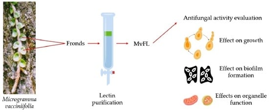
The lectin from Microgramma vacciniifolia fronds (MvFL) is a multifunctional protein that exhibits both carbohydrate-binding and trypsin inhibitor activities. It has demonstrated immunomodulatory effects on human peripheral blood mononuclear cells [29] and in vivo antitumor activity against sarcoma 180 models, with no observed toxicity in mice [30]. In addition, MvFL showed activity against Leishmania amazonensis, impairing promastigote replication and macrophage infection [31]. Despite these promising biological effects, its impact on fungal cells remains unexplored. In this study, MvFL was isolated and evaluated for its antifungal properties on yeast strains, including growth inhibition, cell proliferation, lysosomal integrity, mitochondrial membrane potential, antibiofilm activity, and synergistic interaction with fluconazole. This investigation represents the first report to explore the antifungal potential of MvFL, thereby expanding its known spectrum of biological activities.

2. Materials and Methods

2.1. Plant Material

The fronds of Microgramma vacciniifolia were collected on the campus of the Universidade Federal de Pernambuco (UFPE), Recife, Brazil, under authorization number 72,024 from the Instituto Chico Mendes de Conservação da Biodiversidade (ICMBio). Taxonomic identification was confirmed by the Dárdano de Andrade Lima Herbarium at the Instituto Agronômico de Pernambuco (Recife), where a voucher specimen was deposited under number 63,291. The plant material was thoroughly washed with tap water followed by distilled water to remove surface residues (approximately 2 L per 500 g of plant material, for about 10 min). The fronds were then air-dried at 26 ± 3 °C for seven days and subsequently ground using a blender (LQL-4 multiprocessor, Metvisa, Brusque, Brazil).

2.2. Isolation of MvFL

Proteins were extracted from the powdered fronds by homogenizing the material in 0.15 M NaCl at a ratio of 10% (w/v) for 16 h at 26 ± 3 °C using a magnetic stirrer (Fisatom, São Paulo, Brazil). The resulting extract was obtained by filtering the homogenate and centrifuging it at 9000× g for 15 min at 4 °C, and the supernatant was collected. Protein concentration was determined using the method of Lowry et al. [32], with bovine serum albumin (31.25 to 500 µg/mL) as the standard. Lectin activity was assessed by a hemagglutination assay using rabbit erythrocytes, as described by Procópio et al. [33].
MvFL was isolated following the protocol described by Patriota et al. [29]. Briefly, 2.0 mL of frond extract containing 2.7 mg of protein was applied to a Sephadex G-75 column (30.0 × 1.5 cm; GE Healthcare Life Sciences, Uppsala, Sweden) pre-equilibrated with distilled water. The column was eluted with distilled water at a flow rate of 0.5 mL/min, and 3.0 mL fractions were collected. Absorbance at 280 nm and lectin activity were measured for each fraction. The protein peak exhibiting hemagglutinating activity (P1) was lyophilized and resuspended in 0.1 M Tris-HCl buffer (pH 8.0). Subsequently, 1.0 mL of this solution, containing 2.2 mg of protein, was applied to a DEAE-Sephadex A25 column (3.0 × 2.0 cm) equilibrated with the same buffer. After washing with Tris-HCl buffer, MvFL was eluted using 1.0 M NaCl in 0.1 M Tris-HCl (pH 8.0). Fractions of 3.0 mL were collected at a flow rate of 0.5 mL/min and analyzed for absorbance at 280 nm and hemagglutinating activity.

2.3. Antifungal Activity

2.3.1. Broth Microdilution Assay

Yeast strains of Candida albicans (URM-5901; https://specieslink.net/rec/568/5901, accessed on 15 September 2025), Candida krusei (URM-6391; https://specieslink.net/rec/568/6391, accessed on 15 September 2025), Nakaseomyces glabratus (Candida glabrata, URM-4246; https://specieslink.net/rec/568/4246, accessed on 15 September 2025), Candida tropicalis (URM-6551; https://specieslink.net/rec/568/6551), and Candida parapsilosis (URM-6951; https://specieslink.net/rec/568/6951, accessed on 15 September 2025), were used, provided by the University Recife Mycology Culture Collection (URM) of the Department of Mycology at UFPE. The yeasts were cultured on Sabouraud Dextrose Agar for 24 h at 30 °C. Subsequently, the colonies were suspended in sterile saline solution (0.9% NaCl) to obtain a suspension equivalent to 106 colony-forming units (CFU) per mL. The minimum inhibitory concentration (MIC) was determined using the microdilution assay according to Procópio et al. [33] and corresponded to the lowest concentration of the sample capable of promoting ≥50% growth reduction compared to the control. To assess the minimum fungicidal concentration (MFC), 10 µL samples from wells with concentrations at or above the MIC were plated onto Sabouraud Dextrose Agar and incubated at 30 °C. The MFC was determined as the lowest concentration at which the sample reduced the viable colony-forming units (CFU) by 99.9% relative to the starting inoculum. Each test was carried out in triplicate across three independent trials. Fluconazole (Sigma-Aldrich, St. Louis, MO, USA) was used as positive control.

2.3.2. Cell Proliferation Assessment

Nakaseomyces glabratus cells were cultured for 48 h in RPMI medium, then washed with PBS and stained with carboxyfluorescein succinimidyl ester (CFSE; Sigma-Aldrich, St. Louis, MO, USA) at 25 μg/mL for 30 min at 30 °C. Following staining, the cells were washed with PBS containing 2% bovine serum albumin (BSA) to remove excess CFSE and incubated in RPMI medium with or without MvFL at 0.5 × MIC for 24 h at 30 °C. Data acquisition was performed on a FACSCalibur flow cytometer (BD Biosciences, Franklin Lakes, NY, USA), and analyses were conducted using CellQuest software version 5.1 (BD Biosciences). For each sample, 10,000 events were recorded. Results were reported as arbitrary fluorescence units.

2.3.3. Evaluation of Lysosomal Stability and Mitochondrial Membrane Potential

Lysosomal membrane stability and mitochondrial membrane potential (ΔΨm) were assessed by flow cytometry using acridine orange (AO) and rhodamine 123 (Rh 123) fluorescent probes, respectively. The assays employed N. glabratus. Cells were resuspended at a density of 1.0 × 10⁶ cells/mL in 500 µL of RPMI 1640 medium buffered with MOPS. MvFL was added at concentrations equivalent to the MIC and 2 × MIC, and the suspensions were incubated for 24 h at 37 °C. After incubation, cells were centrifuged at 3800× g for 10 min (Centrifuge 5810R, Eppendorf, Hamburg, Germany), washed three times with PBS (pH 7.2), and resuspended in 500 µL PBS. Cells were then stained with AO (1 µg/mL, in the dark for 20 min) or Rh 123 (10 µg/mL, in the dark for 10 min). Following staining, cells were washed three times with PBS, resuspended in PBS, and analyzed by flow cytometry.

2.3.4. Antibiofilm Activity

Biofilm formation was evaluated using the crystal violet assay in sterile 96-well polystyrene microplates to determine the ability of MvFL to prevent or reduce biofilm development. Each well received 80 µL of microbial suspension (108 CFU/mL in 0.15 M NaCl), 80 µL of MvFL at various concentrations based on the MIC, and 40 µL of Sabouraud Dextrose Broth. Wells containing Milli-Q water instead of MvFL served as the 100% biofilm formation control (untreated cells). Fungal growth was monitored by measuring the optical density at 600 nm after 24 h of incubation at 37 °C. Following incubation, non-adherent cells were removed by washing the wells three times with 0.15 M NaCl. Biofilms were fixed with absolute methanol for 30 min, heat-fixed at 50 °C for 60 min, and stained with 0.4% (w/v) crystal violet for 25 min at 25 °C. Excess stain was rinsed off with distilled water, and the bound dye was solubilized using absolute ethanol for 25 min. Absorbance was measured at 570 nm to quantify biofilm biomass. Biofilm formation was classified according to Sharan et al. [34]. All experiments were performed in triplicate. Caspofungin diacetate (Sigma-Aldrich) at 2 µg/mL was used as positive control.

2.3.5. Assessment of Combinatory Effects of MvFL and Fluconazole on Yeast Growth

To evaluate the interaction between MvFL and the antifungal fluconazole, the method described by Pillai et al. [35] was followed. Briefly, 96-well microplates were prepared by adding 100 µL of Sabouraud Dextrose Broth to each well. In the first well of each row, 100 µL of lectin at 4 × MIC was added and subjected to a serial twofold dilution across the row up to the penultimate well. Simultaneously, 100 µL of fluconazole at 4 × MIC was added to the last well of the same row and serially diluted in the opposite direction toward the second well. Consequently, the first well contained lectin alone, the last well contained fluconazole alone, and the intermediate wells contained varying combinations of both agents. A negative control representing 100% growth (no treatment) was included in a separate row. To determine the interaction between treatments, fractional inhibitory concentration (FIC) values and the FIC index (FICI) were calculated using the equations:
FICfluconazole = (MICfluconazole in combination)/(MICfluconazole alone)
FICMvFL = (MICMvFL in combination)/(MICMvFL alone)
FICI = FICfluconazole + FICMvFL
The FICI values were interpreted according to the classification: FICI ≤ 0.5 indicates synergism; 0.5 < FICI ≤ 1 indicates an additive effect; 1 < FICI ≤ 2 denotes no interaction; and FICI > 2 signifies antagonism.

2.4. Statistical Analysis

Data are presented as mean or mean percentage ± standard deviation (SD). Statistical differences were evaluated using Tukey’s multiple comparison test, with a significance threshold set at p < 0.05. For the growth kinetics assay, statistical analyses were conducted at each time point, comparing treated groups to the negative control.

3. Results and Discussion

In response to growing microbial resistance, studies have focused on identifying natural products with antimicrobial properties, either by combining plant-derived compounds with conventional antimicrobials or using them independently [36]. In this study, we investigated the effects of M. vacciniifolia frond lectin (MvRL) on the survival of Candida and Nakaseomyces species.
The antifungal assays demonstrated that MvFL effectively inhibited the growth of the yeast species, with minimum inhibitory concentrations (MICs) detailed in Table 1. According to Ríos and Recio [37], isolated compounds with MIC values below 10 μg/mL are considered promising. In this context, the potent activity of MvFL against Candida krusei (MIC = 1.25 μg/mL) and N. glabratus (MIC = 0.625 μg/mL) highlights its significant potential as an antifungal agent, offering new prospects for the development of therapeutic molecules targeting fungal infections. The minimal fungicidal concentration (MFC) for MvFL was not detected, indicating it is above 80 µg/mL. The MIC values for positive control (fluconazole) are shown in Table 2.
The MIC values obtained for MvFL are comparable to those reported for lectins from other plant species. Santos et al. [38] found that the water-soluble Moringa oleifera seed lectin (WSMoL) inhibited the growth of C. albicans, N. glabratus, C. krusei, and C. parapsilosis with an MIC of 20 μg/mL. Similarly, Silva et al. [16] reported that Dioclea violacea seed lectin (DVL) reduced the growth of C. albicans, C. krusei, and C. parapsilosis, with MICs of 0.6 μg/mL, 9.8 μg/mL, and 0.6 μg/mL, respectively, although no inhibitory effect was observed against C. tropicalis.
Our data revealed that MvFL exhibited the strongest antifungal activity against C. glabrata, making this species the focus of subsequent experiments investigating the mechanism of action. To assess the effect of MvFL on N. glabratus cell proliferation, we employed the CFSE marker, which binds covalently to intracellular proteins, remaining stably conjugated over time. During cell division, the dye is evenly distributed between daughter cells, resulting in a reduction in fluorescence intensity proportional to the proliferation rate [39]. Figure 1a shows that treatments with MvFL at 2× and 4× MIC led to higher fluorescence intensity compared to the untreated control, indicating reduced cell proliferation. These findings suggest a fungistatic effect of MvFL, likely through interference with cell division.
The effects of MvFL on lysosomal membrane integrity in N. glabratus cells were also investigated. As shown in Figure 1b, untreated control cells exhibited strong fluorescence, indicating intact lysosomal membranes. In contrast, treatment with MvFL resulted in a marked reduction in fluorescence intensity, suggesting that the lectin compromised lysosomal membrane integrity. Lysosomes are acidic organelles that contain hydrolytic enzymes involved in several essential cellular processes, including post-translational protein maturation, receptor degradation, and the extracellular release of active enzymes. Disruption of the lysosomal membrane leads to the release of proteolytic enzymes such as cathepsin D and cysteine proteases into the cytosol. These lysosomal proteases play a direct role in the initiation of apoptosis [40].
The mitochondrial membrane potential of N. glabratus cells was evaluated using Rho 123, a dye that selectively accumulates in active mitochondria in a membrane potential-dependent manner. Figure 1c shows that control cells exhibited high fluorescence intensity, reflecting intact mitochondrial membrane potential. In contrast, treatment with MvFL at 2× and 4× MIC significantly reduced fluorescence, indicating mitochondrial membrane depolarization. Such disruption of transmembrane potential can lead to membrane collapse, transient pore formation, and the release of pro-apoptotic factors into the cytosol [41,42].
Analyses of the effects of WSMoL revealed that cells treated with the lectin exhibited higher Rho 123 fluorescence compared to the control, suggesting that the initial inhibitory effect induced mitochondrial membrane hyperpolarization, followed by a reduction in membrane potential to levels comparable to untreated cells in C. albicans and N. glabratus [38]. The same study also assessed the impact of WSMoL on lysosomal structure and function in Candida species. However, unlike MvFL, WSMoL treatment did not cause lysosomal damage.
To explore the potential of MvFL in combating fungal biofilms, an antibiofilm assessment was conducted. Prior to evaluating the antibiofilm activity of MvFL, the biofilm-forming capacity of the Candida isolates was assessed. As shown in Figure 2, C. albicans, C. krusei, and C. tropicalis were classified as strong biofilm formers, while N. glabratus exhibited moderate biofilm formation. The C. parapsilosis isolate did not form biofilm under the tested conditions. MvFL demonstrated a biofilm-reducing effect only against C. tropicalis, and only at a concentration of ¼ × MIC. Nevertheless, the biofilm produced by C. tropicalis at this concentration was still classified as strong (Figure 2). This shows that, although there was a measurable reduction, it was not sufficient to significantly weaken the overall biofilm structure. Caspofungin inhibited biofilm formation by C. albicans, C. krusei, C. tropicalis and N. glabratus strains in 42.5 ± 4.8%, 37.3 ± 3.9%, 30.4 ± 3.1% and 29.7 ± 2.4%, respectively.
The potential of lectins to inhibit Candida biofilm formation has been explored in several studies. Ferreira et al. [43] reported that the lectin from the inflorescence of Alpinia purpurata (ApuL) significantly inhibited biofilm development of C. albicans at all tested concentrations, with inhibition levels reaching up to 70%. In contrast, the effect on C. parapsilosis was minimal, with biofilm inhibition not exceeding 25% following ApuL treatment.
The limited antibiofilm efficacy of MvFL should be considered when assessing its potential as an antifungal or anti-virulence agent. Notably, no antibiofilm effects were observed in the other tested yeast species, suggesting a species-specific response. This interspecies variation may arise from differences in cell wall composition, extracellular matrix structure, or the regulatory pathways involved in biofilm formation. For example, C. tropicalis may form a biofilm that is more susceptible to disruption by MvFL, whereas C. albicans, C. krusei, and N. glabratus may produce more robust or chemically distinct matrices that confer greater resistance. Further studies are needed to investigate these structural and molecular differences and to clarify the basis of this selective activity.
The increased use of antifungal agents has contributed to a rise in both the number and diversity of drug-resistant fungal strains, highlighting the urgent need for alternative therapies to curb antifungal resistance. This is particularly important given that many commonly used antifungal treatments are associated with toxicity [44,45]. Safer and more effective agents, especially those derived from plants, may act synergistically with existing drugs and offer promising alternatives to help address this growing challenge [46].
Finally, the combinatory effects of MvFL and fluconazole, a commonly used antifungal in the treatment of candidiasis, were investigated to assess potential synergism or antagonism. As shown in Table 2, an additive effect was observed against C. krusei and C. tropicalis, suggesting that fluconazole enhances the antifungal efficacy of MvFL compared to either agent alone. More notably, a synergistic effect was detected against C. parapsilosis, indicating a potential therapeutic advantage when MvFL is used in combination with fluconazole for this species. In contrast, an antagonistic interaction was observed for C. albicans and N. glabratus, where both MvFL and fluconazole lost their antifungal activity when combined.
This result underscores the complexity of drug-lectin interactions and suggests that the combination may interfere with the uptake, binding, or activity of one or both compounds. The variation in combinatory effects of MvFL and fluconazole across yeast species likely reflects interspecies differences in drug susceptibility, cell wall composition, and resistance mechanisms. The synergistic effect against C. parapsilosis may result from complementary actions of the two drugs, potentially exploiting vulnerabilities in this species’ membrane or stress response pathways. The additive effects observed with C. krusei and C. tropicalis suggest independent mechanisms of action that do not interfere with each other but also do not enhance activity. In contrast, the antagonism seen with C. albicans and N. glabratus could stem from induced stress responses, efflux pump activation, or metabolic interference, reducing overall antifungal efficacy [47,48]. Antagonism could also stem from competition for the same cellular target or from MvFL-induced changes in membrane permeability that diminish fluconazole efficacy [48]. These results reinforce the importance of species-specific responses.
Ferreira et al. [43] observed a synergistic interaction between fluconazole and the lectin ApuL, which aligns with the synergistic result seen for MvFL and fluconazole against C. parapsilosis in the present study. These findings highlight the potential of plant-derived lectins as adjuvants in antifungal therapy, especially in overcoming resistance or enhancing the efficacy of existing drugs. However, the species-specific nature of these interactions also emphasizes the need for careful evaluation of combinations to avoid unintended antagonism.
The overall data shows that MvFL appears to inhibit cell proliferation in N. glabratus by targeting key organelles like mitochondria and lysosomes, which are vital for energy production and intracellular recycling in planktonic cells. However, MvFL did not affect the formation of biofilm by N. glabratus. Retrograde signaling pathways in yeasts act to promote changes in gene expression that promote a metabolic readjustment [49]. It is possible that mitochondrial dysfunction caused by MvFL in biofilm-associated cells is mitigated by a metabolic shift that reduces reliance on mitochondrial function. In addition, some biofilm cells exhibit slow-growing characteristics or become metabolically inactive (dormant cells) [50], making them less affected by altered dynamics of organelles such as lysosomes. Importantly, biofilm development involves distinct gene regulation, including the activation of transcription factors and adhesins [51], which may remain unaffected by MvFL. Biofilm cells also display compensatory responses, such as upregulation of stress resistance pathways and phenotypic heterogeneity, which can allow parts of the biofilm to persist despite MvFL-induced damage.

4. Conclusions

This study demonstrates the promising antifungal potential of MvFL against yeast species. The fungistatic effect of MvFL on N. glabratus appears to involve interference with cell proliferation and disruption of key cellular organelles, including lysosomes and mitochondria. The lectin’s combinatory use with fluconazole revealed additive to synergistic antifungal effects against select Candida species, highlighting its potential as an adjuvant in antifungal therapy. On the other hand, MvFL showed limited efficacy in reducing biofilm formation (maximum of 40% and only against Candida tropicalis). Given the increasing prevalence of antifungal resistance and toxicity associated with conventional treatments, plant-derived lectins like MvFL represent promising candidates for developing new antifungal strategies.

Author Contributions

Conceptualization, L.L.d.S.P., L.C.N.d.S. and T.H.N., methodology, R.M.V.V.d.O., A.R.C.J., P.M.d.S., G.R.S.F., L.C.N.d.S., L.L.d.S.P. and T.H.N., validation, P.M.d.S., A.R.C.J., P.M.G.P., L.C.N.d.S., L.L.d.S.P., E.V.P. and T.H.N., formal analysis, R.M.V.V.d.O., A.R.C.J., P.M.d.S., L.C.N.d.S., L.L.d.S.P., E.V.P. and T.H.N., investigation, R.M.V.V.d.O., A.R.C.J., P.M.d.S., G.R.S.F. and L.L.d.S.P., resources, P.M.G.P., L.C.N.d.S. and T.H.N., data curation, R.M.V.V.d.O., P.M.d.S., A.R.C.J. and L.L.d.S.P., writing—original draft preparation, R.M.V.V.d.O. and L.L.d.S.P., writing—review and editing, P.K.A., E.V.P. and T.H.N., visualization, R.M.V.V.d.O., A.R.C.J., P.M.d.S., L.C.N.d.S. and T.H.N., supervision, E.V.P. and T.H.N., project administration, T.H.N., funding acquisition, P.M.G.P., L.C.N.d.S., E.V.P. and T.H.N. All authors have read and agreed to the published version of the manuscript.

Funding

This research was funded by the Conselho Nacional de Desenvolvimento Científico e Tecnológico (CNPq), grant number 305251/2023-6, the Coordenação de Aperfeiçoamento de Pessoal de Nível Superior (CAPES), grant number 1724/2025, and the Fundação de Amparo à Ciência e Tecnologia do Estado de Pernambuco (FACEPE), grant numbers APQ-2144-2.08/24; IBPG-0913-2.00/22.

Institutional Review Board Statement

Not applicable.

Data Availability Statement

The original contributions presented in this study are included in the article. Further inquiries can be directed to the corresponding author.

Conflicts of Interest

The authors declare no conflicts of interest.

References

  1. Garcia-Marin, L.E.; Juarez-Moreno, K.; Vilchis-Nestor, A.R.; Castro-Longoria, E. Highly antifungal activity of biosynthesized copper oxide nanoparticles against Candida albicans. Nanomaterials 2022, 12, 3856. [Google Scholar] [CrossRef]
  2. Azadi, S.; Azizipour, E.; Amani, A.M.; Vaez, A.; Zareshahrabadi, Z.; Abbaspour, A.; Firuzyar, T.; Dortaj, H.; Kamyab, H.; Chelliapan, S.; et al. Antifungal activity of Fe3O4@SiO2/Schiff-base/Cu(II) magnetic nanoparticles against pathogenic Candida species. Sci. Rep. 2024, 14, 5855. [Google Scholar] [CrossRef] [PubMed]
  3. Padmavathi, A.R.; Murthy, P.S.; Das, A.; Priya, A.; Sushmitha, T.J.; Pandian, S.K.; Toleti, S.R. Impediment to growth and yeast-to-hyphae transition in Candida albicans by copper oxide nanoparticles. Biofouling 2020, 36, 56–72. [Google Scholar] [CrossRef]
  4. Badrane, H.; Cheng, S.; Dupont, C.L.; Hao, B.; Driscoll, E.; Morder, K.; Liu, G.; Newbrough, A.; Fleres, G.; Kaul, D.; et al. Genotypic diversity and unrecognized antifungal resistance among populations of Candida glabrata from positive blood cultures. Nat. Commun. 2023, 14, 5918. [Google Scholar] [CrossRef]
  5. Ruiz-Duran, J.; Torres, R.; Stashenko, E.E.; Ortiz, C. Antifungal and antibiofilm activity of Colombian essential oils against different Candida strains. Antibiotics 2023, 12, 668. [Google Scholar] [CrossRef]
  6. Pappas, P.G.; Lionakis, M.S.; Arendrup, M.C.; Ostrosky-Zeichner, L.; Kullberg, B.J. Invasive candidiasis. Nat. Rev. Dis. Primers 2018, 4, 18026. [Google Scholar] [CrossRef]
  7. Talattof, Z.; Azad, A.; Zahed, M.; Shahradnia, N. Antifungal activity of xylitol against Candida albicans: An in vitro study. J. Contemp. Dent. Pract. 2018, 19, 125–129. [Google Scholar] [CrossRef]
  8. Guinea, J. Global trends in the distribution of Candida species causing candidemia. Clin. Microbiol. Infect. 2014, 20, 5–10. [Google Scholar] [CrossRef] [PubMed]
  9. Muñoz-Escobar, A.; Reyes-López, S.Y. Antifungal susceptibility of Candida species to copper oxide nanoparticles on polycaprolactone fibers (PCL-CuONPs). PLoS ONE 2020, 15, e0228864. [Google Scholar] [CrossRef]
  10. Yadav, V.; Heitman, J. On fruits and fungi: A risk of antifungal usage in food storage and distribution in driving drug resistance in Candida auris. mBio 2022, 13, e00739-22. [Google Scholar] [CrossRef] [PubMed]
  11. Meagher, R.B.; Lewis, Z.A.; Ambati, S.; Lin, X. DectiSomes: C-type lectin receptor-targeted liposomes as pan-antifungal drugs. Adv. Drug Deliv. Rev. 2023, 196, 114776. [Google Scholar] [CrossRef]
  12. Sabino, R.; Veríssimo, C.; Pereira, Á.A.; Antunes, F. Candida auris, an agent of hospital-associated outbreaks: Which challenging issues do we need to have in mind? Microorganisms 2020, 8, 181. [Google Scholar] [CrossRef] [PubMed]
  13. Revie, N.M.; Iyer, K.R.; Robbins, N.; Cowen, L.E. Antifungal drug resistance: Evolution, mechanisms and impact. Curr. Opin. Microbiol. 2018, 45, 70–76. [Google Scholar] [CrossRef]
  14. Patterson, T.F.; Thompson, G.R., III; Denning, D.W.; Fishman, J.A.; Hadley, S.; Herbrecht, R.; Kontoyiannis, D.P.; Marr, K.A.; Morrison, V.A.; Nguyen, M.H.; et al. Practice guidelines for the diagnosis and management of aspergillosis: 2016 update by the Infectious Diseases Society of America. Clin. Infect. Dis. 2016, 63, e1–e60. [Google Scholar] [CrossRef]
  15. Fu, N.; Wang, S.; Zhang, Y.; Zhang, C.; Yang, D.; Weng, L.; Zhao, B.; Wang, L. Efficient click chemistry towards fatty acids containing 1,2,3-triazole: Design and synthesis as potential antifungal drugs for Candida albicans. Eur. J. Med. Chem. 2017, 136, 596–602. [Google Scholar] [CrossRef]
  16. Silva, R.R.; Malveira, E.A.; Aguiar, T.K.; Neto, N.A.; Roma, R.R.; Santos, M.H.C.; Santos, A.L.E.; Silva, A.F.B.; Freitas, C.D.T.; Rocha, B.A.M.; et al. DVL, lectin from Dioclea violacea seeds, has multiple mechanisms of action against Candida spp. via carbohydrate recognition domain. Chem. Biol. Interact. 2023, 348, 109649. [Google Scholar] [CrossRef] [PubMed]
  17. Del Rio, M.; Radicioni, M.B.; Mello, É.O.; Ribeiro, S.F.; Taveira, G.B.; Carvalho, A.O.; la Canal, L.; Gomes, V.M.; Regente, M. A plant mannose-binding lectin and fluconazole: Key targets combination against Candida albicans. J. Appl. Microbiol. 2022, 132, 4310–4320. [Google Scholar] [CrossRef] [PubMed]
  18. Coriolano, M.C.; Brito, J.S.; Ferreira, G.R.S.; Moura, M.C.; Melo, C.M.L.; Soares, A.K.A.; Lorena, V.M.B.; Figueiredo, R.C.B.Q.; Paiva, P.M.G.; Napoleão, T.H.; et al. Antibacterial lectin from Moringa oleifera seeds (WSMoL) has differential action on growth, membrane permeability and protease secretory ability of Gram-positive and Gram-negative pathogens. S. Afr. J. Bot. 2020, 129, 198–205. [Google Scholar] [CrossRef]
  19. Silva, W.S.F.L.; Amorim, P.K.; Ferreira, G.R.S.; Alves, R.R.V.; Silva, C.E.S.; Santos, P.E.M.; Paiva, P.M.G.; Ingmer, H.; Silva, S.P.; Silva, P.M.; et al. Moringa oleifera seed preparations containing the lectin WSMoL inhibit growth, cell aggregation, and biofilm production of Listeria monocytogenes. Microb. Pathog. 2025, 204, 107600. [Google Scholar] [CrossRef]
  20. Barbosa, F.E.V.; Lima, D.B.; Santos, A.L.E.; Sousa, V.C.; Carvalho, R.D.C.V.; Alves, M.M.M.; Lima, M.A.P.; Souza, R.O.S.; Silva, C.G.L.; Teixeira, C.S. Dioclea violacea lectin has potent in vitro leishmanicidal activity against Leishmania infantum via carbohydrate recognition domain. Int. J. Biol. Macromol. 2024, 280, 135665. [Google Scholar] [CrossRef]
  21. Santos, N.D.L.; Santos, P.E.M.; Lira, T.L.S.; Santos, A.R.S.; Silva, J.N.O.; Santos, A.N.S.; Amorim, M.M.R.; Barros, M.R.; Coelho, L.C.B.B.; Paiva, P.M.G.; et al. Insecticidal activity of lectin preparations from Moringa oleifera Lam. (Moringaceae) seeds against Alphitobius diaperinus (Panzer) (Coleoptera: Tenebrionidae). Plants 2025, 14, 511. [Google Scholar] [CrossRef]
  22. Mesquita, J.X.; Brito, T.V.; Fontenelle, T.P.C.; Damasceno, R.O.S.; Souza, M.H.L.P.; Lopes, J.L.S.; Beltramini, L.M.; Barbosa, A.L.R.; Freitas, A.L.P. Lectin from red algae Amansia multifida Lamouroux: Extraction, characterization and anti-inflammatory activity. Int. J. Biol. Macromol. 2021, 170, 532–539. [Google Scholar] [CrossRef]
  23. Brito, J.S.; Marinho, A.O.; Coelho, L.C.B.B.; Oliveira, A.M.; Paiva, P.M.G.; Patriota, L.L.S.; Napoleão, T.H. Toxicity and antitumor activity of the water-soluble lectin from Moringa oleifera Lam. seeds (WSMoL) in sarcoma 180-bearing mice. Toxicon 2023, 234, 107306. [Google Scholar]
  24. Ferreira, L.R.D.; Silva, A.F.; Siqueira, J.S.; Santos, R.C.; Lima, V.S.; Oliveira, A.M.; Soares, L.A.L.; Silva, G.C. Estudo do perfil fitoquímico e avaliação dos efeitos citotóxicos do rizoma da Microgramma vacciniifolia. Braz. J. Health Rev. 2020, 3, 1185–1202. [Google Scholar]
  25. Tryon, R.M.; Tryon, A.F. Family Pteridaceae: From Ferns and Allied Plants; Springer: Berlin/Heidelberg, Germany, 1982. [Google Scholar]
  26. Santos, M.G.; Sylvestre, L.D.S. Floristics and economics aspects of the pteridophytes of rocky outcrop from Rio de Janeiro State, Brazil. Acta Bot. Bras. 2006, 20, 115–124. [Google Scholar] [CrossRef]
  27. Agra, M.F.; Silva, K.N.; Basílio, I.J.L.D.; Freitas, P.F.; Barbosa-Filho, J.M. Survey of medicinal plants used in the region Northeast of Brazil. Rev. Bras. Farmacogn. 2008, 18, 472–508. [Google Scholar] [CrossRef]
  28. Peres, M.T.L.P.; Simionatto, E.; Hess, S.C.; Bonani, V.F.L.; Candido, A.C.S.; Castelli, C.; Poppi, N.R.; Honda, N.K.; Cardoso, C.A.L.; Faccenda, O. Chemical and biological studies of Microgramma vacciniifolia (Langsd. & Fisch.) Copel (Polypodiaceae). Quim. Nova 2009, 32, 897–901. [Google Scholar]
  29. Patriota, L.L.S.; Procópio, T.F.; Brito, J.S.; Sebag, V.; Oliveira, A.P.S.; Soares, A.K.A.; Moreira, L.R.; Lima, T.A.; Soares, T.; Silva, T.D.; et al. Microgramma vacciniifolia (Polypodiaceae) fronds contain a multifunctional lectin with immunomodulatory properties on human cells. Int. J. Biol. Macromol. 2017, 103, 36–46. [Google Scholar] [CrossRef]
  30. Patriota, L.L.S.; Ramos, D.B.M.; Silva, Y.A.; Santos, A.C.L.A.; Araujo, M.T.M.F.; Brito, J.S.; Torres, D.J.L.; Oliveira, A.M.; Silva, D.C.N.; Lorena, V.M.B.; et al. Microgramma vacciniifolia frond lectin (MvFL) exhibits antitumor activity against sarcoma 180 in mice. Phytomed. Plus 2021, 1, 100013. [Google Scholar] [CrossRef]
  31. Aguiar, L.M.S.; Alves, M.M.M.; Sobrinho Júnior, E.P.C.; Paiva, P.M.G.; Carvalho, F.A.A.; Albuquerque, L.P.; Patriota, L.L.S.; Napoleão, T.H. Microgramma vacciniifolia Frond Lectin: In Vitro Anti-leishmanial Activity and Immunomodulatory Effects Against Internalized Amastigote Forms of Leishmania amazonensis. Acta Parasitol. 2023, 68, 869–879. [Google Scholar] [CrossRef]
  32. Lowry, O.H.; Rosebrough, N.J.; Farr, A.L.; Randall, R.J. Protein measurement with the Folin phenol reagent. J. Biol. Chem. 1951, 193, 265–275. [Google Scholar] [CrossRef]
  33. Procópio, T.F.; Patriota, L.L.S.; Moura, M.C.; Silva, P.M.; Oliveira, A.P.S.; Carvalho, L.V.N.; Lima, T.A.; Soares, T.; Silva, T.D.; Coelho, L.C.B.B.; et al. CasuL: A new lectin isolated from Calliandra surinamensis leaf pinnulae with cytotoxicity to cancer cells, antimicrobial activity and antibiofilm effect. Int. J. Biol. Macromol. 2017, 98, 419–429. [Google Scholar] [CrossRef]
  34. Sharan, M.; Dhaka, P.; Bedi, J.S.; Mehta, N.; Singh, R. Assessment of biofilm-forming capacity and multidrug resistance in Staphylococcus aureus isolates from animal-source foods: Implications for lactic acid bacteria intervention. Ann. Microbiol. 2024, 74, 22. [Google Scholar] [CrossRef]
  35. Pillai, S.K.; Moellering, R.C.; Eliopoulos, G.M. Antimicrobial combinations. In Antibiotics in Laboratory Medicine; Lorian, V., Ed.; The Lippincott Williams & Wilkins Co.: Philadelphia, PA, USA, 2005; pp. 365–440. [Google Scholar]
  36. Silva, P.M.; Silva, B.R.; Silva, J.N.O.; Moura, M.C.; Soares, T.; Feitosa, A.P.S.; Brayner, F.A.; Alves, L.C.; Paiva, P.M.G.; Damborg, P.; et al. Punica granatum sarcotesta lectin (PgTeL) has antibacterial activity and synergistic effects with antibiotics against β-lactamase-producing Escherichia coli. Int. J. Biol. Macromol. 2019, 135, 931–939. [Google Scholar] [CrossRef]
  37. Rios, J.L.; Recio, M.C. Medicinal plants and antimicrobial activity. J. Ethnopharmacol. 2005, 100, 80–84. [Google Scholar] [CrossRef]
  38. Santos, L.M.M.; Silva, P.M.; Moura, M.C.; Junior, A.C.; Amorim, P.K.; Procópio, T.F.; Coelho, L.C.B.B.; Silva, L.C.N.; Paiva, P.M.G.; Santos, N.D.L.; et al. Anti-Candida activity of the water-soluble lectin from Moringa oleifera seeds (WSMoL). J. Med. Mycol. 2021, 31, 101074. [Google Scholar] [CrossRef] [PubMed]
  39. Lašt’ovička, J.; Budinsky, V.; Spisek, R.; Bartunkova, J. Assessment of lymphocyte proliferation: CFSE kills dividing cells and modulates expression of activation markers. Cell. Immunol. 2009, 256, 79–85. [Google Scholar] [CrossRef] [PubMed]
  40. Hwang, B.; Hwang, J.-S.; Lee, J.; Kim, J.-K.; Kim, S.R.; Kim, Y.; Lee, D.G. Induction of yeast apoptosis by an antimicrobial peptide, Papiliocin. Biochem. Biophys. Res. Commun. 2011, 408, 89–93. [Google Scholar] [CrossRef]
  41. Silva, C.R.; Neto, J.B.A.; Campos, R.S.; Figueiredo, N.S.; Sampaio, L.S.; Magalhães, H.I.F.; Cavalcanti, B.C.; Gaspar, D.M.; Andrade, G.M.; Lima, I.S.P.; et al. Synergistic effect of the flavonoid catechin, quercetin, or epigallocatechin gallate with fluconazole induces apoptosis in Candida tropicalis resistant to fluconazole. Antimicrob. Agents Chemother. 2014, 58, 1468–1478. [Google Scholar] [CrossRef]
  42. Neto, J.B.; Silva, C.R.; Neta, M.A.; Campos, R.S.; Siebra, J.T.; Silva, R.A.; Gaspar, D.M.; Magalhães, H.I.F.; Moraes, M.O.; Lobo, M.D.P.; et al. Antifungal activity of naphthoquinoidal compounds in vitro against fluconazole-resistant strains of different Candida species: A special emphasis on mechanisms of action on Candida tropicalis. PLoS ONE 2014, 9, e93698. [Google Scholar] [CrossRef] [PubMed]
  43. Ferreira, G.R.S.; Brito, J.S.; Procópio, T.F.; Santos, N.D.L.; De Lima, B.J.R.C.; Coelho, L.C.B.B.; Navarro, D.M.A.F.; Paiva, P.M.G.; Soares, T.; Moura, M.C.; et al. Antimicrobial potential of Alpinia purpurata lectin (ApuL): Growth inhibitory action, synergistic effects in combination with antibiotics, and antibiofilm activity. Microb. Pathog. 2018, 124, 152–162. [Google Scholar] [CrossRef]
  44. Pfaller, M.A.; Amaral, R.C.R.; Biasi, R.P.; Guilhermetti, E.; Svidzinski, T.I.E.; Baeza, L.C. Antifungal resistance: Mechanisms, epidemiology and consequences for treatment. Am. J. Med. 2012, 125, 3–13. [Google Scholar] [CrossRef]
  45. Manoraj, A.; Thevanesam, V.; Bandara, B.M.R.; Ekanayake, A.; Liyanapathirana, V. Synergistic activity between Triphala and selected antibiotics against drug resistant clinical isolates. BMC Complement. Med. Ther. 2019, 19, 199. [Google Scholar] [CrossRef]
  46. Ayaz, M.; Ullah, F.; Sadiq, A.; Ullah, F.; Ovais, M.; Ahmed, J.; Devkota, H.P. Synergistic interactions of phytochemicals with antimicrobial agents: Potential strategy to counteract drug resistance. Chem. Biol. Interact. 2019, 308, 294–303. [Google Scholar]
  47. Bollenbach, T. Antimicrobial interactions: Mechanisms and implications for drug discovery and resistance evolution. Curr. Opin. Microbiol. 2015, 27, 1–9. [Google Scholar] [CrossRef]
  48. Chaachouay, N. Synergy, Additive Effects, and Antagonism of Drugs with Plant Bioactive Compounds. Drugs Drug Candidates 2025, 4, 4. [Google Scholar] [CrossRef]
  49. Bui, T.H.D.; Labedzka-Dmoch, K. RetroGREAT signaling: The lessons we learn from yeast. IUBMB Life 2024, 76, 26–37. [Google Scholar]
  50. Napoleão, T.H.; Lira, T.L.S.; Pontual, E.V.; Ferreira, G.R.S.; Silva, P.M. Lectins as Natural Antibiofilm Agents in the Fight Against Antibiotic Resistance: A Review. Molecules 2025, 30, 3395. [Google Scholar] [CrossRef] [PubMed]
  51. Li, F.; Svarovsky, M.J.; Karlsson, A.J.; Wagner, J.P.; Marchillo, K.; Oshel, P.; Andes, D.; Palecek, S.P. Eap1p, an adhesin that mediates Candida albicans biofilm formation in vitro and in vivo. Eukaryot. Cell 2007, 6, 931–939. [Google Scholar] [CrossRef] [PubMed]
Figure 1. Effects of Microgramma vacciniifolia frond lectin (MvFL) on Nakaseomyces glabratus cells. (a) Fluorescence of CFSE (a cell proliferation marker that covalently binds intracellular proteins and decreases in intensity with each cell division) in N. glabratus cells treated or untreated with MvFL. (b) Fluorescence of acridine orange, AO (a lysosomal membrane integrity probe that emits strong fluorescence in intact acidic lysosomes) in N. glabratus cells treated or untreated with MvFL. (c) Fluorescence of rhodamine 123, Rho 123 (a mitochondrial membrane potential-sensitive dye that accumulates in active mitochondria) in N. glabratus cells treated or untreated with MvFL. MIC: minimum inhibitory concentration. (*) Significant difference (p < 0.05) compared to the control.
Macromol 05 00044 g001
Figure 2. Evaluation of the antibiofilm activity of Microgramma vacciniifolia frond lectin (MvFL) on Candida albicans (a), Nakaseomyces glabratus (b), Candida tropicalis (c) and Candida krusei (d). Bacterial growth and biofilm formation are represented. Biofilm formation was assessed by crystal violet staining and classified as strong, moderate or weak according to Sharan et al. [34]. MIC: minimum inhibitory concentration. (*) Significant difference (p < 0.05) compared to the control.
Macromol 05 00044 g002
Table 1. Antifungal activity of Microgramma vacciniifolia frond lectin (MvFL) against yeast isolates.
YeastsMinimal Inhibitory Concentration (µg/mL)
Candida albicans20.00
Candida krusei1.25
Candida parapsilosis40.00
Candida tropicalis40.00
Nakaseomyces glabratus0.625
Table 2. Evaluation of the combinatory effect of Microgramma vacciniifolia frond lectin (MvFL) and fluconazole against yeast species.
YeastsMIC (Alone/Combination)FICICombination Effect
MvFLFluconazole
C. albicans20/ND0.25/NDNCAntagonistic
C. krusei1.25/0.00232.0/32.01.0Additive
C. parapsilosis40.0/0.62564.0/8.00.14Synergism
C. tropicalis40.0/0.0784.0/4.01.0Additive
N. glabratus0.625/ND64.0/NDNCAntagonistic
MIC: minimal inhibitory concentration, expressed in µg/mL. FICI: fractional inhibitory concentration index: ND: not detected. NC: not calculated.
Disclaimer/Publisher’s Note: The statements, opinions and data contained in all publications are solely those of the individual author(s) and contributor(s) and not of MDPI and/or the editor(s). MDPI and/or the editor(s) disclaim responsibility for any injury to people or property resulting from any ideas, methods, instructions or products referred to in the content.