Article

Upcycling Microalgal Residues: Physicochemical Insights and Biocomposite Enhancement

1
Department of Chemical Science and Technologies, University of Rome “Tor Vergata”, Via della Ricerca Scientifica 1, 00133 Rome, Italy
2
Splastica srl spinoff, Via del Lavoro 13, 00045 Rome, Italy
3
Department of Chemical Sciences and Materials Technology, Institute of Crystallography, National Research Council, Via Salaria Km 29.300, 00015 Rome, Italy
*
Authors to whom correspondence should be addressed.
Macromol 2025, 5(3), 32; https://doi.org/10.3390/macromol5030032
Submission received: 9 May 2025 / Revised: 5 June 2025 / Accepted: 1 July 2025 / Published: 8 July 2025

Abstract

The growing concern for environmental sustainability has led to an increased interest in biodegradable materials derived from renewable resources. This study explores the innovative use of residual biomass from the green photosynthetic microalga Chlamydomonas reinhardtii, left over after polysaccharide extraction, as a natural filler in the development of the compostable protein-based material SP-Milk®. The microalgal biomass was characterized using Fourier transform infrared spectroscopy (FTIR) and UV-Visible Spectroscopy to assess its chemical and structural composition. Subsequently, it was incorporated into a biodegradable protein matrix, and the resulting biocomposites were evaluated for mechanical and thermal properties. The results demonstrate that the incorporation of algal filler improves the mechanical strength and elasticity of the material while reducing its glass transition temperature, highlighting its potential for use in sustainable applications as a possible substitute for conventional plastics. The biocomposite materials developed, based on the protein-based material SP-Milk® and residual microalgal biomass, are environmentally friendly, contributing to the reduction in pollution and the risks associated with plastic accumulation. Thus, this study offers a simple, effective, and sustainable strategy for the valorization of microalgal biomass, enabling the production of biodegradable materials with enhanced mechanical performance, suitable for applications such as sustainable packaging within a circular economy framework.

1. Introduction

The increasing demand for sustainable and compostable materials has driven research toward the use of residual biomass as a renewable resource for the development of functional biocomposites.
Materials intended for bioplastic production must exhibit strong mechanical performance, including high strength, toughness, and durability, to withstand demanding environmental conditions. According to recent studies, the incorporation of natural additives into protein-based bioplastics has been shown to significantly improve their mechanical properties, enhancing key features such as tensile strength, flexibility, and durability [1,2].
Among these, microalgae and macroalgae represent a particularly promising source due to their high photosynthetic efficiency, fast growth rates, and capacity to accumulate value-added biomolecules such as proteins, pigments, and polysaccharides [3,4,5,6]. In this context, macroalgal and microalgal biomass represent a promising multifunctional component for bioplastic formulations. Depending on their physicochemical characteristics and loading levels, they can act as reinforcing biofillers, improving mechanical performance, and as functional additives, contributing to color, processability, and thermal behavior. This dual functionality supports the development of advanced biocomposites aligned with circular bioeconomy principles. Recent advances have demonstrated that algal biomass can be incorporated directly into biopolymer matrices or used as a source of bioactive fillers to enhance the functional properties of biomaterials. Key benefits include improved biodegradability, mechanical reinforcement, UV resistance, and in some cases, antimicrobial activity owing to the natural pigments and secondary metabolites present in microalgae (e.g., chlorophylls, carotenoids) [6]. Various strategies have been reported in the literature for the integration of algal biomass, including direct blending with polymers such as polylactic acid (PLA), polyhydroxyalkanoates (PHAs), and starch-based plastics [5,6]; the extraction and functionalization of specific algal components (e.g., alginate, carrageenan, ulvan) to serve as film-forming agents or reinforcement phases [5]; and the use of whole dried algal biomass as biofillers in composites, contributing both to mechanical performance and biodegradability [6]. Challenges remain, particularly related to mechanical strength optimization and material stability during storage and use. It has been shown that introducing microalgae in a gluten-based bioplastic can potentially improve the tensile modulus and tensile strength [7]. Chlamydomonas reinhardtii, a unicellular model green photosynthetic microalga used as a model organism for photosynthesis studies and extensively studied in molecular and synthetic biology, has gained attention as a biotechnological platform for the production of bioactive compounds [8,9]. Another major constituent of C. reinhardtii are glycoproteins, which constitute the cell wall [10]. These compounds have significant potential for various applications, including food, cosmetics, pharmaceuticals, and for the production of natural dyes and pigments.
Despite the growing use of microalgae in industrial processes, the management of post-extraction biomass residues remains a major challenge. Recent studies highlight how the valorization of algal waste can contribute to closing the production loop, thereby reducing environmental impact and promoting a circular bioeconomy [11]. In particular, microalgal proteins are emerging as potential building blocks for biodegradable materials due to their film-forming ability, biodegradability, and compatibility with various additives [12]. The incorporation of algal biomass into biomaterials intended as plastic replacements offers additional functional benefits due to the presence of chlorophyll. Several studies have demonstrated that chlorophyll exhibits antimicrobial activity against bacteria and fungi, particularly when activated by light through photodynamic processes. This property can enhance the hygienic quality of bioplastics, making them particularly attractive for applications in food packaging, medical devices, and cosmetics [13].
However, the direct use of microalgal biomass residues in powder form as a functional filler in compostable protein-based matrices remains an underexplored area.
In this study, the potential of C. reinhardtii as a functional additive and reinforcing filler for biobased polymeric materials was explored. A compostable and biodegradable protein-based material named SP-Milk® was employed as the “base” material [1,14]. Two different types of microalgal biomasses were considered: untreated microalgae, used in their native form, and microalgal residues obtained after polysaccharide extraction, representing a valorization of waste material. The microalgal biomass was spectroscopically characterized (FTIR, UV-Vis) and then blended with SP-Milk® in a composite bioplastic. The effect of these microalgal biomasses on the material properties was systematically investigated to assess their feasibility and performance within bioplastic formulations.
This study demonstrated the effectiveness of C. reinhardtii residual biomass powder as a natural dye with reinforcing- and processing-improving activities on composite bioplastic material.

2. Materials and Methods

Ethanol of spectroscopic grade was purchased from Sigma Aldrich (St. Louis, MO, USA). MQ water (18.2 MΩ·cm) was obtained from a Millipore filtration system. The SP-Milk® material is produced by the Splastica spinoff (Rome, Italy) in the form of granules.
The study was conducted on samples of microalgae belonging to the species C. reinhardtii, collected and cultivated at the Institute of Crystallography, National Research Council of Italy, Department of Chemical Sciences and Materials Technologies. In particular, two strains of C. reinhardtii were examined: CC125, referred to as strain 1 (S1), and SAG 11-32b, referred to as strain 2 (S2). Both strains underwent polysaccharide extraction (indicated with the suffix “-P”) according to a previously published protocol [15] which involved thermal treatment. Biomass from the CC125 strain that did not undergo polysaccharide extraction was used as a reference control. For simplicity, the samples and acronyms are summarized in Table 1. All the samples were freeze-dried for 24 h before using.
Extraction and spectroscopic characterization of pigments. For the extraction of pigments, we used the procedure proposed by Farobie et al. [16]. In particular, 10 mg of freeze-dried microalgae were dissolved into 1.2 mL of EtOH, and sonication was performed for 30 min using an ultrasonicator Elmasonic ONE, Elma Schmidbauer GmbH (Singen, Germany), with a power of 30 W. Each Eppendorf was vortexed for 2 min and placed for 48 h at 4 °C. Centrifugation was performed at 2500 RPM for 10 min (CAPPRondo microcentrifuge, Model CR-68X, Nordhausen, Germany), and the supernatant, containing the pigments, was separated from the pellet. The extraction procedure was repeated 3 times. For measurements, diluted solutions were prepared by taking 20 μL of supernatant stock solution and adding 1 mL of EtOH. All these solutions were characterized using UV-visible and fluorescence spectroscopic measurements. UV-visible absorption measurements were carried out at room temperature using a Varian Cary 100 Scan spectrophotometer (Middelburg, The Netherlands). The absorption spectra were recorded using a 10 mm path length quartz cuvette. Steady-state fluorescence measurements were carried out using a Fluoromax-4 (HORIBA, Jobin Yvon, Milan, Italy) spectrofluorometer, using a 0.5 cm × 0.5 cm quartz cuvette at 25.0 ± 0.1 °C, maintained by a temperature-controlled cuvette holder. Fluorescence emission spectra were measured at the excitation wavelengths of 436, 440, 460, 505, 535, 608, and 656 nm. Excitation at 505, 535, and 608 nm varied from 1 to 2 nm based on the maximum peak in the UV spectrum, while the last excitation wavelength ranged from 620 to 656 nm based on the absorbance value corresponding to the UV maximum peak (656–664 nm), which should be 0.03 and 0.05. All spectra were recorded using 3 nm bandwidths for both the excitation and emission monochromators, with an integration time of 0.1 s and an increment of 1 nm. For IR absorption measurements, a Thermo Scientific Nicolet iS50 FT-IR Spectrometer (Waltham, MA, USA) and OMNIC 9 v.9.2 software were used. The experimental setup was a 4000–600 cm−1 wavenumber interval, 4 cm−1 as the resolution, and 32 as the number of scans. The spectra were all blank-subtracted.
Mechanical test specimen preparation. The specimens for the mechanical tensile tests were prepared as a blend between microalgae and a protein-based bioplastic, called SP-Milk®. After being lyophilized for 24 h, 0.5, 1, and 2% (% w/w) of each microalga sample were added to the bioplastic granules together with 100 mL of distilled water; the mixture was placed on a heated plate until it reached a temperature of 80 °C to form an amalgam. The blend obtained was homogenized by mixing until complete uniformity. The bioplastic material, still at 80 °C, was then molded in dog-dumbbell-shaped specimens (Figure 1a–d) by casting it inside the appropriate PLA molds, specially prepared by 3D printing (Figure 1e). The specimens were air-dried for 48 h at room temperature. The specimens were named as reported in Table 2.
Figure 1 shows the visual appearance of the specimens containing 1 wt% of microalgae, together with the 3D-printed mold used (1 wt% microalga content samples were taken as an example).
Tensile tests. All the formulations prepared were analyzed by tensile tests to investigate the effect of microalgae as reinforcing fillers for the properties of SP-Milk®. Tensile tests were performed on a Quasar 2.5 Galdabini (Cardano al Campo, Italy), with a strain rate of 2 mm/min. Tensile tests were concluded when the stress value registered reached 80% of the maximum stress reached by the sample. Young moduli under tensile stress were calculated following the ISO 527-1 [17], as the slope of the graph between ε = 0.05% and ε = 0.25%. Tensile tests were performed on 3 samples for each formulation, and the results were mediated.
Differential Scanning Calorimetry. Thermal analyses of the formulations containing 1% (w/w) of microalgae were conducted to observe the effects of microalgae on the glass transition temperature of the bioplastic. DSC scans were conducted on a DSC 25 TA instrument (New Castle, DE, USA) on heat-cool-heat mode, from −40 °C to 120 °C, using a heating rate of 20 °C/min under a nitrogen atmosphere and using hermetic aluminium to avoid water evaporation that could potentially cover the glass transition temperature.

3. Results and Discussion

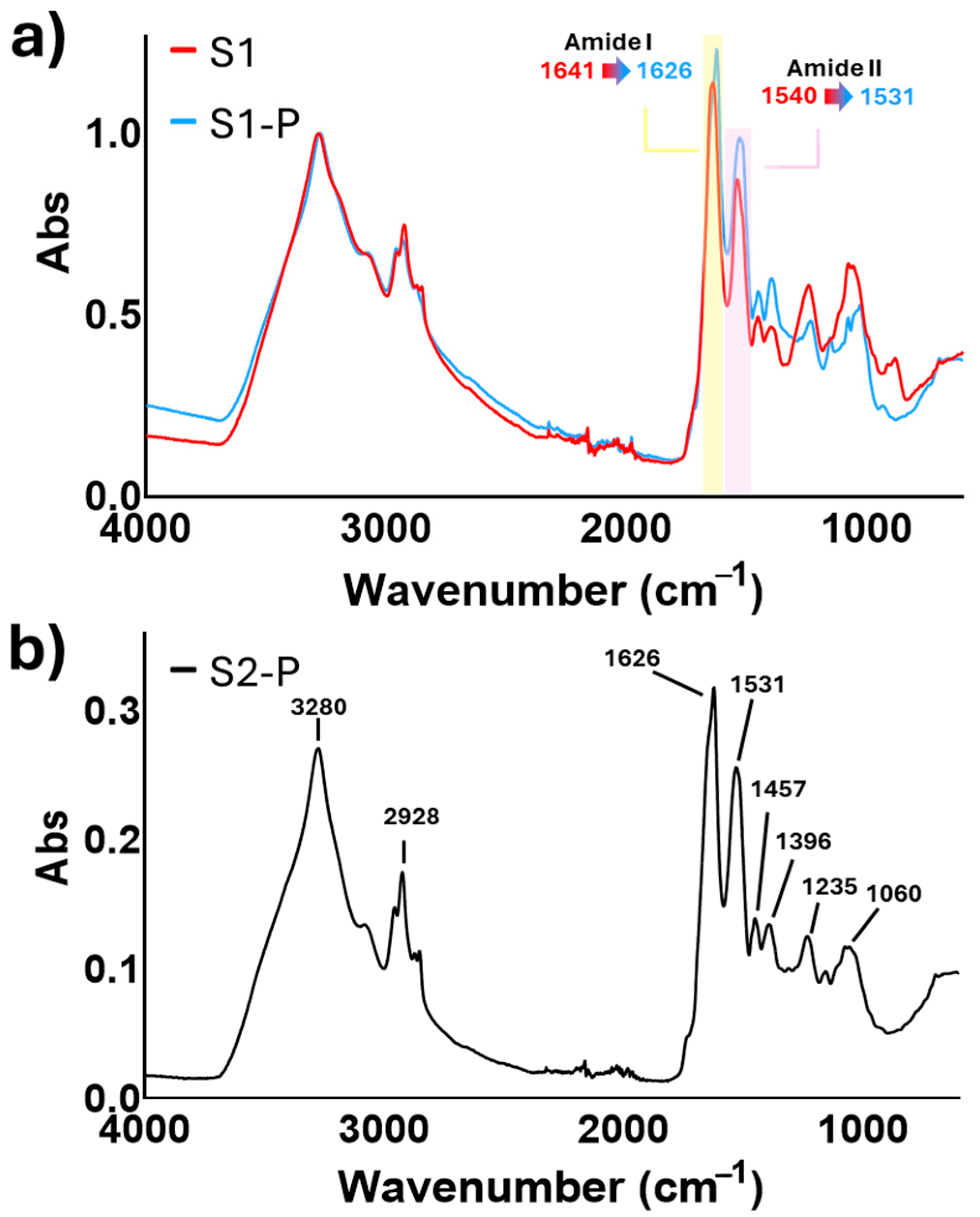
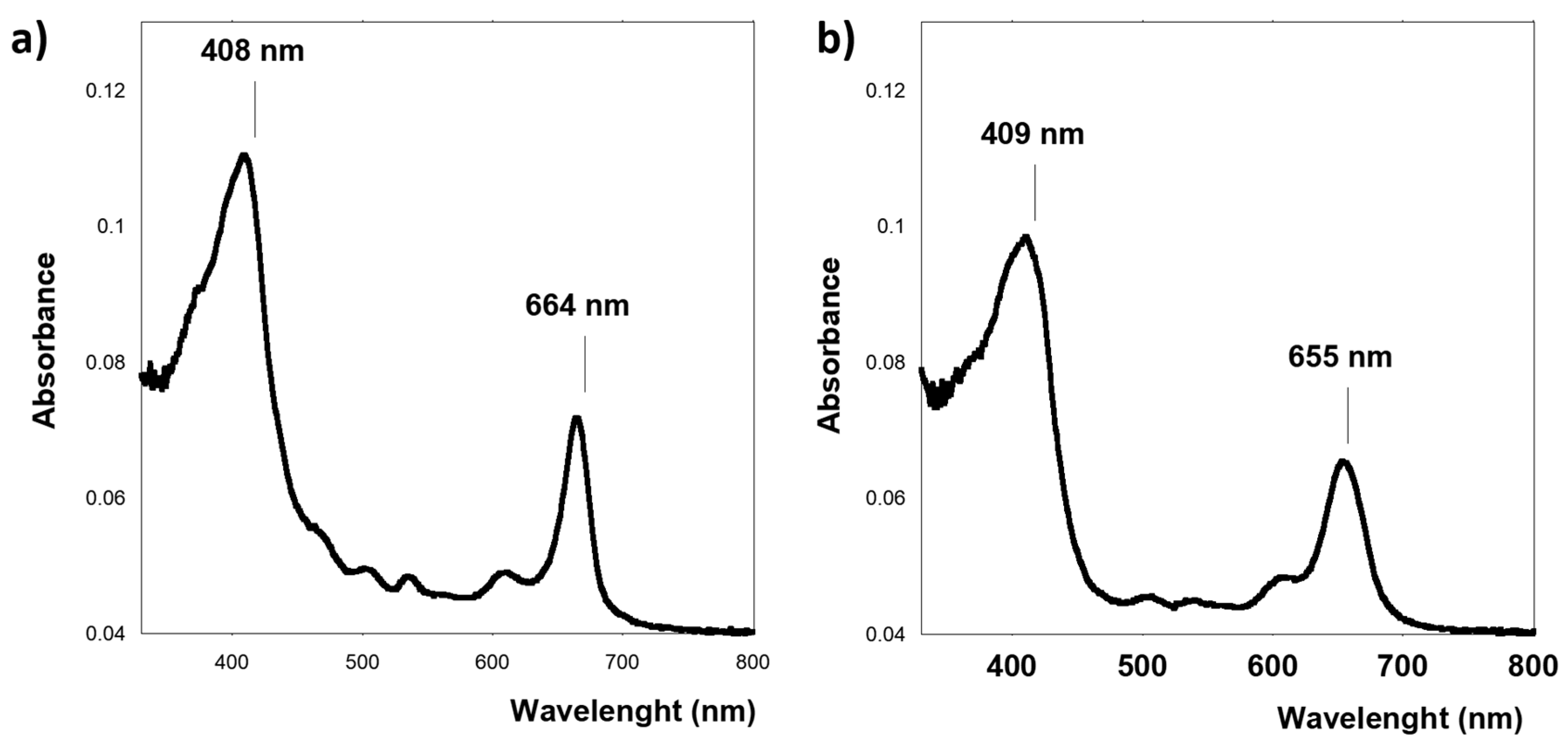
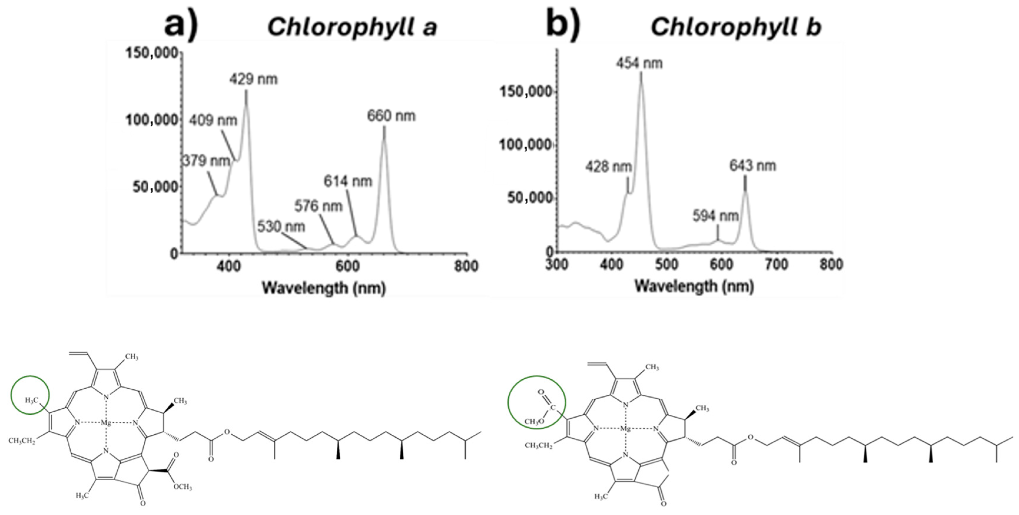
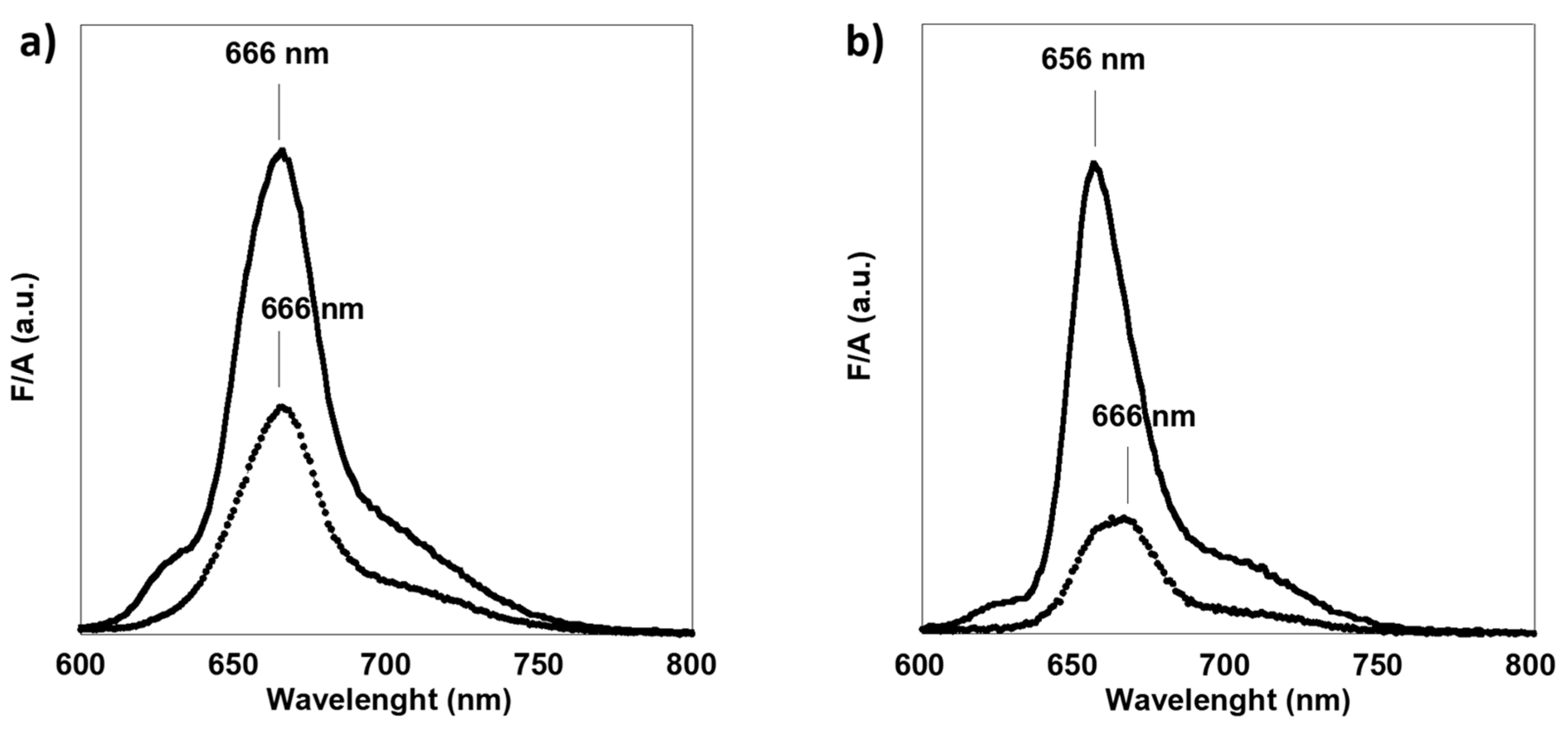
3.1. Spectroscopical Analysis of Microalgae

Some interesting information about the structural features and composition of all three samples can be deduced from FTIR spectra of the microalgae in the form of dry powder (Figure 2).
The FTIR spectrum of the microalga samples revealed a sharp and well-defined absorption band in the region of 3280–3282 cm−1, which can be attributed to the N–H stretching vibrations of protein functional groups. The narrow and intense nature of this band suggests a limited distribution of hydrogen bond strengths, indicative of a well-organized hydrogen bonding network. Such spectral features are commonly associated with highly ordered protein structures, where N–H groups are involved in regular and less dispersed hydrogen bonding interactions [18,19,20]. These observations are consistent with the known protein composition of microalgae and confirm structurally organized protein domains within the algal biomass. The group of signals around 2928 cm−1 corresponds to the CH2 and CH3 stretching vibrations of fatty acids. In the S1 sample, the peak at 1641–1626 cm−1 is assigned to the C=O stretching vibration of amide groups in proteins (amide I band). A sharp and well-defined band centered at 1540–1531 cm−1 can be primarily attributed to the amide II vibrations of the proteins, involving N–H bending and C–N stretching modes [18]. Additionally, the presence of unsaturated and polyunsaturated fatty acids within the microalgal biomass may contribute to this signal through C=C stretching vibrations, leading to a partial overlapping that influences the sharpness and intensity of the band [21,22]. These observations are consistent with the structural organization of the protein components in the microalga cell walls and the significant content of unsaturated lipid chains present in their composition. The peak at 1453 cm−1 is related to the CH3 and CH2 bending vibrations of lipids and proteins. The peak at 1245 cm−1 is attributed to the P=O stretching of phosphorylated proteins. Peaks from 1200 cm−1 onward are mainly associated with the C–O–C stretching of polysaccharides and the C–O stretching of carbohydrates. In alignment with what is reported in the literature [23], all characteristic chlorophyll peaks were found to be present. When comparing the IR spectra of the S1 samples containing polysaccharides with those without, a hypsochromic shift (commonly referred to as a “blue shift”) was observed in the amide I and II bands. To understand the cause of this shift, some structural features of microalgae must be considered: the cells are composed of polysaccharides, which play an important structural role, and a complex protein fraction. In particular, light-harvesting complexes (LHCs), composed of hydrophobic helices bound to pigments (such as chlorophyll a and b, lutein, neoxanthin, and carotenoids), function as the antennae for photosystems I and II. The extraction of polysaccharides involves a thermal treatment of the microalgae, which may cause degradation in the protein fraction of the microorganism. Therefore, it is likely that polysaccharide extraction also affects protein–polysaccharide interactions, altering the chemical environment surrounding the proteins and resulting in spectral shifts. Furthermore, a comparison of the spectra reveals changes in the fingerprint region associated with carbohydrates and polysaccharides, along with a noticeable decrease in band intensity, which is consistent with the removal of these compounds. Analyzing the FTIR spectrum of C. reinhardtii strain 2 after polysaccharide extraction (S2-P), reported in Figure 2b, the same spectral characteristics already observed for strain 1 (S1-P, Figure 2a) were found. The complete FTIR characterization is reported in Supplementary Materials (Figures S1 and S2).
Among the components of microalgal biomasses, chlorophylls play a significant role, as they can enhance the properties of bioplastic materials when used as additive and/or reinforcing biofillers. The incorporation of chlorophyll derivatives into bioplastics has been shown to influence mechanical strength, thermal stability, and flexibility. For instance, studies have demonstrated that adding chlorophyll derivatives to polyhydroxybutyrate (PHB) alters the amorphous phase of the polymer, affecting its mechanical properties and thermal behavior [24]. To investigate the types of chlorophylls present in the biomass under study, UV-visible absorption and fluorescence spectroscopy studies were performed on solutions obtained by dissolving dried microalgal biomasses in ethanol. This method allows chlorophylls to be extracted into the solution, facilitating the analysis of their UV-visible absorption profiles and the identification of their spectral characteristics within this region of the electromagnetic spectrum. Such spectroscopic analyses are commonly employed to assess pigment content and composition in microalgal cultures [25,26,27]. Figure 3a,b show the UV-visible absorption spectra of the two solutions. Since chlorophylls are tetrapyrrolic macrocycles (porphyrinoids) with a central Mg2+ ion (Figure 4), their absorption spectrum is the one of a metallized porphyrin, that is, it is composed of a Soret band, responsible for the absorption at 408–409 nm related to the π → π* (S0 → S2) electronic transition, and for different Q bands at 610 and 664 nm for the S1 sample or 608 and 655 nm for the S1-P sample, related to the π → π* (S0 → S1) electronic transition. The difference in the absorption maxima suggests, for the Soret band aggregation while for the Q band, a different content of chlorophyll a and b, since in general the Q band of chlorophyll a has a maximum at 662 nm, while that of chlorophyll b has a maximum at 642 nm (Figure 4). The difference between a and b is due to a formyl group (-CHO) in chlorophyll b that alters the conjugation and lowers the energy of the transitions (bathochromic shift). Our experimental results suggest that the thermal treatment used to remove polysaccharides in some way influences the spectroscopic properties of the pigments, giving rise to two different tonalities of green.
To confirm this hypothesis, fluorescence emission spectra of samples S1 and S1-P were produced and the results are reported in Figure 5. The spectra show that the emission spectra are the mirror image of the absorption spectra, and that the S1 sample shows a maximum at 666 nm, independent of the excitation wavelength, while the S1-P shows a spectrum that depends on the excitation wavelength. In particular, by exciting at 440 or 460 nm, a different emission spectrum can be obtained (Figure 5b). According to the literature [28], excitation at 440 nm and 460 nm preferentially excites chlorophyll a and chlorophyll b, respectively, but when they are together in solution, an energy transfer process takes place from chlorophyll b to a, making it possible to see mainly the emission spectrum of chlorophyll a.
In S1-P, the maximum fluorescence emission obtained by exciting the sample at 440 nm was shifted to 656 nm, compared to S1, the emission peak of which was centered at 666 nm. This result suggests a higher presence of chlorophyll b in the S1-P sample, compared to the S1. Since the initial chlorophyll content in both the samples should be the same, and the S1-P sample was thermally treated to remove polysaccharides, this result suggests that the thermal stability of chlorophyll b is higher compared to that of chlorophyll a, as confirmed by the literature. This is likely due to the formyl group at C7 of chlorophyll b, which contributes to the greater resonance stabilization of the macrocycle. This hypothesis is in accordance with the absorption spectra, which suggest that the S1-P sample contains more chlorophyll b. Furthermore, literature reports have shown that thermal treatments in the range of 40–80 °C promote the degradation of chlorophyll–protein complexes and chlorophyll molecules themselves, resulting in the formation of degradation products such as pheophytin and pheophorbide that can be responsible for the slightly different spectroscopical properties [29,30]. The fluorescence emission and UV-Vis spectra of all replicates of the microalgae samples, obtained after sequential ethanol extractions, are available in the Supplementary Materials (Figures S3–S11).

3.2. Utilization of Microalgae as Additives for Biobased Materials

The feasibility of employing spent microalgae as fillers in the compostable biobased material SP-Milk® was investigated, with a particular focus on assessing their potential role in enhancing mechanical and thermal performance. To enable a more systematic evaluation, composite materials were prepared using both untreated microalgae (i.e., without prior polysaccharide extraction) and spent microalgae, representing the actual production waste. A series of composite samples was prepared by incorporating different concentrations of the additive (0.5, 1.0, and 2.0 wt%), as detailed in Table 2. A direct comparison between untreated and spent microalgae was carried out using the C. reinhardtii strain 1, corresponding to samples SP-Milk/S1 and SP-Milk/S1-P, respectively. The mechanical and thermal properties of the resulting composites were then compared to elucidate the influence of the microalgae’s processing state on material performance.

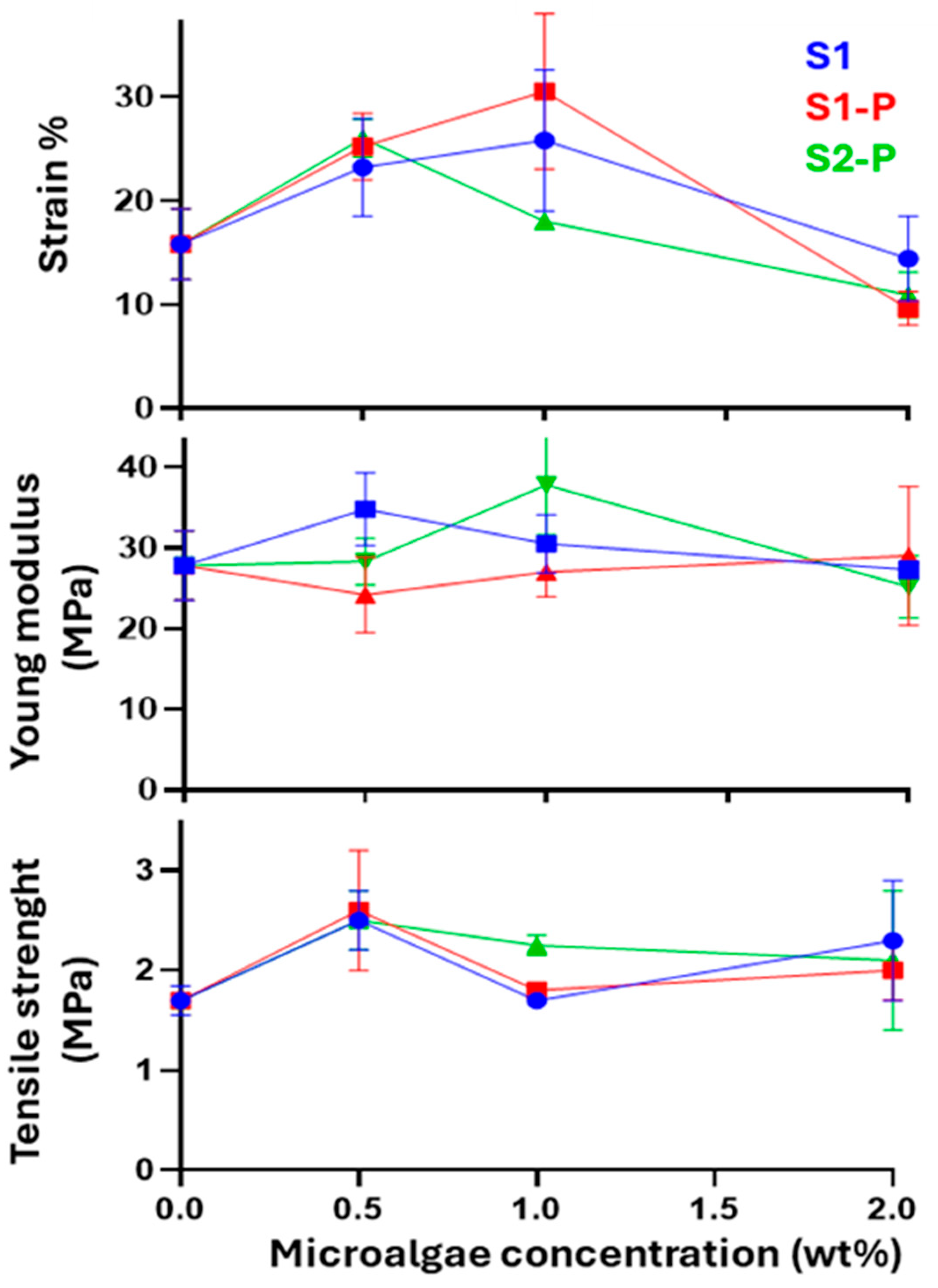
3.2.1. Mechanical Properties

To evaluate the mechanical performance, pure SP-Milk® and the bioplastic reinforced with varying concentrations of microalgal biomass (ranging from 0.5 wt% to 2.0 wt%) were tested. Key mechanical properties, such as tensile strength, elongation, and Young moduli, were assessed by tensile tests reported in Figure 6. The results trend is presented in Figure 7. As expected, considering the plasticizing effect of microalgae observed on the glass transition temperature of SP-Milk®, the addition of microalgae also affects the mechanical properties of the bioplastic. Overall, the tensile test results revealed that the incorporation of microalgae as fillers can enhance the mechanical performance of SP-Milk®, particularly its elasticity, although improvements occur only at certain concentrations.
The addition of 0.5 wt% S1 leads to an increase in all three measured mechanical parameters of SP-Milk (the elastic modulus passes from 27.8 ± 4.3 MPa to 34.8 ± 4.5, tensile strength from 1.7 ± 0.2 to 2.5 ± 0.3 MPa, and the elongation from 15.8 ± 3.4 to 23.2 ± 4.7 strain %). With further increases in S1 concentration, the Young’s modulus and tensile strength returned to values comparable to those of pure SP-Milk (30.5 ± 3.6 MPa and 1.7 ± 0.1 MPa, respectively). Elongation at break continued to increase with the addition of 1 wt% S1, reaching 25.8 ± 6.8%, but decreased to initial values when 2 wt% S1 was incorporated (14.4 ± 4.1%). The addition of S1-P microalgae followed a similar trend to that observed for S1, except for the Young’s modulus, whose mean value slightly decreased (from 27.8 ± 4.3 MPa of SP-Milk to 24.2 ± 4.7 MPa of SP-Milk/S1-P (0.5%)) and stabilized at levels comparable to those of pure SP-Milk as the concentration increased. The main difference observed between microalgal biomass with and without polysaccharides at 0.5 wt% was the increased stiffness of the composite when using S1 (which retained the polysaccharide fraction). This behavior is attributed to polysaccharides’ ability to form hydrogen-bond networks, which reinforce the bioplastic matrix [31]. When considering the sample belonging to strain 2 without its polysaccharide fraction (S2-P), the addition of 0.5 wt% results in an increase in both elongation at break (from 15.8 ± 3.4% of SP-Milk to 26.0 ± 1.8%) and tensile strength (from 1.7 ± 0.2 MPa of SP-Milk to 2.5 ± 0.3 MPa), without significantly altering the stiffness of the material (the elastic modulus remains practically unchanged, passing from 27.8 ± 4.3 MPa to 28.3 ± 2.9 MPa). However, further increases in S2-P concentration led to a reduction in elasticity and a concurrent increase in stiffness, as indicated by the rising values of the elastic modulus. In the samples supplemented with microalgae deprived of polysaccharides (SP-Milk/S1-P and SP-Milk/S2-P), the presence of glycoproteins, which constitute one of the major components of the cell wall, could contribute to influencing the mechanical properties of the material. When added at concentrations greater than 1 wt%, the observed increase in the stiffness of the bioplastic is likely due to more extensive hydrogen bond formation, facilitated by the glycoproteins. Another key factor is the presence of chlorophyll, demonstrated by spectroscopic analyses, which can enhance load distribution and thereby improve mechanical strength [27]. Additionally, chlorophyll exhibits antioxidant activity, which may slow down the thermal degradation of the polymer and thus prolong the lifetime of the material [32,33]. However, if added in excessive amounts, chlorophyll may not be well dispersed, acting as a structural defect and reducing mechanical strength and flexibility [34]. The tensile strength observed in our study, which varied between 1.7 and 2.5 MPa, was in reasonable agreement compared to values reported in the literature for microalga-reinforced biocomposites based on proteic matrices. It has been reported that tensile strength ranges from 2.6 to 6.5 MPa for gluten-based bioplastics [7], and around 3.1 MPa for starch-based bioplastics [35]. Also, a clear stiffening effect of microalgae has been observed when incorporated into protein-based bioplastics, as indicated by a rise in the modulus of elasticity [7].
These results highlight that even microalgae deprived of polysaccharides, when added within an appropriate concentration range, are effective in enhancing the mechanical properties of a biobased material. In particular, at low concentrations, the incorporation of microalgae and microalgal residues significantly improves the elasticity of the bioplastic without drastically compromising the tensile modulus and strength. Conversely, excessive amounts may lead to composite inhomogeneity and reduced compatibility with the polymer matrix. The best performance is observed at 0.5 wt%, while 1 wt% appears to represent the upper limit for effective reinforcement.

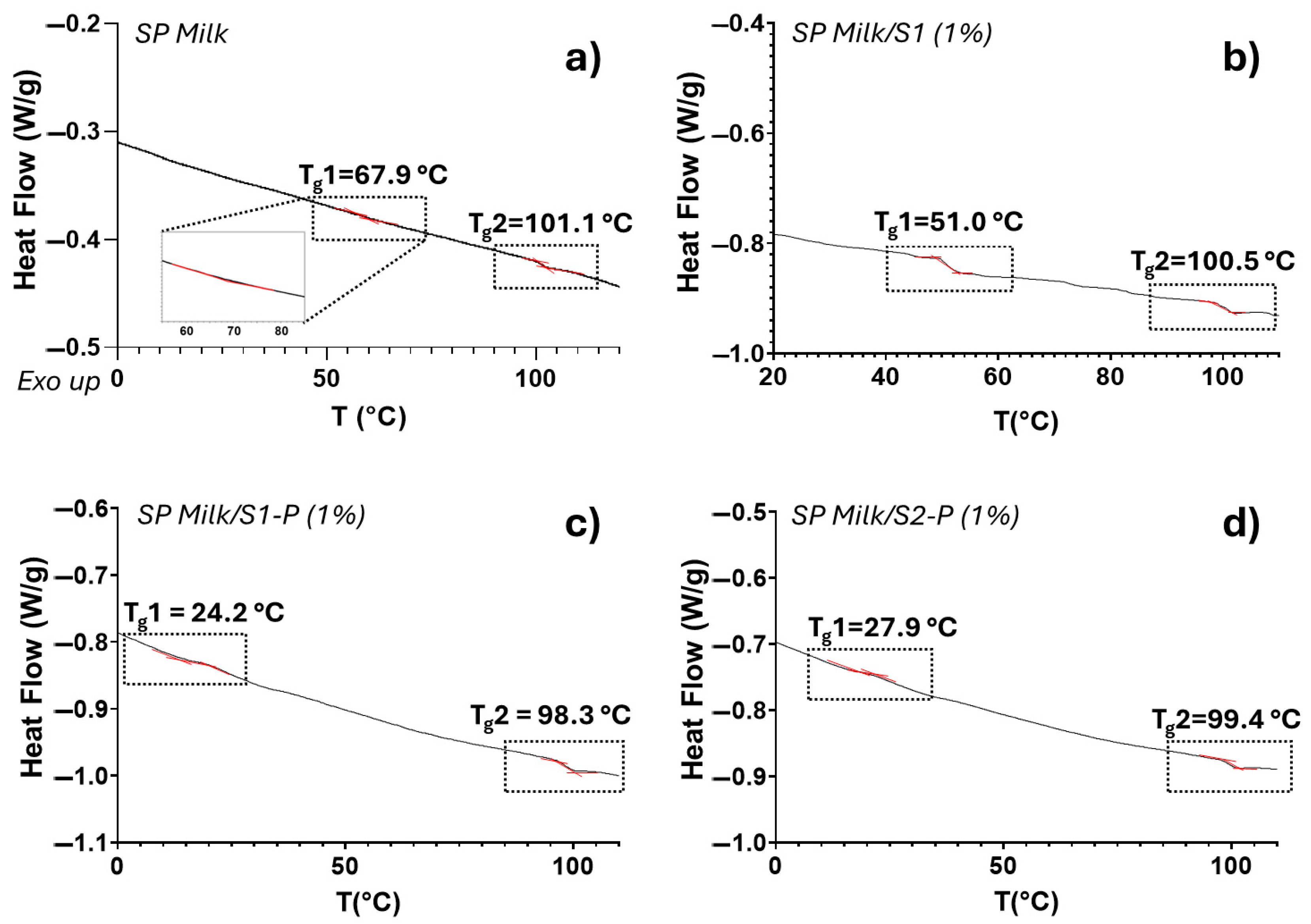
3.2.2. Thermal Analysis

To evaluate the impact of microalga additives on the thermal behavior of the biobased composites, a detailed thermal characterization was carried out. Differential Scanning Calorimetry (DSC) analyses were performed on selected samples, focusing on those containing 1 wt% of microalga content, chosen as the highest among the tested amounts that demonstrated a positive effect on the performance of the composites. This approach allowed for a direct comparison between composites formulated with untreated and spent microalgae, providing insights into the role of biomass processing on the thermal transitions of the material. Therefore, the thermal profile of the bioplastic samples containing 1 wt% of untreated (S1) and spent (S1-P) microalgae was investigated using Differential Scanning Calorimetry. A heat-cool-heat cycle was performed to eliminate the thermal history of the material and to accurately determine the glass transition temperature (Tg). This characteristic temperature is crucial for the processing of thermoplastic materials, as it defines the temperature range at which the material softens and can be shaped or molded into the final product. Figure 8 presents the DSC thermograms of SP-Milk/S1 (1%) and the corresponding material containing spent microalgae, SP-Milk/S1-P (1%).
The bioplastic sample without microalgae exhibited two glass transition temperatures (Tg1 and Tg2) at 67.9 and 101.1 °C (Figure 8a). The observation of two distinct glass transition temperatures in the casein-based bioplastic suggests the presence of structurally heterogeneous amorphous regions within the polymer matrix. Casein, as a complex, intrinsically disordered protein, tends to form a non-uniform network during film formation. This can result in the development of separate domains with differing degrees of molecular mobility. Specifically, the lower-temperature Tg (Tg1) is likely associated with more flexible, loosely packed regions where protein chains experience greater segmental motion. In contrast, the higher-temperature Tg (Tg2) may correspond to more densely packed or partially crosslinked domains, where restricted chain mobility requires more thermal energy to initiate the glass transition. Also, the formulation of SP-Milk containing 1 wt% of untreated microalga (S1), referred to as SP-Milk/S1 (1%), displayed two glass transition temperatures, located at approximately 51 °C and 100.5 °C, respectively (Figure 8b), showing a considerable shifting of Tg1 to lower values (−16.9 °C). In the formulation containing 1 wt% of S1 spent microalgae, SP-Milk/S1-P (1%), Tg1 was further shifted to lower temperatures (24.2 °C), while Tg2 appeared at 98.3 °C (Figure 8c). Despite the presence of two distinct glass transitions, the incorporation of microalgae exerted a beneficial effect on the thermal behavior of SP-Milk, leading to the decrease in Tg1 to significantly lower values compared to those of the neat bioplastic. This change is advantageous, as it facilitates easier processing and molding of the material and reduces the energy consumption required to reach the processing temperature in industrial applications. Furthermore, the incorporation of spent microalgae resulted in an additional shift in Tg1 to even lower temperatures, further enhancing the material’s processability. This reduction in glass transition temperature suggests that the spent microalgae not only act as a natural dye and reinforcement agent but also contribute to improving the processing of the material, making it workable at lower temperatures. The reduction in glass transition temperature observed upon the incorporation of microalgal biomass is primarily attributed to a plasticizing effect. In fact, the SP-Milk bioplastic is protein-based, with its structure stabilized mainly by hydrogen bonds and ionic interactions between polymer chains. The addition of microalgal biomass disrupts these interactions by intercalating between the protein chains, thereby increasing free volume and chain mobility, which results in a lower Tg. Furthermore, the microalgal residues contain low-molecular-weight compounds, such as short-chain fatty acids, amino acids, short peptides, and low-molecular-weight polysaccharides, which act as natural plasticizers [36]. These components can insert themselves between polymer chains, weakening intermolecular forces and further enhancing molecular mobility, thus contributing to the observed reduction in Tg. In SP-Milk/microalga samples, this plasticizing behavior is reasonably attributed to the residual unsaturated and polyunsaturated fatty compounds within the microalgae, whose presence was confirmed by FT-IR analysis (signal group in the 2800–3000 cm−1 region). It has been reported in the literature that the dry biomass of C. reinhardtii can contain up to 20 wt% of lipids, primarily comprising unsaturated and polyunsaturated fatty acids [37,38]. Moreover, the presence of chlorophyll that acts as a plasticizer also has a role in decreasing the glass transition temperature [39]. The effective interaction between the bioplastic’s protein-based matrix and the microalgae is crucial for achieving good material performance. As observed in polymer blends or plastics, poor compatibility between phases typically leads to phase separation and a deterioration of mechanical and thermal properties [40]. In the present study, the gradual transition of Tg in SP-Milk composites suggests a favorable compatibility between the protein matrix and the microalgal component, likely due to intimate molecular interactions. This hypothesis is further supported by the absence of phase separation in the obtained samples, as also evidenced by SEM micrographs (Figure S12, Supplementary Materials), which reveal a homogeneous dispersion and good adhesion in the microalgal phase within the protein matrix. We should add that the specimens were prepared by an artisanal, laboratory-scale process. We are sure that by industrial processing, the mechanical performance of this material can be further improved. Similar trends were observed for the SP-Milk/S2-P sample (Figure 8d), where Tg1 was reduced to 27.9 °C and Tg2 to 99.4 °C. The greater decrease in Tg observed in the formulations containing S2-P and S1-P microalgae, compared to the formulation with S1 microalgae, can be attributed to both the residual biopolymer composition and the treatments the microalgae underwent. As previously discussed, the unsaturated and polyunsaturated fats in the microalgae serve as the primary plasticizing agents, reducing the glass transition temperature. Moreover, it is likely that the S1-P and S2-P microalgae, having undergone polysaccharide extraction treatments, experienced cell wall disruption. This disruption likely enhanced the interaction between the residual lipid component and the SP-Milk matrix. Furthermore, the removal of polysaccharides, which are capable of forming hydrogen-bonded networks that can stiffen bioplastics, further facilitated the reduction in Tg compared to the formulation containing S1 microalgae, where polysaccharides remained intact. DSC analyses, showing the trend of the glass transition temperature, indicate good compatibility between microalgae and the bioplastic matrix in the samples with 1 wt% of microalgae, and this result is in accordance with the mechanical test results where 1 wt% appears to represent the upper limit for effective material reinforcement.

4. Conclusions

In this study, residual biomass from C. reinhardtii, left over after polysaccharide extraction, was repurposed as a natural multifunctional component in SP-Milk®, a compostable protein-based material. The goal was to enhance mechanical properties and provide natural green pigmentation. FTIR and UV-Vis spectroscopy confirmed the chemical composition of the biomass, while subsequent incorporation into the protein matrix yielded biocomposites with improved performance.
Thermal treatment for polysaccharide removal selectively reduced chlorophyll a content, modifying both the optical spectrum and visual appearance. Despite this, even depleted microalgae, particularly at low loadings (<1 wt%), enhanced tensile strength and elasticity without increasing stiffness. The observed improvements in tensile strength and elasticity at low biomass concentrations support its role as a mechanical reinforcing agent, consistent with biofiller behavior in composite materials. At the same time, the reduction in glass transition temperature and the introduction of green pigmentation confirmed the biomass’s function as a functional additive, facilitating processing at lower energy costs and providing a natural aesthetic.
These findings underscore the multifunctionality of the biomass, positioning microalgal residues not as waste, but as functional bio-components, capable of improving workability, mechanical resilience, and the aesthetic appeal of bioplastics. Moreover, chlorophyll serves as a natural, non-toxic pigment, providing a sustainable alternative to synthetic dyes, particularly in eco-friendly packaging. This approach embodies circular bioeconomy principles, converting cultivation by-products into value-added components for next-generation, biodegradable materials.

Supplementary Materials

The following supporting information can be downloaded at: https://www.mdpi.com/article/10.3390/macromol5030032/s1, Figure S1: FTIR spectra of S1 with (S1, in red) and without polysaccharides (S1-P, in blue) repeated for 3 replicates (R1, R2, and R3). Figure S2: FTIR spectra of S1-P and S2-P repeated for 3 replicates (R1, R2, and R3). Figure S3: Fluorescence emission spectra (a) and UV-Vis spectra (b) for 3 replicates (R1, R2, and R3) of S1 microalgae sample, after the first extraction in ethanol. Figure S4: Fluorescence emission (a) and UV-Vis spectra (b) for 3 replicates (R1, R2, and R3) of S1 microalgae sample after the second extraction in ethanol. Figure S5: Fluorescence emission (a) and UV-Vis spectra (b) for 3 replicates (R1, R2, and R3) of S1 microalgae sample after the third extraction in ethanol. Figure S6: Fluorescence emission (a) and UV-Vis spectra (b) for 2 replicates (R1, R2) of S1-P microalgae sample after the first extraction in ethanol. Figure S7: Fluorescence emission (a) and UV-Vis spectra (b) for 2 replicates (R1, R2) of S1-P microalgae sample after the second extraction in ethanol. Figure S8: Fluorescence emission (a) and UV-Vis spectra (b) for 2 replicates (R1, R2,) of S1-P microalgae sample after the third extraction in ethanol. Figure S9: Fluorescence emission (a) and UV-Vis spectra (b) for 3 replicates (R1, R2, and R3) of S2-P microalgae sample after the first extraction in ethanol. Figure S10: Fluorescence emission (a) and UV-Vis spectra (b) for 3 replicates (R1, R2, and R3) of S2-P microalgae sample after the second extraction in ethanol. Figure S11: Fluorescence emission (a) and UV-Vis spectra (b) for 3 replicates (R1, R2, and R3) of S2-P microalgae sample after the third extraction in ethanol. Figure S12: SEM micrographs of the fracture surface following tensile testing of SP Milk® bioplastic incorporating 1 wt% microalgal biomass (sample SP Milk/S1-P(1%)), acquired at different magnifications. Table S1: Young moduli, tensile strength, and strain% mean values (± SD) for all the specimens used for tensile tests.

Author Contributions

Conceptualization, E.G., R.L. and V.C.; methodology, E.G., V.C. and A.C.; software, E.G.; validation, E.G. and R.L.; formal analysis, A.C., M.C. and V.C.; investigation, A.C., M.C. and V.C.; resources, E.G., V.S. and A.A.; data curation, A.C., M.C., E.G. and V.C.; writing—original draft preparation, E.G., R.L. and A.C.; writing—review and editing, E.G., R.L. and A.C.; supervision, E.G.; project administration, E.G. and V.S.; funding acquisition, E.G. and V.S. All authors have read and agreed to the published version of the manuscript.

Funding

This research was funded by the European Union-Next Generation EU, Mission 4, Component 1, Project PRIN-2022EPWFER-NANOgrab (CUP B53D23017540006) and Project PRIN-2022R5RATP-BIODUST (CUP B53D23006350006).

Data Availability Statement

Data is contained within the article or Supplementary Material.

Acknowledgments

The authors would like to thank Cadia D’Ottavi and Leonardo Duranti (Department of Chemical Science and Technologies, University of Rome “Tor Vergata”, Via della Ricerca Scientifica 1, 00133 Rome, Italy) for their valuable support in acquiring the SEM images.

Conflicts of Interest

Viviana Scognamiglio and Amina Antonacci are employed by the Institute of Crystallography, Department of Chemical Sciences and Materials Technology. Emanuela Gatto and Raffaella Lettieri are the founders of the Splastica spinoff, who patented the SP-Milk material. Alice Caravella, Valerio Cuboni, and Martina Corvino declare that the research was conducted in the absence of any commercial or financial relationships that could be construed as a potential conflict of interest.

References

  1. Lettieri, R.; Fazio, V.; Abruzzese, D.; di Bartolomeo, E.; D’Ottavi, C.; Micheletti, A.; Tiero, A.; Duranti, L.; Armuzza, V.; Licoccia, S.; et al. Influence of natural additives on the properties of a milk-based compostable bioplastic. RSC Adv. 2024, 14, 3737–3755. [Google Scholar] [CrossRef] [PubMed]
  2. Boey, J.Y.; Lee, C.K.; Tay, G.S. Factors Affecting Mechanical Properties of Reinforced Bioplastics: A Review. Polymers 2022, 14, 3737. [Google Scholar] [CrossRef] [PubMed]
  3. Brennan, L.; Owende, P. Biofuels from microalgae—A review of technologies for production, processing, and extractions of biofuels and co-products. Renew. Sustain. Energy Rev. 2010, 14, 557–577. [Google Scholar] [CrossRef]
  4. Wijffels, R.H.; Kruse, O.; Hellingwerf, K.J. Potential of industrial biotechnology with cyanobacteria and eukaryotic microalgae. Curr. Opin. Biotechnol. 2013, 24, 405–413. [Google Scholar] [CrossRef]
  5. Trivedi, J.; Aila, M.; Bangwal, D.; Kaul, S.; Garg, M.O. Algae based biorefinery—How to make sense? Renew. Sustain. Energy Rev. 2015, 47, 295–307. [Google Scholar] [CrossRef]
  6. Cheah, W.Y.; Er, A.C.; Aiyub, K.; Mohd Yasin, N.H.; Ngan, S.L.; Chew, K.W.; Khoo, K.S.; Ling, T.C.; Juan, J.C.; Ma, Z.; et al. Current status and perspectives of algae-based bioplastics: A reviewed potential for sustainability. Algal Res. 2023, 71, 103078. [Google Scholar] [CrossRef]
  7. Ciapponi, R.; Turri, S.; Levi, M. Mechanical Reinforcement by Microalgal Biofiller in Novel Thermoplastic Biocompounds from Plasticized Gluten. Materials 2019, 12, 1476. [Google Scholar] [CrossRef]
  8. Merchant, S.S.; Prochnik, S.E.; Vallon, O.; Harris, E.H.; Karpowicz, S.J.; Witman, G.B.; Terry, A.; Salamov, A.; Fritz-Laylin, L.K.; Maréchal-Drouard, L.; et al. The Chlamydomonas genome reveals the evolution of key animal and plant functions. Science 2007, 318, 245–250. [Google Scholar] [CrossRef]
  9. Yunus, I.S.; Wichmann, J.; Wördenweber, R.; Lauersen, K.J.; Kruse, O.; Jones, P.R. Synthetic metabolic pathways for photobiological conversion of CO2 into hydrocarbon fuel. Metab. Eng. 2018, 49, 201–211. [Google Scholar] [CrossRef]
  10. Poulhazan, A.; Arnold, A.A.; Mentink-Vigier, F.; Muszyński, A.; Azadi, P.; Halim, A.; Vakhrushev, S.Y.; Joshi, H.J.; Wang, T.; Warschawski, D.E.; et al. Molecular-level architecture of Chlamydomonas reinhardtii’s glycoprotein-rich cell wall. Nat. Commun. 2024, 15, 986. [Google Scholar] [CrossRef]
  11. Milledge, J.J.; Heaven, S. A review of the harvesting of micro-algae for biofuel production. Rev. Environ. Sci. Biotechnol. 2013, 12, 165–178. [Google Scholar] [CrossRef]
  12. Safi, C.; Zebib, B.; Merah, O.; Pontalier, P.-Y.; Vaca-Garcia, C. Morphology, composition, production, processing and applications of Chlorella vulgaris: A review. Renew. Sustain. Energy Rev. 2014, 35, 265–278. [Google Scholar] [CrossRef]
  13. Suvorov, N.; Pogorilyy, V.; Diachkova, E.; Vasil’ev, Y.; Mironov, A.; Grin, M. Derivatives of Natural Chlorophylls as Agents for Antimicrobial Photodynamic Therapy. Int. J. Mol. Sci. 2021, 22, 6392. [Google Scholar] [CrossRef] [PubMed]
  14. Caravella, A.; Lettieri, R.; Vezza, R.; Gatto, E. Aerobic biodegradation at a seawater-sediment interface of a new bioplastic 100% based on natural polymers. ACS Sustain. Chem. Eng. 2024, 1, 1099–1111. [Google Scholar] [CrossRef]
  15. Masi, A.; Leonelli, F.; Di Luca, D.; Celio, L.; Gasperuzzo, G.; Lunardi, S.; Rigon, C.; Pitocco, M.; Ioele, M.; Storace, M.S.; et al. Extraction and characterization of polysaccharide mixtures from Chlamydomonas reinhardtii: A case study as a consolidant for paper-based artworks. Carbohydr. Polym. Technol. Appl. 2025, 9, 100713. [Google Scholar] [CrossRef]
  16. Farobie, O.; Anis, L.A.; Nurcahyani, P.R.; Hartulistiyoso, E.; Rahman, D.Y.; Fatriasari, W.; Nafisyah, A.L.; Amrullah, A.; Aziz, M. Extraction of Bio-pigments from the Green Microalgae Chlorella pyrenoidosa Under Different Solvent Ratios. IOP Conf. Ser. Earth Environ. Sci. 2023, 1187, 012009. [Google Scholar] [CrossRef]
  17. International Standard ISO 527-1; Plastics—Determination of tensile properties. International Organization for Standardization (ISO): Geneva, Switzerland, 2019.
  18. Barth, A. Infrared spectroscopy of proteins. Biochim. Biophys. Acta Bioenerg. 2007, 1767, 1073–1101. [Google Scholar] [CrossRef]
  19. Jackson, M.; Mantsch, H.H. The use and misuse of FTIR spectroscopy in the determination of protein structure. Crit. Rev. Biochem. Mol. Biol. 1995, 30, 95–120. [Google Scholar] [CrossRef]
  20. Kong, J.; Yu, S. Fourier Transform Infrared Spectroscopic Analysis of Protein Secondary Structures. Acta Biochim. Biophys. Sin. 2007, 39, 549–559. [Google Scholar] [CrossRef]
  21. Guillén, M.D.; Cabo, N. Characterization of edible oils and lard by Fourier transform infrared spectroscopy. Relationships between composition and frequency of concrete bands in the fingerprint region. J. Am. Oil Chem. Soc. 1997, 74, 1281–1286. [Google Scholar] [CrossRef]
  22. Bobroff, V.; Rubio, C.; Vigier, V.; Petibois, C. FTIR spectroscopy characterization of fatty-acyl-chain conjugates. Anal. Bioanal. Chem. 2016, 408, 319–326. [Google Scholar] [CrossRef] [PubMed]
  23. Kareem, J. Effect of Chlorophyll and Anthocyanin on the Secondary Bonds of Poly Methyl Methacrylate (PMMA). Int. J. Tech. Res. Appl. 2014, 2, 73–80. [Google Scholar]
  24. Tyubaeva, P.M.; Gasparyan, K.G.; Romanov, R.R.; Kolesnikov, E.A.; Martirosyan, L.Y.; Larkina, E.A.; Tyubaev, M.A. Biomimetic materials based on poly-3-hydroxybutyrate and chlorophyll derivatives. Polymers 2024, 16, 101. [Google Scholar] [CrossRef] [PubMed]
  25. Hotos, G.N.; Bekiari, V. Absorption Spectra as Predictors of Algal Biomass and Pigment Content of the Cultured Microalgae Amphidinium carterae, Isochrysis galbana, Nephroselmis sp., and Anabaena sp. Int. J. Plant Biol. 2023, 14, 879–895. [Google Scholar] [CrossRef]
  26. Esteves, A.F.; Pardilhó, S.; Gonçalves, A.L.; Vilar, V.J.P.; Pires, J.C.M. Unravelling the impact of light spectra on microalgal growth and biochemical composition using principal component analysis and artificial neural network models. Algal Res. 2025, 85, 103820. [Google Scholar] [CrossRef]
  27. Eregie, S.B.; Sanusi, I.A.; Kana, G.E.B.; Olaniran, A.O. Effect of ultra-violet light radiation on Scenedesmus vacuolatus growth kinetics, metabolic performance, and preliminary biodegradation study. Biodegradation 2024, 35, 71–86. [Google Scholar] [CrossRef]
  28. Na Ayudhya, T.I.; Posey, F.T.; Tyus, J.C.; Dingra, N.N. Using a microscale approach to rapidly separate and characterize three photosynthetic pigment species from fern. J. Chem. Educ. 2015, 92, 920–923. [Google Scholar] [CrossRef]
  29. Lípová, L.; Krchňák, P.; Komenda, J.; Ilík, P. Heat-induced disassembly and degradation of chlorophyll-containing protein complexes in vivo. Biochim. Biophys. Acta 2010, 1797, 63–70. [Google Scholar] [CrossRef]
  30. Östbring, K.; Rayner, M.; Sjöholm, I.; Otterström, J.; Albertsson, P.-Å.; Emek, S.C.; Erlanson-Albertsson, C. The effect of heat treatment of thylakoids on their ability to inhibit in vitro lipase/co-lipase activity. Food Funct. 2014, 5, 2157–2165. [Google Scholar] [CrossRef]
  31. Zhang, Y.; Nian, Y.; Shi, Q.; Hu, B. Protein fibrillation and hybridization with polysaccharides enhance strength, toughness, and gas selectivity of bioplastic packaging. J. Mater. Chem. A 2023, 11, 9884–9901. [Google Scholar] [CrossRef]
  32. Lanfer-Marquez, U.M.; Barros, R.M.; Sinnecker, P. Antioxidant activity of chlorophylls and their derivatives. Food Res. Int. 2005, 38, 885–891. [Google Scholar] [CrossRef]
  33. Pérez-Gálvez, A.; Viera, I.; Roca, M. Carotenoids and chlorophylls as antioxidants. Antioxidants 2020, 9, 505. [Google Scholar] [CrossRef] [PubMed]
  34. Nanni, A.; Parisi, M.; Colonna, M. Wine by-products as raw materials for the production of biopolymers and natural reinforcing fillers: A critical review. Polymers 2021, 13, 381. [Google Scholar] [CrossRef] [PubMed]
  35. Tedeschi, A.M.; Di Caprio, F.; Piozzi, A.; Pagnanelli, F.; Francolini, I. Sustainable Bioactive Packaging Based on Thermoplastic Starch and Microalgae. Int. J. Mol. Sci. 2021, 23, 178. [Google Scholar] [CrossRef]
  36. Alee, M.; Duan, Q.; Chen, Y.; Liu, H.; Ali, A.; Zhu, J.; Jiang, T.; Rahaman, A.; Chen, L.; Yu, L. Plasticization Efficiency and Characteristics of Monosaccharides, Disaccharides, and Low-Molecular-Weight Polysaccharides for Starch-Based Materials. ACS Sustain. Chem. Eng. 2021, 9, 11960. [Google Scholar] [CrossRef]
  37. Li-Beisson, Y.; Beisson, F.; Riekhof, W. Metabolism of acyl-lipids in Chlamydomonas reinhardtii. Plant J. 2015, 82, 504–522. [Google Scholar] [CrossRef]
  38. Masi, A.; Leonelli, F.; Scognamiglio, V.; Gasperuzzo, G.; Antonacci, A.; Terzidis, M.A. Chlamydomonas reinhardtii: A factory of nutraceutical and food supplements for human health. Molecules 2023, 28, 1185. [Google Scholar] [CrossRef]
  39. Dahash, F.K.; Aobaid, A.K.; Al Maamori, M.H. The effect of chlorophyll on the mechanical properties of natural rubber (NR) and the penetration of X-ray. In Recent Trends and Advances in Artificial Intelligence; Garcia, F.P., Jamil, A., Hameed, A.A., Ortis, A., Ramirez, I.S., Eds.; Springer: Cham, Switzerland, 2024; Volume 1138, pp. 1099–1111. [Google Scholar] [CrossRef]
  40. Aversa, C.; Barletta, M.; Cappiello, G.; Gisario, A. Compatibilization strategies and analysis of morphological features of poly(butylene adipate-co-terephthalate) (PBAT)/poly(lactic acid) PLA blends: A state-of-art review. Eur. Polym. J. 2022, 173, 111304. [Google Scholar] [CrossRef]
Figure 1. (a) Pure SP-Milk specimen, (b) SP-Milk/S1 (1%), (c) SP-Milk/S1-P (1%), (d) SP-Milk/S2-P (1%), (e) empty 3D-printed PLA mold.
Macromol 05 00032 g001
Figure 2. FTIR-ATR spectra of microalgae: (a) comparison between S1 with and without (-P) polysaccharides; (b) FTIR-ATR spectrum of S2-P.
Macromol 05 00032 g002
Figure 3. UV-Vis absorption spectra of S1 (a) and S1-P (b) microalga samples, respectively, after the first extraction in ethanol.
Macromol 05 00032 g003
Figure 4. UV-visible reference spectra of chlorophyll a (a) and b (b), respectively, and their corresponding chemical structure.
Macromol 05 00032 g004
Figure 5. Fluorescence emission spectra of S1 (a) and S1-P (b) microalga samples, after the first extraction in ethanol. Continuous line: λex = 440 nm, dotted line: λex = 460 nm.
Macromol 05 00032 g005
Figure 6. Mechanical tensile tests of the four samples analyzed. SP-Milk (blue line), SP-Milk/S1 (1%) (red line), SP-Milk/S1-P (1%) (green line), and SP-Milk/S2-P (1%) (purple line).
Macromol 05 00032 g006
Figure 7. Results of tensile tests on bioplastic samples enhanced with microalgae (S1, S1-P, and S2-P at various concentrations in the SP Milk matrix). Variations in elongation (strain %), Young’s modulus, and tensile strength are reported as a function of the additive weight percentage (wt%) used.
Macromol 05 00032 g007
Figure 8. DSC thermograms of SP-Milk (a) and SP-Milk composites containing 1 wt% of microalga S1 (b), S1-P (c), and S2-P (d). Measurements were performed at a heating/cooling rate of 20 °C/min under a nitrogen atmosphere. The second heating scan is reported and the calculation of glass transition temperatures is shown.
Macromol 05 00032 g008
Table 1. List of abbreviations and description of the samples of C. reinhardtii according to the strain, and treatment undergone.
AcronymStrainTreatment
S1CC125None
S1-PCC125Polysaccharides extracted
S2-PSAG 11-32bPolysaccharides extracted
Table 2. List of abbreviations and the relative amount of microalgal biomass in the composite materials.
Sample Name% w/w Microalgae
SP-Milk0
SP-Milk/S1 (0.5%) 0.5
SP-Milk/S1 (1.0%)1.0
SP-Milk/S1 (2.0%)2.0
SP-Milk/S1-P (0.5%)0.5
SP-Milk/S1-P (1.0%)1.0
SP-Milk/S1-P (2.0%)2.0
SP-Milk/S2-P (0.5%)0.5
SP-Milk/S2-P (1.0%)1.0
SP-Milk/S2-P (2.0%)2.0
Disclaimer/Publisher’s Note: The statements, opinions and data contained in all publications are solely those of the individual author(s) and contributor(s) and not of MDPI and/or the editor(s). MDPI and/or the editor(s) disclaim responsibility for any injury to people or property resulting from any ideas, methods, instructions or products referred to in the content.

