Article

Antibiotic-Coated Melt Electrowritten Polycaprolactone Meshes: Fabrication and In Vitro Antibacterial Evaluation

by
Joana Pinheiro Martins
1,*,
Ana Sofia de Sousa
2,
Sofia Costa de Oliveira
3,
António Augusto Fernandes
1,2 and
Elisabete Teixeira da Silva
1
1
Associate Laboratory of Energy, Transports and Aerospace, Institute of Science and Innovation in Mechanical and Industrial Engineering, 4200-465 Porto, Portugal
2
Faculty of Engineering, University of Porto, 4200-465 Porto, Portugal
3
Rede de Investigação em Saúde (RISE)-Health, Department of Pathology, Faculty of Medicine, University of Porto, 4200-319 Porto, Portugal
*
Author to whom correspondence should be addressed.
Macromol 2025, 5(3), 33; https://doi.org/10.3390/macromol5030033
Submission received: 29 April 2025 / Revised: 20 June 2025 / Accepted: 1 July 2025 / Published: 16 July 2025

Abstract

In recent years, pelvic organ prolapse (POP) cases have been rising, affecting women’s quality of life. Synthetic surgical transvaginal meshes used for POP treatment were withdrawn from the United States market in 2019 due to high risks, including infection, vaginal mesh erosion, and POP reoccurrence. Biodegradable mesh implants with three-dimensional printing technology have emerged as an innovative alternative. In this study, polycaprolactone (PCL) meshes for POP repair were fabricated using melt electrospinning writing (MEW) and mechanically evaluated through uniaxial tensile tests. Following this, they were coated with antibiotics—azithromycin, gentamicin sulfate, and ciprofloxacin—commonly used for genitourinary tract infections. Zone inhibition and biofilm assays evaluated antibiotic effectiveness in preventing mesh infections by Escherichia coli, and methicillin-susceptible (MSSA) and methicillin-resistant (MRSA) Staphylococcus aureus. The meshes presented a mechanical behavior closer to vaginal tissue than commercially available meshes. Fourier transform infrared analysis confirmed antibiotic incorporation. Ciprofloxacin demonstrated antibacterial activity against MRSA, with a 92% reduction in metabolic activity and a 99% biomass reduction. Gentamicin and ciprofloxacin displayed inhibitory activity against MSSA and E. coli. Scanning electron microscopy images support these conclusions. This methodology may offer a more effective, patient-friendly solution for POP repair, improving healing and the quality of life for affected women.

1. Introduction

One of the most common pelvic floor dysfunctions (PFDs) is pelvic organ prolapse (POP), which is described as a protrusion of the pelvic organs, including the bladder, bowel, and uterus, through the vaginal walls and pelvic floor. This happens when the connective tissues, ligaments, and muscles can no longer hold them due to lesions or weakness. There are different risk factors for developing POP, including age, vaginal delivery, menopausal status, and, the most common, pregnancy [1,2].
Regarding the treatment choice for POP, it depends on the symptom and prolapse severity. Treatment strategies can go from non-surgical to surgical treatment. Non-surgical treatments include pelvic floor muscle training and pessaries, which are more focused on decreasing the severity and frequency of symptoms. However, when these are not sufficient, surgery is inevitable [3]. Surgery for POP treatment can be reconstructive, aiming to restore vaginal function, or obliterative, an approach that focuses on repositioning the pelvic organs back into the pelvis and partially or completely closing the vaginal canal [4]. The prevalence of reoperation after reconstructive surgery is 30%, which is mainly caused by using poor-quality tissues to reconstruct pelvic floor defects, leading to an increased use of prosthetic materials to decrease POP recurrence rate [5,6,7]. In this way, surgical meshes for POP treatment have been employed, and in some cases show an improved outcome compared with repair without meshes [8,9]. These meshes can be classified according to porosity, density, mechanical properties, structure, filament type (monofilament or multifilament), and absorption capacity (non-absorbable and absorbable) since these can influence cell attachment and regeneration [10,11]. Multifilament meshes have a higher incidence of infections related to the increased surface area [12]. The introduction of synthetic meshes into the market has enhanced treatment outcomes for pelvic organ prolapse, but it has also led to various complications, including vaginal tissue erosion, pain, infection, bleeding, urinary incontinence, and recurrent prolapse. In 2016, transvaginal meshes for POP repair went from being classified as moderate-risk products, to high-risk ones and by 2019, the FDA prohibited the manufacture and sale of transvaginal surgical meshes for POP in the United States, citing concerns that the risks outweighed the benefits [13]. It is believed that the reported complications result primarily from the insufficient biocompatibility and inappropriate biomechanical properties of these implants [14].
Even though they are relatively rare, mesh-related infections are serious complications that can occur after pelvic floor repair, having been reported to occur in up to 8% of all cases. These infections lead to significant discomfort for patients and may even require the removal of the mesh implant. While mesh infection is considered asymptomatic, it can interfere with the successful integration of the mesh into host tissues, leading to mesh exposure in some cases [15]. Studies have reported that 77% of the tested vaginal meshes explanted due to pain, mesh erosion, mesh infection, and recurrent urinary tract infection (UTI) presented a pathogenic bacterial content [15,16]. A study conducted in 2018 revealed that Staphylococcus aureus (S. aureus) was the most common pathogen in infected vaginal meshes, being present in at least 80% of cases, and comprising mainly methicillin-resistant S. aureus (MRSA), followed by Escherichia coli (E. coli), Enterococcus, and Candida spp. MRSA strains are especially concerning because they are associated with higher mortality rates and longer hospital stays compared with strains that are sensitive to methicillin (MSSA) [15,17,18]. These infections may occur due to the mesh itself or the surgical technique. The former can be related to foreign body reaction, to the adhesion of bacteria to the mesh and consequent biofilm formation, or even to the material and pore size of the mesh, which can be more susceptible to bacterial colonization. The surgical technique can allow bacteria to enter the surgical site and contaminate the mesh [15,19]. When an infection develops in implanted biomaterial, and antibiotics cannot treat it, removing the implanted device may become necessary to prevent the infection from spreading. Consequently, removing the mesh can lead to several complications, such as tissue injury, as well as the return of the symptoms that initially led to its insertion, once again negatively impacting the quality of life of the women. For this reason, it is of extreme importance to study different strategies to prevent infection in these mesh implants [15,20,21].
Polypropylene (PP), a material that has been mainly used in the past to produce surgical meshes, is a non-absorbable biomaterial that is prone to a high inflammatory response and to bacterial adhesion, even though it favors ingrown tissue. The use of biodegradable polymers for medical applications, such as polycaprolactone (PCL), have the advantage of minimizing the amount of material left in the body, therefore minimizing the foreign body reaction [11]. However, PCL meshes present much lower mechanical strengths than commercial meshes. Furthermore, it is believed that meshes with a pore size of over 75 μm can reduce the risk of infection, since they allow certain types of white blood cells, macrophages, and fibroblasts to pass through the mesh in the case of infection. Additionally, lighter and softer meshes are expected to prevent mesh infection as they involve less implanted material and have a lower chance of becoming infected [7]. Three separate studies demonstrated the antimicrobial effects of azithromycin, gentamicin sulfate, and ciprofloxacin, broad-spectrum antibiotics that are used to treat pelvic infections and UTIs [22,23,24], against bacteria present in infections related to surgical meshes when coating 3D-printed implants and meshes [25,26,27].
Electrospinning (ES) is a 3D printing technique used to produce continuous polymer fibers based on the effect of an electrical field, which is created between the extruder and the collector, in a polymer solution or melt. This technique allows ultra-fine fibers to be created and has been extensively explored for POP meshes. Electrospun meshes have been shown to have superior integration than commercial meshes, since they are lightweight meshes [11]. The main issue with ES is that, because of the effect of the electrical field, it is not possible to achieve controlled fiber deposition [28,29]. To counter this, melt electrospinning writing (MEW), a technique that combines ES and AM was developed. MEW allows to built structures at micrometer and nanometer scale with sufficient mechanical strength, without compromising their nontoxicity, biodegradability, and biocompatibility by eliminating the use of solvents [30]. In this way, it is possible to mimic the biological tissues without compromising cellular growth and proliferation, allowing the creation of complex 3D structures, at a low cost from the melted polymers [14]. Recent research has been directed toward creating innovative biodegradable mesh implants with diverse geometries, including auxetic design [31], pore sizes, and filament diameters. However, these efforts remain in the testing and analysis phase [32]. Both medical-grade and non-medical-grade PCL have been investigated for this application, including their degradation properties [33,34]. MEW has also proven to be effective in producing 3D-printed implants with incorporated antimicrobial agents to prevent infections associated with medical devices [35].
The objective of this study is to produce PCL meshes through MEW, with the integration of antibiotic agents, to prevent surgical mesh infection. Before antibiotic incorporation, uniaxial tensile tests were performed on the meshes to understand their mechanical behavior. In these studies, the antibiotics were incorporated into the meshes by coating the mesh after printing. Various analyses were performed to evaluate the efficacy of these antibiotic-loaded meshes, including Fourier transform infrared spectroscopy (FTIR), scanning electron microscopy (SEM), and zone inhibition and biofilm formation assays.

2. Materials and Methods

2.1. Production of Polycaprolactone Meshes

Technical-grade PCL (density: 1.1 g/cm3, ISO 1183 [36] was sourced as a commercial filament, Facilan™ PCL 100 (3D4Makers, Haarlem, The Netherlands), with a 1.75 mm diameter. Its low melting temperature and rapid solidification make this material well suited for MEW, and its expected slow degradation of 2 to 3 makes it appropriate for biomedical applications [37]. The MEW prototype (Figure 1a) developed during the “SPINMESH” project in 2021 was used to print the meshes designed in FullControl GCode Designer (version 3) software [38]. A 240 µm fiber diameter and 1.5 mm pore size squared meshes with 84 × 84 mm size were printed (Figure 1b), after calibration of the printing parameters. Table 1 presents the optimized printing parameters.
The fiber diameter ( D f i b e r ) was calculated using the polymer extrusion rate ( E ) applied in the G-code, as described by Equation (1).
E = D F i b e r 2 × l f i b e r D F i l a m e n t 2
where the length of the print segment is l f i l a m e n t and filament diameter is D filament , while maintaining a constant volume between the market filament and the produced filament.

2.2. Uniaxial Tensile Tests

The uniaxial tensile tests were performed on the Mecmesin Multitest 10-í (Mecmesin GmbH, Freiburg, Germany) along with the EmperorForce Testing System software, configured with a 500 N load cell at a 10 mm/min elongation rate. The tests were performed on 50 × 10 mm samples, obtaining force–displacement curves for three samples. These curves were converted to stress–strain curves, by calculating the stress and strain according to Equation (2) and Equation (3), respectively.
σ = F A
where σ is the stress in MPa, F is the force in N, and A is the cross-sectional area in mm2, which was calculated through the product of the diameter of the filament (since the meshes are only composed of 1 layer) by the width of the sample (10 mm), making the calculated stress apparent.
ε = L L 0
where ε is the strain, ∆L is the displacement in mm, and L0 is the initial length in mm.
Young’s modulus (E) was calculated using Equation (4), given by the ratio between the applied stress and the deformation it suffers in the linear elastic region of a material.
E = σ ε
Young’s modulus is the mechanical property of a material that determines its resistance to mechanical load in the elastic region.

2.3. Preparation of PCL Mesh Implants Loaded with Antibiotic Agents

The antibiotic agents were incorporated by coating the meshes rather than incorporating drugs directly into the filament before its extrusion. This approach was chosen based on previous work [36]. The meshes were cut into 1 cm2 samples using surgical scissors and gloves to avoid contamination. The samples and the necessary material were sterilized for 30 min under UV-C light in the laminated flow cabinet (Thermo Scientific MSC-Advantage Class II biological safety cabinet (Thermo Fisher Scientific, Waltham, MA, USA)). Solutions with 0.9% w/w concentration of ciprofloxacin (Cipro) and with 2% w/w of gentamicin sulfate (GS), obtained from Sigma-Aldrich (Burlington, MA, USA), were prepared by dissolution in distilled water, and a solution with 0.2% w/w azithromycin (Az), also obtained from Sigma-Aldrich, was prepared by dissolution in pure ethanol. These concentrations were chosen to verify if the antibiotics have an effect when in different concentrations than the ones previously tested in different studies [27,35,37]. After sterilization, the samples were immersed for 24 h in the previously prepared solutions, as well as in distilled water and pure ethanol, to analyze if the solvent interfered with the material or the antibiotics, and then airdried for another 24 h in the cabinet.

2.4. FTIR Analysis of Antibiotic-Loaded PCL Mesh Implants

Fourier transform infrared (FTIR) spectroscopy analysis was performed using an Agilent Cary 360 FTIR Spectrometer (Agilent Technologies, Santa Clara, CA, USA) with a diamond attenuated total reflectance (ATR) to evaluate the presence of GS, Cipro, and Az in the meshes after incorporation. The FTIR spectra were obtained for the 6 types of mesh samples: PCL (control), pure ethanol, distilled water, Az 0.2%, Cipro 0.9%, and GS 2%, with a resolution of 4 cm−1 and wavelengths from 900 to 4000 cm−1.

2.5. In Vitro Microbiological Assays

The antimicrobial activity of the meshes was tested against Escherichia coli (E. coli) ATCC 25922, Methicillin-susceptible Staphylococcus aureus (MSSA) ATCC 29213, and Methicillin-resistant Staphylococcus aureus (MRSA) ATCC 43300, which were obtained from the American Type Culture Collection (ATCC, Virginia, USA). These bacteria were cultured in brain–heart infusion broth (BH).

2.5.1. Zone Inhibition Assay

The bacterial suspensions were smeared in triplicate on Muller–Hinton agar plates, where 1 cm2 drug-loaded and unloaded mesh samples were placed and incubated for 24 h at 37 °C, after being sterilized for 30 min on each side under UV-C. The antibacterial activity of the coated mesh samples was determined by measuring an inhibition zone diameter after 24 of incubation, using the unloaded samples (only PCL) as a control.

2.5.2. Biofilm Formation Assays

The bacterial suspensions were diluted to 1:10 in BH and 1 mL of each was added per well, in 12-well plates containing the meshes. These plates were then incubated for 24 h at 37 °C under static conditions. XTT (tetrazolium salt) and Crystal Violet (CV) assays were performed to obtain insight into how the incorporated drugs affect the metabolic activity of the biofilm and their impact on biofilm formation and biomass, respectively.
For the XTT assay, the supernatant was first removed, and the samples were washed 3 times in phosphate-buffered saline (PBS). Two mL of XTT solution were added to each, and then the plates were incubated for 3 h in the dark, with agitation. Afterwards, the optical density (OD) of the immersion solution was measured in the spectrophotometer (SHIMADZU UV-160A (SHIMADZU, Kyoto, Japan)) at 492 nm in triplicate, using the negative control (PCL meshes) as the baseline.
Regarding the CV assay, the samples were washed 3 times in PBS, and 0.5 mL of methanol was added to each well with the mesh samples. Thirty minutes after incubation, the liquid was discharged, and the remaining methanol was left to evaporate for thirty min at room temperature. Following this, 1 mL of CV 0.5% was added and after a 20 min incubation, the CV was removed, and the samples were washed three times with 1.5 mL of water. Next, 1.5 mL of acetic acid 33% (v/v) was added and left to rest for 15 min. Finally, the OD of the solution was registered at 590 nm in triplicate, using the negative control as the baseline.

2.5.3. Scanning Electron Microscopy (SEM)

SEM analyses were carried out to study the presence of bacteria on the meshes coated with antibiotics and to study their morphology. These analyses were carried out using a High-resolution (Schottky) Environmental Scanning Electron Microscope with X-ray microanalysis and Electron Backscattered Diffraction analysis: FEI Quanta 400 FEG ESEM/EDAXGenesis X4M. To perform this analysis, the samples were coated by sputtering with an Au/Pd thin film using the SPI Module Sputter Coater equipment. Images were obtained for 80×, 500×, 4000×, and 13,000× amplifications.

3. Results

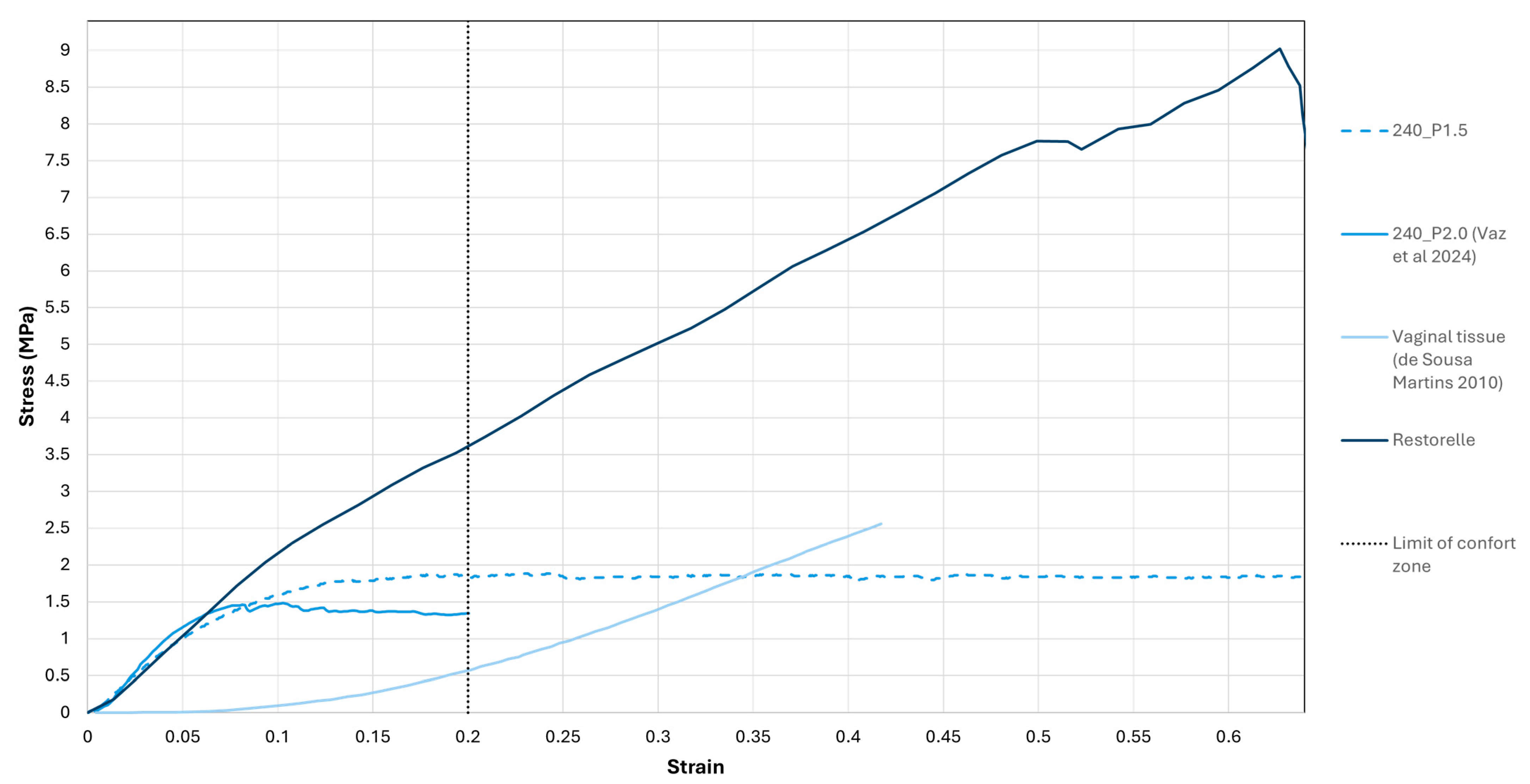
3.1. Uniaxial Tensile Tests

An average stress–strain curve was obtained from the force–displacement data attained from the uniaxial tensile tests and compared with the stress–strain curves of a square mesh with 2.0 mm pore size (240_P2.0), which was studied in previous works [33], a Restorele® PP mesh [33], and the stress–strain curve from vaginal human tissue [38], presented in Figure 2.
The meshes have a similar behavior; however, they reach different maximum stresses, both in and out of the comfort zone. These values are presented in Table 2.

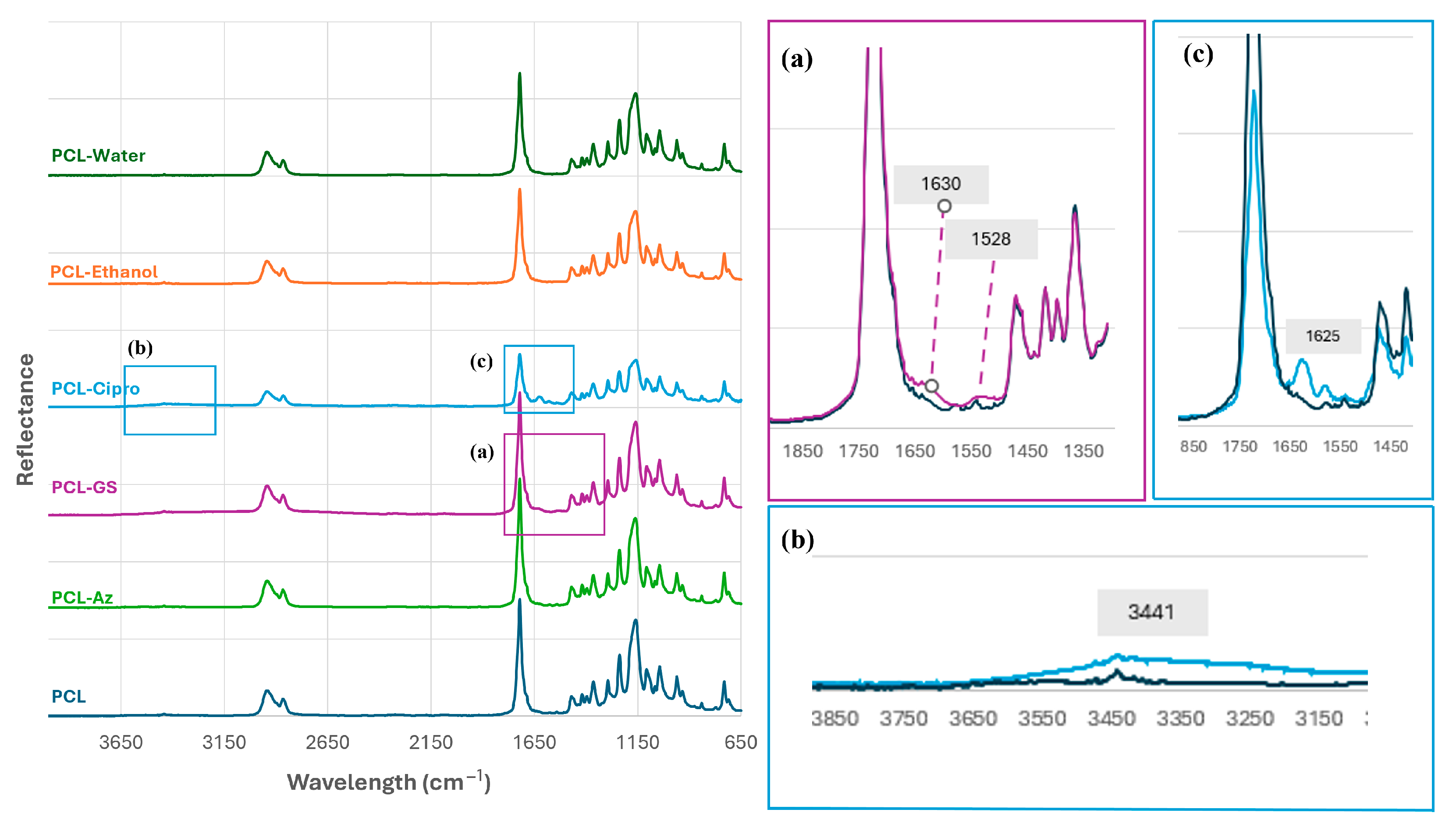
3.2. Preparation of PCL Mesh Implants Loaded with Antibiotic Agents

The FTIR spectra of the PCL–Az, PCL–GS, and PCL–Cipro meshes were obtained along with the spectra of PCL (control), PCL–pure ethanol, and PCL–distilled water, represented in Figure 3. Observing the PCL spectra, it was possible to see the characteristic peaks of this material: 2943 cm−1, 2866 cm−1, 1721 cm−1, and 1471–732 cm−1, related to the asymmetric CH2 stretching, the symmetric CH2 stretching, the C=O carbonyl stretching, and the bending, wagging, twisting, and stretching vibrations of the CH2 groups, C–O, and C–C bonds in the ester linkages of the polymer, respectively [39]. These peaks were consistently present in all samples, although sometimes with lower intensity.
For both samples incorporated with only the solvents, PCL–Ethanol and PCL–Water, as well as for the PCL–Az samples, the peaks of the FTIR spectra obtained coincided with the ones from the control. In the PCL–GS FTIR spectra, two characteristic peaks of GS were identified and are highlighted in Figure 3a: 1630 cm−1, related to the N–H I vibration, and 1528 cm−1, associated with the N–H II vibration. Regarding the PCL–Cipro spectra, 3441 cm−1 and 1625 cm−1 are the wavelengths at which the characteristic peaks of Cipro were found, related to the OH stretch and the C==O vibration and represented in Figure 3b,c, respectively.

3.3. In Vitro Microbiological Assays

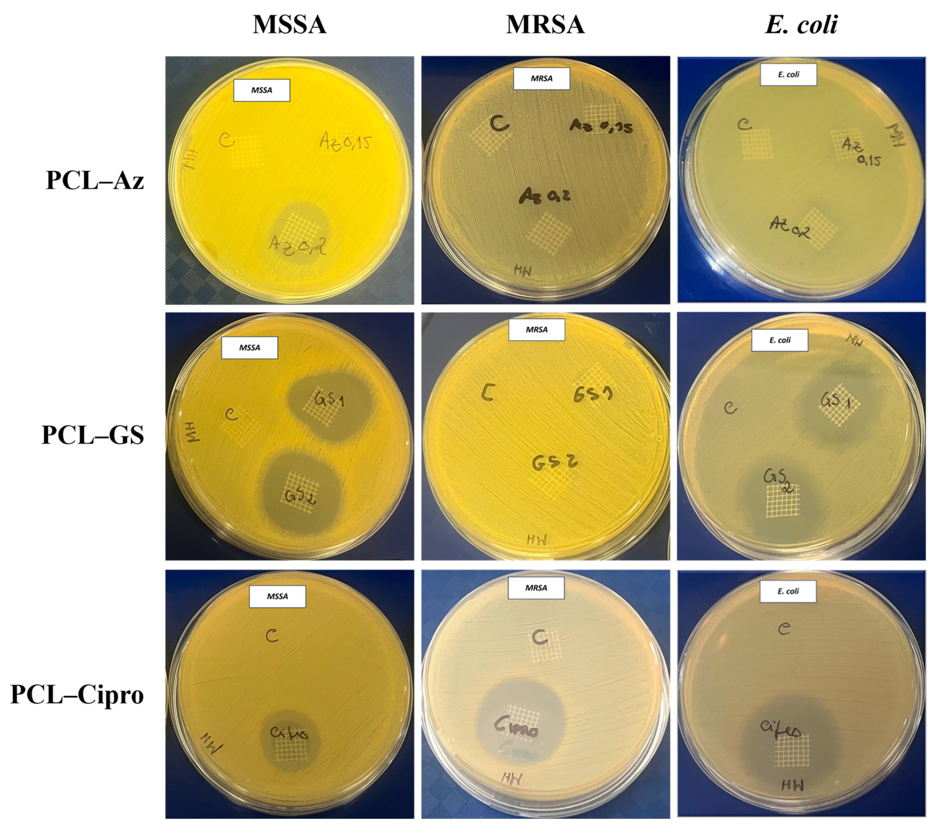
3.3.1. Zone Inhibition Assay

The diameter of inhibition was measured for each mesh coated with antibiotics and compared with the control, the PCL mesh, for each bacterial strain (Table 3) to evaluate the antimicrobial activity of the meshes. Figure 4 shows images of the obtained inhibition zone radius for each bacterial strain.
Regarding MSSA ATCC 29213, all the meshes with incorporated drugs presented inhibition zones, with PCL–Az meshes presenting a greater one (Figure 4). Concerning MRSA ATCC 43300, only ciprofloxacin appeared to have antimicrobial activity against this bacteria strain. Finally, ciprofloxacin and GS presented antimicrobial effects against E. coli, which was not verified in the PCL–Az samples (Figure 4).

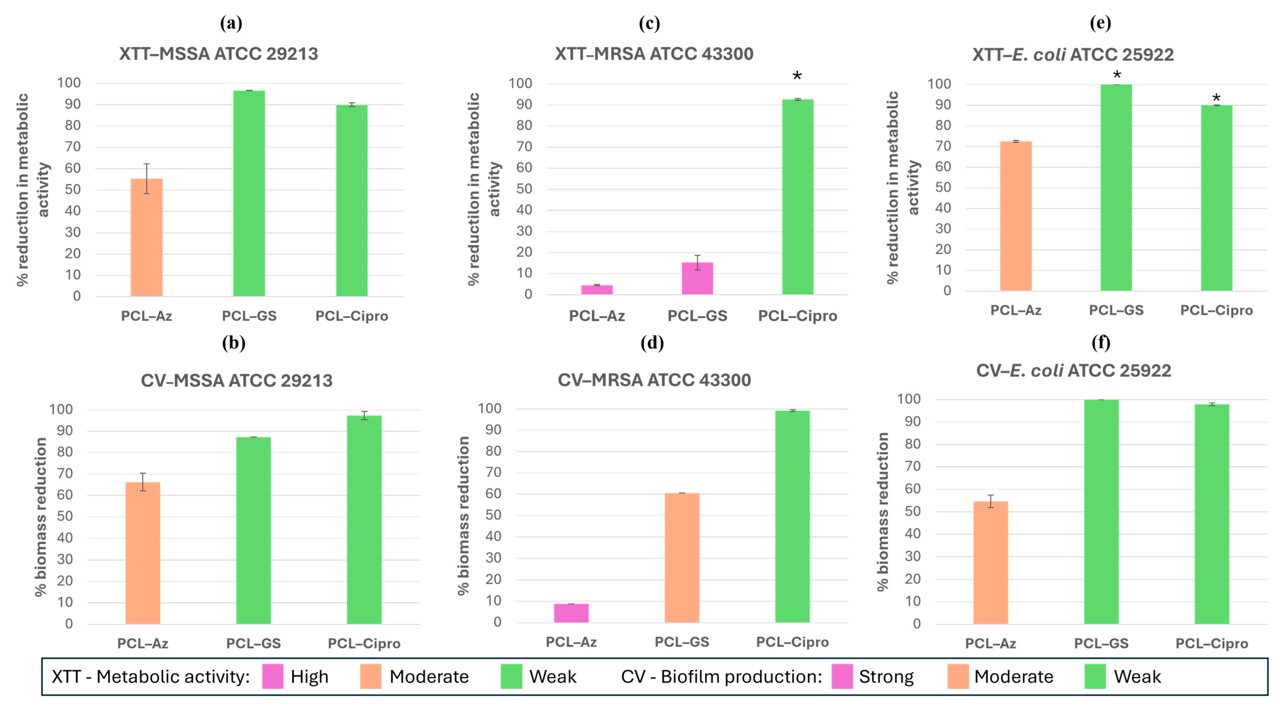
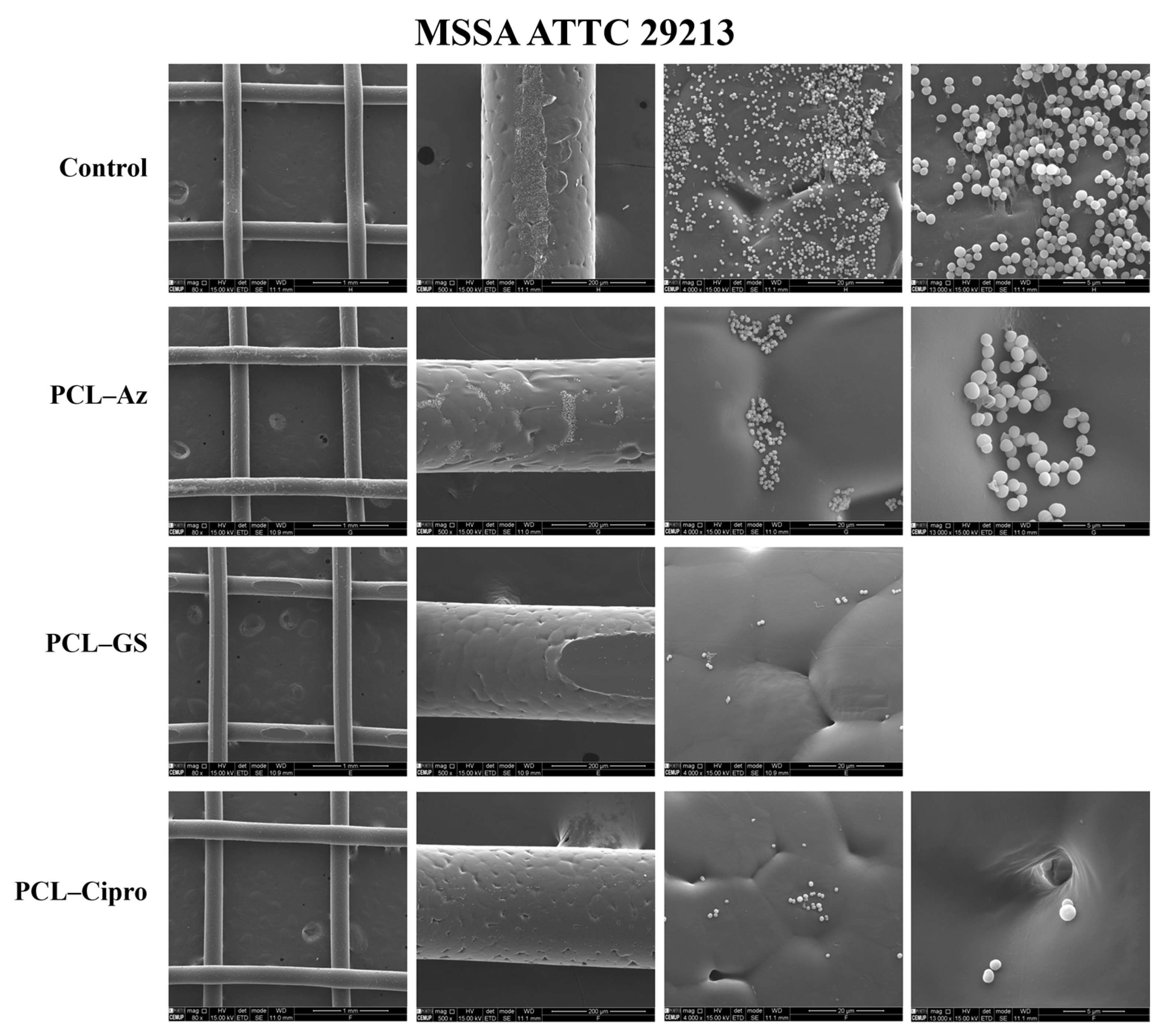
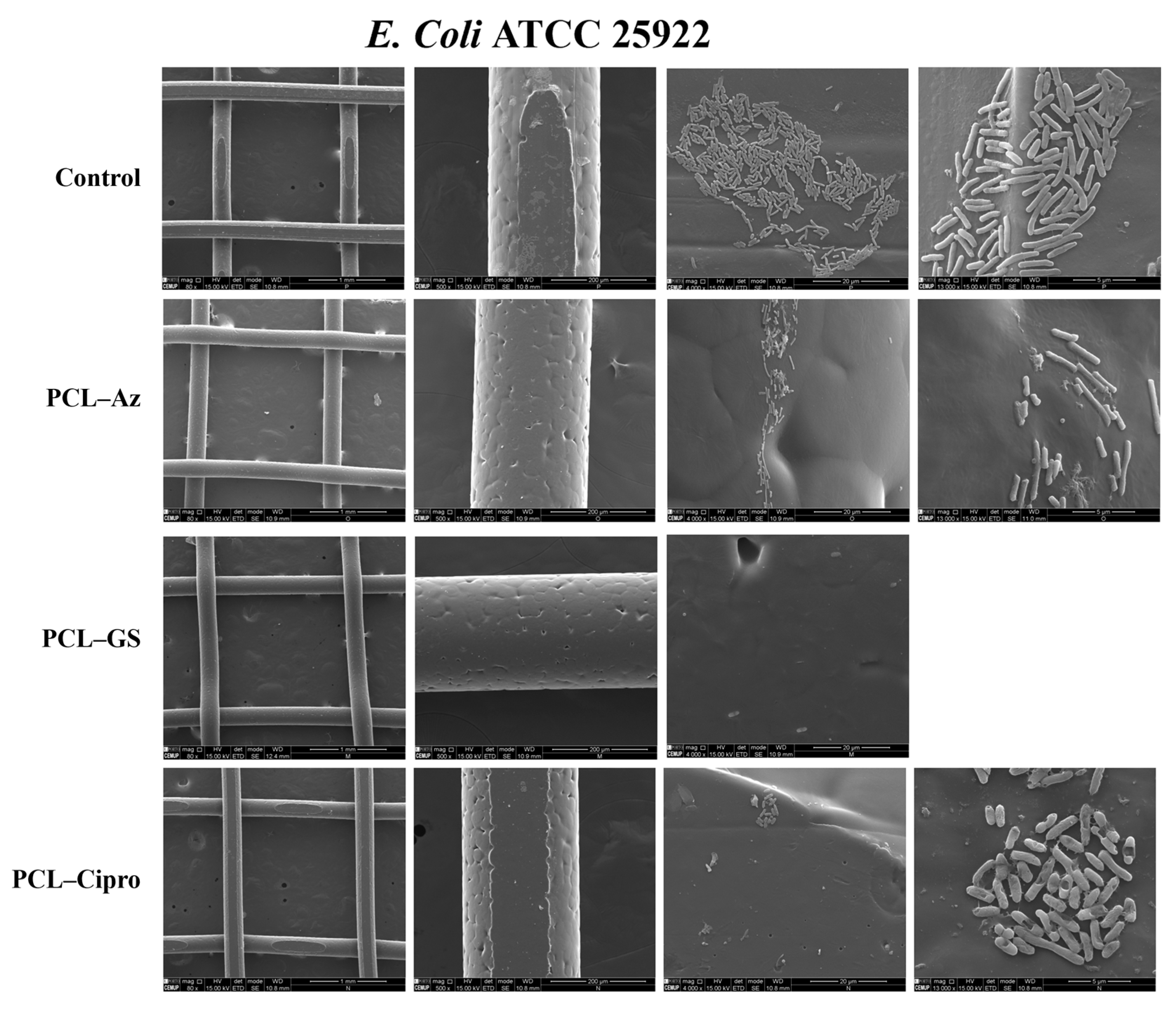
3.3.2. Biofilm Formation Assays

The results of the XTT and CV assays are represented in Figure 5. The graphs depict the percentage of reduction in the metabolic activity of the biofilm (XTT) and biofilm cell concentration (CV) for each antibiotic relative to the positive control. Statistically significant differences in XTT assay were found in the PCL–Cipro meshes with MRSA and all of the drug-loaded meshes with E. coli except for the PCL–Az meshes. However, no statistically significant differences were found in the CV assay. As seen in Figure 5a,b and following the classification by Lade et al. [40], ciprofloxacin and gentamicin presented a high reduction in MSSA biofilm’s metabolic activity and a high reduction in its biofilm biomass, while azithromycin showed a moderate reduction in MSSA biofilm’s metabolic activity and a moderate decrease in its concentration of biofilm cells.
The SEM images represented in Figure 6 show that the control has a high bacteria and biofilm presence, contrary to the meshes incorporated with antibiotics, which present a lower bacterial content, with particularly low bacterial presence in PCL–Cipro and PCL–GS meshes, in accordance with the XTT and CV results.
Regarding MRSA (Figure 5c,d), the PCL–Cipro samples show a high reduction in the biofilm’s metabolic activity and biofilm biomass. In contrast, the PCL–Az and PCL–GS meshes had a weak effect on the reduction in biofilm’s metabolic activity. The PCL–Az samples also presented a low reduction in biofilm biomass, while PCL–GS had a moderate effect. In the SEM images (Figure 7) it is possible to verify a high reduction in bacterial content in the PCL–Cipro meshes, but not in the PCL–Az and PCL–GS samples.
Finally, Figure 5e,f show that all of the antibiotic-incorporated meshes decreased E. coli biofilm’s metabolic activity as well as biofilm production. The SEM images (Figure 8) show that the PCL–GS sample has almost no bacterial presence, while collapsed bacteria can be seen in the PCL–Cipro mesh. For PCL–Az, there is a relatively low bacterial content.

3.3.3. Comparative Analysis of Antibacterial Selectiveness

Figure 9 enables a comparative assessment of each material’s selectiveness across the strains and assay types. PCL–Cipro clearly exhibits broad-spectrum antibacterial activity, with consistently high inhibition and biofilm resistance against all strains. PCL–Az showed higher effectiveness toward MSSA in the zone inhibition assays. PCL–GS, on the other hand, was especially effective against E. coli and MSSA but not MRSA.

4. Discussion

The performance of melt electrospun biodegradable meshes, with a 240 µm fiber diameter and a 1.5 mm pore size, is assessed through uniaxial tensile tests to ensure appropriate mesh stiffness. Comparing these meshes with previously studied meshes with a 2.0 mm pore size, it is suggested that, since they present similar behavior, the plastic behavior is dependent on the material and sample geometry, instead of its pore size. The results obtained suggest that meshes with a smaller pore size have a higher resistance to plastic deformation, with the 1.5 mm pore size meshes enduring 27% more stress than the 2.0 mm pore size meshes. These meshes are significantly less stiff than the Restorelle® ones, having a closer mechanical behavior to vaginal tissues. However, the maximum stress endured by the produced meshes is 70% higher than the vaginal tissue, which can still lead to mechanical mismatches, increasing the risk of tissue erosion. Further research is necessary to optimize the mechanical characteristics of the meshes, including new pore sizes and mesh geometries.
All of the tested meshes presented FTIR peaks characteristic of PCL, confirming its presence. In the meshes coated with antibiotics, a slight decrease in the PCL peak intensity can be seen, which suggests that, although PCL is still present, other substances are also present in the meshes, even if other chemical bonds or functional groups are not noticeable. Analyzing the FTIR spectroscopy results, it was not possible to detect azithromycin’s characteristic peaks. However, this does not necessarily mean that azithromycin is absent since FTIR spectroscopy is not always effective at identifying materials in mixtures [41]. According to the literature, the only expected difference between the FTIR spectrum of PCL and azithromycin-loaded PCL is a peak around 3500–3000 cm−1, which is characteristic of O–H and N–H stretching [42]. Although the peaks identified in the PCL–GS FTIR spectrum are small, the incorporation of this antibiotic was considered successful. In the samples containing ciprofloxacin, two signature peaks were identified besides PCL, confirming its presence in the meshes. It can be challenging to distinguish additional peaks from ciprofloxacin in the PCL spectrum when they overlap or fall within densely populated regions by PCL’s characteristic peaks, for example, the fluorine group C–F stretching, common in fluoroquinolones, which is typically found around the 1050–1000 region [43] and could not be detected. The presence of both gentamicin and ciprofloxacin in the meshes were confirmed, contrary to the presence of azithromycin. However, because the antimicrobial’s FTIR peaks may overlap with PCL’s spectrum, FTIR spectroscopy can be ineffective when identifying materials in mixtures [41], which is the reason why it was not concluded that the drug was not present. A further analysis of the meshes would be necessary to confirm the presence of all incorporated drugs, for example, high-performance liquid chromatography (HPLC) or differential scanning calorimetry (DSC) [44].
The XTT and CV assays complemented each other, providing the necessary information on biofilm formation. It is important to note that only a few samples proved to be statistically significant, which is likely due to the high sensitivity of spectrophotometry analysis to environmental temperature and luminosity [45].
Regarding MSSA, both XTT and CV assays’ results are in accordance with each other. The SEM images in Figure 6 clearly show a high biofilm reduction for all of the samples compared with the control, which is in accordance with the zone inhibition assay where all of the antibiotics showed a radius of inhibition, proving their antibacterial effect against this strain. Gentamicin proved to be effective in reducing biofilm formation and cell viability, confirming its antimicrobial activity against MSSA. Ciprofloxacin, like GS, proved to be highly effective in reducing the biofilm’s metabolic activity and biomass. Once again, this follows the SEM images and zone inhibition assay, proving ciprofloxacin’s antibacterial activity against MSSA. These results were expected since the tested strain is susceptible to both antibiotics [46]. Azithromycin was a less effective antibiotic for MSSA ATCC 29213. There was moderate biofilm production with high metabolic activity and a visible biofilm in the SEM images despite its antibacterial activity seen in the zone inhibition assay. This suggests that azithromycin is effective against bacteria in their planktonic state, but as they grow and aggregate, they develop a biofilm more tolerant to azithromycin [47]. MRSA is known as one of the most serious antibiotic-resistant bacteria, due to its capability of developing multidrug resistance [48]. The SEM images showed a high bacterial content reduction in MRSA ATCC 43300 for ciprofloxacin but not for gentamicin and azithromycin, which is in accordance with the results obtained from the zone inhibition assay, where only ciprofloxacin proved to have antibacterial activity. This strain had high biofilm production with high metabolic activity in the presence of azithromycin, proving its resistance to the antibiotic at the used concentration [48]. MRSA ATCC 43300 is also gentamicin-resistant [49], which was verified for the tested concentration. Although ciprofloxacin’s susceptibility to this bacteria strain is not well documented, in another study, it has been proven to reduce growth by 26% for a lower concentration of the antibiotic, proving the susceptibility of MRSA ATCC 43300 to ciprofloxacin [50]. For the concentration used in this study, ciprofloxacin has proven to be highly effective in reducing the biofilm’s metabolic activity and biomass, which is in accordance with the SEM images, where there is hardly any bacterial presence, and the zone inhibition assay, which proves ciprofloxacin’s antibacterial activity against MRSA ATCC 43300 at the used concentration. Finally, regarding E. coli ATCC 25922, the SEM images show a reduction in bacterial content with the antibiotics, which is in accordance with the zone inhibition assays, which reveal the antibiotics’ antimicrobial activity against this strain. The PCL–Az meshes showed moderate biofilm production and metabolic activity, and formed a small inhibition zone, suggesting that azithromycin has slight antibacterial activity against this strain. Both the PCL–GS and PCL–Cipro meshes highly reduced the biofilm’s biomass and metabolic activity and showed almost no bacterial presence in the SEM images, following the zone inhibition assay results and proving these drugs’ antibacterial activity against E. coli ATCC 25922, as expected [51,52]. Figure 9 gives a comparative analysis of the inhibition zone against the biofilm assays that further supports this analysis.
Overall, the results obtained prove that the antimicrobial incorporation in the PCL meshes was successful. PCL–Cipro was the most effective antibiotic, inhibiting biofilm formation in the three analyzed bacterial strains, as highlighted in Figure 9. Gentamicin was highly effective against E. coli ATCC 25922 and MSSA ATCC 29213. Considering the complications of meshes for POP repair, preventing biofilm formation is important to avoid the infection of these meshes and, therefore, the need to remove them. However, more studies should be performed to understand the antibiotics’ efficacy through mesh degradation. With that in mind, the degradation of the meshes in simulated body fluid (SBF) and in an enzymatic environment (lysozyme, to accelerate the degradation process), to simulate a physiological environment, will be carried out in the future [53,54]. Further work will also focus on the in vivo evaluation of the meshes incorporated with antimicrobial agents.

5. Conclusions

This study successfully demonstrates the fabrication of melt electrowritten polycaprolactone (PCL) meshes coated with broad-spectrum antibiotics, presenting both the mechanical and antibacterial properties suitable for pelvic organ prolapse (POP) repair. Among the tested formulations, PCL–Ciprofloxacin showed the most consistent antimicrobial effect, including a statistically significant reduction in the metabolic activity of MRSA and E. coli biofilms, as demonstrated by the XTT assay. The meshes with a smaller pore size (1.5 mm) exhibited a mechanical response closer to vaginal tissue than conventional meshes, which may reduce complications such as tissue erosion. These findings suggest that antibiotic-coated biodegradable meshes produced via MEW are a promising strategy to prevent mesh-associated infections and improve POP surgical outcomes. Future work will include degradation studies in simulated physiological environments and in vivo validation.

Author Contributions

Conceptualization, S.C.d.O., A.A.F., and E.T.d.S.; methodology, J.P.M., A.S.d.S., S.C.d.O., and E.T.d.S.; software, J.P.M. and A.S.d.S.; investigation, J.P.M., A.S.d.S., S.C.d.O., and E.T.d.S.; resources, S.C.d.O., A.A.F., and E.T.d.S.; writing—original draft preparation, J.P.M.; writing—review and editing, S.C.d.O., A.A.F., and E.T.d.S.; visualization, J.P.M.; supervision, S.C.d.O., A.A.F., and E.T.d.S.; funding acquisition, S.C.d.O., A.A.F., and E.T.d.S. All authors have read and agreed to the published version of the manuscript.

Funding

This work was funded by Stimulus of Scientific Employment 2021.00077.CEECIND and the project PRECOGFIL-PTDC/EMD-EMD/2229/2020, financed through FCT. This work was supported by FCT, through INEGI, under LAETA, project UIDB/50022/2020 and UIDP/50022/2020, LA/P/0079/2020 and CINTESIS, R&D Unit, project UIDP/4255/2020.

Data Availability Statement

The original contributions presented in this study are included in the article. Further inquiries can be directed to the corresponding author.

Acknowledgments

The authors would like to acknowledge and thank the Faculties of Engineering and Medicine of the University of Porto and INEGI for allowing the development of this work and the Materials Center of the University of Porto for their contribution.

Conflicts of Interest

The authors declare no conflicts of interest.

Abbreviations

The following abbreviations are used in this manuscript:
PFDPelvic Floor Dysfunctions
POPPelvic Organ Prolapse
SUIStress Urinary Incontinence
FDAFood and Drug Administration
UTIUrinary Tract Infection
PP Propylene
MRSAMethicillin-Resistant Staphylococcus Aureus
MSSAMethicillin-Susceptible Staphylococcus Aureus
AMAdditive Manufacture
ESElectrospinning
MEWMelt Electrowriting
CiproCiprofloxacin
GSGentamicin Sulfate
AzAzithromycin
FTIRFourier Transform Infrared Spectroscopy
ATRAttenuated Total Reflectance
SEMScanning Electron Microscopy
XTTTetrazolium Salt
CVCrystal Violet
PBSPhosphate-Buffered Saline
ODOptical Density
HPLCHigh-Performance Liquid Chromatography
DSCDifferential Scanning Calorimetry

References

  1. Iglesia, C.B.; Smithling, K.R. Pelvic Organ Prolapse. Am. Fam. Physician 2017, 96, 179–185. [Google Scholar] [PubMed]
  2. Smith, T.A.; Poteat, T.A.; Shobeiri, S.A. Pelvic organ prolapse. J. Am. Acad. Physician Assist. 2014, 27, 20–24. [Google Scholar] [CrossRef] [PubMed]
  3. Weber, A.M.; Richter, H.E. Pelvic Organ Prolapse. Obstet. Gynecol. 2005, 106, 615–634. [Google Scholar] [CrossRef] [PubMed]
  4. Jelovsek, J.E.; Maher, C.; Barber, M.D. Pelvic organ prolapse. Lancet 2007, 369, 1027–1038. [Google Scholar] [CrossRef] [PubMed]
  5. Ko, K.J.; Lee, K.-S. Current surgical management of pelvic organ prolapse: Strategies for the improvement of surgical outcomes. Investig. Clin. Urol. 2019, 60, 413. [Google Scholar] [CrossRef] [PubMed]
  6. Dällenbach, P. To mesh or not to mesh: A review of pelvic organ reconstructive surgery. Int. J. Womens Health 2015, 7, 331–343. [Google Scholar] [CrossRef] [PubMed]
  7. Falagas, M.E.; Velakoulis, S.; Iavazzo, C.; Athanasiou, S. Mesh-related infections after pelvic organ prolapse repair surgery. Eur. J. Obstet. Gynecol. Reprod. Biol. 2007, 134, 147–156. [Google Scholar] [CrossRef] [PubMed]
  8. Maher, C.; Feiner, B.; Baessler, K.; Schmid, C. Surgical management of pelvic organ prolapse in women. In Cochrane Database of Systematic Reviews; Maher, C., Ed.; John Wiley & Sons, Ltd.: Chichester, UK, 2013. [Google Scholar] [CrossRef]
  9. Sand, P.K.; Koduri, S.; Lobel, R.W.; Winkler, H.A.; Tomezsko, J.; Culligan, P.J.; Goldberg, R. Prospective randomized trial of polyglactin 910 mesh to prevent recurrence of cystoceles and rectoceles. Am. J. Obstet. Gynecol. 2001, 184, 1357–1364. [Google Scholar] [CrossRef] [PubMed]
  10. Yuan, M.; Hu, M.; Dai, F.; Fan, Y.; Deng, Z.; Deng, H.; Cheng, Y. Application of synthetic and natural polymers in surgical mesh for pelvic floor reconstruction. Mater. Des. 2021, 209, 109984. [Google Scholar] [CrossRef]
  11. Mancus1o, E.; Downey, C.; Doxford-Hook, E.; Bryant, M.G.; Culmer, P. The use of polymeric meshes for pelvic organ prolapse: Current concepts, challenges, and future perspectives. J. Biomed. Mater. Res. B Appl. Biomater. 2020, 108, 771–789. [Google Scholar] [CrossRef] [PubMed]
  12. Baylón, K.; Rodríguez-Camarillo, P.; Elías-Zúñiga, A.; Díaz-Elizondo, J.A.; Gilkerson, R.; Lozano, K. Past, Present and Future of Surgical Meshes: A Review. Membranes 2017, 7, 47. [Google Scholar] [CrossRef] [PubMed]
  13. Food and Drug Administration. Urogynecologic Surgical Mesh Implants n.d. Available online: https://www.fda.gov/medical-devices/implants-and-prosthetics/urogynecologic-surgical-mesh-implants (accessed on 12 March 2025).
  14. Rynkevic, R.; Silva, M.E.T.; Martins, P.; Mascarenhas, T.; Alves, J.L.; Fernandes, A.A. Characterisation of polycaprolactone scaffolds made by melt electrospinning writing for pelvic organ prolapse correction—A pilot study. Mater. Today Commun. 2022, 32, 104101. [Google Scholar] [CrossRef]
  15. Mangir, N.; Roman, S.; Chapple, C.R.; MacNeil, S. Complications related to use of mesh implants in surgical treatment of stress urinary incontinence and pelvic organ prolapse: Infection or inflammation? World J. Urol. 2020, 38, 73–80. [Google Scholar] [CrossRef] [PubMed]
  16. Mellano, E.M.; Nakamura, L.Y.; Choi, J.M.; Kang, D.C.; Grisales, T.; Raz, S.; Rodriguez, L.V. The Role of Chronic Mesh Infection in Delayed-Onset Vaginal Mesh Complications or Recurrent Urinary Tract Infections. Female Pelvic. Med. Reconstr. Surg. 2016, 22, 166–171. [Google Scholar] [CrossRef] [PubMed]
  17. Boulanger, L.; Boukerrou, M.; Rubod, C.; Collinet, P.; Fruchard, A.; Courcol, R.J.; Cosson, M. Bacteriological analysis of meshes removed for complications after surgical management of urinary incontinence or pelvic organ prolapse. Int. Urogynecol. J. 2008, 19, 827–831. [Google Scholar] [CrossRef] [PubMed]
  18. Feng, C.H.; Stevenson, D.D.; Kahn, B.; Higginbottom, P.A. Methicillin-resistant Staphylococcus aureus pelvic abscesses in a female after gynecologic pelvic surgery. Am. J. Obstet. Gynecol. 2011, 205, e4–e5. [Google Scholar] [CrossRef] [PubMed]
  19. Shah, H.N.; Badlani, G.H. Mesh complications in female pelvic floor reconstructive surgery and their management: A systematic review. Indian J. Urol. 2012, 28, 129–153. [Google Scholar] [CrossRef] [PubMed]
  20. Mardina, Z.; Venezuela, J.; Maher, C.; Shi, Z.; Dargusch, M.S.; Atrens, A. Design, mechanical and degradation requirements of biodegradable metal mesh for pelvic floor reconstruction. Biomater. Sci. 2022, 10, 3371–3392. [Google Scholar] [CrossRef] [PubMed]
  21. Kalelkar, P.P.; Riddick, M.; García, A.J. Biomaterial-based delivery of antimicrobial therapies for the treatment of bacterial infections. Nat. Rev. Mater. 2022, 7, 39–54. [Google Scholar] [CrossRef] [PubMed]
  22. Pfizer Labs. Zithromax® (Azithromycin) for Injection, for Intravenoususe. US Food and Drug Administration, 2022 n.d. Available online: https://www.accessdata.fda.gov/drugsatfda_docs/label/2022/050733Orig1s049lbl.pdf (accessed on 12 March 2025).
  23. Ward, K.; Theiler, R.N. Once-daily Dosing of Gentamicin in Obstetrics and Gynecology. Clin. Obstet. Gynecol. 2008, 51, 498–506. [Google Scholar] [CrossRef] [PubMed]
  24. Christian, J.S. The quinolone antibiotics. Prim. Care Update Ob. Gyns. 1996, 3, 87–92. [Google Scholar] [CrossRef]
  25. Castro, F.J.C.; Yuste, Y.; Pereira, S.; Garvín, M.D.; García, M.Á.L.; Padillo, F.J.; Portilla, F. Proof of concept, design, and manufacture via 3-D printing of a mesh with bactericidal capacity: Behaviour in vitro and in vivo. J. Tissue Eng. Regen. Med. 2019, 13, 1955–1964. [Google Scholar] [CrossRef] [PubMed]
  26. Qamar, N.; Abbas, N.; Irfan, M.; Hussain, A.; Arshad, M.S.; Latif, S.; Mehmood, F.; Ghori, M.U. Personalized 3D printed ciprofloxacin impregnated meshes for the management of hernia. J. Drug Deliv. Sci. Technol. 2019, 53, 101164. [Google Scholar] [CrossRef]
  27. Ren, J.; Murray, R.; Wong, C.S.; Qin, J.; Chen, M.; Totsika, M.; Riddell, A.D.; Warwick, A.; Rukin, N.; Woodruff, M.A. Development of 3D Printed Biodegradable Mesh with Antimicrobial Properties for Pelvic Organ Prolapse. Polymers 2022, 14, 763. [Google Scholar] [CrossRef] [PubMed]
  28. Xue, J.; Wu, T.; Dai, Y.; Xia, Y. Electrospinning and Electrospun Nanofibers: Methods, Materials, and Applications. Chem. Rev. 2019, 119, 5298–5415. [Google Scholar] [CrossRef] [PubMed]
  29. Garg, K.; Bowlin, G.L. Electrospinning jets and nanofibrous structures. Biomicrofluidics 2011, 5, 013403. [Google Scholar] [CrossRef] [PubMed]
  30. Dalton, P.D. Melt electrowriting with additive manufacturing principles. Curr. Opin. Biomed. Eng. 2017, 2, 49–57. [Google Scholar] [CrossRef]
  31. Ferreira, N.M.; Antoniadi, E.; Silva, A.T.; Silva, A.; Parente, M.; Fernandes, A.; Silva, E. Melt Electrowritten Biodegradable Mesh Implants with Auxetic Designs for Pelvic Organ Prolapse Repair. J. Manuf. Mater. Process. 2025, 9, 111. [Google Scholar] [CrossRef]
  32. Sterk, S.; Silva, M.E.T.; Fernandes, A.A.; Huß, A.; Wittek, A. Development of new surgical mesh geometries with different mechanical properties using the design freedom of 3D printing. J. Appl. Polym. Sci. 2023, 140, e54687. [Google Scholar] [CrossRef]
  33. Vaz, M.F.; Martins, J.A.P.; Pinheiro, F.; Ferreira, N.M.; Brandão, S.; Alves, J.L.; Fernandes, A.A.; Parente, M.P.L.; Silva, M.E. Optimizing melt electrowriting prototypes for printing non-medical and medical grade polycaprolactone meshes in prolapse repair. J. Appl. Polym. Sci. 2024, 142, e56408. [Google Scholar] [CrossRef]
  34. Vaz, M.F.R.; Martins, J.A.P.; Pinheiro, F.; Ferreira, N.M.; Brandão, S.; Alves, J.L.; Fernandes, A.A.; Parente, M.P.L.; Silva, M.E.T. Medical- and Non-Medical-Grade Polycaprolactone Mesh Printing for Prolapse Repair: Establishment of Melt Electrowriting Prototype Parameters. Appl. Sci. 2024, 14, 9670. [Google Scholar] [CrossRef]
  35. Martins, J.P.; da Silva, E.T.; Fernandes, A.A.; Costa de Oliveira, S. Three-Dimensional Melted Electrowriting Drug Coating Fibers for the Prevention of Device-Associated Infections: A Pilot Study. Bioengineering 2024, 11, 636. [Google Scholar] [CrossRef] [PubMed]
  36. ISO 1183-1:2025; Plastics—Methods for determining the density of non-cellular plastics—Part 1: Immersion method, liquid pycnometer method and titration method. International Organization for Standardization: Geneva, Switzerland, 2025.
  37. Scaffaro, R.; Botta, L.; Maio, A.; Gallo, G. Incorporation of an Antibiotic in Poly(Lactic Acid) and Polypropylene by Melt Processing. J. Appl. Biomater. Funct. Mater. 2016, 14, e240–e247. [Google Scholar] [CrossRef] [PubMed]
  38. de Sousa Martins, P.A.L. Experimental and Numerical Studies of Soft Biological Tissues. Ph.D. Thesis, Faculty of Engineering of the University of Porto, Porto, Portugal, 2010. [Google Scholar]
  39. Elzein, T.; Nasser-Eddine, M.; Delaite, C.; Bistac, S.; Dumas, P. FTIR study of polycaprolactone chain organization at interfaces. J. Colloid. Interface. Sci. 2004, 273, 381–387. [Google Scholar] [CrossRef] [PubMed]
  40. Lade, H.; Park, J.H.; Chung, S.H.; Kim, I.H.; Kim, J.-M.; Joo, H.-S.; Kim, J.-S. Biofilm Formation by Staphylococcus aureus Clinical Isolates is Differentially Affected by Glucose and Sodium Chloride Supplemented Culture Media. J. Clin. Med. 2019, 8, 1853. [Google Scholar] [CrossRef] [PubMed]
  41. Advantages and Disadvantages of FTIR Spectroscopy n.d. Available online: https://www.chemlabgenius.com/advantages-and-disadvantages-of-ftir-spectroscopy/ (accessed on 15 May 2024).
  42. Bhadra, S. Formulation & process development of azithromycin ophthalmic nanosuspension. Int. J. Pharm. Pharm. Sci. 2013, 5, 490–497. [Google Scholar]
  43. Sahoo, S.; Chakraborti, C.; Mishra, S.; Nanda, U.; Naik, S. FTIR and XRD investigations of some fluoroquinolones. Int. J. Pharm. Pharm. Sci. 2011, 3, 165–170. [Google Scholar]
  44. Yassin, A.E.; Albekairy, A.M.; Omer, M.; Almutairi, A.; Alotaibi, Y.; Althuwaini, S.; Alaql, O.A.; Almozaai, S.S.; Almutiri, N.M.; Alluhaim, W.; et al. Chitosan-Coated Azithromycin/Ciprofloxacin-Loaded Polycaprolactone Nanoparticles: A Characterization and Potency Study. Nanotechnol. Sci. Appl. 2023, 16, 59–72. [Google Scholar] [CrossRef] [PubMed]
  45. Haney, E.; Trimble, M.; Cheng, J.; Vallé, Q.; Hancock, R. Critical Assessment of Methods to Quantify Biofilm Growth and Evaluate Antibiofilm Activity of Host Defence Peptides. Biomolecules 2018, 8, 29. [Google Scholar] [CrossRef] [PubMed]
  46. Nair, S.; Desai, S.; Poonacha, N.; Vipra, A.; Sharma, U. Antibiofilm Activity and Synergistic Inhibition of Staphylococcus aureus Biofilms by Bactericidal Protein P128 in Combination with Antibiotics. Antimicrob. Agents Chemother. 2016, 60, 7280–7289. [Google Scholar] [CrossRef] [PubMed]
  47. Høiby, N.; Henneberg, K.; Wang, H.; Stavnsbjerg, C.; Bjarnsholt, T.; Ciofu, O.; Johansen, U.R.; Sams, T. Formation of Pseudomonas aeruginosa inhibition zone during tobramycin disk diffusion is due to transition from planktonic to biofilm mode of growth. Int. J. Antimicrob. Agents 2019, 53, 564–573. [Google Scholar] [CrossRef] [PubMed]
  48. Bhattacharya, S.; Bir, R.; Majumdar, T. Evaluation of Multidrug Resistant Staphylococcus aureus and their Association with Biofilm Production in a Tertiary Care Hospital, Tripura, Northeast India. J. Clin. Diagn. Res. 2015, 9, DC01-4. [Google Scholar] [CrossRef] [PubMed]
  49. Oduor, J.M.O.; Onkoba, N.; Maloba, F.; Arodi, W.O.; Nyachieo, A. Efficacy of lytic Staphylococcus aureus bacteriophage against multidrug-resistant Staphylococcus aureus in mice. J. Infect. Dev. Ctries. 2016, 10, 1208–1213. [Google Scholar] [CrossRef] [PubMed]
  50. Zawadzka, K.; Nowak, M.; Piwoński, I.; Lisowska, K. The Synergy of Ciprofloxacin and Carvedilol against Staphylococcus aureus–Prospects of a New Treatment Strategy? Molecules 2019, 24, 4104. [Google Scholar] [CrossRef] [PubMed]
  51. Arapović, J.; Kompes, G.; Dedić, K.; Teskeredžić, S.; Ostojić, M.; Travar, M.; Tihić, N.; Delić, J.; Skočibušić, S.; Zekiri-Sivro, M.; et al. Antimicrobial resistance profiles of human Brucella melitensis isolates in three different microdilution broths: The first multicentre study in Bosnia and Herzegovina. J. Glob. Antimicrob. Resist. 2022, 29, 99–104. [Google Scholar] [CrossRef] [PubMed]
  52. Mursyida, E.; Misfa, O.; Pratiwi, S.B.; Shinde, A. In Vitro Sensitivity Test of Escherichia coli ATCC 25922 to Various Antibiotics with Well Diffusion Method. J. Kesehat. Prima 2023, 17, 42. [Google Scholar] [CrossRef]
  53. Domingos, M.; Chiellini, F.; Cometa, S.; De Giglio, E.; Grillo-Fernandes, E.; Bártolo, P.; Chiellini, E. Evaluation of in vitro degradation of PCL scaffolds fabricated via BioExtrusion. Part 1: Influence of the degradation environment. Virtual Phys. Prototyp. 2010, 5, 65–73. [Google Scholar] [CrossRef]
  54. Banerjee, A.; Chatterjee, K.; Madras, G. Enzymatic degradation of polycaprolactone–gelatin blend. Mater. Res. Express 2015, 2, 045303. [Google Scholar] [CrossRef]
Figure 1. (a) MEW prototype; (b) square mesh design.
Macromol 05 00033 g001
Figure 2. Stress–strain curves obtained through uniaxial testing for 240 µm meshes with 1.5 mm and 2.0 mm pore sizes [33], for vaginal human tissue [38] and for the Restorelle® mesh [33], as well as a representation of the comfort zone.
Macromol 05 00033 g002
Figure 3. FTIR spectra of PCL, PCL–Az, PCL–GS, PCL–Cipro, PCL–Ethanol, and PCL–Water meshes’ samples; (a) GS characteristic peaks in PCL–GS meshes compared with PCL meshes; (b,c) Cipro characteristic peaks in PCL–Cipro meshes compared with PCL meshes.
Macromol 05 00033 g003
Figure 4. Inhibition zones of PCL–Az, PCL–Cipro, and PCL–GS meshes against MSSA, MRSA, and E. coli.
Macromol 05 00033 g004
Figure 5. XTT and CV assay for biofilm formation evaluation of MSSA ATCC 29213 (a,b), respectively; of MRSA ATCC 43300 (c,d), respectively; and of E. coli ATCC 25922 (e,f), respectively.
Macromol 05 00033 g005
Figure 6. SEM images of MSSA ATCC 29213 antibiotic-loaded mesh implants.
Macromol 05 00033 g006
Figure 7. SEM images of MRSA ATCC 43300 antibiotic-loaded mesh implants.
Macromol 05 00033 g007
Figure 8. SEM images of E. coli ATCC 25922 antibiotics loaded mesh implants.
Macromol 05 00033 g008
Figure 9. Comparative selectiveness profile of antibiotic-coated PCL meshes (PCL–Az, PCL–Cipro, and PCL–GS) against S. aureus (MSSA and MRSA) and E. coli, based on normalized percentages of zone of inhibition, biofilm metabolic activity reduction (XTT), and biomass reduction (CV).
Macromol 05 00033 g009
Table 1. Process parameters used in the MEW prototype for mesh printing.
ParameterValue
Voltage3.23 kV
Temperature195 °C
Height3 mm
Speed825 mm/min
Table 2. Maximum stress values in the comfort zone derived from the stress–strain curves for the printed meshes and previously studied meshes [34], for vaginal tissue [38] and the Restorelle® mesh [34].
VariablesPore Size (mm)σmax comfort zone (MPa)
240 µm diameter meshes1.51.84
2.01.35
Restorelle®2.03.62
Vaginal Tissue_________0.56
Table 3. Diameter of the inhibition zones of control (PCL), PCL–Az, PCL–Cipro, and PCL–GS meshes.
MSSA
ATCC 29213
MRSA
ATCC 43300
E. coli
ATCC 25922
Control0 mm0 mm0 mm
PCL–Az35 mm0 mm0 mm
PCL–Cipro26 mm29 mm39 mm
PCL–GS31 mm0 mm31 mm
Disclaimer/Publisher’s Note: The statements, opinions and data contained in all publications are solely those of the individual author(s) and contributor(s) and not of MDPI and/or the editor(s). MDPI and/or the editor(s) disclaim responsibility for any injury to people or property resulting from any ideas, methods, instructions or products referred to in the content.

