
Eosinophil-Count-Derived Inflammatory Markers and Psoriasis Severity: Exploring the Link
 , and
, and 
Abstract
1. Introduction
2. Materials and Methods
2.1. Study Population
2.2. Data Collection
2.3. Biomarkers
2.4. Study Outcome
2.5. Statistical Analysis
3. Results
3.1. Study Population Clinical Profile
3.2. Eosinophil-Derived Markers and Disease Severity
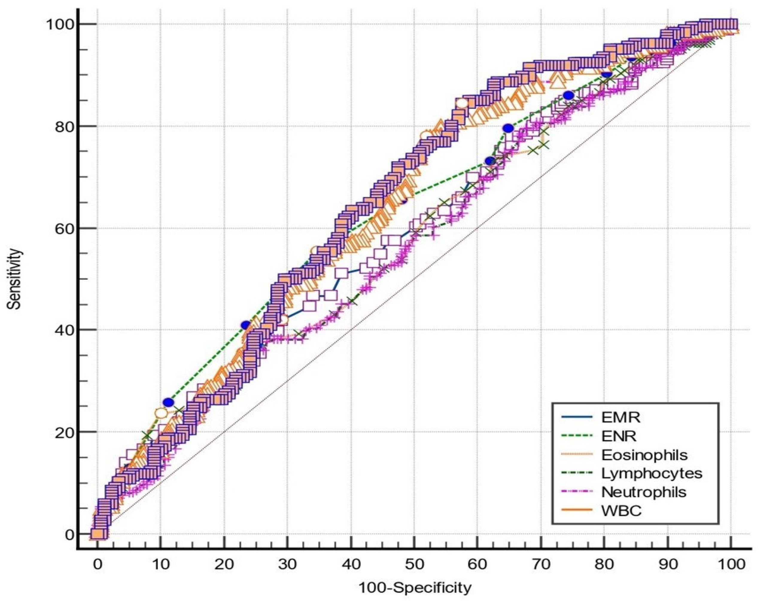
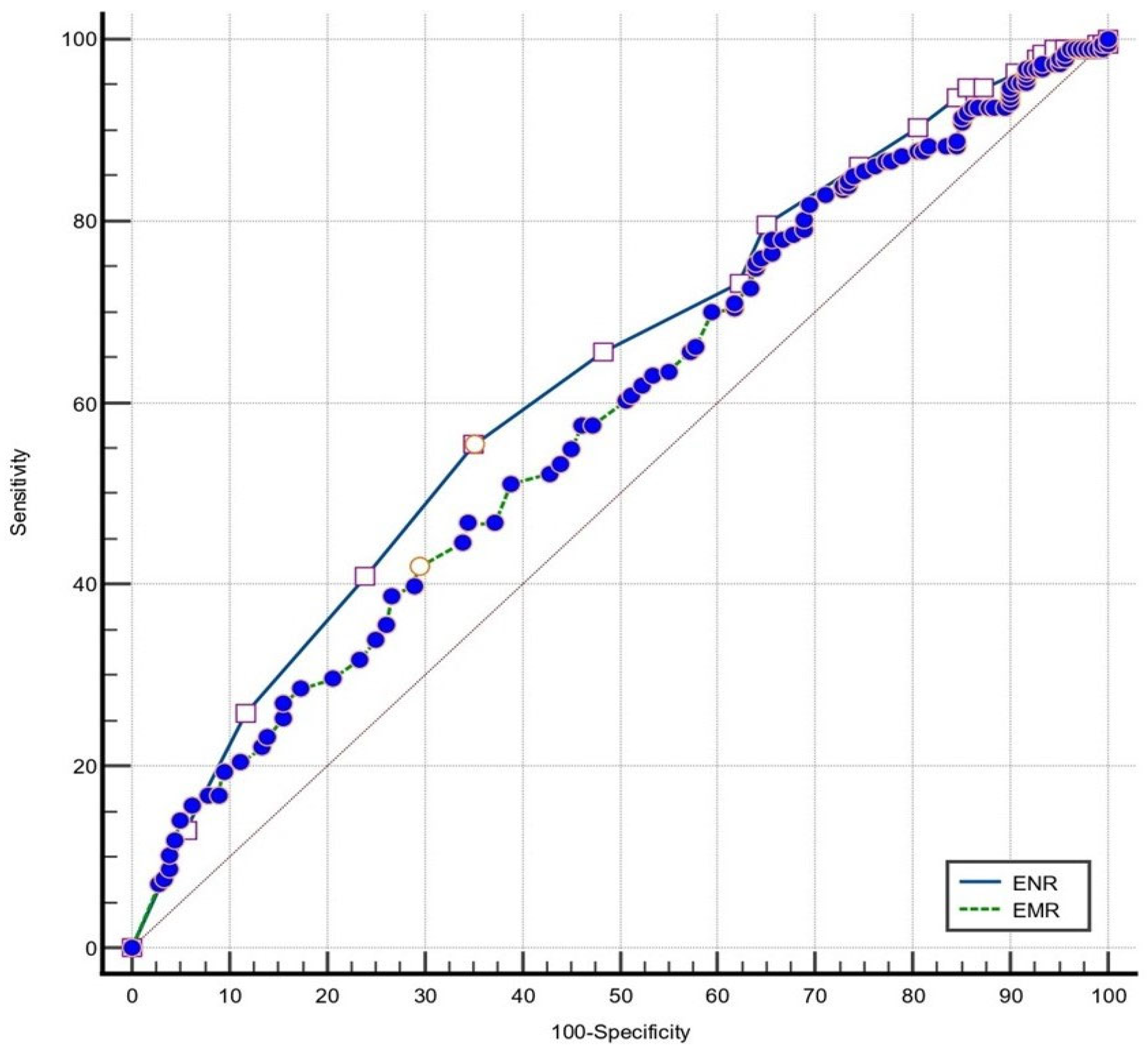
3.3. Eosinophil-Derived Markers Performance for Disease Severity Evaluation
4. Discussion
5. Conclusions
Author Contributions
Funding
Institutional Review Board Statement
Informed Consent Statement
Data Availability Statement
Acknowledgments
Conflicts of Interest
References
- Singh, S.; Young, P.; Armstrong, A.W. Relationship between psoriasis and metabolic syndrome: A systematic review. G. Ital. Dermatol. Venereol. 2016, 151, 663–677. [Google Scholar] [PubMed]
- Luan, L.; Han, S.; Wang, H.; Liu, X. Down-regulation of the Th1, Th17, and Th22 pathways due to anti-TNF-α treatment in psoriasis. Int. Immunopharmacol. 2015, 29, 278–284. [Google Scholar] [CrossRef] [PubMed]
- Canavese, M.; Altruda, F.; Ruzicka, T.; Schauber, J. Vascular endothelial growth factor (VEGF) in the pathogenesis of psoriasis—A possible target for novel therapies? J. Dermatol. Sci. 2010, 58, 171–176. [Google Scholar] [CrossRef] [PubMed]
- Frieder, J.; Ryan, C. Psoriasis and cardiovascular disorders. G. Ital. Dermatol. Venereol. 2016, 151, 678–693. [Google Scholar] [PubMed]
- Grozdev, I.; Korman, N.; Tsankov, N. Psoriasis as a systemic disease. Clin. Dermatol. 2014, 32, 343–350. [Google Scholar] [CrossRef] [PubMed]
- Larmann, J.; Handke, J.; Scholz, A.S.; Dehne, S.; Arens, C.; Gillmann, H.J.; Uhle, F.; Motsch, J.; Weigand, M.A.; Janssen, H. Preoperative neutrophil to lymphocyte ratio and platelet to lymphocyte ratio are associated with major adverse cardiovascular and cerebrovascular events in coronary heart disease patients undergoing non-cardiac surgery. BMC Cardiovasc. Disord. 2020, 20, 230. [Google Scholar] [CrossRef] [PubMed]
- Modica, R.; Minotta, R.; Liccardi, A.; Cannavale, G.; Benevento, E.; Colao, A. Evaluation of Neutrophil-to-Lymphocyte Ratio (NLR), Platelet-to-Lymphocyte Ratio (PLR) and Systemic Immune–Inflammation Index (SII) as Potential Biomarkers in Patients with Sporadic Medullary Thyroid Cancer (MTC). JPM 2023, 13, 953. [Google Scholar] [CrossRef] [PubMed]
- Lin, Z.-Q.; Ma, C.; Cao, W.-Z.; Ning, Z.; Tan, G. Prognostic Significance of NLR, PLR, LMR and Tumor Infiltrating T Lymphocytes in Patients Undergoing Surgical Resection for Hilar Cholangiocarcinoma. Front. Oncol. 2022, 12, 908907. [Google Scholar] [CrossRef]
- Gambardella, C.; Mongardini, F.M.; Paolicelli, M.; Bentivoglio, D.; Cozzolino, G.; Ruggiero, R.; Pizza, A.; Tolone, S.; del Genio, G.; Parisi, S.; et al. Role of Inflammatory Biomarkers (NLR, LMR, PLR) in the Prognostication of Malignancy in Indeterminate Thyroid Nodules. Int. J. Mol. Sci. 2023, 24, 6466. [Google Scholar] [CrossRef]
- Erre, G.L.; Paliogiannis, P.; Castagna, F.; Mangoni, A.A.; Carru, C.; Passiu, G.; Zinellu, A. Meta-analysis of neutrophil-to-lymphocyte and platelet-to-lymphocyte ratio in rheumatoid arthritis. Eur. J. Clin. Investig. 2019, 49, e13037. [Google Scholar] [CrossRef]
- Wu, J.; Yan, L.; Chai, K. Systemic immune-inflammation index is associated with disease activity in patients with ankylosing spondylitis. Clin. Lab. Anal. 2021, 35, e23964. [Google Scholar] [CrossRef] [PubMed]
- Kim, Y.; Choi, H.; Jung, S.M.; Song, J.J.; Park, Y.; Lee, S. Systemic immune-inflammation index could estimate the cross-sectional high activity and the poor outcomes in immunosuppressive drug-naïve patients with antineutrophil cytoplasmic antibody-associated vasculitis. Nephrology 2019, 24, 711–717. [Google Scholar] [CrossRef] [PubMed]
- Uslu, A.U.; Küçük, A.; Şahin, A.; Ugan, Y.; Yılmaz, R.; Güngör, T.; Bağcacı, S.; Küçükşen, S. Two new inflammatory markers associated with Disease Activity Score-28 in patients with rheumatoid arthritis: Neutrophil-lymphocyte ratio and platelet-lymphocyte ratio. Int. J. Rheum. Dis. 2015, 18, 731–735. [Google Scholar] [CrossRef] [PubMed]
- Juhlin, L.; Venge, P. Eosinophilic cationic protein (ECP) in skin disorders. Acta Derm. Venereol. 1991, 71, 495–501. [Google Scholar] [CrossRef] [PubMed]
- Skrzeczynska-Moncznik, J.; Wlodarczyk, A.; Zabieglo, K.; Kapinska-Mrowiecka, M.; Marewicz, E.; Dubin, A.; Potempa, J.; Cichy, J. Secretory Leukocyte Proteinase Inhibitor-Competent DNA Deposits Are Potent Stimulators of Plasmacytoid Dendritic Cells: Implication for Psoriasis. J. Immunol. 2012, 189, 1611–1617. [Google Scholar] [CrossRef] [PubMed]
- Kim, H.J.; Roh, J.Y.; Jung, Y. Eosinophils Accelerate Pathogenesis of Psoriasis by Supporting an Inflammatory Milieu that Promotes Neutrophil Infiltration. J. Investig. Dermatol. 2018, 138, 2185–2194. [Google Scholar] [CrossRef] [PubMed]
- Yenigun, A.; Sezen, S.; Calim, O.F.; Ozturan, O. Evaluation of the Eosinophil-to-lymphocyte Ratio in Pediatric Patients with Allergic Rhinitis. Am. J. Rhinol. Allergy 2016, 30, e21–e25. [Google Scholar] [CrossRef] [PubMed]
- Lan, C.; Su, W.; Yang, M.; Chen, S.; Wu, Y. Predictive role of neutrophil-percentage-to-albumin, neutrophil-to-lymphocyte and eosinophil-to-lymphocyte ratios for mortality in patients with COPD: Evidence from NHANES 2011–2018. Respirology 2023, 28, 1136–1146. [Google Scholar] [CrossRef]
- Ferro, M.; Musi, G.; Serino, A.; Cozzi, G.; Mistretta, F.A.; Costa, B.; Bianchi, R.; Cordima, G.; Luzzago, S.; Di Trapani, E.; et al. Neutrophil, Platelets, and Eosinophil to Lymphocyte Ratios Predict Gleason Score Upgrading in Low-Risk Prostate Cancer Patients. Urol. Int. 2019, 102, 43–50. [Google Scholar] [CrossRef]
- Georgakopoulou, V.E.; Garmpis, N.; Damaskos, C.; Valsami, S.; Dimitroulis, D.; Diamantis, E.; Farmaki, P.; Papageorgiou, C.V.; Makrodimitri, S.; Gravvanis, N.; et al. The Impact of Peripheral Eosinophil Counts and Eosinophil to Lymphocyte Ratio (ELR) in the Clinical Course of COVID-19 Patients: A Retrospective Study. In Vivo 2021, 35, 641–648. [Google Scholar] [CrossRef]
- Chen, Y.; Ren, J.; Yang, N.; Huang, H.; Hu, X.; Sun, F.; Zeng, T.; Zhou, X.; Pan, W.; Hu, J.; et al. Eosinophil-to-Monocyte Ratio is a Potential Predictor of Prognosis in Acute Ischemic Stroke Patients After Intravenous Thrombolysis. Clin. Interv. Aging 2021, 16, 853–862. [Google Scholar] [CrossRef] [PubMed]
- Deng, X.; Wang, X.; Shen, L.; Yao, K.; Ge, L.; Ma, J.; Zhang, F.; Qian, J.; Ge, J. Association of eosinophil-to-monocyte ratio with 1-month and long-term all-cause mortality in patients with ST-elevation myocardial infarction undergoing primary percutaneous coronary intervention. J. Thorac. Dis. 2018, 10, 5449–5458. [Google Scholar] [CrossRef] [PubMed]
- Hagino, T.; Saeki, H.; Fujimoto, E.; Kanda, N. The Eosinophil-to-Lymphocyte Ratio Acts as an Indicator for Improvement of Clinical Signs and Itch by Upadacitinib Treatment in Atopic Dermatitis. JCM 2023, 12, 2201. [Google Scholar] [CrossRef] [PubMed]
- Weissmann, S.; Burrack, N.; Babyev, A.S.; Gordon, M.; Golan-Tripto, I.; Horev, A. Eosinophil–Lymphocyte Ratio, Eosinophil–Neutrophil Ratio, and Eosinophil–Monocyte Ratio in Chronic and Severe Urticaria. Am. J. Clin. Dermatol. 2023, 24, 669–671. [Google Scholar] [CrossRef] [PubMed]
- Asahina, A.; Kubo, N.; Umezawa, Y.; Honda, H.; Yanaba, K.; Nakagawa, H. Neutrophil-lymphocyte ratio, platelet-lymphocyte ratio and mean platelet volume in Japanese patients with psoriasis and psoriatic arthritis: Response to therapy with biologics. J. Dermatol. 2017, 44, 1112–1121. [Google Scholar] [CrossRef] [PubMed]
- Tiucă, O.M.; Morariu, S.H.; Mariean, C.R.; Tiucă, R.A.; Nicolescu, A.C.; Cotoi, O.S. Predictive Performances of Blood-Count-Derived Inflammatory Markers for Liver Fibrosis Severity in Psoriasis Vulgaris. Int. J. Mol. Sci. 2023, 24, 16898. [Google Scholar] [CrossRef] [PubMed]
- Amalia, L.; Dalimonthe, N.Z. Clinical significance of Platelet-to-White Blood Cell Ratio (PWR) and National Institute of Health Stroke Scale (NIHSS) in acute ischemic stroke. Heliyon 2020, 6, e05033. [Google Scholar] [CrossRef] [PubMed]
- Abdulhadi, B.; Naranjo, M.; Krishnamoorthy, P.; Rangaswami, J. White blood cell count to platelet ratio: A novel biomarker for predicting outcomes in patients on circulatory support devices. J. Am. Coll. Cardiol. 2018, 71, A810. [Google Scholar] [CrossRef]
- Yorulmaz, A.; Hayran, Y.; Akpinar, U.; Yalcin, B. Systemic Immune-Inflammation Index (SII) Predicts Increased Severity in Psoriasis and Psoriatic Arthritis. Curr. Health Sci. J. 2020, 46, 352–357. [Google Scholar] [PubMed]
- Yawalkar, N.; Shrikhande, M.; Hari, Y.; Nievergelt, H.; Braathen, L.R.; Pichler, W.J. Evidence for a role for IL-5 and eotaxin in activating and recruiting eosinophils in drug-induced cutaneous eruptions. J. Allergy Clin. Immunol. 2000, 106, 1171–1176. [Google Scholar] [CrossRef]
- Wang, H.B.; Ghiran, I.; Matthaei, K.; Weller, P.F. Airway Eosinophils: Allergic Inflammation Recruited Professional Antigen-Presenting Cells. J. Immunol. 2007, 179, 7585–7592. [Google Scholar] [CrossRef] [PubMed]
- Spencer, L.A.; Szela, C.T.; Perez, S.A.C.; Kirchhoffer, C.L.; Neves, J.S.; Radke, A.L.; Weller, P.F. Human eosinophils constitutively express multiple Th1, Th2, and immunoregulatory cytokines that are secreted rapidly and differentially. J. Leukoc. Biol. 2009, 85, 117–123. [Google Scholar] [CrossRef] [PubMed]
- Langewouters, A.M.G.; Van Erp, P.E.J.; De Jong, E.M.G.J.; Van De Kerkhof, P.C.M. Lymphocyte subsets in peripheral blood of patients with moderate-to-severe versus mild plaque psoriasis. Arch. Dermatol. Res. 2008, 300, 107–113. [Google Scholar] [CrossRef] [PubMed]
- Mattes, J.; Yang, M.; Mahalingam, S.; Kuehr, J.; Webb, D.C.; Simson, L.; Hogan, S.P.; Koskinen, A.; McKenzie, A.N.; Dent, L.A.; et al. Intrinsic Defect in T Cell Production of Interleukin (IL)-13 in the Absence of Both IL-5 and Eotaxin Precludes the Development of Eosinophilia and Airways Hyperreactivity in Experimental Asthma. J. Exp. Med. 2002, 195, 1433–1444. [Google Scholar] [CrossRef]
- MacKenzie, J.R.; Mattes, J.; Dent, L.A.; Foster, P.S. Eosinophils Promote Allergic Disease of the Lung by Regulating CD4+ Th2 Lymphocyte Function. J. Immunol. 2001, 167, 3146–3155. [Google Scholar] [CrossRef] [PubMed]
- Roth, N.; Städler, S.; Lemann, M.; Hösli, S.; Simon, H.U.; Simon, D. Distinct eosinophil cytokine expression patterns in skin diseases—The possible existence of functionally different eosinophil subpopulations. Allergy 2011, 66, 1477–1486. [Google Scholar] [CrossRef] [PubMed]
- Simon, D.; Hoesli, S.; Roth, N.; Staedler, S.; Yousefi, S.; Simon, H.U. Eosinophil extracellular DNA traps in skin diseases. J. Allergy Clin. Immunol. 2011, 127, 194–199. [Google Scholar] [CrossRef] [PubMed]
- Lee, J.J.; Jacobsen, E.A.; McGarry, M.P.; Schleimer, R.P.; Lee, N.A. Eosinophils in health and disease: The LIAR hypothesis. Clin. Exp. Allergy 2010, 40, 563–575. [Google Scholar] [CrossRef] [PubMed]
- Kunsleben, N.; Rüdrich, U.; Gehring, M.; Novak, N.; Kapp, A.; Raap, U. IL-31 Induces Chemotaxis, Calcium Mobilization, Release of Reactive Oxygen Species, and CCL26 in Eosinophils, Which Are Capable to Release IL-31. J. Investig. Dermatol. 2015, 135, 1908–1911. [Google Scholar] [CrossRef]
- Rugeles, M.T.; Trubey, C.M.; Bedoya, V.I.; Pinto, L.A.; Oppenheim, J.J.; Rybak, S.M.; Shearer, G.M. Ribonuclease is partly responsible for the HIV-1 inhibitory effect activated by HLA alloantigen recognition. AIDS 2003, 17, 481–486. [Google Scholar] [CrossRef]
- Roca, E.; Ventura, L.; Zattra, C.M.; Lombardi, C. EOSINOPENIA: An early, effective and relevant COVID-19 biomarker? QJM Int. J. Med. 2021, 114, 68–69. [Google Scholar] [CrossRef] [PubMed]
- Gonlugur, U.; Efeoglu Gonlugur, T. Non-allergic Eosinophilic Inflammation. Immunol. Investig. 2006, 35, 29–45. [Google Scholar] [CrossRef] [PubMed]
- Michaëlsson, G.; Kraaz, W.; Gerdén, B.; Hagforsen, E.; Lundin, I.P.; Lööf, L.; Sjöberg, O.; Scheynius, A. Patients with psoriasis have elevated levels of serum eosinophil cationic protein and increased numbers of EG2 positive eosinophils in the duodenal stroma. Br. J. Dermatol. 1996, 135, 371–378. [Google Scholar] [CrossRef] [PubMed]
- Sueki, H.; Nakada, T.; Iijima, M. A case of psoriasis vulgaris with peripheral blood eosinophilia, parallelling the psoriasis area and severity index (PASI) score. Clin. Exp. Dermatol. 2004, 29, 549–550. [Google Scholar] [CrossRef] [PubMed]
- Shupack, J.L.; Kenny, C.; Jondreau, L.; Eckman, I.; Gropper, C.; Stiller, M.J. Decreased Peripheral Blood Eosinophil Counts in Severe Psoriatic Patients Treated with Low-Dose Cyclosporine A. Dermatology 1992, 185, 202–204. [Google Scholar] [CrossRef] [PubMed]
- Schopf, R.E.; Hultsch, T.; Lotz, J.; Bräutigam, M. Eosinophils, pruritus and psoriasis: Effects of treatment with etretinate or cyclosporin-A. J. Eur. Acad. Dermatol. Venereol. 1998, 11, 234–239. [Google Scholar]
- Akdogan, N.; Dogan, S.; Atakan, N. Long-term effects of biologic therapies on peripheral blood eosinophils in patients with psoriasis: A 3-year single-center study. J. Dermatol. Treat. 2020, 31, 702–706. [Google Scholar] [CrossRef]
- Zhao, Y.; Tian, J.; Gao, C.; Liu, L.; Pan, L.; Song, Z. Retrospective Analysis of 397 Dermatoses Inpatients Associated with Blood Eosinophilia. Clin. Cosmet. Investig. Dermatol. 2023, 16, 3455–3463. [Google Scholar] [CrossRef]
- Mansur, A.; Göktay, F.; Yaşar, Ş. Peripheral blood eosinophilia in association with generalized pustular and erythrodermic psoriasis. Acad. Dermatol. Venereol. 2008, 22, 451–455. [Google Scholar] [CrossRef]
- Patterson, J.W. The psoriasiform reaction pattern. In Weedon’s Skin Pathology, 4th ed.; Elsevier: Philadelphia, PA, USA, 2016; pp. 82–90. [Google Scholar]
- Billings, S.D.; Cotton, J. Psoriasiform dermatitis. In Inflammatory Dermatopathology, 2nd ed.; Springer: Cham, Switzerland, 2016; p. 24. [Google Scholar]
- Moy, A.P.; Murali, M.; Kroshinsky, D.; Duncan, L.M.; Nazarian, R.M. Immunologic Overlap of Helper T-Cell Subtypes 17 and 22 in Erythrodermic Psoriasis and Atopic Dermatitis. JAMA Dermatol. 2015, 151, 753. [Google Scholar] [CrossRef]
- Chau, T.; Parsi, K.K.; Ogawa, T.; Kiuru, M.; Konia, T.; Li, C.; Fung, M.A. Psoriasis or not? Review of 51 clinically confirmed cases reveals an expanded histopathologic spectrum of psoriasis. J. Cutan. Pathol. 2017, 44, 1018–1026. [Google Scholar] [CrossRef] [PubMed]
- Penn, L.; Brinster, N.K. Eosinophils Among the Histological Features of Psoriasis. Am. J. Dermatopathol. 2019, 41, 347–349. [Google Scholar] [CrossRef] [PubMed]
- Rosa, G.; Fernandez, A.P.; Schneider, S.; Billings, S.D. Eosinophils are rare in biopsy specimens of psoriasis vulgaris. J. Cutan. Pathol. 2017, 44, 1027–1032. [Google Scholar] [CrossRef] [PubMed]
- Tai, P.C.; Spry, C.J.F.; Peterson, C.; Venge, P.; Olsson, I. Monoclonal antibodies distinguish between storage and secreted forms of eosinophil cationic protein. Nature 1984, 309, 182–184. [Google Scholar] [CrossRef]
- Lundin, A.; Fredens, K.; Michaelsson, G.; Venge, P. The eosinophil granulocyte in psoriasis. Br. J. Dermatol. 1990, 122, 181–193. [Google Scholar] [CrossRef] [PubMed]
- Kardaun, S.H.; Kuiper, H.; Fidler, V.; Jonkman, M.F. The histopathological spectrum of acute generalized exanthematous pustulosis (AGEP) and its differentiation from generalized pustular psoriasis. J. Cutan. Pathol. 2010, 37, 1220–1229. [Google Scholar] [CrossRef] [PubMed]
- Sharon, V.R.; Konia, T.H.; Barr, K.L.; Fung, M.A. Assessment of the ‘no eosinophils’ rule: Are eosinophils truly absent in pityriasis lichenoides, connective tissue disease, and graft-vs.-host disease? J. Cutan. Pathol. 2012, 39, 413–418. [Google Scholar] [CrossRef] [PubMed]
- Laga, A.C.; Vleugels, R.A.; Qureshi, A.A.; Velazquez, E.F. Histopathologic Spectrum of Psoriasiform Skin Reactions Associated With Tumor Necrosis Factor-α Inhibitor Therapy. A Study of 16 Biopsies. Am. J. Dermatopathol. 2010, 32, 568–573. [Google Scholar] [CrossRef] [PubMed]
- Basavaraj, K.H.; Ashok, N.M.; Rashmi, R.; Praveen, T.K. The role of drugs in the induction and/or exacerbation of psoriasis. Int. J. Dermatol. 2010, 49, 1351–1361. [Google Scholar] [CrossRef]
- Steigleder, G.K.; Inderwisch, R. Eosinophile franulocyten in der efflorescenz bei psoriasis und neurodermitis constitutionalis (atopische dermatitis). Arch. Derm. Res. 1975, 254, 253–255. [Google Scholar] [CrossRef]


| Marker | Formula |
|---|---|
| ELR | Eosinophil count/lymphocyte count (×103/μL) |
| EMR | Eosinophil count/monocyte count (×103/μL) |
| ENR | Eosinophil count/neutrophil count (×103/μL) |
| Variables | All Patients | Mild Disease (n = 180) | Moderate-to-Severe Disease (n = 186) | p-Value * |
|---|---|---|---|---|
| Age | 54.48 ± 16.48 | 53.86 ± 17.45 | 55.08 ± 15.51 | 0.723 |
| Gender | ||||
| Male | 219 | 101 (56%) | 118 (63.4%) | 0.074 |
| Female | 147 | 79 (44%) | 68 (36.6%) | |
| WBC (×103/L) | 7.50 [7.15–7.83] | 6.75 [6.33–7.24] | 8.03 [7.62–8.33] | <0.001 |
| Neutrophils (×103/L) | 4.27 [4.09–4.56] | 3.78 [3.43–4.24] | 4.77 [4.30–5.04] | <0.001 |
| Lymphocytes (×103/L) | 2.10 [1.97–2.23] | 2.22 [1.99–2.30] | 2.02 [1.94–2.21] | 0.043 |
| Monocytes (×103/L) | 0.51 [0.48–0.53] | 0.49 [0.45–0.52] | 0.52 [0.48–0.55] | 0.345 |
| Eosinophils (×103/L) | 0.15 [0.14–0.17] | 0.16 [0.14–0.19] | 0.14 [0.12–0.16] | 0.012 |
| ELR | 0.10 [0.09–0.10] | 0.08 [0.07–0.86] | 0.07 [0.06–0.08] | 0.087 |
| EMR | 0.27 [0.25–0.28] | 0.33 [0.27–0.39] | 0.26 [0.23–0.30] | 0.004 |
| ENR | 0.04 [0.03–0.04] | 0.05 [0.04–0.05] | 0.03 [0.03–0.05] | <0.001 |
| Marker | r | p-Value |
|---|---|---|
| WBC | 0.250 | <0.001 |
| Neutrophil count | 0.236 | <0.001 |
| Lymphocyte count | −0.106 | 0.042 |
| Eosinophil count | −0.127 | 0.015 |
| EMR | −0.198 | <0.001 |
| ENR | −0.188 | <0.001 |
| Parameter | AUC (95% CI) | p-Value | Cut-Off | Se (%) | Sp (%) | Youden Index J | p-Value * |
|---|---|---|---|---|---|---|---|
| WBC | 0.644 [0.593–0.693] | <0.001 | 6.25 | 84.41 | 42.22 | 0.27 | - |
| Neutrophil count | 0.636 [0.585–0.686] | <0.001 | 3.64 | 77.96 | 47.78 | 0.26 | 0.59 |
| Lymphocyte count | 0.561 [0.509–0.613] | 0.041 | 1.7 | 35.48 | 76.67 | 0.12 | 0.09 |
| Eosinophil count | 0.573 [0.521–0.625] | 0.014 | 0.05 | 23.24 | 89.94 | 0.13 | 0.10 |
| EMR | 0.585 [0.536–0.633] | <0.001 | 0.34 | 78.49 | 48.89 | 0.27 | 0.14 |
| ENR | 0.627 [0.575–0.678] | <0.001 | 0.03 | 55.40 | 65 | 0.92 | 0.63 |
Disclaimer/Publisher’s Note: The statements, opinions and data contained in all publications are solely those of the individual author(s) and contributor(s) and not of MDPI and/or the editor(s). MDPI and/or the editor(s) disclaim responsibility for any injury to people or property resulting from any ideas, methods, instructions or products referred to in the content. |
© 2024 by the authors. Licensee MDPI, Basel, Switzerland. This article is an open access article distributed under the terms and conditions of the Creative Commons Attribution (CC BY) license (https://creativecommons.org/licenses/by/4.0/).
Share and Cite
Tiucă, O.M.; Morariu, S.H.; Mariean, C.R.; Tiucă, R.A.; Nicolescu, A.C.; Cotoi, O.S. Eosinophil-Count-Derived Inflammatory Markers and Psoriasis Severity: Exploring the Link. Dermato 2024, 4, 25-36. https://doi.org/10.3390/dermato4020004
Tiucă OM, Morariu SH, Mariean CR, Tiucă RA, Nicolescu AC, Cotoi OS. Eosinophil-Count-Derived Inflammatory Markers and Psoriasis Severity: Exploring the Link. Dermato. 2024; 4(2):25-36. https://doi.org/10.3390/dermato4020004
Chicago/Turabian StyleTiucă, Oana Mirela, Silviu Horia Morariu, Claudia Raluca Mariean, Robert Aurelian Tiucă, Alin Codrut Nicolescu, and Ovidiu Simion Cotoi. 2024. "Eosinophil-Count-Derived Inflammatory Markers and Psoriasis Severity: Exploring the Link" Dermato 4, no. 2: 25-36. https://doi.org/10.3390/dermato4020004
APA StyleTiucă, O. M., Morariu, S. H., Mariean, C. R., Tiucă, R. A., Nicolescu, A. C., & Cotoi, O. S. (2024). Eosinophil-Count-Derived Inflammatory Markers and Psoriasis Severity: Exploring the Link. Dermato, 4(2), 25-36. https://doi.org/10.3390/dermato4020004