


Simple Summary
Prostate cancer is a leading cause of cancer mortalities amongst men, and metastatic castration-resistant prostate cancer (mCRPC) remains particularly difficult to treat. Targeted alpha therapy using actinium-225 (225Ac) shows promise because its alpha particles can target cancer cells while sparing normal surrounding tissue. However, a major challenge is that when 225Ac decays, its daughter radionuclides can break free from the targeting molecule and damage nearby healthy organs. This study explores whether tiny biodegradable particles called nanoparticles, made from a polymer called PLGA and coated with chitosan and folic acid, can trap 225Ac and its daughter radionuclides inside to prevent them from escaping. The researchers prepared the nanoparticles morphology, loaded them with 225Ac-PSMA617-TFA, and assessed their size, structure, and ability to retain 225Ac and its recoiling daughters. The results showed that the nanoparticles were approximately 200 nanometers in size, successfully encapsulated 225Ac, and effectively retained the daughter radionuclides over time.
Abstract
Background: Actinium-225 (225Ac) is receiving major attention as the radionuclide of choice for targeted alpha therapy (TAT) due to its outstanding physical properties such as a long physical half-life of 9.9 days and a short range of alpha (α)-particles which are responsible for the destruction of malignant tumors, whilst sparing normal surrounding tissues. Although the physical properties of 225Ac make it a desirable radionuclide for TAT, its application is challenging due to the lack of chelators available to stabilize its daughter radionuclides, resulting in the recoil effect. This occurs when there is a breakdown between the radionuclide and the chelator, therefore minimizing the therapeutic effects of the radiopharmaceutical. Nanodrug delivery systems (NDDSs) may minimize the challenge of 225Ac’s recoiling daughters and increase tumor penetration. Aim: This study aimed at using poly(lactic-co-glycolic)acid (PLGA) and chitosan (CS) nanoparticles as a delivery vehicle for targeted alpha therapy of prostate cancer in order to increase the therapeutic effect of 225Ac PSMA617-TFA. Methods and Results: PLGA nanoparticles were prepared using a nanoprecipitation method, after which they were functionalized with chitosan and folic acid. Following synthesis of 225Ac PSMA617-TFA, the radiopharmaceutical was loaded onto the nanoparticles. SEM analysis and FTIR were performed for characterization of the nanoparticles, and in-vitro drug release of 225Ac PSMA617-TFA at pH = 6.5 and pH = 7.4, respectively, was measured. The nanoparticles prepared had an average size of 200 nm and had a positive charge. This was further confirmed using a zetasizer and with scanning electron microscope (SEM) analysis. The PLGA-CS nanoparticles indicated a high encapsulation efficiency after 24 h. The results also showed a controlled release of 225Ac PSMA617-TFA over 72 h. The results of this study indicate that PLGA-CS nanoparticles are suitable for retaining 225Ac and its recoiling daughters (221Fr and 213Bi) at the tumor site, potentially providing a platform for future therapeutic evaluation. Conclusions: The results of this study indicate that PLGA-CS nanoparticles demonstrate feasibility as a drug delivery vehicle for 225Ac PSMA617-TFA, with effective retention of 225Ac and its decay daughters. However, biological validation through in vitro cellular studies and in vivo preclinical models is required before therapeutic effectiveness can be established.
1. Introduction
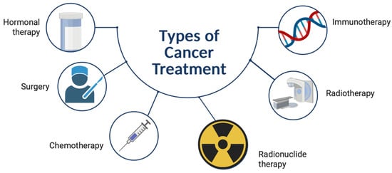
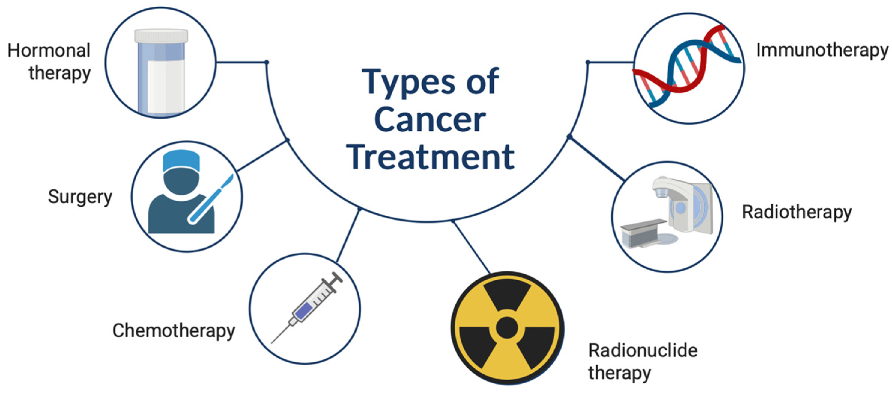
Prostate cancer is the second most common cancer amongst men, accounting for over 300,000 deaths in 2018 [1]. Prostate cancer primarily affects men between the ages of 45 and 60 [2], and accounts for one-third of newly diagnosed cancers amongst men [3]. An average of 15% of all prostate cancers become stubborn or resistant to deprivation therapy, and in turn, develop into metastatic castration-resistant prostate cancer (mCRPC) [4]. mCRPC affects millions of men and has one of the highest mortality rates worldwide [5]. Bone, lymph nodes, and visceral metastases are typical characteristics of mCRPC [6,7]; therefore, finding the best possible approach to patient treatment is desirable. Some of the most common treatment modalities for mCRPC include chemotherapy, radiation therapy, immunotherapy and bone-targeting therapy, and surgery [8], as illustrated in Figure 1.
Figure 1.
Current major therapeutics for cancer [9].
Chemotherapy is one of the most common treatments for metastatic disease, including prostate cancer, and involves the targeting of cancerous cells with high proliferation. Although it is quite effective, one of the major drawbacks of chemotherapy is that normal cells, including leukocytes and erythrocytes, are targeted in the process. This leads to a reduction in those cells, resulting in the development of secondary disorders such as anemia and other infections [10]. Radiation therapy involves the use of ionizing radiation for targeting cancer cells. This is achieved by damaging metastatic cell DNA, which inhibits their ability to proliferate and multiply further, ultimately leading to cell death [11]. One of the main disadvantages of radiation therapy, though, is that healthy cells are also damaged during treatment [12]. With immunotherapy, the treatment can target multiple regulatory pathways and involves the use of individual drugs targeting different targets [13]; however, careful consideration is taken to ensure that the body’s immune system is only activated when required. Although this is a promising treatment modality for mCRPC, immunotherapy is still in its infancy stages and more studies still need to be done to prove its effectiveness [13]. Bone-targeting agents such as bisphosphonates and denosumab are useful for targeting bone metastases in mCRPR patients; however, the pitfalls include that other metastases such as soft tissue metastases are neglected during patient treatment [14]. Although the above treatment modalities are usually effective, mCRPC still remains incurable, with an average survival of less than 2 years, and patient prognosis tends to persevere over time; therefore, their contributions to the field of oncology are limited [4].
Conjugation of targeting molecules (such as peptides, antibodies, or small-molecule ligands) to a radiometal-chelating agent creates a radiopharmaceutical for systemic tumor targeting [15]. The radiopharmaceutical is usually bound to a specific receptor, resulting in tumor-specific binding and retention. Sgorous et al. [16] define radionuclide therapy as the delivery of the radiopharmaceutical to tumor-associated targets. Radionuclide therapy involves the use of radionuclides, which are alpha (α), beta (β), or Auger-electron-emitting, to target tumors [17]. Over the past few years, radionuclide therapy has grown significantly with the use of agents such as Lutetium-177 (177Lu) PSMA for prostate cancer, and Yttrium-90 (90Y) microspheres for the treatment of malignant liver lesions [17]. Radionuclide therapy offers multiple advantages over conventional therapy modalities, such as that the deposition of the therapeutic dose can be accurately measured and controlled since in most cases and the radiation doses are administered intravenously, therefore increasing the therapeutic effect of the dose. This results in a novel approach towards personalized treatment [18]. Radionuclide therapy also allows the delivery of a high and concentrated dose to the target organ whilst sparing normal surrounding tissue. It is non-invasive, the duration of the treatment is quick, and it allows for the treatment of systemic malignancy within a single dose [19]. Various multi-center studies have investigated the use of β-emitting radionuclides, such as 177Lu radiolabeled with PSMA at both the preclinical and clinical level for the treatment of mCRPC. In a study performed by Von Eyben et al. [20], it was concluded that 177Lu PSMA-labeled ligands offer better patient prognosis and fewer side effects as compared to chemotherapy. Although 177Lu PSMA therapy is usually effective for prostate cancer, less than half of the patients with mCRPC have a biochemical response to the treatment [4]. Only 45% of the patients’ PSA levels drop by ≥50% post therapy, and more than a quarter of the patients do not respond to treatment at all [21,22]. Satapathy et al. [23] has also noted a poor response and mortality rate in patients with visceral metastases which have been treated with 177Lu PSMA. 177Lu is a β-emitter possessing a maximum energy of 497 keV and has a maximum tissue penetration of 1.5 mm [24]; this means that although a high dose will be targeted at the tumor site, a low absorbed dose will be deposited into metastatic cells due to the range of electrons being too long [4,25,26]. Behr et al. [26] indicated that radionuclide therapy using α-emitters with high linear energy transfer (LET) and a short range may have significantly more advantages over β-emitters. α-Emitters, such as Actinium-225 (225Ac), hold great promise as therapeutic agents for micro metastases. α-Particles are highly potent cytotoxic agents, potentially capable of tumor killing without limiting morbidity. The increased effectiveness of α-particles is due to the amount of energy deposited per unit distance traveled (high LET), which is approximately 80 keV/μm [27]. Cell survival studies have shown that α-particle-induced killing is independent of oxygenation state or cell cycle during irradiation, and that as minimal as one to three tracks across the nucleus may ultimately result in cell death. Additionally, the 50 μm to 100 μm range is consistent with the dimensions of micro metastases, allowing for localized irradiation of target cells with minimal normal cell irradiation [27].
225Ac is an α-emitter with a long physical half-life (T1/2 = 9.9 days) and a short range of α-particles with a high LET, making it responsible for the destruction of malignant tumors while sparing healthy surrounding tissue, thus making it a promising radionuclide for targeted alpha therapy (TAT). Although the nuclear properties of 225Ac make it an ideal radionuclide for TAT, Thiele et al. [28] argued that its application is challenging due to the lack of chelators available to stabilize its daughter radionuclides, resulting in the recoil effect, which is the breakdown between the radionuclide and the chelator [29], resulting in the transformation of a new daughter radionuclide possessing its own chemical properties which can target healthy organs and tissues, subsequently decreasing the dose administered to the target organ [30,31]. The recoiling issues surrounding 225Ac-labeled radiopharmaceuticals can be minimized by a quick uptake of the product within the tumor, local administration, and encapsulating the product within a nanocarrier, such as nanoparticles [30]. This is because the optimal increase in the cell killing efficacy of 225Ac occurs if all (or most of) the α-emissions occur at the tumor site; otherwise, toxicity may potentially be increased [32]. In order to limit the recoiling impact of 225Ac’s daughters and enable selective deposition of the radionuclides, there is a consensus to develop alternate strategies, such as nano-vehicles. It is interesting to note that the use of nanoparticles in medicine is growing quickly, with several nano-vectors being created to enable targeted therapy, especially in the treatment of cancer.
The aim of this study was to formulate poly(lactic-co-glycolic acid) (PLGA) nanoparticles as a delivery vector for 225Ac, which would potentially increase the therapeutic potential by retaining 225Ac and its decay daughters at the tumor site.
2. Materials and Methods
2.1. Materials
225Ac was supplied by the Joint Research Centre (JRC) of the European Commission. PSMA617-TFA was procured from MedKoo Biosciences Inc, Durham, North Carolina, PLGA, folic acid (FA), ethylene dichloride (EDC), n-hydroxy succinimide (NHS), d-α-tocopherol polyethylene glycol 1000 succinate (TPGS) and chitosan (CS) were procured from Sigma-Aldric, Johannesburg. All other reagents used were of the highest analytical grade.
2.2. Methods
2.2.1. Radiolabeling and Quality Control of 225Ac PSMA617-TFA
Radiolabeling
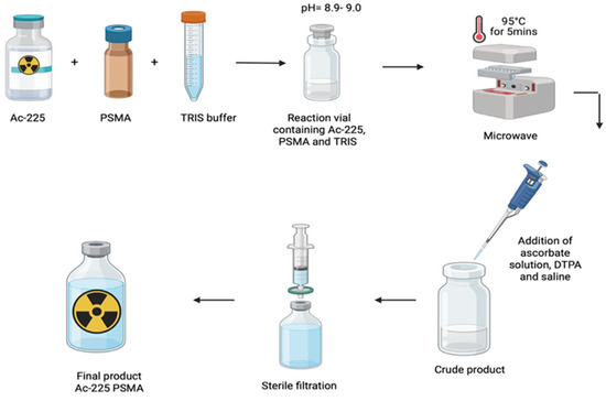
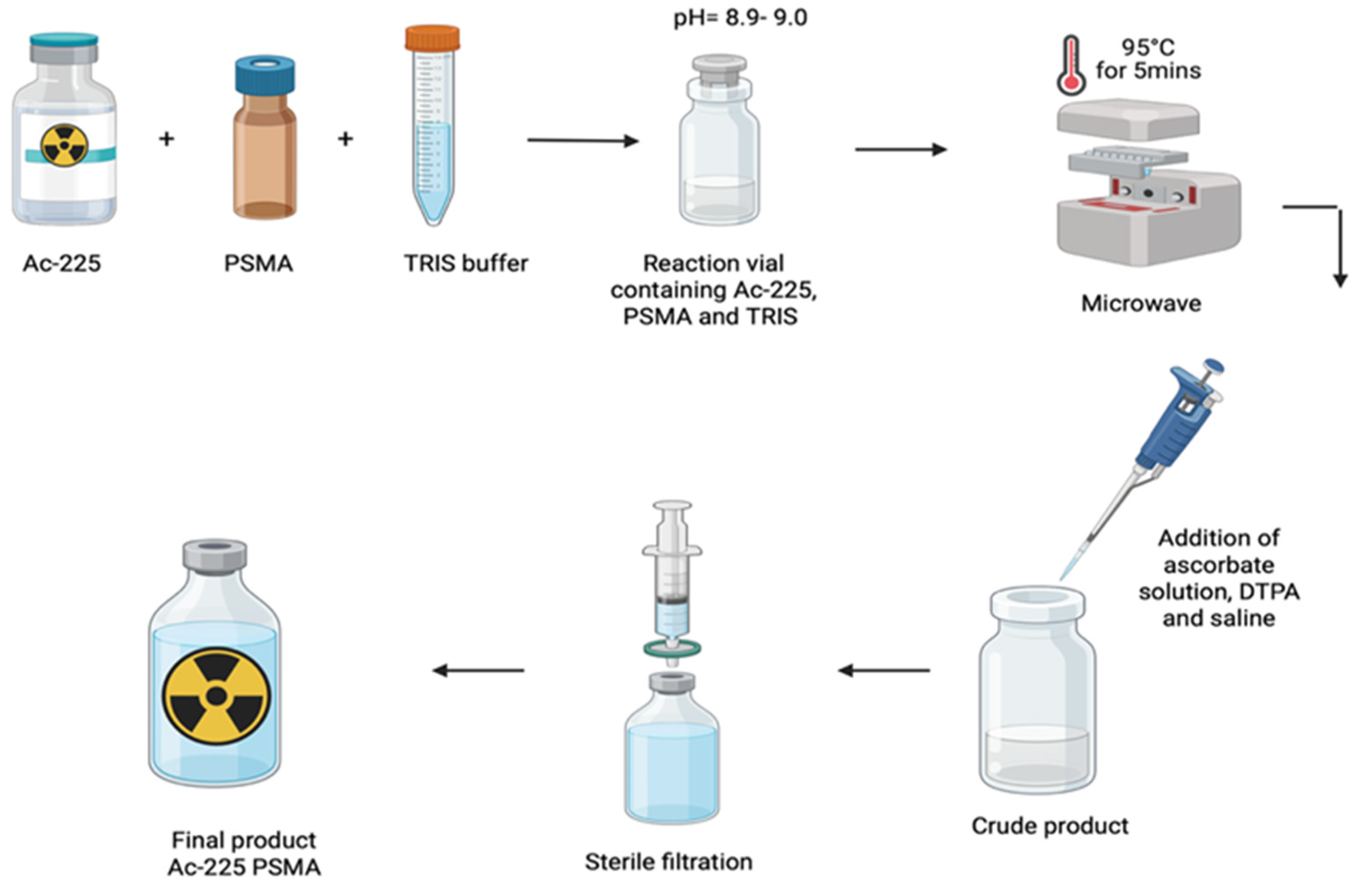
The radiolabeling procedure was carried out using a method outlined by Kratochwil et al. [33], as outlined in Figure 2. In brief, 225Ac(III)Cl3 was dissolved in 0.1 M HCl and allowed to stand at room temperature for 30 min to establish daughter equilibrium prior to radiolabeling. The specific activity was calculated based on the radioactivity of 225Ac (MBq) at the time of formulation per mass of PSMA617-TFA (µg). Based on a formulation ratio of 2 µL of PSMA617-TFA stock solution (7.46 mg/mL) per 1 MBq of 225Ac, the nominal specific activity of the preparation was 67 MBq/mg (0.067 MBq/µg). In a reaction vial, 500 μL of 0.1 M TRIS buffer (pH = 9) was added. This was followed by the addition of PSMA617-TFA (2 μL/MBq) and the desired volume of 225Ac(III)Cl3. The reaction took place for 5 min at 95 °C in a microwave (Biotage ® Initiator+). The resulting mixture was stabilized with the addition of 500 μL of ascorbate solution (20% ascorbic acid + 10 M NaOH) and 100 μL of diethylenetriaminepentaacetic acid (DTPA). Additionally, 4 mL of saline was added to the final product, which was then filtered through a 0.22 μm Millipore sterility filter. The final product’s pH was monitored to be between 6.7 and 7.4, and the entire process took place in a laminar airflow cabinet under sterile conditions.
Figure 2.
Radiolabeling process of 225Ac PSMA617-TFA.
2.2.2. Radiochemical Purity by Cut-And-Count Gamma Counter Method
Radiochemical purity was determined using a cut-and-count method which used thin-layer chromatography (TLC) paper (TLC-SG paper: Agilent Technologies, St Clara, CA, USA). First, 0.5 µL of the product was spotted on silica gel iTLC paper in triplicate and 0.5 M sodium citrate was utilized as the mobile phase to develop the TLC paper, which was cut in into two segments, the top and the bottom. Each segment was counted in a gamma well counter (CaptusTM 4000e well counter, Mirion Medical, Atlanta, GA, USA) in the 221Fr energy window, 218 keV. This was performed in triplicate, and an average was taken as the radiochemical purity (%RCP). The final %RCP results were obtained after circular equilibrium. Although nanoparticles of a large size remain at the origin with 225Ac-PSMA617-TFA in standard stationary phases, the 0.5 M sodium citrate mobile phase used in this study acts as a competitive chelator for uncomplexed radiometals, resulting in any actinium colloids which have been present at the origin to be trans-chelated and solubilized actinium citrate complexes which migrate to the solvent front with any free 225Ac. The single strip citrate mobile phase method has been validated by the European Association of Nuclear Medicine (EANM) and the International Atomic Energy Agency (IAEA) [34,35].
2.2.3. Endotoxin Testing
Final product was sampled at a ratio of 1:100, and a test sample in Limulus Amebocyte Lysate (LAL) pyrogen-free water was prepared. First, 2 µL of 225Ac PSMA617-TFA was diluted in 198 µL of LAL water. After thorough mixing, 25 µL of the sample was added onto each well in the test cartridge (Limulus Amebocyte Lysate Test Cartridges: Charles River Laboratories, Navi Mumbai, Maharashtra), which was inserted onto an Endosafe® Nexgen-PTS™ Kinetic Reader, Fisher Scientific, Johannesburg, South Africa. Bacterial endotoxin results were obtained.
2.2.4. Bubble Point Test
The wetted filter’s upstream pressure was progressively raised by air. Air pressure gradually increased (beginning at 0) through a wetted filter until a continuous stream of bubbles occurred in water, demonstrating the pressure required to overcome capillary forces in the pores. A pass indicated that the pressure was at or above the specified 50 psi (3.5 bar) (or the manufacturer’s specifications).
2.2.5. Preparation of PLGA Nanoparticles
Preparation of PLGA nanoparticles was conducted using various volumes of distilled water between 5 mL and 10 mL, and different surfactants, mainly TPGS and Tween-80. PLGA nanoparticles were prepared using 0.5% TPGS as the surfactant. TPGS was selected as the optimal surfactant based on preliminary screening of different surfactants and concentrations. Although Tween-80 was evaluated during initial optimization (yielding 30 mV zeta potential at 10,000 rpm), TPGS 0.5% was chosen for the final formulation due to its generation of ~200 nm particles with favorable PDI (0.18) and positive surface charge, consistent with its reported advantages as a P-glycoprotein inhibitor and biocompatible surfactant.
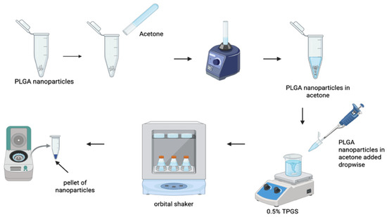
The nanoparticles were stirred at speeds of 3000 rpm and 10,000 rpm following the nanoprecipitation method, which is illustrated in Figure 3. Initially, 25 mg of PLGA was dissolved in 1 mL of acetone. This was followed by vortexing and sonicating until the mixture was completely dissolved. The solution was then added dropwise to 0.5% TPGS (total volume of 5 mL) under a magnetic stirrer at speed 10,000 rpm for 20 min. The solution was then placed in an orbital shaker overnight (±18 h) at a speed of 75 rpm. The nanoparticles were centrifuged at a speed of 2700 rpm for 30 min, followed by ultracentrifugation at 45,000 rpm for 30 min. The supernatant was then removed, and the nanoparticles were washed twice with 5 mL saline before being centrifuged again at 45,000 rpm for 20 min. The supernatant was then removed, leaving behind a pellet of PLGA nanoparticles.
Figure 3.
Preparation of PLGA nanoparticles.
2.2.6. Preparation of Chitosan–Folic Acid (CS-FA)
CS and FA were functionalized using the emulsion solvent diffusion method, where 6 mg of folic acid was dissolved in 1 mL dimethyl sulfoxide (DMSO). First, 100 µL of EDC:NHS, with a total ratio 1:5, was added to the solution. The mixture was vortexed until completely dissolved, followed by incubating in the dark overnight in an orbital shaker (speed: 75 rpm). Then, 20 mg CS was dissolved in 0.1% acetic acid (total volume of 2 mL). The solution was added dropwise to folic acid under stirring conditions. CS-FA was incubated in an orbital shaker for 24 h. The solution was then centrifuged at 4500 rpm for 60 min. The supernatant was removed.
2.2.7. Encapsulation of 225Ac PSMA617-TFA into PLGA Nanoparticles
225Ac PSMA617-TFA (total PSMA617-TFA mass = 5 µg) was added dropwise to the pellet PLGA nanoparticles prepared in Section 2.2.5. This was followed by sonication for 20 min at 35 °C. Following sonication, the 225Ac PSMA617-TFA PLGA nanoparticles were centrifuged at 45,000 rpm for 30 min, leaving behind a pellet of 225Ac PSMA617-TFA PLGA nanoparticles.
2.2.8. Preparation of Loaded PLGA–Chitosan–Folic Acid (PLGA-CS-FA) Nanoparticles
The pellet of CS-FA, which was suspended in Section 2.2.6, was resuspended in 1 mL deionized water and then added dropwise to the pellet of 225Ac PSMA617-TFA PLGA nanoparticles from Section 2.2.7 above. The solution was placed in an orbital shaker for 2 h at a speed of 100. This was followed by centrifugation at 45,000 rpm for 30 min. The supernatant was then sampled and HPLC was utilized to evaluate the encapsulation efficiency, as per Section 2.2.11.
2.2.9. Morphology and Chemical Characterization of Loaded PLGA-CS-FA Nanoparticles
The nanoparticle morphology was examined using scanning electron microscopy (SEM). Powdered samples of the nanoparticles were placed onto an aluminum specimen stub covered with a double-sided carbon adhesive disk and sputter-coated with both palladium and gold for 4 min at 20 KV. SEM images of the loaded PLGA-CS nanoparticle samples were viewed using an SEM (SIGMA VP, Zeiss Electron Microscopy, Carl Zeiss Microscopy Ltd.; Cambridge, UK). Nanoparticle size, polydispersity index (PDI), polarity and zeta potential were determined by loading 0.75 mg of the nanoparticles into a zetasizer Nano-ZS machine (NsnoYtac Wave II), LabX, Midland, Ontario, Canada. The experiments were performed in triplicate at a temperature of 25 °C. The nanoparticles were dispersed in non-ionizing water and filtered through a 0.22 μm Millipore sterility filter.
2.2.10. Componential Analysis of Chemical Structure Integrity Post Nanoparticle Formation
In order to assess the chemical integrity of native and combined components of the 225Ac PSMA617 TFA into PLGA-CS nanoparticles, Fourier-Transform Infrared (FTIR) spectroscopy (PerkinElmer Spectrum 100, Llantrisant, Wales, UK) was used to identify and characterize the pharmaceutical stability of the PLGA, TPGS, chitosan, and folic acid in their native and combined states. Moreover, this technique was used to determine the impact on the chemical stability of loading 225Ac PSMA617 TFA in PLGA–chitosan and to observe any possible significant changes in functional groups. The FTIR spectra were recorded at 20 °C, ranging from 500 to 4000 cm−1 for samples of 225Ac PSMA617 TFA into PLGA-CS-FA.
2.2.11. Radio and UV-VIS-HPLC Analysis for Encapsulated and Released 225Ac PSMA617-TFA
Radio and UV-VIS-HPLC method was developed andutilized to quantify the encapsulated 225Ac PSMA617-TFA, as shown in Figures S1 and S2. The following method and parameters were as follows. Injection volume: 100 µL, column: C18 column, temperature of column oven: 40 °C, gamma channel: 10 keV–2000 keV, and UV channel: 10 keV–2000 keV, as seen in Table 1 below.
Table 1.
Pump program for HPLC method.
The encapsulation efficiency was calculated as follows:
where %E = encapsulation efficiency; At = total activity added; and As = activity in the supernatant [36].
2.2.12. PLGA-CS Retention of 221Fr and 213Bi Evaluation
Evaluation of retention of 221Fr and 213Bi was conducted using thin-layer chromatography, with a TLC scanner (Elysia RayTest miniGINA single), Elysia RayTest, Ougree, Belgium, and Supelco® silica gel aluminum strips, Merck Life Sciences, Johannesburg, South Africa. The TLC was developed using 0.1 M sodium citrate as the mobile phase. The strips were spotted with free 225Ac PSMA617-TFA, 225Ac PSMA617-TFA PLGA nanoparticles, and 225Ac PSMA617-TFA PLGA-CS nanoparticles. iTLC scans were performed and recorded before and after circular equilibrium. This was to ensure that Fransium-221 (221Fr) and Bismuth-213 (213Bi) had completely decayed and that the results obtained were for 225Ac only. The strips were counted immediately, after 30 min and 6 h after spotting, according to the following protocol—run time: 2 min, TLC scan range: 0–140 mm, collimator: free, TLC solvent origin: 10 mm, TLC solvent front: 110 mm and counting limit: None.
2.2.13. In Vitro Drug Release of 225Ac PSMA617-TFA
To investigate the in vitro release of 225Ac PSMA617-TFA from the PLGA-CS nanoparticles, 225Ac PSMA617-TFA PLGA-CS-loaded nanoparticles (n = 3) were incubated in 30 mL of PBS and positioned in an orbital shaking incubator at 37 °C for 72 h.
The prelease of 225Ac PSMA617-TFA was determined at different pH conditions: 7.4 to mimic normal body pH, and 6.5 to mimic microenvironmental tumor pH. The nanoparticles were incubated in the buffer solutions at a temperature of 37 °C to mimic the body’s normal temperature. Then, 150 µL of each sample was taken and run on HPLC using the method described in Section 2.2.4. This was replaced by the same volume of buffer solution. Samples were taken and counted at 30 min, 1 h, 3 h, 6 h, 9 h, 48 h and 72 h post incubation.
3. Results and Discussion
3.1. Radiolabeling and Quality Control of 225Ac PSMA617-TFA
Our work aimed to provide a standardized manual radiolabeling method for producing 225Ac PSMA617-TFA on-site using conventional radiopharmacy equipment and existing regulations. The radiolabeling approach used in our tests has shown constant repeatability, allowing us to create a radiopharmaceutical that fulfills pharmaceutical requirements. Because of actinium’s unusual physical features, identifying the best quality control measures became more complex. Efforts were made to standardize quality control procedures in compliance with pharmacopeia guidelines. The radiochemical purity was done in triplicate, and the average result was calculated to be 99.3%. Final product pH, bacterial endotoxin results, filter integrity testing and visual inspection were all within specification, as shown on Table 2 below.
Table 2.
Quality control of 225Ac PSMA617-TFA (n = 3).
3.2. Preparation and Characterization of PLGA Nanoparticles
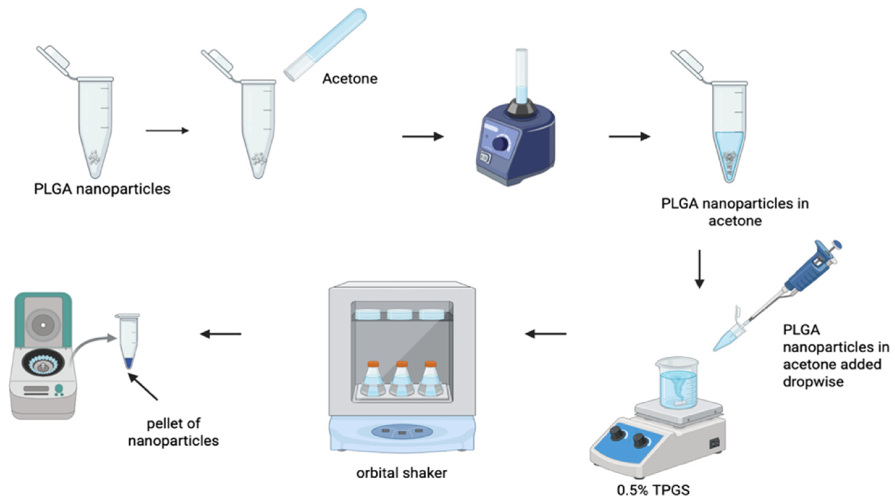
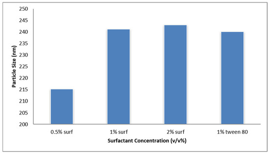
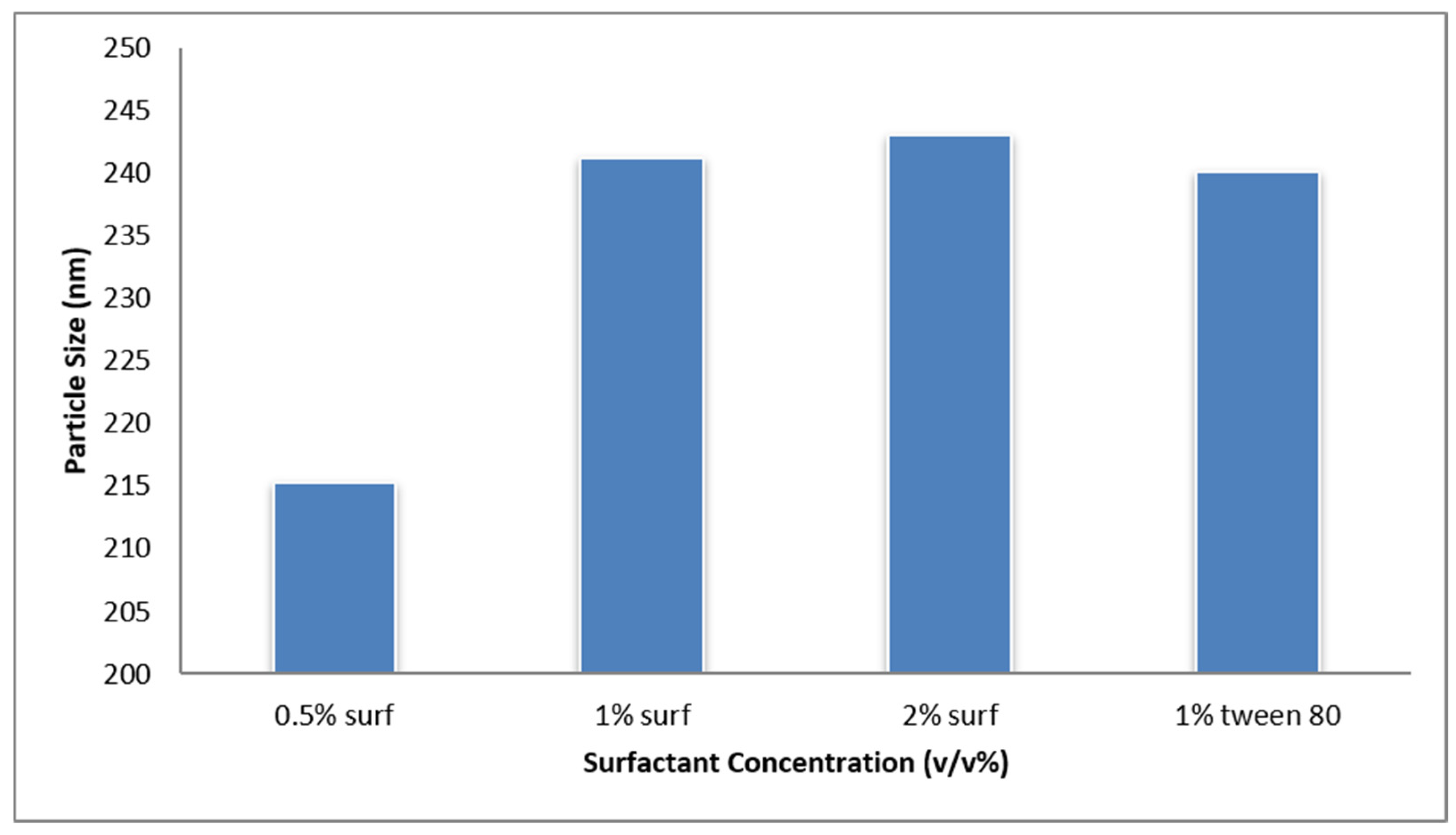
The main reason for choosing PLGA as the polymer was its high biodegradability and biocompatibility. It can be formulated and surface modified quite easily [36]. PLGA has also been approved by the Food and Drug Administration (FDA) of the United States and the European Medicines Agency (EMA) for drug delivery and the safety of humans for pharmaceutical use [37,38,39]. PLGA nanoparticles were prepared using a nanoprecipitation method. The use of different surfactants, their concentration and the speed of homogenization was seen to affect the size, PDI and zeta potential of the nanoparticles, as seen in Figure 4 and Table 3, respectively. Increasing the speed of homogenization has also been seen to decrease the size of the nanoparticles. This was also confirmed by Mulia et al. [39,40].
Figure 4.
Mean particle size impacted by various surfactant concentrations: 0.5% TPGS, 1% TPGS, 2%TPGS and 1% Tween 80, (n = 3).
Table 3.
Average PDI, zeta potential and polarity of PLGA nanoparticles at different volumes of water, concentrations of surfactants and different speeds of homogenization.
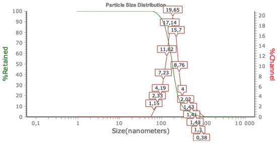
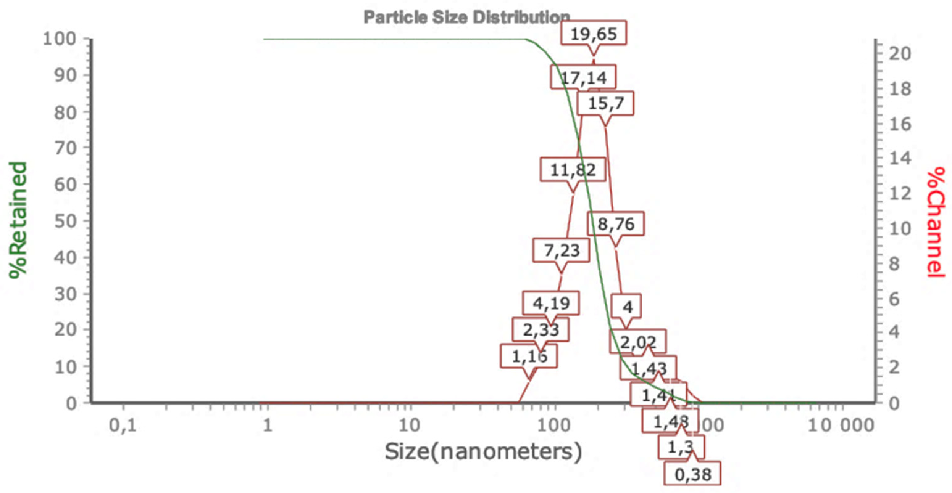
In our study, 0.5% TPGS and a speed of 10,000 rpm were used. This is because it generated a nanoparticle size of approximately 200 nm, which is the desired size for nanomedical applications [38]. Furthermore, the 0.5% TPGS surfactant formulation produced an optimized average hydrodynamic diameter of 215.2 ± 8.2 nm. Elevating the concentration to 1.0% and 2.0% paradoxically expanded the mean size to 241.3 ± 7.9 nm and 243.0 ± 6.1 nm, respectively. This trend indicates that higher stabilizer contents cross the critical micelle threshold, inducing interfacial crowding and localized viscosity increases that impede rapid solvent diffusion, resulting in larger core matrices. The hydrodynamic diameter of ~200 nm places these nanoparticles at the upper limit for optimal intravenous administration. Particles > 150–200 nm in size are increasingly subject to opsonization and subsequent sequestration by Kupffer cells in the liver and macrophages in the spleen, resulting in reduced circulation half-life and increased hepatic/splenic radiation exposure. For 225Ac-loaded nanoparticles, RES accumulation is especially concerning given the high LET alpha emissions and potential for liver toxicity. Future formulation optimization should target a size range of 80–120 nm, which balances an enhanced permeability and retention (EPR) effect with reduced RES uptake. Given the 200 nm particle size and the imperative to avoid RES-mediated 225Ac accumulation, intertumoral or intra-arterial administration may be the most feasible near-term approach, while IV administration would require concurrent nanoparticle size reduction and PEGylation to achieve acceptable biodistribution profiles.
PDI is used to describe the degree of non-uniformity in the distribution of nanoparticle size [41]. PDI values usually range from 0 to 0.7, where the lower the PDI value, the more uniform the sample in terms of the nanoparticle size. In nanodelivery applications, a PDI value of 0.3 and below is deemed acceptable and indicates a homogenous population [41]. PDI values greater than 0.7 are usually unacceptable as they cannot be analyzed by dynamic light scattering. The average PDI value using the chosen method for this study was 0.18, as seen in Figure 5.
Figure 5.
Hydrodynamic size distribution of PLGA-CS-FA by dynamic light scattering (DLS). Intensity-weighted size distribution showing a monomodal peak at ~200 nm with polydispersity index (PDI) = 0.18. Number-weighted distribution confirming uniform particle population. Volume-weighted distribution. Inset: Digital photograph of nanoparticle suspension showing Tyndall effect. Data representative of n = 3 independent measurements.
Zeta potential, which is an essential characteristic of nanoparticle formulation, is described as the number of charges that a particle carries [42]. A zeta potential that is within ±30 mV is acceptable as it can stabilize the nanoparticles through electrostatic repulsion, and a positively charged nanoparticle is preferred for the enhancement of electrostatic interactions; the PLGA-CS nanoparticles produced had a zetapotential of 29 mV with a positive polarity [43].
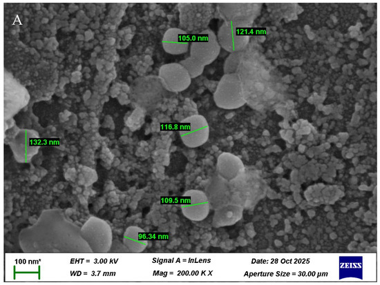
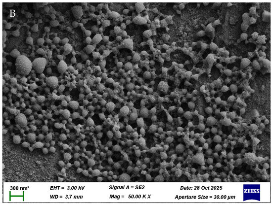
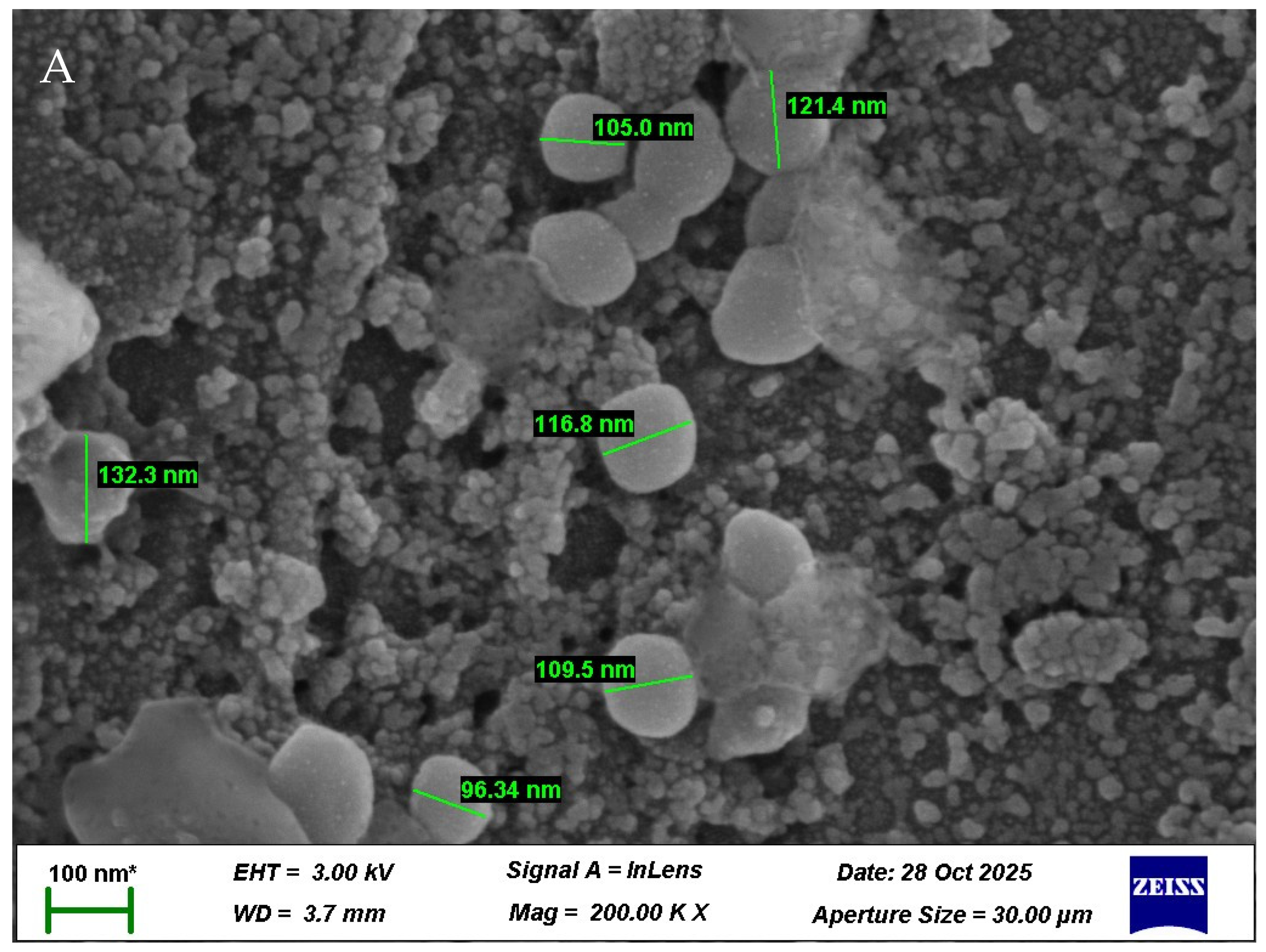
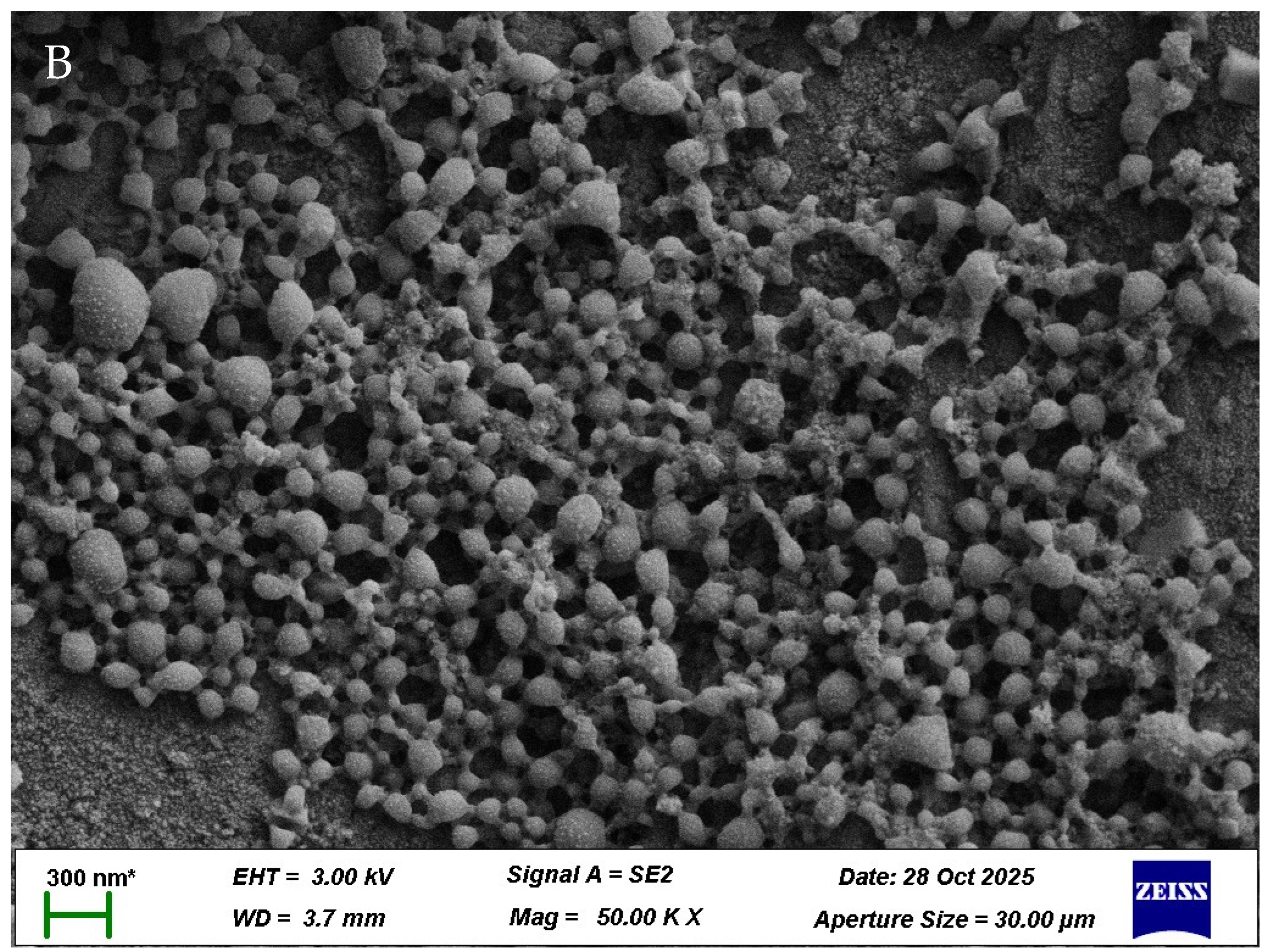
The image in Figure 6 was captured by the scanning electron microscope (SEM), and the nanoparticle formulations were well distributed, with spherical morphologies that had sizes between 96 nm and 132 nm. The particle size established in the SEM was closely aligned with those established in the zeta particle size analysis results.
Figure 6.
Characterization of 225Ac PSMA617-TFA PLGA-CS nanoparticles. Gold–platinum-sputtered SEM micrograph of methotrexate-loaded TPGS-PLGA nanoparticles. (A) Magnification = 100 KX; voltage = 3.00 kV; scale bar = 100 nm (Zoomed Image). (B) Magnification = 50 KX; voltage = 3.00 kV; scale bar 300 nm. SEM of the optimized loaded nanoparticle formulation shows sphericity and a smooth surface of the nanoparticles (* calibrated set scale bar).
The FTIR spectrum of PLGA exhibited intense bands observed in the region between 1770 and 1750 cm−1, which are attributed to the stretching vibration of the carbonyl groups present in the two monomers. Medium intensity bands between 1300 and 1150 cm−1 were attributed to asymmetric and symmetric C–C(=O)–O stretches, respectively. The bands in these regions are useful in the characterization of esters. Bands at 3500 cm−1 and 3450 cm−1 in the FTIR spectra for lactide and glycoside are attributed to stretching vibrations of the OH group. TPGS displayed characteristic bands at 1750 cm−1 and at 2900–3000 cm−1, corresponding to the O–C=O stretching of its ester groups and C–H stretching, respectively. In the infrared spectrum of chitosan, a strong band in the region 3291–3651 cm−1 corresponds to N–H and O–H stretching, as well as the intramolecular hydrogen bonds. The absorption bands at around 2921 and 2877 cm−1 can be attributed to C–H symmetric and asymmetric stretching, respectively. These bands are characteristics typical of polysaccharides and are found in other polysaccharide spectra. The presence of residual N-acetyl groups was confirmed by the bands at around 1645 cm−1 (C=O stretching of amide I) and 1325 cm−1 (C–N stretching of amide III). We did not find the small band at 1550 cm−1 that corresponds to N–H bending of amide II. This is the third band characteristic of typical N-acetyl groups, and it was probably overlapped by other bands. A band at 1589 cm−1 corresponds to the N–H bending of the primary amine. The CH2 bending and CH3 symmetrical deformations were confirmed by the presence of bands at around 1423 and 1375 cm−1, respectively. The absorption band at 1153 cm−1 can be attributed to asymmetric stretching of the C–O–C bridge. The bands at 1066 and 1028 cm−1 correspond to C–O stretching. Folic acid (FA) consisting of three moieties, a pteridine ring, p-amino benzoic acid and glutamate, exhibits peaks corresponding to its functional groups, along with other characteristic peaks in the FTIR spectrum. The peak at 1686 cm−1 corresponds to the carbonyl groups of carboxylic acid moieties of folic acid, while its shoulder peak at 1670 cm−1 and the peaks at 1413 cm−1 and 1602 cm−1 correspond to the carbonyl groups of the amide group of folic acid, the bending vibrations of hydroxyl groups, and the bending vibrations of the N–H groups, respectively. The bands at 3400–3600 cm−1 correspond to the stretching vibrations of O–H carboxylic acid groups of the glutamic acid moiety and N–H group of the pteridine ring, as shown in Figure S3.
3.3. Loading and Encapsulation of 225Ac PSMA into PLGA and PLGA-CS-FA Nanoparticles
Numerous factors were seen to affect the encapsulation efficiency of the nanoparticles. These included the incubation time and the use of different buffer solutions with varying pH levels for in vitro release. In this study, PSMA617-TFA radiolabeled with 225Ac showed an average encapsulation efficiency of 85.6% using HPLC when performed under the same conditions, as seen in Table 4. This is due to the product being more hydrophilic, resulting in the decreased interaction of 225Ac with PLGA, therefore resulting in decreased encapsulation efficiency. Unloaded PLGA nanoparticles which were functionalized with CS and FA showed an increase in encapsulation efficiency, with an average of 99.9%. This is due to increased drug interactions and reduced drug leakage, as observed by [44].
Table 4.
Average encapsulation efficiency at different incubation times (n = 3+).
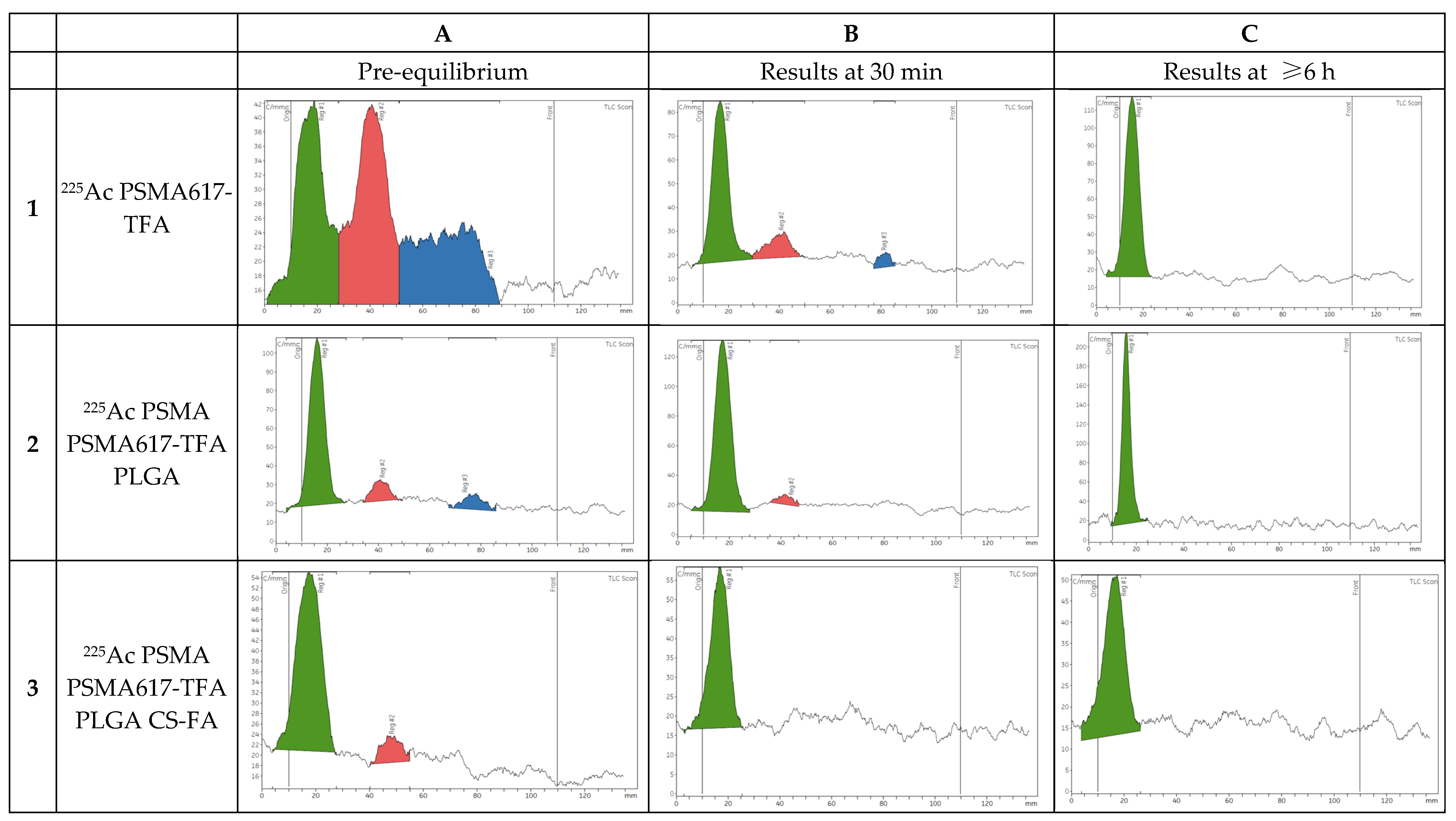
Following HPLC analysis, the encapsulation efficiency was further confirmed using iTLC, as shown in Figure 7 below.
Figure 7.
iTLC analysis of daughter radionuclide retention in nanoparticle formulations. (A) Pre-equilibrium scans showing initial radionuclide distribution. (A1) Free 225Ac-PSMA617-TFA with visible radionuclide daughter peaks (221Fr in blue, 213Bi in red); (A2) 225Ac-PSMA617-TFA PLGA NPs showing partial retention; (A3) 225Ac-PSMA617-TFA PLGA-CS-FA NPs showing near-complete retention. (B) Results 30 min post-spotting: 221Fr decay evident in free formulation (B1), significant retention in PLGA NPs (B2), complete retention in PLGA-CS-FA NPs (B3). (C) ≥6 h post-spotting (circular equilibrium): complete 213Bi decay in free formulation (C1), total encapsulation maintained in PLGA NPs (C2) and PLGA-CS-FA NPs (C3). Green = 225Ac, red = 213Bi, blue = 221Fr.
Following radiolabeling of 225Ac PSMA617-TFA, 0.5 μL of the sample was spotted on iTLC paper and run on an iTLC scanner. Image A1 shows 225Ac (green), alongside its daughters, 213Bi (red) and 221Fr (blue). At 30 min post-spotting, 221Fr is almost decayed due to its short half-life (T1/2 = 4.9 min) (B1); however, the presence of 213Bi is still visible and greater than that of 221Fr, and 6 h post- spotting, the half-life of 213Bi (T1/2 = 45.6 min) had been reached eight times, making 213Bi completely decayed (C1). Following encapsulation of 225Ac PSMA617-TFA using PLGA, both daughters, 221Fr and 213Bi, although still visible, are almost completely retained pre-equilibrium (A2). At 30 min post-equilibrium, the presence of 213Bi is significantly lower than that of the non-encapsulated 225Ac PSMA617-TFA (B2) and total encapsulation is seen at 6 h post-equilibrium (C2). With the PLGA nanoparticles functionalized with CS-FA, total encapsulation of 221Fr and 213Bi is seen as early as 30 min post-equilibrium (B3).
3.4. In Vitro Drug Release of 225Ac PSMA617-TFA
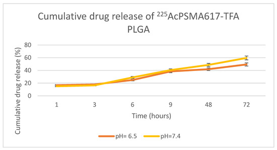
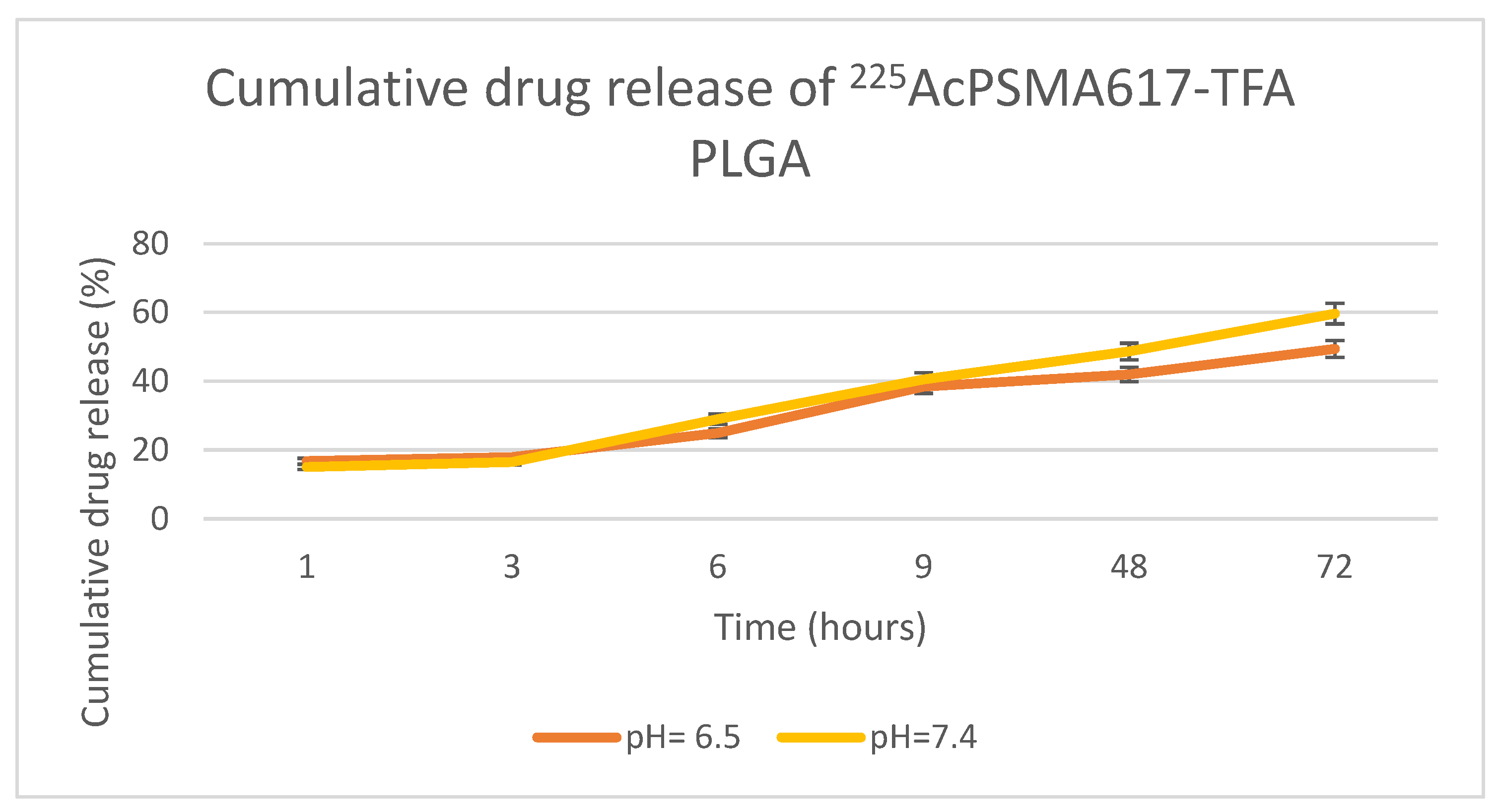
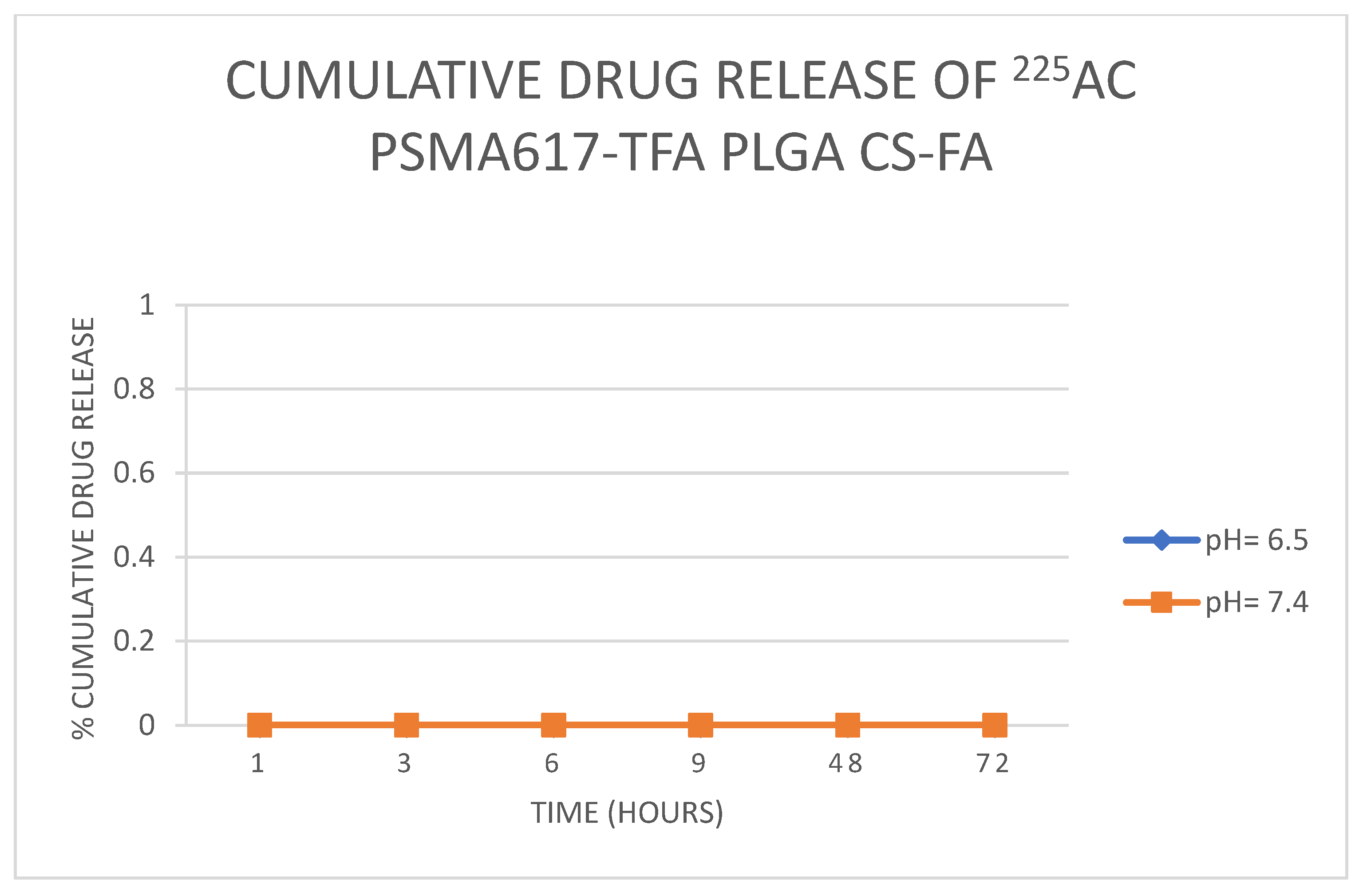
A major concern with when formulating nanocarriers is the burst release for drug delivery [45]. Controlled drug release is, therefore, important because it ensures a strong concentration of the drug in the body over a long period of time, which results in an increased therapeutic effect of the drug, reduces any harsh side effects, and, in turn, improves patient prognosis [46]. In this study, in vitro release kinetics showed a high drug release of 225Ac PSMA617-TFA PLGA nanoparticles at pH 7.4 (60%) compared to at pH 6.5 (49%) after 72 h, as illustrated in Figure 8 below. The absence of detectable 225Ac PSMA617-TFA release from PLGA-CS-FA nanoparticles over 72 h at both pH 6.5 and 7.4 (Figure 9) indicates that the bilayer polymer system (PLGA core + CS-FA shell) creates a highly effective diffusion barrier. While this ensures retention of 225Ac and its recoiling daughters (221Fr and 213Bi), it raises important questions regarding the biological availability of the radiopharmaceutical at the target site. Several mechanisms may facilitate drug availability despite minimal passive release, including intracellular degradation, tumor microenvironment enzymatic activity, and surface erosion. However, these mechanisms remail speculative without cellular uptake and intracellular trafficking studies.
Figure 8.
Cumulative drug release of 225AcPSMA617-TFA PLGA (n = 3).
Figure 9.
Cumulative drug release of 225AcPSMA617-TFA PLGA CS-FA (n = 3).
3.5. Limitations of the Study
This study is limited to the formulation, physicochemical characterization, and in vitro radionuclide retention evaluation of PLGA-CS-FA nanoparticles loaded with 225Ac-PSMA617-TFA. No biological studies were performed to validate therapeutic potential. Specifically, the following critical experiments remain to be conducted: (i) cellular uptake studies in PSMA-expressing prostate cancer cell lines (e.g., LNCaP, C4-2, or PC-3-PSMA) to confirm receptor-mediated internalization and nanoparticle penetration; (ii) cytotoxicity assays to determine the comparative toxicity of free versus nanoparticle-encapsulated 225Ac-PSMA617-TFA; (iii) clonogenic survival assays to quantify long-term reproductive cell death following alpha irradiation; (iv) DNA damage assessment (e.g., γ-H2AX immunofluorescence or comet assay) to verify double-strand break induction; and (v) therapeutic efficacy studies comparing tumor cell killing between free and nano-encapsulated radiopharmaceutical formulations. Such data are essential to establish whether the observed radionuclide retention translates to reduced off-target radiation exposure and enhanced tumor-specific delivery in vivo. Future work will prioritize these biological validation steps before advancing to preclinical animal studies. Additionally, the lack of measurable release of 225Ac PSMA617-TFA at both pH conditions represents a limitation, as it does not fully recapitulate the complex biological environment. Future studies must evaluate nanoparticle stability and drug release in cell culture media, serum-containing conditions, and intracellular compartments.
4. Conclusions
This study demonstrated the successful formulation and physiochemical characterization of PLGA-CS-FA nanoparticles loaded with 225Ac PSMA617-TFA. The nanoparticles exhibited favorable size, positive surface charge, high encapsulation efficiency, and effective retention of 225Ac’s daughter radionuclides (221Fr and 213Bi). In vitro release studies showed minimal passive release, suggesting potential for sustained retention at the target site. It is important to emphasize that this study is limited to formulation feasibility and physiochemical characterization. No in vitro cellular studies or in vivo animal studies were performed. Therefore, while the nanoparticle system shows promise as a delivery platform, future studies must rigorously focus on in vitro evaluation using PSMA-expressing cell lines to assess cellular internalization, cytotoxicity, and DNA damage, followed by preclinical biodistribution and efficacy studies in tumor-bearing animal models before clinical translation can be considered.
Supplementary Materials
The following supporting information can be downloaded at: https://www.mdpi.com/article/10.3390/radiation6030027/s1, Figure S1: Calibration curve of PSMA617-TFA; Figure S2: HPLC chromatogram of 225Ac PSMA617-TFA; Figure S3: The FTIR spectra of PLGA, PLGA nanoparticles, CS and PLGA-CS-FA nanoparticles.
Author Contributions
Designed the experiments: S.M., Y.M., B.A.W. and P.M.; Performed the experiments: S.M., Y.M., B.A.W., P.M. and M.S. (Mbongeni Shungube); Analyzed the data: S.M., Y.M., B.A.W., P.M. and M.S. (Mbongeni Shungube); Wrote the paper: S.M., Y.M., B.A.W., P.M. and M.S. (Mbongeni Shungube), H.N., K.R., S.S., A.M. and M.S. (Mike Sathekge). All authors have read and agreed to the published version of the manuscript.
Funding
This research received no external funding.
Data Availability Statement
The raw data supporting the conclusions of this article will be made available by the authors on request.
Acknowledgments
The authors are grateful to the staff of the department of Nuclear Medicine at the University of Pretoria and Steve Biko Academic Hospital and the staff of the Nuclear Medicine Research Infrastructure and department of Pharmacology at Sefako Makgatho Health Sciences University. The authors are also grateful to the Marie Slodoswka Curie Fellowship Programme of the International Atomic Energy Agency (IAEA).
Conflicts of Interest
The authors declare no conflicts of interest.
Abbreviations
| TAT | Targeted Alpha Therapy |
| NDDS | Nanodrug Delivery Systems |
| PLGA | Poly(lactic-co-glycolic acid) |
| CS | Chitosan |
| FA | Folic Acid |
| CS-FA | Chitosan–Folic Acid |
| PSMA | Prostate-Specific Membrane Antigen |
| LET | Linear Energy Transfer |
| mCRPC | Metastatic Castration-Resistant Prostate Cancer |
| TPGS | d-α-Tocopherol Polyethylene Glycol 1000 Succinate |
| EDC | 1-Ethyl-3-(3-dimethylaminopropyl)carbodiimide |
| NHS | N-Hydroxysuccinimide |
| DTPA | Diethylenetriaminepentaacetic Acid |
| DLS | Dynamic Light Scattering |
| PDI | Polydispersity Index |
| SEM | Scanning Electron Microscopy |
| FTIR | Fourier-Transform Infrared |
| HPLC | High-Performance Liquid Chromatography |
| iTLC | Instant Thin-Layer Chromatography |
| RCP | Radiochemical Purity |
| %EE | Percent Encapsulation Efficiency |
| PBS | Phosphate-Buffered Saline |
| DMSO | Dimethyl Sulfoxide |
| SD | Standard Deviation |
References
- Rawla, P. Epidemiology of Prostate Cancer. World J. Oncol. 2019, 10, 63–89. [Google Scholar] [CrossRef] [Scilit] [PubMed]
- Cheng, S.; Arciero, V.; Goldberg, H.; Tajzler, C.; Manganaro, A.; Kozlowski, N.; Rowbottom, L.; McDonald, R.; Chow, R.; Vasisht, G.; et al. Population-Based Analysis of the Use of Radium-223 for Bone-Metastatic Castration-Resistant Prostate Cancer in Ontario, and of Factors Associated with Treatment Completion and Outcome. Cancer Manag. Res. 2019, 11, 9307–9319. [Google Scholar] [CrossRef] [Scilit] [PubMed]
- Wang, S.; Liu, Y.; Feng, Y.; Zhang, J.; Swinnen, J.; Li, Y.; Ni, Y. A Review on Curability of Cancers: More Efforts for Novel Therapeutic Options Are Needed. Cancers 2019, 11, 1782. [Google Scholar] [CrossRef] [Scilit] [PubMed]
- Juzeniene, A.; Stenberg, V.Y.; Bruland, Ø.S.; Larsen, R.H. Preclinical and Clinical Status of PSMA-Targeted Alpha Therapy for Metastatic Castration-Resistant Prostate Cancer. Cancers 2021, 13, 779. [Google Scholar] [CrossRef] [Scilit] [PubMed]
- Siegel, R.L.; Miller, K.D.; Fuchs, H.E.; Jemal, A. Cancer statistics, 2022. CA Cancer J. Clin. 2022, 72, 7–33. [Google Scholar] [CrossRef] [Scilit] [PubMed]
- Den, R.B.; George, D.; Pieczonka, C.; McNamara, M. Ra-223 Treatment for Bone Metastases in Castrate-Resistant Prostate Cancer. Am. J. Clin. Oncol. 2019, 42, 399–406. [Google Scholar] [CrossRef] [Scilit] [PubMed]
- Parker, C.; Nilsson, S.; Heinrich, D.; Helle, S.I.; O’Sullivan, J.M.; Fosså, S.D.; Chodacki, A.; Wiechno, P.; Logue, J.; Seke, M.; et al. Alpha Emitter Radium-223 and Survival in Metastatic Prostate Cancer. N. Engl. J. Med. 2013, 369, 213–223. [Google Scholar] [CrossRef] [Scilit] [PubMed]
- Cai, M.; Song, X.L.; Li, X.A.; Chen, M.; Guo, J.; Yang, D.H.; Chen, Z.; Zhao, S.C. Current therapy and drug resistance in metastatic castration-resistant prostate cancer. Drug Resist. Updat. 2023, 68, 100962. [Google Scholar] [CrossRef] [Scilit] [PubMed]
- Bellotti, E.; Cascone, M.G.; Barbani, N.; Rossin, D.; Rastaldo, R.; Giachino, C.; Cristallini, C. Targeting Cancer Cells Overexpressing Folate Receptors with New Terpolymer-Based Nanocapsules: Toward a Novel Targeted DNA Delivery System for Cancer Therapy. Biomedicines 2021, 9, 1275. [Google Scholar] [CrossRef] [Scilit] [PubMed]
- Homayouni Tabrizi, M. Fabrication of folic acid-conjugated chitosan-coated PLGA nanoparticles for targeted delivery of Peganum harmala smoke extract to breast cancer cells. Nanotechnology 2022, 33, 495101. [Google Scholar] [CrossRef] [Scilit] [PubMed]
- Jackson, S.P.; Bartek, J. The DNA-damage response in human biology and disease. Nature 2009, 461, 1071–1078. [Google Scholar] [CrossRef] [Scilit] [PubMed]
- Baskar, R.; Lee, K.A.; Yeo, R.; Yeoh, K.W. Cancer and Radiation Therapy: Current Advances and Future Directions. Int. J. Med. Sci. 2012, 9, 193–199. [Google Scholar] [CrossRef] [Scilit] [PubMed]
- Dalibey, H.; Hansen, T.; Zedan, A. The Current Role of Immunotherapy in mCRPC: A Systematic Review. Austin J. Clin. Case Rep. 2021, 8, 1222. [Google Scholar] [CrossRef] [Scilit]
- El-Amm, J.; Freeman, A.; Patel, N.; Aragon-Ching, J.B. Bone-Targeted Therapies in Metastatic Castration-Resistant Prostate Cancer: Evolving Paradigms. Prostate Cancer 2013, 2013, 210686. [Google Scholar] [CrossRef] [Scilit] [PubMed]
- Beddok, A.; Cottu, P.; Fourquet, A.; Kirova, Y. Combination of Modern Radiotherapy and New Targeted Treatments for Breast Cancer Management. Cancers 2021, 13, 6358. [Google Scholar] [CrossRef] [Scilit] [PubMed]
- Sgouros, G.; Bodei, L.; McDevitt, M.R.; Nedrow, J.R. Radiopharmaceutical therapy in cancer: Clinical advances and challenges. Nat. Rev. Drug Discov. 2020, 19, 589–608. [Google Scholar] [CrossRef] [Scilit] [PubMed]
- Chan, T.G.; O’Neill, E.; Habjan, C.; Cornelissen, B. Combination Strategies to Improve Targeted Radionuclide Therapy. J. Nucl. Med. 2020, 61, 1544–1552. [Google Scholar] [CrossRef] [Scilit] [PubMed]
- Gudkov, S.; Shilyagina, N.; Vodeneev, V.; Zvyagin, A. Targeted Radionuclide Therapy of Human Tumors. Int. J. Mol. Sci. 2015, 17, 33. [Google Scholar] [CrossRef] [Scilit] [PubMed]
- Czerwińska, M.; Bilewicz, A.; Kruszewski, M.; Wegierek-Ciuk, A.; Lankoff, A. Targeted Radionuclide Therapy of Prostate Cancer—From Basic Research to Clinical Perspectives. Molecules 2020, 25, 1743. [Google Scholar] [CrossRef] [Scilit] [PubMed]
- von Eyben, F.E.; Roviello, G.; Kiljunen, T.; Uprimny, C.; Virgolini, I.; Kairemo, K.; Joensuu, T. Third-line treatment and 177Lu-PSMA radioligand therapy of metastatic castration-resistant prostate cancer: A systematic review. Eur. J. Nucl. Med. Mol. Imaging 2018, 45, 496–508. [Google Scholar] [CrossRef] [Scilit] [PubMed]
- Rahbar, K.; Ahmadzadehfar, H.; Kratochwil, C.; Haberkorn, U.; Schäfers, M.; Essler, M.; Baum, R.P.; Kulkarni, H.R.; Schmidt, M.; Drzezga, A.; et al. German Multicenter Study Investigating 177Lu-PSMA-617 Radioligand Therapy in Advanced Prostate Cancer Patients. J. Nucl. Med. 2017, 58, 85–90. [Google Scholar] [CrossRef] [Scilit] [PubMed]
- Kratochwil, C.; Bruchertseifer, F.; Rathke, H.; Bronzel, M.; Apostolidis, C.; Weichert, W.; Haberkorn, U.; Giesel, F.L.; Morgenstern, A. Targeted α-Therapy of Metastatic Castration-Resistant Prostate Cancer with 225Ac-PSMA-617: Dosimetry Estimate and Empiric Dose Finding. J. Nucl. Med. 2017, 58, 1624–1631. [Google Scholar] [CrossRef] [Scilit] [PubMed]
- Satapathy, S.; Mittal, B.R.; Sood, A. Visceral Metastases as Predictors of Response and Survival Outcomes in Patients of Castration-Resistant Prostate Cancer Treated with 177Lu-Labeled Prostate-Specific Membrane Antigen Radioligand Therapy. Clin. Nucl. Med. 2020, 45, 935–942. [Google Scholar] [CrossRef] [Scilit] [PubMed]
- Emmett, L.; Willowson, K.; Violet, J.; Shin, J.; Blanksby, A.; Lee, J. Lutetium 177PSMA radionuclide therapy for men with prostate cancer: A review of the current literature and discussion of practical aspects of therapy. J. Med. Radiat. Sci. 2017, 64, 52–60. [Google Scholar] [CrossRef] [Scilit] [PubMed]
- Hindié, E.; Zanotti-Fregonara, P.; Quinto, M.A.; Morgat, C.; Champion, C. Dose Deposits from 90Y, 177Lu, 111In, and 161Tb in Micrometastases of Various Sizes: Implications for Radiopharmaceutical Therapy. J. Nucl. Med. 2016, 57, 759–764. [Google Scholar] [CrossRef] [Scilit] [PubMed]
- Behr, T.M.; Béhé, M.; Stabin, M.G.; Wehrmann, E.; Apostolidis, C.; Molinet, R.; Strutz, F.; Fayyazi, A.; Wieland, E.; Gratz, S.; et al. High-Linear Energy Transfer (LET) α versus Low-LET β Emitters in Radioimmunotherapy of Solid Tumors: Therapeutic Efficacy and Dose-limiting Toxicity of 213Bi- versus 90Y-labeled CO17-1A Fab′ Fragments in a Human Colonic Cancer Model1. Cancer Res. 1999, 59, 2635–2643. [Google Scholar] [PubMed]
- Baidoo, K.E.; Yong, K.; Brechbiel, M.W. Molecular Pathways: Targeted α-Particle Radiation Therapy. Clin. Cancer Res. 2013, 19, 530–537. [Google Scholar] [CrossRef] [Scilit] [PubMed]
- Thiele, N.A.; Wilson, J.J. Actinium-225 for Targeted α Therapy: Coordination Chemistry and Current Chelation Approaches. Cancer Biother. Radiopharm. 2018, 33, 336–348. [Google Scholar] [CrossRef] [Scilit] [PubMed]
- Trujillo-Nolasco, M.; Morales-Avila, E.; Cruz-Nova, P.; Katti, K.; Ocampo-García, B. Nanoradiopharmaceuticals Based on Alpha Emitters: Recent Developments for Medical Applications. Pharmaceutics 2021, 13, 1123. [Google Scholar] [CrossRef] [Scilit] [PubMed]
- De Kruijff, R.; Wolterbeek, H.; Denkova, A. A Critical Review of Alpha Radionuclide Therapy—How to Deal with Recoiling Daughters? Pharmaceuticals 2015, 8, 321–336. [Google Scholar] [CrossRef] [Scilit] [PubMed]
- Silindir-Gunay, M.; Karpuz, M.; Ozer, A.Y. Targeted Alpha Therapy and Nanocarrier Approach. Cancer Biother. Radiopharm. 2020, 35, 446–458. [Google Scholar] [CrossRef] [Scilit] [PubMed]
- Yoshimoto, M.; Yoshii, Y.; Matsumoto, H.; Shinada, M.; Takahashi, M.; Igarashi, C.; Hihara, F.; Tachibana, T.; Doi, A.; Higashi, T.; et al. Evaluation of Aminopolycarboxylate Chelators for Whole-Body Clearance of Free 225Ac: A Feasibility Study to Reduce Unexpected Radiation Exposure during Targeted Alpha Therapy. Pharmaceutics 2021, 13, 1706. [Google Scholar] [CrossRef] [Scilit] [PubMed]
- Kratochwil, C.; Bruchertseifer, F.; Giesel, F.L.; Weis, M.; Verburg, F.A.; Mottaghy, F.; Kopka, K.; Apostolidis, C.; Haberkorn, U.; Morgenstern, A. 225Ac-PSMA-617 for PSMA-Targeted α-Radiation Therapy of Metastatic Castration-Resistant Prostate Cancer. J. Nucl. Med. 2016, 57, 1941–1944. [Google Scholar] [CrossRef] [Scilit] [PubMed]
- International Atomic Energy Agency. Quality Control in the Production of Radiopharmaceuticals; International Atomic Energy Agency (IAEA): Vienna, Austria, 2018. [Google Scholar]
- Gillings, N.; Hjelstuen, O.; Ballinger, J.; Behe, M.; Decristoforo, C.; Elsinga, P.; Ferrari, V.; Peitl, P.K.; Koziorowski, J.; Laverman, P.; et al. Guideline on current good radiopharmacy practice (cGRPP) for the small-scale preparation of radiopharmaceuticals. EJNMMI Radiopharm. Chem. 2021, 6, 8. [Google Scholar] [CrossRef] [Scilit] [PubMed]
- Arora, G.; Shukla, J.; Ghosh, S.; Maulik, S.K.; Malhotra, A.; Bandopadhyaya, G. PLGA Nanoparticles for Peptide Receptor Radionuclide Therapy of Neuroendocrine Tumors: A Novel Approach towards Reduction of Renal Radiation Dose. PLoS ONE 2012, 7, e34019. [Google Scholar] [CrossRef] [Scilit] [PubMed]
- Han, F.Y.; Thurecht, K.J.; Whittaker, A.K.; Smith, M.T. Bioerodable PLGA-Based Microparticles for Producing Sustained-Release Drug Formulations and Strategies for Improving Drug Loading. Front. Pharmacol. 2016, 7, 185. [Google Scholar] [CrossRef] [Scilit] [PubMed]
- Ozeki, T.; Kaneko, D.; Hashizawa, K.; Imai, Y.; Tagami, T.; Okada, H. Improvement of survival in C6 rat glioma model by a sustained drug release from localized PLGA microspheres in a thermoreversible hydrogel. Int. J. Pharm. 2012, 427, 299–304. [Google Scholar] [CrossRef] [Scilit] [PubMed]
- Mulia, K.; Safiera, A.; Pane, I.F.; Krisanti, E.A. Effect of High Speed Homogenizer Speed on Particle Size of Polylactic Acid. J. Phys. Conf. Ser. 2019, 1198, 062006. [Google Scholar] [CrossRef] [Scilit]
- Biswas, A.K.; Islam, M.R.; Choudhury, Z.S.; Mostafa, A.; Kadir, M.F. Nanotechnology based approaches in cancer therapeutics. Adv. Nat. Sci. Nanosci. Nanotechnol. 2014, 5, 043001. [Google Scholar] [CrossRef] [Scilit]
- Danaei, M.; Dehghankhold, M.; Ataei, S.; Hasanzadeh Davarani, F.; Javanmard, R.; Dokhani, A.; Khorasani, S.; Mozafari, M.R. Impact of Particle Size and Polydispersity Index on the Clinical Applications of Lipidic Nanocarrier Systems. Pharmaceutics 2018, 10, 57. [Google Scholar] [CrossRef] [Scilit] [PubMed]
- Sharma, M. Applications of Targeted Nano Drugs and Delivery Systems, 1st ed.; Elsevier: Amsterdam, The Netherlands, 2019; Volume 1. [Google Scholar] [CrossRef] [Scilit]
- Onugwu, A.L.; Nwagwu, C.S.; Onugwu, O.S.; Echezona, A.C.; Agbo, C.P.; Ihim, S.A.; Emeh, P.; Nnamani, P.O.; Attama, A.A.; Khutoryanskiy, V.V. Nanotechnology based drug delivery systems for the treatment of anterior segment eye diseases. J. Control. Release 2023, 354, 465–488. [Google Scholar] [CrossRef] [Scilit] [PubMed]
- Mostafa, M.M.; Amin, M.M.; Zakaria, M.Y.; Hussein, M.A.; Shamaa, M.M.; Abd El-Halim, S.M. Chitosan Surface-Modified PLGA Nanoparticles Loaded with Cranberry Powder Extract as a Potential Oral Delivery Platform for Targeting Colon Cancer Cells. Pharmaceutics 2023, 15, 606. [Google Scholar] [CrossRef] [Scilit] [PubMed]
- Jain, A.K.; Thareja, S. In Vitro and In Vivo characterization of pharmaceutical nanocarriers used for drug delivery. Artif. Cells Nanomed. Biotechnol. 2019, 47, 524–539. [Google Scholar] [CrossRef] [Scilit] [PubMed]
- Hou, X.; Zhong, D.; Chen, H.; Gu, Z.; Gong, Q.; Ma, X.; Zhang, H.; Zhu, H.; Luo, K. Recent advances in hyaluronic acid-based nanomedicines: Preparation and application in cancer therapy. Carbohydr. Polym. 2022, 292, 119662. [Google Scholar] [CrossRef] [Scilit] [PubMed]

Disclaimer/Publisher’s Note: The statements, opinions and data contained in all publications are solely those of the individual author(s) and contributor(s) and not of MDPI and/or the editor(s). MDPI and/or the editor(s) disclaim responsibility for any injury to people or property resulting from any ideas, methods, instructions or products referred to in the content. |
© 2026 by the authors. Licensee MDPI, Basel, Switzerland. This article is an open access article distributed under the terms and conditions of the Creative Commons Attribution (CC BY) license.