Systematic Review

Advancing Nasopharyngeal Carcinoma Diagnosis: A Systematic Review of AI-Driven Machine Learning Techniques for CT, MRI, and WSI Imaging in Bioengineering


by
Muhammad Kabir Abdullahi
1,
Arbab Sufyan Wadood
1,2,
Md Serajun Nabi
1,
Sarina Binti Mansor
1,* and
Mohammad Faizal Ahmad Fauzi
1,3
1
Faculty of Artificial Intelligence and Engineering, Multimedia University, Cyberjaya 63100, Malaysia
2
Department of Computer Science, Balochistan University of IT, Engineering and Management Sciences, Quetta 87300, Pakistan
3
School of Digital Health, KPJ Healthcare University, Nilai 71800, Malaysia
*
Author to whom correspondence should be addressed.
Radiation 2026, 6(2), 16; https://doi.org/10.3390/radiation6020016
Submission received: 2 April 2026 / Revised: 20 May 2026 / Accepted: 21 May 2026 / Published: 25 May 2026

Simple Summary

Nasopharyngeal carcinoma (NPC) is a type of head and neck cancer common in Southeast Asia and Southern China. It is often diagnosed late because early symptoms are vague, which reduces survival rates. Artificial intelligence (AI) can analyze medical images such as CT scans, MRI scans, and digital pathology slides to help detect tumors, plan treatment, and predict patient outcomes. This review examines 55 studies on AI for NPC imaging. We found that AI models show promise in research settings, achieving high accuracy for tasks like tumor segmentation and diagnosis. However, most studies used small, single-center datasets and lacked external validation. No study tested AI in real-world clinical practice. Key limitations include poor reporting of patient demographics, lack of robustness testing, and little attention to AI failures. To move AI into clinics, future research must use larger, diverse datasets, validate models across multiple centers, address bias, and integrate AI into clinical workflows. Our findings guide researchers toward clinically useful AI.

Abstract

Background: Nasopharyngeal carcinoma (NPC) presents significant diagnostic and therapeutic challenges, often due to late-stage detection and its complex anatomical location. The increasing integration of artificial intelligence (AI) into oncology offers potential opportunities to enhance the precision of NPC management. This systematic review aims to synthesise the current evidence of AI applications in NPC diagnosis, prognostication, and treatment planning. Methods: A systematic literature search was conducted following PRISMA guidelines across multiple databases (PubMed, Scopus, Embase, Google Scholar, IEEE Xplore) for studies published up to June 2025. From an initial pool of 2549 articles, 55 studies meeting the inclusion criteria were selected for qualitative analysis. The review focuses on AI models applied to key diagnostic modalities: computed tomography (CT), magnetic resonance imaging (MRI), and histopathological whole-slide images (WSI). Results: AI, particularly deep learning (DL), shows promising performance in automating critical tasks across all modalities. For CT and MRI, models have been reported to achieve accurate tumor and organ-at-risk segmentation, potentially supporting radiotherapy planning, and show strong performance in predicting survival outcomes and treatment toxicity. In digital pathology, AI enables automated diagnosis and facilitates the extraction of prognostic “pathomic” features from WSIs, with some studies suggesting performance comparable to or exceeding traditional radiomics. The most significant advances are seen in multimodal AI systems that integrate radiological, pathological, and clinical data, which, in some studies, show modest improvements in prognostic performance compared to single-modality approaches. However, these findings are preliminary, as none of the reviewed multimodal models underwent rigorous external validation in large, multi-center cohorts. Reported performance varies considerably across studies, and claims of superiority should be interpreted with caution.

1. Introduction

Nasopharyngeal carcinoma (NPC) is an epithelial malignancy arising from the nasopharynx and is strongly associated with Epstein–Barr virus (EBV) infection. It exhibits a distinct geographical distribution, with the highest incidence in Southern China, Southeast Asia, and North Africa [1,2]. Owing to its deep anatomical location and nonspecific early symptoms—such as neck masses, epistaxis, and headaches—NPC is frequently diagnosed at an advanced stage, with over 70% of patients presenting with stage III or IV disease [3]. This delay in diagnosis significantly affects prognosis, highlighting the need for improved strategies for early detection and accurate clinical assessment.
Current management of NPC has evolved considerably, with chemoradiotherapy remaining the standard of care for locoregionally advanced disease. Advances in radiotherapy techniques, including intensity-modulated radiotherapy, proton therapy, and adaptive planning, have contributed to improved treatment outcomes. In parallel, emerging approaches such as immunotherapy are being actively investigated [4]. Despite these developments, challenges remain in optimising diagnosis, prognostication, and treatment planning.
Artificial intelligence (AI)-based approaches have increasingly been applied in oncology to support clinical decision-making. In particular, machine learning and DL techniques have demonstrated potential in analysing complex medical data, especially imaging modalities such as magnetic resonance imaging (MRI), computed tomography (CT), and histopathological whole-slide images (WSI). These methods offer opportunities to enhance tumour detection, segmentation, prognostic assessment, and treatment response prediction.
Although previous reviews have examined AI applications in NPC [5], the rapid evolution of this field necessitates an updated synthesis. Specifically, no prior review has simultaneously (a) covered CT, MRI, and WSI within a unified framework; (b) systematically extracted quantitative Metrics (%) with validation types; (c) analyzed data leakage and external validation practices; and (d) compared single-modality versus multi-modal performance in NPC. This systematic review aims to evaluate current evidence on AI-driven models applied to MRI, CT, and WSI in NPC. Specifically, we assess their roles in diagnosis, prognostication, and treatment planning, while critically examining methodological challenges and limitations. By providing a structured and up-to-date synthesis, this review seeks to inform future research and support the development of clinically robust AI applications in NPC management.

2. Materials and Methods

A systematic literature search was conducted to identify studies applying AI in the context of NPC. The search phrase (“nasopharyngeal carcinoma” OR “nasopharyngeal cancer”) AND (“diagnosis” OR “prognosis” OR “segmentation”) AND (“artificial intelligence” OR “machine learning” OR “deep learning”) was used across PubMed, Scopus, Embase, Google Scholar, and IEEE Xplore databases. The search included publications from the last 28 years, ending in June 2025.
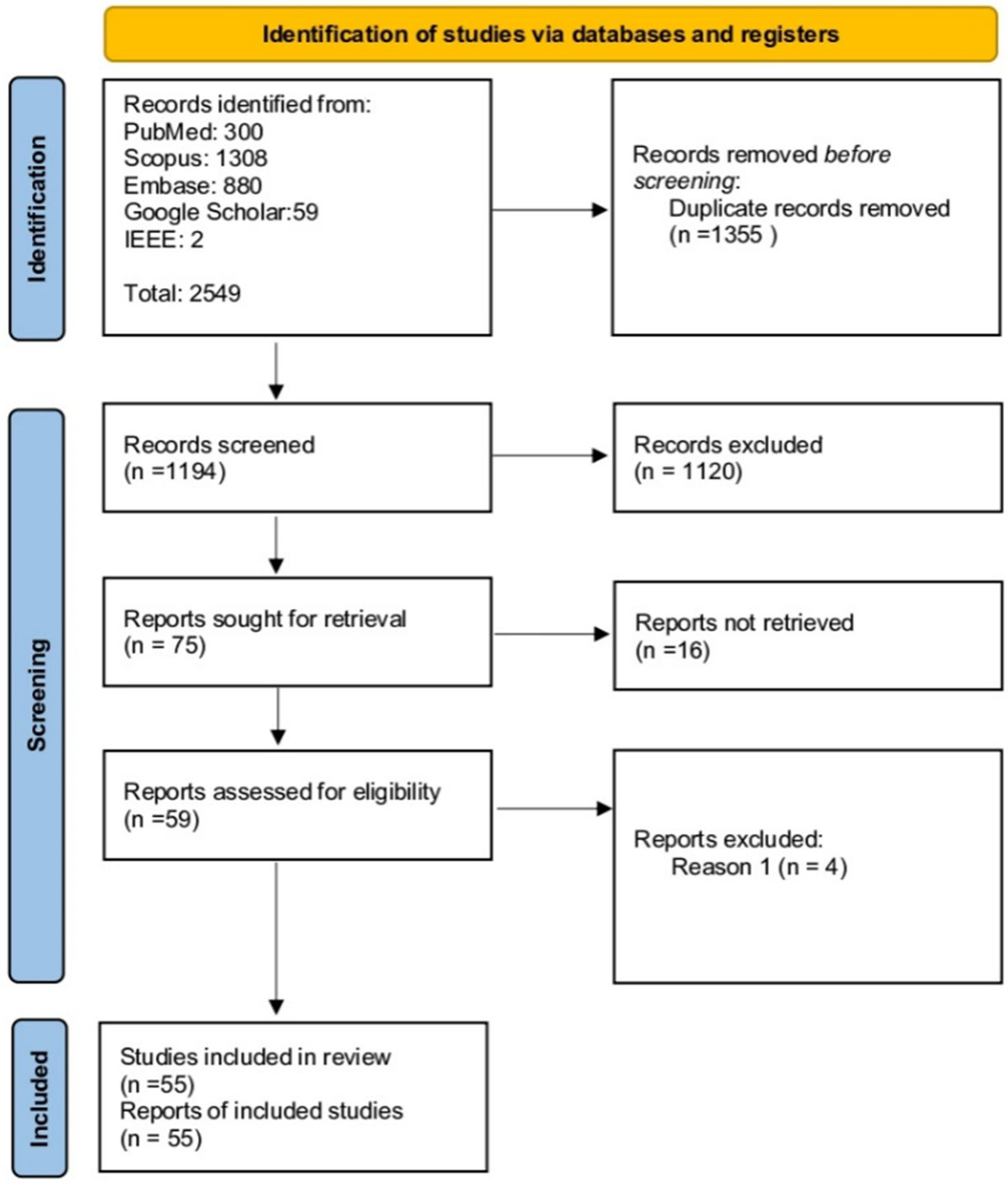
The review process adhered to the Preferred Reporting Items for Systematic Reviews and Meta-Analyses (PRISMA) guidelines [6], and the completed PRISMA 2020 checklist is provided in Supplementary Table S1. This review was not registered with PROSPERO or any other protocol registry. The decision not to register reflects the methodological and exploratory nature of the review, which focuses on AI techniques rather than clinical outcomes; however, we acknowledge this as a limitation. After merging results and removing duplicates, articles were screened based on title and abstract. Exclusion criteria included non-English articles, inaccessible full-texts, and publications that were reviews, editorials, book chapters, or case studies. The remaining articles underwent a full-text examination to ensure they met the inclusion criteria: a primary focus on the application of an AI model to NPC analysis. This screening was performed independently by two individuals. From an initial pool of 2549 articles, 55 studies were ultimately selected for inclusion in this systematic review (Figure 1). Data extracted from each study included the author, publication year, sample size, AI algorithm, application type, and key findings.
For each included study, we extracted information on (1) whether data leakage was prevented (e.g., patient-level separation between training and testing sets, avoiding same-patient images across splits); (2) validation approach (internal validation only, cross-validation, or external validation on an independent dataset); and (3) external validation details (single-center vs. multi-center, geographic distribution, sample size).

3. Clinical and Pathological Landscape of NPC

3.1. Epidemiology and Risk Factors

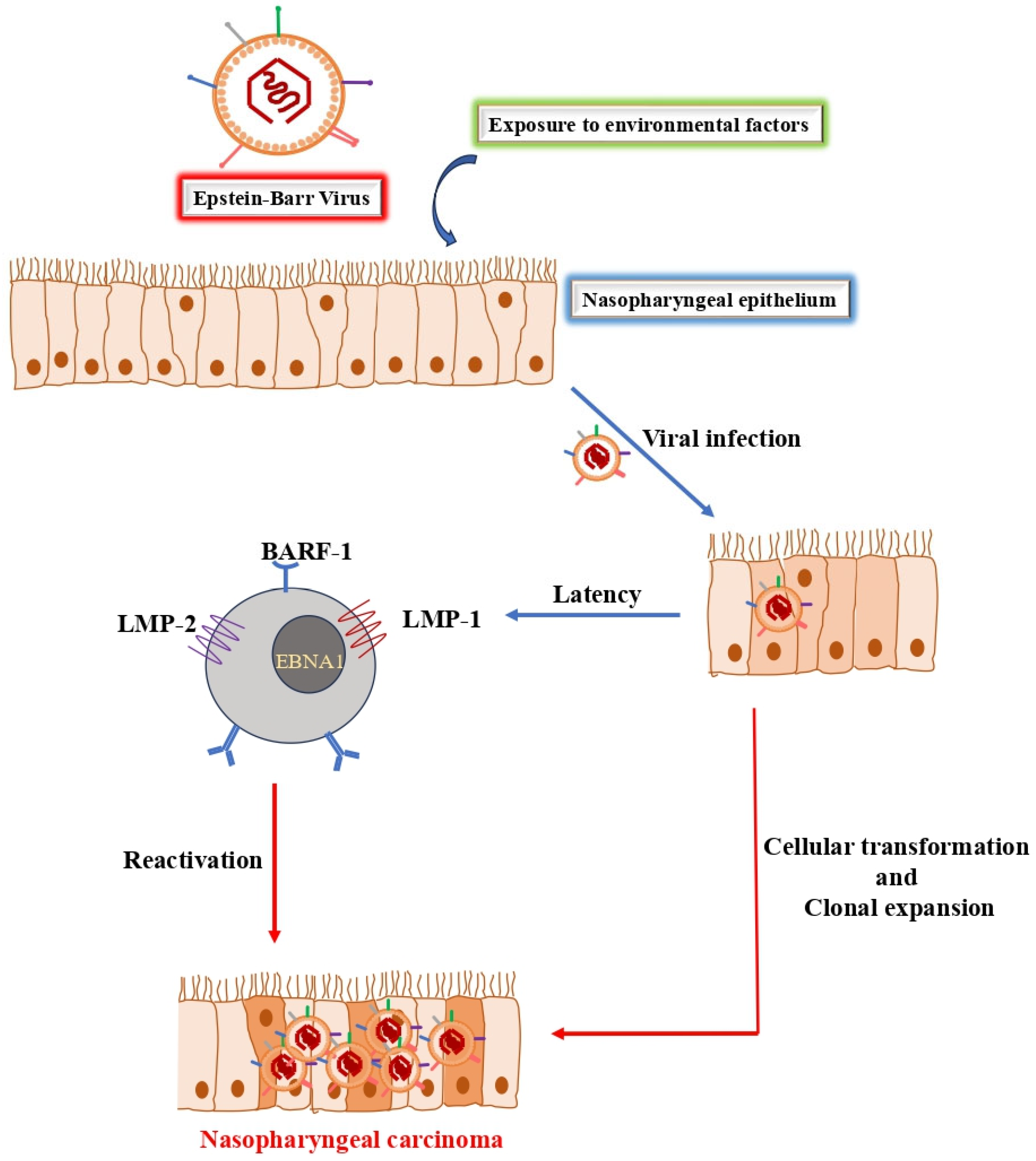
NPC is a malignancy with a unique epidemiology, characterized by low incidence in most of the world, typically under 1 per 100,000 person-years, but with endemic pockets in Southeast Asia, North Africa, the Middle East, and among Arctic Inuit populations [7]. In 2018, an estimated 129,079 new cases were diagnosed globally, with a striking 85% occurring in Asia, making it the 23rd most common cancer by incidence worldwide [8]. Its etiology is multifactorial, involving a complex interplay of genetic susceptibility, environmental exposures, and viral oncogenesis (Figure 2), primarily driven by the EBV and, to a lesser extent, the Human Papillomavirus (HPV) [9].
Incidence varies significantly by ethnicity, even within the same geographic area. In the high-risk Guangdong region of China, the Cantonese-speaking population, particularly the Tanka ethnic group, historically exhibits double the NPC risk compared to other groups like the Hokkien or Hakka [10]. This pattern is mirrored in Southeast Asia; in Singapore, incidence rates are highest among the Chinese population, intermediate among Malays (who have a history of intermarriage with Chinese), and lowest among Indians, correlating with the degree of ethnic admixture with southern Chinese populations [8]. Migration studies further underscore the role of genetics and early-life exposures; when high-risk groups move to low-risk regions, their NPC incidence declines but remains elevated compared to the local population [11]. The disease is two to three times more common in males. In endemic regions, incidence peaks between ages 50 and 59, whereas in low-risk areas, it rises steadily with age, peaking later at 65–69 years [12].

3.2. Histopathology, Anatomy, and Clinical Presentation

The World Health Organization classifies NPC into keratinizing squamous cell carcinoma, nonkeratinizing carcinoma (subdivided into differentiated and undifferentiated), and basaloid squamous cell carcinoma [8]. The undifferentiated nonkeratinizing subtype is overwhelmingly dominant in high-incidence regions and is strongly associated with EBV. In contrast, the keratinizing subtype is more common in low-incidence settings [13]. Beyond histology, genetic and epigenetic alterations are hallmarks of NPC. Somatic mutations, copy number changes, and aberrant DNA methylation of tumor suppressor genes like RASSF1 and CDKN2A are considered key events in its tumorigenesis [14,15].
The clinical presentation of NPC is dictated by the unique anatomy of the nasopharynx. This space communicates anteriorly with the nasal cavity and is bounded superiorly by the base of the skull, which can become eroded in late-stage disease. A critical landmark for local spread is the fossa of Rosenmüller (pharyngeal recess), located posterosuperior to the torus tubarius on the lateral wall; this is the most common site of tumor origin [12,16]. Due to the nasopharynx’s rich lymphatic network, early and frequent cervical lymph node involvement is common, with a painless upper cervical mass being the presenting symptom in 75–80% of patients [17]. Other early symptoms often include ear-related issues (due to Eustachian tube dysfunction), nasal discharge, and epistaxis [17,18]. Because these signs can be non-specific, diagnosis is often delayed until the disease is locally advanced.

3.3. Current Standards for Diagnosis and Treatment

The diagnostic and staging pathway for NPC, as outlined by ESMO-EURACAN guidelines, is multi-modal. It begins with a thorough medical history and physical examination, followed by endoscopic visualization and biopsy of the nasopharynx (nasopharyngoscopy) [19]. Imaging is central to staging, with MRI of the nasopharynx and neck being the preferred modality for assessing soft tissue and intracranial invasion, supplemented by CT for evaluating bony erosion. 18F-fluorodeoxy-glucose-PET/CT imaging is standard for detecting distant metastases and completing the staging process [19]. In addition, circulating cell-free EBV DNA has emerged as a critical biomarker for screening, prognostication, and monitoring treatment response [9].
Treatment has evolved significantly, leading to improved clinical outcomes. Intensity-modulated radiotherapy (IMRT) is the standard radiation technique, offering superior tumor target conformity and reduced toxicity. Advanced techniques like proton therapy are increasingly used for radioresistant or recurrent disease due to their favorable dosimetric profile [20]. For locoregionally advanced NPC, concurrent chemoradiotherapy is the cornerstone of treatment, often preceded by induction chemotherapy. In the metastatic setting, combination chemotherapy regimens and, more recently, immunotherapy have improved survival. Immune checkpoint inhibitors and therapeutic vaccines targeting EBV-associated antigens represent a rapidly advancing frontier in NPC management [21,22].

3.4. The Central Role of Digital Imaging and Pathology in Modern NPC Care

Modern personalized medicine for NPC requires the integration of diverse data, including clinical information, genomics, and, critically, imaging. Digital imaging is indispensable across the entire patient journey, from initial diagnosis and staging to treatment response evaluation. CT provides high-resolution visualization essential for delineating bone infiltration at the skull base and mapping tumor extension into adjacent tissues [23]. MRI is superior to CT for soft tissue contrast, making it invaluable for precisely assessing the boundaries of the primary tumor and its invasion of nearby structures. It is especially crucial for evaluating post-treatment recurrence [24]. WSI, a product of digital pathology, enables computational analysis of histopathological slides. This has opened new avenues for prognosis prediction, risk stratification, and predicting therapeutic response based on microscopic tumor features and the tumor microenvironment [25,26,27,28].
The increasing speed and resolution of imaging technology have led to an explosion of data, far exceeding the capacity for manual analysis by clinicians [29]. This data-rich environment, particularly the fusion of radiologic (CT/MRI) and pathologic (WSI) information, provides a comprehensive, multi-scale view of the tumor [30,31,32]. It is precisely this challenge of complex, large-scale data analysis that has spurred researchers to apply AI, using radiomics and DL to unlock the full diagnostic and prognostic potential contained within these images [33].

4. Applications of AI in NPC Image Analysis

The automated analysis of medical images is a cornerstone of modern oncology, yet significant challenges remain, particularly in image segmentation. The inherent variability in the size, shape, and location of tumors like NPC, often coupled with blurred edges between malignant and healthy tissue, complicates this crucial first step [34]. Manual delineation by experts, while the traditional standard, is both time-consuming and prone to inter-observer variability. Therefore, the development of robust, automated segmentation methods is essential for accelerating diagnosis, standardizing treatment planning, and enabling large-scale quantitative analysis.
DL architectures, particularly those based on the U-Net model, have become the state of the art for this task. The typical workflow involves preprocessing steps such as image resizing, normalization, and data augmentation, followed by feeding the data into a deep neural network for segmentation. Post-processing techniques are then applied to refine the output [35]. However, it is important to note that most existing studies in this field are based on relatively small and heterogeneous datasets, and therefore, the reported results should be interpreted as exploratory and hypothesis-generating rather than definitive clinical evidence. The following sections review the application of these AI-driven methods across the primary imaging modalities used in NPC care.

4.1. AI-Powered Analysis of CT Images

CT is a foundational imaging modality in oncology, providing high-resolution, three-dimensional reconstructions of anatomy that are vital for assessing tumor invasion, particularly into bony structures at the skull base [23]. While historically used for quantifying organ and tissue size [36], its rich informational content has made it a prime target for DL applications aimed at improving NPC diagnosis and predicting outcomes [35].
Early computational approaches to NPC prognosis relied on radiomics, where features are extracted from images and analyzed using machine learning models like support vector machines (SVM) or random forests (RF) [37,38]. However, recent efforts have shifted toward end-to-end DL models that learn relevant features automatically. For instance, Men et al. [39] developed a deep deconvolutional neural network (DDNN) that uses a VGG-16-based encoder to extract features and a decoder to generate pixel-level segmentation maps of the tumor. Taking a different approach, Daoud et al. [40] introduced a cascade system that first eliminates non-target regions from CT scans and then integrates segmentation results from three orthogonal planes (axial, coronal, and sagittal) to achieve high detection performance. Other studies have successfully employed a range of established convolutional neural networks (CNN) architectures, including ResNet50, Xception, and U-Net, to predict treatment outcomes from CT data, demonstrating the versatility of these models in the NPC clinical workflow [41,42].
Nevertheless, these findings are derived from limited sample sizes and relatively few clinical events. Several reported hazard ratios exhibit wide confidence intervals, suggesting potential instability in model estimates. Therefore, these results should be interpreted cautiously as preliminary evidence requiring validation in larger, independent cohorts.
Table 1 summarizes several studies that apply various AI models, predominantly DL networks like U-Net and CNNs, to analyze CT scans. The primary applications were segmentation, image synthesis, and prediction. Many studies use AI to automatically outline tumors and critical organs (organs-at-risk), which streamlines radiotherapy planning and improves consistency [34,39,42,43,44,45]. Models like CycleGAN and deep convolutional neural network (DCNN) are used to generate higher-quality CT images from other sources (e.g., Cone-Beam CT), enhancing the accuracy of treatment planning [46,47]. AI is also used for clinical predictions, including diagnosing diseases [40], forecasting patient prognosis [37,41], and predicting treatment outcomes [48]. Overall, the findings consistently show that these AI methods significantly improve the accuracy, efficiency, and consistency of various clinical tasks compared to traditional approaches. However, human oversight remains essential, and current evidence should not be interpreted as conclusive for clinical deployment.

4.2. Enhancing MRI with DL

For NPC, MRI is the preferred modality for primary tumor assessment due to its unparalleled soft-tissue contrast. This superior resolution allows for precise delineation of the primary tumor, evaluation of local tissue invasion, and detection of retropharyngeal lymph node involvement, which are critical for accurate staging and radiotherapy planning [33,49]. Furthermore, MRI avoids the use of ionizing radiation, making it ideal for repeated imaging during patient follow-up [35].
The detailed anatomical information captured by MRI makes it highly suitable for DL analysis. A recent meta-analysis by Wang et al. [49] confirmed the effectiveness of DL models for NPC segmentation on MRI, reporting a pooled mean Dice score of 78%, which indicates a high degree of precision. Beyond segmentation, AI models are being developed for prognosis. MRI-based radiomic features have shown significant potential for non-invasively predicting progression-free survival (PFS) in patients with advanced NPC by capturing underlying intratumoral heterogeneity [50]. For example, Qiang et al. [51] designed a prognostic system that combines a 3D-CNN to extract MRI features with an XGBoost algorithm to integrate key clinical data. This hybrid approach proved effective in predicting disease-free survival (DFS) and aiding treatment decisions for patients with locally advanced disease. The methodologies, key studies, and findings are summarized in Table 2, including other studies [50,51,52,53,54,55,56,57,58,59,60,61,62,63,64,65,66].
Table 2 aggregates studies employing AI models to analyze MRI data, with the most common application being prognosis and outcome prediction [50,51,52,53,54,55,56,57,58]. These researchers use a variety of models, from radiomics and traditional machine learning algorithms like SVMs to advanced DL networks like 3D CNNs and SE-ResNet for predicting patient outcomes, such as PFS and distant metastasis-free survival (DMFS). Although several studies report improved predictive performance compared to standard clinical staging systems (e.g., TNM/AJCC), these findings are derived from limited and often single-institution datasets. As such, the robustness and generalisability of these models remain uncertain.
These studies found that AI-driven models performed better than the standard clinical staging systems (e.g., TNM/AJCC), allowing for more personalized and accurate patient care. Another major focus is using AI for segmentation, which involves precisely outlining the tumor’s location and size (Gross Tumor Volume or GTV) [59,60,61,62]. Models like 3D CNNs and specialized networks like Dense connectivity embedding U-net (DEU-Net) were frequently used for this task. These automated AI segmentation technique significantly improves the accuracy and consistency of tumor contouring compared to manual methods. However, variability in reported performance and occasional overestimation of target volumes indicate that further refinement is still required. This is crucial for precise radiation therapy planning, although one study noted that models can sometimes overestimate the target volume, indicating a need for continued refinement. Other AI models were also being developed for the early detection of cancer and the prediction of treatment-related side effects [63,64,65,66]. For instance, RF models have been used to predict the likelihood of radiation-induced temporal lobe injury (RTLI). It is clear from these studies that AI demonstrates strong performance in the automated detection of NPC from MRI scans, aiding in early diagnosis. Furthermore, by proactively predicting treatment toxicity, these models allow doctors to adjust treatment plans and intervene in a timely manner to improve patients’ quality of life.
Table 2. Studies employing MRI in the diagnosis, prognosis, or classification of NPC.
RefMediumNModelTaskKey FindingMetrics (%)
MRI
Zeng et al., 2022 [50]MRI110MRI-based RadiomicsPrognosis PredictionRadiomics model was powerful in predicting PFS in advanced NPC.81.4
Qiang et al., 2021 [51]MRI34443D CNNPrognosis PredictionFramework captured complex information to predict prognosis in locally advanced NPC (LA-NPC).77.6
Du et al., 2019 [52]MRI277SVMPrognosis PredictionFindings support the growing evidence for the role of radiomics in evaluating NPC.80
Cui et al., 2020 [53]MRI792AutoML (Ridge, Lasso)Prognosis PredictionAutoML algorithm demonstrated better prognostic performance than the standard TNM/AJCC staging system.79.6
Zhao et al., 2020 [54]MRI123Multiparametric RadiomicsTreatment OutcomeModel showed promise in predicting outcomes for patients receiving induction chemotherapy.86.3
Zhang et al., 2021 [55]MRI233DCNNPrognosis PredictionDL model integrating MRI features and clinical data effectively predicted distant metastasis-free survival (DMFS).79.6
Zhong et al., 2021 [56]MRI1872SE-ResNetPrognosis PredictionNomogram predicts survival for T3N1M0 patients across different treatments, facilitating individualized care.79.6
Li et al., 2022 [57]MRI206DL (Ensembling model)Prognosis PredictionPost-treatment MRI features were shown to be significant for prognosis prediction and clinical decision-making.84.2
Zhang et al., 2023 [58]MRI151DNNPrognosis PredictionModel provided accurate PFS prediction without relying on specific tumor region-of-interest annotations.88.0
Lin et al., 2019 [59]MRI10213D CNNSegmentationModel improved the accuracy of primary gross tumor volume (GTV) contouring.79.0
Ye et al., 2020 [60]MRI44DEU-NetSegmentationA fully automated method using dual-sequence MRI demonstrated accurate and stable NPC segmentation.88.0
Wong et al., 2021 [61]MRI195CNN, U-NetSegmentationCNN showed potential for accurate NPC delineation on non-contrast MRI, but consistently overestimated target volumes.71.0
Luo et al., 2023 [62]MRI1057GTVp segmentation modelSegmentationMethod demonstrated highly accurate GTV segmentation across multi-center MRI images, showing its versatility.88.0
Ke et al., 2020 [63]MRI4100SC-DenseNetDiagnosis/DetectionModel demonstrated strong performance in the automated detection of NPC from MRI scans.77.0
Zhang et al., 2020 [64]MRI242RFToxicity PredictionRadiomic models enabled proactive prediction of RTLI, allowing for timely intervention.83.0
Bin et al., 2022 [65]MRI98SVM, RFToxicity PredictionNomogram developed to predict risk of radiation-induced temporal lobe injury (RTLI) for treatment optimization.82.0
Wong et al., 2021 [66]MRI412CNNDiagnosis/DetectionModel showed good performance for the early detection of NPC from MRI.96.0

4.3. Computational Pathology: AI in WSI Analysis

Histopathological analysis of biopsied tissue remains the definitive gold standard for cancer diagnosis. The accuracy of this process, however, has traditionally depended heavily on the experience of the pathologist [67]. The advent of digital pathology, which involves scanning glass slides to create giga-pixel WSIs, has revolutionized the field by enabling computational analysis. While managing the immense size and complexity of WSIs presents a challenge, DL has emerged as a powerful tool to automate analysis, enhance diagnostic accuracy, and reduce pathologists’ workload [67,68].
These “pathomics” models extract quantitative features from WSIs that can reveal molecular and genetic characteristics correlated with tumor prognosis and therapeutic response [69]. In the context of NPC, Hou et al. demonstrated that a pathomics model derived from WSIs significantly outperformed an MRI-based radiomics model in predicting PFS [69]. This highlights the unique prognostic value contained within the microscopic tissue architecture, which can now be unlocked through AI. The development of such decision support tools is poised to improve the efficiency and consistency of pathological evaluation, ultimately leading to better patient outcomes [68]. Despite the utility of DL models, their lack of interpretability hinders clinical use where transparency is crucial. To solve this, researchers developed a more transparent framework called the Handcrafted Histological Transformer (H2T) [70]. This new H2T model offers competitive performance compared to state-of-the-art methods, is more easily understood, and is up to 14 times faster. Additionally, the development of a novel “mutual transformer learning”, where 2 modules iteratively train each other to generate and clean pseudo-labels without human input, has also helped to mitigate the expensive and labour-intensive manual labelling by pathology experts [71].
A summary of WSI modalities is summarized in Table 3 and highlights the application of AI and DL to analyze WSIs in digital pathology using models like CNNs and Vision Transformers (ViT) to automate and enhance the diagnostic and prognostic workflow. A significant theme across these studies is the drive towards creating a more efficient and less labor-intensive system [70,71]. Newer models are being developed to be faster, more cost-effective, and streamlined for downstream analysis (e.g., H2T). Nevertheless, most existing studies are retrospective and based on limited sample sizes, which restricts the generalisability and robustness of the reported findings. The most common task demonstrated by AI is diagnosis and classification [27,67,72,73]. These models have proven to be robust and accurate for WSI-level classification. They can successfully identify cancerous tissue, often without needing extensive manual annotations from pathologists, and can serve as a valuable “second opinion” to assist in clinical diagnosis. Beyond simple classification, other models were used to predict patient outcomes and identify new biomarkers directly from the tissue morphology in WSIs [74,75,76]. By analyzing microscopic features, models like DeepSurv and MorphResNet can act as reliable prognostic tools for predicting survival risk. Furthermore, AI can quantify protein expression from images, establishing new digital biomarkers that are valuable for forecasting poor prognosis and potentially guiding treatment decisions. However, these findings remain preliminary and should be interpreted as exploratory due to limited validation datasets and variability across studies.

4.4. Multi-Modal Data Integration for Comprehensive Prognostication

While individual modalities provide valuable information, the future of precision medicine in NPC lies in the integration of multi-modal data. By combining clinical variables with data from imaging (radiomics), treatment plans (dosiomics), and pathology (pathomics), AI models can create a more holistic and accurate picture of the disease.
Several studies have demonstrated the power of this approach. Dong et al. [77] developed models that fused clinical data with radiomics and dosiomics information from multiple imaging modalities (CT and MRI) and tumor regions to successfully predict the occurrence of severe acute oral mucositis, a common side effect of radiotherapy. Similarly, Gu et al. [78] created a multi-task deep learning-based radiomic (MTDLR) nomogram that accurately predicts PFS in locally advanced NPC by integrating multiple data types, effectively stratifying patients into distinct risk groups beyond the capabilities of traditional models. Furthermore, frameworks that integrate CT and MRI have been shown to enhance diagnostic accuracy and streamline treatment planning by providing complementary information on bone and soft-tissue invasion [79].
Table 4 highlights a diverse range of studies from which AI models leverage multi-modal data, integrating information from different sources like CT, MRI, PET scans, pathology slides (WSI), endoscopy, and treatment dose plans. The central theme is that combining these data sources provides a more comprehensive view of the patient’s condition, leading to more powerful and accurate AI applications. A dominant application is in prognosis and toxicity prediction [69,77,78,80,81,82,83], where the goal is to forecast patient outcomes or the likelihood of treatment side effects. The key strategy is combining different feature sets for a more robust prediction.
Although multi-modal models often report superior performance compared to single-modality approaches, these improvements are derived from limited datasets and may not consistently generalise across institutions. Moreover, where reported, the absolute improvements in discrimination Metrics (%) (e.g., C-index, AUC) are often modest, and no study included in this review performed robust external validation across independent multi-center cohorts. Therefore, their clinical utility remains to be fully validated.
The synergy of multi-modal data is crucial. Models that integrate imaging features (radiomics), pathology data (pathomics), and treatment plan information (dosiomics) consistently demonstrate superior performance in predicting patient survival and treatment toxicity compared to models using only a single data source. This allows for better patient stratification and more personalized treatment strategies. Another application is segmentation and image synthesis with a focus on improving image analysis and streamlining clinical workflows, particularly for radiation therapy [79,84,85,86]. Here, AI can use information from one imaging modality to improve the analysis of another (e.g., using CT data to accelerate MRI segmentation). Furthermore, DL models like Generative Adversarial Networks (GANs) can accurately synthesize one type of image from another (e.g., creating a pseudo-CT from an MRI), which is essential for accurate dose calculations in treatment planning. Several models are also being used to improve diagnostic accuracy and automate clinical tasks such as treatment planning [87,88,89,90,91]. Recurrent Neural Networks (RNNs) can accurately predict dose-volume histograms (DVHs), automating a key component of treatment planning. In diagnostics, DL models applied to endoscopic images have proven highly effective, in some cases outperforming human oncologists in detecting malignancies and automatically outlining tumors. However, despite these promising results, most findings remain preliminary due to limited sample sizes, retrospective study designs, and a lack of large-scale external validation. These multi-modal strategies represent a significant step toward developing truly personalized treatment plans for patients with NPC.
Table 4. Studies employing multi-modal applications and other tools in the diagnosis, prognosis, or classification of NPC.
RefMediumNModelTaskKey FindingMetrics (%)
Multi-modal Applications and other Tools
Hou et al.,
2024 [69]
MRI, WSI357RadiopathomicsPrognosis PredictionCombined radiomics and pathomics model reliably predicted PFS and stratified patient risk.79.1
Dong et al., 2023 [77]CT, Dose243Radiomics, DosiomicsToxicity PredictionIntegrating radiomics and dosiomics data best predicted severe acute oral mucositis (AOM).81.0
Gu et al., 2023 [78]Imaging, Clinical886MTDLR NomogramPrognosis PredictionMulti-task nomogram provided reliable prognostic prediction and better patient stratification.85.9
Qi et al., 2021 [79]CT, MRI149CNNSegmentationCombining multi-modality images generated reliable segmentations to guide dose painting.88.2
Peng et al., 2019 [80]PET, CT707DCNNPrognosis PredictionDL PET/CT radiomics could serve as a powerful tool for prognosis and treatment planning.75.4
Gu et al., 2020 [81]PET, CT2573D CNNPrognosis PredictionDL radiomics from PET/CT may complement the existing TNM staging system.84.2
Ren et al., 2021 [82]Dose145LR, SVM, RF, KNNToxicity PredictionDosiomics model accurately forecasted radiation-induced hypothalamic toxicity.70.0
Fitton et al., 2011 [83]CT, MRI5Snake AlgorithmSegmentationAlgorithm accelerated delineation on MRI by incorporating morphological information from CT.-
Ma et al., 2021 [84]CT, MRI20U-Net, pix2pix GANImage SynthesisDL model accurately predicted pseudo-CT from MRI for precise dose calculations.-
Chen et al., 2022 [85]MRI, CT206DCNN (Attention)Image SynthesisAttention-based DCNN significantly improved synthesized CT quality for dosimetry.99.1
Li et al., 2018 [86]Endoscopy7951CNN (eNPM-DM)Diagnosis & SegmentationModel outperformed oncologists in diagnosing malignancy and auto-segmented tumors from endoscopic images.88.7
Cao et al., 2020 [87]CT, Dose100Gated Recurrent Unit—Recurrent Neural Network (GRU-RNN)Treatment PlanningGRU-RNN model was capable of accurate DVH prediction.-
Zhuang et al., 2021 [88]Dose124GRU-RNNTreatment PlanningSimplified GRU-RNN performed well even with small DVH samples, showing potential for use with limited data.97.6
Sun et al., 2022 [89]Dose (IMRT)117U-NetTreatment PlanningA hybrid optimization method showed clinical feasibility in generating acceptable IMRT plans.-
Xu et al., 2022 [90]Endoscopy4783DCNNDiagnosis/DetectionProvided a valuable reference for NPC screening using white light and narrow-band imaging.98.6
Zhang et al., 2020 [91]MRI, WSI220DCNNPrognosis PredictionMulti-scale imaging features from MRI and WSI demonstrated complementary prognostic value.94.9

5. Discussion: Challenges and Future Perspectives

Before discussing specific challenges, it is important to clarify the intended scope of this review’s clinical commentary. Where the following sections mention potential applications—such as using AI to tailor follow-up schedules, select patients for clinical trials, or guide treatment decisions—these are presented as hypothetical future directions based on the biological and technical plausibility of the reviewed studies. They are not intended as practical recommendations for current clinical practice. As detailed throughout this review, the overwhelming majority of included studies are retrospective, single-institution, and lack external validation. Consequently, any discussion of clinical utility should be understood as aspirational and contingent upon future prospective, multi-center validation.
While AI has demonstrated considerable promise in NPC research, its translation into routine clinical practice is contingent upon overcoming several significant challenges. The following discussion synthesizes the primary limitations of current research and outlines promising directions for future innovation.

5.1. Overcoming Data-Related Hurdles

A primary limitation of the current body of research is the issue of data generalizability. The vast majority of studies are retrospective and rely on small, private datasets from single institutions [55,80,92,93]. External validation was performed in only a small fraction of studies, and no study reported prospective validation or real-world clinical deployment. Models trained on such homogenous data often fail to perform adequately when applied to external datasets from other institutions, which may use different scanning protocols, resulting in variations in image contrast and field of view [61]. This “domain shift” severely limits the broad applicability of these models. Furthermore, as a relatively uncommon malignancy, NPC research suffers from smaller dataset sizes compared to more prevalent cancers, which poses a particular challenge for data-hungry DL models [2]. Addressing these issues requires a concerted effort toward standardization and collaboration. The establishment of reliable, multicenter data-sharing platforms is crucial. Such initiatives, however, must incorporate robust technical solutions for data security and patient privacy to be successful. In addition, larger, prospectively collected datasets with sufficient event numbers are needed to improve the statistical stability and clinical reliability of AI models. Standardized annotation protocols and large-scale, externally validated studies are paramount to building AI tools with the accuracy and reliability required for clinical adoption.
The reviewer correctly notes that several important clinical variables are under-addressed in the literature we reviewed. While our review focuses on imaging-based AI models, many of the included studies failed to adequately report or adjust for key prognostic factors such as Eastern Cooperative Oncology Group (ECOG) performance status, treatment compliance, HIV co-infection status, or complete HPV subtyping data (beyond EBV, which is the dominant driver in endemic regions). The absence of these variables represents a significant limitation of the primary evidence base, as unmeasured confounding may artificially inflate reported model performance. We strongly recommend that future AI studies in NPC systematically collect and adjust for these clinical covariates to improve real-world generalizability.
Across the studies reviewed that addressed treatment-related toxicity (e.g., RTLI, oral mucositis, hypothyroidism), the reporting of late toxicity was notably limited and inconsistent. Several studies provided only acute toxicity outcomes or lacked standardized follow-up durations. Without systematic, long-term toxicity data—ideally graded by standardized criteria such as CTCAE—the safety profiles of AI-predicted or AI-optimized treatment strategies remain incompletely characterized. We recommend that future AI studies incorporate prospective, longitudinal toxicity assessment with a minimum 2–5 year follow-up where feasible.
We assessed the 55 included studies for validation strategy, presence of external validation, and reporting of data leakage safeguards (e.g., patient-level separation between training and test sets). The majority of studies employed internal validation methods, primarily cross-validation or holdout splitting. Explicit reporting of data leakage prevention measures, such as ensuring that no images from the same patient appeared in both training and test sets, was uncommon; most studies did not mention this critical issue. These observations highlight persistent methodological gaps and underscore the need for more rigorous validation practices and transparent reporting in future AI studies for NPC.

5.2. Advancing Model Sophistication and Interpretability

Current AI models for NPC face technical and conceptual limitations. Many models focus exclusively on the primary tumor region, ignoring the surrounding peritumoral environment, which contains valuable prognostic information. Future models could achieve greater predictive accuracy by incorporating features from this region. Furthermore, the inherent biological complexity of NPC, characterized by a high number of admixed inflammatory cells and poorly differentiated tumor cells, makes automated identification more difficult than in other cancer types. A promising technical solution for WSI analysis is Weakly-Supervised Multiple Instance Learning (MIL). Methods like Clustering-constrained Attention Multiple Instance Learning can process entire gigapixel WSIs by dividing them into smaller patches and using attention mechanisms to identify diagnostically relevant subregions without requiring laborious pixel-level annotation [32,94]. This approach is not only efficient but also enhances model interpretability by generating heatmaps that highlight the areas the model deems important. However, despite these advantages, the clinical utility of such methods remains to be fully established due to limited external validation and variability in study design. Overcoming the “black box” nature of AI is critical for eventual clinical adoption. Should these tools ever be deployed in practice, clinicians will need algorithms whose decision-making processes are transparent and understandable. However, this remains a long-term goal, as current models—even those with interpretability features—have not yet been validated in prospective clinical settings.
Beyond technical performance, several translational barriers must be addressed before AI models can be integrated into routine NPC care. Interpretability remains a key challenge, as most current models function as ‘black boxes’ without transparent reasoning; clinicians require explainable outputs—such as heatmaps or feature attributions—to trust and verify AI recommendations. Workflow integration is also critical: AI tools must seamlessly interface with existing systems such as PACS, radiotherapy planning platforms, and electronic health records without requiring manual data transfer or offline processing. Clinician trust depends on prospective validation and demonstrated clinical benefit, yet no NPC-specific AI tool has undergone such evaluation. Finally, regulatory approval pathways (e.g., FDA, CE marking) require rigorous evidence of safety and efficacy that current retrospective studies cannot provide. Addressing these barriers will require interdisciplinary collaboration among AI researchers, radiologists, oncologists, and regulatory bodies.

5.3. Exploring New Frontiers: Radiogenomics and Multimodality

A largely unexplored yet emerging area of interest for AI in NPC research is radiogenomics. This field aims to build correlations between quantitative imaging features and underlying molecular data, such as genomic signatures and protein expression [95]. By non-invasively predicting tumor subtypes, prognoses, and therapeutic responses, radiogenomics may offer potential as a component of personalized medicine for NPC, mirroring its success in other cancers like glioma and breast cancer [96,97]. Future research should also prioritize the development of AI models capable of multitasking and multi-modal integration. The ultimate goal is to develop systems that are able to integrate data from CT, MRI, WSI, and clinical records to provide a single, comprehensive prediction for diagnosis, prognosis, and treatment response. Recent studies and reviews have further demonstrated the expanding role of AI across diverse cancer types and imaging modalities, highlighting its broader translational potential in precision oncology [98,99,100,101,102].
A systematic analysis of clinical readiness reveals that most studies remain at proof-of-concept or technical validation stages, with only a small minority achieving retrospective clinical validation using external datasets and none reaching prospective validation. Among the few studies with external validation, performance drops are commonly reported, highlighting domain shift across institutions or scanner protocols. Dataset bias is prevalent, as most studies fail to report demographic characteristics or analyze subgroup performance, raising concerns about generalizability across ethnic groups given NPC’s distinct geographic distribution. Robustness testing is rarely performed, and failure modes—such as segmentation errors at tumor boundaries, misclassification of benign hyperplasia as malignant, and poor performance on rare subtypes—are seldom discussed. These gaps collectively underscore the need for rigorous external validation, fairness-aware reporting, and systematic analysis of model failures to advance clinical translation.
We assessed the risk of bias of the included studies using a qualitative adaptation of the QUADAS-AI framework, designed for diagnostic AI studies. Most studies showed high or unclear risk in the ‘index test’ domain because external validation was absent, and model calibration was rarely reported. Patient selection bias was common in retrospective, single-centre studies. Data leakage prevention (e.g., patient-wise splitting) was not explicitly reported in the majority of studies. A study-by-study tabulation was not feasible due to inconsistent reporting across the 55 included studies. Therefore, this qualitative summary reflects the overall methodological limitations of the current evidence base, and results should be interpreted as exploratory.

6. Conclusions

This review highlights the transformative potential of AI in the clinical management of NPC. The key finding is that AI, particularly DL, has demonstrated significant success in automating the analysis of CT, MRI, and WSI data for critical tasks such as tumor segmentation, diagnosis, and survival prediction. The field is rapidly advancing from traditional machine learning based on handcrafted features to sophisticated, end-to-end DL models and integrated multi-modal systems that promise a more holistic approach to patient care.
However, major challenges impede the widespread clinical adoption of these tools. The foremost among these are issues of data generalizability and model robustness, largely due to the prevalence of single-center, retrospective studies with limited data. Additionally, the “black box” nature of many AI algorithms remains a significant barrier to clinical trust and implementation.
Despite these hurdles, the future is promising. Promising directions include the development of interpretable AI frameworks like MIL, the exploration of new frontiers such as NPC-specific radiogenomics, and a necessary shift toward large-scale, multicenter collaborations to create and validate robust models. Ultimately, realizing the long-term potential of AI in this field will require deep and sustained interdisciplinary collaboration between clinicians (radiologists, pathologists, oncologists) and computational scientists. However, at present, the evidence base remains preliminary. None of the reviewed AI models for NPC have undergone the prospective, multi-center validation needed to support routine clinical use. Therefore, while the directions discussed in this review—including automated segmentation, prognostic prediction, and multimodal integration—are scientifically promising, they should be viewed as research priorities rather than practical recommendations. With rigorous validation and continued innovation, AI may one day contribute to personalized NPC care, but that day has not yet arrived.

Supplementary Materials

The following supporting information can be downloaded at: https://www.mdpi.com/article/10.3390/radiation6020016/s1, Table S1: PRISMA 2020 Checklist.

Author Contributions

Conceptualization, M.K.A., S.B.M., and M.F.A.F.; Methodology, M.K.A.; Software, M.K.A.; Validation, M.K.A., A.S.W., and M.S.N.; Formal Analysis, M.K.A.; Investigation, M.K.A.; Data Curation, M.K.A. and A.S.W.; Writing—Original Draft Preparation, M.K.A.; Writing—Review and Editing, S.B.M. and M.F.A.F.; Visualization, M.K.A.; Supervision, S.B.M. and M.F.A.F.; Project Administration, S.B.M.; Funding Acquisition, M.F.A.F. All authors have read and agreed to the published version of the manuscript.

Funding

This research received no external funding.

Institutional Review Board Statement

Not applicable.

Informed Consent Statement

Not applicable.

Data Availability Statement

No new data were created or analyzed in this study.

Acknowledgments

The authors declare that no AI-assisted technologies were used in the preparation of this manuscript.

Conflicts of Interest

The authors declare no conflicts of interest.

Abbreviations

AIartificial intelligence
AutoMLAutomated Machine Learning
AJCCAmerican Joint Committee on Cancer
CTcomputed tomography
CNNconvolutional neural network
DCNNdeep convolutional neural network
DLdeep learning
DEUDense connectivity embedding U-net
DVHdose-volume histogram
EBVEpstein-Barr Virus
eNPM-DMendoscopic image-based nasopharyngeal malignancy detection model
ESMOEuropean Society for Medical Oncology
EURACANEuropean Reference Network for Rare Adult Solid Cancers
GRU-RNNGated Recurrent Unit—Recurrent Neural Network
GANGenerative Adversarial Network
GTVnGross Tumor Volume—nodal
GTVnpGross Tumor Volume of primary NPC
IMRTintensity-modulated radiation therapy
KNNk-nearest Neighbors
LA-NPCLocally Advanced Nasopharyngeal Carcinoma
LRLogistic Regression
MLMachine Learning
MRImagnetic resonance imaging
NPCnasopharyngeal carcinoma
ODS netorgans-at-risk detection and segmentation network
OARsorgans-at-risk
PFSprogression-free survival
PET/CTPositron emission tomography with computed tomography
PTVnPlanning Target Volume—nodal
RS-CNNRaman-specified convolutional neural networks
ResNetResidual Network
RTLIradiation-induced temporal lobe injury
RHTradiation-induced hypothalamic toxicity
RFrandom forest
SE-ResNetSqueeze-and-Excitation Residual Network
SVMsupport vector machines
TNMTumor, Node, Metastasis
WSIWhole slide imaging
WS-T2T-ViTTokens-to-Tokens Vision Transformer
3D-CNNThree-dimensional convolutional neural network
VGGVisual Geometry Group
3DThree dimensions

References

  1. Ferlay, J.; Colombet, M.; Soerjomataram, I.; Parkin, D.M.; Pineros, M.; Znaor, A.; Bray, F. Cancer statistics for the year 2020: An overview. Int. J. Cancer 2021, 149, 778–789. [Google Scholar] [CrossRef]
  2. Ng, W.T.; But, B.; Choi, H.C.W.; de Bree, R.; Lee, A.W.M.; Lee, V.H.F.; Lopez, F.; Makitie, A.A.; Rodrigo, J.P.; Saba, N.F.; et al. Application of Artificial Intelligence for Nasopharyngeal Carcinoma Management—A Systematic Review. Cancer Manag. Res. 2022, 14, 339–366. [Google Scholar] [CrossRef]
  3. Wu, L.; Li, C.; Pan, L. Nasopharyngeal carcinoma: A review of current updates. Exp. Ther. Med. 2018, 15, 3687–3692. [Google Scholar] [CrossRef] [PubMed]
  4. Wong, K.C.W.; Hui, E.P.; Lo, K.W.; Lam, W.K.J.; Johnson, D.; Li, L.; Tao, Q.; Chan, K.C.A.; To, K.F.; King, A.D.; et al. Nasopharyngeal carcinoma: An evolving paradigm. Nat. Rev. Clin. Oncol. 2021, 18, 679–695. [Google Scholar] [CrossRef]
  5. Li, S.; Deng, Y.Q.; Zhu, Z.L.; Hua, H.L.; Tao, Z.Z. A Comprehensive Review on Radiomics and Deep Learning for Nasopharyngeal Carcinoma Imaging. Diagnostics 2021, 11, 1523. [Google Scholar] [CrossRef]
  6. Page, M.J.; McKenzie, J.E.; Bossuyt, P.M.; Boutron, I.; Hoffmann, T.C.; Mulrow, C.D.; Shamseer, L.; Tetzlaff, J.M.; Akl, E.A.; Brennan, S.E.; et al. The PRISMA 2020 statement: An updated guideline for reporting systematic reviews. BMJ 2021, 372, n71. [Google Scholar] [CrossRef]
  7. Schiess, E.; Jensen, K.H.; Kristensen, M.H.; Johansen, J.; Eriksen, J.G.; Maare, C.; Andersen, M.; Farhadi, M.; Hansen, C.R.; Overgaard, J.; et al. Epidemiology and treatment outcome of nasopharyngeal carcinoma in a low-incidence population—A DAHANCA analysis in Denmark 2000–2018. Acta Oncol. 2024, 63, 850–857. [Google Scholar] [CrossRef] [PubMed]
  8. Chang, E.T.; Ye, W.; Zeng, Y.X.; Adami, H.O. The Evolving Epidemiology of Nasopharyngeal Carcinoma. Cancer Epidemiol. Biomark. Prev. 2021, 30, 1035–1047. [Google Scholar] [CrossRef]
  9. Wee, J.T.; Soong, Y.L.; Chua, M.L. Nasopharyngeal carcinoma-past lessons and a glimpse into the future. Chin. Clin. Oncol. 2016, 5, 14. [Google Scholar] [CrossRef]
  10. Wee, J.T.; Ha, T.C.; Loong, S.L.; Qian, C.N. Is nasopharyngeal cancer really a “Cantonese cancer”? Chin. J. Cancer 2010, 29, 517–526. [Google Scholar] [CrossRef] [PubMed]
  11. Mousavi, S.M.; Sundquist, J.; Hemminki, K. Nasopharyngeal and hypopharyngeal carcinoma risk among immigrants in Sweden. Int. J. Cancer 2010, 127, 2888–2892. [Google Scholar] [CrossRef] [PubMed]
  12. Cantu, G. Nasopharyngeal carcinoma. A “different” head and neck tumour. Part A: From histology to staging. Acta Otorhinolaryngol. Ital. 2023, 43, 85–98. [Google Scholar] [CrossRef]
  13. Vaughan, T.L.; Shapiro, J.A.; Burt, R.D.; Swanson, G.M.; Berwick, M.; Lynch, C.F.; Lyon, J.L. Nasopharyngeal cancer in a low-risk population: Defining risk factors by histological type. Cancer Epidemiol. Biomark. Prev. 1996, 5, 587–593. [Google Scholar]
  14. Dai, W.; Cheung, A.K.; Ko, J.M.; Cheng, Y.; Zheng, H.; Ngan, R.K.; Ng, W.T.; Lee, A.W.; Yau, C.C.; Lee, V.H.; et al. Comparative methylome analysis in solid tumors reveals aberrant methylation at chromosome 6p in nasopharyngeal carcinoma. Cancer Med. 2015, 4, 1079–1090. [Google Scholar] [CrossRef]
  15. Dai, W.; Zheng, H.; Cheung, A.K.; Lung, M.L. Genetic and epigenetic landscape of nasopharyngeal carcinoma. Chin. Clin. Oncol. 2016, 5, 16. [Google Scholar] [CrossRef]
  16. Mudry, A. Pharyngeal recess or Rosenmuller’s fossa: Its first description revisited. Head. Neck 2021, 43, 2295–2296. [Google Scholar] [CrossRef]
  17. Lee, A.W.; Foo, W.; Law, S.C.; Poon, Y.F.; Sze, W.M.; O, S.K.; Tung, S.Y.; Lau, W.H. Nasopharyngeal carcinoma: Presenting symptoms and duration before diagnosis. Hong. Kong Med. J. 1997, 3, 355–361. [Google Scholar] [PubMed]
  18. Wang, K.H.; Austin, S.A.; Chen, S.H.; Sonne, D.C.; Gurushanthaiah, D. Nasopharyngeal Carcinoma Diagnostic Challenge in a Nonendemic Setting: Our Experience with 101 Patients. Perm. J. 2017, 21, 16–180. [Google Scholar] [CrossRef] [PubMed]
  19. Bossi, P.; Chan, A.T.; Licitra, L.; Trama, A.; Orlandi, E.; Hui, E.P.; Halamkova, J.; Mattheis, S.; Baujat, B.; Hardillo, J.; et al. Nasopharyngeal carcinoma: ESMO-EURACAN Clinical Practice Guidelines for diagnosis, treatment and follow-up. Ann. Oncol. 2021, 32, 452–465. [Google Scholar] [CrossRef]
  20. Kong, L.; Lu, J.J. Reirradiation of locally recurrent nasopharyngeal cancer: History, advances, and promises for the future. Chin. Clin. Oncol. 2016, 5, 26. [Google Scholar] [CrossRef]
  21. Taylor, G.S.; Steven, N.M. Therapeutic vaccination strategies to treat nasopharyngeal carcinoma. Chin. Clin. Oncol. 2016, 5, 23. [Google Scholar] [CrossRef]
  22. Jain, A.; Chia, W.K.; Toh, H.C. Immunotherapy for nasopharyngeal cancer-a review. Chin. Clin. Oncol. 2016, 5, 22. [Google Scholar] [CrossRef]
  23. Wang, Z.; Fang, M.; Zhang, J.; Tang, L.; Zhong, L.; Li, H.; Cao, R.; Zhao, X.; Liu, S.; Zhang, R.; et al. Radiomics and Deep Learning in Nasopharyngeal Carcinoma: A Review. IEEE Rev. Biomed. Eng. 2024, 17, 118–135. [Google Scholar] [CrossRef]
  24. Gorolay, V.V.; Niles, N.N.; Huo, Y.R.; Ahmadi, N.; Hanneman, K.; Thompson, E.; Chan, M.V. MRI detection of suspected nasopharyngeal carcinoma: A systematic review and meta-analysis. Neuroradiology 2022, 64, 1471–1481. [Google Scholar] [CrossRef]
  25. Niazi, M.K.K.; Parwani, A.V.; Gurcan, M.N. Digital pathology and artificial intelligence. Lancet Oncol. 2019, 20, e253–e261. [Google Scholar] [CrossRef] [PubMed]
  26. Bera, K.; Schalper, K.A.; Rimm, D.L.; Velcheti, V.; Madabhushi, A. Artificial intelligence in digital pathology—New tools for diagnosis and precision oncology. Nat. Rev. Clin. Oncol. 2019, 16, 703–715. [Google Scholar] [CrossRef]
  27. Hu, Z.; Wang, J.; Gao, Q.; Wu, Z.; Xu, H.; Guo, Z.; Quan, J.; Zhong, L.; Du, M.; Tong, T.; et al. Weakly Supervised Classification for Nasopharyngeal Carcinoma With Transformer in Whole Slide Images. IEEE J. Biomed. Health Inform. 2024, 28, 7251–7262. [Google Scholar] [CrossRef] [PubMed]
  28. Cheng, N.; Ren, Y.; Zhou, J.; Zhang, Y.; Wang, D.; Zhang, X.; Chen, B.; Liu, F.; Lv, J.; Cao, Q.; et al. Deep Learning-Based Classification of Hepatocellular Nodular Lesions on Whole-Slide Histopathologic Images. Gastroenterology 2022, 162, 1948–1961 e1947. [Google Scholar] [CrossRef]
  29. Recht, M.P.; Dewey, M.; Dreyer, K.; Langlotz, C.; Niessen, W.; Prainsack, B.; Smith, J.J. Integrating artificial intelligence into the clinical practice of radiology: Challenges and recommendations. Eur. Radiol. 2020, 30, 3576–3584. [Google Scholar] [CrossRef] [PubMed]
  30. Boehm, K.M.; Khosravi, P.; Vanguri, R.; Gao, J.; Shah, S.P. Harnessing multimodal data integration to advance precision oncology. Nat. Rev. Cancer 2022, 22, 114–126. [Google Scholar] [CrossRef]
  31. Li, Z.; Jiang, Y.; Lu, M.; Li, R.; Xia, Y. Survival Prediction via Hierarchical Multimodal Co-Attention Transformer: A Computational Histology-Radiology Solution. IEEE Trans. Med. Imaging 2023, 42, 2678–2689. [Google Scholar] [CrossRef] [PubMed]
  32. Yi, X.; Yu, X.; Li, C.; Li, J.; Cao, H.; Lu, Q.; Li, J.; Hou, J. Deep learning Radiopathomics based on pretreatment MRI and whole slide images for predicting overall survival in locally advanced nasopharyngeal carcinoma. Radiother. Oncol. 2025, 209, 110949. [Google Scholar] [CrossRef] [PubMed]
  33. Chen, Y.P.; Chan, A.T.C.; Le, Q.T.; Blanchard, P.; Sun, Y.; Ma, J. Nasopharyngeal carcinoma. Lancet 2019, 394, 64–80. [Google Scholar] [CrossRef] [PubMed]
  34. Li, S.; Xiao, J.; He, L.; Peng, X.; Yuan, X. The Tumor Target Segmentation of Nasopharyngeal Cancer in CT Images Based on Deep Learning Methods. Technol. Cancer Res. Treat. 2019, 18, 1533033819884561. [Google Scholar] [CrossRef]
  35. Punn, N.S.; Agarwal, S. Modality specific U-Net variants for biomedical image segmentation: A survey. Artif. Intell. Rev. 2022, 55, 5845–5889. [Google Scholar] [CrossRef]
  36. Mazonakis, M.; Damilakis, J. Computed tomography: What and how does it measure? Eur. J. Radiol. 2016, 85, 1499–1504. [Google Scholar] [CrossRef]
  37. Intarak, S.; Chongpison, Y.; Vimolnoch, M.; Oonsiri, S.; Kitpanit, S.; Prayongrat, A.; Kannarunimit, D.; Chakkabat, C.; Sriswasdi, S.; Lertbutsayanukul, C.; et al. Tumor Prognostic Prediction of Nasopharyngeal Carcinoma Using CT-Based Radiomics in Non-Chinese Patients. Front. Oncol. 2022, 12, 775248. [Google Scholar] [CrossRef]
  38. Yang, X.; Wu, J.; Chen, X. Application of Artificial Intelligence to the Diagnosis and Therapy of Nasopharyngeal Carcinoma. J. Clin. Med. 2023, 12, 3077. [Google Scholar] [CrossRef]
  39. Men, K.; Chen, X.; Zhang, Y.; Zhang, T.; Dai, J.; Yi, J.; Li, Y. Deep Deconvolutional Neural Network for Target Segmentation of Nasopharyngeal Cancer in Planning Computed Tomography Images. Front. Oncol. 2017, 7, 315. [Google Scholar] [CrossRef]
  40. Daoud, B.; Morooka, K.; Kurazume, R.; Leila, F.; Mnejja, W.; Daoud, J. 3D segmentation of nasopharyngeal carcinoma from CT images using cascade deep learning. Comput. Med. Imaging Graph. 2019, 77, 101644. [Google Scholar] [CrossRef]
  41. Yang, Y.; Wang, M.; Qiu, K.; Wang, Y.; Ma, X. Computed tomography-based deep-learning prediction of induction chemotherapy treatment response in locally advanced nasopharyngeal carcinoma. Strahlenther. Onkol. 2022, 198, 183–193. [Google Scholar] [CrossRef]
  42. Peng, Y.; Liu, Y.; Shen, G.; Chen, Z.; Chen, M.; Miao, J.; Zhao, C.; Deng, J.; Qi, Z.; Deng, X. Improved accuracy of auto-segmentation of organs at risk in radiotherapy planning for nasopharyngeal carcinoma based on fully convolutional neural network deep learning. Oral. Oncol. 2023, 136, 106261. [Google Scholar] [CrossRef]
  43. Liang, S.; Tang, F.; Huang, X.; Yang, K.; Zhong, T.; Hu, R.; Liu, S.; Yuan, X.; Zhang, Y. Deep-learning-based detection and segmentation of organs at risk in nasopharyngeal carcinoma computed tomographic images for radiotherapy planning. Eur. Radiol. 2019, 29, 1961–1967. [Google Scholar] [CrossRef]
  44. Bai, X.; Hu, Y.; Gong, G.; Yin, Y.; Xia, Y. A deep learning approach to segmentation of nasopharyngeal carcinoma using computed tomography. Biomed. Signal Process. Control 2021, 64, 102246. [Google Scholar] [CrossRef]
  45. He, Y.; Zhang, S.; Luo, Y.; Yu, H.; Fu, Y.; Wu, Z.; Jiang, X.; Li, P. Quantitative Comparisons of Deep-learning-based and Atlas-based Auto- segmentation of the Intermediate Risk Clinical Target Volume for Nasopharyngeal Carcinoma. Curr. Med. Imaging 2022, 18, 335–345. [Google Scholar] [CrossRef] [PubMed]
  46. Li, Y.; Zhu, J.; Liu, Z.; Teng, J.; Xie, Q.; Zhang, L.; Liu, X.; Shi, J.; Chen, L. A preliminary study of using a deep convolution neural network to generate synthesized CT images based on CBCT for adaptive radiotherapy of nasopharyngeal carcinoma. Phys. Med. Biol. 2019, 64, 145010. [Google Scholar] [CrossRef] [PubMed]
  47. Chen, X.; Yang, B.; Li, J.; Zhu, J.; Ma, X.; Chen, D.; Hu, Z.; Men, K.; Dai, J. A deep-learning method for generating synthetic kV-CT and improving tumor segmentation for helical tomotherapy of nasopharyngeal carcinoma. Phys. Med. Biol. 2021, 66, 224001. [Google Scholar] [CrossRef] [PubMed]
  48. Yue, M.; Xue, X.; Wang, Z.; Lambo, R.L.; Zhao, W.; Xie, Y.; Cai, J.; Qin, W. Dose prediction via distance-guided deep learning: Initial development for nasopharyngeal carcinoma radiotherapy. Radiother. Oncol. 2022, 170, 198–204. [Google Scholar] [CrossRef]
  49. Wang, C.K.; Wang, T.W.; Yang, Y.X.; Wu, Y.T. Deep Learning for Nasopharyngeal Carcinoma Segmentation in Magnetic Resonance Imaging: A Systematic Review and Meta-Analysis. Bioengineering 2024, 11, 504. [Google Scholar] [CrossRef]
  50. Zeng, F.; Lin, K.R.; Jin, Y.B.; Li, H.J.; Quan, Q.; Su, J.C.; Chen, K.; Zhang, J.; Han, C.; Zhang, G.Y. MRI-based radiomics models can improve prognosis prediction for nasopharyngeal carcinoma with neoadjuvant chemotherapy. Magn. Reson. Imaging 2022, 88, 108–115. [Google Scholar] [CrossRef]
  51. Qiang, M.; Li, C.; Sun, Y.; Sun, Y.; Ke, L.; Xie, C.; Zhang, T.; Zou, Y.; Qiu, W.; Gao, M.; et al. A Prognostic Predictive System Based on Deep Learning for Locoregionally Advanced Nasopharyngeal Carcinoma. J. Natl. Cancer Inst. 2021, 113, 606–615. [Google Scholar] [CrossRef]
  52. Du, R.; Lee, V.H.; Yuan, H.; Lam, K.O.; Pang, H.H.; Chen, Y.; Lam, E.Y.; Khong, P.L.; Lee, A.W.; Kwong, D.L.; et al. Radiomics Model to Predict Early Progression of Nonmetastatic Nasopharyngeal Carcinoma after Intensity Modulation Radiation Therapy: A Multicenter Study. Radiol. Artif. Intell. 2019, 1, e180075. [Google Scholar] [CrossRef] [PubMed]
  53. Cui, C.; Wang, S.; Zhou, J.; Dong, A.; Xie, F.; Li, H.; Liu, L. Machine Learning Analysis of Image Data Based on Detailed MR Image Reports for Nasopharyngeal Carcinoma Prognosis. Biomed. Res. Int. 2020, 2020, 8068913. [Google Scholar] [CrossRef]
  54. Zhao, L.; Gong, J.; Xi, Y.; Xu, M.; Li, C.; Kang, X.; Yin, Y.; Qin, W.; Yin, H.; Shi, M. MRI-based radiomics nomogram may predict the response to induction chemotherapy and survival in locally advanced nasopharyngeal carcinoma. Eur. Radiol. 2020, 30, 537–546. [Google Scholar] [CrossRef] [PubMed]
  55. Zhang, L.; Wu, X.; Liu, J.; Zhang, B.; Mo, X.; Chen, Q.; Fang, J.; Wang, F.; Li, M.; Chen, Z.; et al. MRI-Based Deep-Learning Model for Distant Metastasis-Free Survival in Locoregionally Advanced Nasopharyngeal Carcinoma. J. Magn. Reson. Imaging 2021, 53, 167–178. [Google Scholar] [CrossRef]
  56. Zhong, L.; Dong, D.; Fang, X.; Zhang, F.; Zhang, N.; Zhang, L.; Fang, M.; Jiang, W.; Liang, S.; Li, C.; et al. A deep learning-based radiomic nomogram for prognosis and treatment decision in advanced nasopharyngeal carcinoma: A multicentre study. EBioMedicine 2021, 70, 103522. [Google Scholar] [CrossRef]
  57. Li, S.; Deng, Y.Q.; Hua, H.L.; Li, S.L.; Chen, X.X.; Xie, B.J.; Zhu, Z.; Liu, R.; Huang, J.; Tao, Z.Z. Deep learning for locally advanced nasopharyngeal carcinoma prognostication based on pre- and post-treatment MRI. Comput. Methods Programs Biomed. 2022, 219, 106785. [Google Scholar] [CrossRef] [PubMed]
  58. Zhang, Q.; Wu, G.; Yang, Q.; Dai, G.; Li, T.; Chen, P.; Li, J.; Huang, W. Survival rate prediction of nasopharyngeal carcinoma patients based on MRI and gene expression using a deep neural network. Cancer Sci. 2023, 114, 1596–1605. [Google Scholar] [CrossRef]
  59. Lin, L.; Dou, Q.; Jin, Y.M.; Zhou, G.Q.; Tang, Y.Q.; Chen, W.L.; Su, B.A.; Liu, F.; Tao, C.J.; Jiang, N.; et al. Deep Learning for Automated Contouring of Primary Tumor Volumes by MRI for Nasopharyngeal Carcinoma. Radiology 2019, 291, 677–686. [Google Scholar] [CrossRef]
  60. Ye, Y.; Cai, Z.; Huang, B.; He, Y.; Zeng, P.; Zou, G.; Deng, W.; Chen, H.; Huang, B. Fully-Automated Segmentation of Nasopharyngeal Carcinoma on Dual-Sequence MRI Using Convolutional Neural Networks. Front. Oncol. 2020, 10, 166. [Google Scholar] [CrossRef]
  61. Wong, L.M.; Ai, Q.Y.H.; Mo, F.K.F.; Poon, D.M.C.; King, A.D. Convolutional neural network in nasopharyngeal carcinoma: How good is automatic delineation for primary tumor on a non-contrast-enhanced fat-suppressed T2-weighted MRI? Jpn. J. Radiol. 2021, 39, 571–579. [Google Scholar] [CrossRef] [PubMed]
  62. Luo, X.; Liao, W.; He, Y.; Tang, F.; Wu, M.; Shen, Y.; Huang, H.; Song, T.; Li, K.; Zhang, S.; et al. Deep learning-based accurate delineation of primary gross tumor volume of nasopharyngeal carcinoma on heterogeneous magnetic resonance imaging: A large-scale and multi-center study. Radiother. Oncol. 2023, 180, 109480. [Google Scholar] [CrossRef]
  63. Ke, L.; Deng, Y.; Xia, W.; Qiang, M.; Chen, X.; Liu, K.; Jing, B.; He, C.; Xie, C.; Guo, X.; et al. Development of a self-constrained 3D DenseNet model in automatic detection and segmentation of nasopharyngeal carcinoma using magnetic resonance images. Oral. Oncol. 2020, 110, 104862. [Google Scholar] [CrossRef]
  64. Zhang, B.; Lian, Z.; Zhong, L.; Zhang, X.; Dong, Y.; Chen, Q.; Zhang, L.; Mo, X.; Huang, W.; Yang, W.; et al. Machine-learning based MRI radiomics models for early detection of radiation-induced brain injury in nasopharyngeal carcinoma. BMC Cancer 2020, 20, 502. [Google Scholar] [CrossRef]
  65. Bin, X.; Zhu, C.; Tang, Y.; Li, R.; Ding, Q.; Xia, W.; Tang, Y.; Tang, X.; Yao, D.; Tang, A. Nomogram Based on Clinical and Radiomics Data for Predicting Radiation-induced Temporal Lobe Injury in Patients with Non-metastatic Stage T4 Nasopharyngeal Carcinoma. Clin. Oncol. 2022, 34, e482–e492. [Google Scholar] [CrossRef]
  66. Wong, L.M.; King, A.D.; Ai, Q.Y.H.; Lam, W.K.J.; Poon, D.M.C.; Ma, B.B.Y.; Chan, K.C.A.; Mo, F.K.F. Convolutional neural network for discriminating nasopharyngeal carcinoma and benign hyperplasia on MRI. Eur. Radiol. 2021, 31, 3856–3863. [Google Scholar] [CrossRef]
  67. Diao, S.; Hou, J.; Yu, H.; Zhao, X.; Sun, Y.; Lambo, R.L.; Xie, Y.; Liu, L.; Qin, W.; Luo, W. Computer-Aided Pathologic Diagnosis of Nasopharyngeal Carcinoma Based on Deep Learning. Am. J. Pathol. 2020, 190, 1691–1700. [Google Scholar] [CrossRef] [PubMed]
  68. Campanella, G.; Hanna, M.G.; Geneslaw, L.; Miraflor, A.; Silva, V.W.K.; Busam, K.J.; Brogi, E.; Reuter, V.E.; Klimstra, D.S.; Fuchs, T.J. Clinical-grade computational pathology using weakly supervised deep learning on whole slide images. Nat. Med. 2019, 25, 1301–1309. [Google Scholar] [CrossRef]
  69. Hou, J.; Yi, X.; Li, H.; Lu, Q.; Lin, H.; Li, J.; Zeng, B.; Yu, X. Integrative radiopathomics model for predicting progression-free survival in patients with nonmetastatic nasopharyngeal carcinoma. J. Cancer Res. Clin. Oncol. 2024, 150, 415. [Google Scholar] [CrossRef]
  70. Vu, Q.D.; Rajpoot, K.; Raza, S.E.A.; Rajpoot, N. Handcrafted Histological Transformer (H2T): Unsupervised representation of whole slide images. Med. Image Anal. 2023, 85, 102743. [Google Scholar] [CrossRef] [PubMed]
  71. Javed, S.; Mahmood, A.; Qaiser, T.; Werghi, N.; Rajpoot, N. Unsupervised mutual transformer learning for multi-gigapixel Whole Slide Image classification. Med. Image Anal. 2024, 96, 103203. [Google Scholar] [CrossRef]
  72. Chuang, W.Y.; Chang, S.H.; Yu, W.H.; Yang, C.K.; Yeh, C.J.; Ueng, S.H.; Liu, Y.J.; Chen, T.D.; Chen, K.H.; Hsieh, Y.Y.; et al. Successful Identification of Nasopharyngeal Carcinoma in Nasopharyngeal Biopsies Using Deep Learning. Cancers 2020, 12, 507. [Google Scholar] [CrossRef]
  73. Lin, T.P.; Yang, C.Y.; Liu, K.J.; Huang, M.Y.; Chen, Y.L. Immunohistochemical Stain-Aided Annotation Accelerates Machine Learning and Deep Learning Model Development in the Pathologic Diagnosis of Nasopharyngeal Carcinoma. Diagnostics 2023, 13, 3685. [Google Scholar] [CrossRef]
  74. Liu, K.; Xia, W.; Qiang, M.; Chen, X.; Liu, J.; Guo, X.; Lv, X. Deep learning pathological microscopic features in endemic nasopharyngeal cancer: Prognostic value and protentional role for individual induction chemotherapy. Cancer Med. 2020, 9, 1298–1306. [Google Scholar] [CrossRef]
  75. Wibawa, M.S.; Zhou, J.Y.; Wang, R.; Huang, Y.Y.; Zhan, Z.; Chen, X.; Lv, X.; Young, L.S.; Rajpoot, N. AI-Based Risk Score from Tumour-Infiltrating Lymphocyte Predicts Locoregional-Free Survival in Nasopharyngeal Carcinoma. Cancers 2023, 15, 5789. [Google Scholar] [CrossRef]
  76. Zhou, L.; Yang, M.; Luo, J.; Zang, H.; Fan, S.; Zhan, Y. Deep learning based analysis of G3BP1 protein expression to predict the prognosis of nasopharyngeal carcinoma. PLoS ONE 2025, 20, e0315893. [Google Scholar] [CrossRef]
  77. Dong, Y.; Zhang, J.; Lam, S.; Zhang, X.; Liu, A.; Teng, X.; Han, X.; Cao, J.; Li, H.; Lee, F.K.; et al. Multimodal Data Integration to Predict Severe Acute Oral Mucositis of Nasopharyngeal Carcinoma Patients Following Radiation Therapy. Cancers 2023, 15, 2032. [Google Scholar] [CrossRef]
  78. Gu, B.; Meng, M.; Xu, M.; Feng, D.D.; Bi, L.; Kim, J.; Song, S. Multi-task deep learning-based radiomic nomogram for prognostic prediction in locoregionally advanced nasopharyngeal carcinoma. Eur. J. Nucl. Med. Mol. Imaging 2023, 50, 3996–4009. [Google Scholar] [CrossRef]
  79. Qi, Y.; Li, J.; Chen, H.; Guo, Y.; Yin, Y.; Gong, G.; Wang, L. Computer-aided diagnosis and regional segmentation of nasopharyngeal carcinoma based on multi-modality medical images. Int. J. Comput. Assist. Radiol. Surg. 2021, 16, 871–882. [Google Scholar] [CrossRef]
  80. Peng, H.; Dong, D.; Fang, M.J.; Li, L.; Tang, L.L.; Chen, L.; Li, W.F.; Mao, Y.P.; Fan, W.; Liu, L.Z.; et al. Prognostic Value of Deep Learning PET/CT-Based Radiomics: Potential Role for Future Individual Induction Chemotherapy in Advanced Nasopharyngeal Carcinoma. Clin. Cancer Res. 2019, 25, 4271–4279. [Google Scholar] [CrossRef]
  81. Gu, B.; Meng, M.; Bi, L.; Kim, J.; Feng, D.D.; Song, S. Prediction of 5-year progression-free survival in advanced nasopharyngeal carcinoma with pretreatment PET/CT using multi-modality deep learning-based radiomics. Front. Oncol. 2022, 12, 899351. [Google Scholar] [CrossRef]
  82. Ren, W.; Liang, B.; Sun, C.; Wu, R.; Men, K.; Xu, Y.; Han, F.; Yi, J.; Qu, Y.; Dai, J. Dosiomics-based prediction of radiation-induced hypothyroidism in nasopharyngeal carcinoma patients. Phys. Med. 2021, 89, 219–225. [Google Scholar] [CrossRef]
  83. Fitton, I.; Cornelissen, S.A.; Duppen, J.C.; Steenbakkers, R.J.; Peeters, S.T.; Hoebers, F.J.; Kaanders, J.H.; Nowak, P.J.; Rasch, C.R.; van Herk, M. Semi-automatic delineation using weighted CT-MRI registered images for radiotherapy of nasopharyngeal cancer. Med. Phys. 2011, 38, 4662–4666. [Google Scholar] [CrossRef]
  84. Ma, X.; Chen, X.; Li, J.; Wang, Y.; Men, K.; Dai, J. MRI-Only Radiotherapy Planning for Nasopharyngeal Carcinoma Using Deep Learning. Front. Oncol. 2021, 11, 713617. [Google Scholar] [CrossRef]
  85. Chen, S.; Peng, Y.; Qin, A.; Liu, Y.; Zhao, C.; Deng, X.; Deraniyagala, R.; Stevens, C.; Ding, X. MR-based synthetic CT image for intensity-modulated proton treatment planning of nasopharyngeal carcinoma patients. Acta Oncol. 2022, 61, 1417–1424. [Google Scholar] [CrossRef]
  86. Li, C.; Jing, B.; Ke, L.; Li, B.; Xia, W.; He, C.; Qian, C.; Zhao, C.; Mai, H.; Chen, M.; et al. Development and validation of an endoscopic images-based deep learning model for detection with nasopharyngeal malignancies. Cancer Commun. 2018, 38, 59. [Google Scholar] [CrossRef]
  87. Cao, W.; Zhuang, Y.; Chen, L.; Liu, X. Application of dose-volume histogram prediction in biologically related models for nasopharyngeal carcinomas treatment planning. Radiat. Oncol. 2020, 15, 216. [Google Scholar] [CrossRef]
  88. Zhuang, Y.; Xie, Y.; Wang, L.; Huang, S.; Chen, L.X.; Wang, Y. DVH Prediction for VMAT in NPC with GRU-RNN: An Improved Method by Considering Biological Effects. Biomed. Res. Int. 2021, 2021, 2043830. [Google Scholar] [CrossRef]
  89. Sun, Z.; Xia, X.; Fan, J.; Zhao, J.; Zhang, K.; Wang, J.; Hu, W. A hybrid optimization strategy for deliverable intensity-modulated radiotherapy plan generation using deep learning-based dose prediction. Med. Phys. 2022, 49, 1344–1356. [Google Scholar] [CrossRef]
  90. Xu, J.; Wang, J.; Bian, X.; Zhu, J.Q.; Tie, C.W.; Liu, X.; Zhou, Z.; Ni, X.G.; Qian, D. Deep Learning for nasopharyngeal Carcinoma Identification Using Both White Light and Narrow-Band Imaging Endoscopy. Laryngoscope 2022, 132, 999–1007. [Google Scholar] [CrossRef]
  91. Zhang, F.; Zhong, L.Z.; Zhao, X.; Dong, D.; Yao, J.J.; Wang, S.Y.; Liu, Y.; Zhu, D.; Wang, Y.; Wang, G.J.; et al. A deep-learning-based prognostic nomogram integrating microscopic digital pathology and macroscopic magnetic resonance images in nasopharyngeal carcinoma: A multi-cohort study. Ther. Adv. Med. Oncol. 2020, 12, 1758835920971416. [Google Scholar] [CrossRef]
  92. Wen, D.W.; Lin, L.; Mao, Y.P.; Chen, C.Y.; Chen, F.P.; Wu, C.F.; Huang, X.D.; Li, Z.X.; Xu, S.S.; Kou, J.; et al. Normal tissue complication probability (NTCP) models for predicting temporal lobe injury after intensity-modulated radiotherapy in nasopharyngeal carcinoma: A large registry-based retrospective study from China. Radiother. Oncol. 2021, 157, 99–105. [Google Scholar] [CrossRef]
  93. Koide, M.; Minamide, M.; Nukina, M.; Nakanishi, H.; Kamiki, T.; Ishikawa, T.; Nagai, K.; Umeda, F.; Shirane, H.; Saitoh, A. Pneumonia caused by Legionella micdadei in a fatal case and its bacteriological characteristics. Kansenshogaku Zasshi 1988, 62, 1–6. [Google Scholar] [CrossRef][Green Version]
  94. Lu, M.Y.; Williamson, D.F.K.; Chen, T.Y.; Chen, R.J.; Barbieri, M.; Mahmood, F. Data-efficient and weakly supervised computational pathology on whole-slide images. Nat. Biomed. Eng. 2021, 5, 555–570. [Google Scholar] [CrossRef]
  95. Shui, L.; Ren, H.; Yang, X.; Li, J.; Chen, Z.; Yi, C.; Zhu, H.; Shui, P. The Era of Radiogenomics in Precision Medicine: An Emerging Approach to Support Diagnosis, Treatment Decisions, and Prognostication in Oncology. Front. Oncol. 2020, 10, 570465. [Google Scholar] [CrossRef]
  96. Jain, R.; Chi, A.S. Radiogenomics identifying important biological pathways in gliomas. Neuro Oncol. 2021, 23, 177–178. [Google Scholar] [CrossRef]
  97. Cho, N. Breast Cancer Radiogenomics: Association of Enhancement Pattern at DCE MRI with Deregulation of mTOR Pathway. Radiology 2020, 296, 288–289. [Google Scholar] [CrossRef]
  98. Chow, J.C.L. Machine learning in cancer imaging for enhanced precision in diagnosis and therapy. Discov. Comput. 2026, 29, 186. [Google Scholar] [CrossRef]
  99. Nabi, M.S.; Tun, S.W.; Alam, S.; Abdullahi, M.K.; Bannah, H.; Santo, I.A.; Wadood, A.S.; Mohiuddin, G.M.; Rehman, Z.U.; Karim, H.B.A. KDH-Net: Explainable Medical AI for Multiclass Kidney Disease Characterization from CT Images. J. Clin. Med. 2026, 15, 3165. [Google Scholar] [CrossRef]
  100. Bannah, H.; Fauzi, M.F.A.; Mansor, S.; Nabi, S.; Hossen, S.; Chiew, S.F.; Cheah, P.L.; Looi, L.M. AI-Driven Breast Cancer Nuclei Segmentation, Classification, and Scoring in PR-IHC Images. Diagnostics 2026, 16, 1295. [Google Scholar] [CrossRef]
  101. Wadood, A.S.; Fauzi, M.F.A.; Wong, L.K.; Lee, J.T.H.; Khor, S.Y.; Looi, L.M. MOSAIC: Multi-scale orientation-aware segmentation and instance classification network for histopathological image analysis. Comput. Methods Programs Biomed. 2026, 280, 109334. [Google Scholar] [CrossRef]
  102. Chow, J.C.L.; Li, K. From Dialogue Systems to Autonomous Agents: A Modeling Framework for Ethical Generative AI in Healthcare. Information 2026, 17, 361. [Google Scholar] [CrossRef]
Figure 1. Flowchart of the article selection process of the systematic review of advancing NPC diagnosis.
Radiation 06 00016 g001
Figure 2. Pathogenesis of NPC. Persistent latent infection with the Epstein–Barr virus, in addition to exposure to environmental factors and genetic susceptibility, results in malignant transformation of the nasopharyngeal epithelial cells.
Radiation 06 00016 g002
Table 1. Studies employing CT in the diagnosis, prognosis, or classification of NPC.
RefMediumNModelTaskKey FindingMetrics (%)
CT
Li et al., 2019 [34]CT502U-NetSegmentationDL models improved accuracy and efficiency for T-stage tumor delineation, though physician input was still needed for lymph nodes.86.0
Intarak et al., 2022 [37]CT197PyRadiomicsPrognosis PredictionIntegrating CT radiomics with clinical data outperformed models using either feature set alone for predicting prognosis.81
Men et al., 2017 [39]CT230DDNNSegmentationDDNN enhanced contouring consistency and workflow, but required human review and editing to ensure accuracy.83.4
Daoud et al., 2019 [40]CT70CNN (Patch-based system)Diagnosis/DetectionThe proposed patch-based system achieved exceptional performance in NPC detection.75.0
Yang et al., 2022 [41]CT208CNNs (ResNet50, etc.)Treatment OutcomeThe DL method provided an efficient prediction of NPC treatment outcomes.81.0
Peng et al., 2023 [42]CT310OrganNet, U-NetSegmentationOrganNet achieved superior segmentation of OARs, particularly for small organs, compared to U-Net.84.0
Liang et al., 2019 [43]CT185ODS NetSegmentationODS Net accurately detected and segmented organs-at-risk (OARs), streamlining radiotherapy planning.86.0
Bai et al., 2021 [44]CTNRResNeXt50 U-NetSegmentationAlgorithm demonstrated strong segmentation performance, outperforming existing methods.62.9
He et al., 2022 [45]CT102DLSegmentationThe DL method provided more accurate and stable segmentation of anatomical areas than traditional atlas-based approaches.84.0
Li et al., 2019 [46]CT70DCNNImage SynthesisDCNN model successfully generated high-quality synthesized CT images from Cone-Beam CT for accurate dose calculation.84.0
Chen et al., 2021 [47]CT270CycleGAN (ResNet, U-Net)Image SynthesisMethod showed potential benefit for adaptive radiotherapy by synthesizing CT images.79.0
Yue et al., 2022 [48]CT1613D U-NetTreatment PlanningA distance-guided method for dose prediction outperformed traditional mask-based methods.-
Table 3. Studies employing WSI in the diagnosis, prognosis, or classification of NPC.
RefMediumNModelTaskKey FindingMetrics (%)
WSI
Hu et al., 2024 [27]WSI802WS-T2T-ViTDiagnosis/ClassificationVision Transformer model demonstrated robust and generalizable performance for WSI-level classification.98.9
Diao et al., 2020 [67]WSI1970CNN (Inception-v3)Diagnosis/ClassificationDL model can assist pathologists by providing a “second opinion” for NPC diagnosis.93.6
Campanella et al., 2019 [68]WSI44,742DL (ResNet34)Classification/EfficiencyTrain accurate classification models at unprecedented scale, laying the foundation for the deployment of computational decision support systems in clinical practice.98.0
Vu et al., 2023 [70]WSI10,042CNN (H2T)EfficiencyDeep CNN Model for faster prediction with lower cost and streamlined for downstream analysis99.0
Javed et al., 2024 [71]WSI3358DLClassification/AutomationFully unsupervised mutual transformer learning algorithm for instance-level predictions for WSI classification.97.5
Chuang et al., 2020 [72]WSI726CNN (Patch-level)Diagnosis/ClassificationDemonstrated that DL algorithms can successfully identify NPC on WSIs.99.0
Lin et al., 2023 [73]WSI251ResNeXtDiagnosis/ClassificationDCNN effectively identified NPC without requiring expert pathologist annotations.89.6
Liu et al., 2020 [74]WSI1229DeepSurvPrognosis PredictionAnalysis of microscopic features proved to be a reliable prognostic tool for survival risk.72.3
Wibawa et al., 2023 [75]WSI385MorphResNetPrognosis PredictionProposed novel digital pathology markers could potentially assist in treatment decisions.-
Zhou et al., 2025 [76]WSI220SOLOv2Biomarker/PrognosisDL-based analysis of G3BP1 protein expression served as a valuable independent biomarker for poor prognosis.-
Disclaimer/Publisher’s Note: The statements, opinions and data contained in all publications are solely those of the individual author(s) and contributor(s) and not of MDPI and/or the editor(s). MDPI and/or the editor(s) disclaim responsibility for any injury to people or property resulting from any ideas, methods, instructions or products referred to in the content.