Review

Antitumor Mechanisms of Pulsed Electromagnetic Fields in Cancer Cells: A Review of Molecular and Cellular Evidence


by
Jesús Antonio Lara-Reyes
1,*,
Libia Xamanek Cortijo-Palacios
2,
María Elena Hernández-Aguilar
1,
Gonzalo E. Aranda-Abreu
1 and
Fausto Rojas-Durán
1,*
1
Instituto de Investigaciones Cerebrales, Universidad Veracruzana, Xalapa 91190, Mexico
2
Centro de EcoAlfabetizacion y Dialogo de Saberes, Universidad Veracruzana, Xalapa 91060, Mexico
*
Authors to whom correspondence should be addressed.
Radiation 2026, 6(1), 12; https://doi.org/10.3390/radiation6010012
Submission received: 19 January 2026 / Revised: 12 March 2026 / Accepted: 15 March 2026 / Published: 18 March 2026

Simple Summary

Cancer management frequently encounters significant hurdles, including drug resistance and adverse side effects. This review explores Pulsed Electromagnetic Fields as a non-invasive and non-thermal therapeutic modality that targets the distinct electrical and biophysical vulnerabilities of cancer cells. Our objective is to synthesize current knowledge regarding the cellular and molecular mechanisms of PEMF, elucidating its selective cytotoxicity toward malignant cells while sparing healthy tissues. This synthesis provides a mechanistic framework to guide the development of more precise and innovative cancer therapies in the future.

Abstract

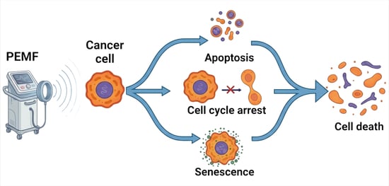
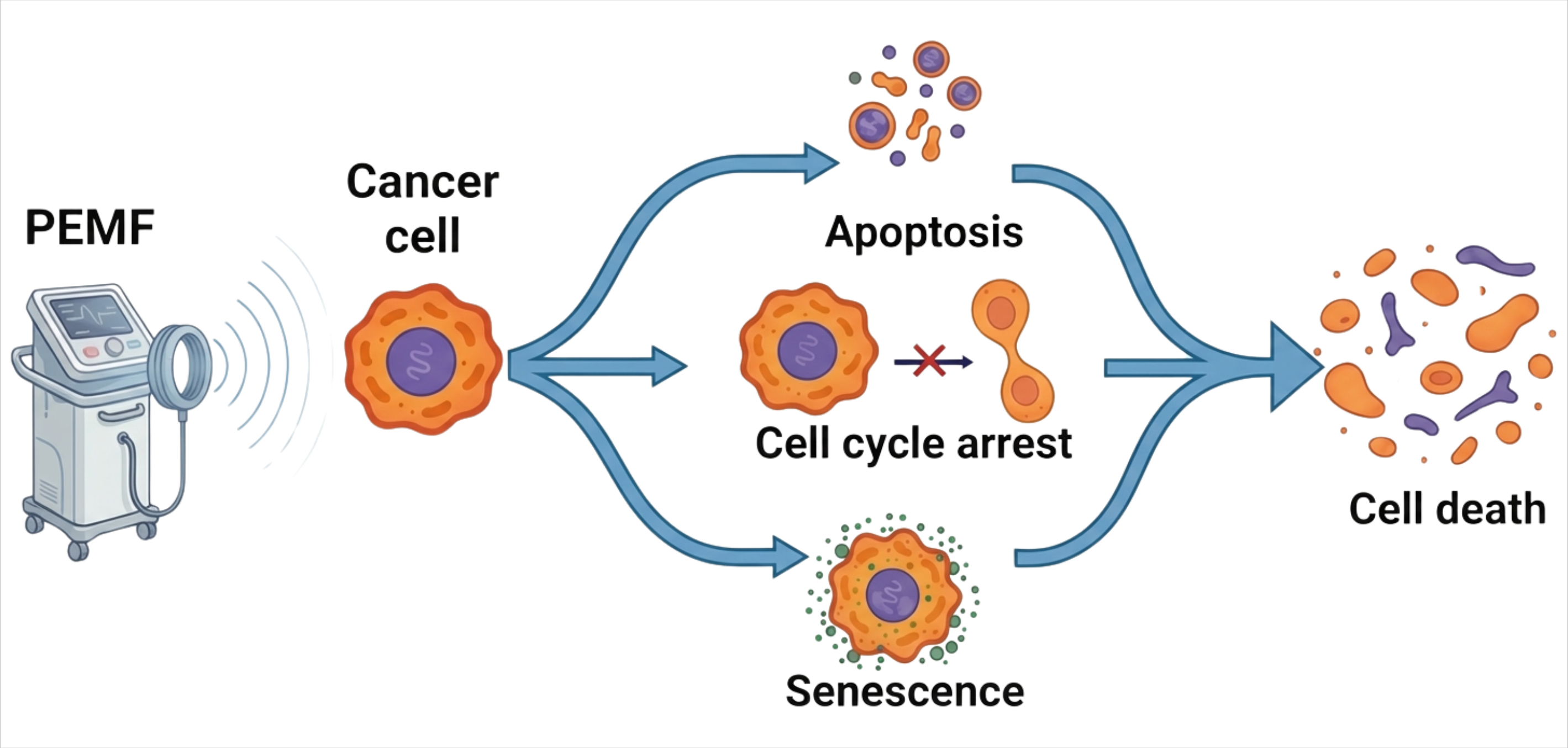
Cancer remains a significant global health burden, often requiring conventional treatments characterized by considerable side effects and limited tumor specificity. This review addresses the critical gap in understanding the non-thermal mechanisms by which Pulsed Electromagnetic fields (PEMFs) exert selective anti-tumor effects. Our primary objective is to analyze the molecular and cellular events through which low-intensity PEMF triggers stress responses and apoptosis in neoplastic cells without impacting normal cell viability. This comprehensive review synthesizes current evidence on the biological effects of PEMFs. Findings indicate that PEMFs disrupts intracellular homeostasis, induces reactive oxygen species-mediated oxidative stress, and activates endoplasmic reticulum stress, collectively driving malignant cells towards apoptosis or cell cycle arrest. Importantly, these effects are preferentially observed in cancer cells due to their inherent biophysical vulnerabilities—such as depolarized membrane potentials—and depend critically on specific PEMFs parameters. In conclusion, PEMFs acts as a multifaceted disruptor of cancer cell homeostasis, representing a promising non-invasive therapeutic modality. Further research is essential to optimize dosimetry and identify primary molecular sensors such as radical pair dynamics, to enhance clinical application and explore synergistic combinations with existing therapies.

Graphical Abstract

1. Introduction

Cancer, a disease characterized by uncontrolled cellular proliferation and the evasion of homeostatic mechanisms, has traditionally been confronted with therapeutic strategies targeting specific biochemical and genetic pathways. However, these conventional modalities, including chemotherapy and radiotherapy, often present significant limitations such as systemic toxicity, the frequent development of drug resistance, and a lack of specific targeting [1,2,3]. Therapeutic resistance, whether intrinsic or acquired during treatment, remains a major challenge, often leading to treatment failure and patient relapse [4,5,6,7]. In this context, a growing understanding of tumor biology has revealed that cancer cells exhibit not only genetic and biochemical anomalies but also profound biophysical alterations that can be therapeutically exploited [8]. These modifications include changes in membrane potential, differential expression of ion channels, and broader alterations in bioelectrical signaling, which together redefine the malignant phenotype [9]. Notably, these alterations include an elevated basal depolarization of the membrane potential and the overexpression of voltage-dependent ion channels in cancer cells, making them particularly sensitive to modulation by external stimuli [8,10].
These biophysical alterations create a unique “window of vulnerability” in cancer cells, making them potentially more susceptible to modulation by external biophysical stimuli than healthy cells [11]. Consequently, Pulsed Electromagnetic Fields (PEMFs) emerge as a noninvasive, nonthermal, and low-energy therapeutic modality. PEMFs differ from other electromagnetic therapies, such as radiofrequency hyperthermia [12], by specifically targeting cellular biophysical vulnerabilities. Their action is mediated through various molecular mechanisms, including the activation of ion channels [13,14,15], modulation of gene expression [9,16], alterations in membrane potential [17,18,19], and changes in cytoskeletal structure and cellular morphology [11,14].
The focus on “pulsed” fields stems from their ability to avoid thermal heating while magnifying specific non-thermal biological effects through high-intensity, short-duration signals [20,21]. This technique builds upon the pioneering mathematical descriptions of electric field-cell interactions by H.P. Schwan [22] and the subsequent discovery and theoretical modeling of membrane electroporation by J.C. Weaver [23,24]. These foundational contributions established the biophysical principles that underpin the selective vulnerability of cancer cells to pulsed electromagnetic stimulation.
Building on this historical foundation, the rationale for focusing PEMFs is rooted in the pioneering biophysical models of the mid-20th century. The theoretical foundation for how external fields interact with the cellular structure was established by the seminal work of Schwan, who described the passive electrical properties and dielectric dispersions of biological cells [23]. This was subsequently expanded by Weaver and colleagues, whose development of the “theory of electroporation” provided the first comprehensive mechanism for how pulsed electric fields induce transient, aqueous pores in the lipid bilayer [23,25]. These early models remain the cornerstone for understanding current PEMFs applications in oncology, where non-thermal pulses are used to manipulate membrane permeability and trigger selective apoptosis.
However, a critical challenge in translating PEMFs therapy to clinical settings is the lack of standardized experimental protocols. Applicator designs vary significantly, ranging from air-core solenoids that induce uniform magnetic fields to loop antennas that generate dominant electric components [26,27]. Consequently, parameters such as frequency (ranging from Extremely Low Frequency (ELF) to Radio Frequency (RF)), magnetic induction (µT to T), and power levels are often non-standardized, complicating the cross-comparison of biological outcomes [28,29,30]. This technical heterogeneity must be addressed to establish reproducible dose–response relationships and to identify the optimal physical parameters for selective anti-tumor effects.
The central hypothesis of this review posits that the abnormal properties of neoplastic cells make them inherently susceptible to electromagnetic forces, a susceptibility not observed in normal cells. This allows low-intensity PEMF to induce stress responses that inhibit growth and promote cell death. The primary objective of this review is to analyze these molecular and cellular mechanisms of PEMF action in neoplastic cells, which is crucial for elucidating the contribution of non-thermal effects, thereby differentiating them from radiofrequency hyperthermia [8].

2. The Cancer Cell as a Biophysical Target

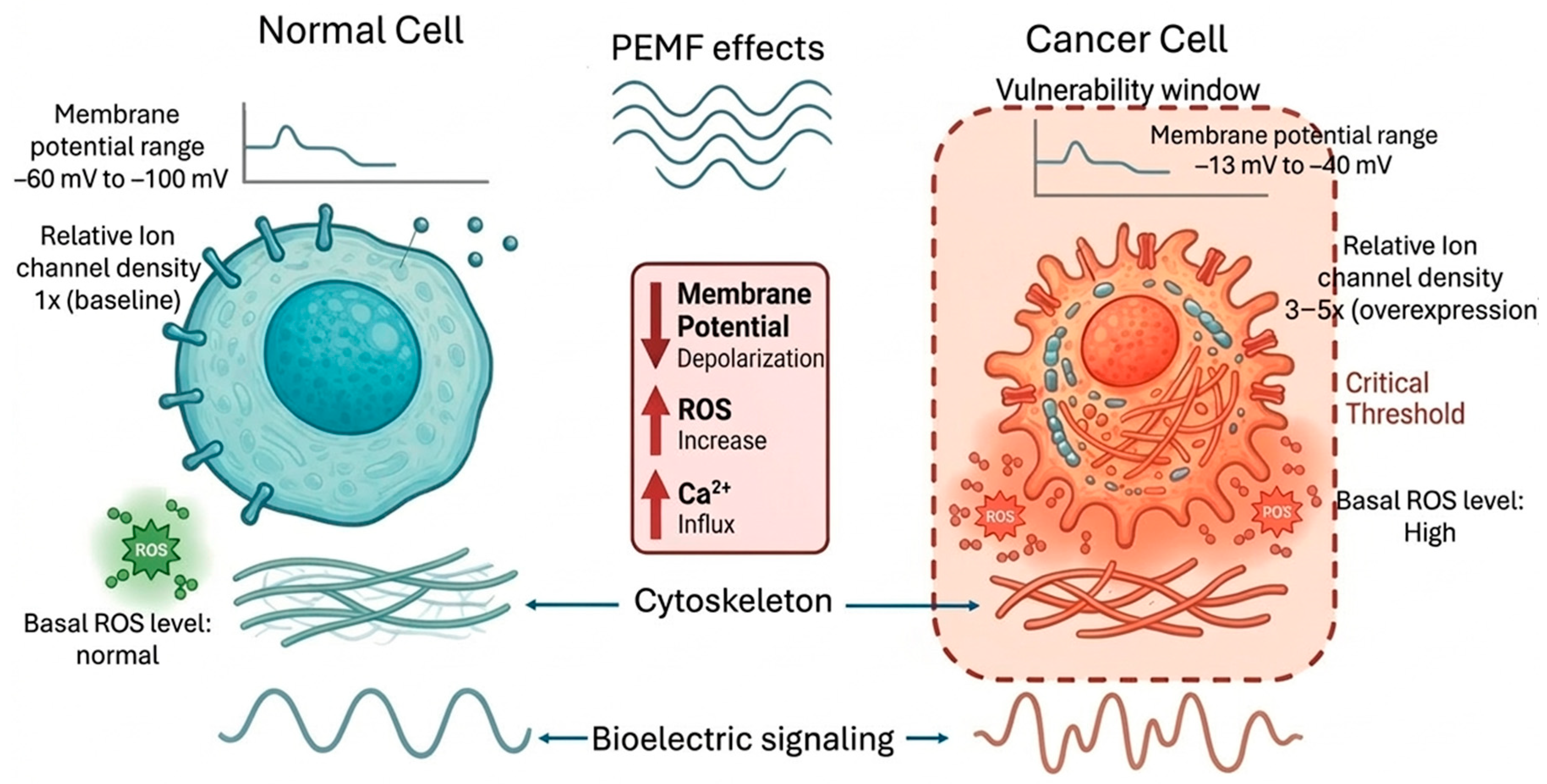
It is increasingly evident that the malignant phenotype cannot be defined solely by a genetic or biochemical signature. A growing body of work, including research by groups such as Costa and Dieper, urges us to consider a third pillar: the cancer cell as an entity with profoundly altered biophysical properties, making it an intriguing target for non-invasive interventions such as PEMFs [8,9]. These biophysical changes create a distinctive “vulnerability window” in cancer cells, rendering them more responsive than healthy cells to external biophysical modulation [11] (Figure 1). But what exactly do these alterations involve? Far from being isolated changes, they represent a systemic reconfiguration that encompasses everything from the resting membrane potential and ion channel activity to mitochondrial dynamics and the fundamental logic of tissue bioelectrical signaling [31,32].
Crucially, the mechanism of cellular interaction is fundamentally dictated by the Electromagnetic field pattern. In solenoid-based systems, the magnetic component is maximized, inducing intracellular eddy currents that can modulate ion channel activity and mitochondrial function, whereas in antenna or electrode setups, the electric field dominates, leading to direct membrane polarization and potential permeabilization [26,33]. Furthermore, the depth of penetration and the specific molecular targets vary with frequency, as higher frequencies may engage different dipolar relaxation mechanisms compared to low-frequency pulsed fields [27,28]. This frequency- and component-dependent variability must be considered when interpreting biological outcomes and designing therapeutic protocols.

2.1. Resting Membrane Potential and Ion Channels

One of the most distinctive and perhaps most underestimated biophysical characteristics of neoplastic cells is the depolarization of the resting membrane potential [31,34]. While the resting plasma membrane potential of healthy cells typically ranges from −60 mV to −100 mV [35], cancer cells tend to be depolarized, exhibiting significantly less negative values, often ranging from −13 mV to −40 mV, thereby promoting cellular proliferation [34]. These data suggest that this depolarized state is not a passive defect but an actively maintained condition. Murugan et al. demonstrated how ion channel expression profiles in cancer are reconfigured to elevate intracellular Na+ at the expense of K+, creating an ionic environment that inherently favors proliferation and migration over differentiation [35]. For example, in breast cancer, a depolarized resting membrane potential correlates with increased cellular activity and a more aggressive tumor phenotype [34].
This reconfiguration brings us to a key determinant: changes in the expression and activity of ion channels, which are central to this aberrant bioelectrical phenotype [35,36]. The crucial question is how can such a seemingly specific dysfunction produce such diverse consequences? The answer lies in the fundamental role of ion channels, Ca2+, K+ and Na+, as master regulators of a wide range of cellular functions, from the proliferative cycle to programmed cell death, a principle thoroughly reviewed by Litan and Langhans and by Tajada and Villalobos [37,38]. In the context of cancer, the expression of these channels is often dysregulated. Aberrant K+ channel expression is observed in multiple neoplasms, with their critical involvement in cell cycle progression well documented [39,40]. On the other hand, Ca2+ channels are positioned as central players, whose dysfunction contributes significantly to key aspects of cancer, such as growth, survival, and migration [38]. However, it is essential to recognize that this landscape is complex. While some studies report consistent dysregulation patterns, tumor heterogeneity indicates that the specific ionic landscape can vary significantly among cancer types [41,42]. Collectively, this perfect ionic storm causes to a profound disruption of cellular signaling pathways, a causal link that Sheth and Esfandiari have helped establish [43].
Cancer cells display distinctive biophysical properties compared to normal cells that favor their proliferation and migration. For example, their resting membrane potential is less negative [34,35,36]. Ion channel expression is regulated in normal cells but aberrant in cancer cells, contributing to depolarization and altered signaling [35,36]. Finally, the intracellular concentration is controlled in normal cells but elevated in cancer cells, which also promotes their proliferation and migration [35].

2.2. Mitochondrial Dynamics and Bioenergetic Metabolism

A simplistic view of mitochondria in cancer that focused only on aerobic glycolysis, or the “Warburg effect,” is insufficient. Although this phenomenon is well established, as confirmed by recent analyses [44,45], a more nuanced body of evidence prompts a reconsideration of its role. Rather than being merely dysfunctional, mitochondria in neoplastic cells undergo complex metabolic reprogramming, a functional adjustment that actively supports proliferation and tumorigenicity [46].
One often-overlooked aspect of this reprogramming is mitochondrial dynamics. How do these organelles manage to adapt to the changing demands of the tumor? The answer appears to lie in the delicate balance between fission and fusion. These processes are crucial for tumor cells to adapt to heterogeneous environments and advance their malignancy [47,48]. Thus, modifications in mitochondrial shape and function collectively regulate bioenergetic metabolism, inhibit cell death pathways, and enhance migration and proliferation, all contributing to the tumorigenic phenotype [48].
Beyond morphology, mitochondrial dysfunction in cancer influences disease progression through a more subtle and prolonged mechanism of retrograde signaling. Mitochondria are not merely energy producers; they serve as hubs of cellular communication. Their dysfunction contributes to cancer progression by regulating retrograde signaling pathways mediated by mitochondria-derived molecules [49]. This phenomenon positions mitochondria not as isolated organelles but as central components in the signaling network that redefines cancer cells.

2.3. Bioelectrical Signaling in Tumor Development

It is increasingly evident that cellular communication in cancer transcends purely biochemical models. We must consider a more fundamental level of regulation: bioelectrical signaling, a sophisticated cellular language mediated by ionic gradients and membrane voltage that coordinates crucial cellular functions, as eloquently presented by Shrivastava and colleagues [32]. But how is this language perturbed in oncogenesis? The answer appears to reside in a phenomenon aptly termed “bioelectrical dysregulation”, a determinant factor profoundly affecting the initiation, promotion, and progression of the disease [43]. This dysregulation is not a mere passive marker; rather, variations in bioelectrical membrane potentials actively alter cellular ionic homeostasis, a hallmark of numerous pathologies, among which cancer occupies a prominent place [32].
One aspect that is often overlooked is how this information is transmitted. Here, endogenous electromagnetic fields, generated by the coordinated movement of ions across membranes, emerge as long-range messengers. These fields are detected by neighboring cells and stand as potent modulators of bioelectrical signaling [35].
However, this communication network is extraordinarily sensitive to its environment. The notorious tumor microenvironment, with its acidic pH and complex biochemical milieu, is therefore expected to cause significant interference. This hostile microenvironment can drastically alter ion channel activity and, consequently, distort bioelectrical signaling, decisively influencing cancer growth and progression [35,50].
PEMFs interfere with this “bioelectrical language” of cells by disrupting endogenous electron transfer processes and the autonomous oscillations essential for maintaining homeostasis [51]. By perturbing these physiological networks, PEMFs disrupt the cellular network that promotes uncontrolled proliferation in cancer cells, effectively hijacking the very communication system that malignant cells exploit for survival and growth.
Extending this concept of environmental sensitivity, the biological response to PEMFs is significantly influenced by the ambient magnetic environment. Experimental evidence demonstrates that weak radiofrequency fields, when combined with specific background static magnetic fields, can modulate cancer cell growth rates, intracellular pH, and membrane potentials [17,52]. This dependency highlights that the “window of vulnerability” is not only a function of the applied pulse but also of the local geomagnetic and static field context, which can alter the hyperfine couplings in radical intermediates [17,53].
This dynamic interaction between the extracellular context and the intracellular bioelectrical state points to a new and promising therapeutic frontier.

3. Effects on Ionic Homeostasis and Signal Transduction

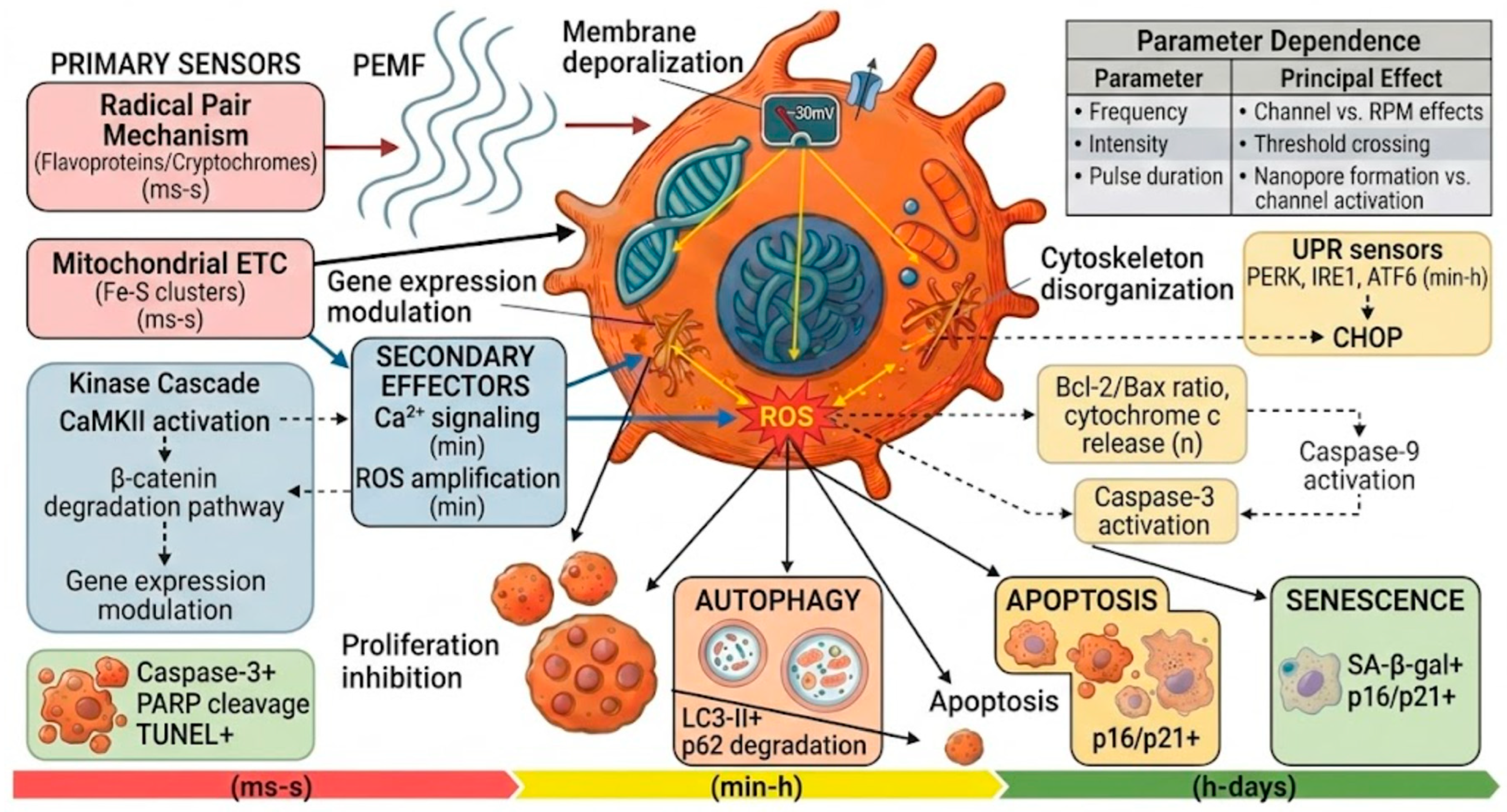
It is difficult to overstate the importance of Ca2+ as a master regulator of cellular function. This ion governs a multitude of fundamental processes, including proliferation, differentiation, metabolism, and apoptosis, a widely recognized biological principle [54,55]. However, in cancer cells, this delicate ionic balance is often profoundly disrupted. It is in this vulnerability that the modulation of Ca2+ fluxes by PEMFs emerges as a notable antitumor mechanism. PEMFs interact with the plasma membrane to activate ion channels and depolarize the membrane potential, initiating an intracellular cascade that includes ROS generation, gene expression modulation, and cytoskeletal disruption, ultimately leading to cell cycle arrest and apoptosis [12,56] (Figure 2).

3.1. Mechanisms of Ca2+ Homeostasis Alteration by PEMF

The key question is: how do PEMFs manage to hijack the Ca2+ machinery in neoplastic cells? Current evidence suggests a multifaceted mechanism. However, it is crucial to distinguish between the primary biophysical sensor and downstream biological messengers. While PEMF exposure consistently modulates intracellular Ca2+ concentrations, the calcium ion itself lacks the magnetic dipole moment required for first-order sensing of weak fields [57]. Instead, Ca2+ serves as a secondary messenger whose concentration is altered following the primary interaction of the field with spin-correlated radical pairs in enzymes like cryptochromes or within mitochondrial complexes [57]. First, it involves the direct activation of voltage-dependent Ca2+ channels, but through a more sophisticated physical interaction than simple membrane depolarization. The activation of voltage-gated calcium channels by PEMFs is driven by the exertion of direct physical forces on the voltage sensor domain (VSD). The VSD contains approximately 20 positively charged amino acid residues that act as the centerpiece of voltage-gating [58,59,60]. Unlike random thermal motion, the highly coherent nature of PEMFs allows for the simultaneous application of force on these charges, dragging the VSD to its ‘out’ position and opening the pore domain gate [61]. This effect is further amplified by the high electrical resistance of the plasma membrane, which concentrates the electric field across the bilayer [60]. Recent studies identify the Cav1.2 L-type and Cav3.2 T-type subtypes as molecular targets particularly sensitive to PEMF stimulation [8,62,63]. An illustrative example comes from glioblastoma multiforme, where stimulation with alternating electric fields causes a substantial increase in cytoplasmic Ca2+, an effect completely abolished by the Ca2+ channel blocker benidipine, confirming the channel-dependent opening mechanism [8]. This supports the hypothesis that the frequent overexpression of channels in cancer cells, rather than providing an advantage, paradoxically makes them more vulnerable to lethal Ca2+ overload [64].
However, this process is not limited to the plasma membrane. An often-overlooked aspect is the release of Ca2+ from the endoplasmic reticulum (ER). Although direct evidence implicating specific inositol triphosphate (IP3) or ryanodine receptors is not conclusive in all contexts, some studies have identified the ER as a significant source of transient Ca2+ spikes induced by electrical pulses, possibly through electrical permeabilization of its membranes [65,66].
At the physicochemical level, nanopore formation is driven by the reorganization of interfacial water dipoles, which, under the influence of pulsed fields, align and penetrate the lipid bilayer to create transient conductive pathways [67,68]. This electroporative effect significantly alters membrane permeability, facilitating a massive influx of extracellular Ca2+ that bypasses voltage-gated channels and leads to intracellular overload. Research indicates that this permeabilization, when combined with ions, drastically potentiates cytotoxic effects in lung cancer cells, highlighting a potent therapeutic synergy between electroporation and sensitization [56].

3.2. Downstream Consequences of Ca2+ Influx

The Ca2+ entry initiated by PEMFs is not a terminal event but instead triggers a precisely orchestrated cascade of molecular consequences. First, it acts as a second messenger that can modulate gene expression and influence key enzymes. Recent studies show that this accumulation activates calcium/calmodulin kinase II, which leads to the degradation of β-catenin, a central regulator of proliferation and metastasis [8].
Similarly, calpains, Ca2+-dependent proteases, are activated by this influx and proceed to remodel the cytoskeleton, destabilizing cellular structure and critically compromising the migratory and invasive capacity of the cancer cell [69].
Extending this cytoskeletal disruption to the mitotic machinery, the electric component of the PEMF interacts directly with the high dipole moment of tubulin dimers. The resulting rotational torque prevents the correct alignment of dimers during polymerization, leading to a reduced fraction of polymerized microtubules and subsequent mitotic catastrophe [70,71,72]. This dual mechanism, both Ca2+-dependent proteolysis and direct physical interference with microtubule dynamics, synergistically compromises cytoskeletal integrity and cell division.
A crucial effect is its role in mitochondrial dysfunction and apoptosis. Excess of cytosolic Ca2+ is taken up by mitochondria, leading to their overload. This event triggers mitochondrial membrane depolarization, the release of pro-apoptotic factors, and the irreversible activation of apoptosis [63,64].
Ca2+ influx can also stimulate the production of reactive oxygen species (ROS) and lipid peroxidation, adding another layer of cellular damage, as observed in lung cancer models [56].
Summing this evidence outlines a compelling picture: Ca2+ serves as the central conductor of the PEMF signal. Cells normally maintain a very strict Ca2+ gradient; a sudden influx drastically disrupts this homeostasis, creating an intracellular signaling storm [63]. The strongest evidence of its crucial mediating role comes from loss-of-function experiments, in which the inhibitory effect of electromagnetic fields on cell proliferation disappears when calcium homeostasis is disrupted [73]. PEMFs use the cancer cell’s own communication system to redirect it towards death.
The robustness of this potential mechanism is confirmed across a variety of cellular models: in breast cancer (MCF-7), where Ca2+-assisted electroporation disrupts homeostasis and triggers cell death; in glioblastoma, where stimulation increases cytoplasmic Ca2+, leading to mitochondrial dysfunction and apoptosis; in lung cancer, where the combination of pulsed fields with Ca2+ enhances oxidative damage; and in hepatocellular carcinoma, where a significant influx of Ca2+ induced by RF-EMF has been reported [8,56,63].
This focus on Ca2+ dysregulation provides both a solid justification and a molecular roadmap for the development of PEMFs as an innovative and deeply targeted therapeutic strategy.

4. Induction of Cell Stress and Regulated Death: A Convergence of Cytotoxic Pathways

Beyond the disruption of ionic and bioelectric homeostasis, a crucial question arises: how do cancer cells translate the physical signal of PEMFs into a lethal biological response? Current evidence indicates that PEMFs trigger a cascade of cellular stress responses that culminate coordinately in regulated cell death. Specifically, the induction of oxidative and ER stress by PEMFs converges on mechanisms of apoptosis, autophagy, and senescence.

4.1. Oxidative Stress

A recurrent and fundamental finding in the literature is that PEMFs can generate reactive oxygen species (ROS), inducing a state of oxidative stress in cancer cells [11,56,74]. But what are the primary sources of this ROS surge? The proposed mechanisms are intriguingly diverse. Firstly, it is well known that mitochondria are a primary source of ROS [75]. However, PEMFs appear to exacerbate this function through mechanisms such as the Radical Pair Mechanism (RPM), profoundly altering mitochondrial function [76]. This mechanism explains the generation of reactive oxygen species. Weak magnetic fields modulate the coherent spin dynamics between singlet and triplet states of radical pairs in flavoproteins, such as the FAD-superoxide pair in cryptochromes [77,78]. This intersystem crossing alters the biochemical yield of O2•− and H2O2, providing a quantum biological basis for PEMF-induced oxidative stress [79,80]. Building on this quantum spin dynamics, PEMFs further inhibit mitochondrial respiration by perturbing spin-correlated electron transfer within the redox centers of the respiratory chain [74]. This “Magnetic Electron Perturbation” occurs when the field aligns electron spins in the iron-sulfur clusters of Complex II, hindering the pairing required for efficient electron flow and leading to a surge in mitochondrial-derived oxidative stress [74,76]. In fact, it has been robustly demonstrated that oscillating magnetic fields can inhibit mitochondrial respiration, promote oxidative stress, and cause a catastrophic loss of organelle integrity, which is associated with a rapid increase in ROS to cytotoxic levels [74]. An aspect that often reinforces this phenomenon is PEMF-induced Ca2+ overload, which depolarizes the mitochondrial membrane, directly impacting ROS production and activating the programmed cell death pathway [8,64].
On the other hand, it has been proposed that the activation of cation channels and their association with ROS-generating NADPH oxidases is a key mechanism [81]. However, it is important to address a contradiction in the literature: contrary to this view, Ehnert and collaborators indicated that extremely low-frequency PEMFs (ELF-PEMFs) might not affect the expression of NADPH oxidases, suggesting that other sources of ROS need to be identified [82].
The concept of “lethal redox stress” is, therefore, central to the anti-tumor action of PEMFs. It is plausible that this vulnerability of cancer cells, which already operate under higher basal ROS levels [83], is exploited by this technology. While a small increase in ROS can promote proliferation, an excess inevitably leads to cellular damage [84]. Reinforcing this quantum biological perspective, the suppression of background magnetic fields (hypomagnetic conditions) has been shown to reduce hydrogen peroxide production in various cancer cell lines, further supporting the idea that the absolute magnetic context is a critical variable in PEMF-induced oncolysis [53]. These effects on ROS partitioning provide a quantum biological explanation for the changes in cell proliferation observed across various frequencies and intensities [78,85].
It is here that a key determinant lies: some studies support the hypothesis that PEMFs can selectively increase intracellular ROS to cytotoxic levels in cancer cells, without affecting normal cells [11,74]. This lethal induction of ROS damages macromolecules and, as shown, can be enhanced by the entry of Ca2+ ions, which stimulate ROS release and lipid peroxidation [56].

4.2. Endoplasmic Reticulum Stress

The ER, a vital center for protein synthesis and storage, becomes a focal point of PEMF-induced disruption. The alteration of homeostasis [63] and the increase in ROS levels [86,87] act as synergistic forces that disrupt ER function, inducing ER stress and triggering the Unfolded Protein Response (UPR) [88]. The UPR has a pro-survival phase in which, under moderate stress, it activates an adaptive program to restore homeostasis, improving protein folding capacity and degradation, allowing the cell to adapt and survive stress [89,90,91]. It also presents a pro-apoptotic phase, which occurs if ER stress is severe or prolonged [89,92,93]. PEMFs appear to deliberately tip this balance. This tipping in the endoplasmic reticulum refers to the transition from a survival-oriented Unfolded Protein Response to terminal apoptosis. Persistent depletion and biophysical stress induce the upregulation of the C/EBP homologous protein (CHOP), triggering a caspase cascade through dependent proteases and mitochondrial membrane permeabilization [94]. Activation of the key effector CHOP promotes the transcription of pro-apoptotic genes and can link ER stress with mitochondrial apoptosis [95]. Two studies provide a critical link by demonstrating that ROS accumulation induces ER stress, which leads to CHOP activation and apoptosis in cancer cells [86,96].
In addition, the release of Ca2+ from the ER to the cytosol is a significant trigger. Electrical permeabilization of internal membranes, including the ER, can cause transient intracellular Ca2+ spikes [65,66]. This large increase in intracellular Ca2+ concentration, originating from both the extracellular environment and ER reserves, can, in the context of high ROS production, trigger apoptosis [63,88].

4.3. Mechanisms of Cell Death

The convergence of these stress pathways results in various forms of regulated cell death. Among them, apoptosis is the most consistently reported mechanism. PEMF exposure shifts the ER stress response from a survival-oriented UPR to a terminal pro-apoptotic state. This is characterized by the upregulation of CHOP and the subsequent activation of the caspase cascade, specifically through Ca2+-dependent proteases and mitochondrial signaling [97,98]. This is a key point, and it is important to note that PEMF-induced apoptosis is characterized by well-defined molecular events. These events include caspase activation, where it has been established that PEMFs can activate both executor and initiator caspases, such as caspase-3 and caspase-9 [12,99]; alteration of the Bcl-2/Bax ratio, in which PEMFs shift the molecular balance toward cell death by increasing Bax expression and reducing Bcl-2 [14,99], with Bax migrating to the mitochondria and triggering the release of cytochrome c, thereby irreversibly activating the apoptotic cascade [12]; similarly, Ca2+ overload in the cytosol, which is taken up by mitochondria, leads to a harmful mitochondrial overload causing their dysfunction. This can result in membrane depolarization, release of pro-apoptotic factors, and final activation of the programmed cell death pathway [8,63,64].
In addition to apoptosis, another mechanism of significant importance, though still not fully understood, is PEMF-induced autophagy. Although autophagy is an essential catabolic process, its role in cancer is ambivalent, potentially being both cytoprotective and cytotoxic [100]. While some studies suggest that PEMFs can induce autophagy, their overall contribution to cytotoxicity or resistance mechanisms remains unclear [101].
A particularly promising finding is the induction of senescence. It has been reported that PEMFs can selectively induce a state of permanent cell growth arrest only in cancer cells, without affecting normal fibroblasts [102]. This suggests that PEMF-induced senescence could represent a distinctive and valuable anti-tumor strategy.
But under which PEMF parameters is one cellular fate favored over another? This fundamental question of how PEMFs determine final cellular fate—whether apoptosis, autophagy, or senescence—does not have a simple answer. Rather than being straightforward, this process is governed by a delicate balance of physical and biological parameters, including exposure duration, flux density, and, importantly, the target cell type [84]. The heterogeneity of responses is not surprising given the significant variability in PEMF-generating devices, experimental conditions, and cellular models used in the literature, all of which contribute substantially to the diversity of observed results [11].
A substantial body of evidence, particularly from the work of Bergandi and collaborators, robustly demonstrates that the effects of PEMFs are cell-type specific and related to exposure characteristics [73]. An eloquent example of this is a work by Zabaleta and collaborators, in which they observed that exposure to low-frequency magnetic fields can increase ROS production in breast cancer cells, an effect that intensifies in a dose-dependent manner with higher field intensities and longer exposure times [84]. An eloquent example is that Zabaleta and collaborators observed that exposure to low-frequency magnetic fields increases ROS production in breast cancer cells, with the effect intensifying in a dose-dependent manner as field intensity and exposure time increase [84]. This raises a key question: where is the turning point? The answer may lie in the cell’s capacity of adaptation. As Dieper and collaborators persuasively argue, modulation of membrane potential or induction of ER stress can inexorably lead to cell death, but only when the cell’s adaptive mechanisms are overwhelmed and collapse [8]. Therefore, the final cellular fate is simply the integrated consequence of the intensity and duration of the stress imposed on the unique resistance threshold of each biological system.

5. Selectivity and Differential Sensitivity

The fundamental premise supporting the use of PEMFs in oncology is not based on non-specific cytotoxic effects but on a selective mechanism: the altered biophysical properties of cancer cells provide a rational basis for differential action. A particularly revealing finding is that the distinctive response of neoplastic cells to electromagnetic stimulation, ranging from proliferative and migratory inhibition to the potentiation of cell death, is simply not reproducible in healthy, non-rapidly dividing cells [8,102].
But what is the molecular origin of this selectivity? Current evidence suggests that it may result from an inherent increased susceptibility of cancer cells to PEMF-induced damage, a vulnerability directly conferred by their unique bioelectric and mechanical characteristics [8]. A key mechanism appears to be the basal depolarization of the membrane potential, which influences the behavior of voltage-gated ion channels (VGICs). As is known, VGICs exhibit high sensitivity to disturbances in membrane potential [8]. Thus, it is logical to deduce that the frequent upregulation of these very VGICs, observed in oncogenesis, is not a mere coincidence but could significantly enhance the effect of PEMFs on ion movement by providing a much larger number of molecular targets [8].
This selectivity is a key differentiator from conventional therapies, which often lack specificity and cause systemic toxicity [8]. Critical analysis of in vitro evidence is essential to understand the consistency and underlying mechanisms of this differential sensitivity.

5.1. In Vitro Evidence: Cancer Cells vs. Healthy Cells

This finding of PEMF selectivity has been consistently observed in in vitro studies, which have determined that PEMFs exert more pronounced effects on cancer cells than on their non-malignant counterparts. However, it is important to recognize that this selectivity is not an absolute but rather variable and highly dependent on specific exposure parameters and the cellular context [84].
The most direct evidence of this selectivity appears in the inhibition of growth and viability. It has been consistently observed that PEMFs suppress the proliferation of various cancer cell lines, while normal cells, such as fibroblasts or MCF-10A mammary epithelial cells, show considerable resistance or even, in some paradoxical contexts, a slight promotion of growth [84]. A particularly interesting example is a study reporting that PEMFs specifically induce cellular senescence in cancer cells, without affecting normal fibroblasts [102]. Similarly, another work demonstrated that radiofrequency radiation inhibits neuroblastoma cells but does not significantly affect the viability of normal human keratinocytes [12].
The next level of selectivity is seen in the induction of cell death. Apoptotic and senescent mechanisms appear to be preferentially activated in the neoplastic context, where oxidative stress seems to be a key determinant. Interestingly, in independent studies, they found that the increase in reactive oxygen species to cytotoxic levels occurs selectively in cancer cells, without significantly affecting normal cells [11,74]. This mechanism is so potent that magnetic fields can induce oncolysis through oxidative stress in glioma cells, a response that is significantly more effective in cancer cells than in healthy cells [11].

5.2. Integrative Hierarchical Model of PEMF Selectivity

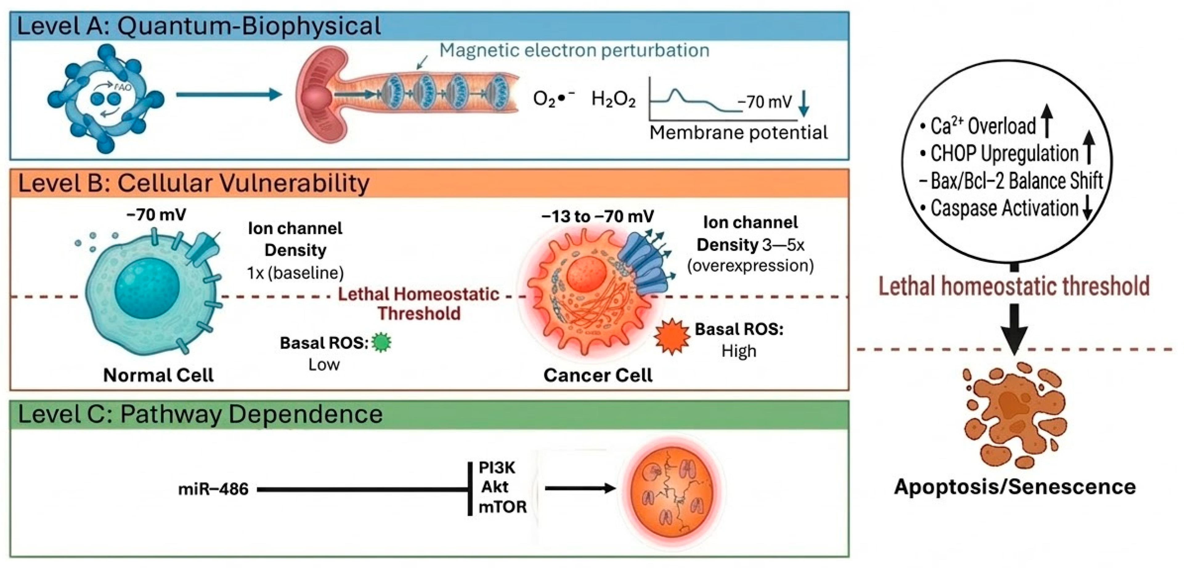
With this evidence, a central question arises: what is the biological basis of this differential vulnerability? Current evidence suggests that there is no single explanation, but rather a set of interrelated mechanisms that operate at distinct levels of biological organization. We propose an integrative hierarchical model that synthesizes these mechanisms into a cohesive framework, explaining how a weak physical stimulus is captured, amplified, and ultimately triggers programmed cell death selectively in malignant cells (Figure 3).

5.2.1. Level A: Quantum-Biophysical Sensing via the Radical Pair Mechanism

At the apex of this hierarchy is the primary biophysical sensor. Building on the quantum biological principles discussed in Section 4.1, the selectivity of PEMF action originates at the level of primary physical sensing. The RPM operates in flavoproteins such as cryptochromes and within mitochondrial electron transport chains, where weak magnetic fields modulate the coherent spin dynamics between singlet and triplet states of radical pairs [77,85]. This intersystem crossing alters the biochemical yield of reactive oxygen species, particularly O2 and H2O2, providing a quantum biological basis for differential cellular responses [79,80]. Crucially, this primary sensing must be distinguished from downstream signaling: while PEMF exposure consistently modulates intracellular Ca2+ concentrations, the calcium ion itself lacks the magnetic dipole moment required for first-order sensing of weak fields [57]. Instead, Ca2+ serves as a secondary messenger whose concentration is altered following the primary interaction of the field with spin-correlated radical pairs [57]. This hierarchical organization, quantum sensing followed by biological amplification, explains how weak fields can produce measurable cellular effects.

5.2.2. Level B: Cellular Vulnerability and the Allostatic Load Threshold

The differential sensitivity to PEMFs can be further explained by the concept of “allostatic load”, the elevated basal stress under which cancer cells operate. Due to accelerated proliferation, abnormal metabolism, and hostile microenvironments, malignant cells maintain higher basal levels of reactive oxygen species, increased demand for protein folding in the endoplasmic reticulum, and inherent mitochondrial instability [76]. This chronic stress state positions cancer cells closer to a critical homeostatic threshold. When PEMFs impose additional biophysical perturbation, whether through radical pair-mediated ROS generation [80,85], mitochondrial electron transport chain disruption [74], or Ca2+ overload [103], cancer cells cross this threshold into cytotoxicity. In contrast, normal cells with lower basal stress and greater buffering capacity absorb the same perturbation without reaching the lethal threshold [104]. This threshold model explains why PEMF effects are dose-dependent and why the “window of vulnerability” represents the parameter space where cancer cells, but not normal cells, exceed their homeostatic capacity [102,104].

5.2.3. Level C: Pathway Dependence—Targeting Oncogenic Addiction

A third level of selectivity emerges from the concept of “oncogenic addiction”, the dependence of cancer cells on hyperactive signaling pathways for survival and proliferation. The PI3K/Akt/mTOR pathway, frequently upregulated in malignancies, represents such an addiction [105,106]. PEMFs exploit this vulnerability through specific molecular interference: low-frequency magnetic fields have been shown to induce autophagy-associated cell death in lung cancer cells via miR-486-mediated inhibition of the Akt/mTOR pathway [67]. This modulation destabilizes tumor survival circuits without significantly affecting normal cells, which do not depend on pathway overactivation [72,102]. The selectivity arises because cancer cells have co-opted these pathways to manage their allostatic load; disrupting them removes a critical adaptive mechanism, pushing cells toward death [107,108]. This pathway-level interference operates in concert with the quantum and cellular stress mechanisms described above, creating a multi-layered attack on cancer cell homeostasis.

5.2.4. Integrative Convergence and the Definition of the Vulnerability Window

The selectivity of PEMFs toward cancer cells emerges from the convergence of the three interconnected levels described above, which together overwhelm the adaptive capacity of malignant cells while sparing normal tissues [102,104]. This integrative model can be conceptualized as a multi-hit mechanism operating across spatial and temporal scales: quantum sensing (Level A) provides the primary physical interaction, cellular vulnerability (Level B) determines the threshold for cytotoxicity, and pathway dependence (Level C) removes adaptive survival circuits. This triple convergence—quantum sensing, threshold crossing, and pathway disruption—explains why PEMFs achieve selectivity: normal cells, with lower basal stress and intact buffering capacity, absorb the perturbation without reaching the lethal threshold.
This model accounts for the observed context dependence of PEMF effects, including variations with frequency, intensity, and exposure duration [104]. The “window of vulnerability” is therefore formally defined as the multi-dimensional parameter space where quantum sensing is maximized, cellular thresholds are exceeded, and pathway dependencies are exploited, all while remaining below the stress threshold of healthy cells [102,104].

5.3. Consistency of Evidence and Context Dependence

Various studies robustly support the principle of PEMF selectivity in vitro [11,12,74,84,102]. However, it must be emphasized that the magnitude and specificity of this effect are highly variable and depend on three factors: PEMF parameters, the type of cancer cell, and the type of normal cell.
Regarding PEMF parameters, frequency, intensity, duration, and waveform are not mere technical details but crucial biological determinants. Different parameters can recruit distinct mechanisms of stress and cell death, and selectivity can be optimized through fine-tuning [73,84].
As for the type of cancer cell, it plays a preponderant role, since the genotypic and phenotypic heterogeneity of tumors is reflected in the response they can present. Not all cancer cells will react identically to the same PEMF regimen. The type of cancer cell plays a major role, as the genotypic and phenotypic heterogeneity of tumors is reflected in their response.
Finally, the type of normal reference cell is important. As selecting the control cells is fundamental. Cells with greater stress resilience or specific biological characteristics may show a different degree of response, influencing the perception of the therapeutic window.
We are therefore clear that selectivity is a promising and biologically grounded characteristic of PEMFs. The hypotheses of Allostatic Load, Dependence on Signaling Pathway, and Biophysical Alteration provide a solid explanatory framework that capitalizes on the inherent weaknesses of cancer. However, the remaining challenge, and the focus of the most advanced research, is the systematic optimization of PEMF parameters to maximize and universalize this therapeutic window, given the challenging heterogeneity of the disease.

5.4. Classification of PEMF Therapeutic Applications by Frequency Range

As discussed throughout this review, the biological effects of PEMFs are not uniform but rather highly dependent on their physical parameters. Among these, frequency stands as a critical determinant of therapeutic efficacy, with distinct physiological effects observed across different regions of the electromagnetic spectrum. In response to the diverse clinical requirements of oncology and regenerative medicine, therapeutic applications are generally classified into three primary frequency domains (Table 1).

5.4.1. Extremely Low Frequency

Currently, the majority of current research into selective anti-tumor mechanisms focuses on the ELF range, typically between 15 Hz and 75 Hz. Importantly, within this “window,” PEMFs have been shown to selectively impair the viability of breast and lung cancer cells by inducing autophagy and apoptosis [104]. Mechanistically, the high selectivity of ELF-PEMFs stems from their ability to interact with the quantum-level dynamics of radical pairs, particularly within mitochondrial complexes, which are already under high allostatic load in malignant cells [74,85].
However, it is essential to emphasize that the efficacy of PEMFs depends not only on frequency but also on a precise combination with magnetic flux density. In this study, the intensity values employed, ranging from µT to mT as shown in Table 2, are significantly higher than ultra-weak fields, such as Schumann resonances (~90 nT) [109], yet remain far below the intensities used in clinical magnetic resonance imaging (1.5–3 T) [110]. These moderate levels are biologically relevant because they operate within the ‘parameter window’ required to influence the Radical Pair Mechanism and spin dynamics in mitochondrial flavoproteins. Unlike strong fields that act via macroscopic forces, intensities in the µT to mT range can modulate reactive oxygen species production and alter ion flux through membrane channels, selectively sensitizing cancer cells that already operate under elevated basal stress. Consequently, intensity is not a passive parameter but a critical determinant that, alongside frequency, defines the threshold for therapeutic vulnerability.

5.4.2. Intermediate Frequencies and TT Fields

Building on this multi-parametric understanding, intermediate frequencies (100–500 kHz) and Tumor-Treating Fields represent a distinct clinical modality. Specifically, unlike ELF-PEMFs that primarily target metabolic and oxidative homeostasis, intermediate frequencies exploit the high dipole moment of tubulin and septin molecules [107]. As a result, by disrupting the assembly of the mitotic spindle, these fields induce mitotic arrest and subsequent cell death, providing a mechanical rather than purely biochemical interference [72,107].

5.4.3. Radiofrequency PEMF

Finally, the radiofrequency range (1–30 MHz) has been less extensively studied in oncology compared to ELF-PEMFs but shows promise in non-thermal pain management and tissue repair [111,112]. For instance, clinical and preclinical evidence suggests that RF-PEMFs, particularly when amplitude-modulated at tumor-specific frequencies (e.g., 27.12 MHz), can modulate calcium-calmodulin signaling pathways and activate anti-inflammatory responses [8,113,114]. These interactions facilitate the regulation of cytokine release and secondary messengers like NO and cGMP, potentially offering supportive benefits in cancer patients undergoing conventional therapies by reducing postoperative pain and improving systemic homeostasis [111,113,115]. Nevertheless, the application of RF-PEMFs as a direct anti-tumor modality remains limited due to challenges in consistent field penetration across diverse tissues and the inherent risk of thermal effects at higher intensities [8]. Thus, future research must identify specific “windows” within this frequency range where non-thermal effects can be harnessed—such as the disruption of mitotic spindles or the selective induction of apoptosis—although this will require rigorous dosimetry and validation in refined preclinical models [8,114].

6. Discussion

Research into PEMF as an anti-tumor therapy has revealed a remarkably complex mechanistic landscape. Rather than a single biophysical effect, the anti-tumor action of PEMFs results from an interconnected network of events that collectively exploit the intrinsic vulnerabilities of cancer cells.
At the center of this network is the alteration of calcium homeostasis, which acts as a master “gateway.” As some studies have shown, PEMFs provoke an influx and intracellular redistribution of Ca2+ by activating voltage-dependent channels in the plasma membrane or by inducing its release from the ER [8,56,63]. This cytosolic increase serves as an essential second messenger, transmitting and amplifying the initial physical signal, prolonging its biological effects long after the stimulus ends, and influencing a range of processes, from enzymatic modulation to cytoskeletal remodeling and mitochondrial function [8].
Simultaneously, and often synergistically, the generation of reactive oxygen species occurs, exacerbating cellular stress [56,74,83,84]. This accumulation of ROS, along with dysfunction, induces ER stress. While the ER initially attempts to restore homeostasis through the Unfolded Protein Response, persistent or severe biophysical perturbation by PEMFs shifts the balance toward its pro-apoptotic arm, activating effectors such as CHOP that promote cell death [96].
Finally, this triad of ionic, oxidative, and ER stress converges to modulate cell fate pathways. Apoptosis, indicated by classic markers, is a frequent outcome [12,14]. However, it is important to recognize that the response is not singular; other modes of regulated cell death, such as autophagy and senescence, are also observed depending on the context [102]. This integration of events underscores that PEMFs function as multifaceted disruptors of cancer cell homeostasis, leveraging the interconnections among membrane biophysics, ionic signaling, redox metabolism, and organelle biology.
By synthesizing the multi-layered interactions discussed above, we propose an Integrative Hierarchical Model of PEMF selectivity that operates at three convergent levels. First, at the quantum level, selectivity begins with the modulation of radical pair dynamics in cryptochromes and mitochondrial complexes [74,77,85]. This primary biophysical event is subsequently amplified at the cellular level, where the elevated allostatic load of cancer cells facilitates crossing a lethal homeostatic threshold [76]. Finally, at the signaling level, PEMFs exploit oncogenic addictions, such as the PI3K/Akt/mTOR pathway, to trigger programmed cell death [100,107]. This hierarchical framework suggests that experimental inconsistencies in the literature often arise when parameters fall outside the ‘window of vulnerability’ where these three levels converge [102,104]. Consequently, clinical efficacy depends on selecting precise electromagnetic parameters that maximize quantum perturbation and systemic collapse in malignant cells without exceeding the buffering capacity of healthy tissues [104].
Currently available evidence shows that PEMF efficacy is strongly influenced by many factors, which explain much of the reported variability. This context dependence is not a weakness but reflects the biological precision of the phenomenon and presents an opportunity for optimization.
First, the physical parameters of PEMFs are critical determinants. Frequency, intensity, duration, and waveform are not arbitrary variables but regulators that modulate the cellular response, as Bergandi has highlighted [73]. This suggests that a specific regimen might induce apoptosis in one cell type, while another might cause senescence or have no effect [84]. The current lack of standardization in these parameters is undoubtedly the greatest obstacle to comparability and reproducibility. It is important to emphasize that there is no single parameter for PEMF therapy; rather, the concept of “parameter windows” must be considered, where specific combinations of frequency, magnetic flux density, pulse duration, and repetition rate recruit distinct molecular mechanisms and determine the final cellular outcome. These windows must be optimized for each cancer type, as the biological response is highly context-dependent. Second, the intrinsic heterogeneity of cancer is an unavoidable factor. Different tumor lineages, and even subpopulations within a tumor, exhibit distinct profiles of ion channels, redox states, and signaling pathway dependencies. This biological diversity leads to variable sensitivity to the same electromagnetic stimulus. To provide a systematic overview, we summarize the typical parameter ranges reported in the literature and their associated biological effects (Table 2).
Table 2. Summary of PEMF parameters and associated biological effects.
ParameterTypical RangeBiological EffectReferences
Low frequency1–300 HzModulation of ion channels and Ca2+ flow[58,103]
High frequency100 kHz–1 MHzMitotic spindle disruption[70,116]
Magnetic flux densityµT–mTROS production and oxidative stress[53,84]
Pulse durationns–µsElectroporation and nanopore formation[117,118]
Repetition rateHz–kHzAccumulation of stress and activation of UPR[119,120]
Third, experimental conditions modulate the response. Factors such as cell density and culture medium composition can alter cells’ capacity to manage stress, influencing the final outcome. These variables underscore the urgent need for a more systematic and standardized approach to precisely unravel biophysical dose–response relationships.
Despite this complexity, the robustness of in vitro evidence regarding selectivity is undeniable and represents the most promising pillar of this therapy. A compelling body of work, including studies by Hambarde, Hernández-Bule, Pantelis, Sharpe, and Zabaleta, robustly demonstrates preferential effects on cancer cells [11,12,74,84,102]. The “Allostatic Load” [90], “Dependence on Signaling Pathway” [8,9], and “Biophysical Alteration” [8,9] hypotheses provide coherent conceptual frameworks that explain this differential vulnerability. In essence, cancer cells, operating at the limit of their homeostasis and relying on hyperactive and physically altered survival circuits, are intrinsically more susceptible to the additional biophysical stimulus that PEMFs represent.

7. Conclusions

Accumulated preclinical evidence demonstrates that low-intensity PEMFs can exert significant and selective anti-tumor effects by activating a coordinated network of cellular stress pathways. Disruption of homeostasis, induction of oxidative stress, and consequent activation of ER stress are interconnected events that differentially drive malignant cells toward death or cell cycle arrest, while largely preserving the viability of normal cells. This action profile positions PEMFs as a non-invasive therapeutic modality with a potentially superior safety profile compared to conventional therapies.
Despite significant advances, translating PEMFs into clinical practice requires addressing key challenges and exploring new research directions. It is crucial to develop high-throughput studies to systematically map the responses of cancerous and non-cancerous cells to a wide range of PEMF frequencies, intensities, durations, and waveforms. This approach would generate specific “sensitivity signatures” for each cancer type, enabling precise and personalized dosing that maximizes the anti-tumor effect and minimizes adverse effects on healthy tissues, directly addressing the lack of standardization that currently limits the field.
A major mechanistic challenge is to identify the primary molecular sensor that initially detects the electromagnetic field, as it remains largely unknown. Identifying this sensor, whether it is a membrane protein, a specific ion channel, or a cytoplasmic component, is crucial. Such a discovery would not only clarify the fundamental mechanism of action but also enable the rational design of highly specific next-generation PEMF devices or the development of combined therapies that sensitize this sensor.
A deep understanding of PEMF-induced stress mechanisms provides a solid foundation for exploring synergistic combinations. Potentiating chemotherapies or targeted therapies by pre-sensitizing cellular stress pathways represents a promising strategy to overcome resistance and reduce systemic toxicity.
PEMFs are much more than a potential physical therapy; they are a systems biology tool of great power. Their ability to selectively perturb the cancer signaling network provides a unique opportunity not only to combat the disease but also to unravel the biophysical principles governing the malignant phenotype, paving the way for smarter, targeted, and fundamentally different oncotherapeutic interventions.

Author Contributions

Conceptualization, J.A.L.-R. and F.R.-D.; investigation, J.A.L.-R.; writing—original draft preparation, J.A.L.-R., and F.R.-D.; writing—review and editing, F.R.-D., L.X.C.-P., M.E.H.-A. and G.E.A.-A.; supervision, F.R.-D.; funding acquisition, J.A.L.-R.; visualization, J.A.L.-R. All authors have read and agreed to the published version of the manuscript.

Funding

This work was supported by the Mexican Secretariat of Science, Humanities, Technology and Innovation (SECIHTI), through the postdoctoral stays for Mexico program with CVU 815262.

Data Availability Statement

Not applicable.

Acknowledgments

The authors wish to extend their sincere gratitude to Antonio Gómez Yepes. His pioneering dedication to the development and application of PEMFs therapy for health and well-being was the foundational inspiration for this work. His commitment continues to motivate our research in this field.

Conflicts of Interest

The authors declare no conflicts of interest. The funders had no role in the design of the study; in the collection, analyses, or interpretation of data; in the writing of the manuscript; or in the decision to publish the results.

Abbreviations

The following abbreviations are used in this manuscript:
Bcl-2B-cell lymphoma 2
Cav1.2Voltage-dependent L-type calcium channel subtype 1.2
Cav3.2Voltage-dependent T-type calcium channel subtype 3.2
CHOPC/EBP homologous protein
DNADeoxyribonucleic acid
ELF-PEMFsExtremely Low Frequency Pulsed Electromagnetic Fields
EMFElectromagnetic Field
EREndoplasmic Reticulum
IP3Inositol trisphosphate
NADPHNicotinamide adenine dinucleotide phosphate
PEMFsPulsed Electromagnetic Fields
PI3K/Akt/mTORPhosphoinositide 3-kinase/Protein kinase B/Mammalian target of rapamycin
RF-EMFRadiofrequency Electromagnetic Field
ROSReactive Oxygen Species
UPRUnfolded Protein Response
VGICsVoltage-Gated Ion Channels

References

  1. Gyanani, V.; Haley, J.C.; Goswami, R. Challenges of Current Anticancer Treatment Approaches with Focus on Liposomal Drug Delivery Systems. Pharmaceuticals 2021, 14, 835. [Google Scholar] [CrossRef]
  2. Imtiaz, S.; Ferdous, U.T.; Nizela, A.; Hasan, A.; Shakoor, A.; Zia, A.W.; Uddin, S. Mechanistic Study of Cancer Drug Delivery: Current Techniques, Limitations, and Future Prospects. Eur. J. Med. Chem. 2025, 290, 117535. [Google Scholar] [CrossRef]
  3. Thongpon, P.; Tang, M.; Cong, Z. Peptide-Based Nanoparticle for Tumor Therapy. Biomedicines 2025, 13, 1415. [Google Scholar] [CrossRef]
  4. Angelis, M.L.D.; Francescangeli, F.; Torre, F.L.; Zeuner, A. Stem Cell Plasticity and Dormancy in the Development of Cancer Therapy Resistance. Front. Oncol. 2019, 9, 626. [Google Scholar] [CrossRef]
  5. Eslami, M.; Memarsadeghi, O.; Davarpanah, A.; Arti, A.; Nayernia, K.; Behnam, B. Overcoming Chemotherapy Resistance in Metastatic Cancer: A Comprehensive Review. Biomedicines 2024, 12, 183. [Google Scholar] [CrossRef] [PubMed]
  6. Fernández-Lázaro, D. General Mechanisms of Resistance to Pharmacological Therapy Applied to Tumor Cells. Cancer Res. J. 2018, 6, 101. [Google Scholar] [CrossRef]
  7. Tian, Y.; Wang, X.; Wu, C.; Qiao, J.; Jin, H.; Li, H. A protracted war against cancer drug resistance. Cancer Cell Int. 2024, 24, 326. [Google Scholar] [CrossRef]
  8. Dieper, A.; Scheidegger, S.; Füchslin, R.M.; Veltsista, P.D.; Stein, U.; Weyland, M.S.; Gerster, D.; Beck, M.; Bengtsson, O.; Zips, D.; et al. Literature review: Potential non-thermal molecular effects of external radiofrequency electromagnetic fields on cancer. Int. J. Hyperth. 2024, 41, 2379992. [Google Scholar] [CrossRef] [PubMed]
  9. Costa, F.; Wiedenmann, B.; Schöll, E.; Tuszyński, J.A. Emerging cancer therapies: Targeting physiological networks and cellular bioelectrical differences with non-thermal systemic electromagnetic fields in the human body—A comprehensive review. Front. Netw. Physiol. 2024, 4, 1483401. [Google Scholar] [CrossRef]
  10. Baumgärtner, C. The world’s first digital cell twin in cancer electrophysiology: A digital revolution in cancer research? J. Exp. Clin. Cancer Res. 2022, 41, 298. [Google Scholar] [CrossRef]
  11. Hambarde, S.; Manalo, J.; Baskin, D.S.; Sharpe, M.A.; Helekar, S.A. Spinning magnetic field patterns that cause oncolysis by oxidative stress in glioma cells. Sci. Rep. 2023, 13, 19264. [Google Scholar] [CrossRef]
  12. Hernández-Bule, M.L.; Medel, E.; Colastra, C.; Roldán, R.; Úbeda, A. Response of neuroblastoma cells to RF currents as a function of the signal frequency. BMC Cancer 2019, 19, 889. [Google Scholar] [CrossRef]
  13. Aguilar, A.A.; Ho, M.C.; Chang, E.; Carlson, K.W.; Natarajan, A.; Marciano, T.; Bomzon, Z.; Patel, C.B. Permeabilizing Cell Membranes with Electric Fields. Cancers 2021, 13, 2283. [Google Scholar] [CrossRef] [PubMed]
  14. Naud, C.; Thébault, C.; Carrière, M.; Hou, Y.; Morel, R.; Berger, F.; Diény, B.; Joisten, H. Cancer treatment by magneto-mechanical effect of particles, a review. Nanoscale Adv. 2020, 2, 3632–3655. [Google Scholar] [CrossRef]
  15. Tai, Y.K.; Chan, K.K.W.; Fong, C.H.H.; Ramanan, S.V.; Yap, J.L.Y.; Yin, J.N.; Yip, Y.S.; Tan, W.; Koh, A.P.F.; Tan, N.S.; et al. Modulated TRPC1 Expression Predicts Sensitivity of Breast Cancer to Doxorubicin and Magnetic Field Therapy: Segue Towards a Precision Medicine Approach. Front. Oncol. 2022, 11, 783803. [Google Scholar] [CrossRef]
  16. Askaripour, K.; Żak, A. A mechanistically approached review upon assorted cell lines stimulated by athermal electromagnetic irradiation. Cell Cycle 2023, 22, 1319–1342. [Google Scholar] [CrossRef]
  17. Gurhan, H.; Barnes, F.S. Impact of weak radiofrequency and static magnetic fields on key signaling molecules, intracellular pH, membrane potential, and cell growth in HT-1080 fibrosarcoma cells. Sci. Rep. 2023, 13, 14223. [Google Scholar] [CrossRef]
  18. Ochoo, L.O.; Migwi, C.M.; Okumu, J. Important parameters for optimized metal nanoparticles-aided electromagnetic field (EMF) effect on cancer. Cancer Nanotechnol. 2018, 9, 2. [Google Scholar] [CrossRef] [PubMed]
  19. Wust, P.; Stein, U.; Ghadjar, P. Non-thermal membrane effects of electromagnetic fields and therapeutic applications in oncology. Int. J. Hyperth. 2021, 38, 715–731. [Google Scholar] [CrossRef]
  20. Debouzy, J.-C.J.C.; del Vecchio, F.F.; Minier, L.L.P.; Jaoui, R.R.; d Crouzier, D. Are Pulsed Millimetre Waves for Biological/Therapeutic Use Suitable to Avoid Thermal Effects and Magnify Specific Electromagnetic Effects? HAL (Le Centre pour la Communication Scientifique Directe): Villeurbanne, France, 2022. [Google Scholar]
  21. Pierro, V.; Vita, A.D.; Croce, R.P.; Pinto, I.M. Membrane Heating in Living Tissues Exposed to Nonthermal Pulsed EM Fields. IEEE Trans. Plasma Sci. 2014, 42, 2236–2244. [Google Scholar] [CrossRef][Green Version]
  22. Burke, R.C. Investigating the Role of Voltage-Gated Ion Channels in Pulsed Electric Field Effects in Excitable and Non-Excitable Cell Lines; HAL (Le Centre pour la Communication Scientifique Directe): Villeurbanne, France, 2017. [Google Scholar]
  23. Son, R.S.; Smith, K.C.; Gowrishankar, T.R.; Vernier, P.T.; Weaver, J.C. Basic Features of a Cell Electroporation Model: Illustrative Behavior for Two Very Different Pulses. J. Membr. Biol. 2014, 247, 1209–1228. [Google Scholar] [CrossRef]
  24. Weaver, J.C. Electroporation Theory: Concepts and Mechanisms; Humana Press Ebooks: New York, NY, USA, 2003; Volume 55, pp. 3–28. [Google Scholar] [CrossRef]
  25. Casciola, M. Interaction of Pulsed Electric Fields with Membrane Models for Controlled Release of Drugs; HAL (Le Centre pour la Communication Scientifique Directe): Villeurbanne, France, 2016. [Google Scholar]
  26. Bordelon, D.E.; Goldstein, R.C.; Nemkov, V.; Kumar, A.; Jackowski, J.; DeWeese, T.L.; Ivkov, R. Modified Solenoid Coil That Efficiently Produces High Amplitude AC Magnetic Fields With Enhanced Uniformity for Biomedical Applications. IEEE Trans. Magn. 2011, 48, 47–52. [Google Scholar] [CrossRef]
  27. Kirson, E.D.; Gurvich, Z.; Schneiderman, R.S.; Dekel, E.; Itzhaki, A.; Wasserman, Y.; Schatzberger, R.; Palti, Y. Disruption of Cancer Cell Replication by Alternating Electric Fields. Cancer Res. 2004, 64, 3288–3295. [Google Scholar] [CrossRef]
  28. Fuster, M.M. Integrating electromagnetic cancer stress with immunotherapy: A therapeutic paradigm. Front. Oncol. 2024, 14, 1417621. [Google Scholar] [CrossRef]
  29. Pasi, F.; Fassina, L.; Mognaschi, M.E.; Lupo, G.; Corbella, F.; Nano, R.; Capelli, E. Pulsed Electromagnetic Field with Temozolomide Can Elicit an Epigenetic Pro-apoptotic Effect on Glioblastoma T98G Cells. Anticancer Res. 2016, 36, 5821–5826. [Google Scholar] [CrossRef] [PubMed]
  30. Lara-Reyes, J.A.; Zarate-Calderon, C.; Aranda-Abreu, G.E.; García, L.; Rojas-Durán, F. Effectiveness of Pulsed Electromagnetic Field Therapy on Neuropathic Pain: A Systematic Review and Meta-Analysis. Neurol. Int. 2026, 18, 28. [Google Scholar] [CrossRef] [PubMed]
  31. Gregorio, E.D.; Israel, S.L.; Staelens, M.; Tankel, G.; Shankar, K.; Tuszyński, J.A. The distinguishing electrical properties of cancer cells. Phys. Life Rev. 2022, 43, 139–188. [Google Scholar] [CrossRef]
  32. Shrivastava, A.; Kumar, A.; Aggarwal, L.M.; Pradhan, S.; Choudhary, S.; Ashish, A.; Kashyap, K.; Mishra, S.B. Evolution of Bioelectric Membrane Potentials: Implications in Cancer Pathogenesis and Therapeutic Strategies. J. Membr. Biol. 2024, 257, 281–305. [Google Scholar] [CrossRef] [PubMed]
  33. Ravin, R.; Cai, T.X.; Pursley, R.; Garmendia-Cedillos, M.; Pohida, T.; Freidlin, R.Z.; Wang, H.; Zhuang, Z.; Giles, A.; Williamson, N.H.; et al. A Novel In Vitro Device to Deliver Induced Electromagnetic Fields to Cell and Tissue Cultures. Biophys. J. 2020, 119, 2378–2390. [Google Scholar] [CrossRef]
  34. Mahapatra, C.; Kishore, A.; Gawad, J.; Al-Emam, A.; Kouzeiha, R.; Rusho, M.A. Review of electrophysiological models to study membrane potential changes in breast cancer cell transformation and tumor progression. Front. Physiol. 2025, 16, 1536165. [Google Scholar] [CrossRef]
  35. Murugan, N.J.; Cariba, S.; Abeygunawardena, S.; Rouleau, N.; Payne, S.L. Biophysical control of plasticity and patterning in regeneration and cancer. Cell. Mol. Life Sci. 2023, 81, 9. [Google Scholar] [CrossRef]
  36. Bell, D.C.; Leanza, L.; Gentile, S.; Sauter, D. News and views on ion channels in cancer: Is cancer a channelopathy? Front. Pharmacol. 2023, 14, 1258933. [Google Scholar] [CrossRef]
  37. Litan, A.; Langhans, S.A. Cancer as a channelopathy: Ion channels and pumps in tumor development and progression. Front. Cell. Neurosci. 2015, 9, 86. [Google Scholar] [CrossRef]
  38. Tajada, S.; Villalobos, C. Calcium Permeable Channels in Cancer Hallmarks. Front. Pharmacol. 2020, 11, 968. [Google Scholar] [CrossRef] [PubMed]
  39. Serrano-Novillo, C.; Capera, J.; Colomer-Molera, M.; Condom, E.; Ferreres, J.C.; Felipe, A. Implication of Voltage-Gated Potassium Channels in Neoplastic Cell Proliferation. Cancers 2019, 11, 287. [Google Scholar] [CrossRef] [PubMed]
  40. Younes, S.; Mourad, N.; Salla, M.; Rahal, M.; Halat, D.H. Potassium Ion Channels in Glioma: From Basic Knowledge into Therapeutic Applications. Membranes 2023, 13, 434. [Google Scholar] [CrossRef]
  41. Dupuy, M.; Guéguinou, M.; Potier-Cartereau, M.; Lézot, F.; Papin, M.; Chantôme, A.; Rédiní, F.; Vandier, C.; Verrecchia, F. SKCa- and Kv1-type potassium channels and cancer: Promising therapeutic targets? Biochem. Pharmacol. 2023, 216, 115774. [Google Scholar] [CrossRef]
  42. Fnu, G.; Weber, G.F. Alterations of Ion Homeostasis in Cancer Metastasis: Implications for Treatment. Front. Oncol. 2021, 11, 765329. [Google Scholar] [CrossRef]
  43. Sheth, M.; Esfandiari, L. Bioelectric Dysregulation in Cancer Initiation, Promotion, and Progression. Front. Oncol. 2022, 12, 846917. [Google Scholar] [CrossRef] [PubMed]
  44. Barba, I.; Carrillo-Bosch, L.; Seoane, J. Targeting the Warburg Effect in Cancer: Where Do We Stand? Int. J. Mol. Sci. 2024, 25, 3142. [Google Scholar] [CrossRef]
  45. Niu, N.; Ye, J.; Hu, Z.; Zhang, J.; Wang, Y. Regulative Roles of Metabolic Plasticity Caused by Mitochondrial Oxidative Phosphorylation and Glycolysis on the Initiation and Progression of Tumorigenesis. Int. J. Mol. Sci. 2023, 24, 7076. [Google Scholar] [CrossRef]
  46. Lee, Y.G.; Park, D.H.; Chae, Y.C. Role of Mitochondrial Stress Response in Cancer Progression. Cells 2022, 11, 771. [Google Scholar] [CrossRef]
  47. Boulton, D.P.; Caino, M.C. Mitochondrial Fission and Fusion in Tumor Progression to Metastasis. Front. Cell Dev. Biol. 2022, 10, 849962. [Google Scholar] [CrossRef]
  48. Kashatus, D.F. The regulation of tumor cell physiology by mitochondrial dynamics. Biochem. Biophys. Res. Commun. 2017, 500, 9–16. [Google Scholar] [CrossRef]
  49. Wang, S.; Tseng, L.; Lee, H. Role of mitochondrial alterations in human cancer progression and cancer immunity. J. Biomed. Sci. 2023, 30, 61. [Google Scholar] [CrossRef]
  50. Gentile, R.; Feudi, D.; Sallicandro, L.; Biagini, A. Can the Tumor Microenvironment Alter Ion Channels? Unraveling Their Role in Cancer. Cancers 2025, 17, 1244. [Google Scholar] [CrossRef]
  51. Jain, A.; Gosling, J.H.; Liu, S.-C.; Wang, H.; Stone, E.M.; Chakraborty, S.; Jayaraman, P.-S.; Smith, S.; Amabilino, D.B.; Fromhold, T.M.; et al. Wireless electrical–molecular quantum signalling for cancer cell apoptosis. Nat. Nanotechnol. 2023, 19, 106–114. [Google Scholar] [CrossRef]
  52. Gurhan, H.; Bajtoš, M.; Barnes, F.S. Weak Radiofrequency Field Effects on Chemical Parameters That Characterize Oxidative Stress in Human Fibrosarcoma and Fibroblast Cells. Biomolecules 2023, 13, 1112. [Google Scholar] [CrossRef] [PubMed]
  53. Martino, C.F.; Castello, P.R. Modulation of Hydrogen Peroxide Production in Cellular Systems by Low Level Magnetic Fields. PLoS ONE 2011, 6, e22753. [Google Scholar] [CrossRef] [PubMed]
  54. Danese, A.; Leo, L.S.; Rimessi, A.; Więckowski, M.R.; Fiorica, F.; Giorgi, C.; Pinton, P. Cell death as a result of calcium signaling modulation: A cancer-centric prospective. Biochim. Biophys. Acta Mol. Cell Res. 2021, 1868, 119061. [Google Scholar] [CrossRef] [PubMed]
  55. Moon, D. Calcium’s Role in Orchestrating Cancer Apoptosis: Mitochondrial-Centric Perspective. Int. J. Mol. Sci. 2023, 24, 8982. [Google Scholar] [CrossRef]
  56. Novickij, V.; Rembiałkowska, N.; Kasperkiewicz, P.; Baczyńska, D.; Rzechonek, A.; Błasiak, P.; Kulbacka, J. Pulsed electric fields with calcium ions stimulate oxidative alternations and lipid peroxidation in human non-small cell lung cancer. Biochim. Biophys. Acta Biomembr. 2022, 1864, 184055. [Google Scholar] [CrossRef]
  57. Nair, P.; Zadeh-Haghighi, H.; Simon, C. Radical pair model for magnetic field effects on NMDA receptor activity. Sci. Rep. 2024, 14, 3628. [Google Scholar] [CrossRef]
  58. Abed, T.; Ganser, K.; Eckert, F.; Stransky, N.; Huber, S.M. Ion channels as molecular targets of glioblastoma electrotherapy. Front. Cell. Neurosci. 2023, 17, 1133984. [Google Scholar] [CrossRef]
  59. Pall, M.L. Scientific evidence contradicts findings and assumptions of Canadian Safety Panel 6: Microwaves act through voltage-gated calcium channel activation to induce biological impacts at non-thermal levels, supporting a paradigm shift for microwave/lower freque. Rev. Environ. Health 2015, 30, 99–116. [Google Scholar] [CrossRef]
  60. Pall, M.L. Millimeter (MM) wave and microwave frequency radiation produce deeply penetrating effects: The biology and the physics. Rev. Environ. Health 2021, 37, 247–258. [Google Scholar] [CrossRef]
  61. Piasta, K.N.; Miller, C. Feel the force: Bio-electricity and the sensing of electric fields. Biochem. 2011, 33, 26–29. [Google Scholar] [CrossRef]
  62. Pall, M.L. Electromagnetic fields act via activation of voltage-gated calcium channels to produce beneficial or adverse effects. J. Cell. Mol. Med. 2013, 17, 958–965. [Google Scholar] [CrossRef] [PubMed]
  63. Bieżuńska-Kusiak, K.; Kulbacka, J.; Choromańska, A.; Rembiałkowska, N.; Michel, O.; Saczko, J. Evaluation of the Anticancer Activity of Calcium Ions Introduced into Human Breast Adenocarcinoma Cells MCF-7/WT and MCF-7/DOX by Electroporation. Pharmaceuticals 2023, 16, 809. [Google Scholar] [CrossRef] [PubMed]
  64. Wust, P.; Kortüm, B.; Strauß, U.; Nadobny, J.; Zschaeck, S.; Beck, M.; Stein, U.; Ghadjar, P. Non-thermal effects of radiofrequency electromagnetic fields. Sci. Rep. 2020, 10, 13488. [Google Scholar] [CrossRef] [PubMed]
  65. Baker, C.L.; Willis, A.; Milestone, W.; Baker, M.; Garner, A.L.; Joshi, R.P. Numerical assessments of geometry, proximity and multi-electrode effects on electroporation in mitochondria and the endoplasmic reticulum to nanosecond electric pulses. Sci. Rep. 2024, 14, 23854. [Google Scholar] [CrossRef]
  66. Hanna, H.; Denzi, A.; Liberti, M.; André, F.M.; Mir, L.M. Electropermeabilization of Inner and Outer Cell Membranes with Microsecond Pulsed Electric Fields: Quantitative Study with Calcium Ions. Sci. Rep. 2017, 7, 13079. [Google Scholar] [CrossRef] [PubMed]
  67. Betal, S.; Shrestha, B.; Dutta, M.; Cótica, L.F.; Khachatryan, E.; Nash, K.; Tang, L.; Bhalla, A.S.; Guo, R. Magneto-elasto-electroporation (MEEP): In-vitro visualization and numerical characteristics. Sci. Rep. 2016, 6, 32019. [Google Scholar] [CrossRef]
  68. Tokman, M.; Lee, J.H.; Levine, Z.A.; Ho, M.-C.; Colvin, M.E.; Vernier, P.T. Electric Field-Driven Water Dipoles: Nanoscale Architecture of Electroporation. PLoS ONE 2013, 8, e61111. [Google Scholar] [CrossRef]
  69. Cui, C.; Zhang, Y.; Liu, G.; Zhang, S.; Zhang, J.; Wang, X. Advances in the study of cancer metastasis and calcium signaling as potential therapeutic targets. Explor. Target. Anti-Tumor Ther. 2021, 2, 266–291. [Google Scholar] [CrossRef]
  70. Giladi, M.; Schneiderman, R.S.; Voloshin, T.; Porat, Y.; Munster, M.; Blat, R.; Sherbo, S.; Bomzon, Z.; Urman, N.; Itzhaki, A.; et al. Mitotic Spindle Disruption by Alternating Electric Fields Leads to Improper Chromosome Segregation and Mitotic Catastrophe in Cancer Cells. Sci. Rep. 2015, 5, 18046. [Google Scholar] [CrossRef] [PubMed]
  71. Vadalà, M.; Morales-Medina, J.C.; Vallelunga, A.; Palmieri, B.; Laurino, C.; Iannitti, T. Mechanisms and therapeutic effectiveness of pulsed electromagnetic field therapy in oncology. Cancer Med. 2016, 5, 3128. [Google Scholar] [CrossRef]
  72. Xu, W.; Xie, X.; Wu, H.; Wang, X.; Cai, J.; Xu, Z.; Shiju, E. Pulsed electromagnetic therapy in cancer treatment: Progress and outlook. VIEW 2022, 3, 20220029. [Google Scholar] [CrossRef]
  73. Bergandi, L.; Lucia, U.; Grisolia, G.; Granata, R.; Gesmundo, I.; Ponzetto, A.; Paolucci, E.; Borchiellini, R.; Ghigo, E.; Silvagno, F. The extremely low frequency electromagnetic stimulation selective for cancer cells elicits growth arrest through a metabolic shift. Biochim. Biophys. Acta Mol. Cell Res. 2019, 1866, 1389–1397. [Google Scholar] [CrossRef]
  74. Sharpe, M.A.; Baskin, D.S.; Pichumani, K.; Ijare, O.B.; Helekar, S.A. Rotating Magnetic Fields Inhibit Mitochondrial Respiration, Promote Oxidative Stress and Produce Loss of Mitochondrial Integrity in Cancer Cells. Front. Oncol. 2021, 11, 768758. [Google Scholar] [CrossRef]
  75. Miyata, Y.; Mukae, Y.; Harada, J.; Matsuda, T.; Mitsunari, K.; Matsuo, T.; Ohba, K.; Sakai, H. Pathological and Pharmacological Roles of Mitochondrial Reactive Oxygen Species in Malignant Neoplasms: Therapies Involving Chemical Compounds, Natural Products, and Photosensitizers. Molecules 2020, 25, 5252. [Google Scholar] [CrossRef]
  76. Egg, M.; Kietzmann, T. Little strokes fell big oaks: The use of weak magnetic fields and reactive oxygen species to fight cancer. Redox Biol. 2024, 79, 103483. [Google Scholar] [CrossRef]
  77. Denton, M.C.J.; Smith, L.D.; Xu, W.; Pugsley, J.; Toghill, A.; Kattnig, D.R. Magnetosensitivity of tightly bound radical pairs in cryptochrome is enabled by the quantum Zeno effect. Nat. Commun. 2024, 15, 10823. [Google Scholar] [CrossRef]
  78. Zadeh-Haghighi, H.; Rishabh, R.; Simon, C. Hypomagnetic field effects as a potential avenue for testing the radical pair mechanism in biology. arXiv 2022, arXiv:2208.10465. [Google Scholar] [CrossRef]
  79. Austvold, C.K.; Keable, S.M.; Procopio, M.; Usselman, R.J. Quantitative measurements of reactive oxygen species partitioning in electron transfer flavoenzyme magnetic field sensing. Front. Physiol. 2024, 15, 1348395. [Google Scholar] [CrossRef]
  80. Usselman, R.J.; Chavarriaga, C.; Castello, P.R.; Procopio, M.; Ritz, T.; Dratz, E.A.; Singel, D.J.; Martino, C.F. The Quantum Biology of Reactive Oxygen Species Partitioning Impacts Cellular Bioenergetics. Sci. Rep. 2016, 6, 38543. [Google Scholar] [CrossRef]
  81. Georgiou, C.D.; Margaritis, L.H. Oxidative Stress and NADPH Oxidase: Connecting Electromagnetic Fields, Cation Channels and Biological Effects. Int. J. Mol. Sci. 2021, 22, 10041. [Google Scholar] [CrossRef]
  82. Ehnert, S.; Fentz, A.; Schreiner, A.J.; Birk, J.; Wilbrand, B.; Ziegler, P.; Reumann, M.K.; Wang, H.; Falldorf, K.; Nüssler, A.K. Extremely low frequency pulsed electromagnetic fields cause antioxidative defense mechanisms in human osteoblasts via induction of •O2 and H2O2. Sci. Rep. 2017, 7, 14544. [Google Scholar] [CrossRef]
  83. Woo, K.; Padalhin, A. Reactive oxygen species overload: A review of plasma therapy and photobiomodulation for cancer treatment. Med. Lasers 2023, 12, 18–28. [Google Scholar] [CrossRef]
  84. Zabaleta, M.; Lazzarini, R.; Tartaglione, M.F.; Piva, F.; Ciarapica, V.; Busilacchi, E.M.; Poloni, A.; Valentino, M.; Santarelli, L.; Bracci, M. A 50 Hz magnetic field influences the viability of breast cancer cells 96 h after exposure. Mol. Biol. Rep. 2022, 50, 1005–1017. [Google Scholar] [CrossRef] [PubMed]
  85. Zadeh-Haghighi, H.; Simon, C. Magnetic field effects in biology from the perspective of the radical pair mechanism. J. R. Soc. Interface 2022, 19, 20220325. [Google Scholar] [CrossRef]
  86. Huang, R.; Chen, H.; Liang, J.; Li, Y.; Yang, J.; Luo, C.; Tang, Y.; Ding, Y.; Liu, X.; Yuan, Q.; et al. Dual Role of Reactive Oxygen Species and their Application in Cancer Therapy. J. Cancer 2021, 12, 5543–5561. [Google Scholar] [CrossRef]
  87. Sola-Leyva, A.; Jabalera, Y.; Chico, M.Á.; Carrasco, M.P.; Iglesias, G.R.; Jiménez-López, C. Reactive oxygen species (ROS) production in HepG2 cancer cell line through the application of localized alternating magnetic field. J. Mater. Chem. B 2020, 8, 7667–7676. [Google Scholar] [CrossRef]
  88. Mittal, L.; Aryal, U.K.; Camarillo, I.G.; Mohallem, R.; Sundararajan, R. Quantitative proteomic analysis of enhanced cellular effects of electrochemotherapy with Cisplatin in triple-negative breast cancer cells. Sci. Rep. 2019, 9, 13916. [Google Scholar] [CrossRef] [PubMed]
  89. Pereyra, C.E.; Dantas, R.F.; Ferreira, S.B.; Gomes, L.P.; Silva, F.P. The diverse mechanisms and anticancer potential of naphthoquinones. Cancer Cell Int. 2019, 19, 207. [Google Scholar] [CrossRef]
  90. Riha, R.; Gupta-Saraf, P.; Bhanja, P.; Badkul, S.; Saha, S. Stressed Out—Therapeutic Implications of ER Stress Related Cancer Research. Oncomedicine 2017, 2, 156–167. [Google Scholar] [CrossRef]
  91. Cole, D.; Svider, P.F.; Shenouda, K.; Lee, P.B.; Yoo, N.; McLeod, T.M.; Mutchnick, S.; Yoo, G.H.; Kaufman, R.J.; Callaghan, M.U.; et al. Targeting the unfolded protein response in head and neck and oral cavity cancers. Exp. Cell Res. 2019, 382, 111386. [Google Scholar] [CrossRef]
  92. Eigner, K.; Filik, Y.; Mark, F.; Schütz, B.; Klambauer, G.; Moriggl, R.; Hengstschläger, M.; Stangl, H.; Mikula, M.; Röhrl, C. The unfolded protein response impacts melanoma progression by enhancing FGF expression and can be antagonized by a chemical chaperone. Sci. Rep. 2017, 7, 17498. [Google Scholar] [CrossRef] [PubMed]
  93. Konuk, E.; Okan, A.; Hidişoğlu, E.; Özen, Ş.; Yargıçoğlu, P.; Demır, N. Short and long-term 2100 MHz radiofrequency radiation causes endoplasmic reticulum stress in rat testis. Histochem. Cell Biol. 2024, 162, 311–321. [Google Scholar] [CrossRef]
  94. Filipič, B.; Gradišnik, L.; Kovacs, K.; Somogyvári, F.; Mazija, H.; Sandor, T. Square-Wave Electric Impulses of 10 ms and 100 V/cm of Field Force, Produced by PGen-1 Impulse Generator Device, Affect the Proliferation Patterns of Different Animal Cells. In IntechOpen eBooks; IntechOpen: London, UK, 2020. [Google Scholar]
  95. Ma, Z.; Yang, Y.; Di, S.; Xiao, F.; Liu, D.; Jiang, S.; Hu, W.; Qin, Z.; Li, Y.; Lv, J.; et al. Pterostilbene exerts anticancer activity on non-small-cell lung cancer via activating endoplasmic reticulum stress. Sci. Rep. 2017, 7, 8091. [Google Scholar] [CrossRef]
  96. Zheng, L.; Shan, F.; Jiang, H.; Rajamanickam, V.; Chen, M.; Weng, Q.; Wu, X.; Zhao, Z.; Ji, J. Triptonide Modulates MAPK Signaling Pathways and Exerts Anticancer Effects via ER Stress-Mediated Apoptosis Induction in Human Osteosarcoma Cells. Cancer Manag. Res. 2020, 12, 5919–5929. [Google Scholar] [CrossRef] [PubMed]
  97. Beltran-Huarac, J.; Yamaleyeva, D.N.; Dotti, G.; Hingtgen, S.; Sokolsky-Papkov, M.; Kabanov, A.V. Magnetic Control of Protein Expression via Magneto-Mechanical Actuation of ND-PEGylated Iron Oxide Nanocubes for Cell Therapy; Carolina Digital Repository, CDR (University of North Carolina at Chapel Hill): Chapel Hill, NC, USA, 2023. [Google Scholar] [CrossRef]
  98. Fano, W.G.; Razzitte, A.C.; Larocca, P.A. Electromagnetic Field Radiation in Matter; IntechOpen: London, UK, 2019. [Google Scholar]
  99. Aghdam, A.R.; Hamedeyazdan, S. Promising leads against lung cancer from the plants in Lamiaceae family. Bioimpacts 2024, 15, 30074. [Google Scholar] [CrossRef]
  100. Xu, Y.; Wang, Y.; Yao, A.; Xu, Z.; Dou, H.; Shen, S.; Hou, Y.; Wang, T. Low Frequency Magnetic Fields Induce Autophagy-associated Cell Death in Lung Cancer through miR-486-mediated Inhibition of Akt/mTOR Signaling Pathway. Sci. Rep. 2017, 7, 11776. [Google Scholar] [CrossRef]
  101. Szklener, K.; Bilski, M.; Nieoczym, K.; Mańdziuk, D.; Mańdziuk, S. Enhancing glioblastoma treatment through the integration of tumor-treating fields. Front. Oncol. 2023, 13, 1274587. [Google Scholar] [CrossRef]
  102. Pantelis, P.; Theocharous, G.; Veroutis, D.; Vagena, I.-A.; Polyzou, A.; Thanos, D.-F.; Kyrodimos, E.; Kotsinas, A.; Evangelou, K.; Lagopati, N.; et al. Pulsed Electromagnetic Fields (PEMFs) Trigger Cell Death and Senescence in Cancer Cells. Int. J. Mol. Sci. 2024, 25, 2473. [Google Scholar] [CrossRef]
  103. Buckner, C.A.; Buckner, A.L.; Koren, S.A.; Persinger, M.A.; Lafrenie, R.M. Inhibition of Cancer Cell Growth by Exposure to a Specific Time-Varying Electromagnetic Field Involves T-Type Calcium Channels. PLoS ONE 2015, 10, e0124136. [Google Scholar] [CrossRef]
  104. Crocetti, S.; Beyer, C.; Schade, G.; Egli, M.; Fröhlich, J.; Franco-Obregón, A. Low Intensity and Frequency Pulsed Electromagnetic Fields Selectively Impair Breast Cancer Cell Viability. PLoS ONE 2013, 8, e72944. [Google Scholar] [CrossRef] [PubMed]
  105. Xu, A.; Wang, Q.; Lin, T. Low-Frequency Magnetic Fields (LF-MFs) Inhibit Proliferation by Triggering Apoptosis and Altering Cell Cycle Distribution in Breast Cancer Cells. Int. J. Mol. Sci. 2020, 21, 2952. [Google Scholar] [CrossRef] [PubMed]
  106. Zhang, L.; Ji, X.; Yang, X.; Zhang, X. Cell type- and density-dependent effect of 1 T static magnetic field on cell proliferation. Oncotarget 2017, 8, 13126–13141. [Google Scholar] [CrossRef]
  107. Kim, E.H.; Jo, Y.; Sai, S.; Park, M.-J.; Kim, J.-Y.; Kim, J.S.; Lee, Y.; Cho, J.-M.; Kwak, S.; Baek, J.; et al. Tumor-treating fields induce autophagy by blocking the Akt2/miR29b axis in glioblastoma cells. Oncogene 2019, 38, 6630–6646. [Google Scholar] [CrossRef]
  108. Sandberg, M.; Whitman, W.; Bissette, R.; Ross, C.; Tsivian, M.; Walker, S.J. Pulsed Electromagnetic Field Therapy Alters the Genomic Profile of Bladder Cancer Cell Line HT-1197. J. Pers. Med. 2025, 15, 143. [Google Scholar] [CrossRef]
  109. Elhalel, G.; Price, C.; Fixler, D.; Shainberg, A. Cardioprotection from stress conditions by weak magnetic fields in the Schumann Resonance band. Sci. Rep. 2019, 9, 1645. [Google Scholar] [CrossRef]
  110. Yamaguchi-Sekino, S.; Sekino, M.; Ueno, S. Biological Effects of Electromagnetic Fields and Recently Updated Safety Guidelines for Strong Static Magnetic Fields. Magn. Reson. Med. Sci. 2011, 10, 1–10. [Google Scholar] [CrossRef]
  111. Cristiano, L.; Tiziano, P. Mechanisms of Action And Effects of Pulsed Electromagnetic Fields (PEMF) in Medicine. J. Med. Res. Surg. 2020, 1, 1–4. [Google Scholar] [CrossRef]
  112. Yadollahpour, A.; Rashidi, S. Electromagnetic field as a pain relieving modality: A review of the current literature. Int. J. Green Pharm. 2017, 11, S76–S80. [Google Scholar]
  113. Gajewski, M.; Rzodkiewicz, P.; Maśliński, S.; Wojtecka-Lukasik, E. The role of physiological elements in future therapies of rheumatoid arthritis. III. The role of the electromagnetic field in regulation of redox potential and life cycle of inflammatory cells*. Reumatol. Rheumatol. 2015, 53, 219–224. [Google Scholar] [CrossRef] [PubMed]
  114. Zimmerman, J.W.; Jimenez, H.; Pennison, M.J.; Brezovich, I.; Morgan, D.E.; Mudry, A.; Costa, F.; Barbault, A.; Pasche, B. Targeted treatment of cancer with radiofrequency electromagnetic fields amplitude-modulated at tumor-specific frequencies. Chin. J. Cancer 2013, 32, 573–581. [Google Scholar] [CrossRef] [PubMed]
  115. Frantsiyants, E.M.; Sheiko, E.A. Antitumor Effect of Electromagnetic Fields and Their Effect on Pain in Experimental and Clinical Oncology. Res. Pract. Med. J. 2019, 6, 86–99. [Google Scholar] [CrossRef]
  116. Moser, J.C.; Salvador, E.; Deniz, K.; Swanson, K.D.; Tuszyński, J.A.; Carlson, K.W.; Karanam, N.K.; Patel, C.B.; Story, M.D.; Lou, E.; et al. The Mechanisms of Action of Tumor Treating Fields. Cancer Res. 2022, 82, 3650–3658. [Google Scholar] [CrossRef]
  117. Ruiz-Fernández, A.R.; Campos, L.; Gutiérrez-Maldonado, S.E.; Núñez, G.; Villanelo, F.; Pérez-Acle, T. Nanosecond Pulsed Electric Field (nsPEF): Opening the Biotechnological Pandora’s Box. Int. J. Mol. Sci. 2022, 23, 6158. [Google Scholar] [CrossRef]
  118. Silve, A.; Leray, I.; Poignard, C.; Mir, L.M. Impact of external medium conductivity on cell membrane electropermeabilization by microsecond and nanosecond electric pulses. Sci. Rep. 2016, 6, 19957. [Google Scholar] [CrossRef] [PubMed]
  119. Hamada, Y.; Furumoto, Y.; Izutani, A.; Taniuchi, S.; Miyake, M.; Oyadomari, M.; Teranishi, K.; Shimomura, N.; Oyadomari, S. Nanosecond pulsed electric fields induce the integrated stress response via reactive oxygen species-mediated heme-regulated inhibitor (HRI) activation. PLoS ONE 2020, 15, e0229948. [Google Scholar] [CrossRef] [PubMed]
  120. Hirata, M.; Tanioka, S.; Hamada, Y.; Oyadomari, S.; Shimomura, N. Study on Selection of Appropriate Conditions of Nanosecond Pulsed Electric Field for Activation of Unfolded Protein Response Using GFP-Expressing Cells. In Proceedings of the 2024 IEEE International Power Modulator and High Voltage Conference (IPMHVC), Indianapolis, IN, USA, 28 May–1 June 2024; pp. 1–4. [Google Scholar] [CrossRef]
Figure 1. The Biophysical Vulnerability Window in Cancer Cells. Comparative diagram illustrating the key bioelectrical, structural, and redox differences between a normal cell (left) and a cancer cell (right). The cancer cell is characterized by a depolarized membrane potential (ranging from −13 to −40 mV, compared to −60 to −100 mV in normal cells), overexpression and clustering of ion channels, and elevated basal levels of ROS indicative of oxidative stress. It also exhibits a disorganized and less rigid cytoskeleton, along with desynchronized bioelectrical signaling. These distinctive features define a “biophysical vulnerability window,” represented by a “critical threshold” line positioned closer to the cancer cell. This threshold marks the point at which these accumulated biophysical alterations can be therapeutically exploited (created with BioRender.com).
Radiation 06 00012 g001
Figure 2. Proposed molecular and cellular mechanisms of PEMF. A weak physical stimulus is transformed into a lethal biological response through hierarchical signal amplification. PEMFs trigger a multi-layered anticancer signaling cascade in cancer cells. It is initially detected by quantum-level sensors: the Radical Pair Mechanism in flavoproteins/cryptochromes and magnetic electron perturbation in mitochondrial Fe–S clusters. These primary events depolarize the membrane potential and activate secondary effectors—Ca2+ influx and ROS generation—which amplify the signal. Downstream consequences include cytoskeletal disorganization, CaMKII-mediated β-catenin degradation, and endoplasmic reticulum stress via PERK/IRE1/ATF6 activation, leading to CHOP upregulation. These integrated stress pathways converge on three regulated cell fates: apoptosis (caspase-3/PARP cleavage), autophagy (LC3-II conversion, p62 degradation), and senescence (SA-β-gal activity, p16/p21 expression) (created with BioRender.com).
Radiation 06 00012 g002
Figure 3. Hierarchical model of PEMF selectivity toward cancer cells. Selectivity arises from the convergence of three interconnected levels. Level A (Quantum-Biophysical Sensing): PEMF modulates radical pair spin dynamics and disrupts electron flow in mitochondrial complexes, increasing ROS and altering membrane potential. Level B (Cellular Vulnerability—Allostatic Load): Cancer cells (red) exhibit depolarized membrane potential (−13 to −40 mV), elevated basal ROS, and ion channel overexpression, positioning them closer to a lethal homeostatic threshold. PEMF exposure pushes them beyond this limit, while normal cells (blue) with greater buffering capacity remain below it. Level C (Pathway Dependence): PEMF exploits oncogenic addiction by inhibiting survival pathways such as PI3K/Akt/mTOR, removing critical adaptive mechanisms. The triple convergence of quantum sensing, threshold crossing, and pathway disruption defines the therapeutic “window of vulnerability” where cancer cells are selectively eliminated (created with BioRender.com).
Radiation 06 00012 g003
Table 1. Classification of PEMF frequencies and their primary therapeutic applications.
Frequency RangeDesignationTherapeutic ApplicationsPrimary Mechanism
0.1–300 HzExtremely Low FrequencyBone healing, inflammation, and selective oncology [104].Radical Pair Mechanism, ROS modulation, and ion channel gating [85,103]
100–500 kHzIntermediate FrequencyTumor-Treating Fields for glioblastoma and solid tumors [72]Dipolar interference with tubulin polymerization during mitosis [72,107].
1–30 MHzRadiofrequencyNon-thermal pain management and deep tissue repairModulation of calcium-calmodulin signaling and anti-inflammatory pathways [72].
Disclaimer/Publisher’s Note: The statements, opinions and data contained in all publications are solely those of the individual author(s) and contributor(s) and not of MDPI and/or the editor(s). MDPI and/or the editor(s) disclaim responsibility for any injury to people or property resulting from any ideas, methods, instructions or products referred to in the content.