Communication

Spermidine Binding to the Acetinobacter baumannii Efflux Protein AceI Observed by Near-UV Synchrotron Radiation Circular Dichroism Spectroscopy

by
Simon G. Patching
School of Biomedical Sciences and Astbury Centre for Structural Molecular Biology, University of Leeds, Leeds LS2 9JT, UK
Radiation 2022, 2(2), 228-233; https://doi.org/10.3390/radiation2020016
Submission received: 8 April 2022 / Revised: 19 May 2022 / Accepted: 23 May 2022 / Published: 26 May 2022

Abstract

:

Simple Summary

The aim of this work was to use highly intense light produced by a synchrotron facility to detect binding of the polyamine compound spermidine to a protein called AceI from the pathogenic bacterium Acinetobacter baumannii, which is a leading cause of hospital-acquired infections. AceI is an efflux pump in the cell membrane of A. baumannii that extrudes the antiseptic compound chlorhexidine from the cell. Efflux pumps are one of the major mechanisms that bacteria use for resistance against antimicrobial agents such as chlorhexidine. Because chlorhexidine is an industrially produced compound, it is not a natural substrate of AceI. This work was the start of our investigation into whether polyamine compounds such as spermidine are naturally occurring substrates of AceI.

Abstract

The aim of this work was to test polyamines as potential natural substrates of the Acinetobacter baumannii chlorhexidine efflux protein AceI using near-UV synchrotron radiation circular dichroism (SRCD) spectroscopy. The Gram-negative bacterium A. Baumannii is a leading cause of hospital-acquired infections and an important foodborne pathogen. A. Baumannii strains are becoming increasingly resistant to antimicrobial agents, including the synthetic antiseptic chlorhexidine. AceI (144-residues) was the founding member of the recently recognised PACE family of bacterial multidrug efflux proteins. Using the plasmid construct pTTQ18-aceI(His6) containing the A. baumannii aceI gene directly upstream from a His6-tag coding sequence, expression of AceI(His6) was amplified in E. coli BL21(DE3) cells. Near-UV (250–340 nm) SRCD measurements were performed on detergent-solubilised and purified AceI(His6) at 20 °C. Sample and SRCD experimental conditions were identified that detected binding of the triamine spermidine to AceI(His6). In a titration with spermidine (0–10 mM), this binding was saturable and fitting of the curve for the change in signal intensity produced an apparent binding affinity (KD) of 3.97 ± 0.45 mM. These SRCD results were the first experimental evidence obtained for polyamines as natural substrates of PACE proteins.

1. Introduction

The Gram-negative bacterium Acinetobacter baumannii has become a highly successful pathogen and a major cause of hospital-acquired infections [1]. A. baumannii is also an important foodborne pathogen [2] and strains of A. baumannii are becoming increasingly resistant to antimicrobial agents, including last-resort antibiotics such as carbapenems [3]. Consequently, A. baumannii is on the World Health Organisation (WHO) “priority pathogens” list for research and development of new antibiotics (https://www.who.int/news/item/27-02-2017-who-publishes-list-of-bacteria-for-which-new-antibiotics-are-urgently-needed, accessed on 20 March 2022) as “Priority 1: CRITICAL”. The A. baumannii gene A1S_2063 (https://www.ncbi.nlm.nih.gov/gene/4919307, accessed on 20 March 2022) was found to be upregulated in response to the bisbiguanide antiseptic chlorhexidine (1,6-bis(4-chloro-phenyl-biguanido)hexane) through transcriptomic analysis. This gene, later renamed aceI (https://www.ncbi.nlm.nih.gov/gene/66396841, accessed on 20 March 2022), was shown to code for a chlorhexidine efflux protein through sequence and biochemical analysis [4].
The 144-residue AceI protein (Acinetobacter chlorhexidine efflux protein I) (https://www.uniprot.org/uniprot/P0DUT9, accessed on 20 March 2022) was the founding member of the PACE (proteobacterial antimicrobial compound efflux) family of bacterial multidrug efflux proteins [5,6]. PACE proteins contain ~150 amino acids and sequence analysis suggests that they form four transmembrane-spanning α-helices arranged as two tandem bacterial transmembrane pair (BTP) domains [7]. The relatively small size of PACE proteins means that they are likely to function as an oligomer. A mass spectrometry study demonstrated that AceI exists in a monomer-dimer equilibrium in solution, where AceI forms dimers at high pH and binding to chlorhexidine facilitates the functional form of the protein [8]. A high-resolution three-dimensional structure of a PACE protein is yet to be elucidated.
Because chlorhexidine is a synthetic biocide used only since the 1950s, chlorhexidine efflux will not be the physiological function of AceI, so the resistance to chlorhexidine that PACE proteins provide is fortuitous. Possible natural substrates of AceI that we investigated included polyamines such as spermidine, spermine, putrescine and cadaverine that have some chemical and structural similarity to chlorhexidine. Polyamines are primordial compounds found abundantly in eukaryotes, bacteria and archaea [9], and they have multiple roles in bacterial pathogens [10,11]. Polyamines may exist at high (millimolar) concentrations in cells, and they can inhibit cell growth when they are in excess, so it makes sense for cells to have detoxification mechanisms for polyamines such as active efflux.
One of the first types of biophysical experimental evidence that we obtained for chlorhexidine binding to AceI was performed through near-UV synchrotron radiation circular dichroism (SRCD) spectroscopy using methods developed with and applied to other membrane proteins [12,13,14,15,16]. By this method, chlorhexidine binding to detergent-solubilised and purified AceI had an apparent binding affinity (KD) of 5.8 μM [4]. We therefore used near-UV SRCD spectroscopy to test for polyamines binding to AceI, and here we report the first experimental evidence obtained for a polyamine binding to a PACE protein.

2. Materials and Methods

2.1. Protein Expression and Purification

The A1S_2063/aceI gene was previously introduced into expression plasmid pTTQ18 immediately upstream of a His6-coding sequence [4]. For producing sufficient quantities of purified AceI(His6), E. coli BL21(DE3) cells carrying plasmid pTTQ18-aceI(His6) were grown in Luria-Bertani broth containing ampicillin (100 μg/mL) in a 30-L fermentor and AceI(His6) expression was induced with isopropyl-β-d-galactopyranoside (IPTG) [4]. Cells were harvested, resuspended in 20 Tris-HCl (20 mM, pH 7.6), EDTA (0.5 mM), and glycerol (10% v/v) and stored at −80 °C. At a later time, cells were thawed at 4 °C and disrupted using a Constant Systems cell disruptor. Inner membranes were isolated by sucrose density gradient ultracentrifugation, snap frozen in Tris-HCl (20 mM, pH 7.6) and stored at −80 °C. Membranes were then solubilized in the mild detergent n-dodecyl-β-d-maltoside (DDM) and purified by Ni-NTA affinity chromatography [17]. Protein concentration was determined using a bicinchoninic acid (BCA) 96-well plate assay (Pierce™) immediately following protein purification and determined again using a NanoDrop™ 2000 spectrophotometer (Thermo Scientific™, Waltham, MA, USA) in sample preparations before performing SRCD measurements.

2.2. Near-UV SRCD Spectroscopy

Measurements were performed using a nitrogen-flushed CD instrument on Beamline B23 at the Diamond Light Source Ltd. (Oxfordshire, UK) [18,19]. Samples contained purified AceI(His6) (20 μM or 40 μM) in potassium phosphate buffer (10 mM, pH 7.6) plus 5% glycerol and 0.05% DDM. The sample cell had a path length of 1 cm and aperture 2 mm (minimum volume 60 μL) and used a 1 mm collimated beam. Spectra in the wavelength range 250–340 nm were obtained at 20 °C using slit widths of 1 mm, increment 0.5 nm and integration time of 1 s. All spectra were an average of 10 scans acquired over approximately 1 h. Spectra were zeroed at a wavelength of 335 nm and corrected for any signals coming from buffer or from the added polyamines themselves by subtraction of the relevant spectra acquired on these sample components alone, as appropriate. Spectra are presented in units of mean residue ellipticity ([θ]MRE, deg.cm2.dmol−1) and all measurements had PMT values below 600 V. For quantifying spermidine binding, the polyamine was titrated with AceI up to a concentration of 10 mM. At each titration point, the sample cuvette was removed from the instrument, an appropriate volume of a higher concentration spermidine solution was added, followed by gentle mixing with a pipette before returning to the instrument and recording the next spectrum. The change in [θ]MRE at each titration point was measured at six different wavelengths in the region 255–260 nm. Average values for the change in [θ]MRE were fitted to the Michaelis–Menten equation using GraphPad Prism 6 software to produce the apparent dissociation constant. Stability measurements confirmed that there was no change in spectra of the protein alone when continuously measured over a period of 12 h, which is longer than that of an entire titration with ligand.

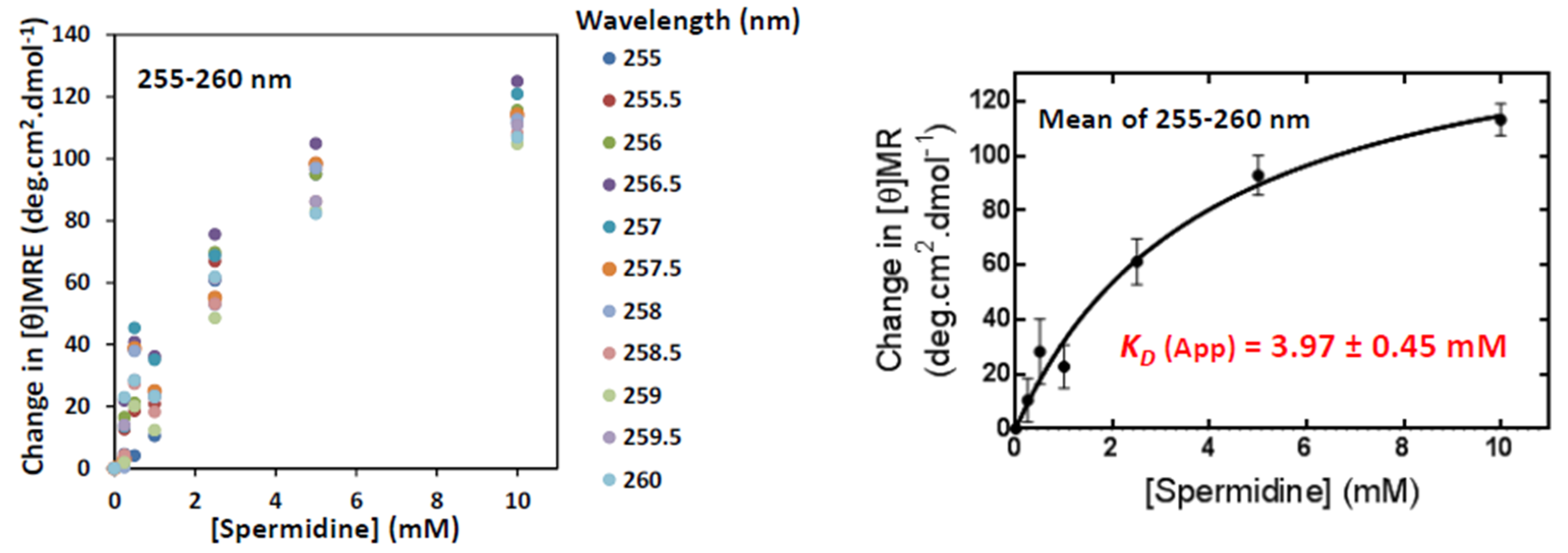
3. Results and Discussion

Using a trial and error approach, sample and near-UV SRCD experimental conditions were identified and optimised for observing the triamine spermidine binding to DDM-solubilised and purified AceI(His6). At concentrations of 20 μM and 40 μM, AceI(His6) the binding of 5 mM spermidine was detected in spectra recorded in the near-UV region (250–340 nm), with the most prominent effects on the spectra at 255–295 nm. Under the same conditions, binding of the longer-chain tetraamine spermine was not detected (Figure 1). SRCD spectra of proteins in the near-UV region represent and are sensitive to changes in their tertiary structure, which can be influenced by their conformational state, mobility and environment. During our extensive experience in developing and performing SRCD experiments on various membrane proteins, we have noticed that separate preparations of the same protein under the same sample conditions can produce starting near-UV spectra with different shapes. In these types of experiments, it is therefore important to monitor any changes in the shape and magnitude of spectra in the same sample over time or on addition of ligands, rather than comparing spectra coming from different preparations.
For protein samples, SRCD spectra in the near-UV region especially report information from the aromatic groups of phenylalanine (255–270 nm), tyrosine (275–282 nm) and tryptophan (290–305 nm) residues, with fine structure originating from vibronic transitions [20]. The 144 residues of AceI contain 12 (8.3%) phenylalanine, 6 (4.2%) tyrosine and 3 (2.1%) tryptophan residues. The contents of phenylalanine and tyrosine residues are notably high when compared with their average contents in secondary transport proteins from Escherichia coli (6.1% and 2.7%, respectively) [21]. The high content of phenylalanines in AceI gives them a greater chance of undergoing conformational changes on the binding of a ligand that can be detected in the spectra. In order to quantify the binding of spermidine, a titration of 40 μM AceI(His6) with spermidine up to 10 mM was performed (Figure 1). Stability measurements confirmed that there was no change in spectra of the protein alone when continuously measured over a period of 12 h, which is longer than the entire titration with ligand.
A saturable change in signal intensity on titration with spermidine was measured in the region 255–260 nm, which corresponds to the wavelength range for phenylalanine residues. Fitting a mean of the change in signal intensity in this region gave an apparent binding affinity (KD) of 3.97 ± 0.45 mM (Figure 2). Whilst the binding of spermidine to AceI has a three-fold lower affinity than the binding of chlorhexidine, a lower binding affinity for a natural substrate of a transporter is expected. The SRCD results presented here were the first experimental evidence obtained for a polyamine binding to a PACE protein.

4. Conclusions

CD measurements on proteins in the near-UV region that report on tertiary structure can be used to detect ligand binding, which may not be possible by using CD in the far-UV region or by other biophysical techniques (e.g., fluorimetry). Due to insufficient sensitivity, such CD measurements in the near-UV region are not possible using a benchtop instrument, so a longer pathlength of 1 cm and the intense light produced by synchrotron radiation are required to detect the near-UV spectra of membrane proteins. The results presented in this paper initiated further study on polyamines as potential natural substrates of AceI and other PACE proteins. Future work on the relatively uncharacterised PACE family should include gene cloning, recombinant expression, purification, reconstitution and stability screening of further PACE proteins for application of a multitude of chemical, biochemical, biophysical and computational techniques to elucidate high-resolution structures and to investigate their oligomeric state, physiological function, ligand interactions and substrate specificity, molecular mechanism and dynamics. This may provide information to assist the development of agents or strategies to block the biocide efflux function of PACE proteins.

Funding

This work was funded by the Diamond Light Source Ltd. (Oxfordshire, UK) and The University of Leeds, UK.

Institutional Review Board Statement

Not applicable.

Informed Consent Statement

Not applicable.

Data Availability Statement

The data that support this study are presented in the manuscript or available from the corresponding author upon reasonable request.

Acknowledgments

We thank David Sharples (University of Leeds, UK) for culturing cells in a fermentor and the Diamond Light Source Ltd. (Oxfordshire, UK) for instrument time on SRCD Beamline B23 with technical assistance from Giuliano Siligardi, Rohanah Hussain and Tamás Jávorfi.

Conflicts of Interest

The author declares no conflict of interest.

References

  1. Lee, C.R.; Lee, J.H.; Park, M.; Park, K.S.; Bae, I.K.; Kim, Y.B.; Cha, C.J.; Jeong, B.C.; Lee, S.H. Biology of Acinetobacter baumannii: Pathogenesis, antibiotic resistance mechanisms, and prospective treatment options. Front. Cell Infect. Microbiol. 2017, 7, 55. [Google Scholar] [CrossRef] [PubMed] [Green Version]
  2. Amorim, A.M.; Nascimento, J.D. Acinetobacter: An underrated foodborne pathogen? J. Infect. Dev. Ctries 2017, 11, 111–114. [Google Scholar] [CrossRef] [PubMed] [Green Version]
  3. Hamidian, M.; Nigro, S.J. Emergence, molecular mechanisms and global spread of carbapenem-resistant Acinetobacter baumannii. Microb. Genom. 2019, 5, e000306. [Google Scholar] [CrossRef] [PubMed]
  4. Hassan, K.A.; Jackson, S.M.; Penesyan, A.; Patching, S.G.; Tetu, S.G.; Eijkelkamp, B.A.; Brown, M.H.; Henderson, P.J.; Paulsen, I.T. Transcriptomic and biochemical analyses identify a family of chlorhexidine efflux proteins. Proc. Natl. Acad. Sci. USA 2013, 110, 20254–20259. [Google Scholar] [CrossRef] [PubMed] [Green Version]
  5. Hassan, K.A.; Liu, Q.; Henderson, P.J.; Paulsen, I.T. Homologs of the Acinetobacter baumannii AceI transporter represent a new family of bacterial multidrug efflux systems. mBio 2015, 6, e01982-14. [Google Scholar] [CrossRef] [PubMed] [Green Version]
  6. Ahmad, I.; Nawaz, N.; Dermani, F.K.; Kohlan, A.K.; Saidijam, M.; Patching, S.G. Bacterial multidrug efflux proteins: A major mechanism of antimicrobial resistance. Curr. Drug Targets 2018, 19, 1–13. [Google Scholar] [CrossRef]
  7. Hassan, K.A.; Liu, Q.; Elbourne, L.D.H.; Ahmad, I.; Sharples, D.; Naidu, V.; Chan, C.L.; Li, L.; Harborne, S.P.D.; Pokhrel, A.; et al. Pacing across the membrane: The novel PACE family of efflux pumps is widespread in Gram-negative pathogens. Res. Microbiol. 2018, 169, 450–454. [Google Scholar] [CrossRef]
  8. Bolla, J.R.; Howes, A.C.; Fiorentino, F.; Robinson, C.V. Assembly and regulation of the chlorhexidine-specific efflux pump AceI. Proc. Natl. Acad. Sci. USA 2020, 117, 17011–17018. [Google Scholar] [CrossRef] [PubMed]
  9. Michael, A.J. Polyamines in eukaryotes, bacteria, and archaea. J. Biol. Chem. 2016, 291, 14896–14903. [Google Scholar] [CrossRef] [PubMed] [Green Version]
  10. Shah, P.; Swiatlo, E. A multifaceted role for polyamines in bacterial pathogens. Mol. Microbiol. 2008, 68, 4–16. [Google Scholar] [CrossRef] [PubMed]
  11. Banerji, R.; Kanojiya, P.; Patil, A.; Saroj, S.D. Polyamines in the virulence of bacterial pathogens of respiratory tract. Mol. Oral. Microbiol. 2021, 36, 1–11. [Google Scholar] [CrossRef] [PubMed]
  12. Patching, S.G.; Edara, S.; Ma, P.; Nakayama, J.; Hussain, R.; Siligardi, G.; Phillips-Jones, M.K. Interactions of the intact FsrC membrane histidine kinase with its pheromone ligand GBAP revealed through synchrotron radiation circular dichroism. Biochim. Biophys. Acta—Biomembranes 2012, 1818, 1595–1602. [Google Scholar] [CrossRef] [PubMed] [Green Version]
  13. Phillips-Jones, M.K.; Patching, S.G.; Edara, S.; Nakayama, J.; Hussain, R.; Siligardi, G. Interactions of the intact FsrC membrane histidine kinase with the tricyclic peptide inhibitor siamycin I revealed through synchrotron radiation circular dichroism. Phys. Chem. Chem. Phys. 2013, 15, 444–447. [Google Scholar] [CrossRef] [PubMed]
  14. Siligardi, G.; Hussain, R.; Patching, S.G.; Phillips-Jones, M.K. Ligand- and drug-binding studies of membrane proteins revealed through circular dichroism spectroscopy. Biochim. Biophys. Acta—Biomembranes 2014, 1838, 34–42. [Google Scholar] [CrossRef] [PubMed] [Green Version]
  15. Phillips-Jones, M.K.; Channell, G.; Kelsall, C.J.; Hughes, C.S.; Ashcroft, A.E.; Patching, S.G.; Dinu, V.; Gillis, R.B.; Adams, G.G.; Harding, S.E. Hydrodynamics of the VanA-type VanS histidine kinase: An extended solution conformation and first evidence for interactions with vancomycin. Sci. Rep. 2017, 7, 46180. [Google Scholar] [CrossRef] [PubMed]
  16. Patching, S.G. Recent developments in Nucleobase Cation Symporter-1 (NCS1) family transport proteins from bacteria, archaea, fungi and plants. J. Biosci. 2018, 43, 797–815. [Google Scholar] [CrossRef] [PubMed]
  17. Ward, A.; Sanderson, N.M.; O’Reilly, J.; Rutherford, N.G.; Poolman, B.; Henderson, P.J.F. The amplified expression, identification, purification, assay, and properties of hexahistidine-tagged bacterial membrane transport proteins. In Membrane Transport: A Practical Approach; Baldwin, S.A., Ed.; Oxford University Press: Oxford, UK, 2000; pp. 141–166. [Google Scholar]
  18. Hussain, R.; Jávorfi, T.; Siligardi, G. Circular dichroism beamline B23 at the Diamond Light Source. J. Synchrotron Rad. 2012, 19, 132–135. [Google Scholar] [CrossRef] [PubMed]
  19. Hussain, R.; Siligardi, G. Characterisation of conformational and ligand binding properties of membrane proteins using synchrotron radiation circular dichroism (SRCD). Adv. Exp. Med. Biol. 2016, 922, 43–59. [Google Scholar] [PubMed]
  20. Kelly, S.M.; Jess, T.J.; Price, N.C. How to study proteins by circular dichroism. Biochim. Biophys. Acta 2005, 1751, 119–139. [Google Scholar] [CrossRef] [PubMed]
  21. Saidijam, M.; Patching, S.G. Amino acid composition analysis of secondary transport proteins from Escherichia coli with relation to functional classification, ligand specificity and structure. J. Biomol. Struct. Dyn. 2015, 33, 2205–2220. [Google Scholar] [CrossRef] [PubMed]
Figure 1. Spermidine binding to the A. baumannii efflux protein AceI observed by near-UV synchrotron radiation circular dichroism spectroscopy. Near-UV SRCD spectra (250–340 nm) at 20 °C for samples of DDM-solubilised AceI(His6) (20 μM or 40 μM in 10 mM KPi pH 7.6, 5% glycerol, 0.05% DDM) showing the effect of spermidine (5 mM) (top) or spermine (5 mM) (bottom left) and a titration with spermidine (0–10 mM) (bottom right). All spectra are an average of 10 scans acquired over approximately 1 h. Spectra were corrected for any signals coming from buffer or from the polyamines themselves by subtraction of the relevant spectra acquired on these sample components alone, as appropriate.
Radiation 02 00016 g001
Figure 2. Quantification of spermidine binding to the A. baumannii efflux protein AceI by near-UV synchrotron radiation circular dichroism spectroscopy. From the spectra showing the titration of AceI(His6) with spermidine (Figure 1, bottom right), the change in mean residue ellipticity ([θ]MRE) at each titration point was measured at six different wavelengths in the region 255–260 nm (left). Average values for the change in [θ]MRE were fitted to the Michaelis–Menten equation using GraphPad Prism 6 software to produce the given apparent dissociation constant (right). Stability measurements confirmed that there was no significant change in spectra of the protein alone when continuously measured over a period of 12 h, which was longer than that of an entire titration with spermidine.
Radiation 02 00016 g002
Publisher’s Note: MDPI stays neutral with regard to jurisdictional claims in published maps and institutional affiliations.

